2002;43:957-967. J Nucl Med. Janina Baranowska-Kortylewicz Takashi Kurizaki, Shinji Okazaki, Sam D. Sanderson, David Colcher, Charles A. Enke, Margaret A. Tempero and Human C5a Potentiation of Radioimmunotherapy with Response-Selective Peptide Agonist of http://jnm.snmjournals.org/content/43/7/957 This article and updated information are available at: http://jnm.snmjournals.org/site/subscriptions/online.xhtml Information about subscriptions to JNM can be found at: http://jnm.snmjournals.org/site/misc/permission.xhtml Information about reproducing figures, tables, or other portions of this article can be found online at: (Print ISSN: 0161-5505, Online ISSN: 2159-662X) 1850 Samuel Morse Drive, Reston, VA 20190. SNMMI | Society of Nuclear Medicine and Molecular Imaging is published monthly. The Journal of Nuclear Medicine © Copyright 2002 SNMMI; all rights reserved. by on August 1, 2014. For personal use only. jnm.snmjournals.org Downloaded from by on August 1, 2014. For personal use only. jnm.snmjournals.org Downloaded from

Welcome message from author
This document is posted to help you gain knowledge. Please leave a comment to let me know what you think about it! Share it to your friends and learn new things together.
Transcript
2002;43:957-967.J Nucl Med. Janina Baranowska-KortylewiczTakashi Kurizaki, Shinji Okazaki, Sam D. Sanderson, David Colcher, Charles A. Enke, Margaret A. Tempero and Human C5aPotentiation of Radioimmunotherapy with Response-Selective Peptide Agonist of
http://jnm.snmjournals.org/content/43/7/957This article and updated information are available at:
http://jnm.snmjournals.org/site/subscriptions/online.xhtml
Information about subscriptions to JNM can be found at:
http://jnm.snmjournals.org/site/misc/permission.xhtmlInformation about reproducing figures, tables, or other portions of this article can be found online at:
(Print ISSN: 0161-5505, Online ISSN: 2159-662X)1850 Samuel Morse Drive, Reston, VA 20190.SNMMI | Society of Nuclear Medicine and Molecular Imaging
is published monthly.The Journal of Nuclear Medicine
© Copyright 2002 SNMMI; all rights reserved.
by on August 1, 2014. For personal use only. jnm.snmjournals.org Downloaded from by on August 1, 2014. For personal use only. jnm.snmjournals.org Downloaded from
Potentiation of Radioimmunotherapy withResponse-Selective Peptide Agonist of Human C5aTakashi Kurizaki, MD, PhD1; Shinji Okazaki, MD1; Sam D. Sanderson, PhD2; David Colcher, PhD3;Charles A. Enke, MD1; Margaret A. Tempero, MD4; and Janina Baranowska-Kortylewicz, PhD1
1Department of Radiation Oncology, University of Nebraska Medical Center, Omaha, Nebraska; 2Eppley Institute for Research inCancer, University of Nebraska Medical Center, Omaha, Nebraska; 3Corixa, South San Francisco, California;and 4UCSF Cancer Center, University of California at San Francisco, San Francisco, California
Physiologic barriers to the delivery of macromolecules to solidtumors are a major obstacle to the clinical success of radioim-munotherapy (RIT). Only a small fraction of the injected dose ofthe radiolabeled monoclonal antibody (mAb) localizes at thetumor site. This situation worsens as the tumor burden in-creases. It is hypothesized that improvements to RIT of adeno-carcinoma can be realized by inclusion of vasoactive agents, inparticular agents able to increase the vascular permeability oftumor capillaries. In these studies, a response-selective peptideagonist of human C5a, GCGYSFKPMPLaR (AP), was used totransiently increase tumor vascular permeability in an effort toimprove RIT of solid tumors. Methods: Athymic mice xenograftedwith human colorectal adenocarcinoma LS174T were treatedintravenously with low doses (9.25 MBq) of 131I-labeled mAbB72.3 in combination with various intravenous doses of AP. Theprogression of the disease or the loss of �20% body weightwas taken as the endpoint. Biodistribution and tumor uptakekinetics were studied in the same tumor–antibody system. Re-sults: The uptake of 125I-B72.3 in LS174T xenografts increasedin a dose-dependent manner with an apparent maximal effectbetween 3 and 6 h after intravenous administration of AP. Aug-menting the dose of 9.25 MBq 131I-B72.3 with a single administra-tion of 0.1 mg AP delayed tumor growth nearly 2-fold; the tumorquadrupling time (Tq) was 14.2 � 3.3 d for 131I-B72.3 alone versus26.0 � 3.6 d for 131I-B72.3 plus 0.1 mg AP (P � 0.001). Anadditional dose of 0.1 mg AP 24 h after 131I-B72.3 further improvedthe therapeutic outcome (Tq � 48.5 � 7.9 d; P � 0.001) and resultedin several cases of tumor regression. Conclusion: The inclusion ofagonist peptides of human C5a in the RIT scheme results inimproved tumor responses without any manifest side effects.
Key Words: radioimmunotherapy; vascular permeability; C5aagonist; tumor uptake; adenocarcinoma.
J Nucl Med 2002; 43:957–967
Monoclonal antibodies (mAbs) have significantly im-proved the targeted delivery of various therapeutic agentsand radioisotopes to tumors. However, gains in selectivity
are strongly counteracted by problems related to the heter-ogeneity of the structure and physiology of solid tumors.These physiologic barriers to the delivery of macromole-cules are a major impediment to the clinical success ofradioimmunotherapy (RIT) of adenocarcinoma. Only asmall fraction of the injected dose of the radiolabeled mAblocalizes at the tumor site. This situation worsens as thetumor burden increases (1–4). Even in experimental cancermodels in mice, in which most small tumors are treatedsuccessfully with a single dose of radioimmunoconjugate,the therapeutic response to RIT abates as the tumor sizeincreases (5). The accretion of radiolabeled mAbs and,consequently, radiation doses deposited in tumors rely onthe blood flow in and around tumor masses, the tumorvascular volume, and the vascular permeability (VP) oftumor vessels to macromolecules (6–8). All of these factorsare particularly impaired in large tumors, which have dis-ordered vasculature and lack draining lymphatic systems.Methods to transiently change the tumor VP have beensuggested as a means to increase the delivery of mAbs totumors. Increases in tumor uptake, some significant, arereported, when the mAb delivery is aided by external beamirradiation (9), hyperthermia (10), and systemically admin-istered vasoactive agents, such as angiotensin II (11), inter-leukin-2 (12), tumor necrosis factor-� (13), and, mostrecently, �v�3 antagonist cyclo-RGD-D-phenylalanine-1-aminocyclohexane carboxylic acid (14). However, to date,the improved therapeutic outcome resulting from the mod-ulation of the VP during RIT has not been documented.
The human anaphylatoxin C5a is a multifunctional phlo-gistic molecule that is generated as a cleavage product of thefifth component of complement during serum complementactivation. One characteristic of the C5a-mediated activityis the increase of the VP to facilitate the recruitment ofcirculating neutrophils, monocytes, eosinophils, and ba-sophils to sites of tissue injury or infection (15,16). C5a-derived small agonist peptides, which maintain this biologicproperty, are expected to improve the delivery of radiola-beled mAb to tumors. On the basis of the structural analysisof a panel of conformationally restricted peptides with ho-mology to the C-terminal end of human C5a, a biologicallypreferred peptide conformation was defined. Peptides with
Received Aug. 6, 2001; revision accepted Mar. 5, 2002.For correspondence or reprints contact: Janina Baranowska-Kortylewicz,
PhD, Department of Radiation Oncology, J. Bruce Henriksen Laboratories forCancer Research, 981050 Nebraska Medical Center, Omaha, NE 68198-1050.
E-mail: [email protected]
AMENDED RADIOIMMUNOTHERAPY • Kurizaki et al. 957
by on August 1, 2014. For personal use only. jnm.snmjournals.org Downloaded from
optimal biologic activities have residues 65–69 in a twisted,helix-like conformation, whereas residues 70–71 exist in anextended backbone conformation, and residues 71/72–74exist in a B-turn of either type II or type V for spasmogenicresponses and a B-turn of type V for neutrophil responses(17,18). One analog with a Pro-for-Asp substitution at po-sition 71 and a D-Ala-for-Gly substitution at position 73,YSFKPMPLaR (C5a65–74, Y65, F67, P69, P71, D-Ala73),a � D-Ala, expressed 15%–20% of the full C5a activity inspasmogenic assays and approximately 5% in polymorpho-nuclear (PMN) assays. YSFKPMPLaR was also shown tobe stable in the presence of mouse and human serum car-boxypeptidases (19). In this study a response-selective pep-tide agonist of human C5a, GCGYSFKPMPLaR (AP), wasused to transiently increase tumor the VP in an effort toimprove RIT of solid tumors. AP is an analog of YSFK-PMPLaR, a conformationally biased, response-selective ag-onist of human C5a that produces marked increases in theVP in the skin of guinea pigs and hypotensive effects in rats(19,20). Structure–activity studies have shown that the con-formational features expressed by YSFKPMPLaR and itsanalog AP are more amenable to interacting with and acti-vating C5a receptors expressed on cells responsible forhemodynamic activities than for C5a receptors expressed oninflammatory PMN cells (17–20). This report shows un-equivocally that RIT combined with the VP-enhancing C5aagonist peptide GCGYSFKPMPLaR improves the outcomeof RIT.
MATERIALS AND METHODS
Peptides and mAbsAP was synthesized by standard solid-phase methods, purified,
and characterized according to previously described procedures(17).
mAb B72.3 was produced by the University of Nebraska Med-ical Center Monoclonal Antibody Facility and purified from miceascites by protein-G affinity chromatography. B72.3 recognizes ahigh-molecular-weight glycoprotein complex designated as tumor-associated glycoprotein 72 (TAG-72) and shows reactivity with�85% of adenocarcinomas with minimal reactivity to normaltissues (21).
Iodination of mAbsB72.3 was labeled with Na125I (biodistribution) or Na131I (ther-
apy) using the IODO-GEN (1,3,4,6-tetrachloro-3�,6�-diphenyl-glycoluril; Pierce, Rockford, IL) method. Briefly, to prepare 131I-B72.3 for RIT studies 2 mg B72.3 were reacted with 370 MBqNa131I in the presence of 0.1 mg IODO-GEN as the oxidant. Theunbound radioisotope was removed on a desalting column (Econo-Pac 10DG; Bio-Rad, Hercules, CA) equilibrated with 0.01 mol/Lphosphate buffer, 0.0027 mol/L KCl, 0.137 mol/L NaCl (phos-phate-buffered saline [PBS]), pH 7.4, at 25°C. Quality assessmentusing instant thin-layer chromatography with 20% aqueous meth-anol as the solvent indicated that �95% of the radioiodine wasroutinely protein bound. Specific activities of 150–190 MBq 131Iper milligram B72.3 and 290–330 MBq 125I per milligram B72.3were typical. The immunoreactivity of radiolabeled B72.3 wastested using bovine submaxillary gland mucin ([BSM] Sigma, St.
Louis, MO) attached to Reacti-Gel beads (Reacti-Gel HW-65F;Pierce). Beads coated with bovine serum albumin (BSA) wereused as the negative control. Radiolabeled B72.3 (100,000 countsper minute [cpm]) was incubated for 1 h with 0.1 mL BSM- andBSA-beads at room temperature. The unbound B72.3 was re-moved by repeated washing with PBS containing 1% BSA and0.1% Tween 20. The radioactivity in the bead pellet was deter-mined in a �-counter, and the percentage bound to BSM-coatedbeads was calculated.
Animal and Tumor ModelsAthymic female mice (nu/nu; National Institutes of Health,
Bethesda, MD) (average weight, 18 g; age, 4–6 wk) were used.Mice were housed in the University of Nebraska Medical CenterAnimal Facility, which is fully accredited by the American Asso-ciation for Accreditation of Laboratory Animal Care. Mice hadfree access to food and water and were kept on a 12-h light cycle.Potassium iodide-supplemented water was provided 3 d before andfor 7 d after any treatment with radioiodinated antibodies. Subcu-taneous tumors (20–200 mm3) were produced in these mice ap-proximately 10 d after a right flank injection of 5 � 106 LS174Thuman colon carcinoma cells in 0.2 mL minimum essential me-dium ([MEM] GIBCO-BRL, Gaithersburg, MD). The cells wereobtained from subconfluent monolayers grown in MEM supple-mented with 10% fetal bovine serum.
Immunogenicity of APSwiss Albino (n � 6) and C57BL/6 (n � 6) mice were given 3
daily doses (either intradermally or intravenously) of 0.1 mg AP;2 wk after the last dose, blood was collected through a cardiacpuncture and serum was separated and tested for 125I-AP binding.Serum samples were incubated with 300,000 cpm 125I-C5a (spe-cific activity, 740 MBq/mg) for 2 h and tested using either asize-exclusion centrifugation or a 96-well plate binding assay (datanot shown). Serum samples were also analyzed by nondenaturing,nonreducing polyacrylamide gel electrophoresis (PAGE). Thesesamples were incubated with 300,000 cpm 125I-C5a (specific ac-tivity, 740 MBq/mg) for 12 h at 0°C–4°C. Mouse IgG, IgM, andIgM were used as negative controls.
Biodistribution of 125I-C5aTo investigate the in vivo fate of AP, the peptide was radioio-
dinated with no-carrier-added 125I to a specific activity of 0.37MBq/0.1 mg using IODO-GEN as an oxidant. Mice bearing sub-cutaneous LS174T xenografts were treated with 0.1-mg doses of125I-AP (0.37 MBq) administered intravenously in 0.2 mL PBSthrough a tail vein and euthanized at designated times.
Biodistribution StudiesMice with LS174T xenografts received an intravenous dose of
AP in 0.2 mL 0.1% albumin in PBS through the tail vein. Todetermine the optimum dosing sequence for AP and B72.3, a timeinterval between the injection of AP and 125I-B72.3 was variedfrom 0.5 to 24 h. AP was injected intravenously through a tail vein0.5, 1, 3, 6, and 24 h before the dose of 125I-B72.3 was given. Atthe times indicated above after AP, an intravenous dose of 0.37MBq 125I-B72.3 in 0.2 mL 0.1% albumin in PBS was administered.Control mice were treated with 125I-B72.3 alone. Additional dosesof AP were administered at 24 and 48 h after injection of 125I-B72.3 in 2 groups of mice. Seventy-two hours later, mice wereeuthanized and the necropsy was performed. The weight andradioactive content of blood, lung, heart, spleen, liver, kidney,
958 THE JOURNAL OF NUCLEAR MEDICINE • Vol. 43 • No. 7 • July 2002
by on August 1, 2014. For personal use only. jnm.snmjournals.org Downloaded from
uterus, muscle, stomach, small intestine, large intestine, skin, andtumor were determined.
To examine the overall effect of AP on the distribution ofmacromolecules and to access the dose dependence, a nonspecificmouse IgG labeled with 125I was used as a tracer. Athymic mice(without tumors) received a simultaneous injection of AP and125I-IgG (0.37 MBq per mouse) through the tail vein. Thirtyminutes later, the aforementioned tissues were harvested and theirradioactive content was determined.
Tumor Therapy StudiesMice with 10-d-old subcutaneous LS174T xenografts were as-
signed to 3 therapy groups (9.25 MBq 131I-B72.3 plus 0.1 mg AP,9.25 MBq 131I-B72.3 plus 2 � 0.1 mg AP, and 9.25 MBq 131I-B72.3 plus 2 � 0.01 mg AP) and 2 control groups (9.25 MBq131I-B72.3 alone or 0.1 mg AP alone) so that the tumor sizedistribution was similar in each group. AP in 0.2 mL 0.1% albuminin PBS was administered intravenously, which was followed 3 hlater by an intravenous dose of 9.25 MBq 131I-B72.3 in 0.1%albumin in PBS. Control groups received a sham injection of 0.2mL 0.1% albumin in PBS in place of either AP or 131I-B72.3. Instudies with 2 AP doses, the second administration of AP was 24 hafter 131I-B72.3 administration. Drinking water was supplementedwith 0.04% potassium iodide 3 d before treatment and for 7 d aftertreatment to protect thyroid function. The tumor size and bodyweight were measured 3 times a week. Tumor volume ([V] mm3)was calculated as: V � 4/3� � (1⁄2 of longer tumor dimension inmm) � (1⁄2 of shorter tumor dimension in mm)2. All therapy exper-iments were terminated when the size of tumor in control groupswas 2,000 mm3. Premature euthanasia was also indicated if anyulcer formation was observed or the weight loss was 20% of theinitial body weight. Serum collected during the necropsy wasanalyzed by gel-filtration high-performance liquid chromatogra-phy (HPLC) (TSK G2000SW and TSK G3000SW, 0.75 � 30 cm;Toso Haas, Tokyo, Japan; connected in tandem with PBS at a flowrate of 0.5 mL/min) and sodium dodecyl sulfate–PAGE ([SDS-PAGE] 4%–15% acrylamide gradient) to determine the effect of
AP on the metabolic fate of 131I-B72.3. The immunoreactivity ofall radioactive fractions collected from an HPLC column also wasmeasured using the bead-binding assay described above.
Data EvaluationEach tumor quadrupling time (Tq) was calculated as the average
of the Tq of all LS174T tumors in a given group of mice. Com-parison was made using the 2-tailed t test for difference in means.
RESULTS
Dose-Dependence StudiesPermeability of mouse tissues to immunoglobulin was
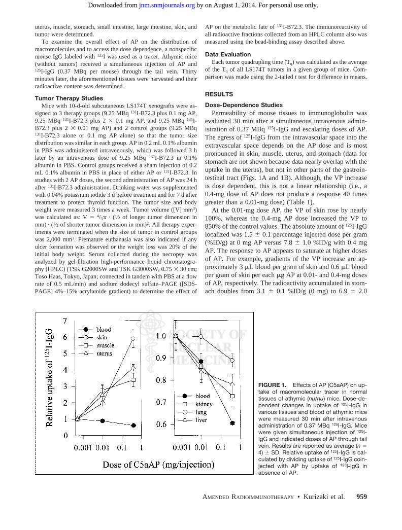
evaluated 30 min after a simultaneous intravenous admin-istration of 0.37 MBq 125I-IgG and escalating doses of AP.The egress of 125I-IgG from the intravascular space into theextravascular space depends on the AP dose and is mostpronounced in skin, muscle, uterus, and stomach (data forstomach are not shown because data nearly overlap with theuptake in the uterus), but not in other parts of the gastroin-testinal tract (Figs. 1A and 1B). Although, the VP increaseis dose dependent, this is not a linear relationship (i.e., a0.4-mg dose of AP does not produce a response 40 timesgreater than a 0.01-mg dose) (Table 1).
At the 0.01-mg dose AP, the VP of skin rose by nearly100%, whereas the 0.4-mg AP dose increased the VP to850% of the control values. The absolute amount of 125I-IgGlocalized was 1.5 � 0.1 percentage injected dose per gram(%ID/g) at 0 mg AP versus 7.8 � 1.0 %ID/g with 0.4 mgAP. The response to AP appears to saturate at higher dosesof AP. For example, gradients of the VP increase are ap-proximately 3 �L blood per gram of skin and 0.6 �L bloodper gram of skin per each �g AP at 0.01- and 0.4-mg dosesof AP, respectively. The radioactivity accumulated in stom-ach doubles from 3.1 � 0.1 %ID/g (0 mg) to 6.9 � 2.0
FIGURE 1. Effects of AP (C5aAP) on up-take of macromolecular tracer in normaltissues of athymic (nu/nu) mice. Dose-de-pendent changes in uptake of 125I-IgG invarious tissues and blood of athymic micewere measured 30 min after intravenousadministration of 0.37 MBq 125I-IgG. Micewere given simultaneous injection of 125I-IgG and indicated doses of AP through tailvein. Results are reported as average (n �4) � SD. Relative uptake of 125I-IgG is cal-culated by dividing uptake of 125I-IgG coin-jected with AP by uptake of 125I-IgG inabsence of AP.
AMENDED RADIOIMMUNOTHERAPY • Kurizaki et al. 959
by on August 1, 2014. For personal use only. jnm.snmjournals.org Downloaded from
%ID/g (0.4 mg). There is also a significant perivascularleakage of radioactivity in uterus, approximately 1.5-fold,reaching 8.9 � 1.0 %ID/g at this dose of AP. The bloodlevel of 125I-IgG decreases with the increasing dose of APstarting at 44.4 � 1.1 %ID/g when no AP is administeredand plummeting to about one half of this value at 0.4 mgAP. The levels of radioactivity recovered in large and smallintestine, liver, spleen, kidney, heart, and lung remain rel-atively constant with only a slight increase at the highestdose of AP.
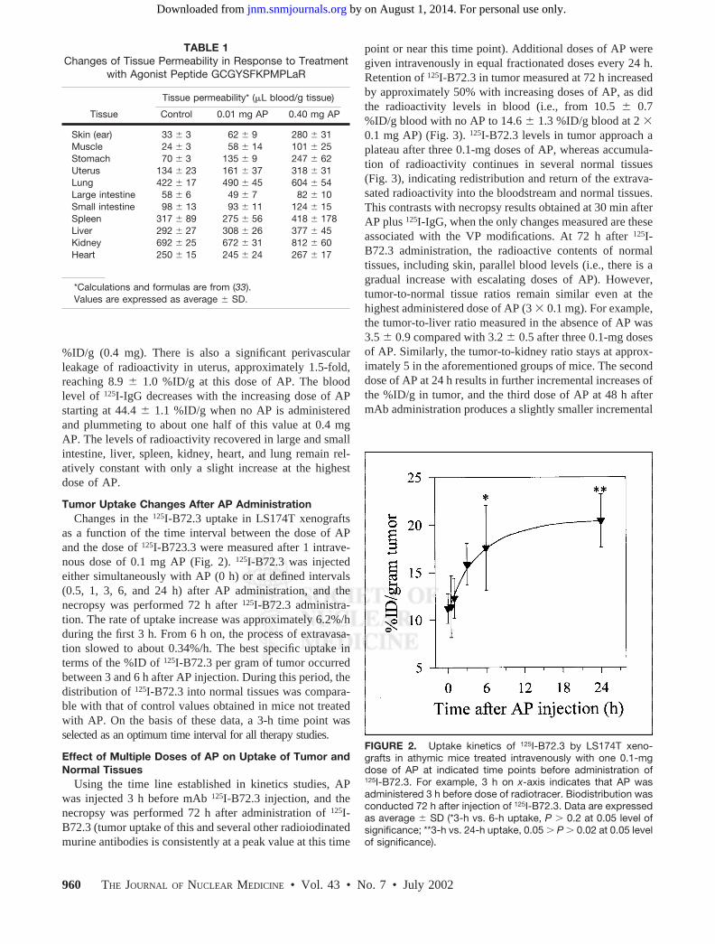
Tumor Uptake Changes After AP AdministrationChanges in the 125I-B72.3 uptake in LS174T xenografts
as a function of the time interval between the dose of APand the dose of 125I-B723.3 were measured after 1 intrave-nous dose of 0.1 mg AP (Fig. 2). 125I-B72.3 was injectedeither simultaneously with AP (0 h) or at defined intervals(0.5, 1, 3, 6, and 24 h) after AP administration, and thenecropsy was performed 72 h after 125I-B72.3 administra-tion. The rate of uptake increase was approximately 6.2%/hduring the first 3 h. From 6 h on, the process of extravasa-tion slowed to about 0.34%/h. The best specific uptake interms of the %ID of 125I-B72.3 per gram of tumor occurredbetween 3 and 6 h after AP injection. During this period, thedistribution of 125I-B72.3 into normal tissues was compara-ble with that of control values obtained in mice not treatedwith AP. On the basis of these data, a 3-h time point wasselected as an optimum time interval for all therapy studies.
Effect of Multiple Doses of AP on Uptake of Tumor andNormal Tissues
Using the time line established in kinetics studies, APwas injected 3 h before mAb 125I-B72.3 injection, and thenecropsy was performed 72 h after administration of 125I-B72.3 (tumor uptake of this and several other radioiodinatedmurine antibodies is consistently at a peak value at this time
point or near this time point). Additional doses of AP weregiven intravenously in equal fractionated doses every 24 h.Retention of 125I-B72.3 in tumor measured at 72 h increasedby approximately 50% with increasing doses of AP, as didthe radioactivity levels in blood (i.e., from 10.5 � 0.7%ID/g blood with no AP to 14.6 � 1.3 %ID/g blood at 2 �0.1 mg AP) (Fig. 3). 125I-B72.3 levels in tumor approach aplateau after three 0.1-mg doses of AP, whereas accumula-tion of radioactivity continues in several normal tissues(Fig. 3), indicating redistribution and return of the extrava-sated radioactivity into the bloodstream and normal tissues.This contrasts with necropsy results obtained at 30 min afterAP plus 125I-IgG, when the only changes measured are theseassociated with the VP modifications. At 72 h after 125I-B72.3 administration, the radioactive contents of normaltissues, including skin, parallel blood levels (i.e., there is agradual increase with escalating doses of AP). However,tumor-to-normal tissue ratios remain similar even at thehighest administered dose of AP (3 � 0.1 mg). For example,the tumor-to-liver ratio measured in the absence of AP was3.5 � 0.9 compared with 3.2 � 0.5 after three 0.1-mg dosesof AP. Similarly, the tumor-to-kidney ratio stays at approx-imately 5 in the aforementioned groups of mice. The seconddose of AP at 24 h results in further incremental increases ofthe %ID/g in tumor, and the third dose of AP at 48 h aftermAb administration produces a slightly smaller incremental
FIGURE 2. Uptake kinetics of 125I-B72.3 by LS174T xeno-grafts in athymic mice treated intravenously with one 0.1-mgdose of AP at indicated time points before administration of125I-B72.3. For example, 3 h on x-axis indicates that AP wasadministered 3 h before dose of radiotracer. Biodistribution wasconducted 72 h after injection of 125I-B72.3. Data are expressedas average � SD (*3-h vs. 6-h uptake, P � 0.2 at 0.05 level ofsignificance; **3-h vs. 24-h uptake, 0.05 � P � 0.02 at 0.05 levelof significance).
TABLE 1Changes of Tissue Permeability in Response to Treatment
with Agonist Peptide GCGYSFKPMPLaR
Tissue
Tissue permeability* (�L blood/g tissue)
Control 0.01 mg AP 0.40 mg AP
Skin (ear) 33 � 3 62 � 9 280 � 31Muscle 24 � 3 58 � 14 101 � 25Stomach 70 � 3 135 � 9 247 � 62Uterus 134 � 23 161 � 37 318 � 31Lung 422 � 17 490 � 45 604 � 54Large intestine 58 � 6 49 � 7 82 � 10Small intestine 98 � 13 93 � 11 124 � 15Spleen 317 � 89 275 � 56 418 � 178Liver 292 � 27 308 � 26 377 � 45Kidney 692 � 25 672 � 31 812 � 60Heart 250 � 15 245 � 24 267 � 17
*Calculations and formulas are from (33).Values are expressed as average � SD.
960 THE JOURNAL OF NUCLEAR MEDICINE • Vol. 43 • No. 7 • July 2002
by on August 1, 2014. For personal use only. jnm.snmjournals.org Downloaded from
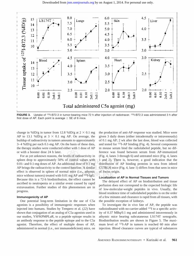
change in %ID/g in tumor from 12.8 %ID/g at 2 � 0.1 mgAP to 13.1 %ID/g at 3 � 0.1 mg AP. On average, thebuildup of radioactivity in tumors amounts to approximately3–4 %ID/g per each 0.1-mg AP. On the basis of these data,the therapy studies were conducted either with 1 dose of APor with a booster dose 24 h later.
For as yet unknown reasons, the levels of radioactivity inspleen drop to approximately 50% of control values with0.01- and 0.1-mg doses of AP. An additional dose of 0.1 mgAP brings the radioactivity to the control baseline. A similareffect is observed in spleen of normal mice (i.e., athymicmice without tumors) treated with 0.01 mg AP and 125I-IgG.Because this is a 72-h biodistribution, the effect cannot beascribed to neutropenia or a similar event caused by rapidextravasation. Further studies of this phenomenon are inprogress.
Immunogenicity of APOne potential long-term limitation in the use of C5a
agonists is a possibility of immunogenic responses wheninjected into humans. Studies by Tempero et al. (22) haveshown that conjugation of an analog of C5a agonists used inour studies, YSFKPMPLaR, to a peptide epitope results inan antibody response to the peptide epitope and to the C5aagonist. Therefore, the effect of multiple doses of AP,administered in normal (i.e., not immunodeficient) mice, on
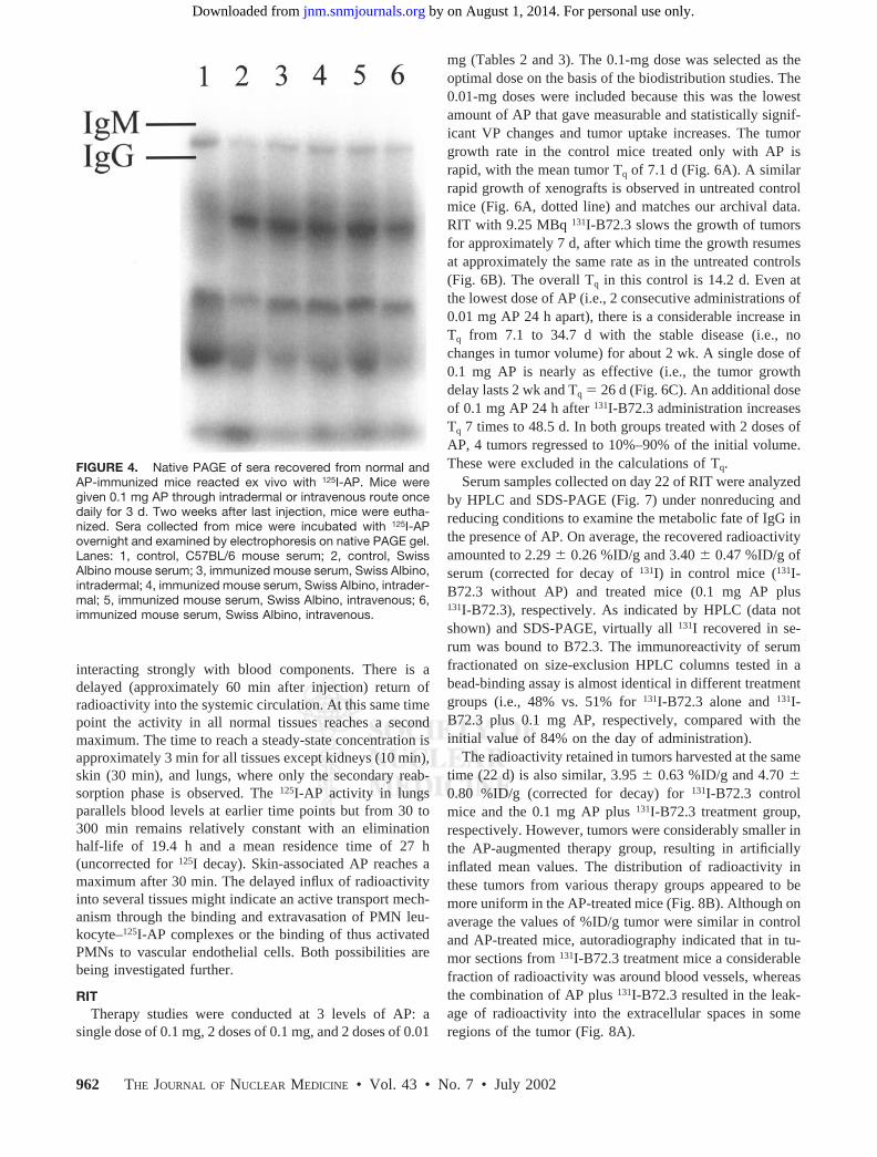
the production of anti-AP response was studied. Mice weregiven 3 daily doses (either intradermally or intravenously)of 0.1 mg AP; 2 wk after the last dose, blood was collectedand tested for 125I-AP binding (Fig. 4). Several componentsin mouse serum bind the radiolabeled peptide, but no dif-ference was found between serum from AP-immunized(Fig. 4, lanes 3 through 6) and untreated mice (Fig. 4, lanes1 and 2). There is, however, a good indication that thedistribution of AP binding proteins in sera from inbredC57BL/6 mice (Fig. 4, lane 1) differs from that seen in miceof Swiss origin.
Localization of AP in Normal Tissues and TumorsThe delayed effect of AP on biodistribution and tissue
perfusion does not correspond to the expected biologic lifeof low-molecular-weight peptides in vivo. Usually, theblood residence time of radiolabeled peptides is on the orderof a few minutes and clearance is rapid from all tissues, withthe possible exception of kidneys.
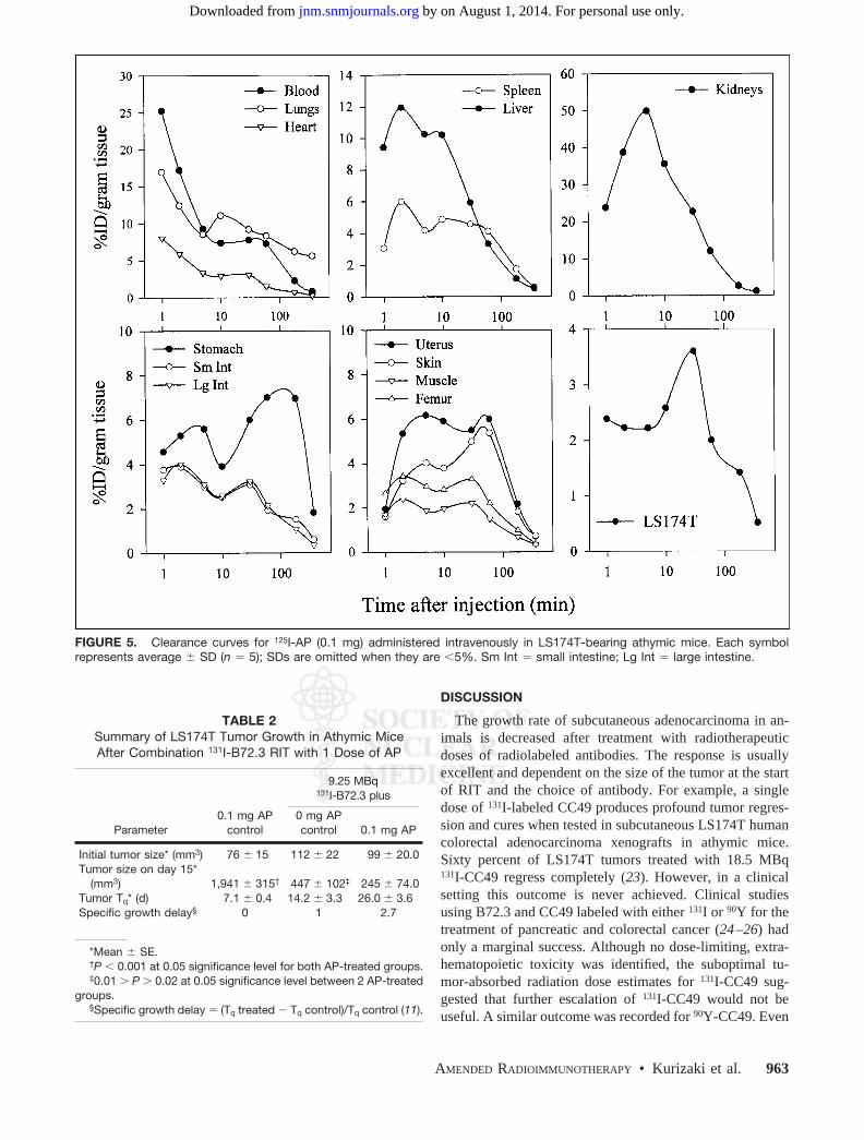
To investigate the in vivo fate of AP, the peptide wasradioiodinated with no-carrier-added 125I to a specific activ-ity of 0.37 MBq/0.1 mg and administered intravenously inathymic mice bearing subcutaneous LS174T xenografts.Biodistribution results are shown in Figure 5. The maxi-mum level of 125I-AP in tumors is reached 60 min afterinjection. Blood clearance curves are typical of substances
FIGURE 3. Uptake of 125I-B72.3 in tumor-bearing mice 72 h after injection of radiotracer. 125I-B72.3 was administered 3 h afterfirst dose of AP. Each point is average � SE of 6 mice.
AMENDED RADIOIMMUNOTHERAPY • Kurizaki et al. 961
by on August 1, 2014. For personal use only. jnm.snmjournals.org Downloaded from
interacting strongly with blood components. There is adelayed (approximately 60 min after injection) return ofradioactivity into the systemic circulation. At this same timepoint the activity in all normal tissues reaches a secondmaximum. The time to reach a steady-state concentration isapproximately 3 min for all tissues except kidneys (10 min),skin (30 min), and lungs, where only the secondary reab-sorption phase is observed. The 125I-AP activity in lungsparallels blood levels at earlier time points but from 30 to300 min remains relatively constant with an eliminationhalf-life of 19.4 h and a mean residence time of 27 h(uncorrected for 125I decay). Skin-associated AP reaches amaximum after 30 min. The delayed influx of radioactivityinto several tissues might indicate an active transport mech-anism through the binding and extravasation of PMN leu-kocyte–125I-AP complexes or the binding of thus activatedPMNs to vascular endothelial cells. Both possibilities arebeing investigated further.
RITTherapy studies were conducted at 3 levels of AP: a
single dose of 0.1 mg, 2 doses of 0.1 mg, and 2 doses of 0.01
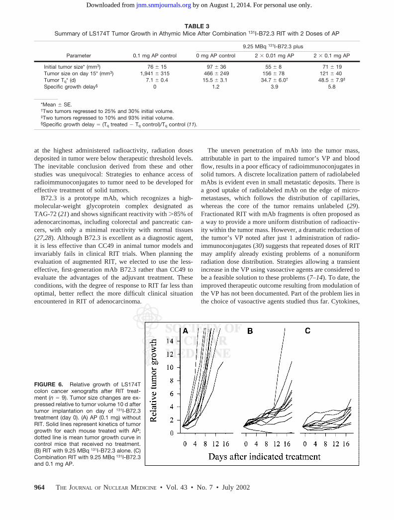
mg (Tables 2 and 3). The 0.1-mg dose was selected as theoptimal dose on the basis of the biodistribution studies. The0.01-mg doses were included because this was the lowestamount of AP that gave measurable and statistically signif-icant VP changes and tumor uptake increases. The tumorgrowth rate in the control mice treated only with AP israpid, with the mean tumor Tq of 7.1 d (Fig. 6A). A similarrapid growth of xenografts is observed in untreated controlmice (Fig. 6A, dotted line) and matches our archival data.RIT with 9.25 MBq 131I-B72.3 slows the growth of tumorsfor approximately 7 d, after which time the growth resumesat approximately the same rate as in the untreated controls(Fig. 6B). The overall Tq in this control is 14.2 d. Even atthe lowest dose of AP (i.e., 2 consecutive administrations of0.01 mg AP 24 h apart), there is a considerable increase inTq from 7.1 to 34.7 d with the stable disease (i.e., nochanges in tumor volume) for about 2 wk. A single dose of0.1 mg AP is nearly as effective (i.e., the tumor growthdelay lasts 2 wk and Tq � 26 d (Fig. 6C). An additional doseof 0.1 mg AP 24 h after 131I-B72.3 administration increasesTq 7 times to 48.5 d. In both groups treated with 2 doses ofAP, 4 tumors regressed to 10%–90% of the initial volume.These were excluded in the calculations of Tq.
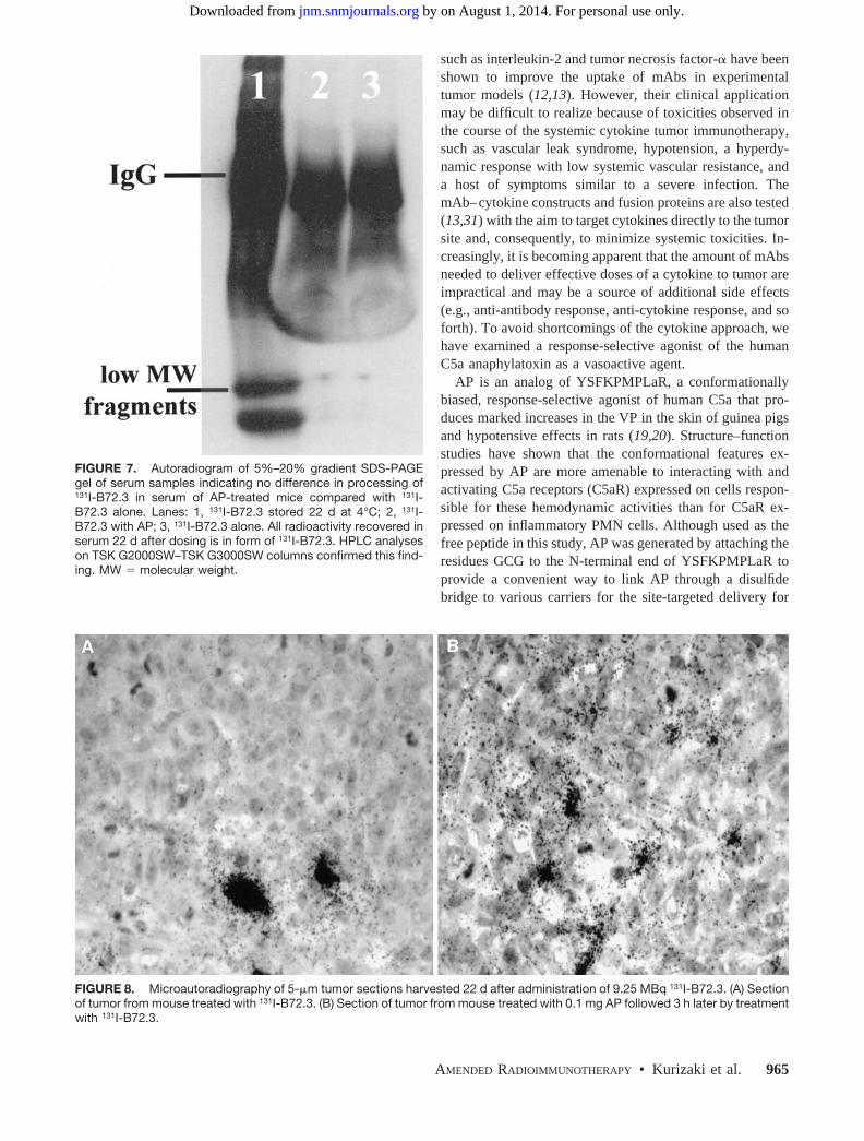
Serum samples collected on day 22 of RIT were analyzedby HPLC and SDS-PAGE (Fig. 7) under nonreducing andreducing conditions to examine the metabolic fate of IgG inthe presence of AP. On average, the recovered radioactivityamounted to 2.29 � 0.26 %ID/g and 3.40 � 0.47 %ID/g ofserum (corrected for decay of 131I) in control mice (131I-B72.3 without AP) and treated mice (0.1 mg AP plus131I-B72.3), respectively. As indicated by HPLC (data notshown) and SDS-PAGE, virtually all 131I recovered in se-rum was bound to B72.3. The immunoreactivity of serumfractionated on size-exclusion HPLC columns tested in abead-binding assay is almost identical in different treatmentgroups (i.e., 48% vs. 51% for 131I-B72.3 alone and 131I-B72.3 plus 0.1 mg AP, respectively, compared with theinitial value of 84% on the day of administration).
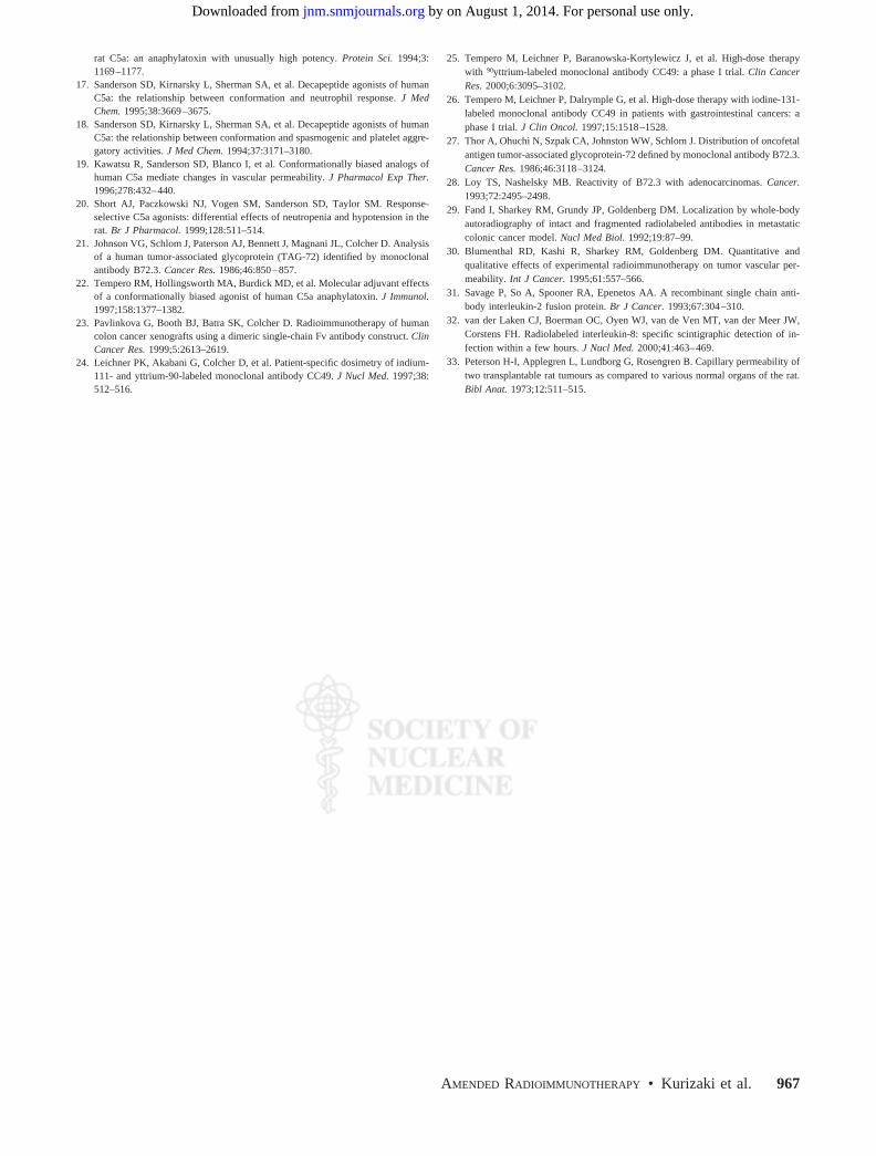
The radioactivity retained in tumors harvested at the sametime (22 d) is also similar, 3.95 � 0.63 %ID/g and 4.70 �0.80 %ID/g (corrected for decay) for 131I-B72.3 controlmice and the 0.1 mg AP plus 131I-B72.3 treatment group,respectively. However, tumors were considerably smaller inthe AP-augmented therapy group, resulting in artificiallyinflated mean values. The distribution of radioactivity inthese tumors from various therapy groups appeared to bemore uniform in the AP-treated mice (Fig. 8B). Although onaverage the values of %ID/g tumor were similar in controland AP-treated mice, autoradiography indicated that in tu-mor sections from 131I-B72.3 treatment mice a considerablefraction of radioactivity was around blood vessels, whereasthe combination of AP plus 131I-B72.3 resulted in the leak-age of radioactivity into the extracellular spaces in someregions of the tumor (Fig. 8A).
FIGURE 4. Native PAGE of sera recovered from normal andAP-immunized mice reacted ex vivo with 125I-AP. Mice weregiven 0.1 mg AP through intradermal or intravenous route oncedaily for 3 d. Two weeks after last injection, mice were eutha-nized. Sera collected from mice were incubated with 125I-APovernight and examined by electrophoresis on native PAGE gel.Lanes: 1, control, C57BL/6 mouse serum; 2, control, SwissAlbino mouse serum; 3, immunized mouse serum, Swiss Albino,intradermal; 4, immunized mouse serum, Swiss Albino, intrader-mal; 5, immunized mouse serum, Swiss Albino, intravenous; 6,immunized mouse serum, Swiss Albino, intravenous.
962 THE JOURNAL OF NUCLEAR MEDICINE • Vol. 43 • No. 7 • July 2002
by on August 1, 2014. For personal use only. jnm.snmjournals.org Downloaded from
DISCUSSION
The growth rate of subcutaneous adenocarcinoma in an-imals is decreased after treatment with radiotherapeuticdoses of radiolabeled antibodies. The response is usuallyexcellent and dependent on the size of the tumor at the startof RIT and the choice of antibody. For example, a singledose of 131I-labeled CC49 produces profound tumor regres-sion and cures when tested in subcutaneous LS174T humancolorectal adenocarcinoma xenografts in athymic mice.Sixty percent of LS174T tumors treated with 18.5 MBq131I-CC49 regress completely (23). However, in a clinicalsetting this outcome is never achieved. Clinical studiesusing B72.3 and CC49 labeled with either 131I or 90Y for thetreatment of pancreatic and colorectal cancer (24–26) hadonly a marginal success. Although no dose-limiting, extra-hematopoietic toxicity was identified, the suboptimal tu-mor-absorbed radiation dose estimates for 131I-CC49 sug-gested that further escalation of 131I-CC49 would not beuseful. A similar outcome was recorded for 90Y-CC49. Even
TABLE 2Summary of LS174T Tumor Growth in Athymic MiceAfter Combination 131I-B72.3 RIT with 1 Dose of AP
Parameter0.1 mg AP
control
9.25 MBq131I-B72.3 plus
0 mg APcontrol 0.1 mg AP
Initial tumor size* (mm3) 76 � 15 112 � 22 99 � 20.0Tumor size on day 15*
(mm3) 1,941 � 315† 447 � 102‡ 245 � 74.0Tumor Tq* (d) 7.1 � 0.4 14.2 � 3.3 26.0 � 3.6Specific growth delay§ 0 1 2.7
*Mean � SE.†P � 0.001 at 0.05 significance level for both AP-treated groups.‡0.01 � P � 0.02 at 0.05 significance level between 2 AP-treated
groups.§Specific growth delay � (Tq treated Tq control)/Tq control (11).
FIGURE 5. Clearance curves for 125I-AP (0.1 mg) administered intravenously in LS174T-bearing athymic mice. Each symbolrepresents average � SD (n � 5); SDs are omitted when they are �5%. Sm Int � small intestine; Lg Int � large intestine.
AMENDED RADIOIMMUNOTHERAPY • Kurizaki et al. 963
by on August 1, 2014. For personal use only. jnm.snmjournals.org Downloaded from
at the highest administered radioactivity, radiation dosesdeposited in tumor were below therapeutic threshold levels.The inevitable conclusion derived from these and otherstudies was unequivocal: Strategies to enhance access ofradioimmunoconjugates to tumor need to be developed foreffective treatment of solid tumors.
B72.3 is a prototype mAb, which recognizes a high-molecular-weight glycoprotein complex designated asTAG-72 (21) and shows significant reactivity with �85% ofadenocarcinomas, including colorectal and pancreatic can-cers, with only a minimal reactivity with normal tissues(27,28). Although B72.3 is excellent as a diagnostic agent,it is less effective than CC49 in animal tumor models andinvariably fails in clinical RIT trials. When planning theevaluation of augmented RIT, we elected to use the less-effective, first-generation mAb B72.3 rather than CC49 toevaluate the advantages of the adjuvant treatment. Theseconditions, with the degree of response to RIT far less thanoptimal, better reflect the more difficult clinical situationencountered in RIT of adenocarcinoma.
The uneven penetration of mAb into the tumor mass,attributable in part to the impaired tumor’s VP and bloodflow, results in a poor efficacy of radioimmunoconjugates insolid tumors. A discrete localization pattern of radiolabeledmAbs is evident even in small metastatic deposits. There isa good uptake of radiolabeled mAb on the edge of micro-metastases, which follows the distribution of capillaries,whereas the core of the tumor remains unlabeled (29).Fractionated RIT with mAb fragments is often proposed asa way to provide a more uniform distribution of radioactiv-ity within the tumor mass. However, a dramatic reduction ofthe tumor’s VP noted after just 1 administration of radio-immunoconjugates (30) suggests that repeated doses of RITmay amplify already existing problems of a nonuniformradiation dose distribution. Strategies allowing a transientincrease in the VP using vasoactive agents are considered tobe a feasible solution to these problems (7–14). To date, theimproved therapeutic outcome resulting from modulation ofthe VP has not been documented. Part of the problem lies inthe choice of vasoactive agents studied thus far. Cytokines,
TABLE 3Summary of LS174T Tumor Growth in Athymic Mice After Combination 131I-B72.3 RIT with 2 Doses of AP
Parameter 0.1 mg AP control
9.25 MBq 131I-B72.3 plus
0 mg AP control 2 � 0.01 mg AP 2 � 0.1 mg AP
Initial tumor size* (mm3) 76 � 15 97 � 36 55 � 8 71 � 19Tumor size on day 15* (mm3) 1,941 � 315 466 � 249 156 � 78 121 � 40Tumor Tq* (d) 7.1 � 0.4 15.5 � 3.1 34.7 � 6.0† 48.5 � 7.9‡
Specific growth delay§ 0 1.2 3.9 5.8
*Mean � SE.†Two tumors regressed to 25% and 30% initial volume.‡Two tumors regressed to 10% and 93% initial volume.§Specific growth delay � (Tq treated Tq control)/Tq control (11).
FIGURE 6. Relative growth of LS174Tcolon cancer xenografts after RIT treat-ment (n � 9). Tumor size changes are ex-pressed relative to tumor volume 10 d aftertumor implantation on day of 131I-B72.3treatment (day 0). (A) AP (0.1 mg) withoutRIT. Solid lines represent kinetics of tumorgrowth for each mouse treated with AP;dotted line is mean tumor growth curve incontrol mice that received no treatment.(B) RIT with 9.25 MBq 131I-B72.3 alone. (C)Combination RIT with 9.25 MBq 131I-B72.3and 0.1 mg AP.
964 THE JOURNAL OF NUCLEAR MEDICINE • Vol. 43 • No. 7 • July 2002
by on August 1, 2014. For personal use only. jnm.snmjournals.org Downloaded from
such as interleukin-2 and tumor necrosis factor-� have beenshown to improve the uptake of mAbs in experimentaltumor models (12,13). However, their clinical applicationmay be difficult to realize because of toxicities observed inthe course of the systemic cytokine tumor immunotherapy,such as vascular leak syndrome, hypotension, a hyperdy-namic response with low systemic vascular resistance, anda host of symptoms similar to a severe infection. ThemAb–cytokine constructs and fusion proteins are also tested(13,31) with the aim to target cytokines directly to the tumorsite and, consequently, to minimize systemic toxicities. In-creasingly, it is becoming apparent that the amount of mAbsneeded to deliver effective doses of a cytokine to tumor areimpractical and may be a source of additional side effects(e.g., anti-antibody response, anti-cytokine response, and soforth). To avoid shortcomings of the cytokine approach, wehave examined a response-selective agonist of the humanC5a anaphylatoxin as a vasoactive agent.
AP is an analog of YSFKPMPLaR, a conformationallybiased, response-selective agonist of human C5a that pro-duces marked increases in the VP in the skin of guinea pigsand hypotensive effects in rats (19,20). Structure–functionstudies have shown that the conformational features ex-pressed by AP are more amenable to interacting with andactivating C5a receptors (C5aR) expressed on cells respon-sible for these hemodynamic activities than for C5aR ex-pressed on inflammatory PMN cells. Although used as thefree peptide in this study, AP was generated by attaching theresidues GCG to the N-terminal end of YSFKPMPLaR toprovide a convenient way to link AP through a disulfidebridge to various carriers for the site-targeted delivery for
FIGURE 7. Autoradiogram of 5%–20% gradient SDS-PAGEgel of serum samples indicating no difference in processing of131I-B72.3 in serum of AP-treated mice compared with 131I-B72.3 alone. Lanes: 1, 131I-B72.3 stored 22 d at 4°C; 2, 131I-B72.3 with AP; 3, 131I-B72.3 alone. All radioactivity recovered inserum 22 d after dosing is in form of 131I-B72.3. HPLC analyseson TSK G2000SW–TSK G3000SW columns confirmed this find-ing. MW � molecular weight.
FIGURE 8. Microautoradiography of 5-�m tumor sections harvested 22 d after administration of 9.25 MBq 131I-B72.3. (A) Sectionof tumor from mouse treated with 131I-B72.3. (B) Section of tumor from mouse treated with 0.1 mg AP followed 3 h later by treatmentwith 131I-B72.3.
AMENDED RADIOIMMUNOTHERAPY • Kurizaki et al. 965
by on August 1, 2014. For personal use only. jnm.snmjournals.org Downloaded from

future investigations. AP’s pharmacokinetics in mice paral-lel these of other agents able to interact with blood compo-nents (32). All organs except lungs clear 125I-AP with theelimination half-life of �60 min.
The effect of an enhanced VP on the tumor uptake ofmAbs is known and usually manifests itself as the increaseof %ID/g tumor in several tumor models (11–14,31). How-ever, the actual augmentation of RIT in the presence ofVP-modulating agents has yet to be shown. A recent reporton the effect of angiotensin II–induced hypertension com-bined with kininase inhibition indicates that altered tumorcirculation can enhance tumor targeting of radiolabeledmAb and RIT. The Tq of colorectal carcinoma xenograftsincreases 1.4 times when treated with RIT in combinationwith angiotensin II and kininase inhibitor. However, in theabsence of studies with each of these agents individually, itis difficult the attribute the overall therapeutic effect tochanges in the VP (11).
Our study convincingly shows an improved RIT outcomein response to the controlled changes in the tumor’s VP. Asingle dose of AP administered before RIT increases Tq
nearly 2 times. A therapeutic protocol with 2 equal doses of0.01 mg 24 h apart is more effective than a single dose of0.1 mg AP. A booster dose of 0.1 mg AP results in a furtherdelay in tumor growth, 3.5 times compared with 131I-B72.3alone or nearly 7 times when no treatment is given. Indeed,the tumor response to a well-tolerated dose of 9.25 MBq131I-B72.3 combined with AP produces therapeutic effectsnormally observed in this tumor model at the dose-limitinglevel of 37 MBq 131I-B72.3.
The possibility was also considered that the response of atumor to combination radioimmunoconjugate–AP therapymay be secondary to both (a) the increased radiation dose totumor cells caused by the enhanced VP and (b) the syner-gistic or additive effects of radiation and activated effectorcell mechanisms. However, the absence of any tumor re-sponse in control mice treated with AP alone indicates thatthe immunoinflammatory effect of AP does not contributeto RIT. Additional studies are in progress to explore ingreater detail the effect of AP on the tumor biology. On thebasis of the currently available data, the enhanced responseto RIT appears to be solely a result of the increased VP oftumor vessels.
Any therapeutic scheme involving substances that mayelicit other than desired biologic responses may be difficultto translate to the clinic. The AP-augmented RIT is noexception. Of particular concern in this approach is thepotential for immune responses to AP (22), especially whenseveral administrations of the peptide are to be used. AP’schemical structure is derived from a human C5a; therefore,if immunogenic, the responses in a rodent model would bea good indication of future problems. To date, in the studieswith multiple challenges of AP administered intravenouslyor intradermally in normal mice, there were no detectableantibody responses. These results are encouraging and in-
dicate that RIT with several doses of AP should not pose aproblem in clinical protocols.
CONCLUSION
RIT of solid tumors can be improved significantly whenjudiciously selected biologic response modifiers are in-cluded in the therapy scheme. Because no blood-bornemolecule can reach tumor cells without passing through thetumor’s vascular and interstitial compartments, transientincreases in the VP engendered by small, nonimmunogenicpeptides such as discussed in this article have a significantimpact on the egress of radioimmunoconjugate into theextravascular spaces in a tumor and, consequently, improvethe outcome of RIT.
ACKNOWLEDGMENT
This work was supported by a grant from the NationalInstitutes of Health (Specialized Programs of Research Ex-cellence 1P50CA72712).
REFERENCES
1. Boerman OC, Sharkey RM, Blumenthal RD, Aninipot RL, Goldenberg DM. Thepresence of a concomitant bulky tumor can decrease the uptake and therapeuticefficacy of radiolabeled antibodies in small tumors. Int J Cancer. 1992;51:470–475.
2. Macey DJ, DeNardo SJ, DeNardo GL, Goodnight JK, Unger MW. Uptake ofindium-111-labeled monoclonal antibody ZME-018 as a function of tumor size ina patient with melanoma. Am J Physiol Imaging. 1988;3:1–6.
3. DeNardo GL, DeNardo SJ, Shen S, et al. Factors affecting 131I-Lym-1 pharma-cokinetics and radiation dosimetry in patients with non-Hodgkin’s lymphoma andchronic lymphocytic leukemia. J Nucl Med. 1999;40:1317–1326.
4. Welt S, Ritter G. Antibodies in the therapy of colon cancer. Semin Oncol.1999;26:683–690.
5. Chiou RK. The impact of tumor size on the efficacy of monoclonal antibody-targeted radiotherapy: studies using a nude mouse model with human renal cellcarcinoma xenografts. J Urol. 1991;146:232–237.
6. Buchsbaum DJ. Experimental approaches to increase radiolabeled antibody lo-calization in tumors. Cancer Res. 1995;55(suppl):5729s–5732s.
7. DeNardo SJ, Mirick GR, Kroger LA, et al. The biologic window for chimeric L6radioimmunotherapy. Cancer. 1994;73:1023–1032.
8. Blumenthal RD, Sharkey RM, Kashi R, Natale AM, Goldenberg DM. Physio-logical factors influencing radioantibody uptake: a study of four human coloniccarcinomas. Int J Cancer. 1992;51:935–941.
9. Kalofonos H, Rowlinson G, Epenetos AA. Enhancement of monoclonal antibodyuptake in human colon tumor xenografts following irradiation. Cancer Res.1990;50:159–163.
10. Cope DA, Dewhirst MW, Friedman HS, Bigner DD, Zalutsky MR. Enhanceddelivery of a monoclonal antibody F(ab)2 fragment to subcutaneous humanglioma xenografts using local hyperthermia. Cancer Res. 1990;50:1803–1809.
11. Kinuya S, Yokoyama K, Kawashima A, et al. Pharmacologic intervention withangiotensin II and kininase inhibitor enhanced efficacy of radioimmunotherapy inhuman colon cancer xenografts. J Nucl Med. 2000;41:1244–1249.
12. Epstein AL, Khawli LA, Hornick JL, Taylor CR. Identification of a monoclonalantibody, TV-1, directed against the basement membrane of tumor vessels, andits use to enhance the delivery of macromolecules to tumors after conjugationwith interleukin-2. Cancer Res. 1995;55:2673–2680.
13. Folli S, Pelegrin A, Chalandon Y, et al. Tumor-necrosis factor can enhanceradio-antibody uptake in human colon carcinoma xenografts by increasing vas-cular permeability. Int J Cancer. 1993;53:829–836.
14. DeNardo SJ, Burke PA, Leigh BR, et al. Neovascular targeting with cyclic RGDpeptide (cRGDf-ACHA) to enhance delivery of radioimmunotherapy. CancerBiother Radiopharm. 2000;15:71–79.
15. Hugli TE. Biochemistry and biology of anaphylatoxins. Complement. 1986;3:111–127.
16. Cui L, Carney DF, Hugli TE. Primary structure and functional characterization of
966 THE JOURNAL OF NUCLEAR MEDICINE • Vol. 43 • No. 7 • July 2002
by on August 1, 2014. For personal use only. jnm.snmjournals.org Downloaded from
rat C5a: an anaphylatoxin with unusually high potency. Protein Sci. 1994;3:1169–1177.
17. Sanderson SD, Kirnarsky L, Sherman SA, et al. Decapeptide agonists of humanC5a: the relationship between conformation and neutrophil response. J MedChem. 1995;38:3669–3675.
18. Sanderson SD, Kirnarsky L, Sherman SA, et al. Decapeptide agonists of humanC5a: the relationship between conformation and spasmogenic and platelet aggre-gatory activities. J Med Chem. 1994;37:3171–3180.
19. Kawatsu R, Sanderson SD, Blanco I, et al. Conformationally biased analogs ofhuman C5a mediate changes in vascular permeability. J Pharmacol Exp Ther.1996;278:432–440.
20. Short AJ, Paczkowski NJ, Vogen SM, Sanderson SD, Taylor SM. Response-selective C5a agonists: differential effects of neutropenia and hypotension in therat. Br J Pharmacol. 1999;128:511–514.
21. Johnson VG, Schlom J, Paterson AJ, Bennett J, Magnani JL, Colcher D. Analysisof a human tumor-associated glycoprotein (TAG-72) identified by monoclonalantibody B72.3. Cancer Res. 1986;46:850–857.
22. Tempero RM, Hollingsworth MA, Burdick MD, et al. Molecular adjuvant effectsof a conformationally biased agonist of human C5a anaphylatoxin. J Immunol.1997;158:1377–1382.
23. Pavlinkova G, Booth BJ, Batra SK, Colcher D. Radioimmunotherapy of humancolon cancer xenografts using a dimeric single-chain Fv antibody construct. ClinCancer Res. 1999;5:2613–2619.
24. Leichner PK, Akabani G, Colcher D, et al. Patient-specific dosimetry of indium-111- and yttrium-90-labeled monoclonal antibody CC49. J Nucl Med. 1997;38:512–516.
25. Tempero M, Leichner P, Baranowska-Kortylewicz J, et al. High-dose therapywith 90yttrium-labeled monoclonal antibody CC49: a phase I trial. Clin CancerRes. 2000;6:3095–3102.
26. Tempero M, Leichner P, Dalrymple G, et al. High-dose therapy with iodine-131-labeled monoclonal antibody CC49 in patients with gastrointestinal cancers: aphase I trial. J Clin Oncol. 1997;15:1518–1528.
27. Thor A, Ohuchi N, Szpak CA, Johnston WW, Schlom J. Distribution of oncofetalantigen tumor-associated glycoprotein-72 defined by monoclonal antibody B72.3.Cancer Res. 1986;46:3118–3124.
28. Loy TS, Nashelsky MB. Reactivity of B72.3 with adenocarcinomas. Cancer.1993;72:2495–2498.
29. Fand I, Sharkey RM, Grundy JP, Goldenberg DM. Localization by whole-bodyautoradiography of intact and fragmented radiolabeled antibodies in metastaticcolonic cancer model. Nucl Med Biol. 1992;19:87–99.
30. Blumenthal RD, Kashi R, Sharkey RM, Goldenberg DM. Quantitative andqualitative effects of experimental radioimmunotherapy on tumor vascular per-meability. Int J Cancer. 1995;61:557–566.
31. Savage P, So A, Spooner RA, Epenetos AA. A recombinant single chain anti-body interleukin-2 fusion protein. Br J Cancer. 1993;67:304–310.
32. van der Laken CJ, Boerman OC, Oyen WJ, van de Ven MT, van der Meer JW,Corstens FH. Radiolabeled interleukin-8: specific scintigraphic detection of in-fection within a few hours. J Nucl Med. 2000;41:463–469.
33. Peterson H-I, Applegren L, Lundborg G, Rosengren B. Capillary permeability oftwo transplantable rat tumours as compared to various normal organs of the rat.Bibl Anat. 1973;12:511–515.
AMENDED RADIOIMMUNOTHERAPY • Kurizaki et al. 967
by on August 1, 2014. For personal use only. jnm.snmjournals.org Downloaded from
Related Documents