doi:10.1182/blood-2006-12-063412 Prepublished online May 4, 2007; Jacobsen and Bernd K Fleischmann Fries, Klaus Tiemann, Heribert Bohlen, Juergen Hescheler, Armin Welz, Wilhelm Bloch, Sten Eirik W Martin Breitbach, Toktam Bostani, Wilhelm Roell, Ying Xia, Oliver Dewald, Jens M Nygren, Jochen WU Potential risks of bone marrow cell transplantation into infarcted hearts (1881 articles) Transplantation (166 articles) Stem Cells in Hematology Articles on similar topics can be found in the following Blood collections http://bloodjournal.hematologylibrary.org/site/misc/rights.xhtml#repub_requests Information about reproducing this article in parts or in its entirety may be found online at: http://bloodjournal.hematologylibrary.org/site/misc/rights.xhtml#reprints Information about ordering reprints may be found online at: http://bloodjournal.hematologylibrary.org/site/subscriptions/index.xhtml Information about subscriptions and ASH membership may be found online at: digital object identifier (DOIs) and date of initial publication. the indexed by PubMed from initial publication. Citations to Advance online articles must include final publication). Advance online articles are citable and establish publication priority; they are appeared in the paper journal (edited, typeset versions may be posted when available prior to Advance online articles have been peer reviewed and accepted for publication but have not yet Copyright 2011 by The American Society of Hematology; all rights reserved. 20036. the American Society of Hematology, 2021 L St, NW, Suite 900, Washington DC Blood (print ISSN 0006-4971, online ISSN 1528-0020), is published weekly by For personal use only. by guest on June 6, 2013. bloodjournal.hematologylibrary.org From

Welcome message from author
This document is posted to help you gain knowledge. Please leave a comment to let me know what you think about it! Share it to your friends and learn new things together.
Transcript
doi:10.1182/blood-2006-12-063412Prepublished online May 4, 2007;
Jacobsen and Bernd K FleischmannFries, Klaus Tiemann, Heribert Bohlen, Juergen Hescheler, Armin Welz, Wilhelm Bloch, Sten Eirik W Martin Breitbach, Toktam Bostani, Wilhelm Roell, Ying Xia, Oliver Dewald, Jens M Nygren, Jochen WU Potential risks of bone marrow cell transplantation into infarcted hearts
(1881 articles)Transplantation � (166 articles)Stem Cells in Hematology �
Articles on similar topics can be found in the following Blood collections
http://bloodjournal.hematologylibrary.org/site/misc/rights.xhtml#repub_requestsInformation about reproducing this article in parts or in its entirety may be found online at:
http://bloodjournal.hematologylibrary.org/site/misc/rights.xhtml#reprintsInformation about ordering reprints may be found online at:
http://bloodjournal.hematologylibrary.org/site/subscriptions/index.xhtmlInformation about subscriptions and ASH membership may be found online at:
digital object identifier (DOIs) and date of initial publication. theindexed by PubMed from initial publication. Citations to Advance online articles must include
final publication). Advance online articles are citable and establish publication priority; they areappeared in the paper journal (edited, typeset versions may be posted when available prior to Advance online articles have been peer reviewed and accepted for publication but have not yet
Copyright 2011 by The American Society of Hematology; all rights reserved.20036.the American Society of Hematology, 2021 L St, NW, Suite 900, Washington DC Blood (print ISSN 0006-4971, online ISSN 1528-0020), is published weekly by
For personal use only. by guest on June 6, 2013. bloodjournal.hematologylibrary.orgFrom
1
Potential risks of bone marrow cell transplantation into infarcted hearts
1*Martin Breitbach, 1*Toktam Bostani, 2*Wilhelm Roell, 3Ying Xia, 2Oliver Dewald,
4Jens M. Nygren, 5Jochen W.U. Fries, 6Klaus Tiemann, 7Heribert Bohlen, 3Juergen
Hescheler, 2Armin Welz, 8Wilhelm Bloch, 4Sten Eirik W. Jacobsen and 1Bernd K.
Fleischmann#
1Institute of Physiology I, 2Department of Cardiac Surgery and 6Department of Internal
Medicine II, University of Bonn, Bonn, Germany, 3Institute of Neurophysiology and
5Department of Pathology, University of Cologne, Cologne, Germany, 4Hematopoietic Stem
Cell Laboratory, Lund Strategic Research Center for Stem Cell Biology and Cell Therapy,
Lund University, Sweden, 7Axiogenesis AG, Cologne, Germany, 8Department of Molecular
and Cellular Sport Medicine, German Sport University, Cologne, Germany
*Authors contributed equally to the manuscript
#Correspondence to: B.K. Fleischmann, Institute of Physiology I, Live & Brain Center,
University of Bonn, Sigmund-Freud-Str. 25, D-53105 Bonn, Tel: xx49-228-6885-200; Fax:
xx49-228-6885-201, e-mail: [email protected]
Running head: Potential risks of BMT into infarcted hearts
Scientific category: Transplantation
Word counts: total text 4685 words, abstract 197 words, references 956 words
Blood First Edition Paper, prepublished online May 4, 2007; DOI 10.1182/blood-2006-12-063412
Copyright © 2007 American Society of Hematology
For personal use only. by guest on June 6, 2013. bloodjournal.hematologylibrary.orgFrom
2
Abstract:
Cellular replacement therapy has emerged as a novel strategy for the treatment of heart
failure. The aim of our study was to determine the fate of injected mesenchymal stem cells
(MSCs) and whole bone marrow (BM) cells in the infarcted heart. MSCs were purified from
BM of transgenic mice and characterized using flow cytometry and in vitro differentiation
assays. Myocardial infarctions were generated in mice and different cell populations including
transgenic MSCs, un-fractionated BM cells or purified hematopoietic progenitors were
injected. Encapsulated structures were found in the infarcted areas of a large fraction of hearts
after injecting MSCs (22/43, 51.2%) and un-fractionated BM cells (6/46, 13.0%). These
formations contained calcifications and/or ossifications. In contrast, no pathological
abnormalities were found after injection of purified hematopoietic progenitors (0/5, 0.0%),
fibroblasts (0/5, 0.0%), vehicle only (0/30, 0.0%) or cytokine induced mobilization of BM
cells (0/35, 0.0%). We conclude that the developmental fate of BM-derived cells is not
restricted by the surrounding tissue post-myocardial infarction and that the MSC fraction
underlies the extended bone formation in the infarcted myocardium. These findings seriously
question the biological basis and clinical safety of utilizing whole BM and in particular MSCs
to treat non-hematopoietic disorders.
For personal use only. by guest on June 6, 2013. bloodjournal.hematologylibrary.orgFrom
3
Introduction:
Severe heart failure is caused by an irreversible loss of cardiomyocytes and has a poor
prognosis regardless of the underlying disease.1 Since medical treatment is only of limited
help, solid organ transplantation was considered until recently the only effective therapy.
However, as organ availability decreases there is an urgent need for alternative treatments.
Studies in mice have suggested that myocardial infarctions can be repaired following
transplantation of bone marrow (BM)-derived cells into the lesioned myocardium, either
through generation of cardiomyocytes or angiogenesis.2 An underlying assumption of this
approach is that the environment will instruct as well as restrict the developmental fate of
adult stem cells after their transplantation (for review see Laflamme and Murry3 or Murry et
al4). However, the original findings in mouse have recently been put into question since we
and others have demonstrated that BM-derived hematopoietic cells do not transdifferentiate
into cardiomyocytes in the infarcted myocardium.5-7
In this study we focussed on the potential of an enriched population of mesenchymal stem
cells (MSCs) which are known to be present in the BM and are multipotent.8 In contrast to
hematopoietic progenitors, MSCs are easy to obtain and to expand in vitro and have therefore
emerged as attractive candidates for cellular therapies in heart and other organs.9,10 However,
recent reports have questioned their “transdifferentiation” potential after injection into the
myocardium and rather propose benefits via paracrine mechanisms.11,12 Herein we
investigated and provide novel insights with regard to the fate of enriched populations of BM-
derived MSCs as well as whole BM cells comprising both hematopoietic and mesenchymal
progenitors after transplantation into the infarcted heart.
For personal use only. by guest on June 6, 2013. bloodjournal.hematologylibrary.orgFrom
4
Methods:
All experiments were approved by the local ethical care committees at Bonn, Cologne and
Lund Universities. Cells for transplantation were isolated from transgenic C57Bl/6 mice
expressing enhanced green fluorescent protein (EGFP) under control of the β-actin
promoter.13
Cell isolation and culture
Fibroblasts were prepared from EGFP+ transgenic mouse embryos (E13.5/E14.5) using
standard protocols. Adult BM cells were obtained by flushing femur and tibia of 2-3-month-
old mice with a 27 gauge needle. Purification of lin-/c-kit+ hematopoietic progenitors and lin-
/c-kit+/Sca-1+ hematopoietic stem cells was performed as described earlier.5 MSCs were
generated using standard protocols.8 Briefly, BM cells were plated on plastic dishes and
adherent cultures grown in Dulbecco's Modified Eagle Medium (DMEM) supplemented with
15% fetal calf serum (FCS). Cells were serially passaged before reaching confluence.
Cultures of circulating MSCs were established accordingly by plating of peripheral blood
cells. In vitro differentiation of cultured MSCs was performed as described earlier8 with
minor modifications. Briefly, adipogenic differentiation was induced with dexamethasone,
isobutylmethylxanthine, hydrocortisone, indomethacine and insulin in DMEM low
glucose/10% FCS; chondrogenic differentiation was induced in cell pellets with
dexamethasone, ascorbic acid, proline, sodium pyruvate and transforming growth factor-β3 in
DMEM/F-12/ITS-Supplement; osteogenic differentiation was induced with dexamethasone,
ascorbic acid and β-glycerol phosphate in DMEM low glucose/10% FCS. All chemicals were
obtained from Sigma-Aldrich, St. Louis, MO, USA, except transforming growth factor-β3
from PeproTech, Rocky Hill, NJ, USA.
Flow cytometry analysis
For personal use only. by guest on June 6, 2013. bloodjournal.hematologylibrary.orgFrom

5
Flow cytometry analysis was performed on a FACSCalibur using CellQuest (BD Biosciences,
Franklin Lakes, NJ, USA). MSCs were trypsinized and stained using PE-labeled antibodies
against CD11b, CD44, CD45, CD49e, CD73, Sca-1 (all from BD Pharmingen, Franklin
Lakes, NJ, USA), CD105 and CD106 (Santa Cruz Biotechnology, Santa Cruz, CA, USA).
Myocardial infarction, reconstitution and BM mobilization
Myocardial infarctions were generated in syngeneic 3-month-old C57Bl/6 wild type mice
using a cryolesion or left coronary artery (LCA) ligation (for detail see Nygren et al5). Briefly,
the mice were anesthetized, their chest opened and the heart exposed. Cryolesions were
generated by pushing a liquid nitrogen cooled copper probe of 4 mm diameter on the free left
ventricular wall three times twenty seconds each. For LCA, a Prolene 8/0 suture was placed
around the left coronary artery just distally to the left atrium and tightened, thereafter the
myocardium distally to the ligation became immediately pale. Both lesion models lead to
transmural infarctions and formation of the typical scar tissue a few days after the
operation.5,14 In small rodents the cryoinjury has distinct advantages compared to LCA, as the
lesions are uniform in size and post-operation mortality is much lower. Importantly, in both of
these lesion models pathological changes in form of ossifications/calcifications could be
observed after injection of bone marrow-derived MSCs. Since there was not difference in
regard to this critical finding, we have combined both lesion types in the statistics.
The cardiac injury was followed immediately by two injections of a total amount of 5-6 µl
cell suspension or vehicle into the center and the border zone of the infarcted area; the precise
location of the injection sites varies due to anatomical differences of the hearts. In some mice
we also performed cryolesions and injected the MSCs in a second operation 4 days later. For
mobilization experiments, lethally irradiated mice were reconstituted with EGFP+ transgenic
whole BM cells or purified lin-/c-kit+/Sca-1+ hematopoietic stem cells and engraftment was
evaluated by flow cytometry. After six weeks myocardial infarction was induced and the mice
For personal use only. by guest on June 6, 2013. bloodjournal.hematologylibrary.orgFrom
6
were mobilized by five daily injections of 5 µg recombinant human Flt-3 ligand and 5 µg
recombinant mouse GM-CSF (gifts of Immunex, Seattle, WA, USA) in PBS with 0.02%
serum starting one hour after infarction (for detail see Nygren et al5).
Histology and immunohistochemistry
Transplanted hearts were harvested and cell engraftment documented using a fluorescent
stereomicroscope. After fixation in 4% paraformaldehyde and cryopreservation in 20%
sucrose, hearts were embedded in OCT compound (Tissue-Tek, Sakura Finetek, Torrance,
CA, USA) and cryosectioned (6-10 µm) using a cryostat (Leica CM3050S, Leica
Microsystems, Wetzlar, Germany). Alternatively, hearts were fixed in zinc-formalin (Z-fix,
Anatech, Battle Creek, MI, USA), embedded in paraffin and sections prepared using a
microtome (Leica SM2000R, Leica Microsystems). Semi thin sections were generated on an
ultra-microtome (Leica Reichert Ultracut R, Leica Microsystems) after fixation of hearts in
cacodylate buffer and osmium tetroxide, dehydration to propylene oxide and embedding in
araldite. Histological stainings (methylene blue, toluidin blue, van Gieson, von Kossa,
hematoxylin/eosin, oil red O, alcian blue) were done using standard protocols.
Immunostainings were performed as described before,14 using antibodies against alpha-
actinin, alpha-smooth muscle-actin, desmin (all Sigma-Aldrich), platelet/endothelial cell
adhesion molecule (PECAM) (Pharmingen), Osteocalcin (Santa Cruz) and CD45 (Lab Vision,
Fremont, CA, USA). Visualization was accomplished with secondary antibodies conjugated
to Cy3 or Cy5 (Jackson ImmunoResearch, West Grove, PA, USA) and nuclei were stained
with Hoechst 33342 (Sigma-Aldrich).
Image acquisition and preparation
Images for figures 2B-E, 3E, 4A, B inset, C inset and 5F were obtained with a Zeiss Axiovert
200M fluorescent microscope equipped with an Zeiss ApoTome (Carl Zeiss Microimaging,
For personal use only. by guest on June 6, 2013. bloodjournal.hematologylibrary.orgFrom

7
Oberkochen, Germany) using EC Plan-Neofluar 2.5x/0.075 (2B) or Plan-Neofluar 40x/1.3 Oil
(all other images) objectives. Images were photographed with a Zeiss AxioCam MRm
camera, acquired by Zeiss AxioVision image acquisition software.
Images for figures 1D-E, 2A and A inset, 3B, B inset, C-F, F inset, 4B and 5C-D were
obtained with a Leica MZ 16F fluorescent stereomicroscope. Images were photographed with
a JenOptik ProgRes C10 plus camera (JenOptik AG, Jena, Germany), acquired by JenOptik
ProgRes Capture Pro image acquisition software.
Images for figures 1A-B, D inset, E inset, F, 4A inset and 5A, E were obtained with a Zeiss
Axiovert 40 CFL fluorescent microscope using Zeiss LD A-Plan 20x/0.3 Ph1 (5E) or Zeiss A-
Plan 10x/0.25 Ph1 (all other images) objectives. Images were photographed with a Canon
PowerShot G5 digital camera.
Images for figures 3A and A inset were obtained with a Olympus BX50 microscope
(Olympus, Center Valley, PA, USA) using a 40x objective and an Olympus U-POT
polarization filter. Images were photographed with a Panasonic XC-003P camera (Panasonic
Deutschland, Hamburg, Germany), acquired by Sybex Intervideo WinDVR3 (Sybex,
Cologne, Germany) image acquisition software.
Images for figure 4C were obtained with an Olympus BX51 fluorescent microscope using a
5x objective. Images were photographed with a Olympus DP70 camera and processed in
Adobe Photoshop (Adobe Systems GmbH, Munich, Germany) to compose a full image of the
whole section.
Image processing was done with Adobe Photoshop or CorelDRAW Graphics Suite (Corel,
Unterschleissheim, Germany). Linear adjustments of brightness, contrast, or color balance
were applied to the whole image and did not obscure, eliminate, or misrepresent any
information present in the original.
Statistics
For personal use only. by guest on June 6, 2013. bloodjournal.hematologylibrary.orgFrom
8
Only hearts were included which contained engrafted cells and were analyzed 12 days or later
post-surgery. Engraftment of EGFP+ cells was proven by fluorescence microscopy up to day
35, at later stages the EGFP fluorescence often declined and engraftment could not be
determined. For all groups of animals percentages are given and frequency estimates are
provided with 95% confidence intervals (CI). Statistical analysis was performed using the
two-sided Fisher’s exact test to compare frequencies between groups of animals (Figure 4D).
For personal use only. by guest on June 6, 2013. bloodjournal.hematologylibrary.orgFrom
9
Results:
Murine MSC-lines were established from whole BM of β-actin-EGFP-mice (Figure 1A,B)
using standard protocols.8,9 After three passages the cultured BM cells were devoid of cells of
the hematopoietic lineage and highly enriched for MSCs as shown by lack of the
hematopoietic markers CD11b and CD45 and expression of Sca-1, CD44, CD49e, CD73,
CD105 and CD106 using flow cytometry (Figure 1C). The multipotent nature and functional
integrity of the MSCs was further confirmed by their in vitro differentiation capacity into the
osteogenic (Figure 1D), adipogenic (Figure 1E) as well as chondrogenic (Figure 1F) lineage.
Then, MSCs (3-11th passage) were injected (1-2x10⁵ cells) into the lesioned area of left
coronary artery (LCA) occluded or cryoinfarcted mouse hearts.5 Prominent engraftment of
MSCs into and beyond the injured areas was seen in 94.0% (47/50, CI 0.84-0.98) of hearts 7
to 132 days after transplantation (Figure 2A,B). However, neither EGFP+ cardiomyocytes
(Figure 2C,D) nor EGFP+ endothelial or smooth muscle cells of vessels (Figure 2E) were
detected ruling out a transdifferentiation of MSCs into cardiac and vascular lineages. In order
to further determine the fate of the MSCs we used araldite embedding and semi-thin sections.
Surprisingly, in a large fraction of the hearts we detected extended pathological abnormalities
in form of encapsulated structures in the myocardial lesions (Figure 3A). These formations
consisted of injected EGFP+ cells within amorphous material and often showed almost
transmural distribution in the scar. Polarization microscopy revealed the presence of
calcifications within these structures (Figure 3A inset) and this finding was confirmed by von
Kossa staining (Figure 3B). Further analysis with combined von Kossa and van Gieson
staining proved that the calcifications were restricted to the scar (Figure 3C) and subsequent
sections demonstrated that these areas contained high numbers of the injected EGFP+ MSCs
(Figure 3D). In fact, von Kossa positive areas were found as early as 14 days after
transplantation in 51.2% (22/43, CI 0.37-0.65, median=28 days) of LCA occluded (4/4) as
well as cryoinfarcted (18/39) hearts. Because of these findings and our earlier results that the
For personal use only. by guest on June 6, 2013. bloodjournal.hematologylibrary.orgFrom

10
fate and long term engraftment of embryonic cardiomyocytes and BM-derived cells are
identical in the cryoinjury and LCA model,5,15 both lesion models are combined in the
statistics (Figure 4D). To better imitate earlier studies in rodents16,17 and also currently used
protocols in patients we have infarcted mice and injected the MSCs in a second operation 4
days later. In 5 out of 7 mice EGFP+ MSCs were found after 4 weeks and in all of these 5
hearts von Kossa positive areas were detected (data not shown). Thus, the observed results are
independent of both, lesion type and time point of cell injection.
In order to determine the precise nature of the amorphous material and the potential source of
calcifications we next used the bone-specific marker osteocalcin. The encapsulated structures
stained positive for osteocalcin proving ossification. The injected MSCs were cemented
within the bone structures (Figure 3E) suggesting that these cells generated the amorphous
material. Besides osteogeneic differentiation, we still identified EGFP+ cells which were
located at the periphery beyond the calcifications/ossifications. These had a fibroblast-like
morphology and neither adopted a cardiomyocyte, endothelial nor smooth muscle fate (data
not shown). We also performed stainings using oil red O and alcian blue as well as
histological analysis of hearts after injection of MSCs. We could neither identify fat cells in
and around the infarcted areas nor chondrocytes. Although slightly positive alcian blue
staining was found in the scar tissue, it was clearly weaker than control stainings of cartilage.
Moreover, histologically no evidence of cartilage differentiation was found within and around
the infarcted areas.
Although MSCs are known to differentiate into mesenchymal tissue types and preferentially
into osteoblasts10 we established three MSC lines to exclude atypical behaviour of a single
preparation. All the highly enriched MSC lines were able to produce bone in vitro and found
to generate ossifications in vivo. Importantly, in untreated MSC cultures no signs of
osteogenic differentiation were found using von Kossa and osteocalcin staining, excluding
For personal use only. by guest on June 6, 2013. bloodjournal.hematologylibrary.orgFrom
11
that the ossifications in the infarcted hearts did result from transplantation of pre-
differentiated osteoblasts. To rule out a dose-escalation effect as underlying mechanism we
also injected lower amounts of MSCs (1x10⁴) into myocardial infarctions and detected again
calcifications/ossifications in the majority of those hearts (4/6, 66.7%, CI 0.30-0.90,
median=29 days), although of smaller size (Figure 3F). The encapsulated structures
containing the calcifications/ossifications were not induced by the surgical procedure and/or
the lesion since none were observed in infarcted control hearts injected with vehicle (0/30, CI
0.00-0.11, median=28.5 days). Moreover, application of other cell types into the lesion did
also not cause these alterations as proven by the injection of syngeneic fibroblasts (0/5, CI
0.00-0.43, median=56 days) into infarctions. Thus, pre-selected MSCs engraft into injured
myocardium, do not transdifferentiate into cardiomyocytes, endothelial or smooth muscle
cells and can generate large calcified bone-like structures due to apparent lack of tissue
restricted differentiation.
Since the MSC lines we developed might be transformed during culture expansion we next
investigated the incidence of pathological abnormalities after injection of 1-5x10⁶ un-
fractionated BM cells into the infarcted myocardium. By using this cell population we could
analyze the differentiation fate of both, hematopoietic progenitors and un-manipulated MSCs.
This approach appeared also important since most patients in ongoing clinical trials receive
whole BM rather than pre-selected cells. Strikingly, we detected encapsulated structures in as
much as 6 hearts, representing 13.0% of transplanted mice (6/46, CI 0.06-0.26, median=18
days, Figure 4D), three of those displayed von Kossa and osteocalcin positive areas (Figure
4A,B). We found evidence of an early stage of ossification 13 days after transplantation with
osteocalcin staining not only in the extracellular space, but also within transplanted EGFP+
cells (Figure 4A). This clearly demonstrated that the injected cells were the source of bone
formation and that the osseous material was subsequently released by those cells. At later
For personal use only. by guest on June 6, 2013. bloodjournal.hematologylibrary.orgFrom

12
stages the von Kossa/osteocalcin positive areas appeared larger and devoid of EGFP+ MSCs
and other cells (Figure 4B). The delayed onset and minor extent of bone formation in these
hearts compared to those transplanted with the enriched MSC population suggested that the
appearance of bone tissue was dependent on the quantity of MSCs in the un-fractionated cell
suspension from BM. This was also supported by the observation of smaller pathological
abnormalities after injecting 1x10⁴ MSCs. Importantly, the massive bone formation at later
stages clearly shows that the ossification is an ongoing time-dependent process.
Although our experiments using cultured MSCs support the involvement of this cell fraction
in the generation of the pathological abnormalities observed after injection of whole BM cells,
we performed additional experiments to distinguish between the role of hematopoietic cells
and MSCs. We injected 1-2x10⁵ lin-/ckit+ hematopoietic progenitors5 directly into the
infarction after LCA ligation and in none of these hearts (0/5, CI 0.00-0.43, median=28 days)
pathological abnormalities were detected (data not shown). In recent clinical trials cytokine
treatment rather than direct injection of BM cells was tested in myocardial infarction
patients18,19 and thus we used a similar experimental approach. In mice reconstituted with
EGFP+ whole BM (n=29) or purified lin-/c-kit+/Sca-1+ hematopoietic stem cells (n=6),
infarctions were induced by LCA ligation (n=27) or cryoinjury (n=8) and followed by
cytokine treatment. This led in accordance with our earlier findings5 to prominent
mobilization of cells and progenitors of hematopoietic lineages from the BM as shown by
strong infiltration of the infarction area exclusively with CD45+ cells (Figure 4C inset).
However, despite the massive engraftment of EGFP+ cells (Figure 4C) no pathological
abnormalities (0/35, CI 0.00-0.11, median=28 days) were found, suggesting that the related
osteogenic cell population was not directed to the heart. Since it is still unclear whether
cytokine treatment induces mobilization of MSCs from the BM, we harvested and cultured
peripheral blood from mice with (n=6) and without (n=4) cytokine application. Similar
For personal use only. by guest on June 6, 2013. bloodjournal.hematologylibrary.orgFrom
13
numbers of colonies were obtained from both groups of mice and the cells displayed typical
morphology and markers of MSCs (Figure 5A,B). Their nature was further corroborated by
successful in vitro differentiation (data not shown). Thus, standard cytokine treatment does
not mobilize additional MSCs from the BM into peripheral blood and this result correlates
with the lack of encapsulated structures in the hearts of cytokine treated mice.
As only direct injection of cultured BM-derived MSCs or un-fractionated BM cells into the
infarction induced calcifications/ossifications, whereas the MSCs present in peripheral blood
appeared to not give rise to pathological abnormalities, we next tried to reconcile this apparent
discrepancy by injecting cultured MSCs intravenously (104-106) into mice post-cryoinjury.
The mice died (n=5, most likely of right heart failure) when injecting the cells into the jugular
or femoralis vein but survived when slowly injecting the cells into the tail vein (n=5). No
EGFP+ cells were found in the hearts of these two groups directly or up to 42 days after
application while most MSCs were trapped in the lung and in the spleen (Figure 5C,D). This
suggests that MSCs do not migrate into the injured heart muscle following myocardial
infarction and that direct administration to the infarcted myocardium is required to induce the
observed adverse pathology. The importance of the injury was further investigated by
injection of cultured MSCs (1.2x10⁵, passage 6) into intact myocardium of mice (n=7).
Notably, in the majority (6/7, 85.7%, CI 0.49-0.97, median=27 days) of these hearts we
observed calcifications/ossifications, but exclusively restricted to the injection channel
(Figure 5E) where damage to cardiomyocytes and inflammation were detected (Figure 5F).
These data demonstrate that tissue injury and direct access of MSCs to the injury site are the
two necessary requirements to induce the pathological abnormalities.
For personal use only. by guest on June 6, 2013. bloodjournal.hematologylibrary.orgFrom
14
Discussion:
Our data do not support the common assumption that the damaged tissue will direct and
restrict the cellular fate of transplanted adult multipotent stem cells.3,4 In contrast, we show
that these cells can adapt with high frequency fates with potentially deleterious effects in the
engrafted tissue. This finding is well established for embryonic stem cells which are
pluripotent and known to develop teratomas upon transplantation into adult recipients20 while
it is a rather unexpected and disturbing finding that the injection of whole BM cells into
infarcted myocardium carries a considerable risk for bone formation.
The pathological abnormalities were only seen after direct injection of MSCs alone or un-
fractioned BM cells but not vehicle, fibroblasts or hematopoietic progenitors. This excluded
tissue-derived heterotopic calcifications as underlying cause which were found after acute
myocardial infarction in a few human patients21,22 and more frequently in rats.23 Direct
involvement of the transplanted cells in the generation of calcifications/ossifications rather
than an unspecific tissue response was clearly supported by the observation that transplanted
cells generated osteocalcin at an early stage whereas at later stages the massive bone
formation led to de-cellularized central areas. Calcifications after injection of BM cells into
the infarcted rat myocardium were reported earlier.24 However, this study had little impact on
ongoing clinical trials because of the lack of information in respect to origin and mechanisms
underlying the observed calcifications due to the fact that they could neither determine the
cellular origin (recipient or donor-derived), nor the responsible cell type. We here addressed
these important questions by using (i) genetically labelled cells, (ii) whole BM and different
fractions of BM-derived cell populations, (iii) careful characterization of the cell biological
properties of the MSCs and their in vitro differentiation potential, (iv) different types of
myocardial infarction models, (v) a large number of different control conditions and (vi)
direct injection of cells as well as BM mobilization. Our present study in mice and the earlier
For personal use only. by guest on June 6, 2013. bloodjournal.hematologylibrary.orgFrom

15
in rats24 could not identify, as reported recently for rat hearts 23, a high incidence of
calcifications after myocardial infarction independent of cell transplantation.
The involvement of the MSC fraction of BM was clearly demonstrated by injection of
purified hematopoietic progenitor cells and by the cytokine treatment where the lack of MSC
mobilization was accompanied by lack of pathological abnormalities in infarcted hearts.
Similarly, intravenous injections of cultured MSCs did not reach the myocardium because of
trapping of the cells in the lung and spleen. Since in healthy individuals no calcifications are
observed our experimental evidence postulates that a special microenvironment in
combination with MSC enrichment is needed to give rise to these pathological abnormalities.
This is clearly supported by the experiments where MSCs were injected into the intact
myocardium, proving that tissue damage and inflammation apparently represent the required
conditions for calcifications/ossifications. We also identified peripheral MSCs, which in
regard to marker expression and in vitro differentiation potential showed similar properties as
the BM-derived MSCs. Future work is required to determine their provenience, multipotency
and long term fate in vivo. Thus, the most important and novel findings of our study are the
demonstration that the calcifications/ossifications originate from the injected cells and that the
responsible cell fraction of BM are the MSCs and that tissue injury and/or inflammation are
required to induce the calcifications/ossifications.
Overall, our findings are not that surprising in light of the fact that MSCs are known to have
the potential to differentiate into mesenchymal tissue types including bone.8 Moreover,
cultured MSCs have been reported to bear karyotype alterations and to develop osteosarcomas
in the lung upon in vivo injection.25 In accordance with this study on mice25 and previous
studies on human MSCs26 we also identified karyotype changes in our MSC lines with longer
culture times (unpublished observations) further suggesting that clinical trials employing
For personal use only. by guest on June 6, 2013. bloodjournal.hematologylibrary.orgFrom
16
MSCs could be affected not only by the herewith reported lack of fate restriction but also by
genetic instability. The karyotype alterations at higher passage numbers do not explain,
however, the observed calcifications/ossifications as these were also detected after injection
of un-manipulated, not-cultured BM cells into the infarcted myocardium. The demonstration
of the inability of injured myocardium to restrict the fate of MSCs makes it likely that
unwanted differentiation fates of MSCs could occur also upon transplantation into other
tissues and makes the findings of considerable relevance for the planned use of MSCs for cell
replacement therapy in a wide variety of different diseases.10
Although in this study the pathological abnormalities were found consistently in infarcted
hearts it is surprising that similar findings were not reported by other studies. We could not
identify major differences in the isolation and the cell biological characteristics of the MSCs
and their transplantation into infarcted hearts. However, epigenetic modulation of MSCs with
5-azacytidine27-29 could influence the fate of the cells. After application of cell fractions that
were enriched for different types of progenitors (e.g. AC13330, c-kit2, side-population31), the
lack of calcifications/ossifications can be explained in accordance with our data with the lack
of MSCs in those transplanted cell populations. The most obvious difference was that in the
other studies a rather short follow up of the fate of injected cells was used. Therefore, the
potential abnormalities would be most likely small in size and difficult to identify. In fact, we
detected these abnormalities accidentally by analyzing semi thin sections and only after
becoming aware of the alterations we found these on a regular basis also in cryosections and
paraffin embedded material.
This study, as well as reported clinical32 and experimental33 incidents reveal potential risks of
the clinical trials using BM transplantation in patients with myocardial infarction34-38 and in
particular with regard to ongoing or planned trials with purified MSCs.3,39 Although a direct
For personal use only. by guest on June 6, 2013. bloodjournal.hematologylibrary.orgFrom

17
comparison of mouse data with the human situation is not possible, in the first clinical study
using MSCs for the treatment of heart infarction published so far, very high numbers of cells
(8-10x109) were injected into the afflicted coronary artery.39 This high dosage could
potentially increase the risk of early onset and large extent of calcifications/ossifications since
not only direct intra-myocardial application but also intra-coronary injection leads to
enrichment of BM-derived stem cells in the infarcted myocardium.40 The fact that in most of
the other clinical trials un-fractionated BM cells which contain a very low percentage of
MSCs (approximately 1 in 100.000) are injected may reduce the number of engrafting MSCs
into the lesion and delay but not prevent the potential formation of calcifications/ossifications
as demonstrated by our dose-dependency and whole BM transplantation experiments. In any
case, it is reasonable to advise long term follow up for patients enrolled in the ongoing clinical
trials using un-fractionated BM for possible calcifications/ossifications which may negatively
affect the electrical and mechanical stability of the heart. On a positive note, our findings
suggest that patients receiving cytokine treatment appear not at increased risk of
calcifications/ossifications in the heart. Since we identified the MSC population being
responsible for bone formation, also enrichment of MSC-free BM cells or the use of other
progenitor populations in BM2,31,41 may lower the risk of uncontrolled differentiation.
Conversely, enrichment into cells of the hematopoietic lineage may reduce the therapeutic
potential as the MSCs appear to be because of their multipotency biologically the most
attractive subpopulation. In fact, our study does not exclude that MSCs may prove a helpful
cell source for the treatment of heart infarction and other disorders.11 Also pluripotent
embryonic stem cells are known for their lack of fate restriction after transplantation that
leads to tumor formation. Therefore novel in vitro differentiation protocols as well as lineage
selection techniques are being developed.15 Similar approaches ought to be pursued for the
further clinical use of multipotent BM-derived cells.
For personal use only. by guest on June 6, 2013. bloodjournal.hematologylibrary.orgFrom

18
Overall, our data further demonstrate that the mechanisms involved in wound healing and
stem cell differentiation are complex and that the developmental fate of adult multipotent BM
cells after transplantation in non hematopoietic organs requires further experimental studies.
For personal use only. by guest on June 6, 2013. bloodjournal.hematologylibrary.orgFrom
19
Acknowledgements:
We thank A. Koester und M. Ghilav for technical help, Drs. A. and A. Zimmer (University of
Bonn, Germany) for support with karyotyping, L. Mürtz for cell culture work and Dr. I. P.
Hall (University of Nottingham, UK) for helpful comments on an earlier version of the
manuscript.
This study was supported by a grant from the Deutsche Forschungsgemeinschaft to B. K.
Fleischmann and W. Roell (FL-276/4-2/3), from the Juvenile Diabetes Research Foundation
and Swedish Research Council to S. E. W. Jacobsen, from the scientific exchange program
North Rhine Westphalia-Sweden to B. K. Fleischmann and in part by the EC-FP6-project
DiMI, LSHB-CT-2005-512146. All experiments were approved by the ethical committees at
Bonn, Cologne and Lund universities. The Lund Stem Cell Center is supported by a center of
excellence grant from the Swedish Foundation for strategic research.
Author contributions:
M. Breitbach undertook generation, cultivation and characterization of MSC clones,
harvesting and fixation of mouse hearts, stainings, analysis and manuscript preparation. T.
Bostani and W. Roell did mouse operations, harvesting and fixation of hearts, preparation of
cryosections, stainings, analysis. Y. Xia contributed to mouse operations, harvesting and
fixation of hearts. O. Dewald generated and analyzed paraffin sections. J. M. Nygren
performed mouse operations, harvesting and fixation of hearts. K. Tiemann provided
functional measurements of hearts. J. W. U. Fries did pathological analysis of hearts and
stainings; H. Bohlen accomplished characterization of MSCs with flow cytometry. S. E. W.
Jacobsen and J. Hescheler supervised stem cell work and were involved in design of
experimental protocols, in analysis of data and writing of the manuscript. A. Welz supervised
the microsurgery. W. Bloch performed ultrastructural analysis and histological analysis of the
semi thin- and paraffin sections, stainings and preparation of figures. B. K. Fleischmann is
For personal use only. by guest on June 6, 2013. bloodjournal.hematologylibrary.orgFrom

20
initiator and supervisor of the project (PI), responsible for the experimental and analytical
proceedings and the writing of the ms.
Conflict of interest:
The authors declare that they have no competing financial interests.
For personal use only. by guest on June 6, 2013. bloodjournal.hematologylibrary.orgFrom

21
References:
1. Jessup M, Brozena S. Heart failure. N.Engl.J.Med. 2003;348:2007-2018.
2. Orlic D, Kajstura J, Chimenti S et al. Bone marrow cells regenerate infarcted
myocardium. Nature 2001;410:701-705.
3. Laflamme MA, Murry CE. Regenerating the heart. Nat.Biotechnol. 2005;23:845-856.
4. Murry CE, Field LJ, Menasche P. Cell-based cardiac repair: reflections at the 10-year
point. Circulation 2005;112:3174-3183.
5. Nygren JM, Jovinge S, Breitbach M et al. Bone marrow-derived hematopoietic cells
generate cardiomyocytes at a low frequency through cell fusion, but not
transdifferentiation. Nat.Med. 2004;10:494-501.
6. Balsam LB, Wagers AJ, Christensen JL et al. Haematopoietic stem cells adopt mature
haematopoietic fates in ischaemic myocardium. Nature 2004;428:668-673.
7. Murry CE, Soonpaa MH, Reinecke H et al. Haematopoietic stem cells do not
transdifferentiate into cardiac myocytes in myocardial infarcts. Nature 2004;428:664-
668.
8. Pittenger MF, Mackay AM, Beck SC et al. Multilineage potential of adult human
mesenchymal stem cells. Science 1999;284:143-147.
9. Pittenger MF, Martin BJ. Mesenchymal stem cells and their potential as cardiac
therapeutics. Circ.Res. 2004;95:9-20.
10. Javazon EH, Beggs KJ, Flake AW. Mesenchymal stem cells: paradoxes of passaging.
Exp.Hematol. 2004;32:414-425.
For personal use only. by guest on June 6, 2013. bloodjournal.hematologylibrary.orgFrom

22
11. Gnecchi M, He H, Liang OD et al. Paracrine action accounts for marked protection of
ischemic heart by Akt-modified mesenchymal stem cells. Nat.Med. 2005;11:367-368.
12. Nagaya N, Kangawa K, Itoh T et al. Transplantation of mesenchymal stem cells
improves cardiac function in a rat model of dilated cardiomyopathy. Circulation
2005;112:1128-1135.
13. Okabe M, Ikawa M, Kominami K, Nakanishi T, Nishimune Y. 'Green mice' as a source
of ubiquitous green cells. FEBS Lett. 1997;407:313-319.
14. Roell W, Lu ZJ, Bloch W et al. Cellular cardiomyoplasty improves survival after
myocardial injury. Circulation 2002;105:2435-2441.
15. Kolossov E, Bostani T, Roell W et al. Engraftment of engineered ES cell-derived
cardiomyocytes but not BM cells restores contractile function to the infarcted
myocardium. J.Exp.Med. 2006;203:2315-2327.
16. Dai W, Hale SL, Martin BJ et al. Allogeneic mesenchymal stem cell transplantation in
postinfarcted rat myocardium: short- and long-term effects. Circulation 2005;112:214-
223.
17. Davani S, Marandin A, Mersin N et al. Mesenchymal progenitor cells differentiate into
an endothelial phenotype, enhance vascular density, and improve heart function in a rat
cellular cardiomyoplasty model. Circulation 2003;108 Suppl 1:II253-II258.
18. Zohlnhofer D, Ott I, Mehilli J et al. Stem cell mobilization by granulocyte colony-
stimulating factor in patients with acute myocardial infarction: a randomized controlled
trial. JAMA 2006;295:1003-1010.
For personal use only. by guest on June 6, 2013. bloodjournal.hematologylibrary.orgFrom
23
19. Ripa RS, Jorgensen E, Wang Y et al. Stem cell mobilization induced by subcutaneous
granulocyte-colony stimulating factor to improve cardiac regeneration after acute ST-
elevation myocardial infarction: result of the double-blind, randomized, placebo-
controlled stem cells in myocardial infarction (STEMMI) trial. Circulation
2006;113:1983-1992.
20. Erdo F, Buhrle C, Blunk J et al. Host-dependent tumorigenesis of embryonic stem cell
transplantation in experimental stroke. J.Cereb.Blood Flow Metab 2003;23:780-785.
21. Bloom S, Peric-Golia L. Geographic variation in the incidence of myocardial
calcification associated with acute myocardial infarction. Hum.Pathol. 1989;20:726-
731.
22. Mullens W, Keyser JD, Droogne W. Images in cardiology. Myocardial calcification: a
rare cause of diastolic dysfunction. Heart 2006;92:195.
23. Ribeiro KC, Mattos EC, Werneck-de-castro JP et al. Ectopic ossification in the scar
tissue of rats with myocardial infarction. Cell Transplant. 2006;15:389-397.
24. Yoon YS, Park JS, Tkebuchava T, Luedeman C, Losordo DW. Unexpected severe
calcification after transplantation of bone marrow cells in acute myocardial infarction.
Circulation 2004;109:3154-3157.
25. Tolar J, Nauta AJ, Osborn MJ et al. Sarcoma Derived from Cultured Mesenchymal
Stem Cells. Stem Cells. Prepublished online on Oct 12, 2006, as DOI
10.1634/stemcells.2005-0620.
26. Rubio D, Garcia-Castro J, Martin MC et al. Spontaneous human adult stem cell
transformation. Cancer Res. 2005;65:3035-3039.
For personal use only. by guest on June 6, 2013. bloodjournal.hematologylibrary.orgFrom
24
27. Tomita S, Li RK, Weisel RD et al. Autologous transplantation of bone marrow cells
improves damaged heart function. Circulation 1999;100:II247-II256.
28. Tomita S, Mickle DA, Weisel RD et al. Improved heart function with myogenesis and
angiogenesis after autologous porcine bone marrow stromal cell transplantation.
J.Thorac.Cardiovasc.Surg. 2002;123:1132-1140.
29. Bittira B, Kuang JQ, Al-Khaldi A, Shum-Tim D, Chiu RC. In vitro preprogramming of
marrow stromal cells for myocardial regeneration. Ann.Thorac.Surg. 2002;74:1154-
1159.
30. Agbulut O, Vandervelde S, Al AN et al. Comparison of human skeletal myoblasts and
bone marrow-derived CD133+ progenitors for the repair of infarcted myocardium.
J.Am.Coll.Cardiol. 2004;44:458-463.
31. Jackson KA, Majka SM, Wang H et al. Regeneration of ischemic cardiac muscle and
vascular endothelium by adult stem cells. J.Clin.Invest 2001;107:1395-1402.
32. Kang HJ, Kim HS, Zhang SY et al. Effects of intracoronary infusion of peripheral
blood stem-cells mobilised with granulocyte-colony stimulating factor on left
ventricular systolic function and restenosis after coronary stenting in myocardial
infarction: the MAGIC cell randomised clinical trial. Lancet 2004;363:751-756.
33. Vulliet PR, Greeley M, Halloran SM, MacDonald KA, Kittleson MD. Intra-coronary
arterial injection of mesenchymal stromal cells and microinfarction in dogs. Lancet
2004;363:783-784.
34. Assmus B, Schachinger V, Teupe C et al. Transplantation of Progenitor Cells and
Regeneration Enhancement in Acute Myocardial Infarction (TOPCARE-AMI).
Circulation 2002;106:3009-3017.
For personal use only. by guest on June 6, 2013. bloodjournal.hematologylibrary.orgFrom
25
35. Wollert KC, Meyer GP, Lotz J et al. Intracoronary autologous bone-marrow cell
transfer after myocardial infarction: the BOOST randomised controlled clinical trial.
Lancet 2004;364:141-148.
36. Janssens S, Dubois C, Bogaert J et al. Autologous bone marrow-derived stem-cell
transfer in patients with ST-segment elevation myocardial infarction: double-blind,
randomised controlled trial. Lancet 2006;367:113-121.
37. Schachinger V, Erbs S, Elsasser A et al. Intracoronary bone marrow-derived progenitor
cells in acute myocardial infarction. N.Engl.J.Med. 2006;355:1210-1221.
38. Lunde K, Solheim S, Aakhus S et al. Intracoronary injection of mononuclear bone
marrow cells in acute myocardial infarction. N.Engl.J.Med. 2006;355:1199-1209.
39. Chen SL, Fang WW, Ye F et al. Effect on left ventricular function of intracoronary
transplantation of autologous bone marrow mesenchymal stem cell in patients with
acute myocardial infarction. Am.J.Cardiol. 2004;94:92-95.
40. Hofmann M, Wollert KC, Meyer GP et al. Monitoring of bone marrow cell homing into
the infarcted human myocardium. Circulation 2005;111:2198-2202.
41. Stamm C, Westphal B, Kleine HD et al. Autologous bone-marrow stem-cell
transplantation for myocardial regeneration. Lancet 2003;361:45-46.
For personal use only. by guest on June 6, 2013. bloodjournal.hematologylibrary.orgFrom

26
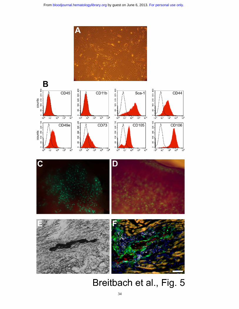
Figure Legends:
Figure 1: Characterization of MSCs. (A) MSCs (passage 4) displayed a fibroblast-like
morphology in culture and were (B) EGFP+ as shown by fluorescence microscopy. (C) flow
cytometry analysis of enriched MSCs (passage 5) proved typical expression of surface
markers; note the lack of the hematopoietic cell markers CD45 and CD11b. (D) Osteogenic
differentiation of MSCs (passage 10) in vitro led to formation of aggregates and trabecular
structures (inset, 17 days); Ca2+ deposition was demonstrated by von Kossa staining. (E) In
vitro differentiation of MSCs (passage 7) into adipocytes. The accumulation of lipid droplets
in vacuoles (inset, 2 weeks in culture) was confirmed by staining with oil red O. (F)
Chondrogenic differentiation of MSCs (passage 10) in vitro was determined using combined
alcian blue/nuclear fast red staining. Bar = 180 µm (A,B), 860 µm (D), 600 µm (D inset), 100
µm (D), 300 µm (E inset), 550 µm (F).
Figure 2: Engraftment of MSCs into the infarcted murine heart; lack of
transdifferentiation of MSCs. (A) Massive engraftment of EGFP+ MSCs (green) in a
cryoinfarcted heart 29 days after injection of 2x10⁶ cells (passage 3); inset shows a
transmission light picture of the heart, the infarcted area is marked by a white dotted line. (B)
Cryosection of border zone of the infarction demonstrating prominent engraftment of EGFP+
MSCs (green) 7 days after injecting 1x10⁵ cells (passage 4). (C) Engrafted EGFP+ MSCs
(green) in the infarcted area 24 days after transplantation of 1x10⁵ cells (passage 3); these
displayed round or elongated shape but were negative for alpha-actinin (red) and desmin
(white). (D) Cardiomyocytes in the intact heart (same section as shown in c) displayed typical
shape and distinct cross-striation. (E) PECAM (red, endothelial marker) and alpha-smooth
muscle-actin (magenta) positive small vessels were identified in vicinity to engrafted MSCs
(green) within and around the lesioned area. None of the vessels showed EGFP+ endothelial
For personal use only. by guest on June 6, 2013. bloodjournal.hematologylibrary.orgFrom
27
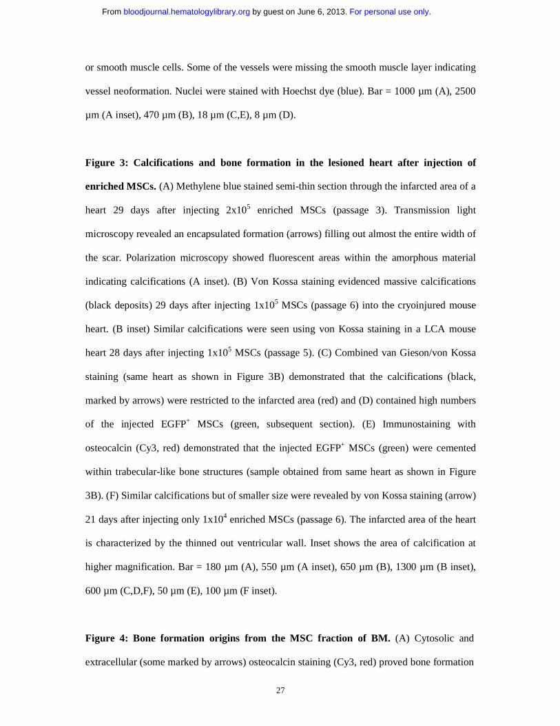
or smooth muscle cells. Some of the vessels were missing the smooth muscle layer indicating
vessel neoformation. Nuclei were stained with Hoechst dye (blue). Bar = 1000 µm (A), 2500
µm (A inset), 470 µm (B), 18 µm (C,E), 8 µm (D).
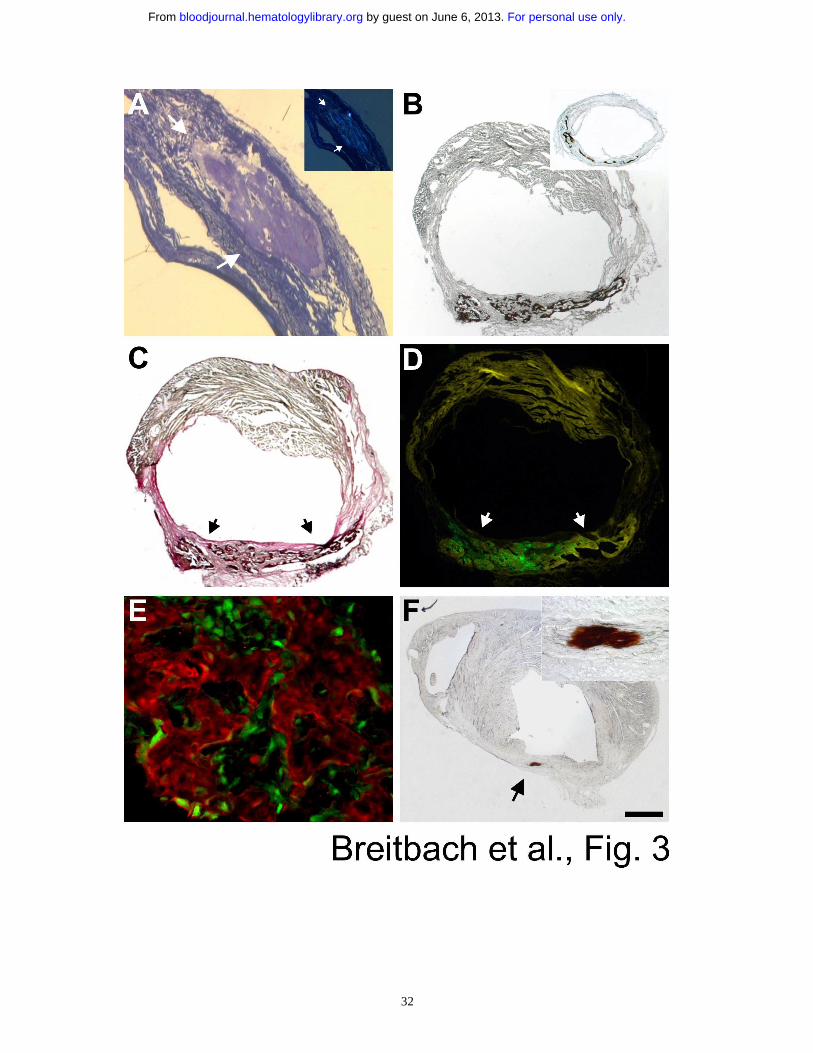
Figure 3: Calcifications and bone formation in the lesioned heart after injection of
enriched MSCs. (A) Methylene blue stained semi-thin section through the infarcted area of a
heart 29 days after injecting 2x10⁵ enriched MSCs (passage 3). Transmission light
microscopy revealed an encapsulated formation (arrows) filling out almost the entire width of
the scar. Polarization microscopy showed fluorescent areas within the amorphous material
indicating calcifications (A inset). (B) Von Kossa staining evidenced massive calcifications
(black deposits) 29 days after injecting 1x10⁵ MSCs (passage 6) into the cryoinjured mouse
heart. (B inset) Similar calcifications were seen using von Kossa staining in a LCA mouse
heart 28 days after injecting 1x10⁵ MSCs (passage 5). (C) Combined van Gieson/von Kossa
staining (same heart as shown in Figure 3B) demonstrated that the calcifications (black,
marked by arrows) were restricted to the infarcted area (red) and (D) contained high numbers
of the injected EGFP+ MSCs (green, subsequent section). (E) Immunostaining with
osteocalcin (Cy3, red) demonstrated that the injected EGFP+ MSCs (green) were cemented
within trabecular-like bone structures (sample obtained from same heart as shown in Figure
3B). (F) Similar calcifications but of smaller size were revealed by von Kossa staining (arrow)
21 days after injecting only 1x10⁴ enriched MSCs (passage 6). The infarcted area of the heart
is characterized by the thinned out ventricular wall. Inset shows the area of calcification at
higher magnification. Bar = 180 µm (A), 550 µm (A inset), 650 µm (B), 1300 µm (B inset),
600 µm (C,D,F), 50 µm (E), 100 µm (F inset).
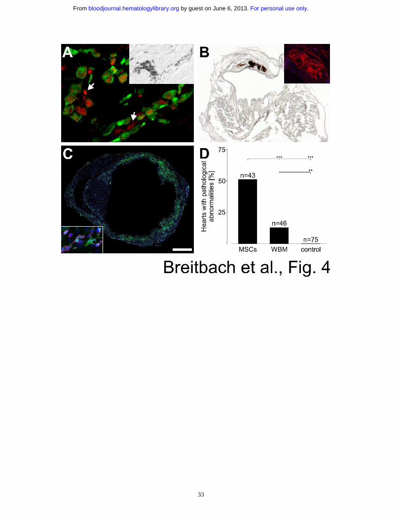
Figure 4: Bone formation origins from the MSC fraction of BM. (A) Cytosolic and
extracellular (some marked by arrows) osteocalcin staining (Cy3, red) proved bone formation
For personal use only. by guest on June 6, 2013. bloodjournal.hematologylibrary.orgFrom
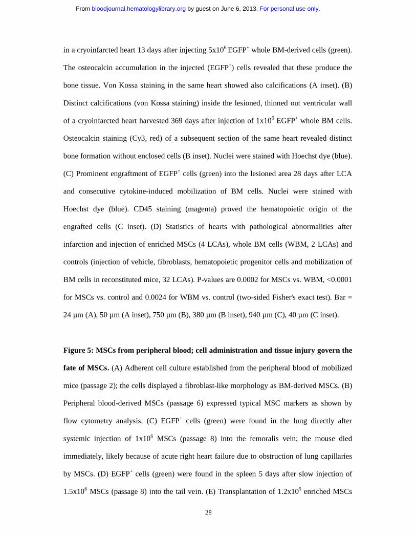
28
in a cryoinfarcted heart 13 days after injecting 5x10⁶ EGFP+ whole BM-derived cells (green).
The osteocalcin accumulation in the injected (EGFP+) cells revealed that these produce the
bone tissue. Von Kossa staining in the same heart showed also calcifications (A inset). (B)
Distinct calcifications (von Kossa staining) inside the lesioned, thinned out ventricular wall
of a cryoinfarcted heart harvested 369 days after injection of 1x10⁶ EGFP+ whole BM cells.
Osteocalcin staining (Cy3, red) of a subsequent section of the same heart revealed distinct
bone formation without enclosed cells (B inset). Nuclei were stained with Hoechst dye (blue).
(C) Prominent engraftment of EGFP+ cells (green) into the lesioned area 28 days after LCA
and consecutive cytokine-induced mobilization of BM cells. Nuclei were stained with
Hoechst dye (blue). CD45 staining (magenta) proved the hematopoietic origin of the
engrafted cells (C inset). (D) Statistics of hearts with pathological abnormalities after
infarction and injection of enriched MSCs (4 LCAs), whole BM cells (WBM, 2 LCAs) and
controls (injection of vehicle, fibroblasts, hematopoietic progenitor cells and mobilization of
BM cells in reconstituted mice, 32 LCAs). P-values are 0.0002 for MSCs vs. WBM, <0.0001
for MSCs vs. control and 0.0024 for WBM vs. control (two-sided Fisher's exact test). Bar =
24 µm (A), 50 µm (A inset), 750 µm (B), 380 µm (B inset), 940 µm (C), 40 µm (C inset).
Figure 5: MSCs from peripheral blood; cell administration and tissue injury govern the
fate of MSCs. (A) Adherent cell culture established from the peripheral blood of mobilized
mice (passage 2); the cells displayed a fibroblast-like morphology as BM-derived MSCs. (B)
Peripheral blood-derived MSCs (passage 6) expressed typical MSC markers as shown by
flow cytometry analysis. (C) EGFP+ cells (green) were found in the lung directly after
systemic injection of 1x10⁶ MSCs (passage 8) into the femoralis vein; the mouse died
immediately, likely because of acute right heart failure due to obstruction of lung capillaries
by MSCs. (D) EGFP+ cells (green) were found in the spleen 5 days after slow injection of
1.5x10⁶ MSCs (passage 8) into the tail vein. (E) Transplantation of 1.2x10⁵ enriched MSCs
For personal use only. by guest on June 6, 2013. bloodjournal.hematologylibrary.orgFrom
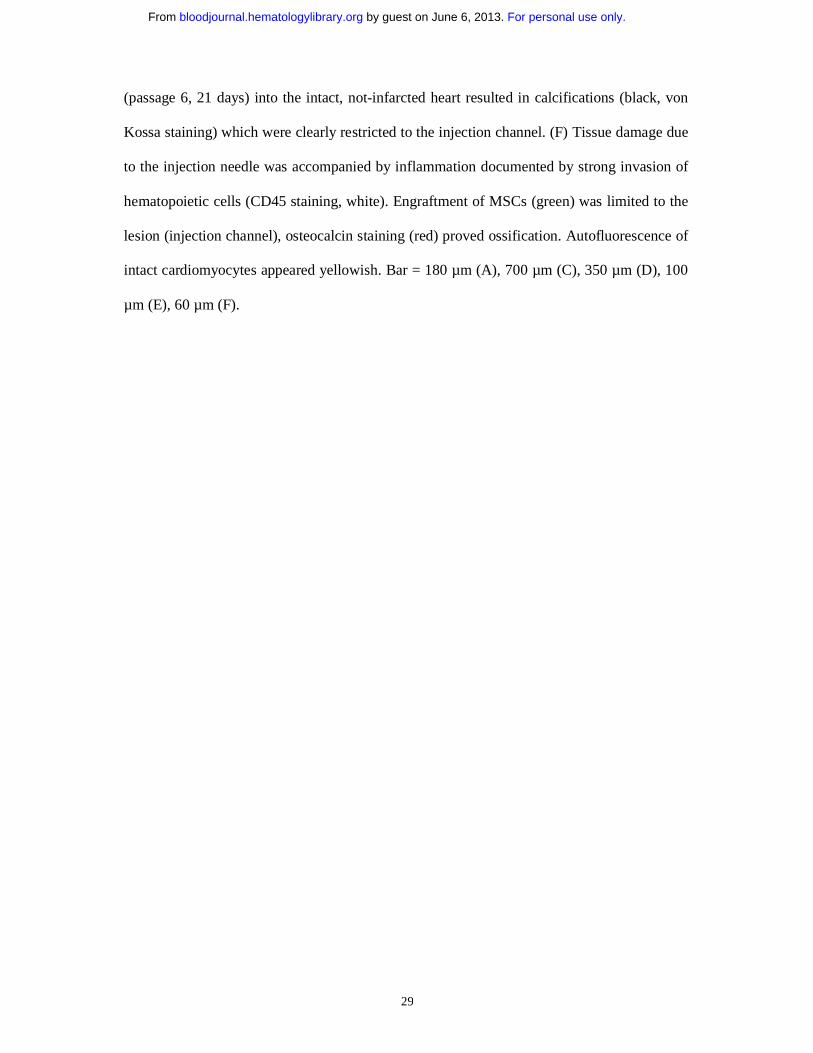
29
(passage 6, 21 days) into the intact, not-infarcted heart resulted in calcifications (black, von
Kossa staining) which were clearly restricted to the injection channel. (F) Tissue damage due
to the injection needle was accompanied by inflammation documented by strong invasion of
hematopoietic cells (CD45 staining, white). Engraftment of MSCs (green) was limited to the
lesion (injection channel), osteocalcin staining (red) proved ossification. Autofluorescence of
intact cardiomyocytes appeared yellowish. Bar = 180 µm (A), 700 µm (C), 350 µm (D), 100
µm (E), 60 µm (F).
For personal use only. by guest on June 6, 2013. bloodjournal.hematologylibrary.orgFrom

30
For personal use only. by guest on June 6, 2013. bloodjournal.hematologylibrary.orgFrom

31
For personal use only. by guest on June 6, 2013. bloodjournal.hematologylibrary.orgFrom
32
For personal use only. by guest on June 6, 2013. bloodjournal.hematologylibrary.orgFrom
33
For personal use only. by guest on June 6, 2013. bloodjournal.hematologylibrary.orgFrom
34
For personal use only. by guest on June 6, 2013. bloodjournal.hematologylibrary.orgFrom
Related Documents