i. ii. iii. iv. v. vi. Potential of Low Voltage Scanning Electron Microscopy Use in Archaeology and History of Art: A Preliminary Study Application Note Introduction Laboratory studies of artifacts have become increasingly common in the last few decades. Archaeologists (both excavators and curators), art historians and conservators have become aware of the importance of analytical studies of these objects for the interpretation, conservation, and restoration of such valuable cultural materials [1–10]. The materials studies usually span the range of metals (bronzes, ferrous alloys, lead, silver, gold), oxide ceramics (pottery, faience, stoneware, porcelain, vitrified materials such as glass, glaze, pâte-de-verre), polymers, and composites. Hence, imaging of artifact microstructure provides invaluable insight for artifacts classification/ characterization, better understanding of their manufacturing technologies, and the best conservation approach or treatment method for their preservation. What is particularly important in the study of archaeological and artistic samples is that not only the examination technique(s) have to be non-destructive, but also that, in most cases, a sample cannot be cut, fractured, or altered. This poses tremendous constraints on the Hamdallah A. Béarat, PhD analyst in sample choice, preparation, and examination, as illustrated by the example sample studied for these papers [9–10]. Therefore, size, shape, and state of the surface dictate the type of analytical technique and instrument chosen. In this application note a few cases are presented where the Agilent 8500 Low Voltage Scanning Electron Microscope (LV-SEM) was used for a given type of artifact to answer specific questions of interest either to archaeology, art history, or conservation science. The samples included in this study are shown in Figure 1. Figure 1. Samples studied in this application note: (i) petrographic thin section of an Iron Age pottery (Hallstatt culture) from Uetliberg, Switzerland; (ii) thin section of a Roman wall painting fragment from Avenches, Switzerland; (iii) Roman wall painting fragment from Pompeii, Italy; (iv) thin sections of glass vessel cores from Tel Amarna, New Kingdom Egypt; (v) and (vi) Egyptian blue and steatite scarabs from Middle Bronze Age Palestine.

Welcome message from author
This document is posted to help you gain knowledge. Please leave a comment to let me know what you think about it! Share it to your friends and learn new things together.
Transcript

i.
ii.
iii. iv.
v. vi.
Potential of Low Voltage Scanning Electron Microscopy Use in Archaeology and History of Art: A Preliminary Study
Application Note
IntroductionLaboratory studies of artifacts have become increasingly common in the last few decades. Archaeologists (both excavators and curators), art historians and conservators have become aware of the importance of analytical studies of these objects for the interpretation, conservation, and restoration of such valuable cultural materials [1–10]. The materials studies usually span the range of metals (bronzes, ferrous alloys, lead, silver, gold), oxide ceramics (pottery, faience, stoneware, porcelain, vitrifi ed materials such as glass,
glaze, pâte-de-verre), polymers, and composites. Hence, imaging of artifact microstructure provides invaluable insight for artifacts classifi cation/characterization, better understanding of their manufacturing technologies, and the best conservation approach or treatment method for their preservation. What is particularly important in the study of archaeological and artistic samples is that not only the examination technique(s) have to be non-destructive, but also that, in most cases, a sample cannot be cut, fractured, or altered. This poses tremendous constraints on the
Hamdallah A. Béarat, PhD
analyst in sample choice, preparation, and examination, as illustrated by the example sample studied for these papers [9–10]. Therefore, size, shape, and state of the surface dictate the type of analytical technique and instrument chosen. In this application note a few cases are presented where the Agilent 8500 Low Voltage Scanning Electron Microscope (LV-SEM) was used for a given type of artifact to answer specifi c questions of interest either to archaeology, art history, or conservation science. The samples included in this study are shown in Figure 1.
Figure 1. Samples studied in this application note: (i) petrographic thin section of an Iron Age pottery (Hallstatt culture) from Uetliberg, Switzerland; (ii) thin section of a Roman wall painting fragment from Avenches, Switzerland; (iii) Roman wall painting fragment from Pompeii, Italy; (iv) thin sections of glass vessel cores from Tel Amarna, New Kingdom Egypt; (v) and (vi) Egyptian blue and steatite scarabs from Middle Bronze Age Palestine.

a. b.
c. d.
Methodology 1. SamplesThe artifacts imaged here are: (1) Iron Age pottery from Switzerland (6th–7th Century BC) [11]; (2) Roman wall painting fragment from Avenches (Aventicum), Switzerland (1st–2nd Century AD) [12]; (3) Roman wall painting fragment from Pompeii, Italy (1st Century AD) [13]; (4) unfi red glass vessel ceramic core from Tel Amarna, New Kingdom Egypt (14th Century BC) [14]; (5) and (6) two scarabs from Middle Bronze Age Palestine (~18th Century BC) made of Egyptian blue and steatite, respectively (unpublished data).
2. Sample Preparation and Imaging Samples were processed and prepared in different ways, but preparation for LV-SEM imaging was minimal. Specimens are simply examined either as-is samples mounted in special holders or as polished sections/thin and adhered via carbon tape to aluminum stubs and loaded onto microscope stage. Samples were prepared for
imaging in different ways. Samples (1), (2) and (4) are thin sections prepared initially for petrographic analysis with polarized light optical microscopy; (3) hand specimen of wall painting fragment examined as is; (5) and (6) hand specimens of scarabs each examined as is and also a small amount was scratched and sprinkled on carbon tape. All samples were imaged in high resolution mode at 1000 V either in SE or BSE modes.
Results and Discussion1. Iron Age pottery from Uetliberg, SwitzerlandThe sample imaged here is an uncovered petrographic thin section of a rare pottery shard from the site of Uetliberg, Switzerland. Many questions are of interest to the archaeologist/archaeometrist when dealing with such rare artifacts. Analyses should provide sound evidence for classifi cation, sourcing, and understanding of fabrication technology. Microscopy has
2
Figure 2. BSE images taken with Agilent 8500 Low Voltage Scanning Electron Microscope (LV-SEM) of a petrographic thin section of Iron Age pottery sample from Uetliberg, Switzerland showing the main morphological features of the composite ceramic material: cementing clayey matrix (M); mineral or non-plastic inclusions (I), macro-cracks and pores (white and black arrows); nano-pores; and striation of the clayey matrix (red arrows). The latter is due to the preferred orientation of clay material during shaping/making of the pot.
the potential to partly answer all three questions. Thus, LV-SEM is used here as non-destructive technique that needs minimal sample preparation while not requiring a coating of the sample. The attempt here is to infer information on the fabrication technology from SEM imaging. Figure 2 includes BSE images showing revealing features of interest. First, qualitative as well as quantitative assessment of naturally occurring non-plastic inclusions (or added temper) relative to the clayey matrix can be done easily on low magnifi cation images. Second, macro- and micro cracks and pores as well as micro-striation or preferred orientation of the matrix certainly indicate less elaborate or skilled treatment of the clay material before shaping of the pot, which is consistent with hand-made vessel technology. However, nano pores are the result of mineral transformation of the clay material during fi ring, such as dehydroxylation/decarbonation that release gases such as H2O and CO2 causing expansion and evolution of the

a. b.
c. d.
3
Figure 3. SE images taken with Agilent 8500 (LV-SEM) of a petrographic thin section of a Roman wall painting fragment from Avenches, Switzerland. Image (a) shows three layers (1, 2, and 3) emphasized in detail in (b), (c) and (d), respectively. (b) Paint layer is composed of vermilion or cinnabar (indicated by P); (c) paint under-layer made of aragonite white pigment rich in micro-fossils, which was a common practice in Roman wall painting; (d) lime plaster made of slaked lime (Ca[OH]2) as binder and calcite or limestone powder as aggregate material. Note the carbonate matrix (binder) in the paint layer (indicated by B) is evidence of a “fresco” painting technique, when the pigment is applied to a fresh layer of lime plaster that causes the lime to diffuse into the paint and carbonate with time thus cementing the paint layer permanently.
porous structure. The evolution of nano-porosity which is certainly a function of the fi ring temperature and atmosphere, and will be discussed in a separate note once the appropriate samples are ready. These remarks indicate a simpler ceramic technology associated with the Iron Age (Hallstatt) culture of pre-Celtic central Europe, compared with Mediterranean World during the same era.
2. Roman wall painting fragment from Avenches, SwitzerlandFigure 3 shows SE images taken of a petrographic thin section of a Roman wall painting fragment from the famous site of Avenches (Aventicum) currently in southwestern Switzerland. Several layers are seen from the paint surface and inward (Fig.3a). The paint layer is composed of a pigment (red cinnabar or �-HgS) and a lime binder. The pigment grains are well rounded, thus indicating that a ground natural pigment was most probably used. A thin layer (Fig. 3b) essentially composed of aragonite
white pigment was applied under the cinnabar. This was a very common practice in Roman painting to apply white aragonite or yellow ochre under cinnabar [8, 12]. The reasons for this technique, is that cinnabar was among the most expensive pigments that unfortunately can turn black under certain environmental conditions [7, 12]. Therefore, it is believed that Roman artists attempted to economize and stabilize the luxurious pigment by applying white or yellow buffering materials such as aragonite (CaCO3) or yellow ochre (goethite-FeO.OH). The aragonite layer imaged here contains numerous micro-fossils (green arrows in Fig. 3c), thus indicating a sedimentary (marine or lacustrous) origin of the white pigment. The third layer (Fig. 3d) is the fi nal coat of lime plaster, which is composed of a carbonate matrix (lime binder) prepared with a carbonate aggregate. Vitruvius, the famous Roman engineer recommended in his “Ten Books of Architecture” for a long lasting construction a plaster made of lime and
an aggregate of spathic calcite or marble dust. The material used here is a powder of a local limestone as substitute.
3. Roman wall painting fragment from Pompeii, ItalyThe sample examined here is a wall painting fragment from the house of Fabio Rufo in Pompeii, Italy [13]. The fragment has light green, white and red bands applied on a yellow background. Only the white area was examined for this study. The fragment shows signs of alteration in the form of deposition/formation of a patina on most of the paint surface. The patina is a coherent and smooth coat of material forming usually on external surface of archaeological materials (and in the pores) as a result of a strong and dynamic chemical interaction with the environment (atmosphere, soil, water). It is simply the product of the interaction between the intrinsic constituents of the sample with extrinsic factors through dissolution (leaching), precipitation, recrystallization on the

a. b.
c. d.
surface and in the pores and cracks. Therefore, this phenomenon is of great importance in archaeological conservation since it alters the physical appearance and integrity of the artifact, but also its chemical and/or mineral composition.
LV-SEM imaging of the white band shows the pigment is aragonite as manifested by the elongated form crystals, typical of aragonite. This is in agreement of XRD analysis [13]. Exposed areas of the fragment shows the plaster is porous and fi ne-grained (Fig. 4a,b), but occasionally with some large crystals of spathic calcite (red arrows in Fig. 4a,c). The paint layer shows both physical and chemical degradation. The former had led to disintegration of the pigment and exposure of the plaster applied underneath (Fig. 4a,b). Chemical degradation is manifested by dissolution of calcium carbonate from the plaster (calcite) and the paint layer (here aragonite) during wet season and
precipitation of calcite during dry season and/or area (Fig. 4 c,d). The high porosity of the plaster (Fig. 4b) facilitates materials transport and crystallization, respectively. The neo-formed calcite smooth surface and rounded shape of grains and their coalescence indicates a dynamic process that had continued over several seasons. The white paint band made of aragonite (Fig. 5a) has also suffered this alteration with different degrees and forms of degradation (Fig. 5b,c,d). Neo-formed calcite continues to grow at the expense of aragonite pigment (Fig. 5d) and plaster calcite (Fig. 4c). It is worth saying that this degradation process that takes place under specifi c environmental conditions pose serious challenges to art conservators and restorers, since transformation of aragonite to calcite is irreversible under such conditions.
4. Unfi red glass vessel ceramic core from Tel Amarna, New Kingdom EgyptAncient Egyptian civilization and material culture in particular have not ceased to fascinate people and impress specialists. In the following examples, three cases of Egyptian pyrotechnology are presented. In the fi rst example, unfi red core samples were investigated. These are pastes shaped and dried before glass was applied to their surface. It has been debated for over a century about the constituents and preparation methods of these glass vessel cores. Sand and clay were proposed by archaeologists despite the inconvenient chemical and/or mechanical properties of the ceramic core produced if sand or clay had been used. In one case, the silica from the sand will melt at the interface with glass vessel wall and it would not have been removable. Use of clay material would have caused a shrinkage in the ceramic core and a misfi t with the glass wall, which would cause its fracture in addition to the diffi culty to remove
Figure 4. SE images taken with Agilent 8500 (LV-SEM) of the top surface of a Roman wall painting fragment from the house of Fabio Rufo in Pompeii, Italy. The paint layer shows both physical and chemical degradation. The former had led to disintegration of the pigment and exposure of the plaster applied underneath (images a and b). Chemical degradation is manifested by dissolution of calcium carbonate from the plaster (calcite) and the paint layer (here aragonite) during wet season and precipitation of calcite during dry season and/or area (images c and d). The high porosity of the plaster (image b) facilitates materials transport and crystallization, respectively. The neo-calcite smooth surface and rounded shape of grains and their coalescence indicates the process had continued over several seasons.
4
a. b.
c. d.
a. b. c.
f. d. e.
Figure 5. SE images taken with Agilent 8500 Low Voltage Scanning Electron Microscope (LV-SEM) of the surface of a white paint layer of the same sample in Figure 4. The paint layer is composed of aragonite pigment, as evidenced by the characteristic elongated crystals (image a and b). Image in (c) shows growth and coalescence of neo-formed, rounded, and smooth calcite crystals. Image in (d) shows the growth of these calcite crystals at the expense of aragonite pigment (red arrows). It is worthy saying that this degradation process that takes place under specifi c environmental conditions does pose serious challenges to art conservators and restorers, since transformation of aragonite to calcite is irreversible under these conditions.
5
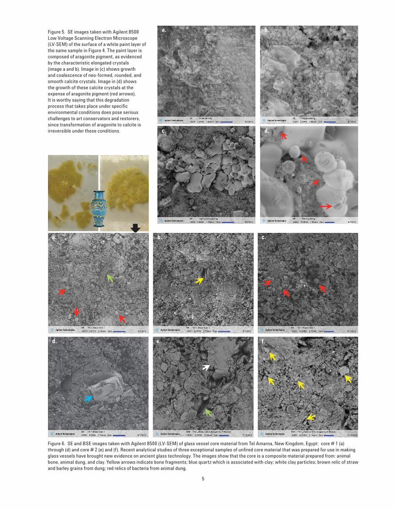
Figure 6. SE and BSE images taken with Agilent 8500 (LV-SEM) of glass vessel core material from Tel Amarna, New Kingdom, Egypt: core # 1 (a) through (d) and core # 2 (e) and (f). Recent analytical studies of three exceptional samples of unfi red core material that was prepared for use in making glass vessels have brought new evidence on ancient glass technology. The images show that the core is a composite material prepared from: animal bone, animal dung, and clay. Yellow arrows indicate bone fragments; blue quartz which is associated with clay; white clay particles; brown relic of straw and barley grains from dung; red relics of bacteria from animal dung.

a. b.
c. d.
the fi red clay (ceramic) from the glass vessel afterward. Some exceptional samples found at Tel Amarna, new capital of King Akhnaten, where glass making workshops were found in the Pharaoh’s palace complex were imaged. Two unfi red samples, core 1 and core 3, already prepared in thin sections (Fig.1, iv) were imaged to elucidate the nature, form, and distribution of its major constituents. Visual and optical microscopy examination have shown the samples were prepared from three major raw materials: animal bone, animal dung, and clay. The latter contained some quartz sand. Figure 5 includes SE and BSE images of the samples. The presence of straw (green arrows), barley grains (separated manually), and relics of bacteria (red arrows) indicate the use of animal dung, and most probably that of horses. Large grains (crystals) of quartz (blue arrow) and of clay minerals (white arrow) indicate the use of some clay as a plastic component to shape the core. However, a larger component is powdered animal bone (yellow arrows),
Figure 7. SE and BSE images taken with Agilent 8500 (LV-SEM) of a scarab made of Egyptian blue from Middle Bronze Age Palestine. Egyptian blue is the common name given to the synthetic blue pigment, which is equivalent to the tetragonal mineral cuprorivaite-CaCuSi4O10). It had been widely used in ancient Egypt both as a pigment in painting and gemstone imitation for inlaid, amulets, scarabs, statuettes, etc. It is widely believed that fabrication was conducted in two fi ring cycles separated by grinding, homogenizing and reshaping of the fi rst frit. Second fi ring is performed to enhance the blue color and adhesion through crystal growth and sintering of the material. Green arrows show examples of crystal growth through solid state reaction.
6
which was most probably that of birds (ducks and chicken). The combination of these ingredient is a fascinating invention, given the properties and availability of these materials. Each would have served different purposes. The animal dung would burn thus providing heat and leaving large pores that prevent shrinkage and ensure a friable product. The animal bone, would provide a refractory strong, but still friable, skeleton for the core. Moreover, it could have been recycled after removal from the glass vessel. The clay, if added as a minor component, would provide a plastic material to help shape the product and give enough strength after drying. Non-adherence to the glass wall was insured through application of a calcium carbonate-rich (marl) material as a slip on the core surface.
5. Egyptian blue scarab from Middle Bronze Age PalestineAmulet, scarabs and small statuettes were very common in ancient Egypt and neighboring countries. They were made in many different materials, whether
natural or synthetic. In most cases, they served as seals with hieroglyphs that have historic and cultural signifi cance. The fi rst scarab examined here comes from a well dated archaeological site in Middle Bronze Age Palestine (corresponding roughly to the 18th Century BC). It is made of Egyptian blue and was imaged as is. The latter is a purely Egyptian invention known from pre-dynastic time through the Roman era. The material was made by mixing quartz, calcite, and copper ore roughly in the stoichiometric oxide composition of the equivalent mineral cuprorivaite (CaCuSi4O10), that is CaO:CuO:SiO2 in the ratios of 1:1:4. As this stoichiometry was hard to control and the reactions take place in the solid state, where diffusion is controlling and limited, craftsmen used to grind the product fi nely, reshape it, and then refi re it to enhance color and crystal (grain) growth. The scarab examined here is friable contrary to vitrifi ed and coherent balls from the Roman times, where crystals with complete facets were seldom seen. Here one sees clearly

a. b.
c. d.
the tetragonal crystals that exceed 10 microns in dimension. Moreover, the blue color is homogeneous throughout the scarab. Hence, the crystal form and size, homogeneity of color, friability, evidence of crystal growth by diffusion (green arrows), all point to a refi ring of the scarab. Another index to refi ring is the fact that the inscription on the fl at surface of the scarab (Fig. 1v). The inscription would have suffered from melting if it was applied before fi rst fi ring.
6. Steatite scarab from Middle Bronze Age PalestineThis sample comes from the same context as the precedent one (Fig. 1vi) and was imaged as is. It is made of steatite or soapstone, which is composed essentially of talc (Mg3Si4O10[OH]2). This is a very convenient material to carve or shape, given its reduced hardness (1 on Moh’s scale). The scripted fl at surface shows traces of light green glaze rich in copper. Therefore, the scarab might have been fully glazed, but the glaze
Figure 8. BSE images taken with Agilent 8500 (LV-SEM) of a scarab made of steatite from Middle Bronze Age Palestine. Steatite is the common name given to a metamorphic rock composed of talc (which is a hydrated magnesium silicate- Mg3Si4O10(OH)2, also called soapstone and soap rock. Due to its low hardness it can be carved and shaped easily and once fi red, it acquires enough strength. Images show the grains to be rounded with a smooth surface; their size varies from submicron to above 10 microns. As the scarab was fi red, the grain surface shows a thin coat of amorphous material produced by dehydroxylation of the talc mineral.
7
has deteriorated as a result of use or burial. The steatite surface is rough and friable. images obtained for this sample show well rounded grains with smooth surface. BSE images (Figure 8) show the grains to be rounded and have a smooth surface; their size vary from submicron to above 10 microns. As the scarab was fi red, the grain surface shows a thin coat of amorphous material produced by dehydroxylation of the talc mineral. Cementing material is absent, which explains the friability of the sintered product. This in turn may explain the motivation of glazing the scarab to minimize its erosion and disintegration. A goal that was met at least in part, since the object has survived almost 4000 years of burial.
ConclusionsUsing Agilent 8500 LV-SEM, it was quite possible to image, with high resolution and excellent contrast, several artifact samples that are too precious to alter, cut or fracture. Given the fact that these insulating materials,
whether inorganic or a mix of organic and inorganic, were quite easy to image without the need for a metal coating or radiation damage to the samples. For instance, a petrographic thin section cannot be coated for imaging, as this will jeopardize further examination with optical microscopy. Other samples such as wall painting fragments or scarabs of Egyptian blue and steatite were imaged as is, without the need for coating or taking a specimen. These analyses are otherwise not as feasible using other FE-SEMs: cold fi eld emission SEM operating at low voltage and E-SEM, respectively. It is fair to say also that Agilent 8500 LV-SEM is proving more helpful in studying valuable archaeological and artistic samples such as painting and pigments, ceramic and sintered materials, textile, glasses and glazes, etc., when compared to other FE-SEMs currently available.

References
[1] Kayani, P.I.; McDonnell, G. An assessment of back-scattered electron petrography as a method for distinguishing Mediterranean obsidians
[2] Klaus, A.V. Museum Application for SEM and X-Ray Microanalysis. In: Industrial Applications of Electron Microscopy, Ed. Z.R. Li. NY, 2003, 259–78.
[3] Burnstock, A.; Jones, C., Scanning electron microscopy techniques for imaging materials from paintings. In: Radiation in Art and Archaeometry. Creagh, D,C., Bradley, D.A., Eds. Amsterdam, 2000, 202–231.
[4] Turner-Walker, G.; Nielsen-Marsh, C.M.; Syversen, U.; Kars, H.; Collins, M.J. Sub-micron Spongiform Porosity is the Major Ultra-structural Alteration Occurring in Archaeological Bone. Int. J. Osteoarchaeol. 12, 2002, 407–414.
[5] Adriaens, A.; Dowsett, M.G. Electron Microscopy and its Role in Cultural Heritage Studies. In: Non-destructive Microanalysis of Cultural Heritage Materials, Volume 42. Janssens, K.H.A. and Van Grieken R. Eds. Amsterdam, 2004, 73–128.
[6] Salomon, H. Vignaud, C., Coquinot, Y., Beck, L., Stringer, C., Strivay, D., D’Errico, F., Selection and heating of colouring materials in the Mousterian level of Es-Skhul (c. 100,000 years BP, Mount Karmel, Israel), Archaeometry 54, 4 (2012) 698–722.
[7] Béarat, H. Chizmeshya, A., Sharma, R., Barbet, A., Fuchs, M., Mechanistic and computational study of cinnabar phase transformation: Applications and implications to the preservation of this pigment in historical painting. In: The 3rd International Conference on “Science and Technology in Archaeology and Conservation”, (December, 7–11 2004, Jordan). Editor: T.S. Akasheh, Foundation El Legado Andalusi, Spain, 2008, 53–70.
[8] Béarat H., 1997, Quelle est la gamme exacte des pigments romains? Confrontation des résultats d’analyse et des textes de Vitruve et de Pline. In: Roman Wall Paintings: Materials, Techniques, Analysis and Conservation. Proceedings of the International Workshop, Fribourg, 7–9 March 1996, H. Béarat, M. Fuchs, M. Maggetti, D. Paunier, (Eds.), 11–34.
[9] Béarat H., 1997, Pigments verts en peinture murale romaine: bilan analytique. In: Roman Wall Paintings: Materials, Techniques, Analysis and Conservation. Proceedings of the International Workshop, Fribourg, 7–9 March 1996, H. Béarat, M. Fuchs, M. Maggetti, D. Paunier, (Eds.), 269–287.
[10] Shannon P. McPherron, Zeresenay Alemseged, Curtis W. Marean, Jonathan G. Wynn, Denné Reed, Denis Geraads, René Bobe, Hamdallah A. Béarat, 2010, Evidence for stone-tool-assisted consumption of animal tissues before 3.39 million years ago at Dikika, Ethiopia. Nature, 466 (12 August) 857–60.
[11] Béarat H., Bauer I., Früheisenzeitliche Keramik von Baarburg ZG und Uetliberg ZH: Eine mineralogisch- petrographische und chemische Untersuchung zur Frage der Herstellungsorte scheibengedrehter Keramik in der ausgehenden Hallstattzeit, Germania 72,1 (1993) 67–93.
[12] Béarat H., Fuchs M., Analyses physico-chimiques et minéralogiques des peintures murales romaines d’Avenches, I: du pigment à Avenches, Bulletin Pro Aventicum 38 (1996) 35–51.
[13] Varone A., Béarat H., 1997, Pittori Romani al lavoro: materiali, strumenti, techniche. Evidenze archeologiche e dati analitici di un recente scavo pompeiano lungo via dell’Abbondanza (Reg. IX. Ins. 12). In: Roman Wall Paintings: Materials, Techniques, Analysis and Conservation. Proceedings of the International Workshop, Fribourg, 7–9 March 1996, H. Béarat, M. Fuchs, M. Maggetti, D. Paunier, (Eds), 199–214.
[14] Neveen A. Salim, A Historical and Technology study of The Ancient Egyptian Glass Core Formed Vessels. Doctoral Dissertation, 2008, Helwan University, Egypt.
Nanomeasurement Systems fromAgilent Technologies
Agilent Technologies, the premier measurement company, offers high-precision, modular nanomeasurement solutions for research, industry, and education. Exceptional worldwide support is provided by experienced application scientists and technical service personnel. Agilent’s leading-edge R&D laboratories ensure the continued, timely introduction and optimization of innovative, easy-to-use nanomeasure system technologies.
www.agilent.com/find/nano
Americas
Canada (877) 894 4414Latin America 305 269 7500United States (800) 829 4444
Asia Pacifi c
Australia 1 800 629 485China 800 810 0189Hong Kong 800 938 693India 1 800 112 929Japan 0120 (421) 345Korea 080 769 0800Malaysia 1 800 888 848Singapore 1 800 375 8100Taiwan 0800 047 866Thailand 1 800 226 008
Europe & Middle East
Austria 43 (0) 1 360 277 1571Belgium 32 (0) 2 404 93 40Denmark 45 70 13 15 15Finland 358 (0) 10 855 2100France 0825 010 700* *0.125 €/minute
Germany 49 (0) 7031 464 6333Ireland 1890 924 204Israel 972-3-9288-504/544Italy 39 02 92 60 8484Netherlands 31 (0) 20 547 2111Spain 34 (91) 631 3300Sweden 0200-88 22 55Switzerland 0800 80 53 53United Kingdom 44 (0) 118 9276201Other European Countries: www.agilent.com/fi nd/contactus
Product specifi cations and descriptions in this document subject to change without notice.
© Agilent Technologies, Inc. 2012Printed in USA, November 8, 20125991-1512EN
Related Documents