Journal of Liposome Research, 2009; 19(4): 332–340 RESEARCH ARTICLE Span and Tween neutral and pH-sensitive vesicles: Characterization and in vitro skin permeation Maria Carafa 1 , Carlotta Marianecci 1 , Federica Rinaldi 1 , Eleonora Santucci 1 , Silvia Tampucci 2 , and Daniela Monti 2 1 Department Of Chimica e Tecnologie del Farmaco, University of Rome “Sapienza,” Rome, Italy, and 2 Department of Chimica Bioorganica e Biofarmaceutica, University of Pisa, Pisa, Italy Address for Correspondence: Maria Carafa, Department of Chimica e Tecnologie del Farmaco, Faculty of Pharmacy, University of Rome “Sapienza,” P.le A. Moro 5, 00185 Rome, Italy; Fax: +390649913133. E-mail: [email protected] (Received 31 January 2009; revised 14 April 2009; accepted 04 May 2009) Introduction Transdermal and topical delivery of drugs provide advantages over conventional oral administration. ese advantages include convenience, improved patient compliance, and elimination of hepatic first-pass effect. Nevertheless, the transdermal administration of a drug is often compromised by its low bioavailability due to the skin-barrier function, represented by the stratum corneum, a complex structure of compact, keratinized cell layers, that presents the greatest barrier to the absorption of topically or transdermally administered drugs. Phospholipid-based artificial vesicles (e.g., lipo- somes, ethosomes, and transfersomes) and niosomes (i.e., nonionic surfactant vesicles) are widely used to enhance drug permeation across the skin in cosmetic and dermatological fields (Fresta and Puglisi, 1996; Banga et al., 1999; Fang et al., 2001; Touitou et al., 2001; Cevc, 2004; Williams and Barry, 2004; Tokumoto et al., 2006; Touitou and Godin, 2006). Further, surfactant vesicles are supposed to interact with human skin by modifying, especially, the horny layer characteristics and to preferentially interact with inflamed epidermal tissue (Junginger et al., 1991). Ibuprofen (IBU), a nonsteroidal anti-inflammatory drug (NSAID), is very effective for the systemic treat- ment of rheumatoid arthritis, osteoarthritis, and anky- losing spondylitis. IBU, a lipophilic drug, was formulated into many topical preparations to reduce adverse side effects and avoid hepatic first-pass metabolism (Stott et al., 1998; Park and Kim, 1999; Irevolino et al., 2001; Heard et al., 2003; Perioli et al., 2004; Cilurzo et al., 2005; Chen et al., 2006; Li et al., 2008). ISSN 0898-2104 print/ISSN 1532-2394 online © 2009 Informa UK Ltd DOI: 10.3109/08982100903014994 Abstract The aim of this work was the preparation, characterization, and comparison of novel pH-sensitive nonphos- pholipid vesicles (niosomes) from two nonionic surfactants, with different hydrophilic-lipophilic balance values (Tween 20-TW20 = 16.7 and Span 60-SP60 = 4.7). Surfactants were mixed with cholesterol (CHOL) and its derivative, cholesteryl hemisuccinate (CHEMS), as a pH-sensitive molecule. Vesicles were character- ized by dynamic light scattering, in order to evaluate their dimensions and vesicle stability, by ζ-potential measurements and by means of electronic microscopy after freeze-fracture. Ibuprofen (IBU) was used as the model drug, and high-performance liquid chromatography analyses were performed to evaluate drug-entrapment efficiency and release in a neutral, acidic environment. The influ- ence of the vesicle composition on skin accumulation and transdermal permeation of IBU across excised hairless rat skin was investigated by using vertical Gummer diffusion cells. When niosomes with SP60 and CHEMS were prepared, there was a statistically significant increase of skin permeation of IBU, while TW20 niosomes did not show statistically significant differences in P app values without the influence of the vesicle size and charge. Keywords: Tween ; Span ; pH-sensitive surfactant vesicles; ibuprofen; skin permeation http://www.informahealthcare.com/lpr

Welcome message from author
This document is posted to help you gain knowledge. Please leave a comment to let me know what you think about it! Share it to your friends and learn new things together.
Transcript
Journal of Liposome Research, 2009; 19(4): 332–340
R E S E A R C H A R T I C L E
Span and Tween neutral and pH-sensitive vesicles: Characterization and in vitro skin permeation
Maria Carafa1, Carlotta Marianecci1, Federica Rinaldi1, Eleonora Santucci1, Silvia Tampucci2, and Daniela Monti2
1Department Of Chimica e Tecnologie del Farmaco, University of Rome “Sapienza,” Rome, Italy, and 2Department of Chimica Bioorganica e Biofarmaceutica, University of Pisa, Pisa, Italy
Address for Correspondence: Maria Carafa, Department of Chimica e Tecnologie del Farmaco, Faculty of Pharmacy, University of Rome “Sapienza,” P.le A. Moro 5, 00185 Rome, Italy; Fax: +390649913133. E-mail: [email protected]
(Received 31 January 2009; revised 14 April 2009; accepted 04 May 2009)
Introduction
Transdermal and topical delivery of drugs provide advantages over conventional oral administration. These advantages include convenience, improved patient compliance, and elimination of hepatic first-pass effect. Nevertheless, the transdermal administration of a drug is often compromised by its low bioavailability due to the skin-barrier function, represented by the stratum corneum, a complex structure of compact, keratinized cell layers, that presents the greatest barrier to the absorption of topically or transdermally administered drugs. Phospholipid-based artificial vesicles (e.g., lipo-somes, ethosomes, and transfersomes) and niosomes (i.e., nonionic surfactant vesicles) are widely used to enhance drug permeation across the skin in cosmetic and dermatological fields (Fresta and Puglisi, 1996;
Banga et al., 1999; Fang et al., 2001; Touitou et al., 2001; Cevc, 2004; Williams and Barry, 2004; Tokumoto et al., 2006; Touitou and Godin, 2006).
Further, surfactant vesicles are supposed to interact with human skin by modifying, especially, the horny layer characteristics and to preferentially interact with inflamed epidermal tissue (Junginger et al., 1991).
Ibuprofen (IBU), a nonsteroidal anti-inflammatory drug (NSAID), is very effective for the systemic treat-ment of rheumatoid arthritis, osteoarthritis, and anky-losing spondylitis.
IBU, a lipophilic drug, was formulated into many topical preparations to reduce adverse side effects and avoid hepatic first-pass metabolism (Stott et al., 1998; Park and Kim, 1999; Irevolino et al., 2001; Heard et al., 2003; Perioli et al., 2004; Cilurzo et al., 2005; Chen et al., 2006; Li et al., 2008).
ISSN 0898-2104 print/ISSN 1532-2394 online © 2009 Informa UK LtdDOI: 10.3109/08982100903014994
AbstractThe aim of this work was the preparation, characterization, and comparison of novel pH-sensitive nonphos-pholipid vesicles (niosomes) from two nonionic surfactants, with different hydrophilic-lipophilic balance values (Tween20-TW20 = 16.7 and Span60-SP60 = 4.7). Surfactants were mixed with cholesterol (CHOL) and its derivative, cholesteryl hemisuccinate (CHEMS), as a pH-sensitive molecule. Vesicles were character-ized by dynamic light scattering, in order to evaluate their dimensions and vesicle stability, by ζ-potential measurements and by means of electronic microscopy after freeze-fracture.Ibuprofen (IBU) was used as the model drug, and high-performance liquid chromatography analyses were performed to evaluate drug-entrapment efficiency and release in a neutral, acidic environment. The influ-ence of the vesicle composition on skin accumulation and transdermal permeation of IBU across excised hairless rat skin was investigated by using vertical Gummer diffusion cells. When niosomes with SP60 and CHEMS were prepared, there was a statistically significant increase of skin permeation of IBU, while TW20 niosomes did not show statistically significant differences in Papp values without the influence of the vesicle size and charge.
Keywords: Tween; Span; pH-sensitive surfactant vesicles; ibuprofen; skin permeation
http://www.informahealthcare.com/lpr

Neutral and pH-sensitive vesicles 333
But, it is difficult to maintain effective concentra-tions by the topical delivery of IBU due to its poor skin-permeation ability (Yano et al., 1986).
The first step of this work was the preparation, characterization, and comparison of novel pH-sensi-tive nonphospholipid vesicles from two nonionic sur-factants, with different hydrophilic-lipophilic balance (HLB) values (Tween20 = 16.7 and Span 60 = 4.7). Surfactants were mixed with cholesterol (CHOL) and its derivative, cholesteryl hemisuccinate (CHEMS), as a pH-sensitive, fusogenic molecule (Hafez and Cullis, 2000).
The inclusion, in vesicle composition, of molecules with fusogenic properties results in the formation of the so-called ‘‘fusogenic’’ or polymorphic liposomes (Simôes et al., 2004), since these undergo a phase tran-sition under acidic conditions, either in the absence or presence of biological membranes.
Also, pH-sensitive surfactant vesicles can be destabi-lized when the external pH is changed, usually from a neutral or slightly alkaline to an acidic pH, such as skin pH, and deliver their content (Carafa et al., 2006).
The second step was the evaluation, by means of in vitro permeation experiments through rat skin, of the effect of the neutral, pH-sensitive nonphospholipid vesicles, in comparison to micellar formulations, on IBU transport into and through the skin.
Materials and methods
Materials
Tween 20 (TW20), Span 60 (SP60), and CHOL were Merck products (Whitehouse Station, USA). CHEMS was an ICN Biomedical product (Eschwege, Germany). Hepes salt {N-(2-idroxyethyl) piperazine-N’-(2-ethanesulfonicacid)}, Sephadex G-75, hydroxypyrene-1,3,6-trisulfonic acid (HPTS), and calcein were Sigma products (Milano, Italy). Ibuprofen was a generous gift of Angelini Farmaceutici (Bologna, Italy). All other prod-ucts and reagents were of analytical grade.
Sample preparation and purification
Composition of the tested samples is reported in Table 1; as can be observed, the TW20 concentrations were always above the critical micellar concentration (CMC in water at 20°C: TW20: 8.04 10−5 M; CMC for SP60: not detectable).
Unilamellar vesicles were obtained by means of the “film” technique, as previously reported (Carafa et al., 2006). The dried film was hydrated by the addition of an aqueous phase (HEPES buffer, 0.01 M; pH 7.4). The dis-persions containing TW20 were vortexed for 15 minutes, and the ones containing SP60 were magnetically stirred
for 1 hour at 60°C; the obtained dispersion were then sonicated (VCX 400; Sonics and Materials, Newtown, Connecticut, USA), at 60°C for 10 minutes.
To obtain drug-loaded vesicles, the drug (1-2-3-5-7%; w/v) was added to the initial organic phase, before “film” formation, because of the IBU lipophilic nature, for the preparation of IBU (1-2-3-5-7%; w/v)-loaded micelles in HEPES, the drug and the surfactants were maintained under magnetic stirring overnight.
In order to separate loaded vesicles from unentrapped drug, the vesicle dispersion was purified by gel filtration on a Sephadex G-75 (glass column, 50 1.2 cm), using HEPES buffer as an eluent; on the contrary, micellar solutions were not purified.
Vesicle characterization
Freeze-fractureVesicles were examined by means of the freeze-fracture microscopy technique: The samples were impregnated in 30% glycerol and then frozen into partially solidified Freon 22 (Rivoira, Rome, Italy), freeze-fractured in a freeze-fracture device (–105°C, 10−6 mmHg), and repli-cated by evaporation from a platinum/carbon gun; the replicas were extensively washed with distilled water, picked up onto Formvar, Polysciences Europe Gmbh (Eppelheim, Germany) coated grids, and examined with a Philips (Amsterdam, The Netherlands) CM 10 trans-mission electron microscope.
Structured surfactant assaysTo evaluate the percentage of surfactant actually struc-tured within the vesicular structure, in purified surfactant
Table 1. Sample compositions.
Sample TW20 (mM) SP60 (mM) CHOL (mM)CHEMS
(mM)
1 15 — 15 —
2 15 — 7.5 7.5
3 15 — 2 13
4 — 33 33 —
5 — 33 16.6 16.5
6 — 33 6.8 26
Table 2. Vesicle dimensions, evaluated by dinamic light scattering, -potential values, and percentage of structured surfactant of “neutral” and pH-sensitive vesicles (reported data are the means of at least three experiments ± SD).
Sample
Diameter (nm)
pH 7.4
Diameter (nm)
pH 5.5 potential
pH 7.4(mV) potential
pH 5.5 (mV)
% of structured surfactant
1 153 ± 5 159 ± 1 –38.71 ± 2.45 –35.66 ± 0.76 40.33 ± 0.10
2 160 ± 3 190 ± 2 –43.16 ± 4.15 –8.45 ± 1.76 39.03 ± 0.16
3 132 ± 5 142 ± 6 –48.04 ± 3.32 –5.31 ± 1.37 43.70 ± 0.07
4 155 ± 5 190 ± 1 –74.71 ± 1.45 –47.66 ± 0.76 97.21 ± 0.11
5 158 ± 3 187 ± 2 –94.16 ± 3.15 –39.47 ± 1.76 88.36 ± 0.10
6 154 ± 3 230 ± 5 –90.04 ± 2.78 –38.31 ± 1.25 98.07 ± 0.05

334 Carafa et al.
vesicle samples, TW20 and SP60 were dosed and the data are reported in Table 2. The concentration of TW20 was determined by the spectrophotometric method after sample treatment with cobalt thiocyanate solution and extraction with CH
2Cl
2; the determination of the absorb-
ance of the organic phase was determined at 620 nm (Kato et al., 1989). The concentration of SP60 was determined by high-performance liquid chromatography (HPLC) (Perkin Elmer, Marza (MI) Italy, 250 LC): the column was a Supelcogel, Sigma-Aldrich (Milano, Italy) ODP-50 ( 15 cm 4.00 mm; L x I.D.); the mobile phase was an 85/15 mixture of isopropanol/H
2O, and the detection was
carried out at 220 nm (Wang and Fingas, 1994).
Size measurements, -potential, and stability testsSize measurements and evaluation of vesicle stability were carried out on vesicle HEPES suspensions by means of dynamic light scattering. The vesicle dispersions were diluted about 100 times in the same buffer used for their preparation. Dust particles were eliminated by filtration (0.45 m) from the buffer solution as well as from the vesicle preparation. Vesicle-size distribution was meas-ured on a Malvern Nano ZS90 (Malvern, UK) at 25°C, with a scattering angle of 90.0 degrees. Before vesicle suspen-sion filtration, often the samples seem to be characterized by the presence of two populations: the first with the same diameter of the filtrate vesicle dispersion and the second with a 5-µm diameter; this one was ascribed to dust parti-cles present in our suspension and probably not to vesicle aggregates. The percentage of this population was also very low, so it was our opinion that their elimination was not very influent on sample characteristics changing.
The polidispersity index (PI) was directly calculated by the software of the apparatus, and the values obtained are in agreement with a mono disperse vesicular system. The same apparatus was used for the evaluation of zeta potential, using a vesicle preparation appropriately diluted (1:10) in HEPES solution at 25°C.
Vesicle stability, in terms of changes in vesicle dimen-sions after aggregation, was evaluated by using the same technique on samples stored, up to 1 month, at 4 and 25°C. This kind of information was also confirmed by means of zeta-potential measurements.
Evaluation of pH-sensitivity in vitro
Fluorescence analyses with a pH-sensitive probe (HPTS)Fluorescence analyses were carried out with a Perkin-Elmer LS50B spectrofluorometer on HPTS-loaded vesicles (Kano and Fendler, 1978), prepared in HEPES buffer (pH 7.4), and diluted with the same buffer or with acetate buffer (pH 5.5) to eliminate the scattering raising from dispersed colloidal particles and to obtain measur-able fluorescence values.
The excitation spectra (300–600 nm) were followed at an emission wavelength of 510 nm, and the emission spec-tra (300–600 nm) were taken at an excitation wavelength of 400 nm. The intensities of the maxima, in the excitation spectra, at 400 and 450 nm were strongly dependent on the hydrogen-ion concentration, and at pH 5.5, the rela-tive intensity of the 450-nm peak is negligible.
Fluorescence analyses with calceinCalcein was encapsulated in surfactant vesicles at a concentration of 10−2M, at which concentration its fluo-rescence is self-quenched (Ohkuma and Poole, 1978). Next, 100 µL of calcein-loaded vesicles was added to 2.5 mL of HEPES buffer (pH = 7.4) or to 2.5 mL of acetate buffer (pH = 5.5), in a fluorometer cuvette at 37°C under constant stirring. Following a 10-minute incubation, cal-cein fluorescence was measured at
ex = 492 nm and
em
= 520 nm, before and after the addition of 20 L of 10% (v/v) Triton X-100, using a Perkin-Elmer LS-5 fluorom-eter. Fluorescence intensities obtained at acidic pH val-ues were corrected for the slight effect of pH on calcein fluorescence, using two different calibration curves at different pH values. To calibrate the fluorescence scale, 100% leakage was achieved by the addition of Triton X-100. The percentage of calcein leakage was calculated according to Equation 1 (Simôes et al., 2004):
% =−
−×leakage
I I
I I100
pH 0
100 0
(1)
where I0 is the fluorescence at neutral pH, I
pH is the
corrected intensity at acidic pH before the addition of Triton X-100, and I
100 is the totally dequenched calcein
fluorescence at neutral pH.
Drug-encapsulation efficiency and in vitro release kinetics
The drug entrapment within the vesicles was assessed by HPLC (Perkin Elmer 250 LC) on purified vesicles, after their disruption by means of freeze-drying. All vesicles suspensions were freeze-dried in the absence of cryoprotectants to obtain a disaggregation of the bilayer structure: the resulting powder was resuspended in acetonitrile to prevent a spontaneous vesicle aggrega-tion; the solution obtained was analyzed by HPLC.
The column was a Supelcogel ODP-50( 15 cm 4.00 mm; L x I.D.); the mobile phase was a 60/40 mixture of CH
3CN/H
2O (pH = 3, with H
3PO
4); the detection was
carried out at 255 nm.Drug-encapsulation efficiency (EE) was espressed as
moles of drug/moles of structured surfactant 100.In vitro release experiments were performed in a
flow-through apparatus (F.U.I. XI ed.; Italian Official

Neutral and pH-sensitive vesicles 335
Pharmacopoeia) at 32 ± 0.5°C in HEPES buffer (pH 7.4). Fixed volumes (200 L) of vesicle samples were included in dialysis sacs (cut-off, 8,000 Da) with a fixed diffusing area (5.5 cm2). The drug concentration was detected in the outer solution at fixed time intervals by means of the HPLC method described above, taking into account the dilution factor.
The release kinetics were assesed by using Equation 2 (Sinclair and Peppas, 1984):
M M Kt t infn⁄ =
(2)
where Mt is the amount of drug released at time t, M
inf is
the loaded drug amount, K is the kinetic constant, and n is the exponent, related to the release mechanism. K- and n-values were calculated by regression analysis.
In vitro skin permeation studies
In order to evaluate the influence of vesicle composition and surface charge on IBU skin accumulation and diffu-sion, we carried out in vitro permeation studies by using rat skin obtained from 6–8-week-old hairless male animals (OFA-hr/hr; Charles River Italia, Lecco, Italy, SpA). The animals were killed by cervical dislocation immediately before the experiments, the skin was carefully excised, and the adhering fat and subcutaneous tissue were removed. Portions of this skin (epidermis+dermis) were then placed as a barrier between the two halves of the permeation cells.
As described in a previous article (Monti et al., 1995), Franz-type cells of the “GH” design specified by Gummer (Gummer et al., 1987), with an available diffusion area of 1.23 cm2 and stratum corneum facing the donor compartment, were used. First, 1.0 mL of each formulation was placed on the skin surface. The formulations tested were niosomal samples 1–6, and as a reference, we used (pH 7.4) HEPES buffer solution of IBU (SOL-R 0.030 mg/mL) and vehicles containing SP60 (3.0 mg/mL; V1) and TW20 (1.5 mg/mL; V2) in the same amount as the niosomal formulation, but not organized in a vesicular structure.
The receptor compartment contained 5.0 mL of isot-onic (66.7 mM; pH 7.4) phosphate buffer solution, main-tained at 37°C and stirred at 600 rpm.
At predetermined time intervals, samples of the medium were withdrawn for analysis and replaced with an equal volume of fresh buffer; the amount of perme-ated IBU was determined by HPLC.
The permeation experiments lasted 20 hours and each test was replicated at least six times.
Linear regression analysis of pseudo–steady-state diffusion data allowed calculation of the steady-state flux, J, given by Q/A.t, where Q is the amount of per-meant diffusing across the area A in time t. The apparent
skin-permeability coefficients (Papp) for IBU were obtained from the relationship shown in Equation 3:
Papp J Cd= /
(3)
derived from Fick’s first law, where Cd is the initial drug
concentration in the donor phase. The permeation lag times (indicating the time taken by the drug to saturate the skin and to reach the receiving compartment) were calculated from the x-axis intercept values of the regres-sion lines. Enhancement factors (EFs) were calculated from the ratio Pb/Pa, where Pb and Pa are the perme-ability coefficients of the vehicles under study and of the reference (SOL-R), respectively.
At the end of the in vitro permeation experiments, the skin was removed (surface: 1.23 cm2), rinsed with dis-tilled water, and the drug accumulated into the skin was extracted by treating homogenized skin with 2 mL of 2% sodium dodecyl sulphate for 20 hours at room tempera-ture. Then, 4 mL of methanol were added under stirring for 3 hours. The dispersion was clarified with 0.22-m filters and 250-µL aliquots were dried in vacuo and, sub-sequently, dissolved in methanol for HPLC analysis.
The HPLC (LC-6A pump and 20-L Reodyne injector, SPD-6AV detector, and computer integrating system; Shimadzu Corp., Kyoto, Japan) analyses were carried out by using a column ( 15 cm 4.6 mm) packed with Synergy fusion–RP 80A C18 (size, 4 m; Phenomenex, Castel Maggiore (BO) Italy). The mobile phase (flow rate, 1.0 mL/min) was 40:60 (v/v) water (pH 2.6, with phosphoric acid):acetonitrile; the retention time and the detection wavelength were 6.0 minutes and 225 nm, respectively.
The amount of IBU in the samples was determined by comparison with a standard curve. In case of biologi-cal materials, standard curve was obtained by adding increasing amounts of IBU to blank biological samples.
For validation of the extraction procedure, a different sample of blank skin was submitted to the assay, and the retention time of endogenous compounds was com-pared with that of IBU.
The extraction recovery was determined by comput-ing the ratio of the drug amount extracted from the skin to the amount added.
Statistical evaluation of data
For characterization studies, results are expressed as the mean of three experiments ± standard deviation. Statistical data analysis were performed by using the t-test, with P < 0.01 as a minimal level of significance. For in vitro skin permeation studies, statistical differ-ences between means were assessed by analysis of variance (ANOVA) (StatView software; Abacus Concepts Inc., Berkeley, CA, USA). The evaluation included the

336 Carafa et al.
calculation of mean and standard errors and group comparisons using Fisher’s PLSD test. Differences were considered statistically significant at P < 0.05.
Results and discussion
Influence of CHEMS on surfactant vesicle formation
In our previous work, we evidenced the fundamental role of CHOL on nonionic surfactant vesicle formation (Santucci et al., 1996). Then, the influence of CHEMS on TW20 vesi-cle and on SP60 vesicle formation was studied. As shown in Figure 1-a and 1-b, at a neutral pH, we obtained unilamellar vesicles comparable to “neutral” formulations, even using a high amount of CHEMS. At an acidic pH (i.e., 5.5), we noticed an increase in vesicle dimensions for pH-sensitive vesicles (Figure 1a D-F) and (Figure 1b D-F), compared to “neutral” samples (Figure 1a B; Figure 1b B).
On the other hand, dynamic light scattering and -potential measurements showed that the presence of CHEMS modifies vesicle dimension and surface charge; these variations (P < 0.05) were strongly affected by pH (Table 2), and these results can be related to the ability of CHEMS to adopt a lamellar organization after hydra-tion in the neutral or alkaline aqueous phase (Lai et al., 1985), while CHOL forms monohydrate crystals in an aqueous environment not influenced by pH variations (Renshaw et al., 1983).
Evaluation of pH sensitivity
For TW20 vesicles, pH sensitivity was confirmed by analyzing HPTS-loaded vesicles. The pH-dependent HPTS release, observed at pH 7.4, was negligible for pH-sensitive vesicles, while, when the same vesicular for-mulations were incubated in acetate buffer (i.e., pH 5.5), the fluorescent probe was released (Figure 2A). Also,
A
a b
B
C D
E F
A B
C D
E F
pH 7.4 pH 5.5 pH 7.4 pH 5.5
Figure 1. a) Trasmission electron micrographs of TW20 vesicles after freeze-fracture: A), C), and E) samples 1, 2, and 3 at neutral pH; B), D), and F) samples 1, 2, and 3 at acidic pH (5.5). b) Trasmission electron micrographs of SP60 vesicles after freeze-fracture: A), C), and E) samples 4, 5, and 6 at neutral pH; B), D), and F) samples 4, 5, and 6 at acidic pH (5.5).

Neutral and pH-sensitive vesicles 337
pH sensitivity was confirmed by evaluating the calcein release at different pH values, calculated according to Equation 1 (Figure 2B). It can be evidenced that in both experiments, the fluorescent probe was released, after vesicular destabilization (i.e., turbidity decrease) due to protonation and/or dehydration of polar head groups of CHEMS (Hafez and Cullis, 2000).
IBU-loaded vesicles: Entrapment efficiency, stability and in vitro release
Vesicular formulations containing different initial amounts of ibuprofen were prepared to choose the
higher EE (espressed as moles of drug/moles of struc-tured surfactant x 100).
For TW20 vesicles, the higher EE was obtained with a 5% (w/v) IBU loading amount, while for SP60 vesicles, it was obtained with a 3% (w/v) IBU loading amount and the results, reported in Table 3, are in agreement with data reported in the literature (Mohammed et al., 2004). The lower EE obtained for SP60 vesicles can be related to the higher negative -potential, probably interfering with the carboxyl group present in partially dissociated IBU. It can be evidenced that IBU encapsulation led to a statistically significant increase of vesicular dimension only for pH-sensitive TW20 formulations (Table 3). This probably related to the insertion of the drug into the vesicle bilayer, after being electrostatically bound, as reported by Du et al. (2006).
In all analyzed samples, no significative variations of -potential values can be noticed; this was probably related to a moderate drug adsorption on the vesicle surface (Table 3), in agreement with data reported in the literature (Mohammed et al., 2004).
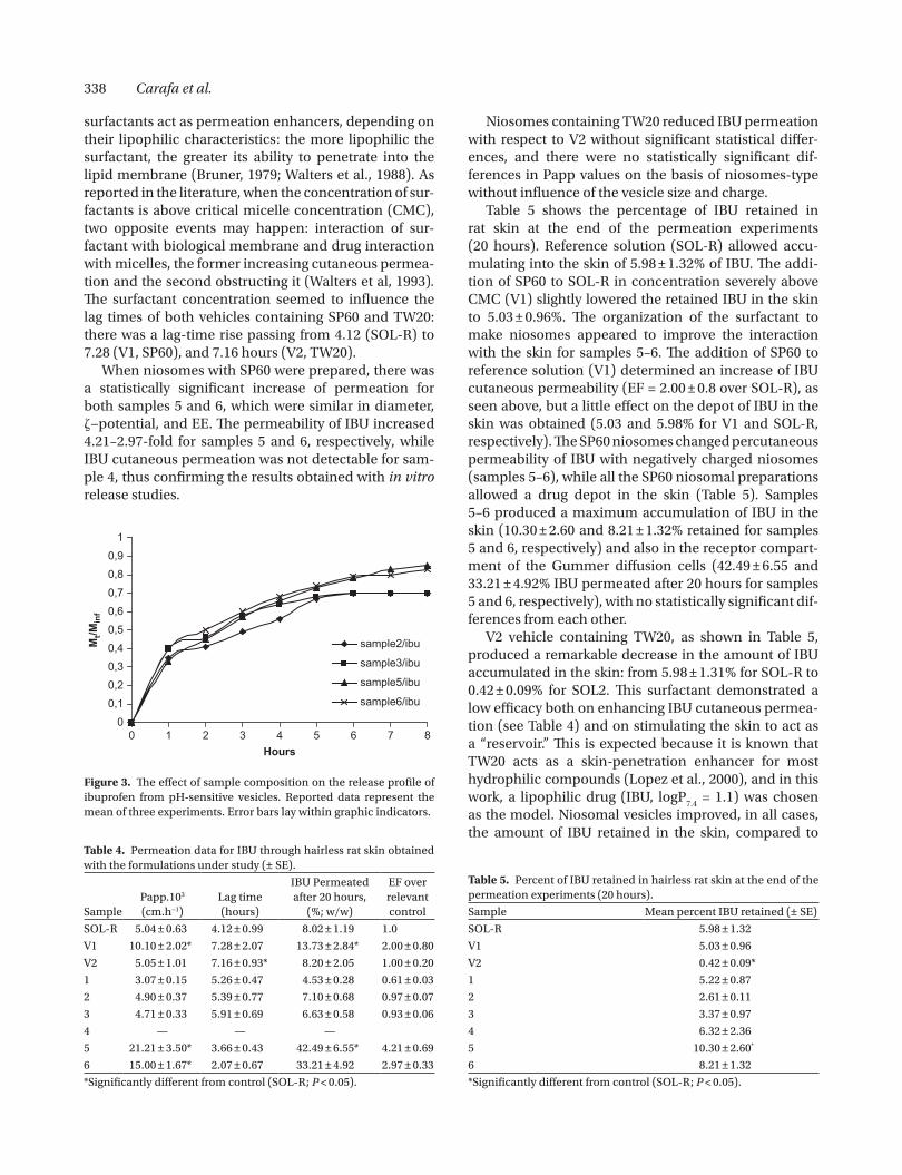
Vesicular dimension and -potential variations were not appreciated at 4 and 25°C for at least 5 months for all the analyzed samples. At 32°C, vesicles remained stable for 24 hours, so IBU release (Figure 3) cannot be related to vesicle disruption.
Further, it can be evidenced that no significant differ-ences in release profile were obtained for pH-sensitive formulations, while neutral vesicles did not show IBU release. The rate of IBU release from vesicular disper-sions decreased as a function of time for all prepared formulations (Figure 3).
In vitro permeation through hairless rat skin
The skin-permeation parameters of IBU from the vehi-cles under study (apparent permeability coefficient, lag time, IBU percent permeated after 20 hours, enhance-ment factor over control) are listed in Table 4.
IBU permeability coefficient for reference (SOL-R) was 5.04.10−3 ± 0.63.10− 3 cm/h. The addition of SP60 (HLB = 4.7; V1) increased Papp value by 2.0-fold, com-pared to SOL-R, unlike TW20 (HLB = 16.7; V2), that did not seem to influence IBU permeation, confirming that
0 1
1 2 3 4 5 6
2 3
50
100
150
200
250 A
B
Fluo
resc
ence
(AU
)
pH 7.4 pH 5.5
**
**
0
20
40
60
80
100
% c
alce
in re
leas
e
pH 7.4 pH 5.5
Tween 20 Span 60
**
**
**
**
Figure 2. (A) Intensities decrease of HPTS excitation max at 450 nm (emission, 510 nm) in function of pH values, for TW20 neutral vesi-cles (sample 1) and pH-sensitive vesicles (samples 2 and 3). Reported data represent the mean of three experiments. **P < 0.01, compared to neutral vesicle (sample 1) incubated at pH 7.4 and 5.5. (B) Influence of CHEMS on pH sensitivity of surfactant vesicles. The vesicles were incubated at pH 7.4 and 5.5, and samples were collected after 3 hours and calcein leakage was measured fluorimetrically. Reported data represent the mean of three experiments. **P < 0.01, compared to vesicle incubated at pH 7.4.
Table 3. Effect of ibuprofen encapsulation on vesicle dimension, -potential, and ibuprofen entrapment efficiency (EE), expressed as moles of drug/moles of structured surfactant x 100 (reported data are the means of at least three experiments ± SD).
Sample Diameter (nm) –potential (mV) EE
1 160 ± 5 –30.35 ± 2.02 24.40 ± 0.01
2 266 ± 3 –41.56 ± 1.45 24.98 ± 0.01
3 350 ± 5 –46.24 ± 2.34 22.72 ± 0.01
4 170 ± 4 –74.33 ± 1.25 2.28 ± 0.01
5 175 ± 3 –87.53 ± 2.64 2.51 ± 0.01
6 160 ± 2 –91.35 ± 1.50 2.25 ± 0.01
338 Carafa et al.
surfactants act as permeation enhancers, depending on their lipophilic characteristics: the more lipophilic the surfactant, the greater its ability to penetrate into the lipid membrane (Bruner, 1979; Walters et al., 1988). As reported in the literature, when the concentration of sur-factants is above critical micelle concentration (CMC), two opposite events may happen: interaction of sur-factant with biological membrane and drug interaction with micelles, the former increasing cutaneous permea-tion and the second obstructing it (Walters et al, 1993). The surfactant concentration seemed to influence the lag times of both vehicles containing SP60 and TW20: there was a lag-time rise passing from 4.12 (SOL-R) to 7.28 (V1, SP60), and 7.16 hours (V2, TW20).
When niosomes with SP60 were prepared, there was a statistically significant increase of permeation for both samples 5 and 6, which were similar in diameter, −potential, and EE. The permeability of IBU increased 4.21–2.97-fold for samples 5 and 6, respectively, while IBU cutaneous permeation was not detectable for sam-ple 4, thus confirming the results obtained with in vitro release studies.
Niosomes containing TW20 reduced IBU permeation with respect to V2 without significant statistical differ-ences, and there were no statistically significant dif-ferences in Papp values on the basis of niosomes-type without influence of the vesicle size and charge.
Table 5 shows the percentage of IBU retained in rat skin at the end of the permeation experiments (20 hours). Reference solution (SOL-R) allowed accu-mulating into the skin of 5.98 ± 1.32% of IBU. The addi-tion of SP60 to SOL-R in concentration severely above CMC (V1) slightly lowered the retained IBU in the skin to 5.03 ± 0.96%. The organization of the surfactant to make niosomes appeared to improve the interaction with the skin for samples 5–6. The addition of SP60 to reference solution (V1) determined an increase of IBU cutaneous permeability (EF = 2.00 ± 0.8 over SOL-R), as seen above, but a little effect on the depot of IBU in the skin was obtained (5.03 and 5.98% for V1 and SOL-R, respectively). The SP60 niosomes changed percutaneous permeability of IBU with negatively charged niosomes (samples 5–6), while all the SP60 niosomal preparations allowed a drug depot in the skin (Table 5). Samples 5–6 produced a maximum accumulation of IBU in the skin (10.30 ± 2.60 and 8.21 ± 1.32% retained for samples 5 and 6, respectively) and also in the receptor compart-ment of the Gummer diffusion cells (42.49 ± 6.55 and 33.21 ± 4.92% IBU permeated after 20 hours for samples 5 and 6, respectively), with no statistically significant dif-ferences from each other.
V2 vehicle containing TW20, as shown in Table 5, produced a remarkable decrease in the amount of IBU accumulated in the skin: from 5.98 ± 1.31% for SOL-R to 0.42 ± 0.09% for SOL2. This surfactant demonstrated a low efficacy both on enhancing IBU cutaneous permea-tion (see Table 4) and on stimulating the skin to act as a “reservoir.” This is expected because it is known that TW20 acts as a skin-penetration enhancer for most hydrophilic compounds (Lopez et al., 2000), and in this work, a lipophilic drug (IBU, logP
7.4 = 1.1) was chosen
as the model. Niosomal vesicles improved, in all cases, the amount of IBU retained in the skin, compared to
00,10,20,30,40,50,60,70,80,9
1
0 1 2 3 4 5 6 7 8Hours
Mt/M
inf
sample2/ibu
sample3/ibu
sample5/ibu
sample6/ibu
Figure 3. The effect of sample composition on the release profile of ibuprofen from pH-sensitive vesicles. Reported data represent the mean of three experiments. Error bars lay within graphic indicators.
Table 4. Permeation data for IBU through hairless rat skin obtained with the formulations under study (± SE).
SamplePapp.103 (cm.h−1)
Lag time (hours)
IBU Permeated after 20 hours,
(%; w/w)
EF over relevant control
SOL-R 5.04 ± 0.63 4.12 ± 0.99 8.02 ± 1.19 1.0
V1 10.10 ± 2.02* 7.28 ± 2.07 13.73 ± 2.84* 2.00 ± 0.80
V2 5.05 ± 1.01 7.16 ± 0.93* 8.20 ± 2.05 1.00 ± 0.20
1 3.07 ± 0.15 5.26 ± 0.47 4.53 ± 0.28 0.61 ± 0.03
2 4.90 ± 0.37 5.39 ± 0.77 7.10 ± 0.68 0.97 ± 0.07
3 4.71 ± 0.33 5.91 ± 0.69 6.63 ± 0.58 0.93 ± 0.06
4 — — —
5 21.21 ± 3.50* 3.66 ± 0.43 42.49 ± 6.55* 4.21 ± 0.69
6 15.00 ± 1.67* 2.07 ± 0.67 33.21 ± 4.92 2.97 ± 0.33
*Significantly different from control (SOL-R; P < 0.05).
Table 5. Percent of IBU retained in hairless rat skin at the end of the permeation experiments (20 hours).
Sample Mean percent IBU retained (± SE)
SOL-R 5.98 ± 1.32
V1 5.03 ± 0.96
V2 0.42 ± 0.09*
1 5.22 ± 0.87
2 2.61 ± 0.11
3 3.37 ± 0.97
4 6.32 ± 2.36
5 10.30 ± 2.60*
6 8.21 ± 1.32
*Significantly different from control (SOL-R; P < 0.05).

Neutral and pH-sensitive vesicles 339
V2, but they did not reach the levels obtained from SOL-R. While niosomes containing TW20 did not determine a change of IBU cutaneous permeability, they allowed a little IBU depot on the skin, particularly in the case of sample 1 (5.22 ± 0.87% IBU retained), smallest niosomes (160 ± 5 nm) consisting of surfactant and cholesterol.
Conclusions
It can be concluded that the addition of CHEMS to SP60 and TW20 vesicular formulation led to pH-sensitive structures, which were stable and able to encapsulate IBU. IBU encapsulation led to a significative increase of vesicular dimension only for TW20 formulations; in all analyzed samples, no significative variations of -potential values could be noticed; this was probably related to a moderate drug adsorption on the vesicle surface. When niosomes with SP60 were prepared, there was a statistically significant increase of permeation for negative charged niosomes (samples 5 and 6).
TW20 niosomes did not show statistically significant differences in Papp values on the basis of niosomes-type without influence of the vesicle size and charge. This behavior could depend on the less lipophilic nature of the surfactant (HLB = 16.7), which makes it more diffi-cult for the vesicles to penetrate or fuse with the skin.
This work pointed out that the topical delivery of IBU might be reachable more efficiently by means of SP60 niosomes with respect to TW20 ones, even if a higher cutaneous permeability was consequently associated with increased drug accumulation in the skin.
Acknowledgments
The authors are grateful to Professor Elka Touitou at the School of Pharmacy of The Hebrew University of Jerusalem (Jerusalem, Israel) for her stimulating sug-gestions and discussion. The authors thank Giuseppe Lucania (Dip. di Medicina Sperimentale e Patologia, Faculty of Medicine, University of Rome “La Sapienza,” Rome, Italy) for freeze-fracture microscopy analyses. MIUR (Ministero dell’ Universitá e della Ricerca) is gratefully acknowledged for financial funding (PRIN 2003-prot. No. 2003034531_005).
Declaration of interest: The authors report no financial conflicts of interest. The authors alone are responsible for the content and writing of this paper.
References
Banga, A.K., Bose, S., Ghost, T.K. (1999). Iontophoresis and electro-poration: comparison and contrast. Int J Pharm 179:1–19.
Bruner, M.M. (1979). The interactions between surfactants and kerat-inous tissues. J Soc Cosm Chem 30:41–64.
Carafa, M., Di Marzio, L., Marianecci, C., Cinque, B., Lucania, G., Kajiwara, K., et al. (2006). Designing novel pH-sensitive non-phospholipid vesicle: characterization and cell interaction. Eur J Pharm Sci 28:385–393.
Cevc, G. (2004). Lipid vesicles and other colloids as drug carriers on the skin. Adv Drug Deliv Rev 56:675– 711.
Chen, H., Chang, X., Du, D., Li, J., Xu, H., Yang, X. (2006). Microemulsion-based hydrogel formulation of ibuprofen for topical delivery. Int J Pharm 315:52–58.
Cilurzo, F., Minghetti, P., Casiraghi, A., Tosi, L., Pagani, S., Montanari, L. (2005). Polymethacrylates as crystallization inhibitors in monolayer transdermal patches containing ibuprofen. Eur J Pharm Biopharm 60:61–66.
Du, L., Liu, X., Huang, W., Wang, E. (2006). A study on the interaction between ibuprofen and bilayer lipid membrane. Electrochim Acta 51:5754–5760.
Fang, J.Y., Hong, C.T., Chiu, W.T., Wang, Y.Y. (2001). Effect of lipo-somes and niosomes on skin permeation of enoxacin. Int J Pharm 219:61–72.
Fresta, M., Puglisi, G. (1996). Application of liposomes as poten-tial cutaneous drug delivery systems. In vitro and in vivo investigation with radioactively labelled vesicles. Drug Targ 4:95–101.
Gummer, C.L., Hinz, R.S., Maibach, H.I. (1987). The skin penetration cell: a design update. Int J Pharm 40:101–104.
Hafez, I.M., Cullis, P.R. (2000). Cholesteryl hemisuccinate exhibits pH-sensitive polymorphic phase behaviour. Biochim Biophys Acta 1463:107–114.
Heard, C.M., Gallagher, S.J., Harwood, J., Maguire, P.B. (2003). The in vitro delivery of NSAIDs across skin was in proportion to the delivery of essential fatty acids in the vehicle—evidence that sol-utes permeate skin associated with their solvation cages? Int J Pharm 261:165–169.
Irevolino, M., Cappello, B., Raghavan, S.L., Hadgraft, J. (2001). Penetration enhancement of ibuprofen from supersaturated solutions through human skin. Int J Pharm 212:131–141.
Junginger, H.E., Hofland, H.E.J., Bouwstra, J.A. (1991). Liposomes and niosomes interactions with human skin. Cosm Toil 106:45–50.
Kano, K., Fendler, J.H. (1978). Pyranine as a sensitive pH probe for liposome interiors and surfaces. Biochim Biophys Acta 509:289–299.
Kato, H., Nagay, H., Yamando, K., Sakaba, Y. (1989). Determination of polisorbate in food by colorimetry with confirmation by IR spectrophotometry, TLC, and gas chromatography. J Assoc Off Anal Chem 72:27–29.
Lai, M.Z., Duzgunes, N., Szoka, F.C. (1985). Effects of replace-ment of the hydroxyl group of cholesterol and tocopherol on the thermotropic behavior of phospholipid. Biochemistry 24: 1646–1653.
Li, X., Nie, S.F., Kong, J., Li, N., Ju, C., Pan, W. (2008). A controlled-release ocular delivery system for ibuprofen based on nanos-tructured lipid carriers. Int J Pharm 363:177–182.
Lopez, A., Llinares, F., Cortell, C., Herraez, M. (2000). Comparative enhancer effects of Span 20 with Tween 20 and azone on the in vitro percutaneous penetration of compounds with different lipophilicities. Int J Pharmaceut 202:133–140.
Mohammed, A.R., Weston, N., Coombes, A.G.A., Fitzgerald, M., Perrie, Y. (2004). Liposome formulation of poorly water-soluble drugs: optimisation of drug loading and ESEM analysis of stabil-ity. Int J Pharm 285:23–34.
Monti, D., Saettone, M.F., Giannaccini, B., Galli-Angeli, D. (1995). Enhancement of transdermal penetration of dapiprazole through hairless mouse skin. J Contr Rel 33:71–77.
Ohkuma, S., Poole, B. (1978). Fluorescence probe measurement of the intralysosomal pH in living cells and the perturbation by various agents. Proc Natl Acad Sci U S A 75:3327–3331.
Park, K.M., Kim, C.K. (1999). Preparation and evaluation of flurbi-profen-loaded microemulsion for parenteral delivery. Int J Pharm 181:173–179.
Perioli, L., Ambrogi, V., Angelici, F., Ricci, M., Giovagnoli, S., Capuccella, M., et al. (2004). Development of mucoadhesive

340 Carafa et al.
patches for buccal administration of ibuprofen. J Contr Rel 99:73–82.
Renshaw, P.F., Janoff, A.S., Miller, K.W. (1983). On the nature of dilute aqueous cholesterol suspensions. J Lipid Res 24:47–51.
Santucci, E., Carafa, M., Coviello, T., Murtas, E., Riccieri, F.M., Alhaique, F., et al. (1996). Vesicles from polysorbate 20 and cholesterol. A simple preparation and a characterization. STP Pharma Sci 6:29–32.
Simões, S., Slepushkin, V., Düzgünes, N., Pedroso, de Lima MC. (2004). On the mechanism of internalization and intracellular delivery medi-ated by pH-sensitive liposomes. Biochim Biophys Acta 1515:23–37.
Sinclair, G.W., Peppas, N.A. (1984). Analisys of non-Fickian transport polymers using semplified exponential expression. J Membr Sci 17:329–331.
Stott, P.W., Williams, A.C., Barry, B.W. (1998). Transdermal delivery from eutectic systems: enhanced permeation of a model drug, ibuprofen. J Contr Rel 50:297–308.
Tokumoto, S., Higo, N., Sugibayashi, K. (2006). Effect of electropora-tion and pH on the iontophoretic transdemal delivery of human insulin. Int J Pharm 326:13–19.
Touitou E., Dayan N., Berelson L., Godin B., Eliaz M., (2001). Ethosomes-Novel Vesicular Carriers for Enchanced Delivery: Characterization and Skin Penetration properties, J Contr Rel 65:403–418.
Touitou E., Godin B., (2006) Vesicles for enchanced delivery into and through the skin. In Touitou and Barry (Eds) Enchancement in drug delivery, pp. 255–278. CRC Press, Taylor and Francis Group. Boca Raton-London-New York.
Walters, K.A., Walters, M., Olejink, O. (1988). Nonionic surfactants on skin permeability characteristics. J Pharm Pharmacol 40:525–529.
Walters, K.A., Bialik, W., Brain,, K.R. (1993). The effects of surfactants on penetration across the skin. Int J Cosm Sci 15:260–271.
Wang, Z., Fingas, M. (1994). Analysis of sorbitan esther surfactants. Part I: high-performance liquid chromatography. J High Res Chrom 17:15–19.
Williams, A.C., Barry, B.W. (2004). Penetration enhancers. Adv Drug Deliv Rev 56:603.
Yano, T., Nakagawa, A., Tsuji, M., Noda, K. (1986). Skin permeabil-ity of various nonsteroidal anti-inflammatory drugs. Life Sci 39:1043–1050.
Related Documents