526 VOLUME 45 | NUMBER 5 | MAY 2013 NATURE GENETICS LETTERS Chronic lymphocytic leukemia (CLL) is the most frequent leukemia in adults 1–3 . We have analyzed exome sequencing data from 127 individuals with CLL and Sanger sequencing data from 214 additional affected individuals, identifying recurrent somatic mutations in POT1 (encoding protection of telomeres 1) in 3.5% of the cases, with the frequency reaching 9% when only individuals without IGHV@ mutations were considered. POT1 encodes a component of the shelterin complex and is the first member of this telomeric structure found to be mutated in human cancer. Somatic mutation of POT1 primarily occurs in gene regions encoding the two oligonucleotide-/oligosaccharide-binding (OB) folds and affects key residues required to bind telomeric DNA. POT1-mutated CLL cells have numerous telomeric and chromosomal abnormalities that suggest that POT1 mutations favor the acquisition of the malignant features of CLL cells. The identification of POT1 as a new frequently mutated gene in CLL may facilitate novel approaches for the clinical management of this disease. A long-standing barrier to understanding leukemogenesis has been an inability to accurately identify the genetic alterations that underlie this disease 4 . To gain further insight into the genetic basis of CLL, we have analyzed the exomes of matched tumor and normal samples from the peripheral blood of 127 untreated individuals with CLL, identifying tumoral somatic mutations using the previously described Sidrón algorithm 5,6 . Several of the driver genes, such as SF3B1, NOTCH1 and MYD88, have previously been assessed for their functional and clini- cal roles in CLL 5–8 . We also observed that POT1 is a frequent genetic target for somatic mutation in CLL, and, given its prominent role in telomere capping and telomerase recruitment 9,10 , we hypothesized that mutations in this gene might be a source of genomic instability and represent a new tumor-driving factor. In fact, when the number of mutations, gene size and codon composition were taken into account, POT1 was the gene that was second most frequently affected by point mutations (P = 4.2 × 10 −6 ), with only SF3B1 having a higher frequency (P = 8.6 × 10 −11 ). POT1 was found to be mutated in approximately 5% of CLL cases (Supplementary Table 1) and, conspicuously, was exclusively altered in samples with wild-type IGHV@, suggesting that inactivation of POT1 could represent a distinct molecular alteration for the development of this aggressive CLL subtype. We next extended our sequence analysis of POT1 by performing Sanger sequencing in a validation set consisting of 214 matched tumor and normal samples from individuals with CLL (Supplementary Table 2), with the find- ing of 5 additional somatic mutations. RNA sequencing (RNA-seq) analysis of available cases confirmed the expression of mutant POT1 alleles in CLL tumor cells (data not shown). Therefore, we have identi- fied 12 somatic point mutations in 341 CLL cases (3.5%; Table 1). This finding also implies that 9% of individuals with wild-type IGHV@ have a POT1 mutation, which is a comparable mutational frequency to that of SF3B1 (11%) in individuals of this CLL subtype 7 . Our findings make POT1 one of the most frequently mutated genes reported so far in CLL along with NOTCH1, SF3B1, TP53 and ATM 5–8 . Notably, 3 of the 12 mutations found in POT1 are predicted to result in a truncated protein. The remaining somatic mutations were all nonsynonymous and were predominantly predicted to be deleterious by a consensus of five bioinformatics tools 11 , supporting the idea that POT1 mutations constitute a driver event in CLL (Table 1). Shelterin proteins (namely TRF1, TRF2, TIN2, TPP1, RAP1 and POT1) are critical regulators of telomere integrity by modulating telomere capping, replication and telomere extension by telomer- ase 12–14 . Structurally, POT1 is the only shelterin that contains two N-terminal OB domains, which confer to this protein affinity for the single-stranded DNA (ssDNA) sequence TTAGGG, in addition to a C terminus that binds TPP1 and anchors POT1 to the shelterin complex. Notably, 9 of the 12 POT1 somatic mutations we have detected in CLL samples reside within gene regions encoding the OB folds (Fig. 1a). Comparative sequence analysis of the OB fold somatic alterations showed that most mutated residues are phyloge- netically conserved and suggests that they have an essential role in POT1 mutations cause telomere dysfunction in chronic lymphocytic leukemia Andrew J Ramsay 1,4 , Víctor Quesada 1,4 , Miguel Foronda 2,4 , Laura Conde 3 , Alejandra Martínez-Trillos 3 , Neus Villamor 3 , David Rodríguez 1 , Agnieszka Kwarciak 1 , Cecilia Garabaya 1 , Mercedes Gallardo 2 , Mónica López-Guerra 3 , Armando López-Guillermo 3 , Xose S Puente 1 , María A Blasco 2 , Elías Campo 3 & Carlos López-Otín 1 1 Departamento de Bioquímica y Biología Molecular, Instituto Universitario de Oncología del Principado de Asturias (IUOPA) Universidad de Oviedo, Oviedo, Spain. 2 Telomeres and Telomerase Group, Molecular Oncology Program, Spanish National Cancer Research Centre (CNIO), Madrid, Spain. 3 Unidad de Hematopatología, Servicio de Anatomía Patológica, Hospital Clínic, Universitat de Barcelona, Institut d’Investigacions Biomèdiques August Pi i Sunyer (IDIBAPS), Barcelona, Spain. 4 These authors contributed equally to this work. Correspondence should be addressed to C.L.-O. ([email protected]), E.C. ([email protected]) or M.A.B. ([email protected]). Received 29 October 2012; accepted 22 February 2013; published online 17 March 2013; doi:10.1038/ng.2584 npg © 2013 Nature America, Inc. All rights reserved.

Welcome message from author
This document is posted to help you gain knowledge. Please leave a comment to let me know what you think about it! Share it to your friends and learn new things together.
Transcript

526 VOLUME 45 | NUMBER 5 | MAY 2013 Nature GeNetics
l e t t e r s
Chroniclymphocyticleukemia(CLL)isthemostfrequentleukemiainadults1–3.Wehaveanalyzedexomesequencingdatafrom127individualswithCLLandSangersequencingdatafrom214additionalaffectedindividuals,identifyingrecurrentsomaticmutationsinPOT1(encodingprotectionoftelomeres1)in3.5%ofthecases,withthefrequencyreaching9%whenonlyindividualswithoutIGHV@mutationswereconsidered.POT1encodesacomponentoftheshelterincomplexandisthefirstmemberofthistelomericstructurefoundtobemutatedinhumancancer.SomaticmutationofPOT1primarilyoccursingeneregionsencodingthetwooligonucleotide-/oligosaccharide-binding(OB)foldsandaffectskeyresiduesrequiredtobindtelomericDNA.POT1-mutatedCLLcellshavenumeroustelomericandchromosomalabnormalitiesthatsuggestthatPOT1mutationsfavortheacquisitionofthemalignantfeaturesofCLLcells.TheidentificationofPOT1asanewfrequentlymutatedgeneinCLLmayfacilitatenovelapproachesfortheclinicalmanagementofthisdisease.
A long-standing barrier to understanding leukemogenesis has been an inability to accurately identify the genetic alterations that underlie this disease4. To gain further insight into the genetic basis of CLL, we have analyzed the exomes of matched tumor and normal samples from the peripheral blood of 127 untreated individuals with CLL, identifying tumoral somatic mutations using the previously described Sidrón algorithm5,6. Several of the driver genes, such as SF3B1, NOTCH1 and MYD88, have previously been assessed for their functional and clini-cal roles in CLL5–8. We also observed that POT1 is a frequent genetic target for somatic mutation in CLL, and, given its prominent role in telomere capping and telomerase recruitment9,10, we hypothesized that mutations in this gene might be a source of genomic instability and represent a new tumor-driving factor. In fact, when the number of mutations, gene size and codon composition were taken into account, POT1 was the gene that was second most frequently affected by point
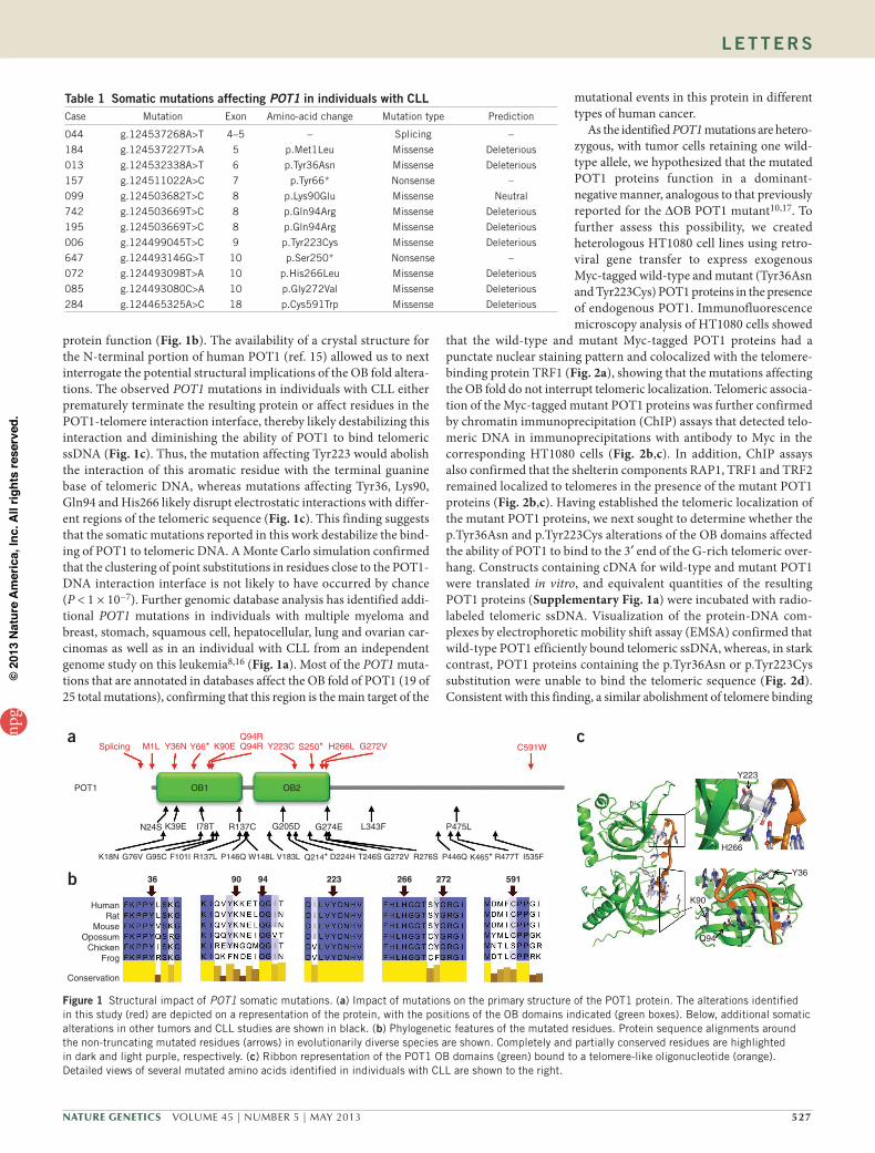
mutations (P = 4.2 × 10⁻⁶), with only SF3B1 having a higher frequency (P = 8.6 × 10⁻¹¹). POT1 was found to be mutated in approximately 5% of CLL cases (Supplementary Table 1) and, conspicuously, was exclusively altered in samples with wild-type IGHV@, suggesting that inactivation of POT1 could represent a distinct molecular alteration for the development of this aggressive CLL subtype. We next extended our sequence analysis of POT1 by performing Sanger sequencing in a validation set consisting of 214 matched tumor and normal samples from individuals with CLL (Supplementary Table 2), with the find-ing of 5 additional somatic mutations. RNA sequencing (RNA-seq) analysis of available cases confirmed the expression of mutant POT1 alleles in CLL tumor cells (data not shown). Therefore, we have identi-fied 12 somatic point mutations in 341 CLL cases (3.5%; Table 1). This finding also implies that 9% of individuals with wild-type IGHV@ have a POT1 mutation, which is a comparable mutational frequency to that of SF3B1 (11%) in individuals of this CLL subtype7. Our findings make POT1 one of the most frequently mutated genes reported so far in CLL along with NOTCH1, SF3B1, TP53 and ATM5–8. Notably, 3 of the 12 mutations found in POT1 are predicted to result in a truncated protein. The remaining somatic mutations were all nonsynonymous and were predominantly predicted to be deleterious by a consensus of five bioinformatics tools11, supporting the idea that POT1 mutations constitute a driver event in CLL (Table 1).
Shelterin proteins (namely TRF1, TRF2, TIN2, TPP1, RAP1 and POT1) are critical regulators of telomere integrity by modulating telomere capping, replication and telomere extension by telomer-ase12–14. Structurally, POT1 is the only shelterin that contains two N-terminal OB domains, which confer to this protein affinity for the single-stranded DNA (ssDNA) sequence TTAGGG, in addition to a C terminus that binds TPP1 and anchors POT1 to the shelterin complex. Notably, 9 of the 12 POT1 somatic mutations we have detected in CLL samples reside within gene regions encoding the OB folds (Fig. 1a). Comparative sequence analysis of the OB fold somatic alterations showed that most mutated residues are phyloge-netically conserved and suggests that they have an essential role in
POT1 mutations cause telomere dysfunction in chronic lymphocytic leukemiaAndrew J Ramsay1,4, Víctor Quesada1,4, Miguel Foronda2,4, Laura Conde3, Alejandra Martínez-Trillos3, Neus Villamor3, David Rodríguez1, Agnieszka Kwarciak1, Cecilia Garabaya1, Mercedes Gallardo2, Mónica López-Guerra3, Armando López-Guillermo3, Xose S Puente1, María A Blasco2, Elías Campo3 & Carlos López-Otín1
1Departamento de Bioquímica y Biología Molecular, Instituto Universitario de Oncología del Principado de Asturias (IUOPA) Universidad de Oviedo, Oviedo, Spain. 2Telomeres and Telomerase Group, Molecular Oncology Program, Spanish National Cancer Research Centre (CNIO), Madrid, Spain. 3Unidad de Hematopatología, Servicio de Anatomía Patológica, Hospital Clínic, Universitat de Barcelona, Institut d’Investigacions Biomèdiques August Pi i Sunyer (IDIBAPS), Barcelona, Spain. 4These authors contributed equally to this work. Correspondence should be addressed to C.L.-O. ([email protected]), E.C. ([email protected]) or M.A.B. ([email protected]).
Received 29 October 2012; accepted 22 February 2013; published online 17 March 2013; doi:10.1038/ng.2584
npg
© 2
013
Nat
ure
Am
eric
a, In
c. A
ll rig
hts
rese
rved
.
Nature GeNetics VOLUME 45 | NUMBER 5 | MAY 2013 527
l e t t e r s
protein function (Fig. 1b). The availability of a crystal structure for the N-terminal portion of human POT1 (ref. 15) allowed us to next interrogate the potential structural implications of the OB fold altera-tions. The observed POT1 mutations in individuals with CLL either prematurely terminate the resulting protein or affect residues in the POT1-telomere interaction interface, thereby likely destabilizing this interaction and diminishing the ability of POT1 to bind telomeric ssDNA (Fig. 1c). Thus, the mutation affecting Tyr223 would abolish the interaction of this aromatic residue with the terminal guanine base of telomeric DNA, whereas mutations affecting Tyr36, Lys90, Gln94 and His266 likely disrupt electrostatic interactions with differ-ent regions of the telomeric sequence (Fig. 1c). This finding suggests that the somatic mutations reported in this work destabilize the bind-ing of POT1 to telomeric DNA. A Monte Carlo simulation confirmed that the clustering of point substitutions in residues close to the POT1-DNA interaction interface is not likely to have occurred by chance (P < 1 × 10⁻⁷). Further genomic database analysis has identified addi-tional POT1 mutations in individuals with multiple myeloma and breast, stomach, squamous cell, hepatocellular, lung and ovarian car-cinomas as well as in an individual with CLL from an independent genome study on this leukemia8,16 (Fig. 1a). Most of the POT1 muta-tions that are annotated in databases affect the OB fold of POT1 (19 of 25 total mutations), confirming that this region is the main target of the
mutational events in this protein in different types of human cancer.
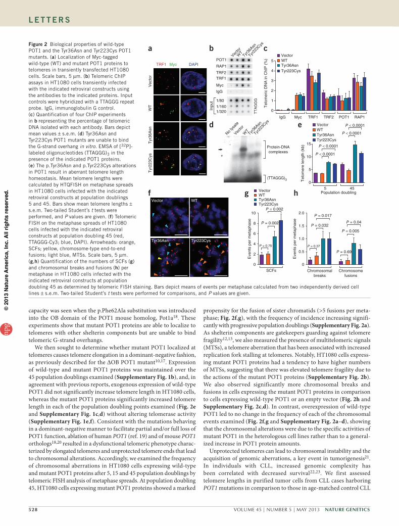
As the identified POT1 mutations are hetero-zygous, with tumor cells retaining one wild-type allele, we hypothesized that the mutated POT1 proteins function in a dominant- negative manner, analogous to that previously reported for the ∆OB POT1 mutant10,17. To further assess this possibility, we created heterologous HT1080 cell lines using retro-viral gene transfer to express exogenous Myc-tagged wild-type and mutant (Tyr36Asn and Tyr223Cys) POT1 proteins in the presence of endogenous POT1. Immunofluorescence microscopy analysis of HT1080 cells showed
that the wild-type and mutant Myc-tagged POT1 proteins had a punctate nuclear staining pattern and colocalized with the telomere-binding protein TRF1 (Fig. 2a), showing that the mutations affecting the OB fold do not interrupt telomeric localization. Telomeric associa-tion of the Myc-tagged mutant POT1 proteins was further confirmed by chromatin immunoprecipitation (ChIP) assays that detected telo-meric DNA in immunoprecipitations with antibody to Myc in the corresponding HT1080 cells (Fig. 2b,c). In addition, ChIP assays also confirmed that the shelterin components RAP1, TRF1 and TRF2 remained localized to telomeres in the presence of the mutant POT1 proteins (Fig. 2b,c). Having established the telomeric localization of the mutant POT1 proteins, we next sought to determine whether the p.Tyr36Asn and p.Tyr223Cys alterations of the OB domains affected the ability of POT1 to bind to the 3′ end of the G-rich telomeric over-hang. Constructs containing cDNA for wild-type and mutant POT1 were translated in vitro, and equivalent quantities of the resulting POT1 proteins (Supplementary Fig. 1a) were incubated with radio-labeled telomeric ssDNA. Visualization of the protein-DNA com-plexes by electrophoretic mobility shift assay (EMSA) confirmed that wild-type POT1 efficiently bound telomeric ssDNA, whereas, in stark contrast, POT1 proteins containing the p.Tyr36Asn or p.Tyr223Cys substitution were unable to bind the telomeric sequence (Fig. 2d). Consistent with this finding, a similar abolishment of telomere binding
table 1 somatic mutations affecting POT1 in individuals with CllCase Mutation Exon Amino-acid change Mutation type Prediction
044 g.124537268A>T 4–5 – Splicing –
184 g.124537227T>A 5 p.Met1Leu Missense Deleterious
013 g.124532338A>T 6 p.Tyr36Asn Missense Deleterious
157 g.124511022A>C 7 p.Tyr66* Nonsense –
099 g.124503682T>C 8 p.Lys90Glu Missense Neutral
742 g.124503669T>C 8 p.Gln94Arg Missense Deleterious
195 g.124503669T>C 8 p.Gln94Arg Missense Deleterious
006 g.124499045T>C 9 p.Tyr223Cys Missense Deleterious
647 g.124493146G>T 10 p.Ser250* Nonsense –
072 g.124493098T>A 10 p.His266Leu Missense Deleterious
085 g.124493080C>A 10 p.Gly272Val Missense Deleterious
284 g.124465325A>C 18 p.Cys591Trp Missense Deleterious
a
b 36
Human
90 94 266223 272 591
Splicing M1L
OB1 OB2POT1
N24S
Y36N K90EQ94RQ94R Y223C H266L G272V C591WS250*Y66*
K39E I78T R137C G205D G274E L343F P475L
K18N G76V G95C F101I R137L P146Q W148L V183L Q214* D224H T246S G272V R276S P446Q K465* R477T I535F
RatMouse
OpossumChicken
Frog
Conservation
c
Y223
H266
Y36
K90
Q94
Figure 1 Structural impact of POT1 somatic mutations. (a) Impact of mutations on the primary structure of the POT1 protein. The alterations identified in this study (red) are depicted on a representation of the protein, with the positions of the OB domains indicated (green boxes). Below, additional somatic alterations in other tumors and CLL studies are shown in black. (b) Phylogenetic features of the mutated residues. Protein sequence alignments around the non-truncating mutated residues (arrows) in evolutionarily diverse species are shown. Completely and partially conserved residues are highlighted in dark and light purple, respectively. (c) Ribbon representation of the POT1 OB domains (green) bound to a telomere-like oligonucleotide (orange). Detailed views of several mutated amino acids identified in individuals with CLL are shown to the right.
npg
© 2
013
Nat
ure
Am
eric
a, In
c. A
ll rig
hts
rese
rved
.
528 VOLUME 45 | NUMBER 5 | MAY 2013 Nature GeNetics
l e t t e r s
capacity was seen when the p.Phe62Ala substitution was introduced into the OB domain of the POT1 mouse homolog, Pot1a18. These experiments show that mutant POT1 proteins are able to localize to telomeres with other shelterin components but are unable to bind telomeric G-strand overhangs.
We then sought to determine whether mutant POT1 localized at telomeres causes telomere elongation in a dominant-negative fashion, as previously described for the ∆OB POT1 mutant10,17. Expression of wild-type and mutant POT1 proteins was maintained over the 45 population doublings examined (Supplementary Fig. 1b), and, in agreement with previous reports, exogenous expression of wild-type POT1 did not significantly increase telomere length in HT1080 cells, whereas the mutant POT1 proteins significantly increased telomere length in each of the population doubling points examined (Fig. 2e and Supplementary Fig. 1c,d) without altering telomerase activity (Supplementary Fig. 1e,f). Consistent with the mutations behaving in a dominant-negative manner to facilitate partial and/or full loss of POT1 function, ablation of human POT1 (ref. 19) and of mouse POT1 orthologs18,20 resulted in a dysfunctional telomeric phenotype charac-terized by elongated telomeres and unprotected telomere ends that lead to chromosomal alterations. Accordingly, we examined the frequency of chromosomal aberrations in HT1080 cells expressing wild-type and mutant POT1 proteins after 5, 15 and 45 population doublings by telomeric FISH analysis of metaphase spreads. At population doubling 45, HT1080 cells expressing mutant POT1 proteins showed a marked
propensity for the fusion of sister chromatids (>5 fusions per meta-phase; Fig. 2f,g), with the frequency of incidence increasing signifi-cantly with progressive population doublings (Supplementary Fig. 2a). As shelterin components are gatekeepers guarding against telomere fragility12,13, we also measured the presence of multitelomeric signals (MTSs), a telomere aberration that has been associated with increased replication fork stalling at telomeres. Notably, HT1080 cells express-ing mutant POT1 proteins had a tendency to have higher numbers of MTSs, suggesting that there was elevated telomere fragility due to the actions of the mutant POT1 proteins (Supplementary Fig. 2b). We also observed significantly more chromosomal breaks and fusions in cells expressing the mutant POT1 proteins in comparison to cells expressing wild-type POT1 or an empty vector (Fig. 2h and Supplementary Fig. 2c,d). In contrast, overexpression of wild-type POT1 led to no change in the frequency of each of the chromosomal events examined (Fig. 2f,g and Supplementary Fig. 2a–d), showing that the chromosomal alterations were due to the specific activities of mutant POT1 in the heterologous cell lines rather than to a general-ized increase in POT1 protein amounts.
Unprotected telomeres can lead to chromosomal instability and the acquisition of genomic aberrations, a key event in tumorigenesis21. In individuals with CLL, increased genomic complexity has been correlated with decreased survival22,23. We first assessed telomere lengths in purified tumor cells from CLL cases harboring POT1 mutations in comparison to those in age-matched control CLL
a b
d e
cPOT1 5
VectorWTTyr36AsnTyr223Cys
VectorWTTyr36AsnTyr223Cys
Vecto
r
WT
Tyr36
Asn
Tyr22
3Cys
Tyr36
Asn
Tyr22
3Cys
RAP1
TRF2TRF1
Myc
IgG
1/80
Inpu
t
Tel
omer
ic D
NA
in C
hIP
(%
)
Tel
omer
e le
ngth
(kb
)
TT
AG
GG
1/160
No lys
ate
Vecto
r
WT
1/320
*V
ecto
rW
TT
yr36
Asn
Tyr
223C
ys
DAPI
4
3
2
1
0
15
IgG
Protein-DNAcomplexes
(TTAGGG)3
Myc TRF1 TRF2 POT1 RAP1
10
P < 0.0001
P < 0.0001
P < 0.0001
P < 0.0001
5 45Population doubling
5
0
g hVectorWTTyr36AsnTyr223Cys
Eve
nts
per
met
apha
se
Eve
nts
per
met
apha
se
10 2.0P = 0.002
P = 0.017
P = 0.032
P = 0.37
P = 0.04
P = 0.005
P = 0.69
SCFs Chromosomalbreaks
Chromosomefusions
P = 0.002
P = 0.75
8
6
4
2
0
1.5
1.0
0.5
0
TRF1 Myc
fVector
Tyr36Asn
WT
Tyr223Cys
Figure 2 Biological properties of wild-type POT1 and the Tyr36Asn and Tyr223Cys POT1 mutants. (a) Localization of Myc-tagged wild-type (WT) and mutant POT1 proteins to telomeres in transiently transfected HT1080 cells. Scale bars, 5 µm. (b) Telomeric ChIP assays in HT1080 cells transiently infected with the indicated retroviral constructs using the antibodies to the indicated proteins. Input controls were hybridized with a TTAGGG repeat probe. IgG, immunoglobulin G control. (c) Quantification of four ChIP experiments in b representing the percentage of telomeric DNA isolated with each antibody. Bars depict mean values ± s.e.m. (d) Tyr36Asn and Tyr223Cys POT1 mutants are unable to bind the G-strand overhang in vitro. EMSA of [32P]-labeled oligonucleotides (TTAGGG)3 in the presence of the indicated POT1 proteins. (e) The p.Tyr36Asn and p.Tyr223Cys alterations in POT1 result in aberrant telomere length homeostasis. Mean telomere lengths were calculated by HTQFISH on metaphase spreads in HT1080 cells infected with the indicated retroviral constructs at population doublings 5 and 45. Bars show mean telomere lengths ± s.e.m. Two-tailed Student’s t tests were performed, and P values are given. (f) Telomeric FISH on the metaphase spreads of HT1080 cells infected with the indicated retroviral constructs at population doubling 45 (red, TTAGGG-Cy3; blue, DAPI). Arrowheads: orange, SCFs; yellow, chromosome-type end-to-end fusions; light blue, MTSs. Scale bars, 5 µm. (g,h) Quantification of the numbers of SCFs (g) and chromosomal breaks and fusions (h) per metaphase in HT1080 cells infected with the indicated retroviral constructs at population doubling 45 as determined by telomeric FISH staining. Bars depict means of events per metaphase calculated from two independently derived cell lines ± s.e.m. Two-tailed Student’s t tests were performed for comparisons, and P values are given.
npg
© 2
013
Nat
ure
Am
eric
a, In
c. A
ll rig
hts
rese
rved
.

Nature GeNetics VOLUME 45 | NUMBER 5 | MAY 2013 529
l e t t e r s
cases with wild-type IGHV@ containing no mutation in POT1, TP53 or ATM (Supplementary Table 3), observing no significant differ-ences in telomere lengths or in telomerase activities (Supplementary Fig. 3a,b). The absence of changes in telomere length due to POT1 mutations potentially derives from the variance in telomere lengths within human populations, a situation that is distinct from those present in the analysis of isogenic cell lines and inbred mouse strains. Next, we examined the integrity of telomeres in the POT1-mutated CLL samples through telomeric FISH analysis of metaphase spreads (Fig. 3a). Notably, similar to observations made in HT1080 cells, we found significantly more sister chromatid–type end-to-end fusions (SCFs) in POT1-mutated samples than in control samples, with up to five fusions occurring per metaphase (P = 0.01) (Fig. 3a,b). We also detected significantly more chromosome-type fusions containing TTAGGG signals in POT1-mutated samples than in control samples (P = 0.042) (Fig. 3c), but the number of chromosome-type fusions with no detectable TTAGGG signal was not significantly different between the two populations (P = 0.072) (Supplementary Fig. 3c), in agreement with the normal telomere lengths observed in POT1-mutated samples. As in HT1080 cells, we detected a modest but sig-nificant increase in the presence of MTSs in POT1-mutated CLL cells compared to the controls (2.2-fold increase, P = 0.03; Fig. 3d), reflect-ing the presence of greater telomere fragility in these individuals.
Collectively, the observation of a high incidence of telomere- containing chromosomal fusions coincident with no change in the frequency of telomere-deficient fusions is consistent with these fusions being produced by POT1 mutation–mediated telomere uncapping rather than by overt telomere shortening, resembling the chromo-somal phenotype of mouse cells with complete ablation of Pot1 activ-ity18,20. This notion is further supported by the fact that we did not find significantly greater numbers of signal-free ends per metaphase in POT1-mutated CLL cells or in HT1080 cells expressing mutant POT1 proteins (Supplementary Figs. 3d and 2e, respectively). We also observed significant increases in the frequency of nuclear aberrations and the presence of unresolved mitotic structures in POT1-mutated samples relative to control samples (Supplementary Fig. 3e–g). Notably, the higher incidence of micronuclei in POT1-mutated CLL cells suggests a potential link between POT1 function and the recently identified phenomenon of chromothripsis24,25. Further,
POT1-mutated CLL cells and HT1080 cells expressing mutant POT1 proteins showed no difference in telomeric DNA damage relative to control cells (Supplementary Fig. 4a–e), as has been observed with complete ablation of POT1 proteins from mammalian cells18,19,21. This discrepancy potentially derives from the presence of wild-type POT1 in both cell systems, which has been implicated in sup-pressing the telomeric DNA damage response in cells expressing the ∆OB POT1 isoform26. Finally, the correlation between genomic insta-bility and poor clinical outcomes, previously observed for individuals with CLL, prompted us to assess the clinical features of individuals with POT1 mutations. Individuals with somatic mutations in POT1 presented with more advanced disease at diagnosis (Binet B: 13% in cases with wild-type POT1 versus 42% in cases with mutated POT1; P = 0.02) and with more adverse biological features, such as the more frequent presence of wild-type IGHV@ (100%
versus 46%; P = 0.001) and the expression of ZAP-70 (75% versus 31%; P = 0.001), than individuals without POT1 somatic mutations (Supplementary Table 4).
The International Cancer Genome Consortium was formed on the idea that next-generation sequencing techniques would reshape and extend understanding of cancer biology to generate novel clinical approaches, including for CLL27. The identification of POT1 as a new frequently mutated gene in CLL and the first shelterin found to be mutated in human cancer will hopefully be useful for improved patient prognosis and will provide additional knowledge for the development of novel therapeutic approaches to treat this disease.
URLs. European Genome-phenome Archive (EGA), https://www.ebi.ac.uk/ega/; Picard, http://picard.sourceforge.net/index.shtml; Protein Data Bank (PDB), http://www.rcsb.org/pdb/.
MeThODSMethods and any associated references are available in the online version of the paper.
Accession code. Sequencing, expression and genotyping array data have been deposited at the European Genome-phenome Archive (EGA), which is hosted at the European Bioinformatics Institute (EBI), under accession EGAS00000000092.
Note: Supplementary information is available in the online version of the paper.
ACKNOwLEDGMENTSWe are grateful to D.A. Puente, S. Guijarro, S. Martín, C. Capdevila, M. Sánchez and L. Plá for excellent technical assistance and to N. Villahoz and C. Muro for excellent work in the coordination of the CLL Spanish Consortium. We thank T. de Lange (The Rockefeller University) for providing the POT1 plasmid. We are also very grateful to all individuals with CLL who have participated in this study. This work was funded by the Spanish Ministry of Economy and Competitiveness through the Instituto de Salud Carlos III (ISCIII) and the Red Temática de Investigación del Cáncer (RTICC) del ISCIII. C.L.-O. is an Investigator of the Botín Foundation. Research in the laboratory of M.A.B. is funded by the Spanish Ministry of Economy and Competitiveness Projects SAF2008-05384 and CSD2007-00017, the Madrid Regional Government Project S2010/BMD-2303 (ReCaRe), the European Union Seventh Framework Programme Project FHEALTH-2010-259749 (EuroBATS), The European Research Council (ERC) Project GA 232854 (TEL STEM CELL), the Körber European Science Award from the Körber Foundation,
a dcb
Signal-free ends
MTSs
SCFs
Breaks and fragments
Chromosome fusions
SCFs
10
8
6
4
2
0
Contro
l
P = 0.01
POT1Mea
n fr
eque
ncy
per
subj
ect
2.0
Contro
l
P = 0.042
POT1
Chromosome fusions(+TTAGGG)
1.5
1.0
0.5
0
6
4
2
0
P = 0.03
Contro
l
POT1
MTSs
Figure 3 Chromosomal aberrations in cells from individuals with CLL expressing mutated POT1. (a) Representative fluorescence microscopy images of telomeric FISH on metaphase spreads from POT1-mutated and control (wild-type POT1) CLL samples (red, TTAGGG-Cy3; blue, DAPI). Scale bars, 5 µm. (b–d) Representative images (red, TTAGGG-Cy3; blue, DAPI; scale bars, 2 µm) (top) and quantitation (bottom) of per-individual chromosomal aberrations are shown for SCFs (b), chromosome-type end-to-end fusions with detectable TTAGGG signal from telomeric FISH (c) and MTSs (d) in CLL cases. Bars depict per-individual means calculated from four controls and six POT1-mutated CLL cell lines ± s.e.m. Two-tailed Student’s t tests were performed for comparisons, and P values are given.
npg
© 2
013
Nat
ure
Am
eric
a, In
c. A
ll rig
hts
rese
rved
.

530 VOLUME 45 | NUMBER 5 | MAY 2013 Nature GeNetics
the Preclinical Research Award from Fundación Lilly (Spain), Fundación Botín (Spain) and the AXA Research Fund.
AUTHOR CONTRIBUTIONSV.Q., A.J.R., A.K. and X.S.P. developed the bioinformatics algorithms and performed the analysis of sequence data. A.J.R., M.F., D.R., C.G. and M.G. performed functional studies. L.C., A.M.-T., N.V., M.L.-G. and A.L.-G. performed clinical analysis. A.J.R., V.Q., M.A.B., E.C. and C.L.-O. conceived and directed the research and wrote the manuscript, which all authors have approved.
COMPETING FINANCIAL INTERESTSThe authors declare no competing financial interests.
Reprints and permissions information is available online at http://www.nature.com/reprints/index.html.
1. Rozman, C. & Montserrat, E. Chronic lymphocytic leukemia. N. Engl. J. Med. 333, 1052–1057 (1995).
2. Zenz, T., Mertens, D., Kuppers, R., Dohner, H. & Stilgenbauer, S. From pathogenesis to treatment of chronic lymphocytic leukaemia. Nat. Rev. Cancer 10, 37–50 (2010).
3. Pekarsky, Y., Zanesi, N. & Croce, C.M. Molecular basis of CLL. Semin. Cancer Biol. 20, 370–376 (2010).
4. Cramer, P. & Hallek, M. Hematological cancer in 2011: new therapeutic targets and treatment strategies. Nat. Rev. Clin. Oncol. 9, 72–74 (2012).
5. Puente, X.S. et al. Whole-genome sequencing identifies recurrent mutations in chronic lymphocytic leukaemia. Nature 475, 101–105 (2011).
6. Quesada, V. et al. Exome sequencing identifies recurrent mutations of the splicing factor SF3B1 gene in chronic lymphocytic leukemia. Nat. Genet. 44, 47–52 (2012).
7. Fabbri, G. et al. Analysis of the chronic lymphocytic leukemia coding genome: role of NOTCH1 mutational activation. J. Exp. Med. 208, 1389–1401 (2011).
8. Wang, L. et al. SF3B1 and other novel cancer genes in chronic lymphocytic leukemia. N. Engl. J. Med. 365, 2497–2506 (2011).
9. Baumann, P. & Cech, T.R. Pot1, the putative telomere end–binding protein in fission yeast and humans. Science 292, 1171–1175 (2001).
10. Loayza, D. & De Lange, T. POT1 as a terminal transducer of TRF1 telomere length control. Nature 423, 1013–1018 (2003).
11. González-Pérez, A. & Lopez-Bigas, N. Improving the assessment of the outcome of nonsynonymous SNVs with a consensus deleteriousness score, Condel. Am. J. Hum. Genet. 88, 440–449 (2011).
12. Tejera, A.M. et al. TPP1 is required for TERT recruitment, telomere elongation during nuclear reprogramming, and normal skin development in mice. Dev. Cell 18, 775–789 (2010).
13. Martínez, P. et al. Increased telomere fragility and fusions resulting from TRF1 deficiency lead to degenerative pathologies and increased cancer in mice. Genes Dev. 23, 2060–2075 (2009).
14. Sfeir, A., Kabir, S., van Overbeek, M., Celli, G.B. & de Lange, T. Loss of Rap1 induces telomere recombination in the absence of NHEJ or a DNA damage signal. Science 327, 1657–1661 (2010).
15. Lei, M., Podell, E.R. & Cech, T.R. Structure of human POT1 bound to telomeric single-stranded DNA provides a model for chromosome end-protection. Nat. Struct. Mol. Biol. 11, 1223–1229 (2004).
16. Forbes, S.A. et al. COSMIC: mining complete cancer genomes in the Catalogue of Somatic Mutations in Cancer. Nucleic Acids Res. 39, D945–D950 (2011).
17. Kendellen, M.F., Barrientos, K.S. & Counter, C.M. POT1 association with TRF2 regulates telomere length. Mol. Cell Biol. 29, 5611–5619 (2009).
18. Wu, L. et al. Pot1 deficiency initiates DNA damage checkpoint activation and aberrant homologous recombination at telomeres. Cell 126, 49–62 (2006).
19. Hockemeyer, D., Sfeir, A.J., Shay, J.W., Wright, W.E. & de Lange, T. POT1 protects telomeres from a transient DNA damage response and determines how human chromosomes end. EMBO J. 24, 2667–2678 (2005).
20. Hockemeyer, D., Daniels, J.P., Takai, H. & de Lange, T. Recent expansion of the telomeric complex in rodents: two distinct POT1 proteins protect mouse telomeres. Cell 126, 63–77 (2006).
21. Martínez, P. & Blasco, M.A. Telomeric and extra-telomeric roles for telomerase and the telomere-binding proteins. Nat. Rev. Cancer 11, 161–176 (2011).
22. Roos, G. et al. Short telomeres are associated with genetic complexity, high-risk genomic aberrations, and short survival in chronic lymphocytic leukemia. Blood 111, 2246–2252 (2008).
23. Lin, T.T. et al. Telomere dysfunction and fusion during the progression of chronic lymphocytic leukemia: evidence for a telomere crisis. Blood 116, 1899–1907 (2010).
24. Stephens, P.J. et al. Massive genomic rearrangement acquired in a single catastrophic event during cancer development. Cell 144, 27–40 (2011).
25. Crasta, K. et al. DNA breaks and chromosome pulverization from errors in mitosis. Nature 482, 53–58 (2012).
26. Barrientos, K.S. et al. Distinct functions of POT1 at telomeres. Mol. Cell Biol. 28, 5251–5264 (2008).
27. Hudson, T.J. et al. International network of cancer genome projects. Nature 464, 993–998 (2010).
l e t t e r s
npg
© 2
013
Nat
ure
Am
eric
a, In
c. A
ll rig
hts
rese
rved
.

Nature GeNeticsdoi:10.1038/ng.2584
ONLINeMeThODSSubjects. All individuals gave informed consent for their participation in the study following the guidelines of the International Cancer Genome Consortium (ICGC). Clinical characteristics of the 341 subjects are shown in Supplementary Table 1.
Collection and preparation of samples. The tumor samples used for exome sequencing were obtained from fresh or cryopreserved mononuclear cells. To purify the CLL fraction, samples were incubated with a cocktail of magnetically labeled antibodies directed against T cells (CD2 and CD3), natural killer (NK) cells (CD11b and CD56), monocytes (CD11b and CD13) and granulocytes (CD11b, CD14 and CD15), with adjustment for the per-centage of each contaminating population (AutoMACS, Miltenyi Biotec). The degree of contamination by non-CLL cells in the CLL fraction was assessed by immunophenotyping and flow cytometry. Whole blood was sedi-mented with 2% dextran, and the leukocyte fraction was obtained. DNA was extracted from purified samples using a FlexiGene DNA kit (Qiagen). The quality of purified DNA was assessed by SYBR Green staining on agarose gels, and DNA was quantified using a Nanodrop ND-100 spectrophotometer. The tumor samples for exome sequencing contained ≥95% neoplastic cells, and the contamination from neoplastic cells in normal DNA was <2%. The samples for the CLL validation cohort contained >30% neoplastic cells.
Exome enrichment. We sheered 3 µg of genomic DNA from each sample and used it for the construction of a paired-end sequencing library as described in the paired-end sequencing sample preparation protocol provided by Illumina. Enrichment of exon sequences was then performed for each library using the SureSelect Human All Exon 50Mb kit (Agilent Technologies) following the manufacturer’s instructions. Exon-enriched DNA was pulled down by magnetic beads coated with streptavidin (Invitrogen), washed and eluted, and 18 additional cycles of amplification of the captured library were per-formed. Exon enrichment was validated by RT-PCR on a 7300 Real-Time PCR System (Applied Biosystems) using a set of two pairs of primers to amplify exons and one pair of primers to amplify an intron. Enriched libraries were sequenced in one lane of an Illumina Genome Analyzer IIx sequencer using the standard protocol.
Read mapping and processing. For exome sequencing, reads from each library were mapped to the human reference genome (GRCh37) using Burrows-Wheeler Aligner (BWA) with the sampe option, and a BAM file was generated using SAMtools. Optical or PCR duplicates were removed using Picard. Bases were ini-tially called using the Mapping and Assembly with Qualities (MAQ) consensus model implemented in SAMtools. For the identification of somatic substitutions, we used the Sidrón algorithm, which has previously been described5,6. This algorithm was adapted to account for sites with very high coverage resulting from exome enrichment. Thus, we added new cutoff values for the S parameter (30 for coverage higher than 50 and 50 for coverage higher than 100).
Sanger sequencing. PCR products were cleaned using ExoSap IT (USB Corporation) and sequenced using ABI Prism BigDye Terminator v3.1 tech-nology (Applied Biosystems) with 5 pmol of each primer (Supplementary Table 5). Sequencing reactions were run on an ABI-3730 automated sequencer (Applied Biosystems). All sequences were manually examined.
Characterization of mutations. The amino-acid sequences of POT1 from evolutionarily diverse species were gathered from NCBI and aligned with CLUSTALX2 (ref. 28). Alignment was depicted with Jalview v2.7 (ref. 29). The structure of the OB domains of POT1 was obtained from the accession 3KJO in the Protein Data Bank (PDB) and was rendered with PyMol v0.99. To statistically assess mutational selection of residues that cluster within the OB folds, we compiled a list of all residues closer than 3.5 Å to the telomeric DNA in the crystal structure of POT1 (PDB 3KJP). This defined an interface between both molecules comprising 24 residues (residues 31, 33, 36, 39–42, 48, 60, 62, 87, 89, 94, 159, 161, 223, 224, 243, 245, 266, 267, 270, 271 and 273). We then performed a Monte Carlo simulation with 10 million sets of 8 ran-dom nonsynonymous mutations. All had less than 6 residues in this interface (only 1 set had 5).
Retroviral expression. The pLPC myc human POT1 plasmid was a gift of T. de Lange (Addgene plasmid 12387)10. The POT1 variants encoding p.Tyr36Asn and p.Tyr223Cys changes were generated by site-directed mutagenesis of the wild-type cDNA using the QuikChange XL site-directed mutagenesis kit (Stratagene) and the corresponding oligonucleotides (Supplementary Table 6). Retroviruses were packaged in HEK 293T cells using a vesicular stomatitis virus G (VSVG)-based package. HT1080 cells were seeded in 6-well plates at 20–30% confluency 24 h before infection. The following day, 1 ml of viral supernatant was added to the growth medium supplemented with 5 µg/ml polybrene (Millipore), and plates were centrifuged at 200g for 1 h at room temperature and incubated for 8 h. This protocol was repeated twice, and cells were allowed to recover for 24 h in growth medium before undergoing selection with puromycin for 3 d. Cells then underwent serial passaging and were collected at the indicated population doubling points.
Confocal microscopy. HT1080 cells expressing Myc-tagged wild-type and mutant (Tyr36Asn and Tyr223Cys) POT1 were plated on sterile glass cover-slips and allowed to adhere overnight. After 24 h, cells were fixed with 2% formaldehyde and were permeabilized by incubation with 1% Triton X-100 in PBS. Cells were washed, blocked with 0.2% cold-water-fish gelatin (Sigma) and 0.5% BSA in PBS and incubated for 4 h at room temperature with 9E10 antibody to Myc (Abcam; 1:1,000 dilution) in combination with an antibody to TRF1 (Abcam, ab10579; 1:1,000 dilution) in blocking buffer. For immuno-fluorescence detection of TIF proteins, HT1080 cells and peripheral blood mononuclear cells (PBMCs) were similarly fixed and permeabilized before incubation with antibodies to 53BP1 (Novus Biologicals, NB100-304; 1:500 dilution) and TRF1. After multiple washes, cells were incubated with Alexa Fluor 488– and Alexa Fluor 568–conjugated goat secondary antibodies to rabbit IgG (1:250 dilutions) for 1 h at room temperature. Nuclei were stained by incubating cells with 4,6-diamidino-2-phenylindole (DAPI), after which coverslips were mounted on slides, and cells were imaged with a Leica SP2 con-focal microscope (Leica Microsystems). Images were processed using ImageJ and displayed using CorelDraw.
Protein blot analysis. Whole-cell lysates were collected in a buffer containing 1% Triton X-100, 50 mM Tris-HCl (pH 7.4), 150 mM NaCl and protease inhibitor cocktail (Roche Applied Science), and protein concentrations were determined by bicinchoninic acid assay (BCA; Pierce). Equal amounts of lysates (20 µg) were separated by SDS-PAGE and transferred to a nitrocellulose membrane that was blocked with 5% nonfat dry milk. Membranes were incubated overnight at 4 °C with 9E10 antibody to Myc (Abcam, ab32; 1:1,000 dilution), antibody to phosphorylated CHK1 (Cell Signaling Technology, 2341; 1:1,000 dilution) or antibody to β-actin (Sigma, A5441; 1:10,000 dilution) and then washed before incubation with species-appropriate horseradish peroxidase (HRP)-conjugated secondary antibodies for 1 h at room temperature. Membranes were analyzed using an LAS-3000 mini imaging system (Fujifilm).
ChIP assays. For ChIP analysis, 3 × 10⁶ cells were used per condition. Formaldehyde was added directly to the tissue culture medium to a final concentration of 1%, and cells were incubated for 15 min at room tempera-ture (25 °C) on a shaking platform. Cross-linking was then stopped by the addition of glycine to a final concentration of 0.125 M. Cross-linked cells were washed twice with cold PBS, collected and lysed at a density of 20 × 106 cells/ml for 10 min at 4 °C in a buffer of 1% SDS, 50 mM Tris-HCl (pH 8.0) and 10 mM EDTA containing protease inhibitors. Lysates were sonicated to obtain chromatin fragments of <1 kb in size and centrifuged for 15 min in a microcentrifuge at room temperature. Chromatin was diluted 1:10 with a buffer composed of 1.1% Triton X-100, 2 mM EDTA, 150 mM NaCl and 20 mM Tris-HCl (pH 8.0) containing protease inhibitors and precleared with a salmon sperm DNA–Protein A–50% agarose slurry (Upstate). Chromatin fragments were incubated with 10 µg of one of the following antibodies at 4 °C overnight on a rotating platform: 9E10 antibody to Myc, antibody to TRF1 (Abcam, ab1423), antibody to TRF2 (Millipore, 05-521), antibody to POT1 (Abcam, ab21382) and antibody to RAP1 (Bethyl Laboratories, A300-306A). Salmon sperm DNA–protein A agarose beads (60 µl) were then added, and incubation was continued for 1 h. Immunoprecipitation pellets were washed with 0.1% SDS, 1% Triton X-100, 2 mM EDTA, 20 mM Tris-HCl
npg
© 2
013
Nat
ure
Am
eric
a, In
c. A
ll rig
hts
rese
rved
.
Nature GeNetics doi:10.1038/ng.2584
(pH 8.0) and 150 mM NaCl (one wash); 0.1% SDS, 1% Triton X-100, 2 mM EDTA, 20 mM Tris-HCl (pH 8.0) and 500 mM NaCl (one wash); 0.25 M LiCl, 1% NP-40, 1% sodium deoxycholate, 1 mM EDTA and 10 mM Tris-HCl (pH 8.0) (one wash); and 10 mM Tris-HCl (pH 8.0) and 1 mM EDTA (two washes). Chromatin was eluted from the beads twice by incubation with 250 µl of 1% SDS and 0.1 M NaHCO3 for 15 min at room temperature with rotation. After adding 20 µl of 5 M NaCl, cross-links were reversed by incubation for 4 h at 65 °C. Samples were supplemented with 20 µl of 1 M Tris-HCl (pH 6.5), 10 µl of 0.5 M EDTA, 20 µg of RNase A and 40 µg of proteinase K and incu-bated for 1 h at 45 °C. DNA was then recovered by phenol-chloroform extrac-tion and ethanol precipitation, slot-blotted onto a Hybond N+ membrane and hybridized with a telomeric probe obtained from a plasmid containing 1.6 kb of TTAGGG repeats. We quantified the signal using ImageJ software. For total input DNA samples, aliquots corresponding to a 1:80 dilution of the amount of lysate used in the immunoprecipitations were processed along with the rest of the samples during the cross-link reversal step. We calculated the amount of telomeric DNA immunoprecipitated in each ChIP assay on the basis of the sig-nal relative to the corresponding total input telomeric DNA signal. In all cases, we represented the ChIP values as a percentage of the total input telomeric DNA, thus correcting for differences in the number of telomere repeats.
In vitro translation and G-strand binding assays. Human POT1 in a T7 expression vector (OriGene) was used to generate the POT1 variants encoding the Tyr36Asn and Tyr223Cys mutants by site-directed mutagenesis as out-lined above. Each of the T7 vectors containing wild-type and mutant human POT1 cDNA was used for in vitro protein expression with the TNT coupled reticulocyte lysate kit (Promega) following the manufacturer’s instructions. Briefly, a 50-µl reaction mixture containing 1 µg of plasmid DNA, 2 µl of EasyTag L-[35S]-methionine (1,000 Ci/mmol; PerkinElmer) and 25 µl of rabbit reticulocyte lysate was incubated at 30 °C for 90 min. A 5-µl fraction of each reaction was analyzed by SDS-PAGE, and proteins were visualized and relative amounts quantified using an FLA 7000 phosphorimager system (Fujifilm). DNA binding assays were performed as described previously with minor modifications30. In 20-µl reaction mixtures, 5 µl of each translation reaction was incubated with 10 nM 5-[32P]-labeled telomeric oligonucleotide (GGTTAGGGTTAGGGTTAGGG) and 1 µg of the nonspecific competitor DNA poly(dI-dC) in binding buffer (25 mM HEPES-NaOH (pH 7.5), 100 mM NaCl, 1 mM EDTA and 5% glycerol). Reactions were incubated for 10 min at room temperature, and protein-DNA complexes were analyzed by electro-phoresis on a 6% polyacrylamide Tris-borate EDTA gel run at 80 V for 3 h. Gels were visualized by exposure to a phosphorimager screen.
Telomere length assays. Using the TeloTAGGG Telomere Length Assay kit (Roche), 2 µg of DNA for each sample were digested and subjected to Southern blot analysis, and determination of mean terminal restriction frag-ment (TRF) length was performed according to the manufacturer’s instruc-tions. Densitometry arrays of 30–40 quadrants were generated across the entire lane for each sample using Multi-Gauge V3.0 software (Fujifilm). After background subtraction, absorbance (Ai) was obtained in each of these quad-rants corresponding to different DNA lengths (Li). Mean TRF was defined as Σ(Ai)/Σ(Ai/Li) for each sample.
TRAP assays. Telomerase activity was measured with a modified telomeric repeat amplification protocol (TRAP) assay as previously described31.
Telomeric FISH on metaphase spreads and HTQFISH. For telomeric FISH on metaphase spreads, 3 × 10⁶ PBMCs or HT1080 cells expressing Myc-tagged wild-type and mutant (Tyr36Asn and Tyr223Cys) POT1 were stimulated with 7.5 ng/ml 12-O-tetradecanoylphorbol-13-acetate (TPA) in RPMI medium supplemented with FBS, penicillin-streptomycin, β-mercaptoethanol, sodium pyruvate, nonessential amino acids and L-glutamine. After 72 h, metaphase spreads were obtained, and telomeric FISH was performed as described32. At least 5–10 metaphase spreads per subject or HT1080 cell line were analyzed, and chromosomal aberra-tions were quantified and represented as frequency per metaphase. High- throughput quantitative telomeric FISH (HTQFISH) was performed as described33, seeding at least 150,000 cells in duplicate wells and analyzing >5,000 nuclei per HT1080 cell line or subject sample.
28. Larkin, M.A. et al. Clustal W and Clustal X version 2.0. Bioinformatics 23, 2947–2948 (2007).
29. Waterhouse, A.M., Procter, J.B., Martin, D.M., Clamp, M. & Barton, G.J. Jalview Version 2—a multiple sequence alignment editor and analysis workbench. Bioinformatics 25, 1189–1191 (2009).
30. Baumann, P., Podell, E. & Cech, T.R. Human Pot1 (protection of telomeres) protein: cytolocalization, gene structure, and alternative splicing. Mol. Cell Biol. 22, 8079–8087 (2002).
31. Blasco, M.A. et al. Telomere shortening and tumor formation by mouse cells lacking telomerase RNA. Cell 91, 25–34 (1997).
32. Samper, E. et al. Normal telomere length and chromosomal end capping in poly(ADP-ribose) polymerase–deficient mice and primary cells despite increased chromosomal instability. J. Cell Biol. 154, 49–60 (2001).
33. Canela, A., Vera, E., Klatt, P. & Blasco, M.A. High-throughput telomere length quantification by FISH and its application to human population studies. Proc. Natl. Acad. Sci. USA 104, 5300–5305 (2007).
npg
© 2
013
Nat
ure
Am
eric
a, In
c. A
ll rig
hts
rese
rved
.
Related Documents


