Sleep Research Society A165 POSTER PRESENTATIONS SLEEP, Vol. 25, Abstract Supplement 2002 Monday, June 10 216.A LOCAL ADMINISTRATION OF SEROTONIN INTO THE PHASIC PONTINE-WAVE (P-WAVE) GENERA- TOR OF THE FREELY MOVING RAT SUPPRESSES P- WAVE ACTIVITYBUT NOT REM SLEEP Mavanji VK, 1 Spoley EE, 1 Patterson EH, 1 Datta S 1 (1) Sleep Research Laboratory, Department of Psychiatry and Behavioral Neuroscience, Boston University School of Medi- cine, Boston, MA, Introduction: Field potentials in the pontine tegmentum, known as PGO- and P-waves, which begin just prior to the onset of REM sleep and continue throughout that state, are prominent phasic signs of REM sleep. Based on a number of indirect studies, it is commonly believed that the P-wave gen- erator is held in inhibitory restraint during wakefulness and slow-wave sleep (SWS) by the serotonergic dorsal raphe neu- rons (1), though the exact mechanism of action is not known. It has been known for a long-time that the P-wave generator is located in the pons, however it is only recently that the P-wave generator has been mapped in the cat and rat (2, 3). The aim of this study was to test the hypothesis that serotonin inhibits P- wave activity by its direct action on the P-wave generator. To test this hypothesis, serotonin (5-HT) or saline was microin- jected unilaterally into the P-wave generator while the effects on wakefulness, sleep, and P-wave activities were quantified in freely moving chronically instrumented rats. Methods: Experiments were performed on 12 male Sprague- Dawley rats weighing between 250 and 350 g. With the use of sterile procedures, cortical electroencephalogram (EEG), dor- sal neck muscle electromyogram (EMG), electrooculogram (EOG), hippocampal EEG (to record theta wave), and bilater- al pontine EEG (to record P-waves in both sides of the brain) recording electrodes were chronically implanted. In addition, bilateral stainless steel guide tubes were stereotaxically implanted for the microinjection of serotonin and control vehi- cle into the P-wave generator. Following a post-surgical recov- ery period of 3-7 days, rats were habituated to a sound attenu- ated recording cage and free moving polygraphic recording conditions for 7 days. All recording sessions were performed between 10:00 and 16:00 h, when rats are normally sleeping. After the adaptation recording sessions, microinjection ses- sions began. Six-hour microinjection recording sessions began after a single, unilateral microinjection of 100 nl control saline (control vehicle) or 5-HT (1.0 nmol) into the P-wave genera- tor. To determine the effects of 5-HT, sleep-wake and P-wave variables were compared between post-control saline (n=6) and post-5-HT (n=6) injections into the P-wave generator. Results: In the six-hour post-injection recording sessions, P- wave density analysis showed that the P-wave density in the injection site was significantly reduced after 5-HT application (70.18% reduction, p<0.001) compared to after control saline. However, the P-wave density in the P-wave generator con- tralateral to the injection side, after 5-HT injection, was mini- mally reduced (8.42%) and was not significantly different compared to the P-wave density in the contralateral P-wave generator after control injection. Surprisingly, the total per- centages of W, SWS, and REM sleep in the 6-hour post-injec- tion recording sessions after 5-HT were not significantly dif- ferent compared with control saline. The results also showed that there were no significant changes in the latency between microinjection and the first episode of REM sleep, total num- ber of REM sleep episodes, and mean duration of REM sleep episodes after injections of 5-HT compared to after control vehicle injections. Conclusions: These results presented here provide direct evi- dence for the first time that the direct application of 5-HT into the P-wave generator of freely moving rats inhibits P-wave activity. These results also demonstrated that the inhibition of the P-wave generator by 5-HT is unable to change the REM sleep in the rat. We suggest that during W and SWS the P- wave generator remains inhibited due to the increased avail- ability of serotonin within the P-wave generator. References: (1) Datta S. Cellular basis of pontine ponto-geniculo-occipital wave generation and modulation. Cell. Mol. Neurobiol. 1997; 17:341-365. (2) Datta S, Hobson JA. Neuronal activity in the caudo-lateral peribrachial pons: relationship to PGO waves and rapid eye movements. J. Neurophysiol. 1994; 71:95-109. (3) Datta S, Siwek DF, Patterson EH, Cipplloni PB. Localiza- tion of pontine PGO wave generation sites and their anatomi- cal projections in the rat. Synapse; 1998, 30:409-423. Research supported by NIH grants: MH59839 and NS34004. 217.A WAKE-SLEEP CYCLE EFFECTS OF GABA-A RECEP- TOR AGONIST MICROINJECTION INTO THE LAT- ERAL HYPOTHALAMUS OF THE FREELY MOVING RAT Spoley EE, 1 Mavanji VK, 1 Patterson EH, 1 Datta S 1 (1) Sleep Research Laboratory, Department of Psychiatry and Behavioral Neuroscience, Boston University School of Medi- cine, Boston, MA, Introduction: Hypocretin, also known as orexin, is a recently discovered neuropeptide that has been implicated in the etiol- ogy of narcolepsy and the general arousal system of the brain. Hypocretin/orexin producing cells in the brain are produced exclusively by neurons located in the lateral hypothalamus (1, 2). Hypocretin/orexin producing cells project to almost the entire brain and spinal cord (2). More recently, it has been shown that the GABA-ergic sleep promoting cells in the pre- optic area project to the perifornical region of the lateral hypo- thalamus (Pf-LH) where those hypocretin/orexin cells are located (3). However, to date, very little is known about how these hypocretin/orexin cells are regulated to influence the normal sleep-wake cycle. The aim of this study was to exam- ine the influence of GABA within the hypocretin/orexin cell area on the normal sleep-wake cycle of freely moving rats. Methods: Experiments were performed on adult male Sprague-Dawley rats weighing between 250 and 350 g. With the use of sterile procedures, cortical electroencephalogram (EEG), dorsal neck muscle electromyogram (EMG), elec-

Welcome message from author
This document is posted to help you gain knowledge. Please leave a comment to let me know what you think about it! Share it to your friends and learn new things together.
Transcript
Sleep Research Society A165
PO
STE
R P
RE
SEN
TA
TIO
NS
SLEEP, Vol. 25, Abstract Supplement 2002
Monday, June 10
216.ALOCAL ADMINISTRATION OF SEROTONIN INTOTHE PHASIC PONTINE-WAVE (P-WAVE) GENERA-TOR OF THE FREELY MOVING RAT SUPPRESSES P-WAVE ACTIVITY BUT NOT REM SLEEPMavanji VK,1 Spoley EE,1 Patterson EH,1 Datta S1
(1) Sleep Research Laboratory, Department of Psychiatry andBehavioral Neuroscience, Boston University School of Medi-cine, Boston, MA,
Introduction: Field potentials in the pontine tegmentum,known as PGO- and P-waves, which begin just prior to theonset of REM sleep and continue throughout that state, areprominent phasic signs of REM sleep. Based on a number ofindirect studies, it is commonly believed that the P-wave gen-erator is held in inhibitory restraint during wakefulness andslow-wave sleep (SWS) by the serotonergic dorsal raphe neu-rons (1), though the exact mechanism of action is not known.It has been known for a long-time that the P-wave generator islocated in the pons, however it is only recently that the P-wavegenerator has been mapped in the cat and rat (2, 3). The aim ofthis study was to test the hypothesis that serotonin inhibits P-wave activity by its direct action on the P-wave generator. Totest this hypothesis, serotonin (5-HT) or saline was microin-jected unilaterally into the P-wave generator while the effectson wakefulness, sleep, and P-wave activities were quantifiedin freely moving chronically instrumented rats.Methods: Experiments were performed on 12 male Sprague-Dawley rats weighing between 250 and 350 g. With the use ofsterile procedures, cortical electroencephalogram (EEG), dor-sal neck muscle electromyogram (EMG), electrooculogram(EOG), hippocampal EEG (to record theta wave), and bilater-al pontine EEG (to record P-waves in both sides of the brain)recording electrodes were chronically implanted. In addition,bilateral stainless steel guide tubes were stereotaxicallyimplanted for the microinjection of serotonin and control vehi-cle into the P-wave generator. Following a post-surgical recov-ery period of 3-7 days, rats were habituated to a sound attenu-ated recording cage and free moving polygraphic recordingconditions for 7 days. All recording sessions were performedbetween 10:00 and 16:00 h, when rats are normally sleeping.After the adaptation recording sessions, microinjection ses-sions began. Six-hour microinjection recording sessions beganafter a single, unilateral microinjection of 100 nl control saline(control vehicle) or 5-HT (1.0 nmol) into the P-wave genera-tor. To determine the effects of 5-HT, sleep-wake and P-wavevariables were compared between post-control saline (n=6)and post-5-HT (n=6) injections into the P-wave generator.Results: In the six-hour post-injection recording sessions, P-wave density analysis showed that the P-wave density in theinjection site was significantly reduced after 5-HT application(70.18% reduction, p<0.001) compared to after control saline.However, the P-wave density in the P-wave generator con-tralateral to the injection side, after 5-HT injection, was mini-mally reduced (8.42%) and was not significantly differentcompared to the P-wave density in the contralateral P-wavegenerator after control injection. Surprisingly, the total per-
centages of W, SWS, and REM sleep in the 6-hour post-injec-tion recording sessions after 5-HT were not significantly dif-ferent compared with control saline. The results also showedthat there were no significant changes in the latency betweenmicroinjection and the first episode of REM sleep, total num-ber of REM sleep episodes, and mean duration of REM sleepepisodes after injections of 5-HT compared to after controlvehicle injections.Conclusions: These results presented here provide direct evi-dence for the first time that the direct application of 5-HT intothe P-wave generator of freely moving rats inhibits P-waveactivity. These results also demonstrated that the inhibition ofthe P-wave generator by 5-HT is unable to change the REMsleep in the rat. We suggest that during W and SWS the P-wave generator remains inhibited due to the increased avail-ability of serotonin within the P-wave generator.References: (1) Datta S. Cellular basis of pontine ponto-geniculo-occipitalwave generation and modulation. Cell. Mol. Neurobiol. 1997;17:341-365.(2) Datta S, Hobson JA. Neuronal activity in the caudo-lateralperibrachial pons: relationship to PGO waves and rapid eyemovements. J. Neurophysiol. 1994; 71:95-109.(3) Datta S, Siwek DF, Patterson EH, Cipplloni PB. Localiza-tion of pontine PGO wave generation sites and their anatomi-cal projections in the rat. Synapse; 1998, 30:409-423.
Research supported by NIH grants: MH59839 andNS34004.
217.AWAKE-SLEEP CYCLE EFFECTS OF GABA-A RECEP-TOR AGONIST MICROINJECTION INTO THE LAT-ERAL HYPOTHALAMUS OF THE FREELY MOVINGRATSpoley EE,1 Mavanji VK,1 Patterson EH,1 Datta S1
(1) Sleep Research Laboratory, Department of Psychiatry andBehavioral Neuroscience, Boston University School of Medi-cine, Boston, MA,
Introduction: Hypocretin, also known as orexin, is a recentlydiscovered neuropeptide that has been implicated in the etiol-ogy of narcolepsy and the general arousal system of the brain.Hypocretin/orexin producing cells in the brain are producedexclusively by neurons located in the lateral hypothalamus (1,2). Hypocretin/orexin producing cells project to almost theentire brain and spinal cord (2). More recently, it has beenshown that the GABA-ergic sleep promoting cells in the pre-optic area project to the perifornical region of the lateral hypo-thalamus (Pf-LH) where those hypocretin/orexin cells arelocated (3). However, to date, very little is known about howthese hypocretin/orexin cells are regulated to influence thenormal sleep-wake cycle. The aim of this study was to exam-ine the influence of GABA within the hypocretin/orexin cellarea on the normal sleep-wake cycle of freely moving rats.Methods: Experiments were performed on adult maleSprague-Dawley rats weighing between 250 and 350 g. Withthe use of sterile procedures, cortical electroencephalogram(EEG), dorsal neck muscle electromyogram (EMG), elec-
A166 American Academy of Sleep Medicine
PO
STE
R P
RE
SEN
TA
TIO
NS
SLEEP, Vol. 25, Abstract Supplement 2002
trooculogram (EOG), hippocampal EEG (to record thetawave), and pontine EEG (to record P-waves) recording elec-trodes were chronically implanted. In addition, bilateral stain-less steel guide tubes were stereotaxically implanted for themicroinjection of a specific GABA-A receptor agonist (Isogu-vacine) and control saline into the Pf-LH. Following a post-surgical recovery period of 3-7 days, rats were habituated to asound attenuated recording cage and free moving polygraphicrecording conditions for 7 days. All recording sessions wereperformed between 10:00 and 16:00 h, when rats are normal-ly sleeping. After the adaptation recording sessions, microin-jection sessions began. Six-hour microinjection recording ses-sions began after bilateral microinjections (one in each side) of100 nl control saline (control vehicle) or Isoguvacine (1.5nmol in 100 nl/injection) into the Pf-LH. To determine theeffects of GABA-A receptor agonist, sleep-wake variableswere compared between post-control saline and post-Isogu-vacine injections into the perifornical area of the LH.Results: In the six-hour post-injection recording sessions,Isoguvacine and saline treated rats had similar amounts ofwakefulness. However, Isoguvacine treated rats spent lesstime (35.23% less) in slow-wave sleep (SWS)-1 and moretime in SWS-2 (28.42% more) compared to control salinetreated rats. Surprisingly, these Isoguvacine treated rats alsospent less time in REM sleep (73.13% less) and in transition-al sleep (between SWS and REM sleep) (22.99% less) com-pared to control saline treated rats.Conclusions: Results presented here provide evidence for thefirst time that the activation of GABA-A receptors within thePf-LH increase deep SWS and decrease REM sleep. Thesepreliminary results show that the function of thehypocretin/orexin system may not be as simple as previouslysuggested. Further studies are being carried out to understandthe mechanisms of hypocretin/orexin system modulation ofnormal REM sleep.References: (1) De Lecea L, Kilduff TS, Peyron C, Gao X, Foye PE,Danielson PE, Fukuhara C, Battenberg EL, Gautvik VT,Bartlett ES, Frankel WN, Van den Pol AN, Gautvik KM, Sut-cliffe JG. The hypocretins: hypothalamus-specific peptideswith neuroexcitatory activity. Proc. Natl. Acad. Sci. (USA)1998; 95:322-327.(2) Nambu T, Sakurai T, Mizukami K, Hosoya Y, YanagisawaM, Goto K. Distribution of orexin neurons in the adult ratbrain. Brain Res. 1999; 827:243-260.(3) Szymusiak R, Gong H, Suntsova N, McGinty D. Sleep-promoting neurons in the median preoptic nucleus: electro-physiology, neurochemistry and efferent projections. Actas deFisiologia 2001; 7:54.
Research supported by NIH research grants MH59839 andNS34004.
218.AREGULATION OF PEDUNCULOPONTINE TEGMEN-TAL NEURONAL ACTIVITY AND REM SLEEP: ROLEOF GABA-ERGIC NEUROTRANSMISSION IN THEFREELY MOVING RATDatta S,1 Patterson EH1
(1) Sleep Research Laboratory, Department of Psychiatry andBehavioral Neuroscience, Boston University School of Medi-cine, Boston, MA,
Introduction: Excitation and inhibition of brainstem pedun-culopontine tegmentum (PPT) cells are important processesfor the regulation of REM sleep (1). Activation of PPT cellscaused by activating glutamate receptors induces REM sleep(2). It has long been suggested that neurotransmitters like nor-epinephrine (NE), serotonin (5-HT), and adenosine (AD) areinvolved in the regulation of REM sleep by inhibiting PPT cellactivity. More recently, another inhibitory neurotransmitter,GABA, in the brainstem has been implicated for the regulationof REM sleep (3). In the present study we examined the sleep-wake responses of freely moving rats following microinjec-tions of 1) Arterenol Bitartrate (NE), 2) 5-hydroxytryptamine(5-HT), 3) Adenosine (AD), 4) Isoguvacine (GABA-A recep-tor agonist), 5) Baclofen (GABA-B receptor agonist), and 6)Cis-4-Aminocrotonic acid (GABA-C receptor agonist) intothe cholinergic cell compartment of the PPT.Methods: Experiments were performed on 31 male Sprague-Dawley rats weighing between 250 and 350 g. With the use ofsterile procedures, cortical electroencephalogram (EEG), dor-sal neck muscle electromyogram (EMG), electrooculogram(EOG), hippocampal EEG (to record theta wave), and bilater-al pontine EEG (to record P-waves in both sides of the brain)recording electrodes were chronically implanted. In addition,bilateral stainless steel guide tubes were stereotaxicallyimplanted for the microinjection of drugs and control vehicleinto the PPT. Following a post-surgical recovery period of 3-7days, rats were habituated to a sound attenuated recordingcage and free moving polygraphic recording conditions for 7days. All recording sessions were performed between 10:00and 16:00 h, when rats are normally sleeping. After the adap-tation recording sessions, microinjection sessions began. Six-hour microinjection recording sessions began after a single,unilateral microinjection of 100 nl control saline (control vehi-cle) or one of the three different doses (0.5, 1.5, and 3.0nmol/100 nl saline) of any one of the following six differentdrugs: 1) NE, 2) 5-HT, 3) AD, 4) GABA-A receptor agonist,5) GABA-B receptor agonist, or 6) GABA-C receptor agonistinto the PPT.Results: In the six-hour post-injection recording sessions,GABA-B receptor agonist suppressed REM sleep dose-dependently. These dose-dependent results showed that 1.5nmol induced the maximum reduction of REM sleep com-pared to control saline (no drug) injection (p<0.001). Afterinjection of GABA-B receptor agonist, within 3-5 minutes,animals went back to deep slow-wave sleep (SWS) and spentmost of the time (>80%) in that state for about 60-90 minutes.Following SWS, animals remained mostly wake for another90-120 minutes. During the first three hours following injec-tion of the optimum dose of GABA-B receptor agonist (1.5
Sleep Research Society A167
PO
STE
R P
RE
SEN
TA
TIO
NS
SLEEP, Vol. 25, Abstract Supplement 2002
nmol in 100 nl), 80% of REM sleep was suppressed comparedto results after control saline injection. None of the NE, 5-HT,AD, GABA-A receptor agonist, or GABA-C receptor agonistinjections into the cholinergic cell compartment of the PPTcaused any significant behavioral state changes compared toresults after control vehicle injection.Conclusions: The results presented here provide direct evi-dence for the first time that the activation of GABA-B recep-tor in the cholinergic cell compartment of the PPT suppressesREM sleep in the freely moving rat. These results suggest thatthe GABA-ergic mechanisms in the brainstem may beinvolved in the regulation of REM sleep. The results of thisstudy do not support the notion that NE, 5-HT, or AD in thePPT inhibit REM sleep in the freely moving rat.References: (1) Datta S. Neuronal activity in the peribrachial area: rela-tionship to behavioral state control. Neurosci. Biobehav. Rev.1995; 19:67-84.(2) Datta S, Spoley EE, Patterson EH. Microinjection of glu-tamate into the pedunculopontine tegmentum induces REMsleep and wakefulness in the rat. Am. J. Physiol. 2001;280:R752-R759.(3) Torterolo P, Yamuy J, Sampogna S, Morales FR, ChaseMH. GABA ergic neurons of the laterodorsal and pedunculo-pontine tegmental nuclei of the cat express c-fos during carba-chol-induced active sleep. Brain Res. 2001; 892:309-319.
Research supported by NIH research grants MH59839 andNS34004.
219.ATRANSCRIPTIONAL REGULATION OF THE MOUSEFATTY ACID AMIDE HYDROLASE GENEWaleh NS,1 Cravatt BF,1 Apte-Deshpande A,2 Terao A,1 KilduffTs1
(1) SRI International, 333 Ravenswood Ave, Menlo Park CA94025, (2) The Skaggs Institute for Chemical Biology andDepartments of Cell Biology and Chemistry, The ScrippsResearch Institute, La Jolla, CA 92037,
Introduction: Fatty acid amide hydrolase (FAAH) is a mem-brane-bound enzyme that inactivates a family of fatty acidamide molecules that are implicated in physiological process-es such as pain and sleep. These bioactive molecules includelipids such as oleamide, a sleep-inducing agent originally iso-lated from the cerebrospinal fluid of sleep-deprived cats, andanandamide, an endogenous ligand for the brain CB1 cannabi-noid receptor. It has been proposed that the metabolic activityof FAAH coupled with endogenous neuromodulatory lipidmolecules play important roles in the CNS by ensuring rapidtermination of specific signaling processes. Mice lackingFAAH are severely impaired in their ability to degradeendogenous anadamide and when treated with this compound,exhibit CB1-dependent behavioral responses, including anal-gesia, catalepsy, hypomotility and hypothermia. Here, wereport the cloning and characterization of FAAH promoter andshow that this sequence has activity in vitro.Methods: The 1.9 kb fragment of the 5’-flanking region of themouse FAAH gene (Genbank Accession No. AF432907) was
cloned into the Kpn1 and Xma1 sites of pGL3-Basic plasmid(Promega) for expression studies. This plasmid, designatedpMP-FAAH, carries 110 bp of the coding sequence includingthe ATG start codon. The luciferase activities of transfectedcells were assayed using a Dual-LuciferaseTM Reporter sys-tem (Promega). The cells were transfected with pMP-FAAH,ER or GR and allowed to recover in growth medium for 24 hbefore treatment. The transcription start site for pMP-FAAHwas determined by primer extension method using wholebrain poly (A+) RNA and a 32P-labeled primer complementa-ry to positions +47 to +64 of the FAAH transcript.Results: The cloned fragment had the ability to promoteluciferase expression in SY5Y, COS-7, and CHO cells. Basedon the expression levels, SY5Ycells were selected for furtherstudies. Primer extension analysis revealed a transcriptionstart site 212 bases upstream from the putative translation startsite. This sequence contained seven AP-1, three AP-3, sevenAP-4, one AP-5, three Sp1, and five E4TF1 sites. It also con-tained one Ets-1 box, one CCAAT box, and an upstream regu-latory element (URE). The sequence also contained six imper-fect EREs and five imperfect GREs. The addition of 17(beta)-estradiol (E2) had no significant effect on the promoter activ-ity. However, the introduction of ER(alpha) or ER(beta) intothe cells, in the absence of external E2, significantly sup-pressed the luciferase activity by about 45%. The addition ofE2 did not affect the reduced luciferase activity. Tamoxifenand ICI 182,780 did not significantly affect promoter activitybeyond that observed in cultures transfected with any of theERs alone. Similar results were obtained when GR was usedin the co-transfections with pMP-FAAH.Conclusions: Our studies demonstrate that 1.9 kb from the 5’-flanking region of the FAAH gene is sufficient to promotegene expression in vitro. We have identified a number of EREsand GREs in this region and shown that the ER and GR down-regulate the expression of FAAH independent of their ligands.This regulatory mechanism provides a system for pharmaco-logical intervention of the physiological pathways that areaffected by this family of fatty-acid amide molecules, such aspain and sleep.
Research supported by NIH grants 1 R01HL/MH59658, 1R01MH61755 and R01DA13173
220.AFOS ACTIVATION IN AMYGDALA AND LOCUSCOERULEUS: IMPLICATIONS FOR FEAR-CONDI-TIONED SUPPRESSION OF REM SLEEPLiu X,1 Tang X,1 Sanford LD1
(1) Sleep Research Laboratory, Department of Pathology andAnatomy,Eastern Virginia Medical School, Norfolk, VA.,
Introduction: Fear-conditioning has a pronounced suppress-ing effect on REM sleep. Shock training and later presentationof fear-conditioned cues alone can suppress REM sleep for upto 6 h post-exposure in BALB/cJ mice, which show greaterreactivity to environmental stimuli (1). The amygdala is criti-cal for fear conditioning, and the central nucleus of the amyg-dala (CNA) is the source of descending projections to brain-stem regions responsible for generating REM sleep (e.g., locus
A168 American Academy of Sleep Medicine
PO
STE
R P
RE
SEN
TA
TIO
NS
SLEEP, Vol. 25, Abstract Supplement 2002
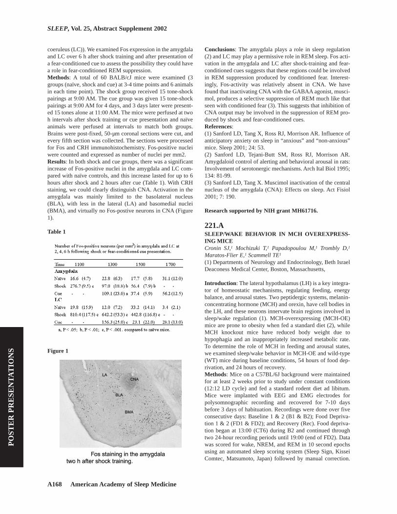
coeruleus (LC)). We examined Fos expression in the amygdalaand LC over 6 h after shock training and after presentation ofa fear-conditioned cue to assess the possibility they could havea role in fear-conditioned REM suppression.Methods: A total of 60 BALB/cJ mice were examined (3groups (naïve, shock and cue) at 3-4 time points and 6 animalsin each time point). The shock group received 15 tone-shockpairings at 9:00 AM. The cue group was given 15 tone-shockpairings at 9:00 AM for 4 days, and 3 days later were present-ed 15 tones alone at 11:00 AM. The mice were perfused at twoh intervals after shock training or cue presentation and naïveanimals were perfused at intervals to match both groups.Brains were post-fixed, 50-µm coronal sections were cut, andevery fifth section was collected. The sections were processedfor Fos and CRH immunohistochemistry. Fos-positive nucleiwere counted and expressed as number of nuclei per mm2.Results: In both shock and cue groups, there was a significantincrease of Fos-positive nuclei in the amygdala and LC com-pared with naïve controls, and this increase lasted for up to 6hours after shock and 2 hours after cue (Table 1). With CRHstaining, we could clearly distinguish CNA. Activation in theamygdala was mainly limited to the basolateral nucleus(BLA), with less in the lateral (LA) and basomedial nuclei(BMA), and virtually no Fos-postive neurons in CNA (Figure1).
Table 1
Figure 1
Conclusions: The amygdala plays a role in sleep regulation(2) and LC may play a permissive role in REM sleep. Fos acti-vation in the amygdala and LC after shock-training and fear-conditioned cues suggests that these regions could be involvedin REM suppression produced by conditioned fear. Interest-ingly, Fos-activity was relatively absent in CNA. We havefound that inactivating CNA with the GABAA agonist, musci-mol, produces a selective suppression of REM much like thatseen with conditioned fear (3). This suggests that inhibition ofCNA output may be involved in the suppression of REM pro-duced by shock and fear-conditioned cues.References: (1) Sanford LD, Tang X, Ross RJ, Morrison AR. Influence ofanticipatory anxiety on sleep in “anxious” and “non-anxious”mice. Sleep 2001; 24: 53.(2) Sanford LD, Tejani-Butt SM, Ross RJ, Morrison AR.Amygdaloid control of alerting and behavioral arousal in rats:Involvement of serotonergic mechanisms. Arch Ital Biol 1995;134: 81-99.(3) Sanford LD, Tang X. Muscimol inactivation of the centralnucleus of the amygdala (CNA): Effects on sleep. Act Fisiol2001; 7: 190.
Research supported by NIH grant MH61716.
221.ASLEEP/WAKE BEHAVIOR IN MCH OVEREXPRESS-ING MICECronin SJ,1 Mochizuki T,1 Papadopoulou M,1 Trombly D,1
Maratos-Flier E,1 Scammell TE1
(1) Departments of Neurology and Endocrinology, Beth IsraelDeaconess Medical Center, Boston, Massachusetts,
Introduction: The lateral hypothalamus (LH) is a key integra-tor of homeostatic mechanisms, regulating feeding, energybalance, and arousal states. Two peptidergic systems, melanin-concentrating hormone (MCH) and orexin, have cell bodies inthe LH, and these neurons innervate brain regions involved insleep/wake regulation (1). MCH-overexpressing (MCH-OE)mice are prone to obesity when fed a standard diet (2), whileMCH knockout mice have reduced body weight due tohypophagia and an inappropriately increased metabolic rate.To determine the role of MCH in feeding and arousal states,we examined sleep/wake behavior in MCH-OE and wild-type(WT) mice during baseline conditions, 54 hours of food dep-rivation, and 24 hours of recovery.Methods: Mice on a C57BL/6J background were maintainedfor at least 2 weeks prior to study under constant conditions(12:12 LD cycle) and fed a standard rodent diet ad libitum.Mice were implanted with EEG and EMG electrodes forpolysomnographic recording and recovered for 7-10 daysbefore 3 days of habituation. Recordings were done over fiveconsecutive days: Baseline 1 & 2 (B1 & B2); Food Depriva-tion 1 & 2 (FD1 & FD2); and Recovery (Rec). Food depriva-tion began at 13:00 (CT6) during B2 and continued throughtwo 24-hour recording periods until 19:00 (end of FD2). Datawas scored for wake, NREM, and REM in 10 second epochsusing an automated sleep scoring system (Sleep Sign, KisseiComtec, Matsumoto, Japan) followed by manual correction.
Sleep Research Society A169
PO
STE
R P
RE
SEN
TA
TIO
NS
SLEEP, Vol. 25, Abstract Supplement 2002
One week after the recordings, the mice were killed followinganother 54-hour period of food deprivation. Brains wereimmunostained for MCH.Results: MCH-OE mice had clear increases in the density andthickness of the MCH fibers throughout the brain, but the dis-tribution of MCH projections was similar to that of the WTmice. During the baseline day (B1), MCH-OE and WT micespent similar amounts of time in NREM, REM, and wakestates. After 54 hours of food deprivation, both groups hadincreased wakefulness during the first 6 hours of the dark peri-od, with more wakefulness in WT mice. During the food dep-rivation days, WT mice had a marked increase in the amountof wakefulness mainly through an increase in the duration ofwake bouts and a proportional loss of NREM and REM sleep.There was no change in spectral power during NREM sleepacross the four days of recordings.Conclusions: We found that food deprivation increased wake-fulness during the first half of the dark period in both WT andMCH-OE mice. These results are consistent with past reportsof nocturnal increases in wakefulness in food deprived rats (3).Although MCH neurons innervate many state regulatoryregions, we did not find substantial differences in thesleep/wake behavior of our MCH-OE mice. Thus, the increasein wakefulness seen with food deprivation may be due to theaction of other systems such as orexin/hypocretin.References: (1) Kilduff TS, Lecea L. Mapping of the mRNAs for thehypocretin/orexin and melanin-concentrating hormone recep-tors: networks for overlapping peptide systems. J Comp Neur,2001, 435:1-5.(2) Ludwig DS, et al. Melanin-concentrating hormone overex-pression in transgenic mice leads to obesity and insulin resist-ance. J Clin Invest, 2001, 107(3): 379-86.(3) Borbely AA. Sleep in the rat during food deprivation andsubsequent restitution of food. Brain Research, 1977, 124:457-471.
Research supported by NIH grants MH01507, HL60292,and MH62589
222.ASLEEP DEPRIVATION INDUCES SPATIAL MEMORYDEFICIT AND DOWN-REGULATES EXTRACELLU-LAR SIGNAL-REGULATED KINASE PHOSPHORY-LATION IN THE HIPPOCAMPUS Guan Z,1* Peng X,1 Harding J,1 Fang J2
(1) Department of Psychiatry and *Department of Compara-tive Medicine, Pennsylvania State University College of Med-icine, Hershey, PA 17033, (2) Department of Veterinary andComparative Anatomy, Pharmacology and Physiology, Wash-ington State University, Pullman, WA 99164,
Introduction: Increasing evidence indicates that loss of sleepmay result in memory impairment. However, little is knownabout the biochemical basis for memory deficits induced bysleep deprivation. Extracellular signal-regulated kinase (ERK)is involved in memory consolidation in different tasks, includ-ing spatial memory (Blum et al, 1999). Phosphorylation ofERK is necessary for its activation and is an important step in
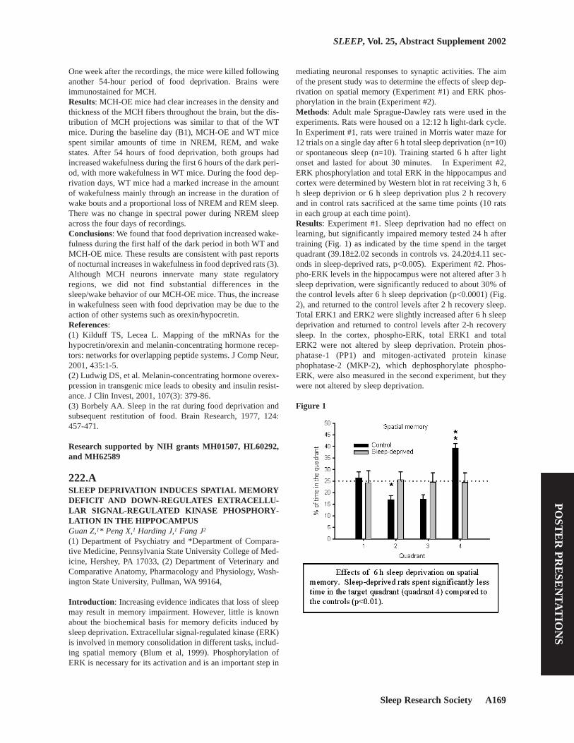
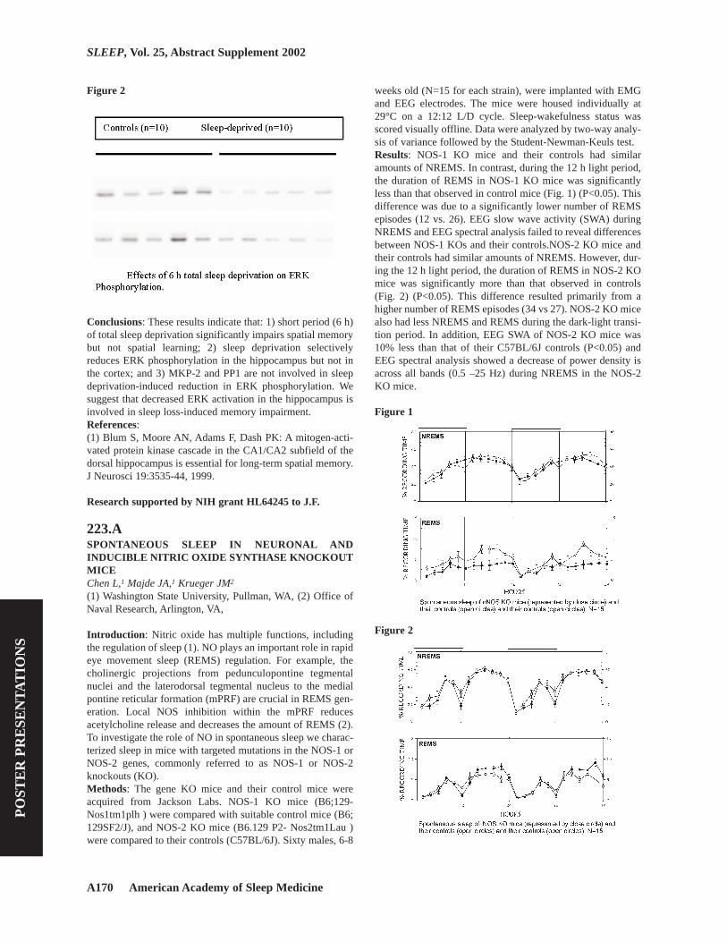
mediating neuronal responses to synaptic activities. The aimof the present study was to determine the effects of sleep dep-rivation on spatial memory (Experiment #1) and ERK phos-phorylation in the brain (Experiment #2).Methods: Adult male Sprague-Dawley rats were used in theexperiments. Rats were housed on a 12:12 h light-dark cycle.In Experiment #1, rats were trained in Morris water maze for12 trials on a single day after 6 h total sleep deprivation (n=10)or spontaneous sleep (n=10). Training started 6 h after lightonset and lasted for about 30 minutes. In Experiment #2,ERK phosphorylation and total ERK in the hippocampus andcortex were determined by Western blot in rat receiving 3 h, 6h sleep deprivion or 6 h sleep deprivation plus 2 h recoveryand in control rats sacrificed at the same time points (10 ratsin each group at each time point).Results: Experiment #1. Sleep deprivation had no effect onlearning, but significantly impaired memory tested 24 h aftertraining (Fig. 1) as indicated by the time spend in the targetquadrant (39.18±2.02 seconds in controls vs. 24.20±4.11 sec-onds in sleep-deprived rats, p<0.005). Experiment #2. Phos-pho-ERK levels in the hippocampus were not altered after 3 hsleep deprivation, were significantly reduced to about 30% ofthe control levels after 6 h sleep deprivation (p<0.0001) (Fig.2), and returned to the control levels after 2 h recovery sleep.Total ERK1 and ERK2 were slightly increased after 6 h sleepdeprivation and returned to control levels after 2-h recoverysleep. In the cortex, phospho-ERK, total ERK1 and totalERK2 were not altered by sleep deprivation. Protein phos-phatase-1 (PP1) and mitogen-activated protein kinasephophatase-2 (MKP-2), which dephosphorylate phospho-ERK, were also measured in the second experiment, but theywere not altered by sleep deprivation.
Figure 1
A170 American Academy of Sleep Medicine
PO
STE
R P
RE
SEN
TA
TIO
NS
SLEEP, Vol. 25, Abstract Supplement 2002
Figure 2
Conclusions: These results indicate that: 1) short period (6 h)of total sleep deprivation significantly impairs spatial memorybut not spatial learning; 2) sleep deprivation selectivelyreduces ERK phosphorylation in the hippocampus but not inthe cortex; and 3) MKP-2 and PP1 are not involved in sleepdeprivation-induced reduction in ERK phosphorylation. Wesuggest that decreased ERK activation in the hippocampus isinvolved in sleep loss-induced memory impairment.References: (1) Blum S, Moore AN, Adams F, Dash PK: A mitogen-acti-vated protein kinase cascade in the CA1/CA2 subfield of thedorsal hippocampus is essential for long-term spatial memory.J Neurosci 19:3535-44, 1999.
Research supported by NIH grant HL64245 to J.F.
223.ASPONTANEOUS SLEEP IN NEURONAL ANDINDUCIBLE NITRIC OXIDE SYNTHASE KNOCKOUTMICEChen L,1 Majde JA,1 Krueger JM2
(1) Washington State University, Pullman, WA, (2) Office ofNaval Research, Arlington, VA,
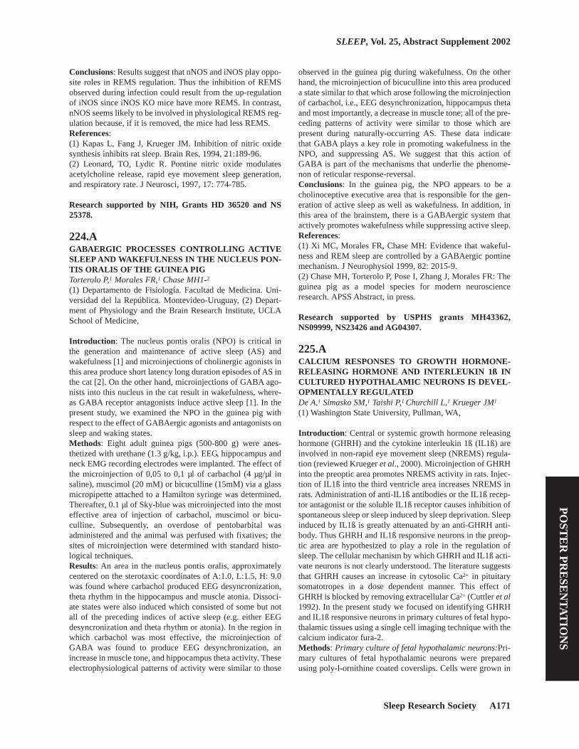
Introduction: Nitric oxide has multiple functions, includingthe regulation of sleep (1). NO plays an important role in rapideye movement sleep (REMS) regulation. For example, thecholinergic projections from pedunculopontine tegmentalnuclei and the laterodorsal tegmental nucleus to the medialpontine reticular formation (mPRF) are crucial in REMS gen-eration. Local NOS inhibition within the mPRF reducesacetylcholine release and decreases the amount of REMS (2).To investigate the role of NO in spontaneous sleep we charac-terized sleep in mice with targeted mutations in the NOS-1 orNOS-2 genes, commonly referred to as NOS-1 or NOS-2knockouts (KO).Methods: The gene KO mice and their control mice wereacquired from Jackson Labs. NOS-1 KO mice (B6;129-Nos1tm1plh ) were compared with suitable control mice (B6;129SF2/J), and NOS-2 KO mice (B6.129 P2- Nos2tm1Lau )were compared to their controls (C57BL/6J). Sixty males, 6-8
weeks old (N=15 for each strain), were implanted with EMGand EEG electrodes. The mice were housed individually at29°C on a 12:12 L/D cycle. Sleep-wakefulness status wasscored visually offline. Data were analyzed by two-way analy-sis of variance followed by the Student-Newman-Keuls test.Results: NOS-1 KO mice and their controls had similaramounts of NREMS. In contrast, during the 12 h light period,the duration of REMS in NOS-1 KO mice was significantlyless than that observed in control mice (Fig. 1) (P<0.05). Thisdifference was due to a significantly lower number of REMSepisodes (12 vs. 26). EEG slow wave activity (SWA) duringNREMS and EEG spectral analysis failed to reveal differencesbetween NOS-1 KOs and their controls.NOS-2 KO mice andtheir controls had similar amounts of NREMS. However, dur-ing the 12 h light period, the duration of REMS in NOS-2 KOmice was significantly more than that observed in controls(Fig. 2) (P<0.05). This difference resulted primarily from ahigher number of REMS episodes (34 vs 27). NOS-2 KO micealso had less NREMS and REMS during the dark-light transi-tion period. In addition, EEG SWA of NOS-2 KO mice was10% less than that of their C57BL/6J controls (P<0.05) andEEG spectral analysis showed a decrease of power density isacross all bands (0.5 –25 Hz) during NREMS in the NOS-2KO mice.
Figure 1
Figure 2
Sleep Research Society A171
PO
STE
R P
RE
SEN
TA
TIO
NS
SLEEP, Vol. 25, Abstract Supplement 2002
Conclusions: Results suggest that nNOS and iNOS play oppo-site roles in REMS regulation. Thus the inhibition of REMSobserved during infection could result from the up-regulationof iNOS since iNOS KO mice have more REMS. In contrast,nNOS seems likely to be involved in physiological REMS reg-ulation because, if it is removed, the mice had less REMS.References: (1) Kapas L, Fang J, Krueger JM. Inhibition of nitric oxidesynthesis inhibits rat sleep. Brain Res, 1994, 21:189-96.(2) Leonard, TO, Lydic R. Pontine nitric oxide modulatesacetylcholine release, rapid eye movement sleep generation,and respiratory rate. J Neurosci, 1997, 17: 774-785.
Research supported by NIH, Grants HD 36520 and NS25378.
224.AGABAERGIC PROCESSES CONTROLLING ACTIVESLEEP AND WAKEFULNESS IN THE NUCLEUS PON-TIS ORALIS OF THE GUINEA PIGTorterolo P,1 Morales FR,1 Chase MH1-2
(1) Departamento de Fisiología. Facultad de Medicina. Uni-versidad del la República. Montevideo-Uruguay, (2) Depart-ment of Physiology and the Brain Research Institute, UCLASchool of Medicine,
Introduction: The nucleus pontis oralis (NPO) is critical inthe generation and maintenance of active sleep (AS) andwakefulness [1] and microinjections of cholinergic agonists inthis area produce short latency long duration episodes of AS inthe cat [2]. On the other hand, microinjections of GABA ago-nists into this nucleus in the cat result in wakefulness, where-as GABA receptor antagonists induce active sleep [1]. In thepresent study, we examined the NPO in the guinea pig withrespect to the effect of GABAergic agonists and antagonists onsleep and waking states.Methods: Eight adult guinea pigs (500-800 g) were anes-thetized with urethane (1.3 g/kg, i.p.). EEG, hippocampus andneck EMG recording electrodes were implanted. The effect ofthe microinjection of 0,05 to 0,1 µl of carbachol (4 µg/µl insaline), muscimol (20 mM) or bicuculline (15mM) via a glassmicropipette attached to a Hamilton syringe was determined.Thereafter, 0.1 µl of Sky-blue was microinjected into the mosteffective area of injection of carbachol, muscimol or bicu-culline. Subsequently, an overdose of pentobarbital wasadministered and the animal was perfused with fixatives; thesites of microinjection were determined with standard histo-logical techniques.Results: An area in the nucleus pontis oralis, approximatelycentered on the sterotaxic coordinates of A:1.0, L:1.5, H: 9.0was found where carbachol produced EEG desyncronization,theta rhythm in the hippocampus and muscle atonia. Dissoci-ate states were also induced which consisted of some but notall of the preceding indices of active sleep (e.g. either EEGdesyncronization and theta rhythm or atonia). In the region inwhich carbachol was most effective, the microinjection ofGABA was found to produce EEG desynchronization, anincrease in muscle tone, and hippocampus theta activity. Theseelectrophysiological patterns of activity were similar to those
observed in the guinea pig during wakefulness. On the otherhand, the microinjection of bicuculline into this area produceda state similar to that which arose following the microinjectionof carbachol, i.e., EEG desynchronization, hippocampus thetaand most importantly, a decrease in muscle tone; all of the pre-ceding patterns of activity were similar to those which arepresent during naturally-occurring AS. These data indicatethat GABA plays a key role in promoting wakefulness in theNPO, and suppressing AS. We suggest that this action ofGABA is part of the mechanisms that underlie the phenome-non of reticular response-reversal.Conclusions: In the guinea pig, the NPO appears to be acholinoceptive executive area that is responsible for the gen-eration of active sleep as well as wakefulness. In addition, inthis area of the brainstem, there is a GABAergic system thatactively promotes wakefulness while suppressing active sleep.References: (1) Xi MC, Morales FR, Chase MH: Evidence that wakeful-ness and REM sleep are controlled by a GABAergic pontinemechanism. J Neurophysiol 1999, 82: 2015-9.(2) Chase MH, Torterolo P, Pose I, Zhang J, Morales FR: Theguinea pig as a model species for modern neuroscienceresearch. APSS Abstract, in press.
Research supported by USPHS grants MH43362,NS09999, NS23426 and AG04307.
225.ACALCIUM RESPONSES TO GROWTH HORMONE-RELEASING HORMONE AND INTERLEUKIN 1ß INCULTURED HYPOTHALAMIC NEURONS IS DEVEL-OPMENTALLY REGULATEDDe A,1 Simasko SM,1 Taishi P,1 Churchill L,1 Krueger JM1
(1) Washington State University, Pullman, WA,
Introduction: Central or systemic growth hormone releasinghormone (GHRH) and the cytokine interleukin 1ß (IL1ß) areinvolved in non-rapid eye movement sleep (NREMS) regula-tion (reviewed Krueger et al., 2000). Microinjection of GHRHinto the preoptic area promotes NREMS activity in rats. Injec-tion of IL1ß into the third ventricle area increases NREMS inrats. Administration of anti-IL1ß antibodies or the IL1ß recep-tor antagonist or the soluble IL1ß receptor causes inhibition ofspontaneous sleep or sleep induced by sleep deprivation. Sleepinduced by IL1ß is greatly attenuated by an anti-GHRH anti-body. Thus GHRH and IL1ß responsive neurons in the preop-tic area are hypothesized to play a role in the regulation ofsleep. The cellular mechanism by which GHRH and IL1ß acti-vate neurons is not clearly understood. The literature suggeststhat GHRH causes an increase in cytosolic Ca2+ in pituitarysomatotropes in a dose dependent manner. This effect ofGHRH is blocked by removing extracellular Ca2+ (Cuttler et al1992). In the present study we focused on identifying GHRHand IL1ß responsive neurons in primary cultures of fetal hypo-thalamic tissues using a single cell imaging technique with thecalcium indicator fura-2.Methods: Primary culture of fetal hypothalamic neurons:Pri-mary cultures of fetal hypothalamic neurons were preparedusing poly-l-ornithine coated coverslips. Cells were grown in
A172 American Academy of Sleep Medicine
PO
STE
R P
RE
SEN
TA
TIO
NS
SLEEP, Vol. 25, Abstract Supplement 2002
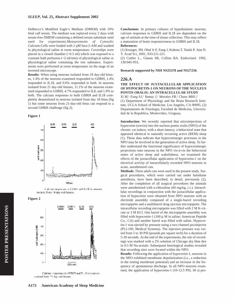
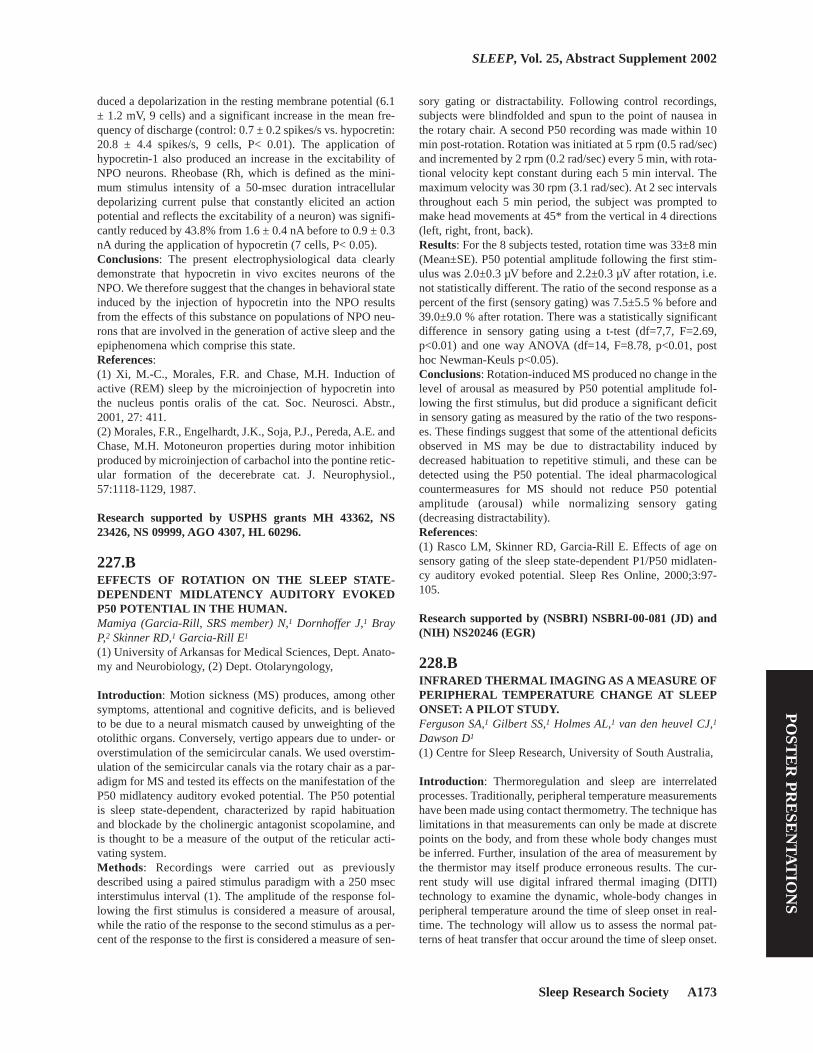
Dulbecco’s Modified Eagle’s Medium (DMEM) with 10%fetal calf serum. The medium was replaced every 2 days withserum-free DMEM containing a defined serum substitute untilused for experiments.Measurements of CytosolicCalcium:Cells were loaded with 2 µM fura-2-AM and washedin physiological saline at room temperature. Coverslips wereplaced in a closed chamber (~0.5 ml) which was exposed to aconstant bath perfusion (~2 ml/min) of physiological saline orphysiological saline containing the test substance. Experi-ments were performed at room temperature on the stage of aninverted microscope. Results: When using neurons isolated from 18 day-old fetus-es, 1.4% of the neurons examined responded to GHRH, 1.4%responded to IL1ß, and 0.6% responded to both. In neuronsisolated from 21 day-old fetuses, 11.1% of the neurons exam-ined responded to GHRH, 4.7% responded to ILß, and 1.9% toboth. The calcium responses to both GHRH and IL1ß com-pletely desensitized in neurons isolated from day 18 fetus (fig1) but some neurons from 21-day-old fetus can respond to asecond GHRH challenge (fig 2).
Figure 1
Figure 2
Conclusions: In primary cultures of hypothalamic neurons,calcium responses to GHRH and IL1ß are dependent on theage of animals at the time of tissue collection. This may reflecta maturation of brain responsiveness to GHRH and IL1ß.References: (1) Krueger, JM, Obal Jr F, Fang J, Kubota T, Taishi P. Ann N.Y. Acad Sci, 2001, 933:211-221.(2) Cuttler L., Glaum SR, Collins BA. Endocrinol. 1992,130:945-953.
Research supported by NIH NS25378 and NS27250.
226.ATHE EFFECT OF JUXTACELLULAR APPLICATIONOF HYPOCRETIN-1 ON NEURONS OF THE NUCLEUSPONTIS ORALIS: AN INTRACELLULAR STUDYXi M,1 Fung SJ,1 Yamuy J,1 Morales FR,1 Chase MH1,2
(1) Department of Physiology and the Brain Research Insti-tute, UCLA School of Medicine, Los Angeles, CA 90095, (2)Departamento de Fisiologia, Facultad de Medicina, Universi-dad de la Republica, Montevideo, Uruguay,
Introduction: We recently reported that microinjections ofhypocretin (orexin) into the nucleus pontis oralis (NPO) of thechronic cat induce, with a short latency, a behavioral state thatappeared identical to naturally occurring active (REM) sleep(1). These data indicate that hypocretinergic processes in theNPO may be involved in the generation of active sleep. To fur-ther understand the functional significance of hypocretinergicprojections onto neurons in the NPO vis-à-vis the behavioralstates of active sleep and wakefulness, we examined theeffects of the juxtacellular application of hypocretin-1 on theelectrical activity of intracellularly recorded NPO neurons inacute, anesthetized cats.Methods: Three adult cats were used in the present study. Sur-gical procedures, which were carried out under halothaneanesthesia, have been described, in detail, previously (2).After the completion of all surgical procedures the animalswere anesthetized with a-chloralose (60 mg/kg, i.v.). Intracel-lular recordings in conjunction with the juxtacellular applica-tion of hypocretin were obtained from NPO neurons with anelectrode assembly composed of a single-barrel recordingmicropipette and a multibarrel drug ejection micropipette. Theintracellular recording micropipette was filled with 2 M K-cit-rate or 3 M KCl. One barrel of the micropipette assembly wasfilled with hypocretin-1 (100 µ M in saline; American PeptideCo., CA) and another barrel was filled with saline. Hypocre-tin-1 was ejected by pressure using a two-channel picoinjector(PLI-100, Medical Systems). The injection pressure was var-ied from 5 to 30 PSI (pounds per square inch) for a duration of5-30 seconds. At the end of the experiments, the site of record-ings was marked with a 2% solution of Chicago sky blue dyein 0.5 M Na-acetate. Subsequent histological studies revealedthat recording sites were located within the NPO.Results: Following the application of hypocretin-1, neurons inthe NPO exhibited membrane depolarization (i.e., a reductionin the resting membrane potential) and an increase in the fre-quency of spontaneous discharge. In all NPO neurons exam-ined, the application of hypocretin-1 (10-12.5 PSI, 30 s) pro-
Sleep Research Society A173
PO
STE
R P
RE
SEN
TA
TIO
NS
SLEEP, Vol. 25, Abstract Supplement 2002
duced a depolarization in the resting membrane potential (6.1± 1.2 mV, 9 cells) and a significant increase in the mean fre-quency of discharge (control: 0.7 ± 0.2 spikes/s vs. hypocretin:20.8 ± 4.4 spikes/s, 9 cells, P< 0.01). The application ofhypocretin-1 also produced an increase in the excitability ofNPO neurons. Rheobase (Rh, which is defined as the mini-mum stimulus intensity of a 50-msec duration intracellulardepolarizing current pulse that constantly elicited an actionpotential and reflects the excitability of a neuron) was signifi-cantly reduced by 43.8% from 1.6 ± 0.4 nA before to 0.9 ± 0.3nA during the application of hypocretin (7 cells, P< 0.05).Conclusions: The present electrophysiological data clearlydemonstrate that hypocretin in vivo excites neurons of theNPO. We therefore suggest that the changes in behavioral stateinduced by the injection of hypocretin into the NPO resultsfrom the effects of this substance on populations of NPO neu-rons that are involved in the generation of active sleep and theepiphenomena which comprise this state.References: (1) Xi, M.-C., Morales, F.R. and Chase, M.H. Induction ofactive (REM) sleep by the microinjection of hypocretin intothe nucleus pontis oralis of the cat. Soc. Neurosci. Abstr.,2001, 27: 411.(2) Morales, F.R., Engelhardt, J.K., Soja, P.J., Pereda, A.E. andChase, M.H. Motoneuron properties during motor inhibitionproduced by microinjection of carbachol into the pontine retic-ular formation of the decerebrate cat. J. Neurophysiol.,57:1118-1129, 1987.
Research supported by USPHS grants MH 43362, NS23426, NS 09999, AGO 4307, HL 60296.
227.BEFFECTS OF ROTATION ON THE SLEEP STATE-DEPENDENT MIDLATENCY AUDITORY EVOKEDP50 POTENTIAL IN THE HUMAN.Mamiya (Garcia-Rill, SRS member) N,1 Dornhoffer J,1 BrayP,2 Skinner RD,1 Garcia-Rill E1
(1) University of Arkansas for Medical Sciences, Dept. Anato-my and Neurobiology, (2) Dept. Otolaryngology,
Introduction: Motion sickness (MS) produces, among othersymptoms, attentional and cognitive deficits, and is believedto be due to a neural mismatch caused by unweighting of theotolithic organs. Conversely, vertigo appears due to under- oroverstimulation of the semicircular canals. We used overstim-ulation of the semicircular canals via the rotary chair as a par-adigm for MS and tested its effects on the manifestation of theP50 midlatency auditory evoked potential. The P50 potentialis sleep state-dependent, characterized by rapid habituationand blockade by the cholinergic antagonist scopolamine, andis thought to be a measure of the output of the reticular acti-vating system.Methods: Recordings were carried out as previouslydescribed using a paired stimulus paradigm with a 250 msecinterstimulus interval (1). The amplitude of the response fol-lowing the first stimulus is considered a measure of arousal,while the ratio of the response to the second stimulus as a per-cent of the response to the first is considered a measure of sen-
sory gating or distractability. Following control recordings,subjects were blindfolded and spun to the point of nausea inthe rotary chair. A second P50 recording was made within 10min post-rotation. Rotation was initiated at 5 rpm (0.5 rad/sec)and incremented by 2 rpm (0.2 rad/sec) every 5 min, with rota-tional velocity kept constant during each 5 min interval. Themaximum velocity was 30 rpm (3.1 rad/sec). At 2 sec intervalsthroughout each 5 min period, the subject was prompted tomake head movements at 45* from the vertical in 4 directions(left, right, front, back).Results: For the 8 subjects tested, rotation time was 33±8 min(Mean±SE). P50 potential amplitude following the first stim-ulus was 2.0±0.3 µV before and 2.2±0.3 µV after rotation, i.e.not statistically different. The ratio of the second response as apercent of the first (sensory gating) was 7.5±5.5 % before and39.0±9.0 % after rotation. There was a statistically significantdifference in sensory gating using a t-test (df=7,7, F=2.69,p<0.01) and one way ANOVA (df=14, F=8.78, p<0.01, posthoc Newman-Keuls p<0.05).Conclusions: Rotation-induced MS produced no change in thelevel of arousal as measured by P50 potential amplitude fol-lowing the first stimulus, but did produce a significant deficitin sensory gating as measured by the ratio of the two respons-es. These findings suggest that some of the attentional deficitsobserved in MS may be due to distractability induced bydecreased habituation to repetitive stimuli, and these can bedetected using the P50 potential. The ideal pharmacologicalcountermeasures for MS should not reduce P50 potentialamplitude (arousal) while normalizing sensory gating(decreasing distractability).References: (1) Rasco LM, Skinner RD, Garcia-Rill E. Effects of age onsensory gating of the sleep state-dependent P1/P50 midlaten-cy auditory evoked potential. Sleep Res Online, 2000;3:97-105.
Research supported by (NSBRI) NSBRI-00-081 (JD) and(NIH) NS20246 (EGR)
228.BINFRARED THERMAL IMAGING AS A MEASURE OFPERIPHERAL TEMPERATURE CHANGE AT SLEEPONSET: A PILOT STUDY.Ferguson SA,1 Gilbert SS,1 Holmes AL,1 van den heuvel CJ,1
Dawson D1
(1) Centre for Sleep Research, University of South Australia,
Introduction: Thermoregulation and sleep are interrelatedprocesses. Traditionally, peripheral temperature measurementshave been made using contact thermometry. The technique haslimitations in that measurements can only be made at discretepoints on the body, and from these whole body changes mustbe inferred. Further, insulation of the area of measurement bythe thermistor may itself produce erroneous results. The cur-rent study will use digital infrared thermal imaging (DITI)technology to examine the dynamic, whole-body changes inperipheral temperature around the time of sleep onset in real-time. The technology will allow us to assess the normal pat-terns of heat transfer that occur around the time of sleep onset.
A174 American Academy of Sleep Medicine
PO
STE
R P
RE
SEN
TA
TIO
NS
SLEEP, Vol. 25, Abstract Supplement 2002
Methods: Sixteen healthy male participants, aged 18-30 wererecruited to the study. Participants spent an adaptation nightand a recording night in the laboratory. We recorded sleepusing conventional PSG and temperature using both contactthermometry (core and feet) and DITI (whole upper body).Participants were lying supine in bed with their hands by theirsides at least 90 minutes prior to their normal lights out. At thetime of lights out they were requested to try and fall asleep inthe same position. From 60 minutes prior to lights out thermalimages were captured every 30 seconds. Splicing the imagestogether into a continuous animation sequence allowed quali-tative assessment of the peripheral temperature changes.Quantitative measurements were taken from regions of inter-est (hands, forearms, face, neck, chest and torso).Results: 1. Infrared thermal images can be captured from par-ticipants around the time of sleep onset with minimal loss ofdata due to movement (fig 1).2. The patterns of heat exchangeacross the period of sleep onset can be readily visualised innormal sleepers.3. Preliminary data analysis indicates that sig-nificant peripheral temperature changes occur in the forearmsand hands, in addition to the lower torso region.
Figure 2—Digital infrared thermal images taken from a sub-ject prior to sleep (top panel) and at sleep onset (bottompanel). The images are grayscaled for better visualisation inblack and white. The change in peripheral temperature is mostevident in the forearms and hands, and the face and neck, withthe darker shade indicating higher temperature. (An animationof the coloured images can be found atwww.unisa.edu.au/sleep/research/default.htm)
Conclusions: The results indicate that digital infrared thermalimaging is a technique that can be readily applied to the inves-tigation of sleep initiation processes. Patterns of heat exchangearound sleep onset, particularly in the arms and hands, andface and neck can be clearly visualised. Images from the pres-ent study will be compared with thermal images collectedfrom sleep-onset insomniacs, with the aim of qualifying thedifferences in temperature exchange between normal sleepersand insomniacs.
229.BALTERATIONS IN BLOOD PRESSURE ACROSS THESLEEP ONSET PERIODCarrington MJ,1 Jones M,1 Crowley KE,1 Colrain IM,1 KimY,1,2 Trinder J1
(1) Department of Psychology, The University of Melbourne,Victoria, Australia, (2) Human Sleep Research Program, SRIInternational, Menlo Park, CA, USA,
Introduction: Blood Pressure (BP) has a 24-hr variation thatis dependent on the sleep-wake cycle, with a reduction occur-ring during sleep. Recent evidence suggests that BP falls rap-idly during sleep onset, reaching its lowest level once stablesleep is attained. The aim of this study was to characterise thepattern of change in BP, Heart Rate (HR) and baroreflex activ-ity during the period from relaxed pre-sleep wakefulness tostable sleep.Methods: Continuous BP and HR recordings were collectedbeginning 2 hrs before lights out until the end of the firstNREM sleep period in 9 young, healthy, male and female sub-jects maintained in a supine position. The data was analysed asa function of 5 consecutive phases: 1) 30 minutes before lightsout; 2) lights out to stage 1 sleep; 3) stage 1 to stage 2 sleep;4) stage 2 sleep to the last micro-arousal before stable sleep;and 5) the first 30 minutes of undisturbed stable sleep. Datawas analysed on a beat-by-beat basis and reported as 2 minperiods for phases 1 and 5, and as 10% epochs for phases 2, 3and 4 (as subjects had variable time periods in these phases).The level of baroreflex activity was assessed by the number of3 beat inverse sequences between BP and HR, while barore-flex sensitivity (BRS) was assessed by the average slope with-in sequences. During phases 3 and 4, the BP and HR responseto arousal from sleep was determined.Results: A combination of inferential and descriptive statisticswere applied to the data. From relaxed wakefulness to stablesleep, HR fell by 7 b/min, whilst systolic blood pressure (SBP)fell by 13 mmHg and diastolic blood pressure (DBP) by 10mmHg, with the minimum values occurring 15 minutes intostable sleep. The fall in BP occurred during phase 2 (followinglights out - SBP fell 8 mmHg) and phase 5 (following theattainment of stable sleep - SBP fell 5 mmHg). There were nosignificant differences in the number of sequences generated,while BRS increased by 3 msec/mmHg during phase 3, butotherwise was unchanged. During phases 3 (stage 1) and 4(stage 2 with micro-arousals), BP did not change during peri-ods of sleep, while arousals were associated with transient,high frequency increases in activity that returned BP and HRback to the pre-lights out wakefulness levels (averageresponse amplitude of 13 mmHg).Conclusions: Data suggest that the fall in BP during sleeponset is due to a rapid downward resetting of the baroreflex, aprocess that is retarded, and may be reversed, by arousalsoccurring during periods of sleep-wake instability. This phe-nomenon may contribute to the “non-dipping” BP profile seenin some sleep and cardiovascular disordered patients.
Research supported by an ARC grant.
Sleep Research Society A175
PO
STE
R P
RE
SEN
TA
TIO
NS
SLEEP, Vol. 25, Abstract Supplement 2002
230.BINCREASED SLEEPINESS AND FINGER SKIN TEM-PERATURE AFTER MELATONIN ADMINISTRA-TION: A TOPOGRAPHICAL INFRARED THERMOM-ETRY ANALYSIS Kräuchi K,1 Pache M,1 von Arb M,2 Wirz-Justice A,1 FlammerJ1
(1) Center for Chronobiology, Psychiatric University Clinic;,(2) University Eye Clinic, Basel, Switzerland,
Introduction: Recent studies have shown a close relationshipbetween sleepiness and the thermolytic effects of melatonin,which induces heat loss via distal skin regions and hence heatloss in the body core (1). Because of the selective increase indistal, but not proximal skin temperatures after melatoninadministration, we suggested a specific vasodilatory action ofmelatonin in skin regions containing arteriovenous anasto-moses (AVAs) (2). However, we previously measured distalskin temperatures on the back of the hand, a region containingno AVAs (2). It is the fingertip, and especially the nail bed,which contains the majority of AVAs in the hand (2). Thus, thefingers provide a good model to study the specificity of actionof melatonin on blood flow and skin temperature.Methods: In a double-blind placebo-controlled 1-weekcrossover study, 11 healthy men (mean age 26y±2 SD, BMI22.2±1.7) received melatonin (mel, 5 mg p.o.) or placebo(plac) at 14:00h in a balanced order. Skin temperatures of thefingers were measured by means of an infrared detector(TH3100mr Thermo Tracer; NEC San-ei InstrumentsLtd.,Tokyo, Japan) 30 min before and 60 min after pill intake(room temperature: 26±1°C). Subjective ratings of sleepiness(mm VAS) and core body temperature (CBT, sublingual) weremeasured in parallel. Subjects remained in a sitting position30min before and during the measurements. Here we reportmean skin temperature of distal and planar skin regions of thefirst, second and third phalanx of the middle finger (ANOVAfor repeated measures, significance level p<0.05).
Figure 1
Results: Melatonin showed a soporific and thermolytic effect(∆ mm VAS sleepiness: +15.4±6.8mm; ∆CBT:-0.07±0.03°C), confirming previous findings under supineconstant routine conditions (1). Baseline skin temperatures(Figure 1a) were highest on the fingernail and lowest on thepalmar side of the third phalanx. No dorsal-palmar differencesin the first phalanx could be observed. Melatonin uniformlyincreased skin temperature independent of skin region (Figure1b, mean temperature of palmar and dorsal skin regions).Conclusions: To achieve a skin temperature increase of simi-lar magnitude at both proximal and distal regions a much larg-er increase of blood flow in the latter is required. Therefore,our results indicate a higher skin blood flow elevation bymelatonin in the fingertip than in the proximal finger, mostprobably by opening of AVAs.References: (1) Kräuchi K, Cajochen C, Wirz-Justice A. Circadian andhomeostatic regulation of core body temperature and alertnessin humans: what is the role of melatonin? Circadian Clocksand Entrainment, In: K-I Honma & S Honma, Hokkaido Uni-versity press, Sapporo, 1998; Vol. 7, 131-146.(2) Grant RT, Bland EF. Observations on arteriovenous anas-tomoses in human skin and in the bird’s foot with special ref-erence to the reaction to cold. Heart 1929-31; 15: 385-411.
231.BGH REPLACEMENT FAILS TO NORMALIZE SLEEPRESPONSES TO VIRAL INFLUENZA INFECTION INGHRH-RECEPTOR DEFICIENT MICE.Alt JA,1 Obal Jr. F,1 Majde JA,2 Krueger JM3
(1) Washington State University, Pullman, WA, (2) Dept.Physiology, Univ. Szeged A. Szent-Gyorgyi Med. Center,Szeged, Hungary, (3) Office of Naval Research, Arlington,VA,
Introduction: Viral infections induce excess non-rapid eyemovement sleep (NREMS) and somnogenic cytokines, includ-ing interleukin-1β (IL-1β). IL-1β stimulates growth hormone-releasing hormone (GHRH), a peptide also implicated inNREMS regulation. Via QTL analysis, the GHRH-receptor(GHRH-R) was identified as a candidate gene responsible forNREMS responses of mice to influenza challenge (1). Wefound that the dwarf lit/lit mouse with a point mutation in theGHRH-R gene (2), responded to influenza with decreasedNREMS (3). GH might be necessary for normal function ofthe immune system and sleep. We determined therefore,whether GH replacement can normalize the sleep response inthe lit/lit mice to influenza infection.Methods: Mice (control C57BL/6, n=12: lit/lit , n=12) wereimplanted with EEG and EMG electrodes. Animals were on a12:12-h light-dark cycle at 29° C. Alzet minipumps wereimplanted ip in lit/lit mice to release 11 µg mouse GH per day(n=4) or 24 µg rat GH per day (n=8). Findings in the 2 groupsdid not differ and were pooled. Controls received saline.Weights were taken to determine if the GH therapy was effec-tive. We recorded spontaneous sleep for 48 h, 8-9 days aftersurgery. Mice were then intranasally infected on postoperativeday 10, with A/PR/8/34 (H1N1) influenza virus (2.5 x10⁶
TCIDs50s in 50 µl) at light onset. Changes in NREMS and
A176 American Academy of Sleep Medicine
PO
STE
R P
RE
SEN
TA
TIO
NS
SLEEP, Vol. 25, Abstract Supplement 2002
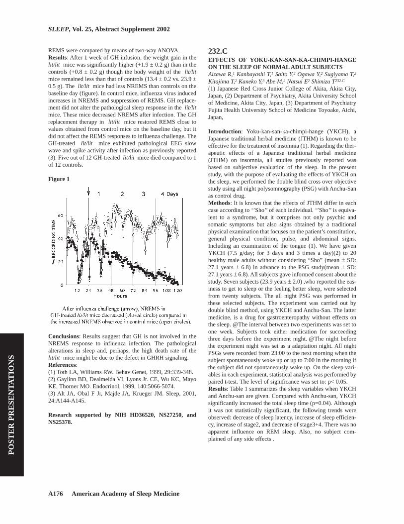
REMS were compared by means of two-way ANOVA.Results: After 1 week of GH infusion, the weight gain in thelit/lit mice was significantly higher (+1.9 ± 0.2 g) than in thecontrols (+0.8 ± 0.2 g) though the body weight of the lit/litmice remained less than that of controls (13.4 ± 0.2 vs. 23.9 ±0.5 g). The lit/lit mice had less NREMS than controls on thebaseline day (figure). In control mice, influenza virus inducedincreases in NREMS and suppression of REMS. GH replace-ment did not alter the pathological sleep response in the lit/litmice. These mice decreased NREMS after infection. The GHreplacement therapy in lit/lit mice restored REMS close tovalues obtained from control mice on the baseline day, but itdid not affect the REMS responses to influenza challenge. TheGH-treated lit/lit mice exhibited pathological EEG slowwave and spike activity after infection as previously reported(3). Five out of 12 GH-treated lit/lit mice died compared to 1of 12 controls.
Figure 1
Conclusions: Results suggest that GH is not involved in theNREMS response to influenza infection. The pathologicalalterations in sleep and, perhaps, the high death rate of thelit/lit mice might be due to the defect in GHRH signaling.References: (1) Toth LA, Williams RW. Behav Genet, 1999, 29:339-348.(2) Gaylinn BD, Dealmeida VI, Lyons Jr. CE, Wu KC, MayoKE, Thorner MO. Endocrinol, 1999, 140:5066-5074.(3) Alt JA, Obal F Jr, Majde JA, Krueger JM. Sleep, 2001,24:A144-A145.
Research supported by NIH HD36520, NS27250, andNS25378.
232.CEFFECTS OF YOKU-KAN-SAN-KA-CHIMPI-HANGEON THE SLEEP OF NORMAL ADULT SUBJECTSAizawa R,1 Kanbayashi T,1 Saito Y,2 Ogawa Y,2 Sugiyama T,2
Kitajima T,2 Kaneko Y,3 Abe M,2 Natsui E2 Shimizu T232.C
(1) Japanese Red Cross Junior College of Akita, Akita City,Japan, (2) Department of Psychiatry, Akita University Schoolof Medicine, Akita City, Japan, (3) Department of PsychiatryFujita Health University School of Medicine Toyoake, Aichi,Japan,
Introduction: Yoku-kan-san-ka-chimpi-hange (YKCH), aJapanese traditional herbal medicine (JTHM) is known to beeffective for the treatment of insomnia (1). Regarding the ther-apeutic effects of a Japanese traditional herbal medicine(JTHM) on insomnia, all studies previously reported wasbased on subjective evaluation of the sleep. In the presentstudy, with the purpose of evaluating the effects of YKCH onthe sleep, we performed the double blind cross over objectivestudy using all night polysomnography (PSG) with Anchu-Sanas control drug.Methods: It is known that the effects of JTHM differ in eachcase according to ‘’Sho’’ of each individual. ‘’Sho’’ is equiva-lent to a syndrome, but it comprises not only psychic andsomatic symptoms but also signs obtained by a traditionalphysical examination that focuses on the patient’s constitution,general physical condition, pulse, and abdominal signs.Including an examination of the tongue (1). We have givenYKCH (7.5 g/day; for 3 days and 3 times a day)(2) to 20healthy male adults without considering “Sho” (mean ± SD:27.1 years ± 6.8) in advance to the PSG study(mean ± SD:27.1 years ± 6.8). All subjects gave informed consent about thestudy. Seven subjects (23.9 years ± 2.0) ,who reported the eas-iness to get to sleep or the feeling better sleep, were selectedfrom twenty subjects. The all night PSG was performed inthese selected subjects. The experiment was carried out bydouble blind method, using YKCH and Anchu-San. The lattermedicine, is a drug for gastroenteropathy without effects onthe sleep. @The interval between two experiments was set toone week. Subjects took either medication for succeedingthree days before the experiment night. @The night beforethe experiment night was set as a adaptation night. All nightPSGs were recorded from 23:00 to the next morning when thesubject spontaneously woke up or up to 7:00 in the morning ifthe subject did not spontaneously wake up. On the sleep vari-ables in each experiment, statistical analysis was performed bypaired t-test. The level of significance was set to: p< 0.05.Results: Table 1 summarizes the sleep variables when YKCHand Anchu-san are given. Compared with Anchu-san, YKCHsignificantly increased the total sleep time (p=0.04). Althoughit was not statistically significant, the following trends wereobserved: decrease of sleep latency, increase of sleep efficien-cy, increase of stage2, and decrease of stage3+4. There was noapparent influence on REM sleep. Also, no subject com-plained of any side effects .
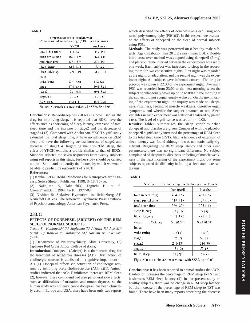
Sleep Research Society A177
PO
STE
R P
RE
SEN
TA
TIO
NS
SLEEP, Vol. 25, Abstract Supplement 2002
Table 1
Conclusions: Benzodiazepines (BDZs) is now used as thedrug for improving sleep. It is reported that BDZs have theeffects such as shortening of sleep latency, extension of totalsleep time and the increase of stage2 and the decrease ofstage3+4 (3). Compared with Anchu-san, YKCH significantlyextended the total sleep time, it have no influence on REMsleep and have the following trends: increase of stage2 anddecrease of stage3+4. Regarding the non-REM sleep, theeffect of YKCH exhibits a profile similar to that of BDZs.Since we selected the seven responders from twenty subjectsusing self reports in this study, further study should be carriedout on ‘’Sho’’, and to identify the factors, by which we wouldbe able to predict the responders of YKCH.References: (1) Kanba S et al: Herbal Medicines for Neuropsychiatric Dis-ease, Seiwa Shoten, Publishers, 1998; 3- 13: Tokyo.(2) Nakajima K, TakeuchiY, Taguchi H, et al:Chem.Pharm.Bull.1994. 42(10), 1977-83.(3) Nishino S: Sedative Hypnotics. in: Schatzberg AF,Nemeroff CB. eds. The American Psychiatric Press Textbookof Psychopharmacology. American Psychiatric Press.
233.CEFFECTS OF DONEPEZIL (ARICEPT) ON THE REMSLEEP OF NORMAL SUBJECTSTetsuo S,1 Kanbayashi T,1 Sugiyama T,1 Aizawa R,1 Abe M,2
Suzuki F,1 Kaneko Y,1 Watanabe M,1 Narumi A1 TakemuraT233.C
(1) Department of Neuropsychiatry, Akita University, (2)Japanese Red Cross Junior College of Akita, Introduction: Donepezil (Aricept) is a therapeutic drug forthe treatment of Alzheimer diseases (AD). Dysfunction ofcholinergic neurons is attributed to cognitive impairment inAD (1). Donepezil effects via activation of cholinergic neu-rons by inhibiting acetylcholin-esterase (ACh-E)(1). Animalstudies indicated that ACh-E inhibitors increased REM sleep(2), however these compound had also peripheral side effects,such as difficulties of urination and mouth dryness, so thehuman study was not easy. Since donepezil has been clinical-ly used in Europe and USA, there have been only two reports
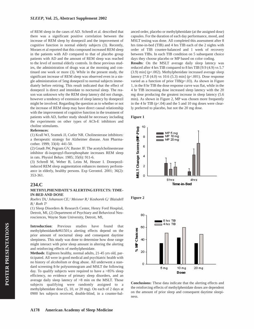
which described the effects of donepezil on sleep using noc-turnal polysomnography (PSG)(3). In this respect, we evaluat-ed the effects of donepezil on the sleep of normal subjectsusing PSG.Methods: The study was performed on 8 healthy male sub-jects. Age distribution was 28 ± 2 years (mean ± SD). Doubleblind cross over method was adopted using donepezil (5 mg)and placebo. Time interval between the experiments was set toone week. Each subject was instructed to sleep in the record-ing room for two consecutive nights. First night was regardedas the night for adaptation, and the second night was the exper-iment night. All subjects gave informed consent. The drug orplacebo was given at 22:30 of the experiment night. OvernightPSG was recorded from 23:00 to the next morning when thesubject spontaneously woke up or up to 8:00 in the morning ifthe subject did not spontaneously wake up. On the next morn-ing of the experiment night, the inquiry was made on: sleepi-ness, dizziness, feeling of muscle weakness, digestive organsymptoms, and whether the subject dreamed or not. Sleepvariables in each experiment was statistical analyzed by pairedt-test. The level of significance was set to: p < 0.05.Results: Table1 summarizes the sleep variables whendonepezil and placebo are given. Compared with the placebo,donepezil significantly increased the percentage of REM sleepto the total sleep time (TST). Also, a tendency of extension ofsleep latency was found although it was not statistically sig-nificant. Regarding the REM sleep latency and other sleepparameters, there was no significant difference. No subjectcomplained of sleepiness, dizziness or feeling of muscle weak-ness in the next morning of the experiment night, but somesubjects reported the difficulty in falling a sleep and increaseddreams.
Table 1
Conclusions: It has been reported in animal studies that ACh-E inhibitor increases the percentage of REM sleep to TST andit shortens REM sleep latency (2). In our present study onhealthy subjects, there was no change in REM sleep latency,but the increase of the percentage of REM sleep to TST wasfound. There have been many reports describing the decrease
A178 American Academy of Sleep Medicine
PO
STE
R P
RE
SEN
TA
TIO
NS
SLEEP, Vol. 25, Abstract Supplement 2002
of REM sleep in the cases of AD. Schredl et al. described thatthere was a significant positive correlation between theincrease of REM sleep by donepezil and the improvement ofcognitive function in normal elderly subjects (3). Recently,Moraes et al reported that this compound increased REM sleepin the patients with AD compared to that of placebo grouppatients with AD and the amount of REM sleep was reachedto the level of normal elderly controls. In these previous stud-ies, the administration of drugs was at the morning and con-tinued one week or more (3). While in the present study, thesignificant increase of REM sleep was observed even in a sin-gle administration of 5mg donepezil to normal subjects imme-diately before retiring. This result indicated that the effect ofdonepezil is direct and immidate to nocturnal sleep. The rea-son was unknown why the REM sleep latency did not change,however a tendency of extension of sleep latency by donepezilmight be involved. Regarding the question as to whether or notthe increase of REM sleep may have direct causal relationshipwith the improvement of cognitive function in the treatment ofpatients with AD, further study should be necessary includingthe experiments on other types of ACh-E inhibitors andcholine stimulants.References: (1) Krall WJ, Sramek JJ, Cutler NR. Cholinesterase inhibitors:a therapeutic strategy for Alzheimer disease. Ann Pharma-cother. 1999; 33(4): 441-50.(2) Gnadt JW, Pegram GV, Baxter JF. The acetylcholinesteraseinhibitor di-isopropyl-fluorophosphate increases REM sleepin rats. Physiol Behav. 1985; 35(6): 911-6.(3) Schredl M, Weber B, Leins M, Heuser I. Donepezil-induced REM sleep augmentation enhances memory perform-ance in elderly, healthy persons. Exp Gerontol. 2001; 36(2):353-361.
234.CMETHYLPHENIDATE’S ALERTING EFFECTS: TIME-IN-BED AND DOSERoehrs TA,1 Johanson CE,1 Meixner R,2 Koshorek G,1 BlaisdellB,1 Roth T1
(1) Sleep Disorders & Research Center, Henry Ford Hospital,Detroit, MI, (2) Department of Psychiary and Behavioral Neu-rosciences, Wayne State University, Detroit, MI,
Introduction: Previous studies have found thatmethylphenidates alerting effects depend on theprior amount of nocturnal sleep and consequent daytimesleepiness. This study was done to determine how dose rangemight interact with prior sleep amount in altering the alertingand reinforcing effects of methylphenidate.Methods: Eighteen healthy, normal adults, 21-45 yrs old, par-ticipated. All were in good medical and psychiatric health withno history of alcoholism or drug abuse. All underwent a stan-dard screening 8-hr polysomnogram and MSLT the followingday. To qualify subjects were required to have a >85% sleepefficiency, no evidence of primary sleep disorders, and anaverage daily sleep latency of >8 min on the MSLT. Thosesubjects qualifying were randomly assigned to amethylphenidate dose (5, 10, or 20 mg). On each of 2 days at0900 hrs subjects received, double-blind, in a counter-bal-
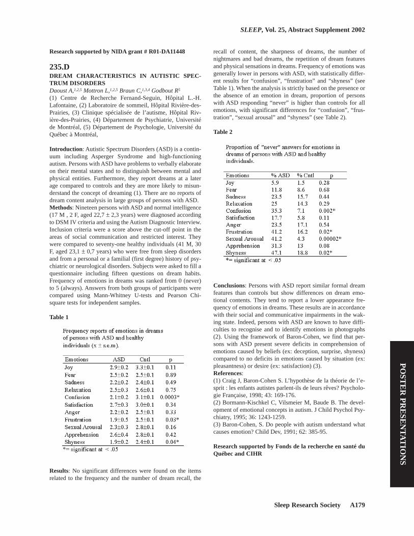
anced order, placebo or methylphenidate (at the assigned dose)capsules. For the duration of each day performance, mood, andMSLT testing was done. All completed this assessment after 8hrs time-in-bed (TIB) and 4 hrs TIB each of the 2 nights withorder of TIB counter-balanced and 1 week of recoverybetween TIBs. In each TIB condition on 5 subsequent choicedays they choose placebo or MP based on color coding.Results: On the MSLT average daily sleep latency wasreduced after 4 hrs TIB compared to 8 hrs TIB [9.9 (4.9) vs 5.7(3.9) min] (p<.002). Methylphenidate increased average sleeplatency [7.8 (4.0) vs 10.6 (5.3) min] (p<.001). Dose responsevaried as a function of prior TIB(p<.03). As shown in Figure1, in the 8 hr TIB the dose response curve was flat, while in the4 hr TIB increasing dose increased sleep latency with the 20mg dose producing the greatest increase in sleep latency (5.6min). As shown in Figure 2, MP was chosen more frequentlyin the 4 hr TIB (p<.04) and the 5 and 10 mg doses were clear-ly preferred to placebo, but not the 20 mg dose.
Figure 1
Figure 2
Conclusions: These data indicate that the alerting effects andthe reinforcing effects of methylphenidate doses are dependenton the amount of prior sleep and consequent daytime sleepi-ness.
Sleep Research Society A179
PO
STE
R P
RE
SEN
TA
TIO
NS
SLEEP, Vol. 25, Abstract Supplement 2002
Research supported by NIDA grant # R01-DA11448
235.DDREAM CHARACTERISTICS IN AUTISTIC SPEC-TRUM DISORDERSDaoust A,1,2,5 Mottron L,1,2,5 Braun C,1,3,4 Godbout R5
(1) Centre de Recherche Fernand-Seguin, Hôpital L.-H.Lafontaine, (2) Laboratoire de sommeil, Hôpital Rivière-des-Prairies, (3) Clinique spécialisée de l’autisme, Hôpital Riv-ière-des-Prairies, (4) Département de Psychiatrie, Universitéde Montréal, (5) Département de Psychologie, Université duQuébec à Montréal,
Introduction: Autistic Spectrum Disorders (ASD) is a contin-uum including Asperger Syndrome and high-functioningautism. Persons with ASD have problems to verbally elaborateon their mental states and to distinguish between mental andphysical entities. Furthermore, they report dreams at a laterage compared to controls and they are more likely to misun-derstand the concept of dreaming (1). There are no reports ofdream content analysis in large groups of persons with ASD.Methods: Nineteen persons with ASD and normal intelligence(17 M , 2 F, aged 22,7 ± 2,3 years) were diagnosed accordingto DSM IV criteria and using the Autism Diagnostic Interview.Inclusion criteria were a score above the cut-off point in theareas of social communication and restricted interest. Theywere compared to seventy-one healthy individuals (41 M, 30F, aged 23,1 ± 0,7 years) who were free from sleep disordersand from a personal or a familial (first degree) history of psy-chiatric or neurological disorders. Subjects were asked to fill aquestionnaire including fifteen questions on dream habits.Frequency of emotions in dreams was ranked from 0 (never)to 5 (always). Answers from both groups of participants werecompared using Mann-Whitney U-tests and Pearson Chi-square tests for independent samples.
Table 1
Results: No significant differences were found on the itemsrelated to the frequency and the number of dream recall, the
recall of content, the sharpness of dreams, the number ofnightmares and bad dreams, the repetition of dream featuresand physical sensations in dreams. Frequency of emotions wasgenerally lower in persons with ASD, with statistically differ-ent results for “confusion”, “frustration” and “shyness” (seeTable 1). When the analysis is strictly based on the presence orthe absence of an emotion in dream, proportion of personswith ASD responding “never” is higher than controls for allemotions, with significant differences for “confusion”, “frus-tration”, “sexual arousal” and “shyness” (see Table 2).
Table 2
Conclusions: Persons with ASD report similar formal dreamfeatures than controls but show differences on dream emo-tional contents. They tend to report a lower appearance fre-quency of emotions in dreams. These results are in accordancewith their social and communicative impairments in the wak-ing state. Indeed, persons with ASD are known to have diffi-culties to recognise and to identify emotions in photographs(2). Using the framework of Baron-Cohen, we find that per-sons with ASD present severe deficits in comprehension ofemotions caused by beliefs (ex: deception, surprise, shyness)compared to no deficits in emotions caused by situation (ex:pleasantness) or desire (ex: satisfaction) (3).References: (1) Craig J, Baron-Cohen S. L’hypothèse de la théorie de l’e-sprit : les enfants autistes parlent-ils de leurs rêves? Psycholo-gie Française, 1998; 43: 169-176.(2) Bormann-Kischkel C, Vilsmeier M, Baude B. The devel-opment of emotional concepts in autism. J Child Psychol Psy-chiatry, 1995; 36: 1243-1259.(3) Baron-Cohen, S. Do people with autism understand whatcauses emotion? Child Dev, 1991; 62: 385-95.
Research supported by Fonds de la recherche en santé duQuébec and CIHR
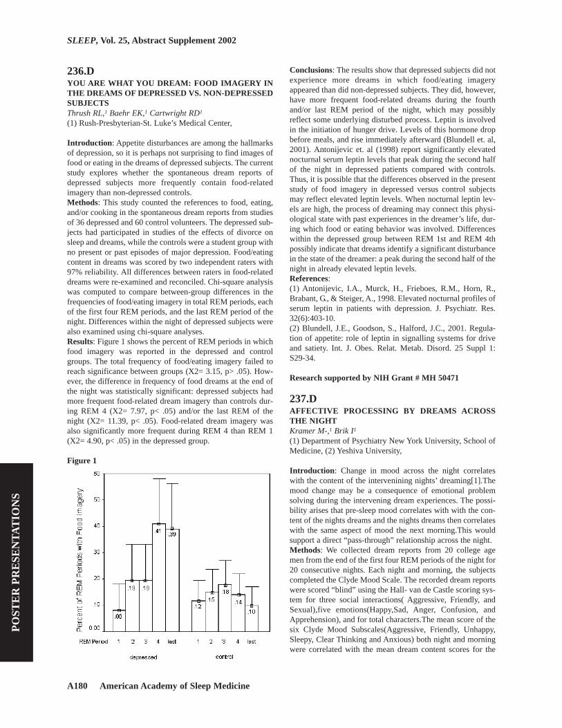
A180 American Academy of Sleep Medicine
PO
STE
R P
RE
SEN
TA
TIO
NS
SLEEP, Vol. 25, Abstract Supplement 2002
236.DYOU ARE WHAT YOU DREAM: FOOD IMAGERY INTHE DREAMS OF DEPRESSED VS. NON-DEPRESSEDSUBJECTSThrush RL,1 Baehr EK,1 Cartwright RD1
(1) Rush-Presbyterian-St. Luke’s Medical Center,
Introduction: Appetite disturbances are among the hallmarksof depression, so it is perhaps not surprising to find images offood or eating in the dreams of depressed subjects. The currentstudy explores whether the spontaneous dream reports ofdepressed subjects more frequently contain food-relatedimagery than non-depressed controls.Methods: This study counted the references to food, eating,and/or cooking in the spontaneous dream reports from studiesof 36 depressed and 60 control volunteers. The depressed sub-jects had participated in studies of the effects of divorce onsleep and dreams, while the controls were a student group withno present or past episodes of major depression. Food/eatingcontent in dreams was scored by two independent raters with97% reliability. All differences between raters in food-relateddreams were re-examined and reconciled. Chi-square analysiswas computed to compare between-group differences in thefrequencies of food/eating imagery in total REM periods, eachof the first four REM periods, and the last REM period of thenight. Differences within the night of depressed subjects werealso examined using chi-square analyses.Results: Figure 1 shows the percent of REM periods in whichfood imagery was reported in the depressed and controlgroups. The total frequency of food/eating imagery failed toreach significance between groups (X2= 3.15, p> .05). How-ever, the difference in frequency of food dreams at the end ofthe night was statistically significant: depressed subjects hadmore frequent food-related dream imagery than controls dur-ing REM 4 (X2= 7.97, p< .05) and/or the last REM of thenight (X2= 11.39, p< .05). Food-related dream imagery wasalso significantly more frequent during REM 4 than REM 1(X2= 4.90, p< .05) in the depressed group.
Figure 1
Conclusions: The results show that depressed subjects did notexperience more dreams in which food/eating imageryappeared than did non-depressed subjects. They did, however,have more frequent food-related dreams during the fourthand/or last REM period of the night, which may possiblyreflect some underlying disturbed process. Leptin is involvedin the initiation of hunger drive. Levels of this hormone dropbefore meals, and rise immediately afterward (Blundell et. al,2001). Antonijevic et. al (1998) report significantly elevatednocturnal serum leptin levels that peak during the second halfof the night in depressed patients compared with controls.Thus, it is possible that the differences observed in the presentstudy of food imagery in depressed versus control subjectsmay reflect elevated leptin levels. When nocturnal leptin lev-els are high, the process of dreaming may connect this physi-ological state with past experiences in the dreamer’s life, dur-ing which food or eating behavior was involved. Differenceswithin the depressed group between REM 1st and REM 4thpossibly indicate that dreams identify a significant disturbancein the state of the dreamer: a peak during the second half of thenight in already elevated leptin levels.References: (1) Antonijevic, I.A., Murck, H., Frieboes, R.M., Horn, R.,Brabant, G., & Steiger, A., 1998. Elevated nocturnal profiles ofserum leptin in patients with depression. J. Psychiatr. Res.32(6):403-10.(2) Blundell, J.E., Goodson, S., Halford, J.C., 2001. Regula-tion of appetite: role of leptin in signalling systems for driveand satiety. Int. J. Obes. Relat. Metab. Disord. 25 Suppl 1:S29-34.
Research supported by NIH Grant # MH 50471
237.DAFFECTIVE PROCESSING BY DREAMS ACROSSTHE NIGHTKramer M-,1 Brik I1
(1) Department of Psychiatry New York University, School ofMedicine, (2) Yeshiva University,
Introduction: Change in mood across the night correlateswith the content of the intervenining nights’ dreaming[1].Themood change may be a consequence of emotional problemsolving during the intervening dream experiences. The possi-bility arises that pre-sleep mood correlates with with the con-tent of the nights dreams and the nights dreams then correlateswith the same aspect of mood the next morning.This wouldsupport a direct “pass-through” relationship across the night.Methods: We collected dream reports from 20 college agemen from the end of the first four REM periods of the night for20 consecutive nights. Each night and morning, the subjectscompleted the Clyde Mood Scale. The recorded dream reportswere scored “blind” using the Hall- van de Castle scoring sys-tem for three social interactions( Aggressive, Friendly, andSexual),five emotions(Happy,Sad, Anger, Confusion, andApprehension), and for total characters.The mean score of thesix Clyde Mood Subscales(Aggressive, Friendly, Unhappy,Sleepy, Clear Thinking and Anxious) both night and morningwere correlated with the mean dream content scores for the
Sleep Research Society A181
PO
STE
R P
RE
SEN
TA
TIO
NS
SLEEP, Vol. 25, Abstract Supplement 2002
three social interaction scales, the five emotion scales, andwith the total characters in the dreams. Between subjects Pear-son correlations were done.We did 108 correlations(six moodsubscales x two times x nine dream content scales).Results: We found ten significant positive correlations atp=.05 or less.Two were with an aspect of night mood (Anxiousnight mood with Sexual Social Interactions and with the emo-tion Confusion).Eight of the significant correlations were withsome aspect of morning mood and the prior nights’dreams:1)Friedly, Unhappy, and Clear Thinking mood withAggressive Social interactions, 2)Aggressive and Unhappymood and the emotion Sad, 3) Friendly mood and the emo-tions Anger and Apprehension, and 4) Unhappy mood and theemotion Confusion.None were with total characters.Conclusions: We were unable to find any direct “pass-through” relationship between mood at night, dream contentand the same aspect of mood in the morning. This is in con-trast to our having found a relationship between the change inmood across the night and the content of the nights’ dreams.This relationship was most striking for the change in Unhappymood across the night and the number and type of charactersin the dreams.The failure to find a direct “pass-through” rela-tionship may be a result of the relatively low dream recall per-centage(63%) and/or the paucity of scoreable dream con-tent:343 social interactions, 224 emotions, and 1194 charac-ters. The low frequency of dream contents precluded a withinsubject correlation, which may be the more appropriate levelof analysis to search for a possible direct “pass-through” rela-tionship.Clearly indirect ones were demonstrated.Three corre-lations were found with Unhappy, but none with characters.Asmore correlations were found with morning mood than nightmood, dreams may be more proactive than reactive.References: (1) Kramer,M. The Selective Moood Regulatory Function ofDreaming. In:Moffitt,A.,Kramer,M.,Hoffmann,R.,eds. TheFunctions of Dreaming.Albany,New York:State University ofNew York Press, 1993:139-195.
238.DNIGHTMARE EXPERIENCE AND FATIGUE IN UNI-VERSITY STUDENTSHicks RA,1 Fernandez C,1 Nguyen TT,1 Picchioni D1
(1) San Jose State University,
Introduction: Recent research and speculation suggests thatnightmares may play a role in the management of stressfulexperience and that stressful experience may be associatedwith the development of fatigue. Our purpose in this study wasto explore the possibility of relationships between extremes infatigue and nightmare experiences (i.e., the frequency of threetypes of nightmares and nightmare distress).Methods: We tested a large group of undergraduate studentsand identified 100 (50 men and 50 women) who scored low infatigue, i.e., the bottom 20% on the FIS-10 and 100 (50 menand 50 women) who scored high in fatigue, i.e., the top 20%on the FIS-10. These groups were matched as closely as pos-sible for age and ethnicity. For these Low and High FatigueGroups, we scored their responses to frequency that they expe-rienced the three types of Nightmares that are included in the
Spadafora and Hunt1 Dream Scale and their responses toBelicki’s2 Nightmare Distress Scale. Then these data wereorganized and used to compare the differences between thenightmare experiences of the Low and High Fatigue Groups.Results: The data for each nightmare parameter for eachfatigue group are summarized in the table. These data indicatethat there is no difference between the means of the twofatigue groups for fantastic nightmares (i.e., the typical REMNightmare or “Bad Dream”). However, for nightmares thatmay be initiated by exposure to major traumatic events (PostTraumatic Nightmares and Night Terrors), the differencesbetween the fatigue groups is both statistically significant andmeaningful with the High Fatigue Group reporting experienc-ing 33% more Post Traumatic Nightmares and 44% moreNight Terrors. Finally, these data indicate that high fatigue wasassociated with nightmares that were perceived as 30% moredistressful than those experienced by the Low Fatigue Group.
Table 1
Conclusions: Collectively, these data suggest that fatigue isassociated with dreaming that may reflect a prolonged attemptto cope with a major traumatic past experience.References: (1) Spadafora A, Hunt HT: The multiplicity of dreams: cogni-tive-affective correlates of lucid, archetypal, and nightmaredreaming. Perceptual and Motor Skills 1990; 71: 627-644.(2) Belicki K: The relationship of nightmare frequency tonightmare suffering with implications for treatment andresearch. Dreaming 1992; 2: 143-148.
Research supported by NIH-MBRS Grant 0819210-21 andNIH-NIGMS MARC Grant 5T34GM08253.
239.EEVENING AND MORNING DIFFERENCES IN WAK-ING EEG REACTIVITY IN YOUNG ADULTSBrière M,1,2 Forest G,1,2 Chouinard S,1,2 Godbout R1
(1) Centre de recherche Fernand-Seguin, Hôp. Louis-H.Lafontaine, (2) Département psychiatrie & psychologie, Uni-versité de Montréal, Canada,
Introduction: A number of studies show that waking EEGpower decreases in the morning, relative to evening. This isgeneraly interpreted as reflecting the restaurative functions ofsleep. Such studies have used eyes closed (EC) or eyes opened(EO) recording conditions. However, it is the EC/EO ratio thatis thought to best represent EEG and cortical reactivity (1).Unfortunately, the restaurative function of sleep has not yetbeen evaluated with this measure. We investigated the topog-raphy of the evening and morning EC/EO ratio as a part of amore extensive project on sleep, EEG and performance.Efforts were devoted to control for confounding variables suchas age, gender, laterality of participants, time of day, lighting
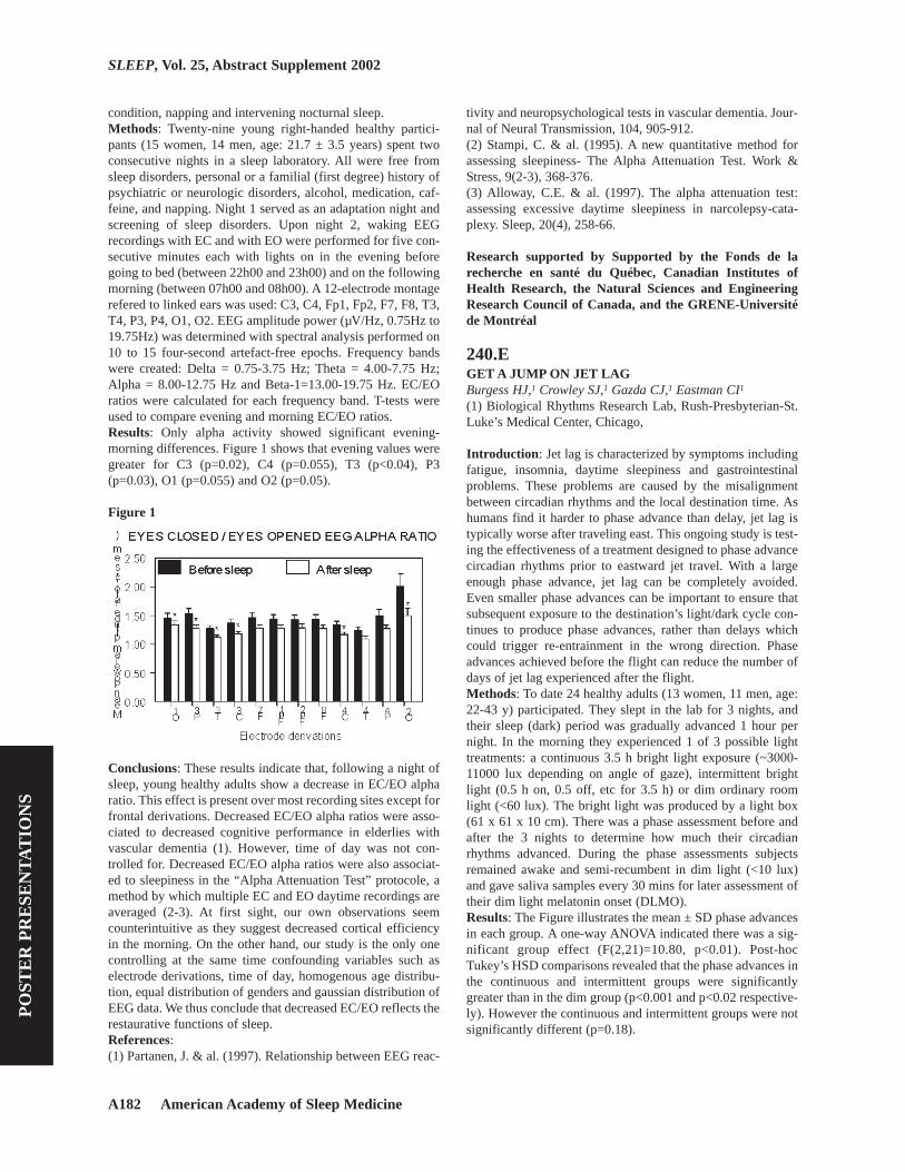
A182 American Academy of Sleep Medicine
PO
STE
R P
RE
SEN
TA
TIO
NS
SLEEP, Vol. 25, Abstract Supplement 2002
condition, napping and intervening nocturnal sleep.Methods: Twenty-nine young right-handed healthy partici-pants (15 women, 14 men, age: 21.7 ± 3.5 years) spent twoconsecutive nights in a sleep laboratory. All were free fromsleep disorders, personal or a familial (first degree) history ofpsychiatric or neurologic disorders, alcohol, medication, caf-feine, and napping. Night 1 served as an adaptation night andscreening of sleep disorders. Upon night 2, waking EEGrecordings with EC and with EO were performed for five con-secutive minutes each with lights on in the evening beforegoing to bed (between 22h00 and 23h00) and on the followingmorning (between 07h00 and 08h00). A 12-electrode montagerefered to linked ears was used: C3, C4, Fp1, Fp2, F7, F8, T3,T4, P3, P4, O1, O2. EEG amplitude power (µV/Hz, 0.75Hz to19.75Hz) was determined with spectral analysis performed on10 to 15 four-second artefact-free epochs. Frequency bandswere created: Delta = 0.75-3.75 Hz; Theta = 4.00-7.75 Hz;Alpha = 8.00-12.75 Hz and Beta-1=13.00-19.75 Hz. EC/EOratios were calculated for each frequency band. T-tests wereused to compare evening and morning EC/EO ratios.Results: Only alpha activity showed significant evening-morning differences. Figure 1 shows that evening values weregreater for C3 (p=0.02), C4 (p=0.055), T3 (p<0.04), P3(p=0.03), O1 (p=0.055) and O2 (p=0.05).
Figure 1
Conclusions: These results indicate that, following a night ofsleep, young healthy adults show a decrease in EC/EO alpharatio. This effect is present over most recording sites except forfrontal derivations. Decreased EC/EO alpha ratios were asso-ciated to decreased cognitive performance in elderlies withvascular dementia (1). However, time of day was not con-trolled for. Decreased EC/EO alpha ratios were also associat-ed to sleepiness in the “Alpha Attenuation Test” protocole, amethod by which multiple EC and EO daytime recordings areaveraged (2-3). At first sight, our own observations seemcounterintuitive as they suggest decreased cortical efficiencyin the morning. On the other hand, our study is the only onecontrolling at the same time confounding variables such aselectrode derivations, time of day, homogenous age distribu-tion, equal distribution of genders and gaussian distribution ofEEG data. We thus conclude that decreased EC/EO reflects therestaurative functions of sleep.References: (1) Partanen, J. & al. (1997). Relationship between EEG reac-
tivity and neuropsychological tests in vascular dementia. Jour-nal of Neural Transmission, 104, 905-912. (2) Stampi, C. & al. (1995). A new quantitative method forassessing sleepiness- The Alpha Attenuation Test. Work &Stress, 9(2-3), 368-376.(3) Alloway, C.E. & al. (1997). The alpha attenuation test:assessing excessive daytime sleepiness in narcolepsy-cata-plexy. Sleep, 20(4), 258-66.
Research supported by Supported by the Fonds de larecherche en santé du Québec, Canadian Institutes ofHealth Research, the Natural Sciences and EngineeringResearch Council of Canada, and the GRENE-Universitéde Montréal
240.EGET A JUMP ON JET LAGBurgess HJ,1 Crowley SJ,1 Gazda CJ,1 Eastman CI1
(1) Biological Rhythms Research Lab, Rush-Presbyterian-St.Luke’s Medical Center, Chicago,
Introduction: Jet lag is characterized by symptoms includingfatigue, insomnia, daytime sleepiness and gastrointestinalproblems. These problems are caused by the misalignmentbetween circadian rhythms and the local destination time. Ashumans find it harder to phase advance than delay, jet lag istypically worse after traveling east. This ongoing study is test-ing the effectiveness of a treatment designed to phase advancecircadian rhythms prior to eastward jet travel. With a largeenough phase advance, jet lag can be completely avoided.Even smaller phase advances can be important to ensure thatsubsequent exposure to the destination’s light/dark cycle con-tinues to produce phase advances, rather than delays whichcould trigger re-entrainment in the wrong direction. Phaseadvances achieved before the flight can reduce the number ofdays of jet lag experienced after the flight.Methods: To date 24 healthy adults (13 women, 11 men, age:22-43 y) participated. They slept in the lab for 3 nights, andtheir sleep (dark) period was gradually advanced 1 hour pernight. In the morning they experienced 1 of 3 possible lighttreatments: a continuous 3.5 h bright light exposure (~3000-11000 lux depending on angle of gaze), intermittent brightlight (0.5 h on, 0.5 off, etc for 3.5 h) or dim ordinary roomlight (<60 lux). The bright light was produced by a light box(61 x 61 x 10 cm). There was a phase assessment before andafter the 3 nights to determine how much their circadianrhythms advanced. During the phase assessments subjectsremained awake and semi-recumbent in dim light (<10 lux)and gave saliva samples every 30 mins for later assessment oftheir dim light melatonin onset (DLMO).Results: The Figure illustrates the mean ± SD phase advancesin each group. A one-way ANOVA indicated there was a sig-nificant group effect (F(2,21)=10.80, p<0.01). Post-hocTukey’s HSD comparisons revealed that the phase advances inthe continuous and intermittent groups were significantlygreater than in the dim group (p<0.001 and p<0.02 respective-ly). However the continuous and intermittent groups were notsignificantly different (p=0.18).
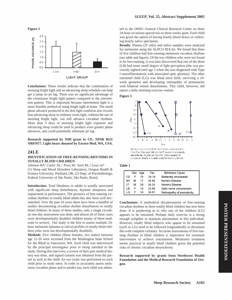
Sleep Research Society A183
PO
STE
R P
RE
SEN
TA
TIO
NS
SLEEP, Vol. 25, Abstract Supplement 2002
Figure 1
Conclusions: These results indicate that the combination ofmorning bright light and an advancing sleep schedule can helpget a jump on jet lag. There was no significant advantage ofthe continuous bright light pattern compared to the intermit-tent pattern. This is important because intermittent light is amore feasible method of using bright light at home. The smallphase advance produced in the dim light condition also revealsthat advancing sleep in ordinary room light, without the use ofmorning bright light, can still advance circadian rhythms.More than 3 days of morning bright light exposure andadvancing sleep could be used to produce even greater phaseadvances, and could potentially eliminate jet lag.
Research supported by NIH grant to CE: NINR RO1NR07677. Light boxes donated by Enviro-Med, WA, USA.
241.EIDENTIFICATION OF FREE-RUNNING RHYTHMS INTOTALLY BLIND CHILDRENJohnson KP,1 Cutler NL,1 Pires M,1 Sack RL,2 Lewy AJ1
(1) Sleep and Mood Disorders Laboratory, Oregon Health &Science University, Portland, OR, (2) Dept. of Psychobiology,Federal University of São Paulo, São Paulo, Brasil,
Introduction: Total blindness in adults is usually associatedwith significant sleep disturbances, daytime sleepiness andimpairment in performance. The presence of free-running cir-cadian rhythms in totally blind adults has also been well doc-umented. Over the past 10 years there have been a handful ofstudies documenting circadian rhythm disturbances in totallyblind children. In many of these studies, only a single circadi-an one-day assessment was done, and almost all of these caseswere developmentally disabled children (many of them mod-erate to severe). Our study is the first to assess multiple 24-hour melatonin (plasma or saliva) profiles in totally blind chil-dren (who were not developmentally disabled).Methods: Five children (three females, two males) betweenage 12-20 were recruited from the Washington State Schoolfor the Blind in Vancouver, WA. Each child was interviewedby the principal investigator prior to being enrolled in thestudy. During this interview, a review of their past medical his-tory was done, and signed consent was obtained from the par-ent as well as the child. An eye exam was performed on eachchild prior to study entry. In order to accurately assess mela-tonin circadian phase and to predict tau, each child was admit-
ted to the OHSU General Clinical Research Center on three24-hour occasions spaced two to three weeks apart. Each childwas given the option of having hourly blood draws or collect-ing hourly saliva specimens.Results: Plasma (JT only) and saliva samples were analyzedfor melatonin using the ALPCO RIA kit. We found that threeof five children had free-running melatonin circadian rhythms(see table and figure). Of the two children who were not foundto be free-running, it was later discovered that one of the them(LB) had some small degree of light perception (she was pre-viously sighted until age 5 when she was diagnosed with TypeI neurofibromatosis with associated optic gliomas). The otherentrained child (LG) was blind since birth, surviving a 24-week gestation and developing retinopathy of prematuritywith bilateral retinal detachments. This child, however, didreport a daily morning exercise routine.
Figure 1
Table 1
Conclusions: A methodical documentation of free-runningcircadian rhythms in three totally blind children has now beendone. It is perplexing as to why one of the children (LG)appears to be entrained. Perhaps daily exercise is a strongenough zeitgeber to maintain entrainment in this individual.However, totally blind subjects who appear to be entrained(such as LG) need to be followed longitudinally to documentthis with complete certainty. Accurate assessments of free-run-ning rhythms in blind children is imperative for treatmentintervention to achieve entrainment. Melatonin treatmentseems practical in totally blind children given the potentialrisks of chronic circadian desynchrony.
Research supported by grants from Northwest HealthFoundation and the Medical Research Foundation of Ore-gon.
A184 American Academy of Sleep Medicine
PO
STE
R P
RE
SEN
TA
TIO
NS
SLEEP, Vol. 25, Abstract Supplement 2002
242.ERECOVERY AFTER SLEEP DEPRIVATION IN SCN-LESIONED RATSMendelson W,1 Bergmann BM1
(1) Sleep Research Laboratory, The University of Chicago,
Introduction: In an ongoing series of studies of baseline andpost-deprivation recovery sleep, we have described a numberof age-related changes. During baseline sleep, for instance,older rats manifest reduced high voltage NREM sleep(“HS2”), EEG delta power, and duration of sleep bouts (1).Following 24 hours of sleep deprivation, the older animals hadsmaller increases in total sleep as well as NREM and REMsleep; moreover the older animals, while starting with lowerbaseline HS2, had a significant rebound increase in this stage,while the younger animals did not (2). While the cause ofthese differences has not been established, one possibility isthat they result from age-related changes in circadian regula-tory mechanisms. In the current study, we have begun toexplore this possibility by examining baseline and recoverysleep in young, “middle aged” and older rats following SCNlesions.Methods: The study involved 54 male Fisher rats in three agegroups (3, 12, and 18 months); in each group, half received aradiofrequency lesion of the SCN, while the other half under-went a sham lesion procedure. They then were given a oneweek recovery period in chambers with a 12:12 L:D cycle, inwhich lights came on at 10:00 AM. Following confirmation ofthe success of the lesion as manifested by 72 hour motor activ-ity recordings, they were given a 3 day acclimatization periodin continuous dim light in the sleep recording chambers, andthen had a 24 hour baseline sleep recording. After 24 hours ofbaseline sleep recordings, they underwent 24 hours of sleepdeprivation using the disk-over-water technique, followed bya 24 hour recovery sleep recording. All recordings, and thesleep deprivation period, began at 10:00 AM. All lesions andsham-lesions were histologically confirmed at the completionof the study. Results: An assessment of the amplitude of the sleep/wakecircadian rhythm in the baseline recordings was carried outusing 3 methods (cosine fit, the “A” statistic, and day/nightratio), and revealed greatly reduced amplitude in the SCN-lesioned animals (p< 0.001). One of the methods (day/nightratio) showed a greater reduction in the 18 month old animals.Lesioned animals, independent of age, manifested a 4%increase in NREM sleep (p< 0.05) and 15% increase in NREMdelta power (p< 0.05), as well as a 10% decrease in REM (p<0.05). SCN lesions had no significant effects on the amount ofNREM or REM during recovery sleep. Delta EEG power inNREM sleep, however, was 85% higher in the lesioned ani-mals (p< 0.01). Conclusions: We found little evidence, then, to indicate thatage-related changes in SCN function might differentiallyaffect the sleep of young, middle-aged and older rats. Ourobservations are consistent with the hypothesis that the SCNinhibits NREM sleep, though the effect was quantitativelysmall. The finding of increased EEG delta power duringrecovery sleep in the lesioned rats is also consistent with theview that the SCN influences homeostatic processes.
References: (1) Mendelson WB, Bergmann BM: Age-related changes insleep in the rat. Sleep 1999;2:5-10.(2) Mendelson WB, Bergmann BM: Age-dependent changesin recovery sleep following 48 hours of sleep deprivation inrats. Neurobiology of Aging 2000;21:689-693.
Research supported by NIH grants 2PO1 AG 11412-03 andK07 HL03640.
243.EEXOGENOUS MELATONIN SHIFTS DIM LIGHTMELATONIN ONSET IN POST-TRAUMATICDELAYED SLEEP PHASE SYNDROMEKayumov L,1 Deb P,1 Serbine O,1 Hossain N,2 Hosn R,1 WangF,1 Shapiro CM1
(1) Sleep Research Laboratory, University Health Network,Toronto, Canada, (2) University at Buffalo, USA,
Introduction: Circadian rhythm sleep disorders may occurafter traumatic brain injury1. Here we present a case of a 44-year-old man who developed a prominent delayed sleep phasesyndrome (DSPS) consequent on his motor vehicle accident.He suffered what appeared to be only minor injuries, includ-ing injuries to a soft tissue of his head, bruises and cuts. Healso lost his consciousness for a few minutes and suffered ananterograde amnesia. He did not have any further neurologicalsequela. However shortly after the accident the patient devel-oped severe sleep onset insomnia (inability to fall asleepbefore 04:00 h), frequent nightmares, and headaches. Thediagnosis of DSPS was established based on several physio-logical markers of the sleep-wake rhythm: Dim Light Mela-tonin Onset (DLMO) test2 using enzyme linked immunosor-bent assays (ELISA), wrist actigraphy, sleep log andpolysomnographic parameters of sleep architecture. Methods: The patient underwent two overnight sleep studies.On the first night the patient was allowed to go to bed when-ever he wanted and to sleep as much as he could. The patientarrived in the sleep clinic at 18:30 h and saliva samples wereobtained at hourly intervals from 19:00 to 03:00 h. Ambientlight intensity was controlled and did not exceed 15 lx. On thesecond night, a 24:00 to 08:00 sleep period was imposed.Results: DLMO test revealed significant delay in the endoge-nous melatonin secretion. DLMO occurred at 23:00 hours.The polysomnographic studies also showed a convincing evi-dence of delayed sleep phase syndrome. There was normalsleep onset latency (12 minutes), normal sleep duration (7.1 h)and normal sleep efficiency (91.4%) on the night when thepatient selected bedtime and rise time. Sleep onset latency was78 minutes on the second night when conventional bedtimewas imposed. Frequent arousals, low sleep efficiency (65.4%)and short sleep duration (5.2 h) were observed on this occa-sion. Administration of exogenous melatonin 5 mg (PennPharmaceutical Ltd) at 18:00 h for 28 days normalised sleep-wake cycle as was evidenced by wrist actigraphy and sleeplog. Post-treatment DLMO occurred at 21:00 h.Conclusions: The learning point of this case is that the DLMOassays can be used as a precise tool to assess the phase of thecircadian pacemaker in patients suspected of having a chrono-

Sleep Research Society A185
PO
STE
R P
RE
SEN
TA
TIO
NS
SLEEP, Vol. 25, Abstract Supplement 2002
biologic component to their sleep disorder. The abnormal tim-ing of DLMO can also provide an indication for the optimaltiming of treatment (medication administration, exposure tolight).References: (1) Sleep Disorders Medicine: Basic Science, Technical Con-siderations, and Clinical Aspects (ed.Chokroverty S). Butterworth-Heinemann 2000.
(2) Lewy AJ. The dim light melatonin onset, melatonin assaysand biological rhythms research in humans. Biol SignalsRecep 1999; 8: 79-83.
244.ECIRCADIAN RHYTHM OF SLEEPINESS AND THERISK OF HIGHWAY VEHICLE ACCIDENTSKim J1
(1) Division of Humanities & Social Sciences, Pohang Uni-versity of Science and Technology, Pohang, Republic ofKorea,
Introduction: Due to the multiple factors involved in most ofthe vehicle accidents, it is extremely difficult to sort out theinfluence of sleepiness on accidents. In the absence of gooddata on the ‘sleepiness-caused’ vehicle accidents in Korea, thepresent study evaluates the impact of sleepiness indirectly byanalyzing the hourly distribution of the Korean highway vehi-cle accidents adjusted to traffic volume in the light of the cir-cadian rhythm of sleepiness.Methods: The database of vehicle accidents and traffic vol-ume on the Korean highway network during the last twoyears(1999-2000) was provided by the Korean Road TrafficSafety Authority and the Korea Highway Corporation. Thepresent analysis used both accidents and traffic volume dataaveraged for weekdays and every 2-hour intervals. To find outhow the rate of accidents adjusted to traffic volume is differ-ent depending on the time of the day, the relative risks of the12 intervals were examined by ANOVA. The relative risk (rel-ative probability of accidents corrected for traffic volume) ofevery 2-hours in the highway vehicle accidents over the 24hours was calculated as follows: % of accidents during the 2-hour interval out of total accidents for 24 hours divided by %of traffic volume during the 2-hour interval out of total trafficvolume for 24 hours.Results: It was found that the relative risks (for all the 3 cate-gories: # of cases, # of the wounded, # of deaths) in the hoursof 00-06h and 22-24h were significantly higher than those inthe rest of the hours, being the highest during the 02-04h, then04-06h / 00-02h, and 22-24h, in order (Fig. 1). It is noticeablethat in the category of # deaths, the relative risk of 02-04h(thepeak) is more than 14 times higher than that of 12-14h(thetrough). The pattern reveals the rapid increase in accidents incorrespondence with high sleep propensity levels occuring atnight, especially in the early morning hours.
Figure 1
Conclusions: The hourly distribution of relative risk in thepresent study closely corresponds with the circadian rhythm ofsleep (sleepiness/alertness) (Carskadon & Dement, 1992:Mitler, 1991), thereby confirming the influence of sleepinesson vehicle accidents. Most important, the present findings,based on the total accidents (not on sleep-ascribed accidentsbut on accidents of all causes), are almost identical to the otherfindings on the sleep-ascribed vehicle accidents (Garbarino etal., 2001: Horne & Reyne, 2001) and the finding on all theaccidents except alcohol-related ones(Akerstedt, Kecklund,Horte, 2001). The present research demonstrates that sleepi-ness is a very important contributing factor in Korean highwayvehicle accidents in the presence of many other contributingfactors(e.g., road conditions, weather, vehicle conditions)thought to be randomly or evenly distributed.References: (1) Garbarino S, Nobili L, Beelke M, Phy FDC, Ferrillo F. Thecontributing role of sleepiness in highway vehicle accidents.Sleep 2001, 24(2):203-206.(2) Akerstedt T, Kecklund G, Horte L. Night driving, season,and the risk of highway accidents. Sleep 2001, 24(4):401-406.(3) Carskadon, MA & Dement WC. Multiple sleep latencytests during the constant routine. Sleep,1992, 15:393-399.
245.ECAN IMPROVED DAYTIME SLEEP WITHTEMAZEPAM HELP OVERCOME THE CIRCADIANPERFORMANCE TROUGH DURING A NIGHT SHIFT?Caldwell L,1 Erickson BS,1 Prazinko BF1
(1) U.S. Army Aeromedical Research Laboratory,
Introduction: It has been well-established that performanceduring the night is lower than daytime performance. The rea-sons for this drop in performance are generally two-fold: thecircadian drive for sleep and the sleep loss experienced frompoor daytime sleep. Some studies have found that an increasein daytime sleep due to administration of a hypnotic does notimprove nighttime performance1 while another study indicat-ed that improved daytime sleep did lead to improved nighttime
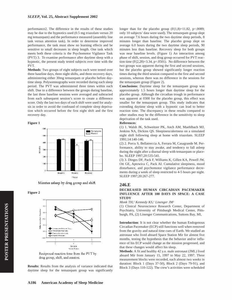
A186 American Academy of Sleep Medicine
PO
STE
R P
RE
SEN
TA
TIO
NS
SLEEP, Vol. 25, Abstract Supplement 2002
performance2. The difference in the results of these studiesmay be due to the hypnotics used (0.5 mg triazolam versus 20mg temazepam) and the performance measured (assembly linetask versus attention task). In order to determine improvedperformance, the task must show no learning effects and besensitive to small decreases in sleep length. One task whichmeets both these criteria is the Psychomotor Vigilance Task(PVT) 3. To examine performance after daytime sleep with ahypnotic, the present study tested subjects over time with thePVT.Methods: Two groups of eight subjects each were tested overthree baseline days, three night shifts, and three recovery days,administering either 30mg temazepam or placebo before day-time sleep. Polysomnographs were recorded during each sleepperiod. The PVT was administered three times within eachshift. Due to a difference between the groups during baseline,the last three baseline sessions were averaged and subtractedfrom each subsequent session’s score to create a differencescore. Only the last two days of each shift were used for analy-sis in order to avoid the confound of complete sleep depriva-tion which occurred before the first night shift and the firstrecovery day.
Figure 1
Figure 2
Results: Results from the analysis of variance indicated thatdaytime sleep for the temazepam group was significantly
longer than for the placebo group (F(1,8)=11.82, p=.0089;only 10 subjects’ data were used). The temazepam group slepton average 7.6 hours during the two daytime sleep periods, 8minutes longer than baseline. The placebo group slept onaverage 6.0 hours during the two daytime sleep periods, 90minutes less than baseline. Recovery sleep for both groupswas near baseline levels. (Figure 1) An interaction amongphase of shift, session, and drug group occurred for PVT reac-tion time (F(2,28)=3.34, p=.0501). No difference between thetwo groups was apparent during the first and second sessions,but the placebo group showed significantly longer reactiontimes during the third session compared to the first and secondsessions, whereas there was no difference in the sessions forthe temazepam group (Figure 2).Conclusions: Daytime sleep for the temazepam group wasapproximately 1.5 hours longer than daytime sleep for theplacebo group. Although the circadian trough in performancewas apparent at 0300 for the placebo group, this effect wassmaller for the temazepam group. This study indicates thatextending daytime sleep with a hypnotic can lead to betterreaction time. The discrepancy in these results compared toother studies may be the difference in the sensitivity to sleepdeprivation of the task used.References: (1) 1. Walsh JK, Schweitzer PK, Anch AM, Muehlbach MJ,Jenkins NA, Dickins QS. Sleepiness/alertness on a simulatednight shift following sleep at home with triazolam. SLEEP1991;14:140-146.(2) 2. Porcu S, Bellatreccia A, Ferrara M, Casagrande M. Per-formance, ability to stay awake, and tendency to fall asleepduring the night after a diurnal sleep with temazepam or place-bo. SLEEP 1997;20:535-541.(3) 3. Dinges DF, Pack F, Williams K, Gillen KA, Powell JW,Ott GE, Aptowica C, Pack AI. Cumulative sleepiness, mooddisturbance, and psychomotor vigilance performance decre-ments during a week of sleep restricted to 4-5 hours per night.SLEEP 1997;20:267-277.
246.EDECREASED HUMAN CIRCADIAN PACEMAKERINFLUENCE AFTER 100 DAYS IN SPACE: A CASESTUDYMonk TH,1 Kennedy KS,1 Linenger JM1
(1) Clinical Neuroscience Research Center, Department ofPsychiatry, University of Pittsburgh Medical Center, Pitts-burgh, PA, (2) Linenger Communications, Suttons Bay, MI,
Introduction: It is not clear whether the human EndogenousCircadian Pacemaker (ECP) still functions well when removedfrom the gravity and natural time cues of Earth. We studied anastronaut who lived aboard Space Station Mir for almost fivemonths, testing the hypothesis that the behavior and/or influ-ence of his ECP would change as the mission progressed, andthat these changes would affect his sleep.Methods: A fit and healthy 42 y.o. male astronaut (JML) livedaboard Mir from January 15, 1997 to May 22, 1997. Threemeasurement blocks were recorded, each almost two weeks induration: Block 1 (Days 37-50), Block 2 (Days 79-91), andBlock 3 (Days 110-122). The crew’s activities were scheduled
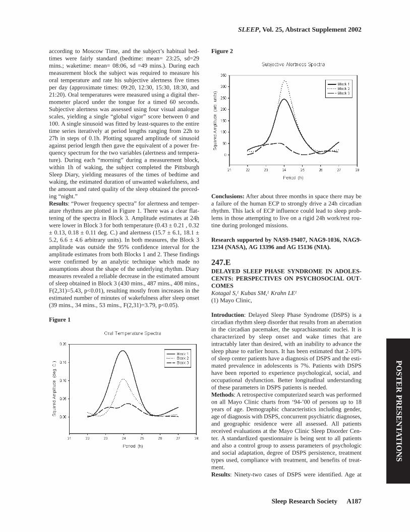
Sleep Research Society A187
PO
STE
R P
RE
SEN
TA
TIO
NS
SLEEP, Vol. 25, Abstract Supplement 2002
according to Moscow Time, and the subject’s habitual bed-times were fairly standard (bedtime: mean= 23:25, sd=29mins.; waketime: mean= 08:06, sd =49 mins.). During eachmeasurement block the subject was required to measure hisoral temperature and rate his subjective alertness five timesper day (approximate times: 09:20, 12:30, 15:30, 18:30, and21:20). Oral temperatures were measured using a digital ther-mometer placed under the tongue for a timed 60 seconds.Subjective alertness was assessed using four visual analoguescales, yielding a single “global vigor” score between 0 and100. A single sinusoid was fitted by least-squares to the entiretime series iteratively at period lengths ranging from 22h to27h in steps of 0.1h. Plotting squared amplitude of sinusoidagainst period length then gave the equivalent of a power fre-quency spectrum for the two variables (alertness and tempera-ture). During each “morning” during a measurement block,within 1h of waking, the subject completed the PittsburghSleep Diary, yielding measures of the times of bedtime andwaking, the estimated duration of unwanted wakefulness, andthe amount and rated quality of the sleep obtained the preced-ing “night.”Results: “Power frequency spectra” for alertness and temper-ature rhythms are plotted in Figure 1. There was a clear flat-tening of the spectra in Block 3. Amplitude estimates at 24hwere lower in Block 3 for both temperature (0.43 ± 0.21 , 0.32± 0.13, 0.18 ± 0.11 deg. C.) and alertness (15.7 ± 6.1, 18.1 ±5.2, 6.6 ± 4.6 arbitrary units). In both measures, the Block 3amplitude was outside the 95% confidence interval for theamplitude estimates from both Blocks 1 and 2. These findingswere confirmed by an analytic technique which made noassumptions about the shape of the underlying rhythm. Diarymeasures revealed a reliable decrease in the estimated amountof sleep obtained in Block 3 (430 mins., 487 mins., 408 mins.,F(2,31)=5.43, p<0.01), resulting mostly from increases in theestimated number of minutes of wakefulness after sleep onset(39 mins., 34 mins., 53 mins., F(2,31)=3.79, p<0.05).
Figure 1
Figure 2
Conclusions: After about three months in space there may bea failure of the human ECP to strongly drive a 24h circadianrhythm. This lack of ECP influence could lead to sleep prob-lems in those attempting to live on a rigid 24h work/rest rou-tine during prolonged missions.
Research supported by NAS9-19407, NAG9-1036, NAG9-1234 (NASA), AG 13396 and AG 15136 (NIA).
247.EDELAYED SLEEP PHASE SYNDROME IN ADOLES-CENTS: PERSPECTIVES ON PSYCHOSOCIAL OUT-COMESKotagal S,1 Kubas SM,1 Krahn LE1
(1) Mayo Clinic,
Introduction: Delayed Sleep Phase Syndrome (DSPS) is acircadian rhythm sleep disorder that results from an aberrationin the circadian pacemaker, the suprachiasmatic nuclei. It ischaracterized by sleep onset and wake times that areintractably later than desired, with an inability to advance thesleep phase to earlier hours. It has been estimated that 2-10%of sleep center patients have a diagnosis of DSPS and the esti-mated prevalence in adolescents is 7%. Patients with DSPShave been reported to experience psychological, social, andoccupational dysfunction. Better longitudinal understandingof these parameters in DSPS patients is needed.Methods: A retrospective computerized search was performedon all Mayo Clinic charts from ‘94-’00 of persons up to 18years of age. Demographic characteristics including gender,age of diagnosis with DSPS, concurrent psychiatric diagnoses,and geographic residence were all assessed. All patientsreceived evaluations at the Mayo Clinic Sleep Disorder Cen-ter. A standardized questionnaire is being sent to all patientsand also a control group to assess parameters of psychologicand social adaptation, degree of DSPS persistence, treatmenttypes used, compliance with treatment, and benefits of treat-ment.Results: Ninety-two cases of DSPS were identified. Age at
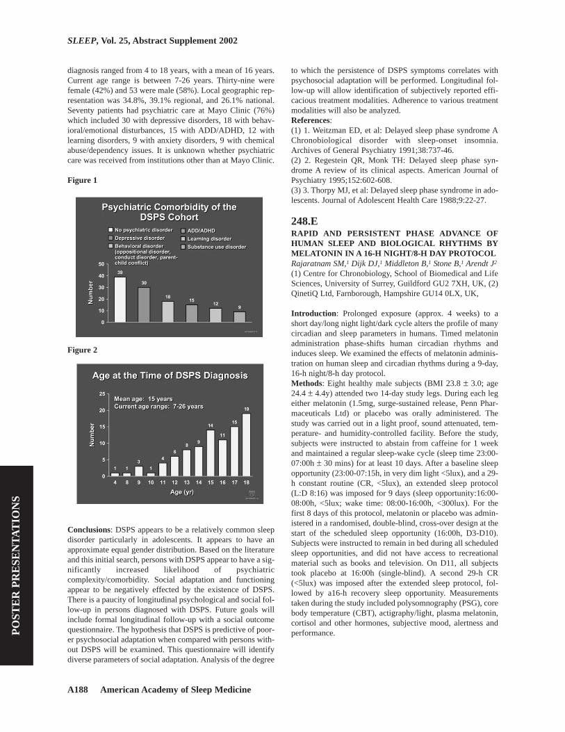
A188 American Academy of Sleep Medicine
PO
STE
R P
RE
SEN
TA
TIO
NS
SLEEP, Vol. 25, Abstract Supplement 2002
diagnosis ranged from 4 to 18 years, with a mean of 16 years.Current age range is between 7-26 years. Thirty-nine werefemale (42%) and 53 were male (58%). Local geographic rep-resentation was 34.8%, 39.1% regional, and 26.1% national.Seventy patients had psychiatric care at Mayo Clinic (76%)which included 30 with depressive disorders, 18 with behav-ioral/emotional disturbances, 15 with ADD/ADHD, 12 withlearning disorders, 9 with anxiety disorders, 9 with chemicalabuse/dependency issues. It is unknown whether psychiatriccare was received from institutions other than at Mayo Clinic.
Figure 1
Figure 2
Conclusions: DSPS appears to be a relatively common sleepdisorder particularly in adolescents. It appears to have anapproximate equal gender distribution. Based on the literatureand this initial search, persons with DSPS appear to have a sig-nificantly increased likelihood of psychiatriccomplexity/comorbidity. Social adaptation and functioningappear to be negatively effected by the existence of DSPS.There is a paucity of longitudinal psychological and social fol-low-up in persons diagnosed with DSPS. Future goals willinclude formal longitudinal follow-up with a social outcomequestionnaire. The hypothesis that DSPS is predictive of poor-er psychosocial adaptation when compared with persons with-out DSPS will be examined. This questionnaire will identifydiverse parameters of social adaptation. Analysis of the degree
to which the persistence of DSPS symptoms correlates withpsychosocial adaptation will be performed. Longitudinal fol-low-up will allow identification of subjectively reported effi-cacious treatment modalities. Adherence to various treatmentmodalities will also be analyzed.References: (1) 1. Weitzman ED, et al: Delayed sleep phase syndrome AChronobiological disorder with sleep-onset insomnia.Archives of General Psychiatry 1991;38:737-46.(2) 2. Regestein QR, Monk TH: Delayed sleep phase syn-drome A review of its clinical aspects. American Journal ofPsychiatry 1995;152:602-608.(3) 3. Thorpy MJ, et al: Delayed sleep phase syndrome in ado-lescents. Journal of Adolescent Health Care 1988;9:22-27.
248.ERAPID AND PERSISTENT PHASE ADVANCE OFHUMAN SLEEP AND BIOLOGICAL RHYTHMS BYMELATONIN IN A 16-H NIGHT/8-H DAY PROTOCOLRajaratnam SM,1 Dijk DJ,1 Middleton B,1 Stone B,1 Arendt J2
(1) Centre for Chronobiology, School of Biomedical and LifeSciences, University of Surrey, Guildford GU2 7XH, UK, (2)QinetiQ Ltd, Farnborough, Hampshire GU14 0LX, UK,
Introduction: Prolonged exposure (approx. 4 weeks) to ashort day/long night light/dark cycle alters the profile of manycircadian and sleep parameters in humans. Timed melatoninadministration phase-shifts human circadian rhythms andinduces sleep. We examined the effects of melatonin adminis-tration on human sleep and circadian rhythms during a 9-day,16-h night/8-h day protocol.Methods: Eight healthy male subjects (BMI 23.8 ± 3.0; age24.4 ± 4.4y) attended two 14-day study legs. During each legeither melatonin (1.5mg, surge-sustained release, Penn Phar-maceuticals Ltd) or placebo was orally administered. Thestudy was carried out in a light proof, sound attenuated, tem-perature- and humidity-controlled facility. Before the study,subjects were instructed to abstain from caffeine for 1 weekand maintained a regular sleep-wake cycle (sleep time 23:00-07:00h ± 30 mins) for at least 10 days. After a baseline sleepopportunity (23:00-07:15h, in very dim light <5lux), and a 29-h constant routine (CR, <5lux), an extended sleep protocol(L:D 8:16) was imposed for 9 days (sleep opportunity:16:00-08:00h, <5lux; wake time: 08:00-16:00h, <300lux). For thefirst 8 days of this protocol, melatonin or placebo was admin-istered in a randomised, double-blind, cross-over design at thestart of the scheduled sleep opportunity (16:00h, D3-D10).Subjects were instructed to remain in bed during all scheduledsleep opportunities, and did not have access to recreationalmaterial such as books and television. On D11, all subjectstook placebo at 16:00h (single-blind). A second 29-h CR(<5lux) was imposed after the extended sleep protocol, fol-lowed by a16-h recovery sleep opportunity. Measurementstaken during the study included polysomnography (PSG), corebody temperature (CBT), actigraphy/light, plasma melatonin,cortisol and other hormones, subjective mood, alertness andperformance.
Sleep Research Society A189
PO
STE
R P
RE
SEN
TA
TIO
NS
SLEEP, Vol. 25, Abstract Supplement 2002
Figure 1
Results: Melatonin administration at 16:00h resulted in ele-vated plasma melatonin levels during the entire 16-h sleepopportunity. Mean sleep efficiency (PSG) in the first half ofthe sleep opportunity, calculated across 7 days of melatonintreatment (D4-D10, 16:00-00:00h) was significantly andmarkedly higher compared to placebo [Fig. 1, effect size (ηs p
s 2 )=0.842]. In contrast, in the second half of the sleep oppor-tunity, sleep efficiency was significantly lower during mela-tonin treatment (ηs p s 2 =0.560). Total sleep time during theentire 16-h sleep opportunity was not significantly differentbetween the conditions (Fig. 1). After melatonin treatment wasstopped (D11), sleep timing remained phase advanced relativeto placebo. Exposure to the extended sleep protocol phaseadvanced plasma melatonin and cortisol rhythms, assessedduring CRs in the placebo condition. In the melatonin condi-tion, phase advances in plasma melatonin, cortisol and CBTwere substantially larger than in the placebo condition.Conclusions: Artificially elevated plasma melatonin levelsthroughout a 16-h sleep opportunity phase advances circadianrhythms and leads to a rapid and persistent redistribution ofsleep to the beginning of the sleep episode without an increasein sleep duration.
Research supported by Medical Research Council/Min-istry of Defence Grant G9810584 and Stockgrand Ltd(University of Surrey).
249.ECHARACTERIZATION OF A NON-24-HOUR SLEEP-WAKE SYNDROME AFTER TRAUMATIC BRAININJURYCaliyurt O,1 James FO,1 Boivin DB1
(1) Douglas Hospital Research Center, Department of Psychi-atry, McGill University, Montreal, Qc, Canada,
Introduction: Sleep disturbances and insomnia occur fre-quently following traumatic brain injuries. Cases of sleep-wake cycle disturbances, particularly delayed sleep phase syn-drome (DSPS), have been observed to occur after head or neck
injuries (1). In the present study, we report the case of a sight-ed woman who developed an inability to properly adjust to the24-hour day after a car accident.Methods: In March 2000, a 38 y.o woman was refered to ourclinic by her neurologist because she developed an unusualsleep-wake pattern. The condition appeared several monthsafter a 1991 car accident with resultant head trauma. She triedunsuccessfully to adopt a regular sleep schedule but wouldsuffer from recurrent sleep disturbances that resulted in aworsening of her epileptic attacks. She therefore resigned her-self to live on longer than 24-hour «days» and her epilepsyimproved. The CT scan, MRI and opthalmologic exams werenormal. At the time of referral, she was taking Valproic Acid375 mg four times a day for her epilepsy, morphine sulphateand dimenhydrinate as needed for her migraines. She kept asleep diary that was confirmed by wrist actigraphy (AW-64,Mini Mitter, Oregon, USA). Sleep onset and offset times werederived from the Sleepwatch software. Sleep episodes report-ed as naps by the patient were excluded from the analysis. If awake episode between two consecutive sleep episodes wasshorter than 30 minutes, the sleep episodes were merged.Descriptive statistics were used to determine the duration ofher sleep-wake cycles.
Figure 1
Results: The sleep-wake cycle of the patient was found to con-sist of two main daily components. Sleep-wake cycles longerthan 24 hours were observed (mean ± SD, 25:05 ± 2:23) andresulted in an hypernychthemeral sleep-wake disorder. Thesleep-wake ratio of these days were similar to that of healthysubjects. In addition, 12.5% of sleep-wake episodes showedrelative coordination to the 24 hour day (Fig.1). A 24-hour pat-tern was observable in times of sleep onset and offset andthese occurred most frequently around 04:00 and 12:00,respectively. The likelihood of being asleep was lowest in theevening and highest at the end of the night, as observed forpatients with DSPS (Fig.2). A number of short sleep episodesfollowed by short wake episodes were also noticed.

A190 American Academy of Sleep Medicine
PO
STE
R P
RE
SEN
TA
TIO
NS
SLEEP, Vol. 25, Abstract Supplement 2002
Figure 2
Conclusions: In this patient, we documented a longer than-24-hour sleep-wake schedule. Her relative coordination to the 24-hour day suggests that environmental synchronizers exert aninfluence too weak to entrain the patient sleep-wake cycle tothe 24-hour day. A daily pattern of sleep onset and offset timesconsistent with DSPS was also observed. This association isconsistent with the prior observation that DSPS might be apredisposing condition for the hypernychthemeral sleep-wakesyndrome in humans (2) .References: (1) 1. Quinto C, Gellido C, Chokroverty S, Masdeu J. Post-traumatic delayed sleep phase syndrome. Neurology 2000 Jan11;54(1):250-2.(2) 2. Oren DA and Wehr TA. Hypernyctohemeral syndromeafter chronotherapy for delayed sleep phase syndrome. N EnglJ Med 1992 Dec 10;327(24):1762.
250.ENONREMS PRESSURE IN DAY-AND NIGHTTIMESLEEP IN WINTER DEPRESSIONPutilov AA, Palchikov V, Danilenko K, Pandi-Perumal SR(1) Institute for Med. and Biol. Cybernetics, 2 TimakovaStreet, 630117 Novosibirsk, RUSSIA, (2) Department ofPhysiology, UCLA School of Medicine, Los Angeles, CA90095, USA,
Introduction: Seasonal affective disorder (SAD) is character-ized by atypical depressive symptoms such as fatigue, daytimehypersomnolence, and difficulties waking up in the morning.It is important to know whether subjective ratings of sleepquality can reflect abnormalities of EEG sleep, and what arethe objectively measured mean levels and 24-hr variations ofsleep propensity and nonREMS pressure in SAD.Methods: The polysomnographic records were obtained insummer (6 patients with SAD, 6 controls), and in winterbefore (n=8 and 5) and after a week of 2-hr bright light treat-
ment (LT) in the evening (n=7 and 4). After an adaptationnight, EEG-sleep parameters were measured during normallyscheduled night sleep (23:00-8:00) and then during multiplesleep latency test in daytime (10:00, 12:00, 14:00, 16:00,18:00) and nighttime (23:00, 1:00, 3:00, 5:00). Clinical symp-toms were rated using the 21-item Hamilton Depression Rat-ing Scale (HDRS) and 29-item SIGH-SAD (HDRS plus 8-item Addendum for the atypical depressive symptoms). Toself-assess subjective vigilance after all night sleep and beforeevery 20-min sleep attempt, the subjects used three subscalesof the so-called WAM-Test (Well-being, Activity, Mood).Each subscale consists of 10 word pairs with a 7-pointresponse scale printed between each pair of words.Results: The comparison of night sleep EEG in patients andcontrols provides evidence of an enhanced total sleep time(TST) in SAD in winter (p<0.05) and summer (p<0.1). Thepercentage of slow-wave sleep (SWS) tended to be lower inpatients than in controls before LT (p<0.1). Moreover, in bothseasons the declining trend of slow-wave activity (SWA) fromthe first to the last hour of night sleep was significantly steep-er in controls compared to patients. It became steeper after LTin patients with good clinical response (n=4). The percentageof sleep stage 2 decreased in patients and increased in controlsby LT (p<0.01), while the percentages of SWS increased inpatients and decreased in controls (p<0.01). Clinical responseto LT correlated positively with TST and negatively with SWSin pretreatment conditions. Neither before nor after LT patientsdiffered from controls on mean level and circadian variationsof sleep latency. The level of SWA in the first 8 min of non-REMS showed an increasing trend from morning sleepattempts to night sleep attempts. The extent of this buildup insleep pressure was in the agreement with the quantitative pre-dictions of the somnostat models1. The clinical response tolight correlated significantly (p<0.05) with the differencebetween daytime and nighttime WAM-scores: in good respon-ders, the difference was low or negative before LT and it sig-nificantly increased following LT.Conclusions: Earlier, very rapid buildup of sleep pressure dur-ing the first 10 hours of wakefulness was found in SAD com-pared to controls2, 3 . Neither these reports of waking EEGnor our recent data on the first 8 min of sleep EEG suggest thepathological changes in time course of sleep propensity inSAD throughout afternoon, evening and nighttime wakeful-ness. We, however, found that all-night sleep in SAD is char-acterized by deficits of SWS and atypically slow decline ofSWA. Besides, when light succeeds in inducing good clinicalresponse, some of sleep the EEG characteristics improve.References: (1) Daan S, Beersma D, Borbely A: Timing of human sleep:Recovery process gated by a circadian pacemaker. Am. J.Physiol., 1984, 246: R161-R178.(2) Cajochen C, Brunner DP, Krauchi K, Graw P, Wirz-JusticeA. EEG and subjective sleepiness during extended wakeful-ness in Seasonal Affective Disorder: Circadian and homeosta-tic influences. Biol Psychiat, 2000, 47:610-607. (3) Putilov AA, Donskaya OG, Jafarova OA, Danilenko KV.Waking EEG power density in hypersomnic winter depres-sion. Soc. Light Treatment Biol. Rhythms. Abst., v. 12, 2000,p. 24.

Sleep Research Society A191
PO
STE
R P
RE
SEN
TA
TIO
NS
SLEEP, Vol. 25, Abstract Supplement 2002
251.EFAILURE TO IDENTIFY PUBERTALLY-MEDIATEDMELATONIN SENSITIVITY TO LIGHT IN ADOLES-CENTSCarskadon MA,1 Acebo C,1 Arnedt JT1
(1) E.P. Bradley Hospital Sleep and Chronobiology ResearchLaboratory.
Introduction: We have tested the hypothesis that the phasedelay of sleep commonly observed in adolescent developmentis related to changes in sensitivity of the circadian timing sys-tem to light signals. Suppression of melatonin secretion tolight was evaluated as a preliminary to assessing phase-shift-ing effects of light. Specifically, we tested whether moremature adolescents suppress melatonin to lower levels of lightduring the phase delaying portion of the phase response curve(evening hours) or require greater light levels to suppressmelatonin during the phase advancing (late-night/early-morn-ing) portion of the phase response curve(1).Methods: Healthy children with relatively regular sleep pat-terns, screened for medical and psychological problems,received brief physical examinations and Tanner staging (2).Thirty-two pre/early pubertal participants (Tanner pubic hairstages 1 or 2; ages 9.1 to 13.9 years (11 girls)) and 34 mid/latepubertal participants (Tanner stages 3-5; ages 10.0 to 15.9 (17girls)) completed the study. Condition assignment (evening(n=40) vs. morning (n=26) light) was based upon participantavailability. After >= 10 consecutive nights sleeping at homewhile wearing eye shades from 2100 to 0700 (confirmed byactigraphy), participants received 1-hr testing at 4 light levelson consecutive nights: ~0.1 lux, 15 lux, 150 lux, and 500 lux,respectively. Saliva was collected at 30-minute intervals,frozen, and melatonin assayed subsequently. Room lights wereoff (0 lux) for sleep or dimmed (~0.1 lux) while awake from2100 to 0730 during in-lab nights, with sleep scheduled asshown in Figure 1. Light administration for the evening groupoccurred from 2300 to midnight and for the morning groupfrom 0300 to 0400. The ~0.1 lux level was included as a con-trol condition.
Figure 1
Table 1
Results: Melatonin data were converted to percentages basedupon the mean of each subjects’ two samples immediatelybefore light exposure. MANOVA within condition showedmain effects for light level but no interaction with pubertalgroup. Planned comparisons within condition and Tannerstage were performed between light levels for samples occur-ring at 30 and 60 minutes of light exposure and are shown inTable 1. Suppression to 15 lux was found only for pre/earlypubertal participants in the Morning light exposure group;otherwise, significant differences were found for 150 and 500lux versus .1 or 15 lux in the morning and evening and for bothpubertal groups.Conclusions: These data indicate that pre/early and mid/latepubertal adolescents do not show differential sensitivity formelatonin suppression at the levels of light tested in thisexperiment. Although unlikely it remains possible that sensi-tivity to light’s phase shifting effects differs pubertally. Weplan to examine this issue in subsequent studies along withwhether developmental changes in other processes, such assleep/wake homeostasis and/or phase angle of entrainment ofsleep to the circadian system may mediate pubertal phasedelay.References: (1) DeCoursey PJ. Daily light sensitivity rhythm in a rodent.Science 1960;131:33-35. (2) Tanner JM. Growth at adolescence. Oxford: Blackwell,1962.
Research supported by MH52415 and MH01358.
252.ERELATIONSHIP BETWEEN INFANTS’ QUIET SLEEPRESPIRATION RATES AND BLOOD OXYGENATION:IMPLICATIONS FOR COGNITIVE DEVELOPMENTMontgomery-Downs HE,1 Thoman EB1
(1) University of Connecticut
Introduction: In a previous study (1) we found a significant,

A192 American Academy of Sleep Medicine
PO
STE
R P
RE
SEN
TA
TIO
NS
SLEEP, Vol. 25, Abstract Supplement 2002
negative relationship between infants’ Quiet Sleep respirationrates (QSRR) on postnatal day 2 and 6 months with their men-tal scores at 6 months and 1 year. Because the purpose ofinvoluntary respiration during Quiet Sleep (QS) is the mainte-nance of blood gas homeostasis, the two goals of the presentstudy of infants over the first 6 postnatal months were: 1) tomeasure changes over age in, and relationships between,infants’ QSRR and their blood oxygenation (SpO2); and 2) tomeasure the relationship between infants’ SpO2 during QS atsuccessive ages and their mental development assessed at 6months.Methods: Sleep and SpO2 were longitudinally recorded in 12healthy infants at 2 days, 2, 6, and 12 weeks, and 6 months.The all-night sleep recordings were made non-intrusivelyusing the Motility Monitoring System (2). From these record-ings, average QSRR was calculated from 1-minute counts dur-ing the first, middle, and last 5-minute periods of each QS bout≥ 15 minutes, SpO2 during QS was simultaneously recorded,averaged over 2-second periods, then averaged over the peri-ods that were synchronous with the respiration counts. At 6months the Bayley Scales of Mental and Motor Developmentwas administered in the subjects’ homes. For each infant,QSRR and SpO2 were correlated at each age, and these r-val-ues were converted to Fisher z’ scores in order to analyze thecorrelation between these relationships and the infants’ mentalscores.Results: QSRR decreased linearly after the Day 2 recording;SpO2 levels showed a linear increase after Week 2 with high-er values from 6 Weeks to 6 Months than on Day 2 and Week2. As expected, QSRR and SpO2 were negatively correlatedwithin individuals at each age. At Week 12, the correlation(using Fisher’s z’ scores) between QSRR and SpO2 was sig-nificantly correlated with 6-month MDI (r= -.68, p<.05). Thus,the more highly related (negatively) QSRR and SpO2 are, thehigher the infants’ mental development scores.
Figure 1
Conclusions: The results are consistent with and extend ourprevious study (1), and they suggest a developmental advan-tage on the part of infants with lower respiration rates in asso-ciation with higher oxygenation levels during QS. While earlysleep characteristics have been linked to mental development(3), our results emphasize that respiration in association withoxygenation during sleep, plays a significant role in the com-
plex neural systems involved in cognitive development.References: (1) Montgomery-Downs H, Thoman EB. Biological andbehavioral correlates of quiet sleep respiration rates in infants.Physiology and Behavior 1998;64:637-43.(2) Thoman EB, Glazier RC. Computer scoring of motilitypatterns for states of sleep and wakefulness: human infants.Sleep 1987;10(2):122-9.(3) Freudigman K, Thoman EB. Infant sleep during the firstpostnatal day: an opportunity for assessment of vulnerability.Pediatrics 1993;92(3):373-9.
Research supported by NICHHD #HD32903, the Universi-ty of Connecticut Research Foundation, the scientificresearch honor society Sigma Xi, and an educational grantfrom Mallinckrodt, Inc.
253.GCHASING THE SLEEP-ADHD LINK: WHAT DOES“NORMAL” MEAN?Danner FW,1 Phillips B,1 Welsh R1
(1) University of Kentucky,
Introduction: Despite a wealth of clinical observations andthe obvious connection between daytime sleepiness and atten-tion problems, there has been very little systematic objectivestudy of sleep and ADHD until recently (Chervin et al., 1997;Picchietti et al., 1998; Harnish et al., 2001). Inconsistency inthe diagnostic criteria used to define ADHD samples makes itdifficult to compare and synthesize findings across studies.Further, there is lack of consensus on what constitutes “nor-mal” sleep in children and how to measure it. The followingoutlines our struggle to address these difficulties in a pilotstudy.Methods: Six boys, aged 8-10, were selected for study. Threewere diagnosed with ADHD by the co-director of a hyperac-tive children’s clinic, based on a detailed history with parents,analysis of information from the Conners Parent and TeacherBehavior Rating Scales, consultation with the classroomteacher, performance on a continuous performance task, and aone-hour diagnostic interview with the child. The control chil-dren were volunteers who received the same screening as theADHD children to rule out obvious ADHD symptomatology.The following assessments were conducted:1.A brief sleephistory and physical examination2.The Pediatric Sleep Ques-tionnaire, filled out by parents3.A standard overnightpolysomnogram (PSG) with simultaneous leg movement mon-itor (PAM-RL) and actigraph (Micro-Mini Motionlogger)recordings.4.A standard Clinical Multiple Sleep Latency Test(MSLT) the day after the PSG.5.The Psychomotor VigilanceTask (PVT)6.Four consecutive in-home nights of sleep moni-toring with the PAM-RL leg movement monitor and theMicro-Mini Motionlogger 7.A sleep log, filled out by the par-ents, for the four in-home assessment daysResults: 1.One of the boys with ADHD exhibited PLM’s dur-ing more than a third of his sleep time, and had 16 PLMarousals. He also averaged 15 PLMs per night at home. He hadseven errors on the PVT (the only child who did). 2.Two of thecontrol boys had RDI’s of > 5 events/hr.3.MSLT scores for all

Sleep Research Society A193
PO
STE
R P
RE
SEN
TA
TIO
NS
SLEEP, Vol. 25, Abstract Supplement 2002
children were greater than 12 minutes.4.Actigraphic scoring ofsleep correlated well with PSG scoring.5.Actigraphic readingsat home averaged 94.5 minutes more total sleep than the labPSG readings and 14.1% greater sleep efficiency.Conclusions: Despite efforts to get distinct samples of ADHDand non-ADHD children, these subjects did not markedly dif-fer on a vigilance task, and control subjects had more sleepproblems than ADHD children. In-lab sleep was poorer than athome. These preliminary results suggest that it may beextremely difficult to select appropriate controls and proce-dures to test the link between sleep disorders and ADHD.References: (1) Chervin R, Dillon J, Bassetti C, Ganoczy D, Pituch K:Symptoms of sleep disorders, inattention, and hyperactivity inchildren. Sleep, 1997, 20, 1185-1192.(2) Picchietti D, England S, Walters A, Willis K, Verrico B:Periodic limb movement disorder and restless legs syndromein children with attention-deficit hyperactivity disorder. JChild Neurol 1998, 13, 588-594.(3) Harnish M, Boyer S, Kukas L, Bowles A, Orr, W: The rela-tionship between sleep disorders and attention deficit hyperac-tivity disorder (ADHD): Objective findings. Sleep, 2001, 24Abstract Supplement A14.
Research supported by Supported by a Sleep AcademicAward 5K07 HL03637-03 from the NIHThe University of Kentucky Research CommitteeThe University of Kentucky Psychiatry DepartmentIM Systems, Inc.
254.GMELATONIN PRODUCTION IN HEALTHY INFANTS:NORMAL VALUES AND SEASONAL VARIATIONSSivan Y,1 Tauman R,1 Laudon M,1 Nechama H,2 Zisapel N3
(1) Pediatric Center for Sleep Disorders, Sleep Laboratory,Dana Children’s Hospital, Tel Aviv Medical Center, Israel, (2)Department of Neurobiochemistry, Tel Aviv University, Facul-ty of Life Sciences, Tel Aviv, Israel, (3) Division of PublicHealth, Tel Aviv Municipality, Israel,
Introduction: In mammals, including humans, the nocturnalproduction of melatonin by the pineal gland reflects the pho-toperiod and plays a key role in seasonal acclimation. There isonly little information on whether the development of mela-tonin production in infants is seasonally regulated. The evalu-ation of melatonin production in infants as a clinical and diag-nostic marker of child development is, however, limited by thelack of a definition of the normal range of melatonin for agiven age group. The urinary metabolite of melatonin, 6-sul-phatoxymelatonin (6SMT), has been proven to be a very reli-able index of melatonin production in humans includinginfants. The purpose of the present study was to establish thenormal range of nocturnal 6SMT excretion in full term infantsat 8 and 16 weeks of age using a large sample of healthyinfants. In addition, we sought to reassess whether the produc-tion of melatonin in infants depends on season. This wasachieved using a recently developed method for extraction ofthe urinary components from disposable diapers and for deter-mination of 6SMT conten.
Methods: 6SMT was assessed in urine samples extracted fromdisposable diapers removed from full-term, 8-week (n=317)and 16-week old (n=93) infants over the nocturnal period(19:00-08:00h). In addition, 6SMT was assessed in 8-week old(n=35) healthy infants over the entire 24-hr period. 6SMT wasdetermined by an ELISA assay.Results: 6SMT excretion at 8 weeks of age exhibited diurnalvariations with (mean+SD) 61+18% of the daily productionexcreted during the nocturnal period regardless of season. Thenocturnal 6SMT values in the entire cohort (at 8 as well as 16weeks of age) were found to significantly depart from normaldistribution (Kolmogorov-Smirnov test). A normal distribu-tion was obtained using a natural base logarithmic (Ln) trans-formation of the data. The normal range (2.5-97.5 percentile ofthe Ln 6SMT excretion per night) was thus defined as 4.66-8.64 (106-5646 ng/night) for 8-weeks old and 5.19-9.67 (180-15820 ng/night) for 16-weeks old infants. A significant effectof the month of birth on 6SMT production at the age of 8weeks was found (ANOVA, p<0.002) with maximal levelsproduced by infants born in June (summer solstice) and mini-mal excretion in infants born in December (winter solstice).Short photoperiod born infants excreted in average about 3fold less 6SMT compared to long photoperiod born infants (t-test, p=0.01). The seasonal variations were no longer presentat 16 weeks of age. No effect of breast-feeding at the time ofsampling on seasonality of 6SMT was found.Conclusions: Normal ranges for the nocturnal urinary excre-tion of 6SMT in full term infants at 8 and 16 weeks of age aredefined. This enables the evaluation of nocturnal 6SMT excre-tion as a prognostic and diagnostic factor for child develop-ment. The strong effect of season on the normal excretion ofnocturnal 6SMT in 8 but not 16 weeks of age suggests prena-tal influence of the photoperiod on the ontogeny of melatonin.
255.GSLEEP HABITS OF INTERNATIONALLY ADOPTEDCHILDRENMindell JA,1,2 Bishop CT1,2
(1) Saint Joseph’s University, (2) Children’s Hospital ofPhiladelphia,
Introduction: Recent research suggests that 25% of childrenwho are adopted as babies are found to have at least onebehavioral or mental health concern later in life. One area thathas never been evaluated, however, is the sleep habits andsleep problems of adopted children. It was hypothesized thatinternationally adopted children would experience significant-ly more sleep disturbances immediately after adoption in com-parison to a matched control group of biological children.Methods: Parents of 17 internationally adopted children and15 nonadopted children between 3 and 30 months (M = 11months) participated in this study. All families were recruitedfrom online list services, local daycares, and referrals fromother families. Parents of the children completed a demo-graphic and parental sleep questionnaire and the Sleep HabitsQuestionnaire (Acebo et al., 1994) that assesses sleep patterns(e.g., usual bedtime, total sleep time) as well as specificbehaviors associated with sleep (e.g., night wakings). Fromthis questionnaire, five subscales are derived: bedtime prob-
A194 American Academy of Sleep Medicine
PO
STE
R P
RE
SEN
TA
TIO
NS
SLEEP, Vol. 25, Abstract Supplement 2002
lems, sleep problems, night waking, morning problems, anddaytime sleepiness. In addition, parents completed the SleepAssociations Scale that assesses co-sleeping and sleeping con-ditions. Adoptive families completed the questionnaires twoweeks post-adoption.Results: To assess differences in sleep patterns while control-ling for the multiple comparisons, a one-way multivariateanalysis of variance (MANOVA) was calculated with type offamily (adopted vs. nonadopted) as the independent variablefor each of the six sleep pattern items of the SHQ (bedtime,latency to sleep onset, total minutes of night wakings, totalsleep time, total nap time, and morning wake time) and fivesleep problem subscales. Overall, only one difference wasfound, with adopted children (M = 16.69) experiencing a sig-nificantly longer duration of night wakings than nonadoptedchildren (M = 6.80), p < .05. No differences were found for theother five sleep pattern variables and the six sleep problemsubscales, p > .05. Additionally, no differences in the parents’sleep were found.Conclusions: The findings indicate that there is little effect offamily status, adopted versus nonadopted, on young children’ssleep patterns. Of 13 variables, differences were only found onone variable, duration of night wakings. It was surprising thatthe infants in this study who were two weeks post-adoptiondisplayed similar sleep patterns and virtually no sleep prob-lems when compared to nonadopted children. However, itwould be beneficial for future research to investigate the sleepof internationally adopted children as they get older since pre-vious research suggests that adopted children typically do notdisplay difficulties in adjustment until they reach post-infancyor early childhood. Overall, this study is the first to examinesleep patterns and behaviors of internationally adopted infantswith results indicating few, if any, sleep problems immediate-ly post-adoption.
256.GCOSLEEPING IN THE UNITED STATES: PARENTS’PRACTICES AND OPINIONS FROM 1992-2001Borkowski MM,1 Johnson CM,1 Famoye F,1 Hether NW1
(1) Central Michigan University, (2) Gerber Products Compa-ny,
Introduction: Cosleeping is a controversial issue because ofperceived benefits such as promoting breastfeeing, reductionof SIDS, and fostering attachment as well as potential risksincluding overlayment, entrapment, and suffocation.(1,2,3)Differences in cosleeping rates of families from different eth-nic backgrounds have been shown and some children whocosleep are more likely to have sleep problems.(2) This studyinvestigated cosleeping practices and how they relate to sleep-ing problems in 3 cohorts within the United States.Methods: Three prospective longitudinal telephone surveyswere conducted over 9 years. Respondents from 1992-1994were mostly White parents (n=201) from rural Michigan,1994-1996 participants were primarily Black mothers (n=206)from cities in the Midwest, and a national sample of Hispanicmothers (n=237) was obtained from 2000-2001. Parents werecalled when their infants were 2-, 4-, 8-, and 12-months-oldand asked if they typically coslept and their opinions of
cosleeping. Data on problems of settling and awakening werealso collected. Repeated measure categorical data analysis wasused to test for changes over time and to examine the differ-ences between the three cohorts. Chi-square tests of independ-ence were run to compare the three groups on their practicesand opinions of cosleeping. Chi-square tests also were used toexamine the relationships between cosleeping and the prob-lems of awakening and settling.
Figure 1
Figure 2
Results: Cosleeping did not change over time within any ofthe three cohorts. White parents, however, coslept less (p <.0001) and liked cosleeping less (p < .0001) than Black or His-panic mothers. In each cohort, cosleepers were not more like-ly to have settling problems. However, within the Whitecohort, cosleepers were more likely to have awakening prob-lems (p < .05).Conclusions: Cosleeping practices and opinions differ signif-icantly between White, Black, and Hispanic parents. Whilethere was no apparent relationship between cosleeping andsettling problems for Black, White, or Hispanic infants, Whitecosleeping infants had more night wakings that were viewedproblematic by their parents.References: (1) Nakamura S, Wind M, Danello C: Review of hazards asso-
Sleep Research Society A195
PO
STE
R P
RE
SEN
TA
TIO
NS
SLEEP, Vol. 25, Abstract Supplement 2002
ciated with children placed in adult beds. Archives of Pediatricand Adolescent Medicine 1999; 153: 1019-1023.(2) Lozoff B, Askew G, Wolf A: Cosleeping and early child-hood sleep problems: Effects of ethnicity and socioeconomicstatus. Journal of Developmental and Behavioral Pediatrics1996; 1:9-15.(3) McKenna J, Thoman E, Anders T, Sadeh A, Schechtman V,Glotzbach S: Infant-parent cosleeping in an evolutionary per-spective: Implications for understanding infant sleep develop-ment and the Sudden Infant Death Syndrome. Sleep 1993;16:263-282.
Research supported by Gerber Products Company
257.GA CROSS-SECTIONAL SURVEY TO ASSESS THEPREVALENCE OF DISTURBED SLEEPAMONG CHIL-DREN WITH CYSTIC FIBROSISQuintero DR,1 Oermann CM,1 Wise M,1 Glaze D1
(1) Baylor College of Medicine, Houston, Texas,
Introduction: Owens and Chervin have established normativedata for the prevalence of disturbed sleep among healthy pedi-atric populations.1,2 Although cystic fibrosis (CF) childrenmay be at increased risk of sleep disturbance due to 1) noctur-nal hypoxemia 2) chronic cough 3) recurrent pain 4) sinusi-tis/polyposis (SP) related to CF, this has not previously beendescribed in the medical literature. The objectives of this studywere to 1) investigate the prevalence of disturbed sleep amongCF children and 2) evaluate the relations between disturbedsleep and pulmonary function and SP.Methods: The Children’s Sleep Habit Questionnaire (CSHQ)validated by Owens et. al and the Pediatric Sleep Question-naire (PSQ) validated by Chervin et. al were given to 107 sub-jects aged 4-18 years with a confirmed diagnosis of CF at theCystic Fibrosis Center at Texas Children’s Hospital. Parentscompleted the questionnaires for subjects less than 13 years ofage whereas older subjects completed the questionnairesthemselves. The control group was taken from the normativedata described by Owens. Statistical analysis was done usingSPSS 10.0. Means subscale scores were compared betweenCF and control groups using the student t-test. ANOVA wasused for post hoc analysis evaluating the effects of pulmonaryfunction and SP on subscale items.Results: 95% of the CF group identified at least one sleeprelated problem compared to 14.9% of the control group. Inthe subscale items, subjects with CF demonstrated increasedbedtime resistance (p= 0.05), delayed sleep onset (p=0.009)and increased daytime sleepiness (p=0.03) compared to con-trols. The CSHQ questionnaire did not reveal significant dif-ferences between CF and control groups for sleep disorderedbreathing; however data from the PSQ was strongly sugges-tive of significant differences. For 18.6% of CF subjects posi-tive responses on the PSQ suggested the occurrence of sleepdisordered breathing. Post hoc analysis of CHSQ subscaleitems did not identify SP as a contributing factor for sleep dis-turbances between CF subjects with or without SP. Post hocanalysis did suggest that significant differences exist betweensubjects with mild (FEV1>80% predicted), moderate (FEV1
60-80% predicted) and severe disease (FEV1 < 60% predict-ed) for some subscale items related to increased daytimesleepiness (p=.012-.028).Conclusions: Children with CF have an increased incidenceof disturbed sleep when compared to healthy children. Thispotentially has a negative impact on overall health, but maynot be adequately assessed by CF caregivers. The CHSQ andPSQ are useful screening tools to identify children with dis-turbed sleep. Formal polysomnography testing maybe indicat-ed for the confirmation of sleep disordered breathing in chil-dren with positive screen.References: (1) Owens JA MD, MPH, Spirito A PhD, McGuinn M BS. TheChildren’s Sleep Habit Questionnaire (CSHQ): Psychometricproperties of a survey instrument for school-aged children.Sleep, vol 23 No 8 2000(2) Chervin RD, Hedger K, Dillon JE, Pituch KJ . PediatricSleep Questionnaire (PSQ): Validity and reliability of scalesfor sleep disordered breathing, snoring, sleepiness and behav-ioral problems. Sleep Medicine 1 (2000) 21-32
258.GACTIGRAPHY MEASURES OF SLEEP IN CHILDRENWITH CANCERGedaly-Duff VR,1 Nicholson HS,1 Lee K,1 Nail L,2 Perko K,1
Moran K,1 Anderson K,1 Johnson KP1
(1) Oregon Health & Science University, Portland, Oregon, (2)University of California, San Francisco,
Introduction: Disturbed sleep in children with acute lym-phoblastic leukemia (ALL), and of the families who care forthem, has had little research. Between 53%\\super1\\/and74%\\super2\\/ children with cancer have reported sleep dis-turbances. Sleep deprivation was a common theme in a quali-tative analysis of 31 parents.\\super3\\/ The purpose of thisreport is to describe sleep disturbances in schoolage childrenduring their chemotherapy. Data were from a pilot study thatcollected 72 hours of actigraph data on target child, primarycaregiver, and sleep partner at home.Methods: Nine families participated in the pilot study. A fam-ily was defined as the target child (patient), caregiver (usuallymother), and sleep partner of caregiver. Children were recruit-ed who were either newly diagnosed or in first remissionundergoing chemotherapy for ALL. Study instruments wereactivity-sleep diaries for the target child, caregiver and adultsleep partner, as well as a medical record review. Objectivesleep activity data for each family member was obtained usinga wrist motion sensor on the non-wrist. Data for this analysisare from the 9 children’s first 3 nights at home after receivingintravenous vincristine at a scheduled out-patient appoint-ment. Data collection occurred on weekdays and weekendsduring summer months. Measures included: total sleep time(minutes from sleep start time to end of scored sleep), sleepafter sleep onset (% of total sleep time), number of awaken-ings, and mean duration of wake episodes in minutes.Results: Average age of the children in the sample was 11years (7 to 16). There was no significant difference betweennight 1, 2 and 3. Average for 3 nights at home after chemother-apy for total sleep time was 482 minutes (SEM = 21.3; range

A196 American Academy of Sleep Medicine
PO
STE
R P
RE
SEN
TA
TIO
NS
SLEEP, Vol. 25, Abstract Supplement 2002
= 367 to 572), wake time averaged 64 minutes (SEM = 16.1;range = 2 to 131), wake after sleep onset was 11.5% (SEM =2.9; range = 0 to 24)], number of awakenings averaged 19.8(SEM = 4.1; range =1 to 37), mean duration of wake episodeswas 3.1 minutes (SEM = 0.6; range = 1 to 6).Conclusions: These pilot data suggest that children duringchemotherapy for ALL range between 6 and 10 hours of sleepwith frequent awakenings. Because children are scheduled forchemotherapy Monday through Friday, the effect ofchemotherapy on sleep activity includes weekends. Largerstudies are needed to examine weekday versus weekendeffects on both the child and the family members. Under-standing the type and severity of sleep disturbance will con-tribute to the development of comprehensive interventions thatcan benefit both children with cancer and their families.References: (1) Hirschfeld S, Moss H, Dragisic K, Smith W, Pizzo PA.Pain in pediatric human immunodeficiency virus infection:incidence and characteristics in a single-institution pilot study[see comments]. Pediatrics 1996;98(3 Pt 1):449-52.(2) Miser AW, McCalla J, Dothage JA, Wesley M, Miser JS.Pain as a presenting symptom in children and young adultswith newly diagnosed malignancy. Pain 1987;29(1):85-90.(3) Ferrell BR, Rhiner M, Shapiro B, Dierkes M. The experi-ence of pediatric cancer pain, Part I: Impact of pain on thefamily. Journal of Pediatric Nursing: Nursing Care of Childrenand Families 1994;9(6):368-79.
Research supported by Lee, K. (2001). Nursing ResearchTraining in Symptom Management. NIH/NINR, No. T32NR07088
259.GSLEEP HABITS OF STUDENTS IN ELEMENTARYSCHOOLS, JUNIOR AND SENIOR HIGH SCHOOLS INJAPAN. Takaubu T,1 Kanbayashi T,1 Tutsui K,1 Saito Y,1 Aizawa R,1
Mikami M,1 Kogawa S,1 Takahashi K,1 Takahashi Y1 ShimizuT259.G(1) Department of Psychiatry, Akita University School ofMedicine, Akita City, Japan,
Introduction: It is widely accepted that children in Japanspend shorter time in sleep than they need. The sleep habits ofstudents have also been altered due to the habit of sitting upuntil late at night. However, epidemiological data on sleephabits among Japanese students are rather scarce (1,2). Theobjective of this paper is to study sleep habits and related prob-lems among children in elementary schools, students in juniorand senior high schools in Akita Prefecture.Methods: A self-rated sleep habit questionnaire (SHQ) wasgiven to 658 schoolchildren in 25 elementary schools (age10),803 students in 24 junior high schools (age13), and 705 stu-dents in 18 senior high schools (age16) randomly selected inAkita Prefecture. The survey was carried out at the time ofregular medical checkup in their schools. The SHQ was a shortversion of the questionnaires designed by Hori et al. (3). Thosewho replied with effective answer to SHQ were: 578 (87.8%)schoolchildren in elementary schools, 495 (61.6%) students in
junior high schools, and 577 (81.8%) students in senior highschools.Results: The schoolchildren in elementary schools spendabout 9h for sleep, and there was no remarkable difference oftheir sleeping habit between weekdays and holidays. Studentsin junior and senior high school spend 7h38m and 6h58m insleep on weekday respectively (Table1). The difference ofsleep time between weekday and weekend were over 80m inthem. Those who replied that they slept too short on weekdayswere: 124 (21.5%) among schoolchildren, 200 (40.4%) amongstudents of junior high schools, and 303 (52.5%) among stu-dents of senior high schools. When those who slept shorterthan 6h a day were defined as “short sleepers”, and those whoslept for 9h or longer are defined as “long sleepers”, the shortsleepers were 3 (0.5%), 34 (6.9%) and 106 (18.4%) respec-tively, and the “long sleepers” were 330 (57.1%), 48 (19.7%)and 30 (5.2%) respectively. Among “short sleepers”, thosewho replied that they had sufficient time for sleep, i.e. thosewho may be regarded as “true short time sleepers”, were 2(0.3%), 8 (1.6%) and 14 (2.4%) respectively. Among “longsleeper”, 45 (7.8%) in primary school, 8 (1.6%) in junior highand 8 (1.6%) in senior high school answered that their sleepwere short or too short. Those who replied that they had takenafternoon naps at home twice or more per week were 31(5.4%), 53 (10.7%), and 114 (19.8%) respectively. Those whonodded off during classes twice or more per week were 45(7.8%), 93 (18.8%), and 251 (43.5%) respectively.
Table 1
Conclusions: The results of the present study reveal thatschoolchildren in elementary schools in Akita Prefecture seemto have spent sufficient time for sleep. However, the studentsof junior and senior high schools were apparently short ofsleep, and they seem to compensate the shortage of sleep byextending sleep on weekends. Those who slept 6 hours or lesson weekdays showed rapid increase among the students ofjunior and senior high schools. Since “true short time sleepers”are not many, most of “short sleepers” are thought to be insuf-ficient sleepers. Consequently, about the half of students of

Sleep Research Society A197
PO
STE
R P
RE
SEN
TA
TIO
NS
SLEEP, Vol. 25, Abstract Supplement 2002
senior high schools answered that they nodded off duringclasses twice or more per week. These results are quite con-cordant with previous reports (2,3). Further study should beneeded to elucidate the relationship between sleep habits andother life styles such as sports habit, attending crammerschool.References: (1) Shinkoda H. Matsumoto K, Park YM, Nagashima H.Sleep-wake habits of schoolchildren according to grade. Psy-chiatry Clin, Neurosci. 2000; 54: 287-289.(2) Arakawa M, Taira K, Tanaka H, et al. A survey of juniorhigh school students’ sleep habit and lifestyle in Okinawa.Psychiatry Clin, Neurosci. 2001; 55: 211-212.(3) Hori T, Miyasita A, Shirakawa S, et al. Survey on thedevelopmental changes of sleep-wake habits and sleep prob-lems from infants to advanced ages in Japanese population.
Research supported by This research supported by a grantfrom the Japanese Ministry of Health and Welfare, andSpecial Coordination Funds for Promoting Science andTechnology from the Science and Technology Agency.
260.GACADEMIC ACHIEVEMENT AND ATTENTION INCHILDREN SCHEDULED FOR ADENOTONSILLEC-TOMY IN COMPARISON TO CONTROLSHuffman JL,1 Giordani B,1 Layne JR,1 Ruzicka D,1 MarriottDJ,1 Weatherly RA,1 Dillon JE,1 Chervin RD1
(1) The University of Michigan,
Introduction: Sleep-disordered breathing (SDB) is among themost common indications for adenotonsillectomy (AT), butchildren who undergo the procedure for other reasons oftenhave enlarged tonsils and may also be at risk for SDB.1 Con-sequences of childhood SDB may include inattention, hyper-activity, and related behaviors, as well as poor school per-formance.2-3 However, the specific deficits in cognition andachievement that may be present in children who undergo AThave not been well defined.Methods: We recruited 51 children (27 girls) aged 5 to 12years (M=7.78, SD=1.90): 43 patients had been scheduled foradenotonsillectomy (AT) and 8 had been seen at a general sur-gery clinic (scheduled for hernia repair). In an ongoingresearch protocol, estimates of verbal and nonverbal intelli-gence were made using the Wechsler Abbreviated Scales ofIntelligence (WASI). Academic achievement was measuredusing the specific subtests of the Wechsler IndividualAchievement Test (WIAT). Attention and concentration wereobjectively measured using the Integrated Visual and Audito-ry (IVA) Continuous Performance Test, and subjective ratingswere obtained using the Conners’ Parent Rating Scale,Revised: Long Version.Results: Group means for included variables are presented inTable 1 below. Independent samples t-tests showed that the ATand control groups did not differ with respect to age or grade.Furthermore, inspection of estimates of Verbal and Perfor-mance IQ scores revealed no significant differences betweenthe groups. However, the AT group performed more poorly ona number of academic achievement tests, including Mathe-
matics Reasoning, Spelling, and Numerical Operations (Table2). A similar trend was shown for Basic Reading performance.No difference between the groups was noted for a measure ofReading Comprehension. Children in the AT group performedmore poorly than the control group on the Auditory AttentionQuotient of the IVA, whereas both groups performed similar-ly on the Visual Attention Quotient. Parent ratings of opposi-tional behaviors and cognitive problems/inattention were sig-nificantly greater for the AT group as compared to the controlgroup. No significant differences were noted between thegroups for ratings of hyperactivity.
Table 1
Table 2
Conclusions: Children who are scheduled to undergo AT, as agroup, demonstrate poorer academic skills and greater audito-ry attention difficulties than children scheduled for other sur-gery. Consistent with these results, behavioral ratings by par-ents demonstrate significant problems with cognition, inatten-tion, and oppositional behaviors among the AT group. Theresults of this study underscore specific academic and behav-ioral vulnerabilities in children who undergo AT.

A198 American Academy of Sleep Medicine
PO
STE
R P
RE
SEN
TA
TIO
NS
SLEEP, Vol. 25, Abstract Supplement 2002
References: (1) Croft CB, Brockbank MJ, Wright A, Swanston AR.Obstructive sleep apnea in children undergoing routine tonsil-lectomy and adenoidectomy. Clin Otolaryngol 1990;15:307-314.(2) 2. Guilleminault C, Winkle R, Korobkin R, Simmon B.Children and nocturnal snoring – evaluation of the effects ofsleep related respiratory resistive load and daytime function-ing. Eur J Pediatr 1982;139:165-171.(3) 3. Gozal, D. Sleep-disordered breathing and school per-formance in children. Pediatrics 1998;102:616-620.
Research supported by the National Institutes of Healthgrants R01 HD38461, K02 NS02009, and M01 RR00042
261.GPARENTAL ASSESSMENT OF SLEEP DIFFICULTIESIN CHILDREN WITH ADHD REFERRED TO A PEDI-ATRIC SLEEP MEDICINE CENTER.Crabtree VM,1 Ivanenko A,1 Gozal D1
(1) Division of Pediatric Sleep Medicine and Kosair Chil-dren’s Hospital Research Institute, University of Louisville,Louisville, KY,
Introduction: Based on parental reports, children with ADHDare believed to be at higher risk for sleep-disordered breathing(SDB), periodic leg movement disorder of sleep, sleep initia-tion and maintenance difficulties, and excessive daytimesleepiness (1-2). To gain more insights into sleep in ADHD,we conducted a systematic assessment of the most frequentcomplaints of parents of children with ADHD referred to apediatric sleep medicine center.Methods: The charts of 86 consecutive children with ADHDwere reviewed. There were 67 male; 75 Caucasian; and theirage range was 3-18 years, with a median age of 8.3 years. Theparents’ primary sleep-related complaint leading to the childreferral, and their responses to a structured clinic-based sleepquestionnaire, the Pediatric Symptom Checklist, and the Clin-ical Attention Problems Scale were extracted.Results: The majority of these 86 children were on medica-tions, namely psychostimulants (65%), sleep-promotingagents (trazodone, clonidine, melatonin; 36%), and other psy-chotropic agents (valproic acid, sertraline; 22%). In 28% ofthe children, co-morbid psychiatric conditions such as moodand/or anxiety disorders were present. The most frequentsleep-related complaints were snoring and SDB (60%), rest-less sleep with or without multiple awakenings (44%), anddelayed sleep onset (43%). Based on the questionnaire, 49%of children were reported to have long sleep latencies (>30minutes at bedtime), and the mean estimate for sleep latencywas 61.7 minutes. Furthermore, 79% of parents reported thattheir children awoke in a negative mood, and 74% thought thattheir child’s sleep problems were moderate to severe. Themost frequently reported sleep problems included restlesssleep (91%), snoring (78%), somnolence (67%), nightmares(60%), and nocturnal enuresis (48%). The Pediatric SymptomChecklist mean score for the sample was 36.3 (normal<28).Similarly, the mean Child Attention Problems Scale scores forthe inattentive and overactive subscales were 10.8 and 8.2,
respectively (normal<3).Conclusions: Children with ADHD present 3 major types ofsleep disturbances. Delayed sleep onset could be related to theseverity of hyperactivity, or alternatively to limit-setting diffi-culties. Snoring was 7-fold more frequent than in a generalpediatric population, such that SDB may be playing a role inthe daytime and nighttime manifestations reported. In addi-tion, the frequency of restless sleep may underlie an intrinsiccomponent of ADHD manifesting during sleep. Thus, system-atic exploration of actigraphic and polysomnographic patternswill be needed in children with ADHD and sleep problems, inorder to obtain objective sleep correlates that can ultimatelyguide the clinical management of sleep disturbances in thesepatients.References: (1) Chervin RD, Dillon JE, Bassetti C, Ganoczy DA, PituchKJ. Sypmtoms of sleep disorders, inattention, and hyperactiv-ity in children. Sleep 1997;20:1185-1192.(2) Corkum P, Tannock R, Moldofsky H, Hogg-Johnson S,Humphries T. Actigraphy and parental ratings of sleep in chil-dren with Attention-Deficit/Hyperactivity Disorder (ADHD).Sleep 2001;24:303-312.
Research supported by NIH Grant HL-65270 and Depart-ment of Education Grant H324E011001
262.GEVENING NAPS AND DELAYED NIGHTTIME SLEEPSCHEDULE TYPICALLY FOUND IN JAPANESE ADO-LESCENTS CAUSES THEIR DAYTIME MALFUNC-TIONINGFukuda K,1 Ishihara K1
(1) Department of Educational Psychology, Fukushima Uni-versity, Japan, (2) Department of Child Welfare, Faculty ofHuman Life Sciences, Notre Dame Seishin University, Japan,
Introduction: Fukuda and Ishihara (2001) administered aquestionnaire concerning sleep related lifestyle to junior highand high school students and revealed that nighttime sleeplength was shortest and several physical and mental conditionswere worst at high school age. About a half of junior high andhigh school students take routine afternoon or evening naps tokeep themselves awake to study at night at home and to com-pensate for their chronic sleep insufficiency (Fukuda and Ishi-hara, 2002). At the same time late naps are known to disturbnighttime sleep. This study aimed at investigating relationshipbetween the schedule of evening naps and delayed nighttimesleep and daytime functioning in the Japanese junior high andhigh school students.Methods: Questionnaire concerning sleep related lifestyle,and physical and mental health was administered through theteachers to the students of junior-high schools (12-15 years)and high schools (15-18 years) in Fukushima (North East) andOkayama (West) prefectures of Japan. About 10,000 students(5,461 junior high school students, 4,880 high school students)participated into the survey as a volunteer subject. Studentswere classified into groups according to their bedtime and fre-quency of evening naps. The classification was made in juniorhigh and high school students separately. MANOVA was con-
Sleep Research Society A199
PO
STE
R P
RE
SEN
TA
TIO
NS
SLEEP, Vol. 25, Abstract Supplement 2002
ducted to investigate the effects of bedtime and frequency ofnaps on several daytime physical and mental symptoms (e.g.,frequency of nodding off, anxiety, depression, fatigue, irri-tability, moody on rise).Results: The statistical test revealed that significant maineffects of evening naps and bedtime were found in the mostdaytime symptoms in the both junior high and high school stu-dents. Fig.1 shows the relationship of frequency of naps andbedtime with frequency of nodding off during daytime in thejunior high school students. The more frequently the studentstake evening naps and the later they go to bed, the more fre-quently they nod off during daytime.
Figure 1
Conclusions: Students who take frequent naps and/or go tobed later are found to suffer from undesirable daytime symp-toms, including daytime sleepiness, suggested by the presenceof frequent nodding. As there is a little individual difference inmorning rise time among the students compared with theirbedtime, the late bedtime means short nighttime sleep conse-quently. It is quite understandable that the students who go tobed later showed daytime sleepiness and other undesirablesymptoms, however, the fact that the students who take addi-tional naps, i.e., longer total sleep time during a day, com-plained stronger daytime sleepiness and other symptomsseems more difficult to understand. This finding may suggestthat a custom of taking late naps, which the students intend tocompensate their insufficiency of nighttime sleep caused theirdaytime malfunctioning. Custom of taking long naps might beconsidered in terms of polyphasic or interrupted sleep andwakefulness regimen.References: (1) Fukuda K, Ishihara K. Age related changes of sleeping pat-tern during adolescence. Psychiatry Clin. Neurosci. 2001, 55,231-232(2) Fukuda, K., Ishihara, K. Routine evening naps and night-time sleep in junior high and high school students. PsychiatryClin. Neurosci. 2002, 56, (in press).
Research supported by a Special Coordination Fund forPromoting Science and Technology from the Ministry ofEducation, Culture, Sports, Science and Technology,Japan.
263.GLATE BEDTIMES MAY BE THE NORM FOR EVENVERY YOUNG, MINORITY CHILDREN IN URBANAMERICASewitch DE,1 Levy PS,1 Bangayan M,2 Chan L,1 Olopade CS1
(1) Center for Sleep and Ventilatory Disorders, University ofIllinois at Chicago Medical Center, College of Medicine, (2)University of Illinois at Chicago School of Public Health,
Introduction: Although the Delay in Adolescent Bedtimes iswell known from large surveys (1-2), it has generally beenassumed that toddlers through pre-teen children go to bedmuch earlier than their teenage counterparts. We questionedthis assumption in current American society and hypothesizedthat the Delay in Adolescent Bedtimes is an acute, exaggera-tion of a Bedtime Delay Pattern that begins very early in child-hood and is then reinforced over time by current societalnorms.Methods: A retrospective analysis of clinical and polysomno-graphic data on 202 children and adolescents (18-months to 18years) evaluated at our Sleep Center for complaints of snoringand daytime somnolence was performed. Parent/child/teenreported, Bedtime (BT) and Total Sleep Duration (TSD) datawere available on 157 children and adolescents. We focusedon weeknight, Bedtime and TSD because weekend sleep pat-terns are usually different (3). We chose a regression analysisapproach to BT and TSD by Age, Sex, and Race. We then sub-divided the database into 3 Age groups of 1-5 years, 6-12 yearsold, and 13-18 years old.
Table 1
Results: Group demographics are presented in Table 1. Therewere 89 males and 68 females with 9% Caucasians and a pre-dominance of minorities (91%). About 64% of the sample wasconsidered below the poverty line due to qualification for Pub-lic Aid Assistance. A regression of reported Sleep Durationwith Age was significant (r=0.4998, F(1,153)=50.95, p=.0000,Adj R-squared=0.2449). About 25% of the variance indecreasing, TSD was accounted for by increasing age. Therewas a weaker gender difference with girls averaging slightlyless TSD (9 hours, 37 minutes) compared to boys (9 hours, 59minutes) across the entire age range by a one-tailed, t-test (
A200 American Academy of Sleep Medicine
PO
STE
R P
RE
SEN
TA
TIO
NS
SLEEP, Vol. 25, Abstract Supplement 2002
p=.0455). Regression of reported Bedtime failed to demon-strate significance by Age, Gender, or Race. Results for Gen-der distribution of BT and TSD across the 3, Age-groups arepresented in Table 2. There were no gender differences ineither BT or TSD by Age groups. However, the Adolescentshad a Later Bedtime (F(2,155)=8.31, p=.0004) and shorterTSD (F(2,154)=17.22, p=.0000) than both of the younger agegroups. While there were no significant differences in BT orTSD between the 1-5 year and 6-12 year age groups, the Late(9:38pm), average Bedtime of the 1-5 year olds was striking(See Table 2).
Table 2.
Conclusions: The Late Bedtime of Adolescence may be anacute exaggeration of a delayed bedtime that is set early inchildhood development and reinforced by current societalpractice. An acute, additional delay specific to the adolescentyears also appears to be present.References: (1) Wolfson AR,Carskadon MA. Sleep schedules and daytimefunctioning in adolescents. Child Devel 1998; 69:875-887.(2) Wahlstrom KL,Davison ML, Choi J, Ross JN. School starttime study: Technical report,Vol. II:Analysis of student surveydata. CAREI, University of Minnesota WebPage.2001,http://education.umn.edu/carei/Programs/start_time/2001exec_sum.html/ (3) Carskadon MA, Wolfson AR, Acebo C et al. Adolescentsleep patterns, circadian timing, and sleepiness at a transitionto early school days. Sleep 1998; 21(8):871-881.
264.GSLEEP ORGANIZATION IN PREMATURE 33/34-WEEK POSTMENSTRUAL AGE INFANTS TREATEDWITH MAINTENANCE-DOSE CAFFEINE.Curzi-Dascalova L,1 Aujard Y,1 Gaultier C,2 Rajguru M1
(1) INSERM, EPI9935, Service de physiologie, HôpitalRobert-Debré, Paris, (2) Service de Néonatologie, HôpitalRobert-Debré, Paris.,
Introduction: Behavioral sleep-wake states, an index of nor-mal brain development and functioning (ref. in 1), may bealtered by caffeine, which is currently used to prevent ordecrease apneas in premature babies (2).Methods: To assess the potential effect on sleep organization
of caffeine in standard maintenance dosages, we performed10-hour polysomnographic recordings (3) in 15 neurological-ly normal and clinically stabilized, appropriate-for-gestation-al-age 33/34-week postmenstrual age (w PMA) neonates, ofwhom ten had been treated for longer than 3 days with once-a-day oral caffeine citrate, 5 mg/kg, given around 2 p.m. Weanalyzed (a) the usual sleep-wake parameters, including wake-fulness (W), active sleep (AS), quiet sleep (QS), and indeter-minate (IS) sleep expressed as the number of episodes, dura-tion, and percentage of total sleep time (TST)b; (b) the dura-tion and order of parameter modifications during transitions(trans) between the main AS and QS states and; (c) the char-acteristics of morning data (before caffeine) compared toevening data (after caffeine). Statistical analyses were per-formed using a multivariate linear model, taking into accountindependent effects of morning versus afternoon, caffeine ver-sus no caffeine, AS versus QS, and transition from AS to QSversus from QS to AS, with the subject factor as a cluster.Because data distribution was non-normal, all analyses weretested also by the nonparametric Wilcoxon rank-sum test. Achi-square test was designed to analyze the order of parameterchanges during between-state transitions.Results: We found no significant differences between the con-trols and the infants on maintenance caffeine. The main dataare shown on the following table:
Table 1
Conclusions: We conclude that caffeine in a standard mainte-nance dosage does not modify sleep organization in neurolog-ically normal and clinically stable 33/34 w PMA infants.References: (1) Curzi-Dascalova L, Challamel MJ: Neurophysiologicalbasis of sleep development.In Loughlin GM, Carroll JL, Marcus CL (eds); Sleep andbreathing in children: a developmental approach. New York,Marcel Dekker, 2000, 3-37.(2) Aranda JV, Turmen T, Davis J, Trippenbach T, Grondin D,Zinman R et al: Effect of caffeine on control of breathing ininfantile apnea. J Pediatr 1983, 103, 975-983.(3) Curzi-Dascalova L, Mirmiran M: Manual of methods ofrecording and analyzing sleep-wakefulness states in pretermand full-term infants. Paris,INSERM, 1996, 160 pp.
Criteria Caffeine No Caffeine P valueW: number 14.9 ± 8.9 20.4 ± 11.3 0.3AS: number 26.7 ± 6.8 33.6 ± 9.8 0.2QS: number 9.7 ± 3 9 ± 3.5 0.9IS: number 30.7 ± 6.9 28.2 ± 8 0.6W:%of record. 6.1 ± 4.3 7.6 ± 4.3 0.5AS: % of TST 64.3 ± 9.7 63 ± 15.2 0.8QS: % of TST 14.8 ± 3.4 14.9 ± 5.4 1IS: % of TST 20.9 ± 8.2 22.1 ± 10.5 0.8State trans(n) 81.6 ± 22.9 91.2 ± 15.1 0.4AS-QS trans(min) 6.4 ± 3.1 6.1 ± 3.7 0.7QS-AS trans(min) 1.7 ± 1.8 1.5 ± 1.8 0.7

Sleep Research Society A201
PO
STE
R P
RE
SEN
TA
TIO
NS
SLEEP, Vol. 25, Abstract Supplement 2002
265.HIMPACT OF MOOD, FUNCTION, AND BEHAVIORALDISTURBANCE ON THE SLEEP OF ALZHEIMER’SDISEASE PATIENTS AND CAREGIVERSMcCurry SM,1 Vitiello MV,1 Logsdon RG,1 Gibbons LE,1 TeriL1
(1) University of Washington,
Introduction: Most of the research on sleep disturbances inpersons with Alzheimer’s disease (AD) has examined thechanges in sleep architecture and circadian rhythms associatedwith the disease. Little is known about the psychosocial fac-tors that may alter an individual’s risk for developing or notdeveloping sleep problems, or that may influence their persist-ence and severity.Methods: Subjects were 24 AD patients (mean age = 78.2years; mean dementia duration = 5.5 years; 65% male) recruit-ed as part of an ongoing study of sleep disturbance in demen-tia (the NITE-AD study). Subjects had at least one sleep prob-lem on the Neuropsychiatric Inventory (NPI) NighttimeBehavior scale occurring three or more times per week. Oneweek of sleep-wake activity was inferred for patients and theirfamily caregivers (mean = 66.8 years; 96% female) using anActillume Sleep Watch. Nocturnal in-bed sleep periods weredetermined from daily sleep logs kept by caregivers during thesame period. Patient behavioral disturbance (Time-BasedBehavioral Disturbance Questionnaire, TBDQ; Revised Mem-ory and Behavior Problem Checklist, RMBPC; AgitatedBehavior in Dementia, ABID), cognitive and functional status(Mini-Mental State Examination, MMSE; Lawton-BrodyPhysical Self-Maintenance [ADL] and Instrumental Activities[IADL] scales), and mood (Cornell Depression Scale) wererated by caregivers and trained interviewers. Patient self-rat-ings of mood (Geriatric Depression Scale, GDS) and caregiv-er self-ratings of sleep (Pittsburgh Sleep Quality Index, PSQI)were also completed.
Table 1
Results: Table 1 shows mean baseline sleep characteristics forpatients and caregivers. Caregivers reported an average of 3.3patient sleep problems on the NPI occurring 3 or more timesper week (range 1 - 5 problems), including waking the care-giver at night (85%), excessive daytime sleepiness (67%), andwandering/inappropriate acts (63%). Patient and caregiverbedtime (r = .66), rising time (r = .62), and acrophase time (r
= .83) were all significantly correlated (p ≤ .01). Decreasedpatient sleep was associated with self-reported depression(Table 2). Decreased caregiver sleep was associated withgreater patient cognitive and functional impairment, and over-all levels of behavioral disturbance. Similar although lessrobust patterns were observed between patient sleep, cogni-tive/functional status, and agitation. Caregiver circadian quo-tient of sleep (24-hr amplitude over mesor) was inverselyrelated to total ABID (r = -.42; p ≤ .05) scores, further sug-gesting the relationship between patient behavior problemsand circadian organization of sleep in caregivers. No associa-tions with patient or caregiver demographic characteristicswere observed.
Table 2
Conclusions: The study provides preliminary evidence of therelationship between depression and sleep disturbance in com-munity-dwelling AD patients, and the influence of patient cog-nitive, functional, and behavioral disturbance on the sleep offamily caregivers. Limitations of the study are the imprecisionof monitoring sleep/wake from wrist activity and the relative-ly small number of participating patient/caregiver dyads todate. Future research should further explore the extent towhich mood, cognitive/functional status, and behavioral dis-turbances interact with disease progression to impact the sleepof AD patients and their family caregivers.
Research supported by the National Institute of MentalHealth, K01 MH01644.
266.HSLEEP AND MEMORY IN OLDER ADULTSMaresky HS, Razmy S, McAndrews MP, Kayumov L, ShapiroCM
Introduction: Sleep disturbances and memory impairment arecommon complaints in older adults and previous studies havesuggested a connection between the two. Poor sleep also leadsto problems with daytime functioning, and the relationshipbetween memory and daytime functioning warrants furtherinvestigation. The aim of this study was to ascertain if poorsleep and impaired daytime functioning (excessive daytimesleepiness (EDS), fatigue, decreased alertness) can affect
A202 American Academy of Sleep Medicine
PO
STE
R P
RE
SEN
TA
TIO
NS
SLEEP, Vol. 25, Abstract Supplement 2002
memory in older adults.Methods: Our independent variables consisting of sleep qual-ity (Pittsburgh Sleep Quality Index), EDS (Epworth Sleepi-ness Scale), fatigue (Fatigue Severity Scale) and alertness(The Toronto Hospital Alertness Test) were subjectivelyassessed using the aforementioned questionnaires. These vari-ables were correlated with memory which was assessed usingthe following tests: Hopkins Verbal Learning Test , Letter –Number Sequencing Test , Digit Symbol - Coding Test andIncidental Learning, and the Animal Naming Test. Subjectswere recruited from community retirement homes.Results: Fifty healthy subjects (average age: 77; range: 60-97)were included in this study. No significant differences werefound between the independent variables and the differentcomponents of memory measured. The results indicated thatpoor sleep quality, EDS and alertness did not significantly cor-relate with each other. However, fatigue was positively corre-lated to poor sleep quality [p<0.001], positively correlated toEDS [p<0.002] and negatively correlated to alertness[p<0.001].Conclusions: Poor sleep quality, EDS, fatigue and impairedalertness did not significantly correlate with the different com-ponents of memory measured. These findings suggest thatthese variables do not contribute to memory impairment inhealthy older adults. Our study further demonstrated that ourmeasures of sleep quality, sleepiness and alertness were sepa-rate and independent constructs while fatigue was not and wasstrongly correlated to sleep quality, sleepiness and alertness.Other factors such as medication use, medical history andalcohol/drug abuse may contribute to memory impairment inolder adults, but these were not investigated in this study.
267.HAGE-RELATED GENE EXPRESSION PROFILES INMURINE HIPPOCAMPUSTerao A,1 Freund YR,1 Apte-Deshpande A,1 Morairty SR,1
Eynon BP,1 Kilduff TS2
(1) SRI International, (2) Silicon Genetics,
Introduction: A frequent correlate of human aging is a declinein neurological functions such as sleep, learning and memory.Functional declines in these parameters have also beenobserved in aged rodents. The hippocampus is clearlyinvolved in both learning and memory and exhibits theta activ-ity during REM sleep. In a companion abstract, we describeage-related changes in the levels of both hypocretin/orexinreceptor 1 and 2 mRNAs in the hippocampus of maleC57BL/6 mice. To obtain a more global view of the changesin gene expression in the aged hippocampus, we examined thisbrain region in young (3 mo), middle-aged (12 and 18 mo) andold (24 mo) male C57BL/6 mice using Affymetrix MurineU74A GeneChips.Methods: Mice (n=3 per age group) were sacrificed between1100-1400h and hippocampi removed and quick-frozen. TotalRNA was isolated and the 3 samples pooled for each agegroup. Double-stranded cDNA was synthesized from 20 µgtotal RNA per pool. Biotinylated cRNA was synthesized usingthese double-stranded cDNAs as templates and was thenchemically fragmented before hybridization. The four cRNA
samples were hybridized to Murine U74A GeneChips(Affymetrix, Santa Clara, CA). The tissue sampling, RNAextraction and hybridization was repeated on another set ofmice (n=3 per age group) so that a total of eight GeneChipswere used in this aging study. Data were analyzed using theAffymetrix Microarray Suite 4.0 and GeneSpring version 4.1(Silicon Genetics, Redwood City, CA).Results: 4514/12488 elements represented on the GeneChip(36.1%) were categorized as “present” in all four age groupsin the two replicate experiments, indicating that only a third ofgenes are constitutively expressed in the hippocampus acrossall ages. Of these, the expression of 74 known genes and 54ESTs changed in direct proportion with age. These 128 ele-ments were subjected to hierarchical cluster analyses includ-ing GeneTree, Experimental Tree, and Self-Organizing Maps(SOM). Using a 2x2 SOM, 95 elements showed increasedexpression and 33 elements showed decreased expression withage. Among those genes indicated to systematically increasewith age are the precursor mRNA for beta2-microglobulin, tis-sue plasminogen activator (tPA), the immediate early geneJun-D and the mRNA for the prion protein. Genes whoseexpression decreases with age include the potassium channelKv4.2, the G protein beta 1 subunit and beta-actin.Conclusions: These results indicate that a small percentage ofgenes either increase or decrease in expression in the hip-pocampus in direct proportion to age. Whether such changesin mRNA levels are reflected in protein levels, as well as thefunctional consequence of such changes, remain to be eluci-dated. However, tPA mRNA has previously been found toincrease in the cerebral cortex after sleep deprivation andprion knockout mice are known to have an altered sleep/wakedistribution.
Research supported by 1RO1MH61755 and 1RO3AG18118
268.HAGE AND SLEEP LATENCY: A META-ANALYSISFloyd JA,1 Janisse JJ,1 Ager JW2
(1) College of Nursing, Wayne State University, (2) Center forHealthcare Effectiveness Research, Wayne State University,
Introduction: The purposes of this meta-analysis were: (a)Precise estimation of the correlation between age and sleeplatency (SL); and, (b) exploration of factors affecting the mag-nitude of this correlation. It is generally accepted that sleepcharacteristics change over the lifespan and are normal in thesense of typical; however, questions remain regarding theextent to which reported sleep changes are due to aging itselfversus other factors. In an earlier meta-analysis we wereunable to account for differences in reported correlationsbetween age and SL (1). This meta-analysis refines andextends the earlier study by exploring the influence of manyuncontrolled health, substance use, and sleep schedule vari-ables.Methods: Studies were included in this meta-analysis if they:(a) were reported in English, (b) measured SL by polysomnog-raphy in a sleep laboratory setting, and (c) provided statisticalinformation required to estimate rho. Search, retrieval, and
Sleep Research Society A203
PO
STE
R P
RE
SEN
TA
TIO
NS
SLEEP, Vol. 25, Abstract Supplement 2002
coding methods have been reported previously (1). Threescores were computed for each study reflecting the extent towhich: (a) unhealthy subjects were eliminated, (b) psychoac-tive substance use was eliminated, and (c) sleep schedule vari-ables (circadian, homeostatic, and fist-night) were controlled.Standard methods for averaging and analyzing correlationswere used including Fisher’s r-to-Z transformations, weight-ing effect sizes by sample size, homogeneity testing, and useof weighted regression analysis to examine possible modera-tors (2).Results: Reported correlations between age and SL (N=24)ranged between -.60 and .81. After winsorizing two outliersthe correlations ranged between -.03 and .48. The subsequenttest for homogeneity was not rejected (Q = 20.13, p=.63). Theweighted mean r was .176 (95% CI = .113-.238). Neither fail-ure to eliminate unhealthy subjects nor lack of control of sleepschedule factors moderated reported correlations. There was atrend toward psychoactive substance use moderating correla-tions (z = -1.86, p=.06). Examination of each psychoactivesubstance showed that failure to control for alcohol useaccounted for a significant amount of variance in reported cor-relations (QBET = 4.23, p=.04). The average correlationbetween age and SL for samples from studies where alcoholuse was controlled (n=10) was .112, while r = .243 for samplesfrom studies failing to report control for alcohol use (n=14).Further examination of studies showed the mean SL foryounger subjects was significantly shorter in uncontrolled(M=12.5 min.) than controlled studies (M=19.1 min.), t(22) =2.84, p = .01, but not significantly different for older subjects.No significant gender differences were found, but few all-female samples were available for analysis.Conclusions: A small but significant positive correlationappears to exist between age and SL. Given the Q-test was notsignificant, it is possible that variance in correlations is due tonormal variation from study to study; however, failure to con-trol for alcohol use during studies may inflate the observedcorrelation between age and SL by shortening younger sub-jects’ SL.References: (1) Floyd JA, Medler SM, Ager JW, Janisse JJ. Age-relatedchanges in initiation and maintenance of sleep: A meta-analy-sis. Res Nurs Health 2000;23:106-17.(2) Cooper H, Hedges LV. The handbook of research synthe-sis. NY, Russell Sage, 1994.
Research supported by RO1 NR 03880, Aging-RelatedSleep Changes: A Meta-Analysis.
269.HARGININE IMPROVES SLEEP IN ELDERLY MENSteiger A,1 Weikel J,1 Held K,1 Schmid D,1 Ziegenbein M1
(1) Max Planck Institute of Psychiatry, Munich, Germany,
Introduction: Somatostatin is the major endogenous antago-nist of growth hormone (GH)-releasing hormone (GHRH) inthe release of GH. GHRH and somatostatin exert also antago-nistic effects on sleep. Pulsatile iv GHRH promoted slowwave sleep and GH and inhibited cortisol in young men. Sleepwas impaired after somatostatin in elderly subjects (1). In old
rats somatostatin activity is not much affected whereas GHRHactivity decreases (2). Hence the influence of somatostatinfunctionally dominates. This may explain the reduced efficacyof exogenous GHRH to promote sleep in the elderly (1) andthe parallel decrease of SWS and GH during ageing as well .Arginine is known to antagonize somatostatin and to facilitatethe effects of GHRH on GH during aging. The aim of ourstudy was to investigate the capacity of arginine to counteractthe age-dependent decline of the influence of endogenous andexogenous GHRH on sleep in elderly men.Methods: In a double blind randomized protocol 8 healthyelderly men (age mean ± SD 69.7 ± 7.3 years) participated in3 sessions, which were divided by one week. Prior to studydiseases were excluded by extensive examinations includingsleep-EEG screening for sleep apnea syndrome and RLS. Eachsession consisted of one night of adaptation and one examina-tion night. During the latter sleep EEG (2300-0700) and thesecretion of ACTH, cortisol and GH (2200-0700) were inves-tigated simultaneously. In order to do this blood specimenswere collected every 20 min via long catheter between 2200and 0700. During the examination nights subjects receivedeither placebo (A) or infusion of 29.5 g L-arginine-hydrochlo-ride (2205-2335) in combination with hourly iv administrationof placebo (B) or 4 x 50 µg GHRH (C). Group differenceswere analyzed by tests with contrasts in MANOVA.Results: Intermittent wake time (min) decreased significantlyin group C (mean ± S.E.M. 102.06 ± 16.64) vs. group A(118.56 ± 19.58; p<0.05). Stage 4 time (min) increased(p<0.05) in groups B (4.25 ± 3.71) and C (2.75 ± 2.13) vs.group A (0.88 ± 0.54). GH secretion was elevated significant-ly (p<0.05) in group C (mean area under the curve [ng/ml x 20min] 30.07 ± 8.28) vs. group A (8.65 ± 2.11). Other sleep-EEGand endocrine variables including ACTH and cortisol secre-tion did not differ between groups.Conclusions: Our data show an increase of stage 4 sleep afterarginine infusion near to sleep onset in elderly men. Combinedadministration of arginine and GHRH prompted increases ofstage 4 sleep and GH and a decrease of wakefulness. Thesleep-promoting effect in this protocol was superior to admin-istration of GHRH only in elderly men and women in our pre-vious study (1). We suggest that somatostatin antagonism byarginine facilitates the promotion of sleep by endogenous andexogenous GHRH in the elderly.References: (1) Steiger A, Holsboer F: Neuropeptides and human sleep.Sleep 1997;20:1038-1052.(2) Sonntag WE, Gottschall PE, Meites J: Increased secretionof somatostatin-28 from hypothalamic neurons of aged rats invitro. Brain Res 1986;380:229-234.
Research supported by a grant from the DeutscheForschungsgemeinschaft (Ste 486/5-1).

A204 American Academy of Sleep Medicine
PO
STE
R P
RE
SEN
TA
TIO
NS
SLEEP, Vol. 25, Abstract Supplement 2002
270.ITHE BODY POSTURE OF SLEEP-DEPRIVED SUB-JECTS AFFECTS PERFORMANCE ON THE PSY-CHOMOTOR VIGILANCE TASKCaldwell JA,1 Prazinko BF,1 Caldwell JL1
(1) Aeromedical Research Laboratory,
Introduction: Relatively little research has examined theimpact of body posture on arousal. However, researchers haveestablished a link between sleep quality, sleepiness, and bodyposture. Nicholson and Stone\\1\\/ found that volunteers sleptmore poorly when reclining than when lying down. Cole\\2\\/found that tilting volunteers from a reclined to an upright posi-tion increased the time to sleep onset and produced more high-frequency EEG. Caldwell, Prazinko, and Hall\\3\\/ found thatsitting versus standing improved alertness as indicated by areduction in slow-wave EEG activity. In the present study, theeffects of posture on psychomotor vigilance were examined.Methods: Sixteen subjects between the ages of 22 and 45were tested during 27 hours of continuous wakefulness fol-lowing a 2-day rotation from day to night shift. Each volunteercompleted a 10-minute Psychomotor Vigilance Task (PVT)after 3, 7, 11, 19, 23, and 27 hours awake. During half of thesessions, volunteers remained seated, and during the otherhalf, they stood (the sit/stand order was counterbalanced).These data are a subset of a larger set of data collected duringthis study.Results: A within-subjects analysis of variance revealed ses-sion main effects on overall reaction time (RT), the slowestRT, the fastest RT, and the percentage of performance lapses(p<.05). These were due to a linear slowing of performancefrom the first to the last session. Posture main effects on thefastest RT and the percentage of lapses (p<.05) resulted frombetter performance under the standing versus the sitting con-dition. Session-by-posture interactions were observed on theoverall RT, the slowest RT and the percentage of lapses as well(p<.05). Analysis of simple effects on reciprocal (but not raw)RT data revealed better performance under standing than sit-ting at the last three sessions. The percentage of lapses wassmaller under standing than sitting at the last two sessions.
Figure 1
Figure 2
Conclusions: These results suggest that the improved physio-logical arousal previously associated with upright posture con-tributes to improved performance as well. This is especiallytrue when subjects are sleepy.References: (1) Nicholson N,Stone,BM: Influence of back angle on thequality of sleep in seats. Ergonomics, 1987;30(7):1033-1041.(2) Cole,RJ: Postural baroreflex stimuli may affect EEGarousal and sleep in humans. J Appl Physiol, 1989;67(6):2369-2375.(3) Caldwell JA,Prazinko BF,Hall K K: The effects of bodyposture on resting electroencephalographic activity in sleep-deprived subjects. Clinical Neurophysiology, 2000:111:464-470.
271.ISLEEP DEPRIVATION ABOLISHES MAINTENANCEOF LONG-TERM POTENTIATION IN CA1Davis CJ,1 Kramar EA,1 Harding JW,2 Wright JW1
(1) Washington State University, (2) Universtiy of California -Irvine,
Introduction: The lack of post-training sleep is detrimental toperformance on a variety of procedural and declarative mem-ory related tasks. In addition, neuronal activity is augmentedduring REM sleep following task acquisition in the hippocam-pus (a known memory area) and several other cortical areas.These findings have led to the conclusion that sleep is crucialfor the acquisition and consolidation of memory. Long-termpotentiation (LTP) is an increase in synaptic efficacy as theresult of repeated stimulation. This electrophysiological phe-nomenon is the putative basis of memory at the cellular level.LTP has a transient “induction phase” sustained by kinaseactivity, followed by a more permanent “maintenance phase”upheld by gene expression and protein synthesis.Methods: Male Sprague-Dawley rats (150-250 g) were divid-ed into three groups: Control (C), Pedestal Control (PC), andSleep Deprived (SD). Prior to sacrificing, SD rats were sleepdeprived for 2 consecutive days for 12 h during the light cycle.Deprivation was accomplished by the flowerpot method usingstacked pots with a base diameter of 4.5cm surrounded bywater (5cm deep). The PC rats were treated identical to the SDrats except their flowerpot base diameter was large enough forthem to rest comfortably (7cm). The C rats spent those two

Sleep Research Society A205
PO
STE
R P
RE
SEN
TA
TIO
NS
SLEEP, Vol. 25, Abstract Supplement 2002
days in their home cage. On the morning of the third day, eachrat was anesthetized and decapitated. Following extraction ofthe hippocampus, slices from the middle third portion (400 umthick) were transferred to a gassed (95%, O2/5%, CO2) incu-bation chamber containing aCSF. After at least 1 h (at 24 °C)single slices were secured in a perfusion-recording chamberand were superfused with gassed aCSF (30-31°C) at 1-1.5ml/min. Glass micropipettes filled with 0.15 M NaCl (2 - 3 M-Ohms) were used to record from the CA1 stratum radiatumlayer. A concentric bipolar stimulating electrode was posi-tioned adjacent to the recording electrode and provided ortho-dromic activation of the Schaffer collaterals. LTP inductionconsisted of 10 theta burst stimulations containing four pulsesat 100 Hz each, separated by 200 ms. High frequency stimula-tion was repeated three times with an inter-train interval of 10s. Extracellular signals were amplified (gain 1000×) and fil-tered (1 kHz). Three successive peak slope measurements ofthe initial phase of the fEPSP response were averaged andrecorded for 30-min post-tetanus.Results: The PD group showed a slight decrement in fEPSPscompared to the C group. The SD group showed normalinduction to 70% above baseline, followed by a markeddecline to 50% below baseline.Conclusions: This suggests an interference with the biochem-ical pathways that sustain the maintenance phase of LTP. Pos-sible mechanisms underlying this sleep-deprivation-inducedimpairment of LTP will be discussed.
272.IDOES SLEEP DEPRIVATION AFFECT EFFORT?Engle-Friedman M,1 Riela S,1 Davis CM,1 Golan R,1 MurthyA2
(1) Baruch College of the City University of New York, (2)Pacific Graduate School of Psychology,
Introduction: Sleep loss increases fatigue and seems todeplete internal resources. Holding (1983) suggests that fortests of fatigue and effort to be maximally sensitive, they mustinclude options to shift to low-effort work preferences. Thisstudy sought to understand how sleep loss of one night affectseffort and motivation by examining choice behaviors whenparticipants are free to increase or decrease task demands.Methods: Twenty-two male and thirty-six female undergradu-ates were randomly assigned to either a no sleep deprivation(NSD) condition or to one night of sleep deprivation (SD). Theparticipants in the NSD group slept in their homes and the par-ticipants in the SD group were kept awake in the Departmentof Psychology for one night. All participants were assessed at9:00 AM. Four tasks assessed effort. The math task presenteda series of 40 consecutive addition problems. The verbal taskpresented a series of 30 consecutive trivia questions. The mathand verbal questions were organized into five levels of diffi-culty. Prior to each math or verbal problem, each participantselected the level of difficulty of the next problem. In a non-academic task choice each participant chose one task, from alist of five tasks, to perform for the next 20 minutes. The fivetasks had been normed for perceived difficulty. A subjectiveeffort question asked participants to rate the effort they hadapplied to the entire assessment. The assessment also included
the Stanford Sleepiness Scale, the Profile of Mood States, asimple reaction time task, and a sleep diary.Results: Analyses of variance indicate that both groups report-ed equivalent effort expenditures. However, the SD group wassignificantly more likely to choose simpler math problemsthroughout the entire assessment [F(1,56) = 5.062, p < .05]beginning with the first trial. There were no differencesbetween groups in the percentage of correct responses. The SDparticipants were also significantly more likely to select theleast difficult non-academic tasks [F(1,56)= 5.486, p < .05].Difficulty of task chosen was negatively correlated with reac-tion time (r= -.277, p<.05) and sleepiness (r= -.429, p < .01).There was no difference between groups on the verbal effortassessment task.Conclusions: Sleep loss results in low effort work prefer-ences. Participants, however, do not perceive a reduction ineffort. The maintenance of correct responses in sleep deprivedparticipants occurs at the expense of task difficulty. Theseresults support the compensatory regulation model (Hockey,Wastel and Sauer, 1998; & Kahneman, 1971) which suggeststhat under the stress of sleep deprivation, active reductions intask difficulty are necessary to maintain effective responding.Methods of assessing sleep deprivation must include taskssensitive to low and high effort strategies.References: (1) Holding DH. Fatigue. In Hockey GRJ, ed. Stress andFatigue in Human Performance. Chichester:Wiley, 1983:145-168.1983, 145-168.(2) Hockey GRJ, Wastell DG, Sauer J. Effects of sleep depri-vation and user interface on complex performance: A multi-level analysis of compensatory control. Human Factors1998;40:233-253.(3) Kahneman D. Attention and Effort. Englewood Cliffs, NJ:Prentice Hall, 1973.
273.INAPPING AND SLOW-RELEASE CAFFEINE ASCOUNTERMEASURES TO DRIVER SLEEPINESS: ADRIVING SIMULATOR STUDYCluydts RJ,1 De Valck E,1 De Groot E1
(1) Dept. of Cognitive and Physiological Psychology, Univer-sity of Brussels, Belgium,
Introduction: Two countermeasures to driver sleepinessinduced by partial sleep deprivation - a 30-minute nap and 300mg slow-release (SR) caffeine - were evaluated experimental-ly by using a driving simulator. Untill today, driving simulatorstudies comparing the value of naps and caffeine as counter-measures were only done using immediate-release caffeine(Horne & Reyner, 1996). Methods: Twelve subjects, with a mean age of 21.9 years,underwent four experimental conditions, a 30-minute napopportunity or no nap opportunity with 300 mg SR caffeine orplacebo, according to a Latin square design and with a one-week washout. The nights before the testing days subjectswere allowed 4.5 hours time in bed at the laboratory. Drivingperformance was measured twice by a 45-minute driving taskon a simulator (De Valck & Cluydts). Subjective

A206 American Academy of Sleep Medicine
PO
STE
R P
RE
SEN
TA
TIO
NS
SLEEP, Vol. 25, Abstract Supplement 2002
sleepiness/alertness and mood were assessed four times, bymeans of the SSS and POMS. A MANOVA procedure wasused for the statistical analysis of this 2(NAP)x2(SUB-STANCE)x2(TIME) design.Results: The level of lane drifting was lower following SRcaffeine intake as compared with placebo and this effect wasmore expressed in the afternoon sessions[F(1,11)=7.60;p<0.05]. The administration of SR caffeine alsosignificantly reduced subjective sleepiness [Rao’sR(3,9)=4.23;p<0.05]. A nap only led to a decrease in lanedrifting in the morning sessions. The speed deviation and acci-dent liability parameters yielded no significant differencesbetween any experimental conditions.
Table 1
Conclusions: A 30-minute nap opportunity and 300 mg of SRcaffeine both turned out to be successful in counteracting driv-er sleepiness. The remedial effect of SR caffeine was moreexpressed and lasted longer as compared to that of the nap.This suggests that it represents a valuable countermeasure that- in some circumstances - is preferred to a nap. However, thesefindings need to be replicated in real traffic situations andsome potential risks related to SR caffeine have to be investi-gated.References: (1) De Valck E, Cluydts R. Slow-release caffeine as a counter-measure to driver sleepiness induced by partial sleep depriva-tion. J Sleep Research 2001;10:203-209(2) Horne JA, Reyner LA. Counteracting driver sleepiness:effects of napping, caffeine, and placebo. Psychophysiology1996;33:306-9
Research supported by University of Brussels, ResearchCouncil, Project OZR560
274.ITHE NIGHT FLOAT ROTATION TO DECREASE RES-IDENT SLEEP DEPRIVATION: GOOD SOLUTION ORA NEW PROBLEM?Cavallo A,1,2 Ris MD,1,2 Succop PA1,2
(1) Children’s Hospital Medical Center, Cincinnati, Ohio, (2)University of Cincinnati College of Medicine,
Introduction: Serious incidents in patient care have been
attributed to work overload and night call duty for physiciansin training (1). To correct these problems residency programsdeveloped the night float rotation characterized by night callduties without daytime work. Shift work, however, is associ-ated with sleep disturbances, mood alterations and diminishedalertness (2). We evaluated the impact of night float on resi-dents’ sleep, attention and mood.Methods: Night float at our hospital consists of work frommidnight to 8-9AM for 2 consecutive weeks with 3 inter-spersed days off. Study participants were 38 residents, 13male, 25 female, ages 28.7 + 1.7 years. The study included 1night float phase and 1 or 2 control phases of 2 consecutiveweeks of daytime work. In all phases residents completed asleep diary daily and performed tests of attention (Conners’continuous performance test) and mood (Profile of MoodSymptoms) 3 times a week. All 38 residents completed onenight float phase; 10 completed one and 28 completed twocontrol daytime phases.Results: The duration of the main sleep period for daytime andnight float was 7.2 ± 1.7h and 6.3 ± 2.5h, respectively, p <0.0001. During daytime 48% of the main sleep periods hadonly 0 or 1 awakening and 52% had 2 or more awakenings. Incontrast, during night float only 38% of the main sleep periodshad 0 or 1 awakening, while 62 % had 2 or more awakenings.Measures of attention were not significantly different betweendaytime and night float and they were, respectively: omissionerrors 1.42 ± 4.52 and 4.04 ± 15.85; commission errors 9.57 ±7.29 and 10.22 ± 8.03; mean hit reaction time in milliseconds337.09 ± 62.2 and 340 ± 64.9; and hit reaction time blockchange -0.00271 ± 0.02809 and 0.00169 ± 0.02. Likewise,mood measures of tension-anxiety, depression-dejection,anger-hostility and confusion-bewilderment were not signifi-cantly different between the two phases. In contrast, the scoresfor vigor-activity and fatigue-inertia were significantly differ-ent between the two phases and they were, respectively, fordaytime and night float: vigor-activity 14.71 ± 7.11 and 10.43± 6.93, p = 0.019, and fatigue-inertia 5.17 ± 4.75 and 8.94 ±6.04, p < 0.0001. There was no significant effect of the order(sequence) of the two study weeks per phase on any of thesleep, attention or mood variables.Conclusions: This study shows that the night float rotation,which is characterized by a sudden reversal of sleep/wakeactivities during residency training, is accompanied by signif-icant alterations in sleep with decreased sleep duration andmore frequent awakenings, increased fatigue and decreasedvigor. Despite these problems, residents were able to maintainadequate alertness in a testing situation. Alertness tests, how-ever, may not fully represent actual work performance, whichmay be compromised by sleep deprivation and shift change.Further studies are needed to examine the full impact of thenight float rotation on resident performance and patient safety.References: (1) McCall TB. The impact of long working hours on residentphysicians. N Engl J Med 1988;318(12):775-778.(2) Czeisler CA, Johnson MP, Duffy JF, Brown EN, RondaJM, Kronauer RE. Exposure to bright light and darkness totreat physiologic maladaptation to night work. N Engl J Med1990;322(18):1253-1259.
Research supported by General Clinical Research Centers

Sleep Research Society A207
PO
STE
R P
RE
SEN
TA
TIO
NS
SLEEP, Vol. 25, Abstract Supplement 2002
Program, NIH (grant RR-08084) and DHHS/CDC/NIOSH(grant 5R18 OH03533)
275.ICAFFEINE CHALLENGE AFTER TOTAL SLEEPDEPRIVATION TO PRIMARY INSOMNIAC PATIENTSAND NORMAL VOLUNTEERS.Salin-Pascual RJ,1,2 Castaño-Meneses A,1,2 Garcí-Ramos G,3
Valencia-Flores M1
(1) Departamento de Neurología y Psiquiatría. InstitutoNacional de Ciencias Médicas y Nutrición, (2) Departamentode Fisiología Facultad de Medicina. Universidad NacionalAutónoma de México, (3) Facultad de Psicología. PUIS. Uni-versidad Nacional Autónoma de México,
Introduction: Insomnia is a common clinical condition with aprevalence of 30-36 % among adults 1. Adenosine is a modu-lator that has a pervasive and generally inhibitory effect onneural activity. Receptor antagonist such as caffeine couldmake relief from that inhibition 2. Since primary insomniacsreported avoidance to caffeine beverage, a sensitivity of thissubstance that could be related to low adenosine receptors insome insomniac patients was explored.Methods: Primary insomniacs patients (PIP) (DSM-IV) andNormal Volunteers (NV) were studied. After the protocol wasexplained an informed consent was obtained. All subjects werecaffeine and nicotine free for at least one year previous to thestudy. They undergo the following sleep procedures: (a) Base-line polysomnographic 8 hrs recordings; Multiple SleepLatency Test (MSLT) starting at 10:00, 12:00, 14:00 and 16:00hrs. The following night all subject were Total Sleep Deprived(TSD). (c) MSLT after sleep deprivation were performed buthalf an hour before the first nap, 200 mg of caffeine or place-bo were administered in a double-blind design. One weekapart TSD and MSLT were repeated, with the administrationof placebo or caffeine capsule at the same time than before.Results: There were three subjects per group. PIP, were allfemale (Age=37.3±19.0) while NV were two female and onemale (Age=30.3±4.9). Baseline Efficiency Index were: PIP =79.6 % vs. NV = 90.23. Table 1 shows no differences in MSLTat baseline and in recovery after TSD+ placebo, but significantdifferences were found when PIP received caffeine after TSD(last two rows), with an increase of the sleep latencies in allthe naps compared to NV (Two-Ways ANOVA for repeatedmeasures. Group effect: F=17.8, p<0.006).
Table 1
Conclusions: The same dose of caffeine that in NV did notstop the shortage of sleep latencies after TSD, produced theopposite effect in PIP group. That may account for a low num-ber of adenosine receptors that can be saturated with the doseof caffeine that was used for both groups. Of course other non-adenosine mechanisms related of caffeine may be importantfor this effect, that also may be different in PIP than NV.References: (1) Ancoli-Israel S, Roth T. Characteristics of insomnia in theUnited States: results of the 1991 national sleep foundationsurvey .I. Sleep 1999;S347-S353.(2) Dunwiddie TV, Masino SA. The role of adenosine in thecentral nervous system. Ann Rev Neurosci., 2001;24:31-55.
276.I“ASLEEP AT THE WHEEL”Peterson RE1,2
(1) Sleep Disorder Professionals NY,
Introduction: Alexandria, VA Oct 28-29 2001—-” SleepFatigue and Medical Training; Optimizing Learning and thePatient Care Environment Conference” One objective of thisconference was to “ evaluate the effectiveness of countermea-sures and other strategies designed to overcome the effects offatigue “ in physician training. The distinguished panel agreedthat fatigue while driving a motor vehicle merits research,public safety being a major concern. Washington, D.C. Sept.26 2001—- With results of a new national survey reportingthat one half of all American adults- particulary young males-admit to driving drowsy in the past year, and one in five actu-ally fell asleep behind the wheel, the National Sleep Founda-tion (NSF) today called for a national consensus to implementsolutions. NSF Executive Director Richard L. Gelula said,“We are facing enormous public consequences. We have morevehicles and more drivers driving more miles each year, and ahuge number are literally asleep at the wheel. This cannot beignored as an unfortunate consequence of hectic, modern dayliving.” “ Safety experts express concern that despite success-ful efforts to get most Americans to buckle up, major reduc-tions in drunk driving, and significant safety advances to vehi-cles themselves, that we continue to have so many deaths andinjuries on our roads. The missing part of the puzzle is toomany drowsy drivers,” Gelula said.Methods: 200 long haul truckers, representing a geographiccross section of American truckers were polled. The truckersaged 20-70 years, 90% male, 10% female were polled Nov 1,2001-Nov 5, 2001 inclusive at 5 truck stops in the Northeast.The truckers were asked the following: How do you keepawake behind the wheel when your tired? Caffeine was so pre-dominant that the question was changed to: What do you dowhen the effects of stimulants, loud music, cold air, tobaccoetc. no longer helped you stay awake?Results: Gender was not a factor. 155 professional driversresponded that they “ put their head on the wheel” or “ sleptover the doghouse “This means drivers would park their trucksand put their head on the steering wheel and go to sleep in thatposition. Drivers state that sleeping in this uncomfortableposition, many times for as little as 15 minutes, markedlyincreased their alertness, allowing them to continue driving. 11

A208 American Academy of Sleep Medicine
PO
STE
R P
RE
SEN
TA
TIO
NS
SLEEP, Vol. 25, Abstract Supplement 2002
drivers used isometric exercise, utilizing the steering wheeland floor, to stimulate arms and legs. 18 drivers would parkand walk around the truck several times. 8 would jog for aperiod of time. 8 would get out of the truck and stretch.Conclusions: An unusually high percentile of professionaldrivers agree:1) Eyes closed, head down resting for as little as15 minutes can add 30 minutes of alertness.2) Parking yourvehicle in a safe area limits distraction and improves rest effi-ciency.The commuting public would benefit from this prac-tice. Informed that 15 minutes of rest can effectively get themhome safely when they feel fatigue behind the wheel.References: (1) NSF: Press Archives/oneinfive.html(2) AASM, “Sleep,Fatigue and Medical Training” Conference.VA(3) TA and Pilot Truckstops NJ, DE
277.IEFFECTIVENESS OF NADH IN ALLEVIATINGEFFECTS OF ACUTE SLEEP DEPRIVATION INHEALTHY MIDDLE-AGED ADULTSMoline ML,1 Rebeta JL,1 Flye BL,1 Broch L,1 Ford T,1 ZendellSM,1 Kay GG1
(1) Sleep-Wake Disorders Center, Department of Psychiatry,New York Presbyterian Hospital-Weill Medical College ofCornell University, (2) Department of Neurology, GeorgetownUniversity School of Medicine,
Introduction: Sleep deprivation is a common problem affect-ing most people at one time or another. It impacts otherwisehealthy individuals who cross time zones, work shifts, or haveinfant children; and patients with sleep disorders, certain psy-chiatric disorders, and medical conditions. Sleep deprivationcan lead to declines in cognitive performance, impacting thequality of waking time and can lead to vehicle collisions andoccupational consequences.Few substances have been shownin rigorously designed studies to improve daytime alertnessfollowing sleep deprivation. Manufacturers of a number ofnutritional supplements claim that their products improvealertness, concentration, or cognitive performance. However,most of these claims are not supported by well-designed clin-ical studies that measure sleepiness and performance.We test-ed the ability of sublingual NADH to improve alertness,mood, and performance on a range of cognitive tasks in mid-dle-aged men and women who had undergone one night oftotal sleep deprivation. NADH was tested since it has beenreported to increase energy in patients suffering from chronicfatigue syndrome (1) and to improve some aspects of cogni-tive performance in people suffering from jet lag (2).Methods: This was a double blind, placebo-controlled, ran-domized crossover study involving twenty-five non-smokingmen and women ages 40-59. Subjects were screened for med-ical, psychiatric, and sleep disorders and maintained a regularsleep schedule. Subjects completed a baseline cognitiveassessment using a computerized battery. Before baseline test-ing, subjects consumed open-label sublingual placebo and hadelectrodes placed to simulate experimental conditions. Theyreturned to the laboratory for one night of enforced wakeful-ness under the supervision of a technologist followed by con-
sumption of sublingual NADH (20 mg) or placebo determinedby random assignment. Cognitive testing, mood assessmentusing the POMS, assessment of subjective sleepiness (Stan-ford Sleepiness Scale and Epworth Sleepiness Scale) andobjective sleepiness (Multiple Sleep Latency Test with threenaps) were then performed. The 45-60 minute cognitive bat-tery (CogScreen-Aeromedical Edition; CogScreen-AE)assessed attention, immediate- and short-term memory, visual-perceptual functioning, sequencing functions, logical problemsolving, calculation skills, reaction time, simultaneous infor-mation processing abilities, and executive functions. After thefirst laboratory testing session, subjects returned to the labora-tory for testing using the other substance.Results: Three composite measures of cognitive performancewere derived by summing speed, accuracy, and throughput(correct answers/min) scores on all subtests of the cognitivebattery with data available from enough subjects. Throughput,a combination of speed and accuracy, was significantly betterfollowing consumption of NADH than after placebo. Overallmeasures of accuracy and speed were not different betweenconditions. Subjective and objective measures of sleepinessand mood did not differ between conditions. Although severalsubjects reported typical effects of total sleep deprivation, noadverse effects were reported after consumption of NADH orplacebo.Conclusions: This study is among the first to rigorously eval-uate a non-prescription substance aside from stimulants inalleviating the effects of sleep deprivation. NADH is one ofthe first non-stimulant non-herbal substances to showincreased aspects of cognitive performance, despite expectedsubjective reports of increased fatigue following sleep depri-vation. NADH may have an important role to play in mitigat-ing the effects of unavoidable sleep deprivation.References: (1) Forsyth LM, Preuss HG, MacDowell AL, Chiazze L, Birk-mayer GD, Bellanti JA: Therapeutic effects of oral NADH onthe symptoms of patients with chronic fatigue syndrome. AnnAllergy Asthma Immunol 1999, 82:185-191.(2) Kay GG, Viirre E, Clark J. Stabilized NADH as a counter-measure for jet lag: Abstract presented and published in theproceedings of the 48th International Congress of Aviation andSpace Medicine, September 2000.
Research supported by This study was sponsored byMENUCO Corporation.
278.ISLEEPINESS AND FATIGUE IN UNIVERSITY STU-DENTSFernandez C,1 Picchioni D,1 Nguyen TT,1 Hicks RA1
(1) San Jose State University,
Introduction: Carskadon1 pointed out that although theseterms are often confused, sleepiness and fatigue are differentand should be considered as separate concepts. In contrast,Lee and her colleagues2 included items in their fatigue scalewhich assessed sleepiness. Our purpose in this study was toprovide data which provide empirical elaboration of the rela-tionships between sleepiness and fatigue.
Sleep Research Society A209
PO
STE
R P
RE
SEN
TA
TIO
NS
SLEEP, Vol. 25, Abstract Supplement 2002
Methods: We tested a large group of undergraduates and iden-tified 309 students who scored low in fatigue, i.e., the bottom20% on the FIS-10, and 353 students who scored high infatigue, i.e., the top 20% on the FIS-10. For these Low andHigh Fatigue Groups, we asked “How long do you sleep on anaverage night?”; “Are you generally satisfied with your sleepeach night?”; and “How much sleep would you like to get eachnight?” Then, the responses of the two fatigue groups to eachof these sleep-related items were compared.Results: The Low Fatigue Group averaged more sleep pernight (M = 7.00 ± 1.09 hrs.) than the High Fatigue Group (M= 6.82 ± 1.09 hrs.) but the difference between these means wasnot either statistically significant (t = 1.79) or meaningful (est.Ο2 = .0034). Thus, in testing the difference between theseextreme fatigue groups, we observed that their normal habitu-al sleep durations were about the same. These data clearly sup-port Carskadon’s view that sleepiness and fatigue should beconsidered as separate variables. However, we also observedsubstantial differences between these Low and High FatigueGroups in their responses to the sleep satisfaction question.The majority of the Low Fatigue Group (n = 186 or 60.2%)reported that they were satisfied with their sleep while the major-ity of the High Fatigue Group (n =239 or 67.7%) reported thatthey were dissatisfied with their sleep. The difference betweenthese response distributions is statistically significant (\chis(1)s= 51.75) and meaningful (\phi2 = .08). Finally, in response tothe need for sleep question, the Low Fatigue Group claimed toneed less additional sleep (M = 1.50 ± 1.05 hrs.) than the HighFatigue Group (M = 1.96 ± 1.18 hrs.). The difference betweenthese means is statistically significant (t = 5.17, p < .001) butthe meaningfulness of this difference is marginal (est. Ο2 =.04).Conclusions: Overall, these data suggest that the quantity ofsleep obtained by these extreme fatigue groups is roughly thesame, but that they perceived the quality of their sleep different-ly. This difference in the perception of sleep quality may bedue to the fact that we typically attribute reduced performanceto treatments for sleepiness or exhaustion rather than tofatigue. Since the treatments for sleepiness and fatigue are dif-ferent, it seems prudent for sleep disorders therapists to rou-tinely distinguish between these states.References: (1) Carkadon MA (Ed) Encyclopedia of sleep and dreaming.New York, Macmillan, 1993.(2) Lee KA, Hicks G, Nino-Murcia G: Validity and reliabilityof a scale to assess fatigue. Psychiatry Research 1991; 36(3):291-298.
Research supported by NIH-MBRS Grant 0819210-21 andNIH-NIGMS MARC Grant 5T34GM08253.
279.ISEX DIFFERENCES IN WORKING MEMORY FOL-LOWING PARADOXICAL SLEEP DEPRIVATIONDelaney K,1 Smith CT1
(1) Department of Psychology, Trent University, Peterbor-ough, Ontario, Canada,
Introduction: Substantial evidence exists in support of the
relationship between paradoxical sleep (PS) and learning(Smith 1996). Research has shown that PS increases exceedbaseline levels during specific intervals following the acquisi-tion of a task. Selective paradoxical sleep deprivation (PSD)administered during these paradoxical sleep windows (PSWs)results in memory impairments affecting task performance.Although the majority of animal research studies investigatingthe effects of PSD on memory have been conducted with malerats, a surprising number of studies (10%) used females.Because the timing of the PSW is sensitive to variations in thespecies and strain of the subject, the type of task and the inten-sity of training (1), it is important to question whether theeffects of PSD may vary as a function of the sex of the subjectas well.Methods: Male (n=35) and female (n=33) Sprague-Dawleyrats were trained on the eight-arm radial maze for ten days.Food rewards were placed in the same four arms and never inthe alternate four arms. Following each training session, ratsreceived either 0, 4 or 12 hours of PSD using the “platform”method. The control group was returned to their home cageimmediately following training. Three different groupsreceived four hours of PSD either immediately (GR1-4), 4hours (GR5-8) or 8 hours (GR9-12) after training. The remain-ing group received PSD for 12 hours beginning 12 hours aftertraining (GR13-24). Re-entries into baited arms after thereward had been taken were scored as working memory errors(WMEs).Results: 1) The analysis of WMEs revealed a significantgroup effect (p<.005). Post-hoc tests indicated that GR9-12and GR13-24 groups made significantly more WMEs than thecontrol group (p<.05). 2) Females committed significantlymore WMEs than males (p<.001). 3) The performance overtrials improved significantly in all groups (learning occurred)(p<.005). 4) The group by sex interaction was also significant(p<.05). Further analyses indicated that the females in GR13-24 made significantly more WMEs than all other groups(p<.05), with the exception of GR9-12 females. This groupalso performed worse than all groups, except the females fromGR5-8 and GR13-24 (p<.05). There were no differences inWMEs between any of the male groups.Conclusions: In the male rats, the lack of effect of PSD onworking memory is consistent with previous research (2).Overall, the females performed worse than the males, regard-less of PSD condition. Most notable is the critical impairmentof working memory in GR13-24 and GR9-12 females. Theseresults indicate the presence of a working memory PSW infemales.References: (1) Smith, C. (1996). Sleep states, memory processes andsynaptic plasticity. Behavioural Brain Research 78:49-56. (2) Smith, C., Conway, J. & Rose, G. (1998). Brief paradoxi-cal sleep deprivation impairs reference, but not working,memory in the radial arm maze task. Neurobiology of Learn-ing and Memory 69:211-217.
Research supported by Natural Sciences and EngineeringResearch Council (NSERC) of Canada
A210 American Academy of Sleep Medicine
PO
STE
R P
RE
SEN
TA
TIO
NS
SLEEP, Vol. 25, Abstract Supplement 2002
280.ISEX DIFFERENCES IN REFERENCE MEMORY FOL-LOWING PARADOXICAL SLEEP DEPRIVATIONDelaney K,1 Smith CT1
(1) Department of Psychology, Trent University, Peterbor-ough, Ontario, Canada,
Introduction: The connection between paradoxical sleep (PS)and memory is supported by several decades of research (1).There are, however, no published data examining sex differ-ences on the relationship between paradoxical sleep depriva-tion (PSD) and memory. Recent findings suggest that sex dif-ferences exist in the quantity and timing of PS (2). Differencesin cognitive abilities, especially those related to spatial mem-ory have also been reported. However, numerous contradicto-ry findings suggest that sex differences in learning can beinfluenced by a number of factors (3). The purpose of the pres-ent study was to observe the effects of PSD on referencememory performance in males and females.Methods: Male (n=35) and female (n=33) Sprague-Dawleyrats were trained on the eight-arm radial maze for ten days.Food rewards were placed in the same four arms and never inany of the alternate four arms. Following each training ses-sion, rats received either 0, 4 or 12 hours of PSD by means ofthe “platform” method. The control group rats were returnedto their home cage immediately following training. Three dif-ferent groups received four hours of PSD either immediately(GR1-4), 4 hours (GR5-8) or 8 hours (GR9-12) after training.The remaining group received PSD for 12 hours beginning 12hours after training (GR13-24). The number of entries intounbaited arms, entries into baited arms without taking the foodbait and failure to enter baited arms were recorded as referencememory errors (RMEs).Results: 1) The analysis of RMEs revealed significant groupdifferences (p<.005). Post hoc tests confirmed in both malesand females that the control group made less errors than GR5-8, GR9-12 and GR-13-24 (p<.05). 2) The females committedsignificantly more RMEs than the males (p<.001). 3) Therewere no initial pre - PSD differences between the variousfemale groups or between the various male groups.4) Thefemale rats were inferior on these scores prior to PSD (p <.001). 5) There was a significant reduction in errors across tri-als (p<.001). Conclusions: Overall, the females performed worse on refer-ence memory than the males, regardless of PSD condition. Animpairment for reference memory was evident in males andfemales from GR5-8 and GR9-12 indicating that there weretwo common PS Windows (PSW) vulnerable to PS depriva-tion. However, the GR13-24 female group performed verybadly compared to the coinciding male group at this time peri-od, indicating the possibility of yet another additional PSW forthe females. Further, more detailed PSD groups must be run toestablish the exact position of the PSWs in the 13-24 posttraining time period.References: (1) Smith, C. (1996). Sleep states, memory processes andsynaptic plasticity. Behavioural Brain Research 78:49-56.(2) Fang, J. & Fishbein, W. (1996). Sex differences in para-doxical sleep: Influences of estrus cycle and ovariectomy.
Brain Research 734:275-285. (3) Roof, R & Stein, D. (1999). Gender differences in Morriswater maze performance depend on task parameters. Physiol-ogy and Behavior 68:81-86.
Research supported by Natural Sciences and EngineeringResearch Council of Canada
281.JSPLIT-NIGHT POLYSOMNOGRAPHY IN CHILDRENWITH SLEEP-DISORDERED BREATHINGEspiritu JD,1 Kotagal S1
(1) Mayo Sleep Disorders Center,
Introduction: Split-night polysomnography (PSG) with con-tinuous positive airway pressure (CPAP) titration has beenroutinely performed in adults since the 1990s to help reducethe cost of diagnosis and management of obstructive sleepapnea (OSA). The split-night protocol is adequate in deter-mining the effective CPAP pressure, particularly when theapnea-hypopnea index (AHI) is >20 1. Its usefulness in chil-dren with sleep disordered breathing, however, has not beenestablished. The objective of this retrospective study is to eval-uate the effectiveness of split-night PSG with CPAP titration inchildren with sleep-disordered breathing.Methods: The medical records and PSG results on all children(up to age 18 years) who were seen from July 1999 to October2001 at our accredited sleep center for suspected sleep-disor-dered breathing were analyzed.Results: Forty-four patients had split-night PSG’s with CPAPtitration ordered between July 1999 and October 2001 at oursleep center for suspicion of sleep disordered breathing. Of the44 patients, 23 did not require CPAP titration because of a nor-mal AHI ≤ 2 (n=19) or low AHI of 3-5 (n=4) during the diag-nostic segment of the study. CPAP titration was attempted inthe remaining 21 patients with AHI > 5, but was unsuccessfulin 4 due to CPAP intolerance. Split-night PSG was successful-ly carried out in 17 patients (10 males, 11 overweight) whohad a mean age of 13 years (range 9-17 years, SD 2) and amean Epworth sleepiness scale score of was 9 (range 0-18, SD5). Primary symptoms in these 17 subjects were snoring (5),hypersomnolence (4), disturbed sleep (3), fatigue (2), apnea(1), headache (1), and insomnia (1), respectively. Significantco-morbidities were noted in 12 patients: neuropsychiatric (9),respiratory/ENT (4), cardiovascular (2), endocrine (2), ortho-pedic (2), and gastrointestinal (1). Six had undergone tonsil-lectomy and adenoidectomy prior to PSG. The mean value ofthe PSG results (n=17) are shown on the table. REM sleepoccurred in 16 of 17 (94%) patients during both the diagnosticand therapeutic segments of the study. A positive diagnosis ofOSA was established in 16 of 17 (94%) during the diagnosticsegment of the study. The overall CPAP titration failure ratewas 4/17 (24%), mainly due to CPAP intolerance. Twelve(71%) reported that they slept more soundly on CPAP. Nasalmask was preferred over nasal pillows (15 to 1), while 1required a full-face mask for leakage. Seven were prescribedCPAP with a mean level of 8 ± 1.5 cmH2O, 5 were referred fortonsillectomy/adenoidectomy, and 5 were managed conserva-tively (weight loss/positional therapy/antihistamine).
Sleep Research Society A211
PO
STE
R P
RE
SEN
TA
TIO
NS
SLEEP, Vol. 25, Abstract Supplement 2002
Table 1
Conclusions: Split-night polysomnography is feasible andmoderately successful in the diagnosis and management ofsleep-disordered breathing in childhood.References: (1) Yamashiro Y, Kryger MH. CPAP titration for sleep apneausing split-night protocol. Chest 1995; 107:62-6.
282.JIS CONTINUOUS POSITIVE AIRWAY PRESSURETHERAPY INNOCUOUS?Mustafa M,1 Nathan M,1 Principe K,1 Kress J,1 Strohl KP1
(1) Louis Stokes VA Medical Center, Case Western ReserveUniversity, Cleveland, Ohio,
Introduction: Use of nasal continuous positive airway pres-sure (CPAP) has been a major advance in the treatment ofobstructive sleep apnea (OSA). Multiple factors influencepatients` acceptance of nasal CPAP. These include mechanicalcharacteristics of the device such as weight, dimensions andportability. Noise level, tolerance by the bed partner, andsocial embarrassment are also important factors. Side effectsrelated to mask and air pressure also influence patients`acceptance. There is paucity of literature regarding long-termconsequences of CPAP use. The purpose of this study was tobetter characterize the long-term patient-based side effects ofCPAP use.Methods: Patients were surveyed at VAMC Sleep center Out-patient Clinic. Survey data included: 1) Demographics (age,height, sex, and race), 2) diagnosis and type of treatment(CPAP/BIPAP, type of mask, type of machine, use of chinstrap, machine settings, oxygen/humidifier use, Epworth SleepScale, CPAP compliance, Length of CPAP use, time to diag-nosis of OSA), and 3) CPAP complications (runny nose,stiffed/blocked/congested, complaints from partner aboutCPAP noise, change in voice, pain on swallowing, jaw pain,toothache, sense of suffocation, excessive salivation, difficul-ty chewing). Data was analyzed using SPSS 10 for windows(SPSS, Inc., Chicago, Illinois).Results: 112 patients completed the surveys over a 3 monthperiod of time. Mean age 50 years (range 26 to 80), 77%males, Mean length of treatment was 21 months (range 1 to180 months) and mean CPAP settings were 7.35 (range 3 to
20). Epworth 10 (range 0 to 23), 75% of patients were onCPAP. 57% knew their prescribed pressures, 37% reported thatat one point, they were given written information on CPAP.10% patients wanted machines with less noise, about 15%wanted improved masks, and 40% would miss CPAP if themachine was taken away. Bivariate analysis of data showedthat, those using CPAP the longest reported fewer symptoms(such as runny nose, nasal congestion, sore throat etc).
Table 1
Conclusions: A significant number of patients being currentlytreated with CPAP for OSA, complain of symptoms (stuffednose, dry nose and throat, headaches, gum aches andswelling). It is interesting to note that about a quarter ofpatients using CPAP complained of a sense of suffocation.Longitudinal studies are neede to determine if longer useresults in fewer symptoms or continued symptoms resulted indisuse.References: (1) Flemons WW, Reimer MA: Development of a disease spe-cific health related quality of life questionnaire for sleepapnea. American Journal of Respiratory and Critical CareMedicine, VOL 158 1998, 494-503.
Research supported by VA Research Service and the NIH(KO7 HL 03650 and HL07913)
283.JDOES OVERNIGHT PSG OR LEVEL 3 TESTING CON-TRIBUTE TO ASSESSMENT OF UNCOMPLICATEDOSA?Whitelaw WA,1 Remmers JE,1 Flemons WW1
(1) University of Calgary,
Introduction: Patients with obstructive sleep apnea (OSA)who have similar sleep study results (apnea-hypopnea index(AHI) cummulative oxygen desaturation, or arousal index),nevertheless show wide variations in severity of symptoms.

A212 American Academy of Sleep Medicine
PO
STE
R P
RE
SEN
TA
TIO
NS
SLEEP, Vol. 25, Abstract Supplement 2002
Selection of patients for treatment therefore depends on bothclinical and laboratory assessment. The apnea-hypopnea indexdoes not accurately predict improvements in patients’ qualityof life or compliance with therapy. We measured the ability ofexperienced sleep physicians to predict whether patientswould improve with CPAP treatment, with and without the aidof overnight sleep testing.Methods: A random sample of patients referred for suspicionof OSA excluding those with complications or major comor-bidity was assessed clinically, including history, a sleep ques-tionnaire and Sleep Apnea Quality of Life Index (SAQLI),Epworth Sleepiness Score and physical exam, after which thephysician estimated the probability of successful treatment as< 25% (category I), 25-50% (category II), 50-75% (categoryIII) or > 75% (category IV). An overnight sleep test was thendone. Half the patients had standard polysomnograms, halfhad home studies with SnoreSat, a level 3 monitor whoseoperating characteristics compared to PSG are well known tothe physicians. With this additional data, the physician made asecond estimate of probability of success. All patients weretreated 4 weeks with automatically adjusting CPAP. Improve-ment was defined as an increase ≥ 1.0 in SAQLI, a criterionestablished as a clinically important difference by patients.230 patients have been studied.Results: For the whole group, compliance defined as mean use> 4 hrs/night was 65%, mean change in SAQLI was 0.84 and45% of the patients improved by ≥ 1.0. The table shows theproportion of patients in each category who improved onCPAP. Although 60% of patients had their category changedafter review of the overnight sleep test, the numbers for thecategories show no significant differences, and no trendtoward improved accuracy. For comparison, the proportion ofpatients who improved according to the quartile in which felltheir initial SAQLI, Epworth Score and AHI are shown. Thecorrect prediction rate, taken as (number in category I and IIwho did not improve, plus number in category III and IV whodid improve)/total patients, was .64 on clinical assessment, .64post test, and using the median values as cutoffs, .65 forSAQLI alone, .62 for Epworth Score alone, .67 using AHIalone.
Table 1
Conclusions: It is not clear that overnight testing improves theability of experienced sleep physicians to predict whichpatients will benefit from treatment for uncomplicated OSA.There may be an important role for therapeutic trials after ini-tial clinical assessment in these patients.
284.JCLINICAL SIGNIFICANCE OF SLEEP-RELATEDRESPIRATORY DISORDERS SYNDROME (SRDS)WITHOUT RESPIRATORY EPISODE DURING REMSLEEPZhang K,1 Zhu S,1 You G1
(1) Sleep Disorders Center, Tangdu Hospital, Xi’an, China, (2)Department of Neurology, Tangdu Hospital, Xi’an, China,
Introduction: The respiratory episodes (RE, apnea and/orhypopnea) in obstructive sleep apnea-hypopnea syndrome(OSAHS) usually occur during sleep stages 1 and 2, are rareduring stages 3 and 4, and are more prevalent and can occursolely during rapid eye movement sleep[1]. Several occasionsof OSAHS with RE bypassing REM sleep and occurring sole-ly during non-REM sleep were encountered in our lab: no ref-erence reporting this phenomenon was found in the literature.A preliminary investigation was performed and potential clin-ical significance is discussed. Methods: Fourteen cases without RE during REM sleep wererandomly selected as study subjects (Group A), another 14cases with similar respiratory disorder indices (RDI) duringREM sleep and non-REM sleep were chosen as controls(Group B) from 503 adults who have been diagnosed withSRDS but lack apparent cardiac and/or pulmonary diseases.All subjects underwent overnight polysomnography (PSG)and were asked to complete a questionnaire. There was no sta-tistical difference between ages of group A and group B (49.71± 13.46, 46.14 ± 14.22 years respectively). BMI (kg/m2) ofgroup A (24.99 ± 3.05) was less than that of group B (27.96 ±3.45); p=0.023 (Table 1). Sleep stage scoring and classifica-tion of respiratory episodes were performed in accordancewith Rechtschaffen & Kales [2]. In addition, hypopnea wasfurther classified as central or obstructive. Irregular oral/nasalairflow was classified as central RE, while others were con-sidered obstructive RE. Symptoms and PSG parametersbetween groups were statistically tested.Results: 1. Snoring presented in both groups, but habitual loudsnoring in group A was significantly reduced compared withgroup B (p<0.01). Insomnia predominated in group A whilesleepiness predominated in group B; more patients withdepression, anxiety and/or cerebrovascular diseases werefound in group A (p<0.01). Other symptoms, such as dizzi-ness, daytime tiredness, fatigue, difficulties with concentrationand memory, sluggish responsiveness, palpitation, shortnessof breath, and hypertension presented equally among bothgroups (p>0.05; Table 1). 2. Sleep structure was abnormal inboth groups, with decreased stages NREM 3 and 4, and REMsleep (p>0.05). However, the incidence of increased NREM 1was higher in group A, and an increased NREM 2 stage washigher in group B (p=0.01). Additionally, numbers of awaken-ing (>5min), sleep efficiency (SE) <80%, and prolonged REMsleep latency were found more often in group A (p<0.05), butepisodes of REM sleep were significantly less than group B(p=0.01). 3. Group A demonstrated lower degrees of sleepoxygen de-saturation and less total RDI, but higher centralRDI (p=0.000). Further, the number of patients without mixedRE in group A was significantly greater than group B (p<0.05;Table 2).
Sleep Research Society A213
PO
STE
R P
RE
SEN
TA
TIO
NS
SLEEP, Vol. 25, Abstract Supplement 2002
Table 1
Table 2
Conclusions: The sleep quality of SRDS with no RE duringREM sleep was poor. PSG indicated that non-REM sleep wasshallow and difficult to maintain. The disturbance of REMsleep patterns was even more prominent, indicating that cer-tain functional disorders of brainstem reticular structures mayexist[3]. Although group A demonstrated a reduced severity ofRE with less O2 saturation, there was a significantly enhancedprobability of finding central RE and cerebrovascular diseasein this group. Thus, when patients who demonstrate SRDS butlack RE during REM sleep are clinically encountered, exami-nation for potential cerebrovascular lesions is indicated.References: (1) American Sleep Disorders Association. The InternationalClassification of Sleep Disorders, Revised 1997; 56.(2) Rechtschaffen A, Kales A. A Manual of Standardized Ter-minology, Techniques and Scoring System for Sleep Stages ofHuman Subject. Respir Care 1995; 40(12):1336-1343.(3) Siegel H, McCutchen C, Dalakas MC, et al. Physiologicevents initiating REM sleep in patients with the postpolio syn-drome. Neurology 1999; 52(3):516-22.
285.JOBSTRUCTIVE SLEEP APNEA (OSA) IN PEDIATRICATTENTION DEFICIT DISORDER (ADD) PATIENTSREPORTING SYMPTOMS OF SNORING AND DAY-TIME SLEEPINESSPagel JF,1 Dawson D1
(1) J.University of Colorado Medical School Department ofFamily Practice, Parkview Neurological Institute, (2) Univer-ity of Colorado Medical Schoold Department of Pediatric Psy-chiatry and Forensic Psychiatry,
Introduction: Pediatric patients diagnosed with OSA canhave symptoms of ADD and display high scores on diagnostictests for ADD (1). Chervin et. al. (2000) have designed andvalidated a questionnaire for assessing snoring, sleepiness andassociated behavioral problems in pediatric OSA patients. Inthis study, a questionnaire based on their results was utilized toselect patients for polysomnography in order to assess the inci-dence of OSA in pediatric patients presenting with ADD, snor-ing and daytime sleepiness.Methods: A questionnaire including eight questions on snor-ing and breathing (snoring more than ½ the time, always snor-ing, snoring loudly, heavy breathing and struggling nocturnalbreathing, dry mouth in AM, mouth breathing during the day,and observed apnea), four questions on daytime sleepiness(waking un-refreshed, sleepiness, difficulty waking, andreports of sleepiness in class) and four constitutional questions(enuresis, morning headaches, overweight, and stoppinggrowth) was administered to thirty-four pediatric (age 4-15)patients being evaluated by a pediatric psychiatrist at a com-munity mental health center with either the diagnosis of ADD,or in the younger patients, referral for symptoms and evalua-tion for presumptive ADD. Patients responding positively tothe questionnaire were referred to a sleep medicine physicianfor evaluation and polysomnography. Patients with a previoushistory of ear nose and throat surgery (T & A) and chromo-some abnormalities known to be associated with upper airwayobstruction (Downs Syndrome) were not included in thisstudy.
Table 1
A214 American Academy of Sleep Medicine
PO
STE
R P
RE
SEN
TA
TIO
NS
SLEEP, Vol. 25, Abstract Supplement 2002
Results: 41.1 percent of these patients (14/34) were found tohave an RDI > 5.0. Study patients are divided into three group-ings for statistical comparison based on Respiratory Distur-bance Index (RDI): (RDI < 2.0 – minimal OSA), (RDI > 2.0and < 5.0 – borderline OSA), and (RDI > 5.0 – diagnosticOSA)[Table 1]. No specific, categorical (snoring, sleepiness orconstitutional), or total number of positive responses to thequestionnaire varied significantly between groups. No signifi-cant variation between groupings was found for age, gender,tonsillar hypertrophy, body mass index (BMI), lowest SaO2,sleep latency, or arousal index. Tonsillar hypertrophy and/orelevated BMI occurred with higher frequency in the RDI > 5.0grouping. Periodic limb movement index (PLMI) was elevat-ed (> 10) in 70% of these patients, however, PLMI did notvary significantly between groupings.Conclusions: In this study, a high percentage (> 40%) of pedi-atric patients presenting with symptoms of ADD, and report-ing daytime sleepiness and snoring on questionnaire werefound to have OSA (polysomnographically defined RDI >5.0).References: (1) (1) Chervin RD. Hedger K. Dillon JE. Pituch KJ. (2000)Pediatric sleep questionnaire (PSQ): Validity and reliability ofscales for sleep-disordered breathing, snoring, sleepiness andbehavioral problems. Sleep Medicine 1(1):21-32.(1) Chervin RD. Hedger K. Dillon JE. Pituch KJ. (2000) Pedi-atric sle
286.JTHE ASSOCIATION BETWEEN SLEEP-DISORDEREDBREATHING AND VENTRICULAR PREMATURECOMPLEXES IN THE WISCONSIN SLEEP COHORTSTUDYKinsey TJ,1 Hla KM,1 Ende DJ,1,2 Austin D,2 Finn LA,1 YoungT1
(1) Department of Preventive Medicine, University of Wis-consin - Madison, (2) Department of Medicine, University ofWisconsin - Madison,
Introduction: Previous studies have shown an associationbetween sleep-disordered breathing (SDB) and cardiacarrhythmias in patients with symptomatic SDB. To date, therehas been no study investigating the association of occult SDBwith resting electrocardiographically (ECG) indicated ventric-ular ectopy in the general population. We investigated theassociation between polysomnographically (PSG) determinedSDB and ECG indicated ventricular premature complexes(VPCs) in a population-based sample of middle-aged men andwomen enrolled in the Wisconsin Sleep Cohort Study.Methods: We performed a cross-sectional analysis on 352overnight in-laboratory PSG studies to compare the occur-rence of VPCs observed in a subgroup of participants withoccult SDB to an age-and sex-matched subgroup of partici-pants with no SDB. Participants taking prescription or over-the-counter medications considered to be potentially arrhyth-mogenic or antiarrhythmic were excluded from the analysis.SDB status was defined by the apnea-hypopnea index (AHI),the number of apneas and hypopneas per hour of sleep, as asummary measure. Occult SDB was defined as an AHI >= 5.
Occurrences of VPCs were recorded from a continuous,overnight ECG rhythm tracing by trained readers blinded tothe participant’s SDB status. The relationship between AHIcategories (AHI < 5, n=195; AHI 5-30, n=139; AHI >= 30,n=18) and 5 or more VPCs per night during sleep was investi-gated using multiple logistic regression analysis, controllingfor age, sex, total sleep time, BMI, history of heart disease,and alcohol and smoking status.Results: The mean age of study participants was 48 years;72% were men, 28% women. The overall prevalence of 5 ormore VPCs per night was 15%. The proportion of participantswith 5 or more VPCs in each AHI category (AHI < 5, 5-30, >=30) was 13%, 14%, and 44%, respectively. As shown in Table1, the adjusted odds ratio of 5 or more VPCs for AHI >= 30 vs.AHI < 5 was 6.9 (95% CI: 2.04, 23.50). The adjusted oddsratio of 5 or more VPCs for AHI 5-30 vs. AHI < 5 was 1.1(95% CI: 0.5, 2.2). Age and history of heart disease were alsosignificantly associated with VPCs.
Table 1
Conclusions: We found a significant association betweenSDB and VPCs in a subgroup analysis of participants from theWisconsin Sleep Cohort Study. This association was limited tothe moderately severe SDB category of AHI >= 30. Thesefindings suggest that moderately severe SDB may be a riskfactor for cardiac electrical instability, which can lead to moreserious cardiac arrhythmias.
287.JAN UPPER AIRWAY CEPHALOMETRIC COMPARI-SON BETWEEN UPRIGHT AND SUPINE BODY POSI-TIONS IN OBSTRUCTIVE SLEEP APNEA PATIENTSAlmeida Fr,1 Tsuiki S,1 Bhalla PS,1 Lowe AA,1 Fleetham JA1
(1) The University of British Columbia, Vancouver, Canada.,
Introduction: A supine-related reduction in upper airway(UA) size is known to increase UA resistance and may lead toa worsening of Obstructive Sleep Apnea (OSA)(1). It is knownthat OSA may worsen, or occurs only when the patient adoptsa supine posture (2). Since the UA acts like a tube structure,UA resistance appears to increase at the narrowest segment.Identification of the most constricted site in the UA of supineOSA patients would useful to determine possible treatmentoptions and to evaluate the effects of a specific therapy (3).The purpose of this study was to locate the area of the UAwhich underwent the greatest constriction after a change inbody position from upright to supine.
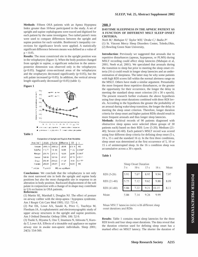
Sleep Research Society A215
PO
STE
R P
RE
SEN
TA
TIO
NS
SLEEP, Vol. 25, Abstract Supplement 2002
Methods: Fifteen OSA patients with an Apnea HypopneaIndex greater than 10/hour participated in the study. A set ofupright and supine cephalograms were traced and digitized foreach patient by the same investigator. Two tailed paired t testswere used to compare differences between the upright andsupine position for each variable. Bonferroni’s inequality cor-rections for significance levels were applied. A statisticallysignificant difference between means was defined as a value ofp < 0.05.Results: The most constricted site in the upright position wasin the velopharynx (figure 1). When the body position changedfrom upright to supine, a significant reduction in the antero-posterior dimension was observed only in the velopharynx(p<0.05). Saggital cross-sectional areas of the velopharynxand the oropharynx decreased significantly (p<0.05), but thesoft palate increased (p<0.05). In addition, the vertical airwaylength significantly decreased (p<0.05) (table 1).
Figure 1
Table 1
Conclusions: We conclude that the velopharynx is not onlythe most narrowed site in both the upright and supine bodypositions but also the most changeable site in response to analteration in body position. Backward displacement of the softpalate in conjunction with a change of in shape may contributeto UA occlusion in OSA patients.References: (1) Martin SE, Marshall I, Douglas NJ. The effect of postureon airway caliber with the sleep-apnea / hypopnea syndrome.Am J Respir Crit Care Med 1995; 152: 721-4.(2) Pae EK, Lowe AA, Sasaki K, Price C, Tsuchiya M,Fleetham JA. A cephalometric and electromyographic study ofupper airway structures in the upright and supine positions.Am J Orthod Dentofac Orthop 1994; 106: 52-9.(3) Tsuiki S, Hiyama S, Ono T, Imamura N, Ishiwata Y, Kuro-da T, Lowe AA. Effects of a titratable oral appliance on supineairway size in awake non-apneic individuals. Sleep 2001;24(5): 554-560.
288.JDAYTIME SLEEPINESS IN THE APNEIC PATIENT ASA FUNCTION OF DIFFERENT MSLT SLEEP ONSETCRITERIA.Neeb M,1 Mahajan V,1 Taylor WH,1 Drake C,1 Badia P2
(1) St. Vincent Mercy Sleep Disorders Center, Toledo,Ohio,(2) Bowling Green State University,
Introduction: Previously we suggested that arousals due torepetitive disturbances (apneas, hypopneas, or PLMS) duringMSLT recording could affect sleep latencies (Mahajan et al,2001; Neeb et.al; 2001). We speculated that arousals duringthe transition to sleep but prior to meeting the sleep onset cri-teria (16 s) could result in longer sleep latencies and an under-estimation of sleepiness. The latter may be why some patientswith high RDI scores fall within the normal alertness range onthe MSLT. Others have made a similar argument. Presumablythe more frequent these repetitive disturbances, or the greaterthe opportunity for their occurrence, the longer the delay inmeeting the standard sleep onset criterion (16 s 30 s epoch).The present research further evaluates the above hypothesisusing four sleep onset durations combined with three RDI lev-els. According to the hypothesis the greater the probability ofan arousal during wake/sleep transition, the longer the delay inmeeting the sleep onset criterion. Therefore, longer durationcriteria for sleep onset and higher patient RDIs should result inmore frequent arousals and thus longer sleep latencies.Methods: Archival records of 90 patients diagnosed withobstructive sleep apnea were selected (three groups of 30patients each) based on their RDI: Low (5-20); Medium (21-40); Severe (41-60). Each patient’s MSLT record was scoredusing four different sleep criteria for defining sleep onset (5 s,10 s, 15 s and the standard 16 s). In the first three conditions,sleep onset was determined at the first occurrence of 5, 10 or15 s of uninterrupted sleep. In the 16 s condition sleep wasaccumulative across a 30 s epoch.
Table 1
Results: Table 1 contains mean sleep latencies for the threeRDI levels and four sleep onset durations. The data reveal thatthe duration criterion used for defining sleep onset has amarked effect on MSLT latency. The shorter the duration of
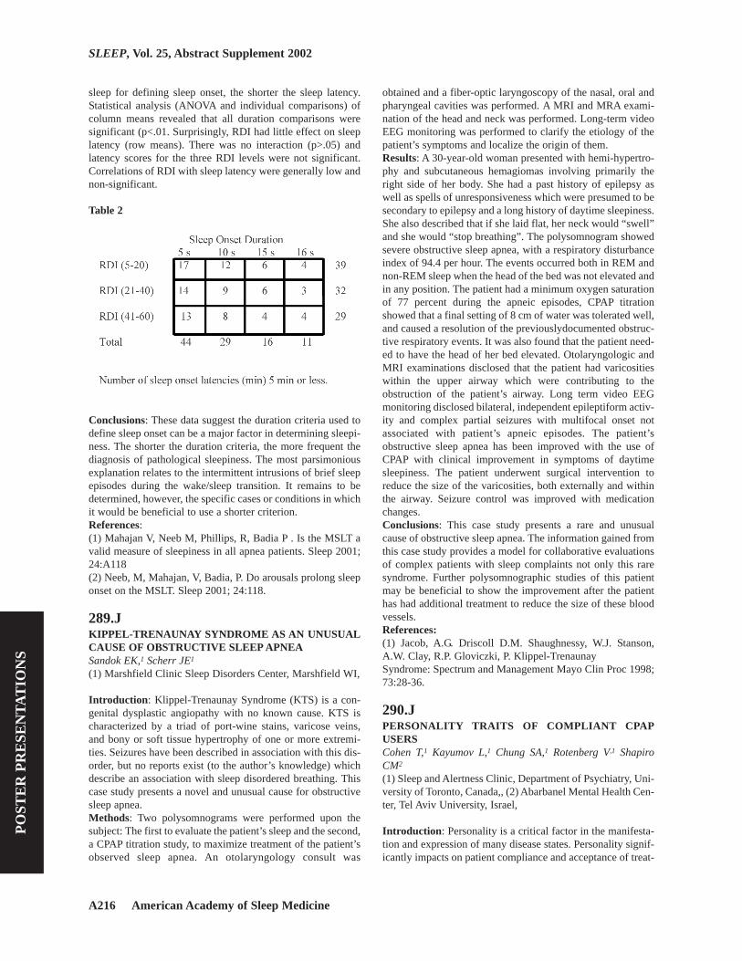
A216 American Academy of Sleep Medicine
PO
STE
R P
RE
SEN
TA
TIO
NS
SLEEP, Vol. 25, Abstract Supplement 2002
sleep for defining sleep onset, the shorter the sleep latency.Statistical analysis (ANOVA and individual comparisons) ofcolumn means revealed that all duration comparisons weresignificant (p<.01. Surprisingly, RDI had little effect on sleeplatency (row means). There was no interaction (p>.05) andlatency scores for the three RDI levels were not significant.Correlations of RDI with sleep latency were generally low andnon-significant.
Table 2
Conclusions: These data suggest the duration criteria used todefine sleep onset can be a major factor in determining sleepi-ness. The shorter the duration criteria, the more frequent thediagnosis of pathological sleepiness. The most parsimoniousexplanation relates to the intermittent intrusions of brief sleepepisodes during the wake/sleep transition. It remains to bedetermined, however, the specific cases or conditions in whichit would be beneficial to use a shorter criterion.References: (1) Mahajan V, Neeb M, Phillips, R, Badia P . Is the MSLT avalid measure of sleepiness in all apnea patients. Sleep 2001;24:A118(2) Neeb, M, Mahajan, V, Badia, P. Do arousals prolong sleeponset on the MSLT. Sleep 2001; 24:118.
289.JKIPPEL-TRENAUNAY SYNDROME AS AN UNUSUALCAUSE OF OBSTRUCTIVE SLEEP APNEASandok EK,1 Scherr JE1
(1) Marshfield Clinic Sleep Disorders Center, Marshfield WI,
Introduction: Klippel-Trenaunay Syndrome (KTS) is a con-genital dysplastic angiopathy with no known cause. KTS ischaracterized by a triad of port-wine stains, varicose veins,and bony or soft tissue hypertrophy of one or more extremi-ties. Seizures have been described in association with this dis-order, but no reports exist (to the author’s knowledge) whichdescribe an association with sleep disordered breathing. Thiscase study presents a novel and unusual cause for obstructivesleep apnea.Methods: Two polysomnograms were performed upon thesubject: The first to evaluate the patient’s sleep and the second,a CPAP titration study, to maximize treatment of the patient’sobserved sleep apnea. An otolaryngology consult was
obtained and a fiber-optic laryngoscopy of the nasal, oral andpharyngeal cavities was performed. A MRI and MRA exami-nation of the head and neck was performed. Long-term videoEEG monitoring was performed to clarify the etiology of thepatient’s symptoms and localize the origin of them.Results: A 30-year-old woman presented with hemi-hypertro-phy and subcutaneous hemagiomas involving primarily theright side of her body. She had a past history of epilepsy aswell as spells of unresponsiveness which were presumed to besecondary to epilepsy and a long history of daytime sleepiness.She also described that if she laid flat, her neck would “swell”and she would “stop breathing”. The polysomnogram showedsevere obstructive sleep apnea, with a respiratory disturbanceindex of 94.4 per hour. The events occurred both in REM andnon-REM sleep when the head of the bed was not elevated andin any position. The patient had a minimum oxygen saturationof 77 percent during the apneic episodes, CPAP titrationshowed that a final setting of 8 cm of water was tolerated well,and caused a resolution of the previouslydocumented obstruc-tive respiratory events. It was also found that the patient need-ed to have the head of her bed elevated. Otolaryngologic andMRI examinations disclosed that the patient had varicositieswithin the upper airway which were contributing to theobstruction of the patient’s airway. Long term video EEGmonitoring disclosed bilateral, independent epileptiform activ-ity and complex partial seizures with multifocal onset notassociated with patient’s apneic episodes. The patient’sobstructive sleep apnea has been improved with the use ofCPAP with clinical improvement in symptoms of daytimesleepiness. The patient underwent surgical intervention toreduce the size of the varicosities, both externally and withinthe airway. Seizure control was improved with medicationchanges.Conclusions: This case study presents a rare and unusualcause of obstructive sleep apnea. The information gained fromthis case study provides a model for collaborative evaluationsof complex patients with sleep complaints not only this raresyndrome. Further polysomnographic studies of this patientmay be beneficial to show the improvement after the patienthas had additional treatment to reduce the size of these bloodvessels.References: (1) Jacob, A.G. Driscoll D.M. Shaughnessy, W.J. Stanson,A.W. Clay, R.P. Gloviczki, P. Klippel-Trenaunay Syndrome: Spectrum and Management Mayo Clin Proc 1998;73:28-36.
290.JPERSONALITY TRAITS OF COMPLIANT CPAPUSERSCohen T,1 Kayumov L,1 Chung SA,1 Rotenberg V,1 ShapiroCM2
(1) Sleep and Alertness Clinic, Department of Psychiatry, Uni-versity of Toronto, Canada,, (2) Abarbanel Mental Health Cen-ter, Tel Aviv University, Israel,
Introduction: Personality is a critical factor in the manifesta-tion and expression of many disease states. Personality signif-icantly impacts on patient compliance and acceptance of treat-
Sleep Research Society A217
PO
STE
R P
RE
SEN
TA
TIO
NS
SLEEP, Vol. 25, Abstract Supplement 2002
ment. Although behavioral attitudes represent heritable tem-peraments based upon neurobiological substrates, theoretical-ly they might be influenced by environmental factors andchronic illnesses. To date, there are very few studies consider-ing personality factors in the diagnosis and treatment of sleepdisorders in general. To the best of our knowledge there are nostudies specifically focused on behavioral attitudes and com-pliance in sleep apnea. The purpose of the present study wasto evaluate personality traits in a sample of compliant CPAPusers as judged by two personality scales – The Temperamentand Character Inventory (TCI)1 and Behavioural Attitudes andSearch Evaluation test (BASE)2,3.Methods: Twenty two patients (12 males and 9 females, aged56.4± 13.4) of the Sleep and Alertness Clinic who have beensuccessfully using CPAP therapy for 1.5-2 years nightly for atleast 7 hours per night participated in the study. All patientsreceived TCI and BASE tests in an interview setting. Person-ality traits such as novelty seeking, harm avoidance, rewarddependence, persistence, self-directedness, cooperativenessand self-transcendence have been assessed in the analysis. Thesemiprojective test BASE was used to assess different behav-ioral attitudes including search activity pattern, stereotyped,panic and passive behaviors. Analysis of variance using a gen-eral linear model procedure was employed to detect statisti-cally significant differences in personalities’ traits. Furtheranalysis included Tukey post hoc paired comparisons and non-parametric Mann-Whitney’s U-test.Results: There were no significant gender differences withrespect to CPAP compliance. All CPAP users had high searchactivity scores as judged by the BASE questionnaire. Howev-er female patients scored significantly lower on search activi-ty than that of the male patients (p=.01). Both males andfemales scored lowest in those patterns classified as passiveand chaotic behaviors. There were no significant differenceson male and female scores for stereotyped behavior. On theTCI, CPAP patients demonstrated a combination of high harmavoidance scores and average scores for novelty seekingbehaviors. Females scored significantly higher on harm avoid-ance scale than male CPAP users (p=.02). It is noteworthy thatthere were no gender differences on novelty seeking patternsof behaviors. Males scored significantly higher than femaleson self-directedness scales (p=.01) There were no any signifi-cant differences on other personalities’ traits as measured bythe TCI.Conclusions: In this study we have shown that a group ofpatients with high CPAP compliance had strong search activi-ty attitudes as judged by the BASE questionnaire. The dis-crepancy between prominent search activity patterns and aver-age scores on novelty seeking behaviors as measured by theTCI implies that these two similar personality traits’ instru-ments measure different behavioral domains. References: (1) Cloninger CR, Przybeck TR, Svrakic DM, Wetzel RD. TheTemperament and Character Inventory (TCI): a guide to itsdevelopment and use. St. Louise, MO: Washington universityCenter for Psychobiology of Personality, 1994.(2) Venger A, Rotenberg V and Desiatnikov Y. Evaluation ofsearch activity and other behavioural attitudes in indefinite sit-uations. Dynamic Psychiatry 1996; 160-161: 368-377.(3) Hossain NK, Kayumov L, Rotenberg V, Shapiro CM.
Behavioural attitudes and sleep patterns in depression andsleep apnea. Sleep Research Online 1999; 2: 308.
290.JPERSONALITY TRAITS OF COMPLIANT CPAPUSERSCohen T,1 Kayumov L,1 Chung SA,1 Rotenberg V,1 ShapiroCM2
(1) Sleep and Alertness Clinic, Department of Psychiatry,University of Toronto, Canada,, (2) Abarbanel Mental HealthCenter, Tel Aviv University, Israel,
Introduction: Personality is a critical factor in the manifesta-tion and expression of many disease states. Personality signif-icantly impacts on patient compliance and acceptance of treat-ment. Although behavioral attitudes represent heritable tem-peraments based upon neurobiological substrates, theoretical-ly they might be influenced by environmental factors andchronic illnesses. To date, there are very few studies consider-ing personality factors in the diagnosis and treatment of sleepdisorders in general. To the best of our knowledge there are nostudies specifically focused on behavioral attitudes and com-pliance in sleep apnea. The purpose of the present study wasto evaluate personality traits in a sample of compliant CPAPusers as judged by two personality scales – The Temperamentand Character Inventory (TCI)1 and Behavioural Attitudes andSearch Evaluation test (BASE)2,3.Methods: Twenty two patients (12 males and 9 females, aged56.4± 13.4) of the Sleep and Alertness Clinic who have beensuccessfully using CPAP therapy for 1.5-2 years nightly for atleast 7 hours per night participated in the study. All patientsreceived TCI and BASE tests in an interview setting. Person-ality traits such as novelty seeking, harm avoidance, rewarddependence, persistence, self-directedness, cooperativenessand self-transcendence have been assessed in the analysis. Thesemiprojective test BASE was used to assess different behav-ioral attitudes including search activity pattern, stereotyped,panic and passive behaviors. Analysis of variance using a gen-eral linear model procedure was employed to detect statisti-cally significant differences in personalities’ traits. Furtheranalysis included Tukey post hoc paired comparisons and non-parametric Mann-Whitney’s U-test.Results: There were no significant gender differences withrespect to CPAP compliance. All CPAP users had high searchactivity scores as judged by the BASE questionnaire. Howev-er female patients scored significantly lower on search activi-ty than that of the male patients (p=.01). Both males andfemales scored lowest in those patterns classified as passiveand chaotic behaviors. There were no significant differenceson male and female scores for stereotyped behavior. On theTCI, CPAP patients demonstrated a combination of high harmavoidance scores and average scores for novelty seekingbehaviors. Females scored significantly higher on harm avoid-ance scale than male CPAP users (p=.02). It is noteworthy thatthere were no gender differences on novelty seeking patternsof behaviors. Males scored significantly higher than femaleson self-directedness scales (p=.01) There were no any signifi-cant differences on other personalities’ traits as measured bythe TCI.
A218 American Academy of Sleep Medicine
PO
STE
R P
RE
SEN
TA
TIO
NS
SLEEP, Vol. 25, Abstract Supplement 2002
Conclusions: In this study we have shown that a group ofpatients with high CPAP compliance had strong search activi-ty attitudes as judged by the BASE questionnaire. The dis-crepancy between prominent search activity patterns and aver-age scores on novelty seeking behaviors as measured by theTCI implies that these two similar personality traits’ instru-ments measure different behavioral domains. References: (1) Cloninger CR, Przybeck TR, Svrakic DM, Wetzel RD. TheTemperament and Character Inventory (TCI): a guide to itsdevelopment and use. St. Louise, MO: Washington universityCenter for Psychobiology of Personality, 1994.(2) Venger A, Rotenberg V and Desiatnikov Y. Evaluation ofsearch activity and other behavioural attitudes in indefinitesituations. Dynamic Psychiatry 1996; 160-161: 368-377.(3) Hossain NK, Kayumov L, Rotenberg V, Shapiro CM.Behavioural attitudes and sleep patterns in depression andsleep apnea. Sleep Research Online 1999; 2: 308.
291.JSUCCESSFUL TREATMENT OF OBSTRUCTIVESLEEP APNEA THROUGH CORRECTION OF DEN-TAL OCCLUSAL ABNORMALITIESDiamond B,1 Knebelman S,1 Deeley AG2
(1) Sleep Disorders Center, Abington Memorial Hospital,Abington, Pennsylvania, USA, (2) Centerline Technology,Craniometrics, Inc., Wynnewood, Pennsylvania, USA,
Introduction: Investigators have demonstrated physiologicand anatomical abnormalities in the upper airway of patientswith obstructive sleep apnea (OSA) including increased day-time genioglossus muscle activity, an alteration in shape of theairway, and cephalometric changes with low placement of thehyoid bone. The ability of the upper airway to change physio-logically and anatomically secondary to longstanding maloc-clusion was eloquently developed by Nove in a series of arti-cles in l946, and by Enlow in his classic text on facial growth.2Nove observed that “When the jaws are incorrectly opposed,this indispensable pivot or fulcrum for normality is out of trueand all of the structural units concerned with mastication, deg-lutition and respiration are compelled to function by means ofadaptation.” Recent experience with several patients withOSA, presenting with malocclusion, have led us to postulatethat the changes described are secondary to abnormal adapta-tion by the upper airway to compensate for a chronically dis-turbed bite resulting in persistence of an infantile (deviate)swallowing pattern; and that correction of the occlusal abnor-malities using daytime and nighttime splints may reduce theseverity of the apnea.Methods: Three patients with obstructive sleep apnea wereevaluated for dental occlusal abnormalities using a techniqueconsisting of an extensive dental examination, craniometricmeasurement and dynamic recordings with a ball-bearing biterecorder. These techniques are described in detail elsewhere.3Appliances to improve mandibular positioning were made.This required several visits with alteration in the appliance asadaptive changes occurred in the musculature. A nighttimeappliance, also designed to maintain normal dental relation-ships was constructed. One of the patients had tried mandibu-
lar advancement devices and surgical intervention with littlesuccess. All tried and were unable or unwilling to use nasalCPAP or BiPAP. Polysomnography was performed before andafter dental therapy.Results: The cases in this report were found to have abnormalmedial, anterior, posterior or vertical alignment. Each patientwas corrected in a specific way designed to address the prob-lem. The three patients all had dramatic drops in RDI, arousalindices and ESS scores on their PSM’s performed after treat-ment (See Table.1). No reduction in BMI occurred. The resultswere comparable to or better than CPAP and BiPAP, in the twopatients who had tried positive-pressure therapy. They experi-enced improved swallowing, posture, and daytime breathingand found the splints quite comfortable.
Table 1.
Conclusions: The benefit observed in these three patients withOSA, treated with correction of occlusal abnormalities, sup-ports an alternative dental approach to this disorder. Correc-tion of the abnormal opposition of the maxilla and mandibleshould, with time, reverse these abnormal adaptations. Whileocclusal therapy requires that an appliance be worn during theday to maintain appropriate craniofacial relationships andmuscle balance, these devices, adjusted over time, providenormal and more comfortable dental relationships and appearto be well accepted in our patients.References: (1) ) Nove, A: Cervico-Facial Othopaedia. The Dental Record1946; 66: 25-35, 49- 57, 86-91, 109-114.(2) Enlow, DH: Facial Growth. W.B. Saunders Company1990; 268-280.(3) Nordstrom, D: Craniometer/Aqualizer Techniques. TheFunctional Orthodontist 2000; Spring: 33-38.
292.JCHANGES IN BECK DEPRESSION INVENTORY (BDI)SYMPTOMS AFTER CONTINUOUS POSITIVE AIR-WAY PRESSURE (CPAP) TREATMENT FOROBSTRUCTIVE SLEEP APNEA (OSA)Means MK,1,2 Lichstein KL,1,2 Edinger JD,1 Taylor DJ,2,3 Dur-rence HH,1 Husain AM,1 Radtke RA2
(1) The University of Memphis, Memphis, TN, (2) VA Med-ical Center, Durham, NC, (3) Duke University Medical Cen-ter, Durham, NC,
Introduction: Many studies have documented elevated
Sleep Research Society A219
PO
STE
R P
RE
SEN
TA
TIO
NS
SLEEP, Vol. 25, Abstract Supplement 2002
depression levels in OSA patients that improve subsequent toCPAP treatment. However, few researchers have consideredtype (i.e., affective/cognitive vs. somatic/vegetative) ofdepressive symptom change in these patients. The overlap insomatic symptoms between OSA and depression has led Leeand colleagues (1) to postulate that depression in OSA patientspredominantly reflects OSA symptoms. The present studyexplored differences in affective versus somatic symptoms ofdepression in OSA patients after CPAP treatment.Methods: Thirty-nine outpatients (90% male; M age=57.8years) undergoing routine diagnostic polysomnography (PSG)for OSA at a sleep disorders center completed the BDI prior toPSG and 3 months following initiation of CPAP. Participantshad no co-existing sleep disorders, no psychiatric history, andwere not taking psychotropic medications. The BDI wasselected for this study because previous factor analyses (2)have yielded two main factors that correspond with the dis-tinction between affective and somatic symptoms.
Table 1
Figure 1
Results: The difference in BDI scores from baseline (M =9.59, SD = 7.63) to follow-up (M = 7.26, SD = 5.85) was sig-nificant (t[38] = 2.92, p < .01). Table 1 presents the distribu-tion of BDI scores before and after CPAP. Although mostpatients were non-depressed, there were fewer moderately andseverely depressed patients after CPAP (Chi-square [6, N =39] = 34.93, p < .001). Factor scores for affective symptoms
(Factor 1) and somatic symptoms (Factor 2) were computed.Symptom change was analyzed using a repeated measuresMANOVA, with total BDI and factor scores entered asdependent variables. There was a significant effect for time(Wilks \lambda [3, 36] = .80, F [3, 36] = 2.98, p < .05). Uni-variate F tests were significant for all three dependent vari-ables: total BDI (F [1, 38] = 8.52, p < .01), Factor 1 (F [1, 38]= 5.39, p < .05), and Factor 2 (F [1, 38] = 4.70, p < .05). Tocompare change in symptoms, z-scores were calculated usingthe overall mean and standard deviation (Figure 1). Theseresults suggest that although scores improve after treatment,affective symptoms improve to the same extent as somaticsymptoms.Conclusions: OSA patients treated with CPAP showedimprovements in BDI scores, although pre-treatment meanscores were in the non-clinical range. Analysis of symptomchange suggests that patients are not only endorsing somaticsymptoms of depression but are expressing affective symp-toms as well. The present study provides suggestive evidencethat depressive symptoms in OSA patients are not entirelyattributable to somatic symptoms shared by OSA and depres-sion.References: (1) Lee S, Wing YK, Chen CN. Obstructive sleep apnoea anddepression. Australian and New Zealand Journal of Psychiatry1995;27:162,165-166.(2) Tanaka JS, Huba GJ. Confirmatory hierarchical factoranalyses of psychological distress measures. Journal of Per-sonality and Social Psychology 1984;46:621-635.
293.JAN EVALUATION OF A NEW BILEVEL MODE FORTHE TREATMENT OF OBSTRUCTIVE SLEEP APNEAHYPOPNEA SYNDROME (OSAHS)Wylie P,1 Crossett D,1 Hardy W2
(1) Arkansas Center for Sleep Medicine, Little Rock AR, (2)Respironics, Inc. Murrysville PA,
Introduction: Bilevel positive airway pressure is a treatmentmodality where expiratory and inspiratory pressures are setindependently to prevent apnea, hypopnea and snoring.BiFlexTM is a bilevel mode that alters the end-inspiratory andinitial expiratory pressure characteristics. BiFlex reducesinspiratory pressure (IPAP) towards end-inspiration smooth-ing the transition from the inspiratory to the expiratory phase.With the BiFlex mode a lower expiratory pressure (EPAP) isdelivered during the beginning of the expiratory phase (1). TheBiFlex feature is expected to improve patient comfort andcompliance with positive pressure therapy without negativelyinfluencing sleep quality. This study was undertaken to deter-mine if BiFlex alters conventional bilevel pressure require-ments.Methods: Ten patients (8 male, 2 female, mean age 58.3 ±(SD) 12.7 years, BMI 33.6 ± 9 (SD)) who were previouslydiagnosed with OSAHS and who were already receivingCPAP therapy were recruited. Full night polysomnographywas performed on two, non-consecutive nights. Patients wereblinded as to which mode they were receiving (bilevel orbilevel with BiFlex). On the first night, patients received con-
A220 American Academy of Sleep Medicine
PO
STE
R P
RE
SEN
TA
TIO
NS
SLEEP, Vol. 25, Abstract Supplement 2002
ventional bilevel therapy to determine pressure requirements.On the second night, patients received bilevel therapy withBiFlex. BiFlex gains were set at a level the patient found to bemost comfortable while awake, prior to the second PSG.Ther-apy pressure requirements, RDI, apnea index, hypopnea index,and mean saturation were compared from the PSG recordingson both nights. Paired data (bilevel versus bilevel with BiFlex)were analyzed with Student’s t-Test assuming unequal vari-ance.Results: Comparing the results of bilevel therapy to bileveltherapy with BiFlex, there were no significant differences inthe frequency and severity of sleep related breathing events(Table 1). The inspiratory and expiratory pressure required totreat the patients did not differ significantly. (Table 2).
Table 1
Table 2
Conclusions: In a group of patients who by age and BMI, aretypical of patients with OSAHS, this evaluation detected nodifference in therapy requirements between standard bileveland bilevel therapy with BiFlex. There were no significant dif-ferences in sleep quality. Twice as many patients preferredbilevel with BiFlex compared to standard bilevel therapy.BiFlex may offer advantages in comfort over conventionalpositive pressure therapy without altering therapy pressurerequirements.References: (1) Juhasz J., Becker H., Proportional Positive Airway Pres-sure: A New Concept to Treat Obstructive Sleep Apnea EurRespir J 2001; 17: 467-473
Research supported by Supported by: Respironics, Inc.
294.JEFFICACY OF ORAL CPAP IN THE MANAGEMENTOF OBSTRUCTIVE SLEEP APNEA.Anderson FE,2 Smith NC,2 Kingshott RN,2 Taylor DR,3 WhyteKF3
(1) Sleep Disordered Breathing Unit, Greenlane Hospital,Auckland, New Zealand, (2) Fisher & Paykel Healthcare,Auckland, New Zealand, (3) Respiratory Research Unit,Dunedin School of Medicine, University of Otago, Dunedin,New Zealand,
Introduction: Continuous positive airway pressure (CPAP) isconsidered the ‘gold standard’ treatment for obstructive sleepapnea (OSA). This positive pressure is traditionally appliedvia a nasal mask or full-face mask, which some patients findintolerable due to nasal discomfort, mask or mouth leaks andcumbersome headgear. These adverse effects can lower patientcompliance, and in some cases, cause patients to cease treat-ment altogether. This randomised, crossover study assessedthe efficacy of an innovative oral device, which allows posi-tive pressure to be delivered via the mouth, compared with tra-ditional nasal CPAP.Methods: Twenty-two CPAP-naive OSA sufferers (18 Male, 4Female) were randomised to receive CPAP treatment via anasal mask (Aclaim™, Sullivan Standard, Mirage® or UltraMirage™) (13 patients, mean age 42.0 ± 10.2 years, meanBMI 41.6 ± 7.5 kg/m2) or the Oracle™ oral interface (9patients, mean age 48.6 ± 11.4 years, mean BMI 43.3 ± 7.1kg/m2). Patients were diagnosed in a sleep laboratory using astandard polysomnography (PSG). When OSA was evident(AHI > 20/hr slept) after a 2-hour minimum diagnostic period,CPAP therapy was initiated with the first treatment arm. CPAPpressure was titrated to eliminate all evidence of upper airwayobstruction. The diagnostic and treatment studies, at the finaltitrated pressure, were analysed in a blinded fashion. The studyend-points were patient compliance with CPAP and subjectiveand objective data on CPAP efficacy with each interface. Cal-culations for significant differences with rejection of the nullhypothesis at a p < 0.05 were carried out using two-tailed stu-dent t-tests. This trial is currently in progress and thereforeonly data from the first treatment arm is presented here.Results: Two patients in each treatment group withdrewbecause they could not tolerate CPAP. One additional patientin the oral group was excluded because compliance data wasunobtainable. Disease severity was similar between the twogroups (Table 1). There was no significant difference in themonitored compliance between the two interfaces with a meancompliance of 3.8 ± 2.3 hrs/night and 3.2 ± 1.5hrs/night fornasal and oral, respectively (p = 0.60). Mean (SD) EpworthSleepiness Scores at baseline were 16.5 (± 3.2) and 18.5 (±2.1) for nasal and oral, respectively (p = 0.24). After fourweeks of CPAP treatment on the randomised interface thisdecreased to 6.4 (± 3.2) and 8.0 (± 6.1), respectively (p =0.53). There was no significant difference in PSG databetween the two treatment groups (Table 1 & 2).
Sleep Research Society A221
PO
STE
R P
RE
SEN
TA
TIO
NS
SLEEP, Vol. 25, Abstract Supplement 2002
Table 1
Table 2
Conclusions: Sleep parameters and CPAP compliancerevealed no significant differences between the nasal and oralinterfaces. CPAP delivery through the mouth is as effective asdelivery through the nose. In addition, oral and nasal CPAPwere equally accepted by patients.
Research supported by Fisher & Paykel Healthcare
295.JTWO-POINT PALATAL DISCRIMINATIONRESPONSE IS NORMAL IN UPPER AIRWAY RESIS-TANCE SYNDROME AND REDUCED IN OBSTRUC-TIVE SLEEP APNEA SYNDROMERobinson A, Li K, Robinson, A, Guilleminault C(1) Sleep Institute Department, UNIFESP-EPM, Sao Paulo,Brazil, (2) Stanford University Sleep Disorders Center, Stan-ford, California, USA,
Introduction: Patients with Upper Airway Resistance Syn-drome (UARS) show increased respiratory effort, arousalsrelated to increased effort and hypopneas. By contrast patientswith Obstructive Sleep Apnea Syndrome experience obstruc-tive apneas and hypopneas associated with oxygen desatura-tion. UARS patients show increased 7 to 9 cps electroen-cephalographic (EEG) power spectrum of the central leadscompared to normals. The EEG power spectrum of patientswith OSAS shows a delayed arousal response. Friberg et al(1).have shown abnormalities including evidence of neurogeniclesions in histological analyses of palatopharyngeal musclebiopsies obtained from OSAS patients. These findings corrob-orate earlier work by Edstrom et al(2). We hypothesize thatthere also may be differences in sensory input in UARS com-pared to OSAS. We theorize sensory differences associatedwith neurogenic lesions may play a role in the delayedresponse to arousal found with abnormal breathing events inOSAS. Our investigation analyzed the response to a two-point
discrimination test applied to the palatal mucosaMethods: Fifteen each of normal subjects, OSAS patients andUARS patients between 35 and 50 years of age were age, sexand body mass index (BMI) matched. Subjects were recruitedfrom the community and patients from the Stanford Sleep Dis-orders clinic. All participants underwent full nocturnalpolysomnogram (PSG) with esophageal manometry followinginterview and physical examination.OSAS patients wereincluded who had an apnea-hypopnea index (AHI) >15events/hour, UARS patients an AHI <5 and normal controlswere included only if the had an AHI < 1 without evidence ofincreased respiratory effort as described for UARS.A prelimi-nary evaluation was performed using a modified 25gauge nee-dle on a tuberculin syringe. The patient was first given a tac-tile stimulus with the needle in contact with the mucosa.at 0.5mm. If no was sensation noted, at two minute intervals pres-sure was applied at 1 mm then 2 mm if needed.After prelimi-nary measurements, a compass type device was then used toevaluate two-point discrimination. Two-point detection wasmeasured twice in the three selected palatal locations in a ran-dom way. The best result of 6 measurement trials is presentedin Table 1.
Table 1
Table 2
A222 American Academy of Sleep Medicine
PO
STE
R P
RE
SEN
TA
TIO
NS
SLEEP, Vol. 25, Abstract Supplement 2002
Results: Results5 OSAS patients needed the 1 mm marker torecognize pressure at one point and 4 with OSAS needed pres-sure to the 2 mm mark to note sensation. One OSAS patientwas an outlier with a two-point discrimination distinction at 15mm. Review of clinical history implicated coexistent GERD,this subject was excluded from the analysis of results.Twopoint distinction evaluation revealed a significant differencebetween the OSAS patients and the other two groups. Themean (±SD) values were3.86 (0.58), 1.66 (1.0) and 1.63 (0.29)for OSAS, UARS and Controls respectively (Mean rank =38.0, 15.9, 15.1). p = .0001.Conclusions: ConclusionsThe normal two-point discrimina-tion responses seen with UARS indicate that these patients aremore capable of transmitting sensory inputs than those withOSAS. This may be one element in explaining the arousalresponse previously documented in UARS compared toOSAS. These conclusions are consistent with the recent workof Kimoff3 et al. who concluded a selective impairment in thedetection of mechanical stimuli in OSAS patients could bypartially reversed with CPAP.References: (1) Friberg D, Gazelius B, Holfelt T, Carlsson- Norlander B.Abnormal afferent nerve endings in the soft palatal mucosa ofsleep apneics and habitual snorers. Regul Pept. 1997; 71: 29-36.(2) Edstrom L, Larsson H, Larsson L. Neurogenic effects onthe palatopharyngeal muscle in patients with obstructive sleepapnoea: a muscle biopsy study. J. Neuro. Psych 1992-55: 916-920(3) Kimoff Jr, Sforza E, Champagne V, Ofiara L, Gendron D.Upper airway sensation in snoring and obstructive sleepapnea. Am J Respir. Crit Care med. 2001-164: 250-255.
296.JA NEW AIRFLOW SENSOR FOR THE DETECTIONOF FLOW LIMITATION IN SLEEP-DISORDEREDBREATHINGMelendrez DC,1 Krakow BJ,1 Hall MD1
(1) Sleep & Human Health Institute, (2) Eastern New MexicoSleep Disorder Center,
Introduction: The diagnosis of Sleep-Disordered Breathinghas become more dependent on the capacity of airflow moni-tors to detect subtle breathing disturbance termed flow limita-tion or respiratory effort related arousal (RERA). Still, themost widely used device to assess SDB is the thermistor/ ther-mocouple, but it does not detect subtle respiratory events.Only esophageal manometry (EM) and nasal cannula pressuretransducer (NC) have been validated for this purpose. Yet,each of these devices has drawbacks. EM, although deemedthe gold standard (1), is invasive and may be poorly toleratedby patients. In comparison to EM invasiveness, NC appearsmore promising, but anecdotally, NC has shown decreasedaccuracy during cases of mouth breathing, nasal congestion,and nasal prong obstruction or displacement/dislodgment.These difficulties in respiratory monitoring indicate the con-tinued need to develop sensors that are non-invasive and accu-rate in detecting subtle breathing disturbance. The newDymedix airflow sensor (DAS) (Dymedix, Minneapolis, MN)
is one such device. The DAS utilizes Polyvinylidene Fluoride(PVDF) film that is a specially processed flexible plastic filmthat responds rapidly to both pressure and temperaturechanges. As a first step prior to formal validation of the DAS,we compared the DAS to both NC and thermocouple in apatient suspected of upper airway resistance syndrome(UARS).Methods: One 32-year-old non-obese female with snoring andsleepiness, suspicious for UARS underwent polysomnographywith simultaneous respiratory monitoring with DAS, NC, andstandard thermocouple at the Eastern New Mexico Sleep Dis-order Center in Roswell, NM. The recording included a 16channel montage: LOC-A2; ROC-A1; C3-A2; C4-A1; O1-A2;O2-A1; chin EMG; EKG; L-R leg EMG; Snore; NC (Biolog-ic Systems, Mudelein, IL); DAS; thermocouple (Protech,Woodinville, WA); chest effort; abdominal effort; pulseoximetry. Respiratory monitoring for each device was assesedfor the presence of flow limitation events. Flow limitationevents were defined as two or more consecutive breaths witha flattened or non-sinusoidal appearance without airflowdecrements meeting 50% reduction criteria for hypopnea.
Figure 1
Results: Figure 1 is a 60-second PSG window displaying thesimultaneous NC, DAS, and thermocouple signals. At thebeginning of the tracing, all sensors show their particular sinu-soidal shape. Then, both the NC and the DC show a flow lim-itation event with a non-sinusoidal flow signal terminating inan arousal. The thermocouple signal shows no decrease inamplitude and remains sinusoidal during the event. Many flowlimitation events seen in this patient were similar to the eventin Figure 1 and confirmed a UARS diagnosis.Conclusions: This preliminary case study shows that the DASmay be an alternative device used to measure subtle breathingdisturbance. Anecdotally, during mouth breathing in thispatient, the DAS sustained its average baseline while the NCsignal was dampened and difficult to interpret. In order to for-mally validate the DAS, future studies will assess intersignal
Sleep Research Society A223
PO
STE
R P
RE
SEN
TA
TIO
NS
SLEEP, Vol. 25, Abstract Supplement 2002
agreement between the DAS and both nasal cannula pressuretransducer and esophageal manometry on an event-by-eventbasis in a larger sample of SDB patients. In the interim, theDAS appears useful for diagnosing SDB, including UARS,and may be appropriate for patients with mouth breathing,nasal congestion, or intolerance to esophageal manometry.References: (1) AASM Task Force. Sleep-related breathing disorders inadults: recommendations for syndrome definition and meas-urement techniques in clinical research. Sleep. 1999 Aug1;22(5):667-89.
Research supported by Dymedix Corporation
297.JPILOT STUDY FOR SAFETY AND EFFICACY OFCORUS-1010 NASAL/THROAT SPRAY IN THETREATMENT OF SNORING AND OBSTRUCTIVESLEEP APNEASchoenhofer B,1 Hofmann T,1 Barchfeld T,2 Simon A,1 Mont-gomery AB,1 Koehler D2
(1) Krankenhaus Kloster Grafschaft, Schmallenberg, Ger-many, (2) Corus Pharma Inc., Seattle, WA, USA,
Introduction: CORUS-1010 (Tyloxapol Nasal Solution) is amucolytic and surface active agent developed for aerosol use.In this pilot study we investigated the effect of nasally appliedCORUS-1010 on snoring and sleep apnea.Methods: In an open study design 10 consecutive habitualsnorers or patients with mild to moderate OSA were included(8 males, 50.2 ± 10.6 years old, RDI: 23.8 ± 18.8/h). After thebaseline polysomnographic recording, in a second night drugeffect was investigated. The intervention consisted of 5 spraysin each nostril and the throat 15-30 minutes prior to lights off.After a treatment period of 4 weeks, both the adverse effectscaused by CORUS-1010 and the snoring intensity (the latterby the bed partner) were evaluated based on a questionnaire.Results: The main findings concerning respiratory events andsleep quality are given in the table. At the end of the 4-weekobservation period the patients did not report adverse effects.Simultaneously, 7/10 spouses reported a mild to moderatereduction of snoring.
Table 1
Conclusions: Although the trial was underpowered in sample
size, we found some non-significant trends to improvement ofboth sleep disordered breathing and sleep quality. Further ade-quately powered, placebo controlled trials in snoring and sleepapnea are ongoing.
Research supported by This work was supported by CorusPharma Inc.
298.JAN EVALUATION OF DME VENDOR PERFOR-MANCE IN THE EDUCATION OF NEW CPAP USERS Honor MJ,1 Kilkenny TM,1 Elsayegh D1
(1) Staten Island University Hospital Sleep Center,
Introduction: Durable Medical equipment (DME) vendorsare assumed to play an important role in the health care net-work, and in terms of sleep disorders, an important role inCPAP compliance. The DME company is the last entity to seethe patient before treatment begins; therefore, it is their per-formance that may directly affect CPAP compliance. Ourobjective was to determine whether the DME vendor providedthe education and service that is required to obtain a satisfac-tory outcome.Methods: A retrospective analysis was performed by contact-ing patients who have undergone sleep testing and haverecently been prescribed CPAP. A phone interview was con-ducted asking the patients several questions about the DMEservice with regard to speed of delivery, equipment setup andeducation. The vendor score was based on the number of affir-mative answers to 13 questions. The questionnaire resultswere broken down into two parts; one part analyzing compo-nents of the machine setup, and the other analyzing compo-nents of patient education.Results: Two hundred and six patients were called with com-plete data available in 104 cases. The interviews were con-ducted ranging from 60 ¨C 90 days after CPAP delivery. Eightvendors were identified. Sixteen patients (36%) were unable toname their vendor. Seventy-five percent (75%) of the patientscontacted were using the CPAP device. Seventy-eight percent(78%) of those patients were using the machine ¡Ý 5 hours pernight with an average usage of 6.7 hrs. per night. The equip-ment was utilized an average of 6 nights per week. The over-all scores for machine setup were higher than that of thepatient education component (86% v. 73%). Based on the 13questions asked, the vendors scored within a range of 86% to96% on all questions except knowledge of how to travel withthe device (67%), knowledge of CPAP pressure (69%), propermask fit (54%), and knowledge of how to manage nasal dry-ness (59%). Mask fit pertained to whether or not the DMEvendor provided only one mask (negative response) or thevendor actually measured the patient¡¯s face before providinga mask (affirmative response).Conclusions: Two major factors that contribute to CPAP com-pliance are proper mask fit and the ability to manage or pre-vent nasal dryness. We believe that further efforts must bemade to properly inform patients regarding the appropriate fitand use of this equipment and that failure to do so may be acontributing factor to poor compliance with therapy.
A224 American Academy of Sleep Medicine
PO
STE
R P
RE
SEN
TA
TIO
NS
SLEEP, Vol. 25, Abstract Supplement 2002
299.JTHE THREAT OF MANDATORY REPORTING TO ADRIVER’S LICENSE AGENCY DISCOURAGESSLEEPY DRIVERS FROM BEING EVALUATED FORSLEEP APNEAFindley LJ1
(1) Sleep Disorders Center of Northern Colorado,
Introduction: Drivers with obstructive sleep apnea have a 2-4 fold higher rate of automobile crashes than controls. Thisincreased risk has prompted some governments to mandatethat physicians report the names of their patients with sleepapnea to a driver’s license agency. However, mandatoryreporting may provoke fear of losing their driving privilegeand discourage drivers from being evaluated for sleep apnea.To evaluate the effect of mandatory reporting we interviewedsubjects undergoing evaluation for sleep apnea.Methods: We interviewed a consecutive group of 30 outpa-tients having an initial evaluation for sleep apnea. These sub-jects were referred for evaluation of snoring and excessivesleepiness or fatigue. We asked each subject if mandatory oftheir name to the state driver’s license agency would causethem to avoid evaluation of their sleepiness.Results: There were 18 males and 12 females with a mean ageof 50 years + 13 (SD). Twenty-one of the 30 subjects (70%)said they would avoid medical evaluation of their sleepinessbecause of fear of losing the ability to drive. There was a trendof a higher percentage of males avoiding evaluation, but thiswas not significant. The subjects who would avoid medicalevaluation were not different in age or score on EpworthSleepiness Scale from those not discouraged by mandatoryreporting.Conclusions: The threat of mandatory reporting of theirnames to a driver’s license agency discourages sleepy driversfrom being evaluated for sleep apnea. The resulting failure todiagnose and treat sleep apnea would result in many prevent-able automobile crashes.
300.JCORRELATION OF OXIMETRY PARAMETERSWITH RESPIRATORY DISTURBANCE INDEXAMONG ADULT PATIENTS WHO UNDERWENTFULLNIGHT DIAGNOSTIC POLYSOMNOGRAPHYCorpuz FC,1 Cua IY,1 Codamos LJ,1 Gappi MS1
(1) Department of Medicine, St. Luke’s Medical Center,
Introduction: Obstructive Sleep Apnea Syndrome (OSAS) isof increasing importance because of its high prevalence anddanger of morbidity to untreated patients. With the cost andinconvenience of doing polysomnography (PSG), oximetryhas been proposed as a tool in the diagnosis of OSAS. The aimof this study is to correlate Respiratory Disturbance Index(RDI) of 5, 20 and 30 with oximetry parameters among adultpatients who underwent fullnight diagnostic polysomnogra-phy (PSG) and to compare these parameters in patients withand without OSAS.Methods: Design: Retrospective, descriptive and analyticalstudyDesign: Retrospective, descriptive and analyticalstudySetting: St. Luke’s Medical Center Sleep Center Materi-
als and Methods: Patients referred to the Sleep Center for PSGwith symptoms suggestive of OSAS were investigated. Theirclinical features and questionnaire responses were reviewed.RDI was calculated for all the patients and were classified asmild (RDI>5 events/hour), moderate (RDI>20 events/hour),and severe (RDI>30 events/hour). Oximetry parameters deter-mined quantitatively and qualitatively from the strip chartrecording include mean lowest oxygen saturation (MLSAT),lowest oxygen saturation (LSAT), and mean oxygen saturation(MSAT).Results: Three hundred one patients (age, 47.04±13.6) wereincluded. A total of 222(74%) OSAS patients were identifiedwith RDI > 5 events/hr of which 24%(54/223) had mildOSAS, 10%(22/223) had moderate OSAS and 66%(146/223)had severe OSAS. Height, weight, body mass index (BMI) andneck circumference were significantly different betweenpatients with and without OSAS at p<0.05. Comparison of theMLSAT, LSAT, and MSAT in patients with and without OSASshowed a significant difference using the Student’s T-test atp<0.05. There was likewise a significant difference (p=0.000)in the above mentioned oximetry parameters between patientswithout OSAS and with mild, moderate, and severe OSAScomputed independently. RDI had significant negative corre-lation with MLSAT (r=-0.441; p=0.000), LSAT (r=-0.247;p=0.003), and MSAT (r=-0.430; p=0.000)using Pearson’s cor-relation.
Table 1
Conclusions: Combined clinical evaluation and the use ofovernight pulse oximetry would allow recognition of a patientwith OSAS in the absence of PSG. It helps determine theseverity of the disease and can be used as a cheaper alternativeto detect sleep apnea.References: (1) Herer B,Roche N, Carton M, Roig C, Pujol V, Houchon G:Value of clinical, functional, and oximetric data for the pre-diction of obstructive sleep apnea in obese patients. Chest1999 Dec; 116(6):1537-44(2) Epstein et al. Cost effectiveness Analysis of NocturnalOximetry as a method of screening for Sleep Apnea-HypopneaSyndrome. Chest 1998; 113:97-103(3) Yamashiro Y, Kryger MH, Nocturnal Oximetry: is it ascreening tool for sleep disorders?, Sleep 1995 Apr;18(3):167-71

Sleep Research Society A225
PO
STE
R P
RE
SEN
TA
TIO
NS
SLEEP, Vol. 25, Abstract Supplement 2002
301.JOBSTRUCTIVE SLEEP APNEA SYNDROME AMONGCITY BUS DRIVERS: DIAGNOSTIC IMPLICATIONSAND DRIVING CAPACITYTelakivi T,1,2 Partinen M,1,2 Hublin C,1,2 Hirvonen K1,2
(1) Haaga Neurological Research Centre, (2) Department ofClinical Neurosciences, University of Helsinki,
Introduction: Excessive daytime sleepiness (EDS) of a pro-fessional motor vehicle conductor is a risk for traffic safety.Risk of an individual with obstructive sleep apnea syndrome(OSAS) to be involved in a traffic accident has been reportedto be significantly greater than for controls. We reported a20.3% prevalence of OSAS and 7.9% prevalence of severeOSAS with EDS (oxygen desaturation index, ODI4 > 30 andS1-latency in MWT < 12.9 minutes) in city bus drivers. Wefurther analyzed these data for the implications on driving per-formance and clinical decision-making.Methods: 421 bus drivers of the City Transportation Depart-ment, Helsinki, Finland answered to a modified Basic NordicSleep Questionnaire. 22 subjects with suspected OSAS(reported loud, intermittent snoring every or nearly every nightand either sleep apneas > 1 night a week or snoring 20 yearsor longer with Epworth Sleepiness Scale (ESS) score > 8) and16 age-matched controls (snoring once a week or less often,ESS < 8) were screened by all-night polysomnography, MWTand driving simulator test (STISIM, Systems Technology Inc.,USA). Definitions: EDS: S1-latency < 12.9 minutes in MWT,OSAS: ODI4 > 10, severe OSAS ODI4 > 30. The subjects’history of traffic accidents in the past 10 years was reviewed.Results: 17/22 with suspected OSAS and 4 /16 of subjectswith no OSAS-suspicion had OSAS in the polysomnography.The performance of subjects with or without OSAS in thedriving test and the past history of accidents did not differ.ESS score did not correlate significantly with ODI4. The S1-latency was shortest in those with OSAS and longest with noOSAS when ESS >10. The sensitivity and specificity of ESS> 10 were 60% and 66% and those of two items, self-reportedsnoring every day or almost every day and reported apneas atleast once a week were 100% and 40%, respectively for severeOSAS with EDS. MWT sleep latency increased with age.ODI4 correlated significantly with reaction time (F = 4.498, P= 0.042), but the reaction times of subjects with OSAS andEDS did not differ significantly from those with OSAS but notwith EDS nor with controls with EDS.Conclusions: The results illustrate the difficulty of clinicalconclusions concerning professional drivers with OSASand/or EDS. Questions concerning habitual snoring and self-reported apneas > once a week did single out all drivers withsevere OSAS and EDS, but there was a weak specificity. ESS>10 was not sensitive in differentiating OSAS patients fromcontrols, nor the driving performance, or history. A significantcorrelation between ODI4 and the mean reaction time wasfound, but it did not differentiate those with or without EDS.Better tools to screen for sleep laboratory examinations and toestimate driving capacity in professional drivers are warrant-ed.
Research supported by The Finnish Work Environment
Fund
302.JEXECUTIVE DYSFUNCTION IN CHILDRENREFERRED FOR OBSTRUCTIVE SLEEP APNEA(OSA) EVALUATIONBeebe DW,1 Groesz L,1 Jeffries J,1 Chini B,1 Amin R1
(1) Children’s Hospital Medical Center, Cincinnati, Ohio,
Introduction: The most consistent area of cognitive deficit inadults with OSA is executive dysfunction (1). Executive dys-function is comprised of deficits in attention, inhibition,immediate working memory, behavior regulation, metacogni-tion (e.g., planning and organization), and mental flexibility.In children, similar cognitive deficits and additional behav-ioral disturbances reminiscent of attention-deficit / hyperactiv-ity disorder (ADHD) have been reported. However, no pub-lished study has targeted executive functioning in a pediatricpopulation referred for evaluation for OSA. We hypothesizedthat, relative to published norms, such children would displaydeficits on tests of executive functioning, despite having nor-mal basic verbal, nonverbal, and long-term memory skills.Methods: We assessed consecutive children aged 6-12 whohad been referred for evaluation of obstructive breathing dur-ing sleep. All were reported by their parents to snore loudlyand chronically. Exclusion criteria included any other chronicmedical condition or developmental delay. Children under-went overnight polysomnogram and, on a separate day, a 1.5hour battery of validated cognitive tests begun between 2 and3:30 p.m.: WISC-III Vocabulary, Block Design and DigitSpan, NEPSY Visual Attention and Verbal Fluency, WRAMLList Learning, Gordon CPT Omissions and Commissions. Par-ents also completed a validated executive functioning form(2). Subjects were classified as poor scorers on a test if theirperformance was <=16th percentile (z<=1) compared to age-based norms. Statistical analyses included single-sample t-tests comparing group means to norms, and binomial testsassessing the likelihood of obtained poor performance rates.
Table 1
Results: To date, 12 children (50% male; median age=7.5)have been enrolled. The results in Table 1 are consistent withpredictions. Mean scores and rates of poor scorers in all three
A226 American Academy of Sleep Medicine
PO
STE
R P
RE
SEN
TA
TIO
NS
SLEEP, Vol. 25, Abstract Supplement 2002
non-executive areas (verbal memory, vocabulary, visual con-struction) did not differ from norms. In contrast, all areas ofexecutive function (verbal fluency, sustained and selectiveattention, working memory, and impulsivity) yielded deficientmean scores or a high rate of poor scores. Parents also report-ed abnormal mean scores and poor performance rates in thebehavior regulation and metacognition of their children. Thesedeficits were present despite relatively mild respiratory pathol-ogy (all but one AHI < 5). AHI correlated only with sustainedattention (rho= -.58, p=.025), but this may be related to thesmall sample.Conclusions: These data suggest specific executive functiondeficits in children referred for evaluation for OSA. If futurework supports these findings, they (a) could account forbehavioral similarities with ADHD, a neurodevelopmentaldisorder that involves executive dysfunction, (b) would extendadult findings to the pediatric age, and (c) would support rou-tine cognitive screening of children suspected of OSA. Thesedata are in line with our recent theoretical model of the patho-genesis of cognitive morbidity in OSA, which posits that sleepand respiratory pathology differentially impact prefrontalbrain regions, resulting in executive dysfunction (1). We arenow expanding this sample and conducting longitudinal fol-low-up.References: (1) Beebe DW, Gozal D: Obstructive sleep apnea and the pre-frontal cortex. Journal of Sleep Research, under review.(2) Gioia GA, Isquith PK, Guy SC, Kenworthy L: BRIEF —Behavior Rating Inventory of Executive Function. Odessa,FL: Psychological Assessment Resources, 2000.
Research supported by University of Cincinnati FacultyResearch Award to D.B., Children’s Hospital Medical Cen-ter Trustee Awards to D.B. and R.A.
303.JDIAGNOSIS OF OBSTRUCTIVE SLEEP APNEA IN APRIMARY CARE POPULATION: Confirmation with Polysomnography and Ambulatory Studies-Nichols DA,1 Kushida CA,1 Simon RD,1 Grauke JH,2 BrownJB,3 Hyde PR,3 Dement WC1
(1) Stanford University Center of Excellence for Sleep Disor-ders, Stanford, California, (2) Kathryn Severyns Dement SleepDisorders Center, Walla Walla, Washington, (3) Moscow Clin-ic, Moscow, Idaho,
Introduction: The Primary Care Sleep Education and Train-ing Project was initiated in 1996 to increase awareness ofsleep disorders within the primary care community and todetermine their prevalence within a specific primary care prac-tice. In 2000, we reported that almost a fourth of the patientswithin a primary care population had symptoms of ObstructiveSleep Apnea (OSA) (1). These preliminary OSA diagnoseswere based on patient responses from interviews and ques-tionnaires. To demonstrate that symptomatology gatheredfrom this study could provide information about the preva-lence of OSA in primary care, it was crucial to solidify thediagnoses for the identified patients by either polysomnogra-phy or ambulatory studies.
Methods: A three-physician primary care practice in Moscow,Idaho was selected for the study. A total of 1,254 patients fromthe Moscow Clinic agreed to complete the consent form andfour sleep questionnaires. Once the questionnaires werescored, a preliminary OSA diagnosis was assigned to anypatient whose responses met criteria. Additionally, an OSAseverity rating was assigned to each of the positive patientsbased on snoring, apnea, and fatigue questions as well asEpworth scores. Once a questionnaire-based OSA diagnosiswas made, the primary care physician as well as the patientwas notified. To encourage treatment, the patients wereoffered a complimentary Edentrace recording to screen forOSA. Polysomnography was recommended for patients with apositive screening test.
Table 1
Results: A total of 296 patients from the original study hadsymptoms of OSA. Either a polysomnogram or an ambulatorystudy was performed on 114 (39%) of these patients. Theseverity based on the Respiratory Disturbance Index (RDI)from the overnight study is shown in Table 1. The groupsformed by the questionnaire-based OSA severity rating aredescribed in Table 2.
Table 2
Conclusions: Our results indicate that the questionnaires usedto screen the Moscow Clinic primary care population wereeffective in identifying patients with at least mild OSA. Strongevidence of OSA, as measured by an RDI of five or more, waspresent in 87% of the patients who underwent eitherpolysomnography or a screening test. Additionally, all 15patients with an RDI less than 5 had airflow limitation as evi-denced by snoring, and 7 out of 15 had evidence of upper air-way resistance syndrome by crescendo breathing. Therefore,sleep disordered breathing was found for all 114 patients whoagreed to overnight testing. We attempted to study all 296
Sleep Research Society A227
PO
STE
R P
RE
SEN
TA
TIO
NS
SLEEP, Vol. 25, Abstract Supplement 2002
patients, yet the majority did not respond to our multiple offersfor complementary screening tests. Upon evaluating thepatients with an OSA severity rating, we found the RDI, theEpworth score, and the BMI were greatest for the severegroup. The most important outcome is the overwhelming num-ber of the patients identified by our questionnaires with evi-dence of OSA, suggesting that this sleep disorder has a highprevalence in primary care. Continued efforts to inform theprimary care community about OSA are essential to insuretreatment for the greatest number of patients.References: (1) Kushida CA, Nichols DA, Simon RD, Young T, GraukeJH, Britzmann JB, Hyde PR, Dement WC. Symptom-BasedPrevalence of Sleep Disorders in an Adult Primary Care Pop-ulation. Sleep and Breathing 2000; 4(No1):11-15.
Research supported by a grant from ResMed, Inc. andNellcor Puritan Bennett donated the supplies.
304.JDISCRIMINATING VALUE OF SIMPLE MORPHOME-TRIC FEATURES AND EPWORTH SLEEPINESSSCALE IN PREDICTING APNEA PLUS HYPOPNEAINDEX ≥≥ 20 IN A SLEEP DISORDERS CENTER POPU-LATION.Shah JS,1 Herzberg P,1 Ungo J,1 Nunez AA,1 Quartin A,1 Che-diak AD2
(1) Sleep Disorders Center, Mount Sinai Medical Center, (2)University of Miami School of Medicine,
Introduction: Several investigators have demonstrated signif-icant association between morphometric features, measures ofsleepiness and severity of obstructive sleep apnea \subset OSA\supset, as determined by the apnea plus hypopnea frequency\subset AHI \supset. We proposed to determine, if applied to asleep disorders center population, could readily obtained mor-phometric features, subjective sleepiness as assessed byEpworth sleepiness scale \subset ESS \supset and simpleassessment of oral pharyngeal aperture by Mallampati oralpharyngeal class \\1\\/ \subset OPC \supset score be used toidentify patients with severe OSA as defined by an AHI ≥ 20.Methods: The study consisted of data prospectively collectedon 222 consecutive patients referred to the sleep disorders cen-ter. All patients had comprehensive sleep consultations prior topolysomnography \subset PSG \supset. 23 were deleted fromanalysis due to missing data points. Of the remaining 199patients, 156 \subset 78% \supset had PSG for suspected OSA\subset 28 split night PSG \supset and the remaining 43 \sub-set 22% \supset for nonapnic sleep disorders. Multiple linearregression was used to examine the relationship between AHIand neck circumference in inches \subset NC \supset, bodymass index in kg/m\\2\\/ \subset BMI \supset, ESS and OPC.Using cut off points for NC ≥ 16, BMI ≥ 28, OPC of 3 and ESS≥ 10, we calculated the specificity, sensitivity, negative pre-dictive value \subset NPV \supset and positive predictivevalue \subset PPV \supset for these variables to predict AHI ≥20.Results: Mean ± standard deviation for age, BMI, NC, OPC,ESS, AHI, % REM sleep and % sleep efficiency were 51.43 ±
13.4, 32.7 ± 8.99, 16.9 ± 1.62, 2.6 ± 0.6, 12.3 ± 6.28, 31.93 ±31.8, 13.79 ± 7.89 and 80.62 ± 15.38, respectively. Multiplelinear regression analysis showed statistically significant asso-ciation \subset p \prec 0.05 \supset between AHI and NC, BMIand ESS. The predictive value of the variables can be found inthe table.
Table 1
Conclusions: Using specific cut off points, all of the variablestested were highly sensitive but not specific for AHI ≥ 20.Combining the variables improves specificity and PPV. OPC,while not linearly related to AHI, improves specificity andPPV at predicting AHI ≥ 20. This data may be useful to sleepdisorders centers when selecting patients for split nightpolysomnography. Since these variables were derived from asleep disorders center population, they should not be extrapo-lated for use in general medical practice.References: (1) Mallampati S, Gatt S, Guigino L, et al: A clinical sign tppredict difficult tracheal intubation: A prospective study. CanAnaesth Soc J 1985;32:429-434
305.JEFFECT OF SURGICALLY INDUCED WEIGHT LOSSON OBSTRUCTIVE SLEEP APNEA IN MORBIDLYOBESE PATIENTS.Valencia-Flores M,1,2 Castaño VA,1,2 Montes J,1 Herrera MF,1
Reséndiz M,1 Santiago V,1 Campos RM,1 Rebollar V,2 García-Ramos G1 Salín-Pascual R(1) Instituto Nacional de Ciencias Médicas y Nutricón, (2)Universidad Nacional Autónoma de México,
Introduction: A high percentage of morbidly obese patientshave Obstructive Sleep Apnea (OSA) (98%) (1). Obesepatients with OSA may respond favorably to weight reduction.The aim of the study was to evaluate the impact of surgicallyinduced weight loss on OSA in morbidly obese patients. Methods: Eighteen morbidly obese patients were evaluated bynocturnal polysomnography and the daytime Multiple SleepLatency Test (MSLT) before and 1-year after bariatric surgery.All patients completed the Sleep Disorders Questionnaire(SDQ) and the Epworth Sleepiness Scale the day afterpolysomnographic evaluation. Pulmonary systolic artery pres-sure (PSAP) by echocardiogram was also evaluated in 8patients. Differences were analyzed using the paired t-Test.There were 8 men and 10 women with a mean age of39.2+11.2 years. Performed surgical procedures were: VBG in6 patients, RYGBP in 4 patients and dRYGBP in 8 patients.Results: After 1-year of surgery, a decrease of 30% in the
A228 American Academy of Sleep Medicine
PO
STE
R P
RE
SEN
TA
TIO
NS
SLEEP, Vol. 25, Abstract Supplement 2002
excess body weight resulted in significant improvement ofOSA and arterial blood gases, with a decrease in the level ofreported snoring severity and reported level of sleepiness.There was not statistically significant change in pulmonaryartery pressure nor objective measurement of sleep tendencyduring the day (see Table 1).
Table 1
Conclusions: A weight loss of 30% after 1-year of surgeryimproves OSA and arterial blood gases in morbidly obesepatients. However, despite the significant weight reduction,this group of obese patients is still at risk for OSA with a sig-nificant level of sleepiness determined by MSLT.References: (1) Valencia-Flores, Orea A, Castaño VA, et al. Prevalence ofSleep Apnea and Electrocardiographic Disturbances in Mor-bidly Obese Patients. Obesity Res 2000;8:262-269.
Research supported by UNAM-DGAPA-IN209500
306.JPREVALENCE OF DYSPNEA IN PATIENTS BEINGASSESSED FOR SLEEP DISORDERED BREATHINGKilkenny LA,1 Givelber RA,1 Atwood CW,1 Sanders MH,1 Strol-lo PJ1
(1) University of Pittsburgh Medical Center, Pittsburgh, PAUSA,
Introduction: Dyspnea is the term used for the subjectivecomplaint of an uncomfortable sensation while breathing.Patients with sleep disordered breathing (Obstructive SleepApnea and Cheyne-Stokes Respirations) frequently have com-plaints of gasping for breath at night1 and shortness of breathat various times throughout the day. Many of these patientshave concomitant obesity that may contribute to the complaintof dyspnea. Dyspnea is commonly experienced by patientswith cardiopulmonary disease but is also noted in normalhealthy subjects2,3. To date, the prevalence of dyspnea in
patients being evaluated for sleep disordered breathing has notbeen well described. The purpose of this investigation was to:1) Determine the prevalence of the complaint of dyspnea inpatients being evaluated for sleep disordered breathing. 2)Ascertain the characteristics of the dyspnea complaint in thispopulation. 3) Examine the impact of obesity on the complaintof dyspnea.Methods: Prospective Case Series. 120 consecutive patients(58 men / 62 women) - to date. Inclusion Criteria: New maleand female patients > 18 years of age referred to the SleepEvaluation Center. A dyspnea questionnaire was administeredto all new patients referred to the University of PittsburghSleep Evaluation Center prior to an assessment by a sleepphysician. The questionnaire asked if during the past monththe patient was short of breath at rest during the daytime, shortof breath with activity/exertion, short of breath when they firstlaid down (orthopnea) or awoke suddenly from sleep withshortness of breath (paroxysmal nocturnal dyspnea). If anaffirmative response was obtained, they were questioned as tohow many days a week they experienced the symptom.Descriptive statistics were utilized and the data was expressedas mean ± standard deviation.Results: Age 46 ± 14; BMI 37.4 ± 12.3; 40% (48/120) haddyspnea at rest; 78% (94/120) reported dyspnea with exertion;27.5% (33/120) reported orthopnea and 33.3% (40/120)reported paroxysmal nocturnal dyspnea (PND).
Figure 1
Figure 2
Conclusions: 1) The complaint of dyspnea is common inpatients being evaluated for sleep disordered breathing. 2)Dyspnea at rest, orthopnea and dyspnea on exertion positively

Sleep Research Society A229
PO
STE
R P
RE
SEN
TA
TIO
NS
SLEEP, Vol. 25, Abstract Supplement 2002
correlated with obesity. 3) The complaint of PND was less fre-quent (33.3% of patients queried) and did not strongly corre-late with the presence of obesity.References: (1) Strollo PJ, Rogers RM. Obstructive Sleep Apnea. N Engl JMed 1996;334: 99-10(2) Harver A, Mahler DA, Schwartzstein RM, Baird J.Descriptors of Breathlessness in Healthy Individuals. Chest2000;118:679-690(3) Manning H, Schwartzstein RM. Mechanisms of Dyspnea.N Engl J Med 1995; 333:1547-1553
307.JANATOMICAL AND PHYSIOLOGICAL DIFFER-ENCES IN THE PHARYNX OF PRE VS POST-MENOPAUSAL WOMENMalhotra A,1,2,4 Lazic SE,1,2,4 Fogel RB,0 Pillar G,1,2,4 Stanchi-na M,1-4 Jakab M,1,2,4 White DP1
(1) Brigham and Women’s Hospital - Division of Sleep Medi-cine, (2) Harvard Medical School, (3) Technion-Israel Instituteof Medicine, Haifa, Israel (Sleep Laboratory), (4) Brighamand Women’s Hospital - Pulmonary/Critical Care Medicine,
Introduction: Obstructive sleep apnea (OSA) increases inprevalence in women following menopause, for unclear rea-sons. The mechanism could be related to anatomical changes,development of pharyngeal muscle control abnormalities, oralterations in ventilatory control stability. This study examineddifferences in upper airway (UA) anatomy and physiologybetween pre and postmenopausal women that might contributeto the development of OSA in the latter group.Methods: Twenty-two female subjects, between the ages of 27and 80 were studied. All women under 50 had regular menses,while women over age 50 had secondary amenorrhea for atleast 2 years. Each underwent standard overnight polysomno-graphic evaluation (with nasal pressure recording), UA physi-ology testing [awake genioglossal EMG (measured as a per-centage of maximum activity), plus genioglossal responsive-ness to negative pressure pulses and CO2 stimulation] and vol-umetric magnetic resonance imaging (MRI) analysis of multi-ple upper airway, soft tissue and bony features.
Figure 1.
Results: Postmenopausal women had a significantly longerairway (p=.012) as measured from the hard palate to the baseof the epiglottis. In addition, the volume of the parapharyngealadipose tissue deposits was larger in post-menopausal (i.e. fatpads; p = .047) than pre-menopausal women (even when BMInormalized p=.06). Apnea Hypopnea Index (AHI) correlatedsignificantly with fat pad volume (r = .491, p = .028) and withairway length (r=.432, p=.06). None of the measured physio-logical variables (GGEMG, response to negative pressurepulses, pharyngeal resistance, or response to CO2) was differ-ent between pre and postmenopausal women.Conclusions: Airway length and fat pad volume (independentof BMI) may be important anatomical variables in the devel-opment of OSA in postmenopausal women. These differencesare not likely due to aging alone as there was no significantdifference in airway length or fat pad volume between youngerand older men (n=27) that underwent the same experimentalprotocol.
Research supported by NIH-RR02635HL60292/HL48531/AHA/MedResCouncilCanada
308.JROLE OF GABA-A RECEPTOR MECHANISMS INTHE CONTROL OF HYPOGLOSSAL MOTOR OUT-FLOW TO GENIOGLOSSUS MUSCLE IN NATURALREM SLEEPMorrison JL,1 Sood S,1 Liu H,1 Park E,1 Liu X,1 Horner RL1
(1) Departments of Medicine and Physiology, University ofToronto, Canada,
Introduction: The genioglossus (GG) muscle of the tongue isimportant for the maintenance of an open airway and effectivebreathing. Rapid eye movement (REM) sleep recruits power-ful neural mechanisms that can abolish GG muscle activity,even during strong reflex respiratory stimulation by elevatedCO2. Such suppression of GG activity during sleep can lead toupper airway narrowing or closure, and the clinical syndromeof obstructive sleep apnea. However, the mechanisms mediat-ing the major suppression of GG activity in natural REM sleepare unknown, particularly the potential role of inhibitory neu-ral mechanisms such as GABA. Indeed, controversy arisesfrom studies in a pharmacologically induced REM-like stateproduced by pontine carbachol in which there is both evidencefor (1) and against (2) post-synaptic inhibition of hypoglossalmotoneurons. However, the carbachol model does not fullyreproduce REM sleep neural events, particularly the effects onbreathing. Accordingly, we have developed a model to chron-ically manipulate neurotransmission at the hypoglossal motornucleus using microdialysis across natural sleep-wake states inrats (3). The present study tests the hypothesis that microdial-ysis perfusion of the GABA-A receptor antagonist bicucullineinto the hypoglossal motor nucleus will prevent the suppres-sion of GG muscle activity in REM sleep during both room airand CO2 stimulated breathing.Methods: Six male Wistar rats were implanted with electrodesto chronically record sleep-wake states from electroen-cephalogram (EEG) and neck muscle (EMG) activities, and
A230 American Academy of Sleep Medicine
PO
STE
R P
RE
SEN
TA
TIO
NS
SLEEP, Vol. 25, Abstract Supplement 2002
respiratory activity of the GG and diaphragm muscles. Micro-dialysis probes were implanted into the hypoglossal motornucleus for perfusion with artificial cerebrospinal fluid(ACSF) or 100uM bicuculline. In initial studies we showedthat this dose of bicuculline blocks the inhibition of hypoglos-sal motor output by GABA-A receptor stimulation with mus-cimol. The effects of ACSF and bicuculline were tested duringboth room air and CO2 stimulated breathing (6.5-7.5%inspired CO2).Results: Microdialysis probes were successfully implanted inthe hypoglossal motor nucleus in each rat as determined frompost mortem histology. Respiratory-related GG activitydecreased from non-REM to tonic REM sleep with this changebeing especially prominent during CO2 breathing (P=0.002,see Figure) but unaffected by GABA-A receptor antagonism atthe hypoglossal motor nucleus (mean decreases = 89.1 and88.3% for ACSF and bicuculline respectively). Of importance,compared to ACSF, antagonism of GABA-A receptors at thehypoglossal motor nucleus did not increase GG activity intonic REM sleep during room air (P=0.34) or CO2 breathing(P=0.19).
Figure 1
Conclusions: We conclude that GABA-A receptor mecha-nisms at the hypoglossal motor nucleus do not play a majorrole in the inhibition of GG muscle activity during tonic REMsleep. Given that GABA-A receptors are present on hypoglos-sal motoneurons and are blocked by 100uM bicuculline (seeMethods), we conclude that GABA pathways inhibiting GGactivity may be recruited in behaviors other than REM sleep.Further studies will determine the role of other neurotransmit-ters in the major suppression of hypoglossal motor outflow innatural REM sleep. Determination of such mechanisms maylead to the development of new treatment strategies forobstructive sleep apnea.References: (1) Fung SJ, Yamuy J, Xi MC, Engelhardt JK, Morales FR,Chase MH: Changes in electrophysiological properties of cathypoglossal motoneurons during carbachol-induced motorinhibition. Brain Res 2000; 885: 2262-272.(2) Kubin L, Kimura H, Tojima H, Davies RO, Pack AI. Sup-pression of hypoglossal motoneurons during carbachol-induced atonia of REM sleep is not caused by fast synapticinhibition. Brain Res 1993; 611: 300-312.(3) Jelev A, Sood S, Liu H, Nolan P, Horner RL. Microdialy-
sis perfusion of 5-HT into hypoglossal motor nucleus differ-entially modulates genioglossus activity across natural sleep-wake states in rats. J Physiol. 2001; 532:467-81.
Research supported by Canadian Institutes of HealthResearch, Grant number 15563
309.JSNORING PREVALENCE AMONG PRESCHOOLERSAT-RISK FOR LEARNING DIFFICULTIES: A PRELIM-INARY REPORT.Montgomery-Downs HE,1 Holbrook CR,1 Gozal D1
(1) Division of Pediatric Sleep Medicine and Kosair Chil-dren’s Hospital Research Institute, University of Louisville,Louisville, KY.,
Introduction: Previous research from our laboratory supportsthe notion that cognitive development in school-age childrenmay be compromised by the presence of sleep-disorderedbreathing and fragmented sleep (1). However, the prevalenceof sleep-disordered breathing (SDB) among preschool-agechildren who are developmentally or financially disadvan-taged is unknown.Methods: Parents of children ages 3-5 years attending JumpStart preschool classes in Jefferson County, Kentucky, wereadministered a previously validated, 5-point Likert-scale ques-tionnaire (1) to identify those children who may have sleep-disordered breathing.Results: Thus far, information on 341 children (52% male) hasbeen collected. Snoring was very frequent, such that 56% ofchildren (53% males) were reported to snore > 1 night/week.Furthermore, habitual snoring (>3 nights/week) was present in15.5%. Of all the snorers, 37% were reported to snore from“medium loudly” to “extremely loudly”. Although we did notfind a significant difference between males and females intheir frequency of snoring (Chi-square=4.2, df=4; p>0.05),males were reported to have more severe levels of snoring(Chi-square=17.6, df=4, p<0.01).Conclusions: In a population of preschool children who are atrisk for lower school achievements, an unusually high propor-tion will be reported to snore by their parents. Since SDB mayadversely impact on neurobehavioral development, identifica-tion of at-risk groups for SDB coupled to an early interventionprogram may result in improved cognitive development andschool-related performance.References: (1) Gozal D. Sleep-disordered breathing and school perform-ance in children. Pediatrics 1998; 102:616-620.
Research supported by NIH Grant HL-65270 and Depart-ment of Education Grant H324E011001

Sleep Research Society A231
PO
STE
R P
RE
SEN
TA
TIO
NS
SLEEP, Vol. 25, Abstract Supplement 2002
310.JDO SUBJECTIVE SLEEPINESS MEASURES PREDICTDIFFERENCES IN OBJECTIVE MEASURES OFSLEEP APNEA BY GENDER IN THE SLEEP HEARTHEALTH STUDY?Baldwin CM,1 Holberg CJ,1 Rosen CL,1 O’Leary B,2 Nieto FJ3
(1) The University of Arizona and the Arizona RespiratoryCenter, (2) Case Western Reserve University, (3) Strong HeartStudy, (4) The Johns Hopkins University,
Introduction: The Epworth Sleepiness Scale has been the‘gold standard’ for assessing subjective daytime sleepiness.Male gender has been associated with higher Epworth scores,however, even after adjusting for confounders (1). In the SleepHeart Health Study (SHHS), the distribution of Epworthscores and sleepiness items from a Sleep Habits Questionnaire(SHQ) also differed by gender (2). The intent of this study isto determine whether differences in the Epworth and SHQ arereflected in gender differences in objective measures of sleepapnea.Methods: SHHS participants (n=6,185; 52.7% women) com-pleted the Epworth scale and a measure of sleepiness thatcombined two SHQ items. No gender differences were notedfor age or BMI. Epworth scores ³11 were defined as daytimesleepiness. SHQ questions (daytime sleepiness and insuffi-cient sleep) were dichotomized such that ‘almost always’ and‘frequently’ indicated daytime sleepiness, while ‘sometimes’,‘rarely’, or ‘never’ indicated its absence. Both measuresserved as independent variables, and were compared acrossobjective continuous dependent variables related to sleepapnea, including respiratory disturbance index (RDI), time indesaturation below 90%, sleep architecture, stage shifts, sys-tolic and diastolic blood pressure (SBP/DBP), and naps perweek. Skewed variables were log transformed. Bivariateanalyses were used controlling for gender.Results: Daytime sleepiness prevalence rates assessed by theEpworth were higher for men than women (29.7% vs. 20.8%),but higher for women as assessed by the SHQ (19.3% vs.24%) (each p<0.0001). Significant gender interactions werenoted with the SHQ, but not the Epworth, for desaturation andDBP respectively. Specifically, men positive for sleepinessusing the SHQ spent a greater percent of their time in desatu-ration below 90% (interaction p<0.05). Men positive forsleepiness showed elevated DBP compared to men withoutsleepiness with the SHQ, whereas women showed no such dif-ferences (interaction p<0.01). After adding gender as a maineffect, the Epworth was not associated with percent time spentin sleep stages 2 and 3/4, suggesting gender acted as a con-founder. The Epworth, SHQ, and gender (males higher) wereindependently associated with RDI and mean number of napsper week (each p<0.0001).Conclusions: Results suggest that the SHHS SHQ measuremay not only serve as a salient and parsimonious subjectivemeasure of sleep apnea-related daytime sleepiness, but alsoappears to show less gender bias. The Epworth, SHQ, andgender were independently associated with RDI, a key objec-tive sleep apnea variable. Gender acted as a confounder usingthe Epworth, however, for percent time spent in some sleepstages. In addition, more men scored ³11 on the Epworth, sug-
gesting bias toward identifying sleepiness in males, as report-ed elsewhere (1). Additional studies are required to assess fur-ther any gender differences, as well as concordance ratesbetween the Epworth and SHQ measures.References: (1) 1. Chervin RD, Aldrich MS. The Epworth Sleepiness Scalemay not reflect objective measures of sleepiness or sleepapnea. Neurology 1999;52:125-131.(2) 2. Baldwin CM, et al. The association of sleep disorderedbreathing and sleep symptoms with quality of life in the SleepHeart Health Study. Sleep 2001;24:96-105.
Research supported by NIH NHLBI cooperative agree-ments #U01HL53940, U01HL53941, U0HL53938 andU01HL53938-0851, U01HL53916, U01HL53934,U01HL53931, U01HL53937, and U01HL63429.
311.JAUTOMATED CONTINUOUS POSITIVE AIRWAYPRESSURE (CPAP) TREATMENT FOR SLEEP DISOR-DERED BREATHING IN CHILDRENPelayo R,1 Palombini L,1 Longford R,2 Guilleminault C2
(1) Stanford University, (2) ResMed,
Introduction: This study will attempt to determine the effec-tiveness of an automated or “smart” nasal continuous positiveairway pressure device (AutoSet Tä) in children with sleepdisordered breathing in an attended setting. (1,2). These typeof devices are available for adults but have not been previous-ly tested in children. This is the first report using automatedCPAP in childrenMethods: Our institution’s review board for human subjectresearch approved the study. Subjects were recruited fromchildren previously diagnosed with sleep disordered breathingwho were scheduled for conventional CPAP titration. AfterCPAP mask selection, the children were monitored with apolysomnogram (EEG, EOG, EMG, EKG, airflow, respiratoryeffort and oximetry). The titration was initiated using an auto-mated CPAP device in automatic mode. The sleep technolo-gists continuously monitored the child during the titration inautomatic mode. If during the recording there was any evi-dence of the device not correcting the subject’s respiratoryevents the technologist were instructed to switch to a conven-tional manual CPAP titration. The raw data was reviewed todetermine the optimal CPAP pressure. The children were thenprescribed conventional nasal CPAP for home use based on thedata collectedResults: Ten children were studied. (Means: age 7.7 years old,range: 8 months-12 year; BMI: 19.2 kg/m2). The AHI showeda significant decrease from baseline (mean 12.9 events/hour,range:2.3-40.6) after titration (1.9 events/hour, range:0- 5.9) (p> .05). There was an improvement in the oxygen saturationnadir from 87.6%(range: 74-100) to 91.6% (range: 88-100)that did not reach statistical significance. The mean pressureprescribed was: 7.9 cm H20 (range 5-12 H20). All children,except for one, were able to tolerate automated CPAP in auto-matic mode during the study. That child was unable to tolerateCPAP even when it was later being titrated manually. Duringthe study the technologists needed to sometimes adjust the
A232 American Academy of Sleep Medicine
PO
STE
R P
RE
SEN
TA
TIO
NS
SLEEP, Vol. 25, Abstract Supplement 2002
children’s masks to minimize air pressure leak. Minimizingthe air leak was the most common problem during the study.This was due in part to relatively few number of CPAP masksstyles and sizes available for this age group. When the airleaks were controlled with a better fitting mask the automatedCPAP was able control the obstructive respiratory events.Conclusions: For a child, automated CPAP can be effectiveand safe in an attended setting to determine the optimal CPAPpressure. It is essential to have proper fitting mask for thedevice to work properly. Greater availability of child sizemasks would be beneficial for children needing CPAP. Furtherresearch is needed to test the suitability of automated CPAP inan unattended home setting.References: (1) Teschler H, Berthon-Jones M. Intelligent CPAP systems:clinical experience. Thorax 1998;53(Suppl 3): S49-54.(2) Rees K, Wraith PK, Berthon-Jones M, Douglas NJ. Detec-tion of apnoeas, hypopneas and arousals by the AutoSet in thesleep apnoea/hypopnoea syndrome. Eur Resp J 1998;12:764-769.
Research supported by This investigation was supportedby ResMed Corp.
312.JTHE RELATIONSHIP BETWEEN SLEEP DISOR-DERED BREATHING AND GASTROESOPHAGEALREFLUXBandla HP,1 Statza T,1 Splaingard ML,1 Rice TB1
(1) Medical College of Wisconsin, Milwaukee, WI,
Introduction: The presence of significant Gastroesophagealreflux (GER) has been reported in adults with Sleep Disor-dered Breathing (SDB)(1). However the relationship betweenGER and SDB in children is unknown. The aim of the study isto determine the prevalence of GER in a cohort of otherwisenormal children with suspected SDB and to investigate therelationship of GER and the severity of SDB.Methods: Children with a history of snoring and possibleapnea who were referred to a tertiary care Children’s Hospitalto rule out SDB were included in this study. Patients with cran-iofacial malformations, neuromuscular diseases, genetic syn-dromes or patients with a prior history of surgery for Gastre-oesophageal Reflux Disease were excluded from the study. Allpatients underwent overnight polysomnographic study in con-junction with 24 hr dual pH probe monitoring. Patients wereconsidered to have SDB if 1) Apnea Hypopnea Index (AHI) isgreater than 1 or 2) Et CO2 >47 torr for greater than 70 % ofthe total study time(obstructive hypoventilation OH). A pHvalue <4 occurring for greater than ≥12% of the study time ininfants and ≥6% in older children was considered das patho-logical reflux (2). Patients with SDB were sub- categorizedinto mild (AHI 1-5), moderate (AHI 5-10), and severe (AHI>10).Results: Over a twenty-four month period 45 children wereincluded in the study. (mean age of 4.5 ± 0.6yrs. M/F 27/18).Thirty children (66%) were diagnosed with SDB (mean AHI5.9). Pathological reflux was identified in 56% patients withSDB (17of 30). In contrast, 20% of the children without SDB
(3 of 15) had pathological reflux (Chisq = 5.44, p= 0.019).There was no relationship between severity of SDB and GER(Table 1). Interestingly children with OH seem to have higherincidence of GER. In children with pathological reflux andSDB, 79 % of the episodes were noted during wakefulness vs21 % during sleep.
Table 1
Conclusions: This preliminary analysis suggests that childrenwith SDB have a higher prevalence of GER than control sub-jects. However there is no relationship between the severity ofSDB and presence of GER. Children with OH appear morelikely to have GER in the small group of patients studied. Themajority of the reflux events are noted during wakefulness andvisual analysis of the polysomnogram reveal no temporal rela-tionship between Obstructive Sleep Apnea (OSA) events andreflux episodes. Thus the role of SDB in the pathogenesis ofGER is unclear and needs additional studies to establish thecause and effect relationship.References: (1) Ing, A., Ngu, M., Breslin, A. 2000. Obstructive SleepApnea and Gastroesophageal reflux. Am J Med. 108 Suppl 4a:120S-125S(2) Guidelines for Evaluation and Treatment of Gasteoe-sophageal Reflux in Infants and Children: Recommendationsof the North American Society for Pediatric Gastroenterologyand Nutrition. JPGN, vol 32; Suppl 2; 2001
313.JTHE RELATIONSHIP BETWEEN CEPHALOMETRICCHARACTERISTICS AND OBSTRUCTIVE SITES INOBSTRUCTIVE SLEEP APNEA SYNDROMESuzuki M,1 Baik U,1 Ikeda K,2 Horiuchi A,1 Okubo M,2 Sug-awara J,2 Mitani H,2 Kobayashi T2
(1) Department of Otolaryngology, Tohoku University Schoolof Medicine, Sendai, Japan, (2) Department of Orthodontics,Tohoku University School of Dentistry, Sendai, Japan,
Introduction: Although it has been widely reported that
Sleep Research Society A233
PO
STE
R P
RE
SEN
TA
TIO
NS
SLEEP, Vol. 25, Abstract Supplement 2002
patients with obstructive sleep apnea syndrome (OSAS) havecharacteristic dentofacial features, the sites of obstruction dif-fer greatly. The purpose of this study was to investigate thedentofacial characteristics of OSAS patients with respect tothe obstructive sites.Methods: The subjects consisted of thirty Japanese adult maleOSAS patients and were divided into three groups; (1)obstruction at the retropalatal and retroglossal region (Rp+Rggroup), (2) obstruction at the retropalatal region (Rp group),(3) obstruction due to tonsillar hypertrophy (tonsil group). Thenumber of each group was ten. To identify the Rp+Rg and Rpgroup, dynamic MRI was used. To identify the tonsil group,Meckenzie fs classification, axial MRI, and the weight of thetonsils were used. The control group was comprised of tenJapanese adult males showing no symptoms suggestive ofOSAS. Lateral cephalometric radiographs were taken for theall subjects and ANOVA was performed for the 46 cephalo-metric parameters.Results: Among the many dentofacial characteristics of OSASpatients, the tendencies for retrognathia, micrognathia, andskeletal Class‡U were most closely related to the Rp+Rggroup and somewhat related to the Rp group. The tendency fora long face was dominant in the tonsil group and the presenceof a long soft palate was strongly related to the Rp group. Allgroups shared the characteristics of having an inferior positionof the hyoid bone.Conclusions: In the current study, we could conclude thatmany features of OSAS are specifically related to eachobstructive type of OSAS.References: (1) Ikeda K et al. Quantitative assessment of the pharyngealairway by dynamic magnetic resonance imaging in obstructivesleep apnea syndrome. Ann. otol. rhinol. and laryngol.2001,110,183-189.
314.JPARTITIONING OF INHALED VENTILATIONBETWEEN THE NOSE AND MOUTH DURING SLEEPIN NORMAL SUBJECTS.Fitzpatrick MF,1 Driver HS,1 Voduc N,1 Chatha N,1 Girard A1
(1) Department of Medicine, Queen’s University, Kingston,Ontario, Canada.,
Introduction: It is not clear how normal humans partitioninhaled ventilation between the nose and mouth during sleep.This is important to understand for two practical reasons: (1)the nasal cannula pressure transducer, which is rapidly becom-ing the standard method of airflow measurement for clinicalsleep studies, may not include any measurement of oral venti-lation; and (2) there is considerable evidence that mouth-breathing increases the propensity to obstructive sleep apnea.Methods: Volunteers were first screened by questionnaire toexclude subjects with systemic or respiratory illness, regularuse of medication, nasal obstruction or allergic rhinitis, andcigarette smokers. Subjects meeting inclusion criteria werefurther screened by (a) acoustic rhinometry, to rule outanatomic nasal obstruction, and (b) by spirometry and flow-volume loop to verify normal lung function. Each subjectunderwent a single overnight sleep study. A two-compartment
facemask with sealed oral and nasal compartments wasemployed to enable independent measurements of oral andnasal ventilation. An identical research pneumotach systemwas attached to each of the two mask compartments to simul-taneously record oral and nasal ventilation. The pneumotachdata was interfaced on-line with the other routine polysomno-graphic data to permit simultaneous visualization of sleepstage and breath-by-breath inhaled ventilation throughout theovernight sleep recording. Subjects: 9 healthy subjects (5M,4F) aged (mean) 37 ± (sd) 13 years (range 22-63y), BMI 23±2 kg/m2 (range 22-28 kg/m\\super2\\/ ).Results: All subjects had normal nasal resistance (mean) 1.84± (sem) 0.2, range 1.0-3.0 cmH2O/l/s. Nasal ventilation [%Total inhaled ventilation (%VI)] was significantly greater thanoral ventilation during wakefulness (90 ± 7 % vs 10 ± 7%; p\prec 0.001) and sleep (95 ± 3 % vs 5 ± 3%; p \prec 0.001).The oral fraction of inhaled ventilation (% VI ) during wake-fulness (10 ± 7%) was higher than that during sleep (5 ± 3%;p \prec 0.001). However, there was no significant difference inthe oral fraction between sleep stages [stage 2 (4 ± 3%); stages3&4 (6 ± 3%); REM sleep (5 ± 3%) p=0.33].
Figure 1
Conclusions: Recumbent normal subjects inhale primarilythrough the nose during wakefulness and sleep. Mouth-breath-ing decreases with the transition from wakefulness to sleep.Once asleep, the oral fraction in normal subjects does notchange significantly between sleep stages.
Research supported by Ontario Thoracic Society Blockterm Grant.
315.JSLEEP APNEAS AND SLEEP HYPOPNEAS DURINGPERIODS OF MICROSLEEP OR TRANSITIONALSLEEP WITHIN 30-SECOND EPOCHS SCORED ASWAKEFULNESSZimmerman JT1
(1) Mountain Medical Sleep Disorders Center,
Introduction: Clinically significant sleep apneas and sleephypopneas may occur during periods of microsleep (alsocalled transitional sleep) within 30-second epochs scored as
A234 American Academy of Sleep Medicine
PO
STE
R P
RE
SEN
TA
TIO
NS
SLEEP, Vol. 25, Abstract Supplement 2002
wakefulness according to R&K criteria (>15 seconds of alpha,beta, or muscle artifact). Such microsleep respiratory distur-bances present a scoring conundrum for calculating the respi-ratory disturbance index: Should such events be included in anRDI using a denominator of total sleep time or sleep periodtime? The sleep period time refers to the period of time fromthe first epoch of sleep to the final epoch of sleep, inclu-sive.The EEG changes during the transition from wakefulnessto sleep have been well documented and consist of a dropoutof alpha and a generalized slowing of the EEG.1,2 The ques-tion arises as to whether or not to score sleep apneas or hypop-neas within epochs scored as awake in which such EEG slow-ings (microsleeps) and obvious respiratory disturbances haveoccurred. It has also previously been recognized that EEG fre-quency changes, specifically a slowing of the EEG frequenciesfrom alpha to theta, occur during sleep apnea episodes.3Methods: Three patients from a clinical sleep disorders centerwere selected to illustrate the occurrence of sleep apneas andhypopneas during microsleep. The RDI figures are comparedbetween using total sleep time versus sleep period time in thedenominator for the RDI calculation.Results:
Conclusions: In certain patients with extremely severeobstructive sleep apnea/hypopnea the respiratory disturbancesmay occur during repetitive hypnogogic or transitional periodsof sleep as defined by brief periods of time of relatively lowvoltage, mixed frequency EEG or sometimes even during peri-ods of just alpha attenuation. In such patients, the duration ofthe EEG patterns defining sleep is so short (less than 15 sec-onds) that it must be called a period of microsleep. This pres-ents a conundrum for calculating the RDI. This paper hasshown that in such cases it is better to determine a RDI/SPT interms of the number of apneas or hypopneas per hour of sleepperiod time rather than the number of apneas or hypopneas perhour of total sleep time.References: (1) Tanaka H, Hayashi M, Hori T. Topographical characteris-tics and principal component structure of the hypnagogic EEG.Sleep 1997;20:523-33.(2) Wright KP Jr.,Badia P, Wauquier A. EEG analyses andtransition from wake: topographical and temporal patterns ofbrain activity during the transition from wakefulness to sleep.Sleep 1995;18:880-88.(3) Svanborg E, Guilleminault C. EEG frequency changes dur-ing sleep apneas. Sleep 1996;19:248-53.
316.KSECONDARY NARCOLEPSY WITH NORMAL CSFHYPOCRETIN (OREXIN) LEVELSKrahn LE,1 Boeve BF,1 Oliver LK,1 Silber MH1
(1) Mayo Clinic,
Introduction: Secondary narcolepsy has been associated withprocesses near the third ventricle and cerebrospinal fluid(CSF) hypocretin deficiency (1,2). We report a case of sec-ondary narcolepsy with normal hypocretin levels after treat-ment of a pineal region tumor.Methods: At the age of 28, a woman with no prior history ofsleep complaints developed headache and diplopia. A MRI ofthe head revealed a cystic pineal tumor later demonstrated tobe a metastatic choroid plexus carcinoma. Treatment com-menced with neurosurgery (resection plus placement of areservoir in the inferior aspect of the third ventricle), localexternal beam radiation (60 Gy to the pineal, hypothalamusand pituitary region) as well as intrathecal chemotherapy. Sheentered full remission from her malignancy but required thy-roid replacement. Immediately after surgery, she developed anirregular sleep/wake rhythm, initial insomnia (2-3 hours) andmiddle insomnia. The patient had an Epworth SleepinessScore (ESS) of 15. She denied cataplexy, sleep paralysis andhypnagogic hallucinations. Over 2 years, over 15 unsuccessfulmedication trials (including melatonin and barbiturates) plussleep hygiene training were undertaken. Eventually all hyp-notic medications were discontinued for two weeks and, afterobtaining IRB approval, comprehensive sleep testing was con-ducted at the GCRC.Results: Actigraphy revealed an irregular sleep/wake cyclewithin the context of the patient carrying out her parentalresponsibilities. Polysomnography showed a brief ISL of 0.5min, initial REM latency of 5.0 min, increased total percent-age of REM sleep (38.4%) with a TST of 503 min. Serummelatonin levels measured every 30 minutes were normal witha dim light melatonin onset at 20:00 with a peak level of 47.6pg/ml. MSLT demonstrated 3/4 SOREMs with a median ISLof 4 minutes. The SOREM’s were not accounted for by sleepdeprivation or the abnormal sleep/wake cycle. CSF hypocretin1 level collected at 2 pm was 518 pg/ml. HLA DQ B1*0602testing was negative. She subsequently responded well (lessinsomnia, ESS 8) to treatment with methylphenidate 100 qd inthree divided doses.Conclusions: Despite an extensive resection, sufficient pinealtissue remained to produce melatonin. Instead of finding thesuspected melatonin deficiency associated with insomnia (3),narcolepsy was diagnosed based on multiple SOREM’s. Nar-colepsy likely resulted from the intracranial radiotherapy orthe instrumentation in the third ventricle disrupting hypothal-amic functioning rather than the pineal resection. In this case,monosymptomatic narcolepsy developed in the absence ofhypocretin deficiency. It is unclear whether narcolepsy wasinduced through dysfunction of some other aspect of thehypocretin system, such as receptor dysfunction as seen incanine narcolepsy, or through another as yet undiscoveredneuromodulating system. The effects of neurosurgical proce-dures and radiotherapy on the diencephalon region need fur-ther study in patients with and without sleep complaints.
Sleep Research Society A235
PO
STE
R P
RE
SEN
TA
TIO
NS
SLEEP, Vol. 25, Abstract Supplement 2002
References: (1) (1) Malik S et al: Narcolepsy associated with other CNSdisorders. Neurology 2001; 57(3): 539-41. (2) (2) Arii J et al: A hypersomnolent girl with decreased CSFhypocretin levels after removal of a hypothalamic tumor. Neu-rology 2001; 56: 1775-6.(3) (3) Lehmann ED et al: Somnolence associated with mela-tonin deficiency after pinealectomy. Lancet 1996; 347 (8997):323.
Research supported by the Mayo Foundation GCRC grant(M01-RR00585) and the discretionary funds of the Depart-ment of Psychiatry and Psychology.
317.KCOMPLICATIONS OF HIGH DOSE STIMULANT USEIN THE TREATMENT OF DISORDERS OF EXCES-SIVE SOMNOLENCEGoodman SH,1 Krahn LE,1 Silber MH1
(1) Sleep Disorders Center, Mayo Clinic, Rochester, Minneso-ta,
Introduction: A subgroup of patients with narcolepsy or idio-pathic hypersomnia requires stimulant therapy at doses thatexceed published guidelines in order to achieve adequate alert-ness. It is controversial whether such doses are associated withunacceptable risks. Among 11 narcoleptics receiving 100 mgor more of methylphenidate daily for at least 5 years, 2 devel-oped psychoses and 54% showed evidence of depression ordysthymia[1]. We performed a retrospective study to deter-mine the effects of high dose stimulants in a population ofpatients with narcolepsy or idiopathic hypersomnia.Methods: We used the Mayo Narcolepsy Research Centerdatabase to identify all patients treated between 1950 and 2000with stimulants at dosages greater or equal to 120% of themaximum recommended by the ASDA Standards of PracticeCommittee [2]. The minimum daily doses for inclusion were:methylphenidate 120 mg, dextroamphetamine 120 mg,methamphetamine 100 mg, pemoline 180 mg. Patients’records were reviewed and the data abstracted and analyzed.Results: Fifty-eight patients were identified, 31 male and 27female. Forty-seven (81%) had narcolepsy with cataplexy, 9(16%) narcolepsy without cataplexy, and 2 (3%) idiopathichypersomnia. Fifty-seven patients had used methylphenidate,2 dextroamphetamine, 5 methamphetamine and 3 pemoline indoses above the inclusion limits. The median highest dailydose of methylphenidate was 200 mg (range 120-1,400 mg).Patients used up to a daily maximum of 375 mg dextroam-phetamine, 225 mg methamphetamine and 300 mg pemoline.The median duration of high dose methylphenidate use was5.9 years (range <1 month- 39 years). Forty patients (69%)received one or more psychiatric diagnosis. These includedpsychoses in 7 (12%), paranoia or disordered thinking in 6(10%), depression in 24 (41%), alcohol or polydrug abuse in10 (18%), anxiety in 26 (45%) and obsessive-compulsive dis-order in 3 (5%). One depressed patient committed suicide, 3attempted suicide and one experienced suicidal ideation.Twelve (21%) needed psychiatric hospitalization at least once,3 were arrested and 4 demonstrated drug seeking behavior
(multiple sources or alleged lost prescriptions). Twenty-onepatients (36%) were hypertensive, 6 (10%) experiencedsupraventricular tachyarrhythmias and 8 (14%) developed car-diac or cerebrovascular disease. Eleven (19%) experiencedloss of appetite or weight.Conclusions: The high frequency of psychiatric disorders inthese patients is alarming. Almost a quarter experienced psy-choses or thought disorder, while over 40% were diagnosedwith depression, a fifth of whom considered or attempted sui-cide. Drug abuse, unrelated to their prescribed stimulants, wascommon. Over a third were diagnosed with hypertension and10% developed tachyarrhythmias. Various interpretations ofthese findings are possible. The disorders described may havebeen induced by the drugs, may be associated with narcolep-sy, or may be risk factors predisposing to the need for higherdoses of stimulants. Whatever the explanation, cliniciansshould prescribe these higher doses only with considerablecaution. A history of prior psychiatric disorders should beobtained. Patients should be carefully monitored for psy-chosis, thought disorder, depression, anxiety, substance abuse,hypertension and cardiovascular disease.References: (1) Pawluk LK, Hurwitz TD, Schluter JL, Ullevig C,Mahowald MW. Psychiatric morbidity in narcoleptics onchronic high dose methylphenidate therapy. J Nerv Ment Dis1995;183:45-48.(2) Standards of practice Committee of the American SleepDisorders Association. Practice parameters for the use of stim-ulants in the treatment of narcolepsy. Sleep 1994;17:348-351.
Research supported by Mayo Foundation (Piscopo Funds)
318.KCSF HYPOCRETIN (OREXIN) LEVELS IN NAR-COLEPSY WITH AND WITHOUT CATAPLEXYKrahn LE,1 Oliver LK,1 Pankratz VS,1 Boeve BF,1 Silber MH1
(1) Mayo Clinic
Introduction: Human narcolepsy with cataplexy is associatedwith hypocretin 1 (also known as orexin A) deficiency basedon undetectable CSF levels and absent hypocretin neuronalexpression in post mortem brains (1,2). To date relatively fewstudies have been published expanding on these significantfindings by looking at important variants and includingpatients without cataplexy or HLA DQB1* 0602 (3). Wehypothesized that CSF hypocretin 1 levels will be unde-tectable (< 40 pg/ml) in narcoleptic patients with cataplexyand the DQB1* 0602 allele compared to other variants.Methods: This ongoing study examines hypocretin 1 CSF lev-els in narcoleptic patients divided in to four groups subdivid-ed by presence of cataplexy and HLA type. Narcolepsy wasdiagnosed based on the Mayo Narcolepsy Research Criteria.Cataplexy was confirmed using a clinical interview and cata-plexy questionnaire. Hypocretin values were also determinedfor 10 control patients undergoing lumbar punctures for indi-cations other than sleep, infectious or possible autoimmunediseases.Results: As seen in Figure 1, 6 out of 7 subjects with cataplexyand the DQB1*0602 allele (86%) had undetectable hypocretin
A236 American Academy of Sleep Medicine
PO
STE
R P
RE
SEN
TA
TIO
NS
SLEEP, Vol. 25, Abstract Supplement 2002
1 levels. The outlier, a probable familial case, had unequivocalcataplexy since areflexia was documented during an episode.The five groups were compared using an ANOVA with Fish-er’s Protected Least Significance Difference test. Significantdifferences were found for pair-wise comparisons of means(pg/mL ± SD) between cataplexy pos, HLA pos (103.1 ±167.1) and each of the other 4 groups: cat pos, HLA neg (585.0± 58.8) (<0.001); cat neg, HLA pos (399.0 ± 203.6) (0.015);cat neg, HLA neg (664.0 ± 144.6) (<0.001); and controls(574.3 ± 125.2) (<0.001).
Figure 1
Conclusions: Undetectable hypocretin levels are associatedspecifically with narcoleptic patients with cataplexy and theDQB1* 0602 allele. However, the one patient with probablefamilial narcolepsy with cataplexy and the DQB1*0602 allelewith normal hypocretin levels was an exception. This findingis consistent with that of Nishino where hypocretin deficiencyis not always found in familial narcolepsy (3). More data alsoneeds to be collected regarding patients without cataplexy whoare DQB1* 0602 positive. Hypocretin deficiency does notexplain the excessive daytime sleepiness for patients withoutcataplexy or this HLA allele and other factors need to beexplored. The definition of narcolepsy should be refined toseparate DQB1*0602 positive patients with cataplexy frommonosymptomatic narcolepsy since these conditions appear tohave distinctly different pathogenesis.References: (1) (1) Nishino S, et al: Hypocretin (orexin) deficiency inhuman narcolepsy. Lancet 2000: 355:39-40.(2) (2) Peyron C et al: A mutation in a case of early onset nar-colepsy and a generalized absence of hypocretin peptides inhuman narcoleptic brains. Nat Med 2000; 6: 991-997.(3) (3) Nishino S et al: Low cerebrospinal fluid hypocretin(orexin) and altered energy homeostasis in human narcolepsy.Annals Neurology 2001; 50 (3): 381-388.
Research supported by the Mayo Foundation and the dis-cretionary funds of the Department of Psychiatry and Psy-chology.
319.KVAGUS NERVE STIMULATION MODULATES CATA-PLEXY IN NARCOLEPTIC DOGSFujiki N,1,2 Yoshida Y,1,2 Shiba T,1 Parnis S,1 Nishino S3
(1) Center for Narcolepsy, Stanford University, Palo Alto, CA,(2) Department of Physiology, Gifu University School ofMedicine, (3) Cyberonics, Inc, Huston, TX,
Introduction: Cataplexy, one of the disabling symptoms ofnarcolepsy, is most typically treated with pharmacologicalcompounds that enhance monoaminergic neurotransmission(i.e. antidepressants), but with a limited success (1). The elec-trical stimulation of the vagus nerve (VNS) has recently beensuccessfully used for the treatment for medication-refractoryepilepsy and depression. Anticonvulsant effects of VNS maybe explained by desynchronization of the cerebral cortexactivity induced by VNS stimulation on afferent pathways,such as through the nucleus solitalis-parabrachial nucleusand/or -locus coeruleus (LC) (2). Mechanisms of antidepres-sant effects of VNS are not known, but may also be involvedin the modulation of the central monoaminergic neurotrans-mission. In this study, we therefore examined the therapeuticeffect of VNS on cataplexy using narcoleptic DobermansMethods: Two genetically (hcrtr2 mutated) narcolepticDobermans (6 y/o male and 4 y/o female) were used. Vagusnerve stimulators (Neurocybernetic Prosthesis System,Cyberonics, Inc., Houston, TX) were implanted on the left cer-vical vagus nerves under the general anesthesia in each ani-mal. Three weeks after the surgery, cataplexy testing was start-ed with the Food Elicited Cataplexy Test (FECT) (three timesa week). After a 2 week baseline session, VNS was initiatedwith an initial stimulation frequency of 20Hz, 0.5mA (cur-rent)/500ms (pulse width) and 30 sec on every 5 minutes. Out-put current was then gradually increased in both dogs. Dog #1 had mild but persistent coughing at 0.75mA, and thus thecurrent was reduced to 0.5mA. The 2nd animal also had mod-erate coughing with the initial setting, and thus the pulse widthwas reduced to 250ms, but the increase in the current was con-tinued. The final setting were 0.5mA (current)/ 500ms (pulsewidth) and 1.5mA /250ms, respectively, and both dogs werewell tolerated. After this 2-week adjusting period, we assessedchanges in the severity of cataplexy for 4 weeks during VNSon (VNS period) and 4 weeks during stimulation off (with-drawal period). These VNS sessions were repeated twice ineach animal. We also performed drug challenges for BHT 920(3 µg/kg i.v.) (an alpha-2 agonist that aggravates cataplexy)and desipramine (0.25 g/kg i.v.) (a noradrenergic uptakeinhibitor that reduces cataplexy) to examine whether VNSmodifies sensitivities to these cataplexy modulating com-pounds.Results: VNS significantly aggravated cataplexy in one dog.The cataplexy aggravating effect of VNS in this animal wasabout 180 (1st session) and 130 % (2nd session), compared torespective baselines (p<0.01, Kruskal-Wallis). In contrast,there were moderate improvements in cataplexy by VNS in thesecond animal. This effect was not statistically significant, butwas observed in both sessions (-20 % and -50 %, respective-ly). Although cataplexy-suppressing effect of desipramine wasstill observed in both animals during VNS, cataplexy-aggra-
Sleep Research Society A237
PO
STE
R P
RE
SEN
TA
TIO
NS
SLEEP, Vol. 25, Abstract Supplement 2002
vating effect of BHT-920 was attenuated by VNS.Conclusions: VNS significantly modified cataplexy in nar-coleptic dogs. We have previously demonstrated thatdopaminergic D2/D3, adrenergic alpha-2 and muscarinicM2/3 receptor systems are critically involved in the occur-rence of cataplexy (1). The results of the drug challenge maysuggest that VNS modulate the central adrenergic neurotrans-mission and significantly modify cataplexy. This interpretationis also consistent with the results of previous animal experi-ments suggesting the involvement of changes in activity of LCneurons for the mediation of the VNS effects (2). The oppositeeffect of VNS on cataplexy in these two animals may be dueto the differences in the VNS stimulation parameters. Consid-ering the fact that VNS was reported to have opposite effectson sleep depends on stimulation parameters (3), it may be pos-sible that we are able to find stimulation parameters that sig-nificantly improve cataplexy by adjusting the VNS frequency,and further experiments are in progress.References: (1) Nishino S et al. Prog Neurobiol. 1997; 52: 27-78(2) Krahl S.E. et al. Epilepsia. 1998; 39(7): 709-14(3) Schachter SC. et al. Epilepsia. 1998; 39(7): 677-86
Research supported by MH01600, NS23724
320.KHOW VALID ARE THE ICSD CRITERIA FOR NAR-COLEPSY?Shneerson JM,1 Pilsworth SN,1 King MA,1 Smith IE1
(1) Sleep Centre, Papworth Hospital, Cambridge, UK,
Introduction: Criteria for the diagnosis of Narcolepsy havebeen proposed and aer widely used (1) but there is little evi-dence regarding their validity (2). The aim of this study is toassess to what extent the physiological criteria apply to agroup of patients with a definite clinical diagnosis of Nar-colepsy.Methods: All patients attending the Papworth Hospital SleepCentre, Cambridge, UK with a definite clinical diagnosis ofNarcolepsy were studied. All patients were HLA DQB1*0602positive, had cataplexy and excessive daytime sleepiness.They underwent polysomnography (Psg) and next day multi-ple sleep latency tests (MSLT) off stimulant medication. Thesleep latency and presence of sleep onset REM during Psg andsleep latency and number of sleep onset REMs during MSLTwere recorded.Results: Sixty-Six patients were studied. The age range was24 to 92 years. There were 30 males and 36 females. No dif-ferences were found between the physiological measurementsfor sex. Polysomnographic and MSLT results are shown inTable 1. Psg alone supported the clinical diagnosis of Nar-colepsy in 82% of patients, while MSLTs alone supported thediagnosis in 88% of patients. At least 1 of the 4 ICSD physio-logical criteria were present in 64 patients. When Psg andMSLT findings were used together 97% of patients with a clin-ical diagnosis met the ICSD criteria.
Table 1
Conclusions: All the subjects had well-established clinicaldiagnostic criteria and supporting HLA type for the diagnosisof Narcolepsy (3). The agreement between the clinical diag-nosis and physiological findings are improved when both Psgand MSLT criteria are used emphasising the importance ofscheduling both investigations when assessing patients fornarcolepsy.References: (1) International Classification of Sleep Disorders. Revised.Diagnostic and Coding Manual. Rochester. American SleepDisorders Association. 1997(2) Aldrich MS, Chervin RD, Malow BA. Value of the Multi-ple Sleep Latency Test (MSLT) for the diagnosis of narcolep-sy. Sleep 1997; 20: 620-629(3) Parkes JD, Chen SY, Clift SJ, Dahlitz MJ, Dunn G. Theclinical diagnosis of the narcoleptic syndrome. J Sleep Res1998; 7: 41-52
No financial or other support has been provided for thisstudy.
321.KWITHDRAWN
322.KADDING A MID-DAY DOSE OF MODAFINIL SUS-TAINS WAKEFULNESS IN NARCOLEPSY PATIENTSWITH RESIDUAL EVENING SLEEPINESS DESPITESATISFACTORY DAYTIME EFFICACYSchwartz JR,1 Veit C,1 Cobb KC,1 Nelson MT,1 Torchio S,2
Hughes RJ2
(1) Integris Southwest and Baptist Medical Centers, Okla-homa City, OK, (2) Cephalon Inc, West Chester, PA,
Introduction: Some patients treated with modafinil for exces-sive daytime sleepiness experience late-afternoon/eveningsleepiness despite satisfactory treatment earlier in the day. Forthese patients, physicians are faced with the decision to add a
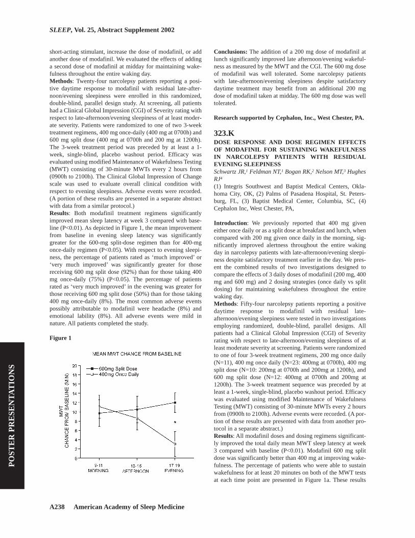
A238 American Academy of Sleep Medicine
PO
STE
R P
RE
SEN
TA
TIO
NS
SLEEP, Vol. 25, Abstract Supplement 2002
short-acting stimulant, increase the dose of modafinil, or addanother dose of modafinil. We evaluated the effects of addinga second dose of modafinil at midday for maintaining wake-fulness throughout the entire waking day.Methods: Twenty-four narcolepsy patients reporting a posi-tive daytime response to modafinil with residual late-after-noon/evening sleepiness were enrolled in this randomized,double-blind, parallel design study. At screening, all patientshad a Clinical Global Impression (CGI) of Severity rating withrespect to late-afternoon/evening sleepiness of at least moder-ate severity. Patients were randomized to one of two 3-weektreatment regimens, 400 mg once-daily (400 mg at 0700h) and600 mg split dose (400 mg at 0700h and 200 mg at 1200h).The 3-week treatment period was preceded by at least a 1-week, single-blind, placebo washout period. Efficacy wasevaluated using modified Maintenance of Wakefulness Testing(MWT) consisting of 30-minute MWTs every 2 hours from(0900h to 2100h). The Clinical Global Impression of Changescale was used to evaluate overall clinical condition withrespect to evening sleepiness. Adverse events were recorded.(A portion of these results are presented in a separate abstractwith data from a similar protocol.)Results: Both modafinil treatment regimens significantlyimproved mean sleep latency at week 3 compared with base-line (P<0.01). As depicted in Figure 1, the mean improvementfrom baseline in evening sleep latency was significantlygreater for the 600-mg split-dose regimen than for 400-mgonce-daily regimen (P<0.05). With respect to evening sleepi-ness, the percentage of patients rated as ‘much improved’ or‘very much improved’ was significantly greater for thosereceiving 600 mg split dose (92%) than for those taking 400mg once-daily (75%) (P<0.05). The percentage of patientsrated as ‘very much improved’ in the evening was greater forthose receiving 600 mg split dose (50%) than for those taking400 mg once-daily (8%). The most common adverse eventspossibly attributable to modafinil were headache (8%) andemotional lability (8%). All adverse events were mild innature. All patients completed the study.
Figure 1
Conclusions: The addition of a 200 mg dose of modafinil atlunch significantly improved late afternoon/evening wakeful-ness as measured by the MWT and the CGI. The 600 mg doseof modafinil was well tolerated. Some narcolepsy patientswith late-afternoon/evening sleepiness despite satisfactorydaytime treatment may benefit from an additional 200 mgdose of modafinil taken at midday. The 600 mg dose was welltolerated.
Research supported by Cephalon, Inc., West Chester, PA.
323.KDOSE RESPONSE AND DOSE REGIMEN EFFECTSOF MODAFINIL FOR SUSTAINING WAKEFULNESSIN NARCOLEPSY PATIENTS WITH RESIDUALEVENING SLEEPINESS Schwartz JR,1 Feldman NT,1 Bogan RK,2 Nelson MT,3 HughesRJ4
(1) Integris Southwest and Baptist Medical Centers, Okla-homa City, OK, (2) Palms of Pasadena Hospital, St. Peters-burg, FL, (3) Baptist Medical Center, Columbia, SC, (4)Cephalon Inc, West Chester, PA,
Introduction: We previously reported that 400 mg giveneither once daily or as a split dose at breakfast and lunch, whencompared with 200 mg given once daily in the morning, sig-nificantly improved alertness throughout the entire wakingday in narcolepsy patients with late-afternoon/evening sleepi-ness despite satisfactory treatment earlier in the day. We pres-ent the combined results of two investigations designed tocompare the effects of 3 daily doses of modafinil (200 mg, 400mg and 600 mg) and 2 dosing strategies (once daily vs splitdosing) for maintaining wakefulness throughout the entirewaking day.Methods: Fifty-four narcolepsy patients reporting a positivedaytime response to modafinil with residual late-afternoon/evening sleepiness were tested in two investigationsemploying randomized, double-blind, parallel designs. Allpatients had a Clinical Global Impression (CGI) of Severityrating with respect to late-afternoon/evening sleepiness of atleast moderate severity at screening. Patients were randomizedto one of four 3-week treatment regimens, 200 mg once daily(N=11), 400 mg once daily (N=23: 400mg at 0700h), 400 mgsplit dose (N=10: 200mg at 0700h and 200mg at 1200h), and600 mg split dose (N=12: 400mg at 0700h and 200mg at1200h). The 3-week treatment sequence was preceded by atleast a 1-week, single-blind, placebo washout period. Efficacywas evaluated using modified Maintenance of WakefulnessTesting (MWT) consisting of 30-minute MWTs every 2 hoursfrom (0900h to 2100h). Adverse events were recorded. (A por-tion of these results are presented with data from another pro-tocol in a separate abstract.)Results: All modafinil doses and dosing regimens significant-ly improved the total daily mean MWT sleep latency at week3 compared with baseline (P<0.01). Modafinil 600 mg splitdose was significantly better than 400 mg at improving wake-fulness. The percentage of patients who were able to sustainwakefulness for at least 20 minutes on both of the MWT testsat each time point are presented in Figure 1a. These results
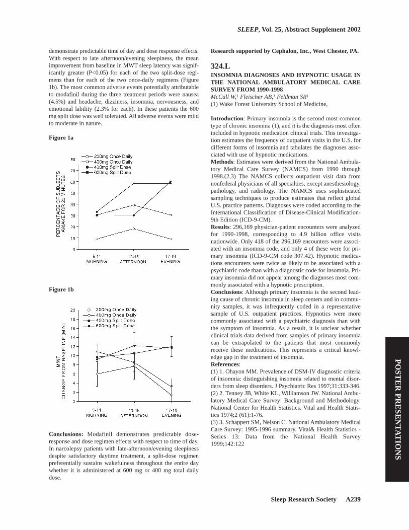
Sleep Research Society A239
PO
STE
R P
RE
SEN
TA
TIO
NS
SLEEP, Vol. 25, Abstract Supplement 2002
demonstrate predictable time of day and dose response effects.With respect to late afternoon/evening sleepiness, the meanimprovement from baseline in MWT sleep latency was signif-icantly greater (P<0.05) for each of the two split-dose regi-mens than for each of the two once-daily regimens (Figure1b). The most common adverse events potentially attributableto modafinil during the three treatment periods were nausea(4.5%) and headache, dizziness, insomnia, nervousness, andemotional lability (2.3% for each). In these patients the 600mg split dose was well tolerated. All adverse events were mildto moderate in nature.
Figure 1a
Figure 1b
Conclusions: Modafinil demonstrates predictable dose-response and dose regimen effects with respect to time of day.In narcolepsy patients with late-afternoon/evening sleepinessdespite satisfactory daytime treatment, a split-dose regimenpreferentially sustains wakefulness throughout the entire daywhether it is administered at 600 mg or 400 mg total dailydose.
Research supported by Cephalon, Inc., West Chester, PA.
324.LINSOMNIA DIAGNOSES AND HYPNOTIC USAGE INTHE NATIONAL AMBULATORY MEDICAL CARESURVEY FROM 1990-1998McCall W,1 Fleischer AB,1 Feldman SR1
(1) Wake Forest University School of Medicine,
Introduction: Primary insomnia is the second most commontype of chronic insomnia (1), and it is the diagnosis most oftenincluded in hypnotic medication clinical trials. This investiga-tion estimates the frequency of outpatient visits in the U.S. fordifferent forms of insomnia and tabulates the diagnoses asso-ciated with use of hypnotic medications.Methods: Estimates were derived from the National Ambula-tory Medical Care Survey (NAMCS) from 1990 through1998.(2,3) The NAMCS collects outpatient visit data fromnonfederal physicians of all specialties, except anesthesiology,pathology, and radiology. The NAMCS uses sophisticatedsampling techniques to produce estimates that reflect globalU.S. practice patterns. Diagnoses were coded according to theInternational Classification of Disease-Clinical Modification-9th Edition (ICD-9-CM).Results: 296,169 physician-patient encounters were analyzedfor 1990-1998, corresponding to 4.9 billion office visitsnationwide. Only 418 of the 296,169 encounters were associ-ated with an insomnia code, and only 4 of these were for pri-mary insomnia (ICD-9-CM code 307.42). Hypnotic medica-tions encounters were twice as likely to be associated with apsychiatric code than with a diagnostic code for insomnia. Pri-mary insomnia did not appear among the diagnoses most com-monly associated with a hypnotic prescription.Conclusions: Although primary insomnia is the second lead-ing cause of chronic insomnia in sleep centers and in commu-nity samples, it was infrequently coded in a representativesample of U.S. outpatient practices. Hypnotics were morecommonly associated with a psychiatric diagnosis than withthe symptom of insomnia. As a result, it is unclear whetherclinical trials data derived from samples of primary insomniacan be extrapolated to the patients that most commonlyreceive these medications. This represents a critical knowl-edge gap in the treatment of insomnia.References: (1) 1. Ohayon MM. Prevalence of DSM-IV diagnostic criteriaof insomnia: distinguishing insomnia related to mental disor-ders from sleep disorders. J Psychiatric Res 1997;31:333-346.(2) 2. Tenney JB, White KL, Williamson JW. National Ambu-latory Medical Care Survey: Background and Methodology.National Center for Health Statistics. Vital and Health Statis-tics 1974;2 (61):1-76.(3) 3. Schappert SM, Nelson C. National Ambulatory MedicalCare Survey: 1995-1996 summary. Vital& Health Statistics -Series 13: Data from the National Health Survey1999;142:122
A240 American Academy of Sleep Medicine
PO
STE
R P
RE
SEN
TA
TIO
NS
SLEEP, Vol. 25, Abstract Supplement 2002
325.LA META ANALYSIS OF SLEEP CHANGES ASSOCIAT-ED WITH PLACEBO IN HYPNOTIC CLINICAL TRI-ALS:METHODOLOGIC AND ETHICAL IMPLICA-TIONSMcCall W,1 D’Agostino R,1 Dunn A,1 Sevick MA1
(1) Wake Forest University School of Medicine, (2) Universi-ty of Pittsburgh School of Nursing,
Introduction: These effects associated with placebo (EAP)have been incompletely described in clinical trials of insomniatreatment. We conducted a meta-analysis of insomnia medica-tion trials for the purpose of estimating the magnitude of sleepEAP after two weeks of placebo administration.Methods: We performed a literature review using Medline forthe period of 1966 through 2000 to gather data for the meta-analysis (1). The subject heading of insomnia restricted to thesubheading of drug therapy was crossed against the results ofa search on the subjects heading placebo and text word place-bo. We selected only papers that examined primary insomnia,incorporating both placebo and active medication therapies ina randomized, double-blind, parallel-group design. Werequired that treatment results be reported for 1, 2, 3, or 4weeks of treatment, and that the outcome variable be reportedin hours/minutes.Results: Only five papers satisfied our requirements for eligi-bility, comprising 213 patients receiving placebo for a two-week interval. Subjective sleep latency demonstrated a signif-icant reduction (mean + s.e.) of 13.1 + 2.0 minutes (95% CI9.2, 17.0) for the placebo group after combining the dataacross studies. Subjective total sleep time demonstrated a sig-nificant increase of 13.5 + 3.2 minutes (95% CI 7.0, 19.6).Polysomnographic sleep latency demonstrated a non-signifi-cant reduction of 2.5 + 4.3 minutes (95% CI -5.9, 10.9).Conclusions: These results are consistent with the generalfinding in the insomnia literature that the magnitude of sub-jective complaints and subjective response to treatment aregreater than PSG measurements. Our present findings havepractical and perhaps ethical ramifications. If we had found noEAP, then it could be argued that an ‘equivalence design’could be employed for testing the efficacy of new medicationsagainst a proven standard treatment in primary insomnia,without a placebo control. Instead, the presence of EAP in pri-mary insomnia supports the idea that new medications be con-trasted against placebo. The strongest arguments againstplacebo are ethical ones. The World Health Organiza-tion’s1964 Declaration of Helsinki effectively prohibits theethical use of placebos in clinical trials when a proven thera-peutic method exists. The ethical dilemma would seem tosolved by appealing to the principal of freely-given informedconsent, allowing patients to make their own choices. Yet thestrongest antagonists of placebo use in clinical trials havequestioned whether freely-given and fully-informed consentever exists. However, if placebo-controlled studies are aban-donned in favor of an ‘equivalence design’, the effects sizewill likely be smaller, requiring the enrollment of more studyparticipants and potentially greater exposure to an ineffectivenew treatment.
References: (1) Hedges L, Olkin I. Statistical Methods for Meta-Analysis.Orlando: Academic Press, Inc., 1985
326.LSLEEP DYNAMIC THERAPY FOR POSTTRAUMATICINSOMNIAKrakow BJ,1 Melendrez D,1 Johnston L,1 Clark JO,1 SantanaE,1 Warner TD,1 Hollifield M,2 Sisley B,2 Lee S1
(1) Sleep & Human Health Institute, (2) U. of New MexicoSchool of Medicine, Department of Psychiatry,
Introduction: Posttraumatic insomnia is a complex disorderoften comprising the triad of psychiatric distress, psychophys-iological conditioning, and physiological sleep fragmentation[e.g. sleep- disordered breathing (SDB)](1). We have devel-oped an integrative, evidence-based, Sleep Dynamic Therapy(SDT) program to address “complex insomnia.” SDT aims toincrease awareness of the multifactorial nature of sleep distur-bance in the aftermath of trauma by emphasizing six basiccomponents: (1) sleep quality self-assessment; (2) cognitive-behavioral therapy (CBT); (3) sleep hygiene; (4) sleep-relatedemotional processing; (5) imagery rehearsal therapy (IRT);and (6) sleep physiological self-assessment. The core thera-peutic aim of SDT is to educate the patient on the value of arigorous and candid assessment of sleep quality through whichone might estimate the degree that distress, conditioning, andphysiological sleep disruption cause or exacerbate insomnia.Because trauma survivors are often inculcated with the beliefthat distress is the primary cause of insomnia, SDT starts withsleep quality, CBT, and sleep hygiene principles to quicklydemonstrate that insomnia responds to standard sleep medi-cine therapies regardless of distress symptoms. Then, sleep-related emotional processing examines how anxiety anddepression exacerbate insomnia, and IRT is taught to decreasenightmares. In the last session, sleep physiological self-assess-ment prepares the patient for a night of polysomnography toassess sleep breathing or movement disorders.Methods: In a pilot study, SDT was administered to a group ofdisaster survivors (n=66), evacuated during the Cerro GrandeFire in Los Alamos, NM (May, 2000). SDT was delivered insix weekly sessions to the entire group (on average, 8 patientsattended makeup sessions each week). The Insomnia SeverityIndex (ISI, range 0 to 28) assessed insomnia at 8 points:intake; immediate pre-treatment (7.5 weeks after intake);weeks 2, 3, 4, and 5 of treatment; posttreatment; and 12-weeksfrom treatment initiation. Repeated measures ANOVA wasconducted and Cohen’s d effect sizes were calculated.Results: ISI showed a small decrease from intake to pre-treat-ment (d = .32) indicative of spontaneous recovery. Additionalsubstantive improvement occurred at posttreatment (d = .62)(F(7,59) = 11.97, P = .0001) with maintenance of change at12-week follow-up. Forty-nine patients improved, 11 wors-ened and 6 reported no change in insomnia. Forty participants’post-treatment ISI scores fell below non-clinical levels (scores≤ 10)(2).Conclusions: SDT was successfully delivered to a singlegroup of disaster survivors. In the early post-treatment follow-up, there was a highly significant, medium-sized decrease in
Sleep Research Society A241
PO
STE
R P
RE
SEN
TA
TIO
NS
SLEEP, Vol. 25, Abstract Supplement 2002
insomnia severity. Additional treatment effects among patientspursuing SDB treatment with CPAP or oral appliances will beforthcoming. Although this study was uncontrolled, sponta-neous recovery was small during a lengthy pre-treatmentassessment period. Moreover, all the techniques comprisingSDT have been found effective in controlled studies. There-fore, it is likely that SDT played an important role in thechanges reported by these patients. Given the current need toassist disaster and other trauma survivors, SDT may be a verypragmatic approach for the treatment of posttraumatic insom-nia in large groups of patients.References: (1) Krakow B, Melendrez D, Pedersen B et al: Complexinsomnia: insomnia and sleep-disordered breathing in a con-secutive series of crime victims with nightmares and PTSD.Biological Psychiatry, 2001;49:948-953.(2) Bastien CH, Vallieres A, Morin CM: Validation of theinsomnia severity index as an outcome measure for insomniaresearch. Sleep Medicine, 2001;2:297-307.
Research supported by RAPID (Rapid Assessment Post-Impact Disaster) grant from NIMH (MH 63477)
327.LBRAIN MUSIC THERAPY FOR TREATMENT OFINSOMNIA AND ANXIETYKayumov L,1 Soare K,1 Serbine O,1 Kotlyar B,2 Simkhovich Z,1
Goldstein Y,1 Gavrilov D,3 Levin Y,3 Shapiro CM3
(1) Sleep Research Laboratory, Department of Psychiatry,University of Toronto, Canada,, (2) University at Buffalo,USA,, (3) Sleep Disorders Center, I. M. Sechenov MoscowMedical Academy, Russia,
Introduction: The close relationship between insomnia andanxiety is well established. Anxious patients have difficultiesmaintaining sleep, they spend less time in deep sleep and theirsleep is more fragmented than that of normals. Traditionalapproaches have emphasized pharmacological treatment ofinsomnia. Benzodiazepines have become the most widely pre-scribed of all pharmaceuticals. Concern has been expressedhowever about their potential to cause dependency associatedwith self-dosing management. A non-pharmacological method– “brain music therapy” has been recently developed for treat-ment of some psychosomatic symptoms. This method allowsestablishing the most effective rhythmic and tonal parameterscreating meditative conditions in patients by influencing thebioelectrical brain activity in the process of music therapydepending on the individual EEG. Subsequently EEG patternsare converted into unique music recorded on a personalizedcompact disc with listening instructions catered to each indi-vidual. Brain music therapy because of its more favorableside-effect profile may represent a possible alternative fortherapeutic management of insomnia and anxiety. The purposeof the present study was to assess the effectiveness of brainmusic therapy for treatment of insomnia in anxious patientsusing objective actigraphic measures and psychometric test-ing.Methods: Eighteen volunteers who had complained of symp-toms of insomnia of at least two years duration and who had
scored above 50 on the Zung Self Rating Anxiety Scale wererecruited for participation in the study. Patients were dividedinto two groups on a double-blind randomized basis. Experi-mental group I comprised ten insomniacs (7 females and 3males, aged 41.6.0±5.8) who were provided with their authen-tic “brain music” (computerized or composed during neuro-feedback session). Placebo group II consisted of eight patients(5 females and 3 males, aged 42.8±7.8) who received compactdisks with brain music of a different subject. The duration ofthe treatment, which entailed listening to the music on a dailybasis, was four weeks. Athens Insomnia Scale and actigraphywere used for assessment of subjective and objective qualityof sleep. Forty eight-hour actigraphic recordings were per-formed before and after 4 weeks of brain music therapy. Aver-age sleep onset latency (SOL), total sleep time (TST), andamount of intervening wakefulness were determined. Affec-tive status of the patients was controlled by using the CES-Depression Scale. Participants from both groups had slightlyelevated scores (19.6±3.8 and 20.1±5.4 respectively; p>.05).Statistical analysis was performed using the independent sam-ples’ t-test in the SPSS statistical software package with sig-nificance set at p<0.05. Bonferroni correction was used formultiple variable analysis.Results: Both authentic and placebo brain music reduced anx-iety scores with more pronounced effects observed in theexperimental group (58.1±2.8 vs.31±4.6 and 60±5.6 vs. 46.5±6.1 respectively, p<.01). There was a dramatic improvementin sleep quality as judged by the Athens Insomnia Scale(p<.001). However there was no significant differencebetween the effects of authentic and placebo brain music.Interestingly some actigraphic parameters characterizinginsomnia in anxious patients were found to be significantlyimproved only in the experimental group of patients who wereusing authentic brain music therapy. Amount of interveningwakefulness was significantly less following brain music ther-apy (p=.02). The patients had a significant increase in TST(p=.004). SOL remained unchanged before and after brainmusic treatment (p>.05).Conclusions: In this study a 4 week regimen of brain musictherapy was shown to be of value in reducing symptoms ofanxiety and insomnia as evidenced by psychometric testing.Objective actigraphic measures of insomnia have beenimproved in the group of anxious insomniacs treated withendogenously generated brain music. Brain music therapy is auseful alternative to pharmaceutical therapy for treating theseconditions.
328.LPRELIMINARY RESULTS OF A PILOT STUDY EVAL-UATING YOGA AS A TREATMENT FOR INSOMNIAKhalsa SS,1 Winkelman JW1
(1) Brigham and Women’s Hospital, Harvard Medical School,
Introduction: There is good evidence that cognitive and/orphysiological arousal, including sustained elevated sympatho-vagal balance, is a characteristic of chronic primary insomnia.Furthermore, relaxation treatments such as progressive relax-ation and meditation, which reduce arousal, have been foundto be effective insomnia treatments. Numerous research stud-
A242 American Academy of Sleep Medicine
PO
STE
R P
RE
SEN
TA
TIO
NS
SLEEP, Vol. 25, Abstract Supplement 2002
ies have documented the effectiveness of yoga in reducingautonomic arousal and in the treatment of specific medical dis-orders. Although yoga has been clinically recommended forthe treatment of insomnia, its effectiveness in treating insom-nia has not been thoroughly tested (1,2). The aim of this pilotstudy is to evaluate the feasibility of applying a daily yogatreatment for chronic insomnia of various etiologies.Methods: Individuals with an insomnia complaint wererecruited primarily from physician referrals. Followinginformed consent and a sleep history interview, study partici-pants kept daily sleep-wake diaries. Following a 2-week base-line evaluation, participants were individually taught a 30-minute sequence of Kundalini yoga (as taught by Yogi Bhajan)including breathing exercises, static postures, and meditation,which they practiced daily at home for 8 weeks. One in-personfollow-up meeting confirmed proper practice of the exerciseswith additional contact by telephone. For quantifying the sub-jective degree of insomnia, daily values of total wake time,total sleep time, sleep efficiency, and subjective sleep qualityon a scale of 1 to 5 were averaged over the 2-week baselineperiod and over the last 2 weeks of the treatment.Results: To date, 10 female participants aged 33 to 64 havecompleted the 10-week protocol. Initially, all of the partici-pants complained of sleep maintenance insomnia, and 7 ofthem also reported sleep onset insomnia. Only one participanthad evidence of a sleep disorder other than insomnia (RLS),three had some evidence of anxiety disorder and one hadmajor depression. Two participants reported occasional hyp-notic use during the protocol. Qualitative analysis of subjec-tive sleep diary characteristics over the course of the treatmentrevealed gradual improvements in the degree of insomnia anddecreases in day-to-day variability for 8 of the 10 participants.Averaged values for all 10 participants comparing pre-treat-ment baseline and end-treatment periods are plotted with stan-dard errors, and show a decrease of 0.9 hr in total wake time(p = 0.02, t-test), an increase of 0.7 hr in total sleep time (p =0.07), an increase of 10% in sleep efficiency (p = 0.02), and anincrease of 0.4 on the sleep quality scale (p = 0.12). In gener-al, most of the participants exhibited a high degree of compli-ance in performing the daily treatment and reported no diffi-culties in executing the exercises.
Figure 1
Conclusions: The yoga treatment applied was well toleratedand the majority of participants reported some degree of ben-efit. These preliminary data suggest that the effectiveness of
this treatment is worthy of further evaluation with a largersample size in a randomized controlled study incorporatingadditional objective outcome measures.References: (1) Joshi KS. Yogic treatment of insomnia: An experimentalstudy. Yoga Mimamsa 1992;30:24-26.(2) Koch U, Volk S, Heidenreich T, Pflug B. Yoga treatment inpsychophysiological insomnia. J Sleep Res1998;7(Suppl.2):137.
Research supported by a Research Scientist DevelopmentAward from the National Center for Complementary andAlternative Medicine (K01AT00066), NIH
329.LSCREENING FOR SLEEP-DISORDERED BREATHINGIN COMPLEX INSOMNIA PATIENTSMelendrez DC,1 Krakow BJ,1 Clark JO1
(1) Sleep & Human Health Institute,
Introduction: Polysomnography (PSG) is not routinely indi-cated for insomnia patients. However, the presumed boundarybetween insomnia and sleep-disordered breathing (SDB) hasbecome less distinct (1,2). Accordingly, it may prove useful toscreen appropriate insomnia patients for SDB prior to under-going PSG. The Autoset II Plus (AS) (ResMed, Australia) is anambulatory device that performs a diagnostic sleep study byautomatically analyzing an airflow tracing from a nasal can-nula. Previous studies have validated the AS apnea/hypopneaindex (AHI) for obstructive sleep apnea (OSA)(3). Addition-ally, the AS measures flattening of the flow-time curve that isdisplayed on an arbitrary scale of 0 to .3 in which a cut-off (≤.15) implies flow limitation (i.e. RERAs). The flattening index(FI) is the percentage below the cut-off. Anecdotally, a FI ≥20% appears consistent with UARS when the AHI is low.Therefore, we hypothesize that the AS might successfullyscreen for UARS in diagnostic mode using the FI. As a pre-liminary step prior to a formal validation of the AS, we col-lected data on a series of patients who underwent PSG afterhaving screened positive with an elevated FI.Methods: Thirty-five consecutive crime victims seeking treat-ment for insomnia who had an AHI ≤ 10 and a FI ≥ 20 wereevaluated for SDB with PSG at various sleep labs in Albu-querque, NM. Some labs utilized thermistor/thermocoupleswhile others used airflow pressure transducers to assess sleep-breathing disturbance. All diagnoses were based on the indi-vidual sleep specialist’s interpretation of the PSG. Eightpatients in the sample were diagnosed by the second author.Results: SDB was diagnosed in 32 of 35 (94%) patients. Ofthose with SDB, 50% had OSA, 47% had UARS, and one bor-derline UARS. Of the OSA patients, 69% technically qualifiedas OSA (i.e. AHI ≥ 5) but had predominantly UARS typeevents. An additional 12 patients, not meeting this study crite-ria (AHI < 10 and a FI < 20), reported clinical symptoms high-ly suspicious for SDB. All 12 were referred for PSG and diag-nosed with SDB (2 OSA and 10 UARS).Conclusions: AS was evaluated and found to be useful as ascreening tool for SDB in crime victims with insomnia. How-ever, there are limitations with respect to false negative AS
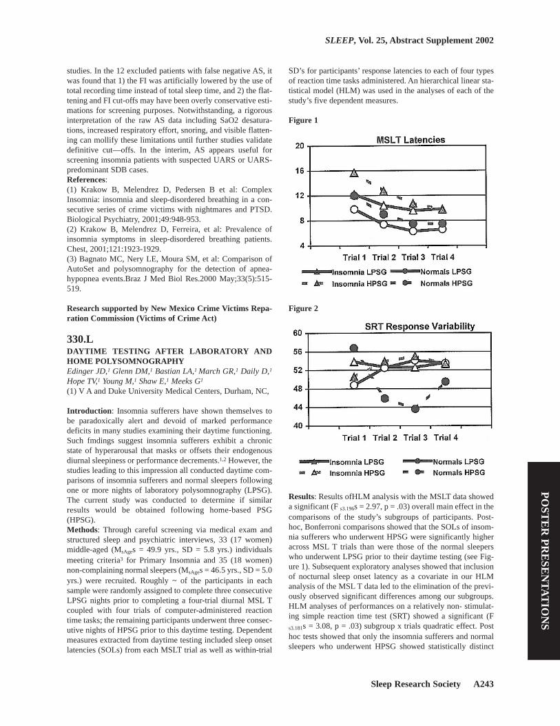
Sleep Research Society A243
PO
STE
R P
RE
SEN
TA
TIO
NS
SLEEP, Vol. 25, Abstract Supplement 2002
studies. In the 12 excluded patients with false negative AS, itwas found that 1) the FI was artificially lowered by the use oftotal recording time instead of total sleep time, and 2) the flat-tening and FI cut-offs may have been overly conservative esti-mations for screening purposes. Notwithstanding, a rigorousinterpretation of the raw AS data including SaO2 desatura-tions, increased respiratory effort, snoring, and visible flatten-ing can mollify these limitations until further studies validatedefinitive cut—offs. In the interim, AS appears useful forscreening insomnia patients with suspected UARS or UARS-predominant SDB cases.References: (1) Krakow B, Melendrez D, Pedersen B et al: ComplexInsomnia: insomnia and sleep-disordered breathing in a con-secutive series of crime victims with nightmares and PTSD.Biological Psychiatry, 2001;49:948-953.(2) Krakow B, Melendrez D, Ferreira, et al: Prevalence ofinsomnia symptoms in sleep-disordered breathing patients.Chest, 2001;121:1923-1929. (3) Bagnato MC, Nery LE, Moura SM, et al: Comparison ofAutoSet and polysomnography for the detection of apnea-hypopnea events.Braz J Med Biol Res.2000 May;33(5):515-519.
Research supported by New Mexico Crime Victims Repa-ration Commission (Victims of Crime Act)
330.LDAYTIME TESTING AFTER LABORATORY ANDHOME POLYSOMNOGRAPHYEdinger JD,1 Glenn DM,1 Bastian LA,1 March GR,1 Daily D,1
Hope TV,1 Young M,1 Shaw E,1 Meeks G1
(1) V A and Duke University Medical Centers, Durham, NC,
Introduction: Insomnia sufferers have shown themselves tobe paradoxically alert and devoid of marked performancedeficits in many studies examining their daytime functioning.Such fmdings suggest insomnia sufferers exhibit a chronicstate of hyperarousal that masks or offsets their endogenousdiurnal sleepiness or performance decrements.1,2 However, thestudies leading to this impression all conducted daytime com-parisons of insomnia sufferers and normal sleepers followingone or more nights of laboratory polysomnography (LPSG).The current study was conducted to determine if similarresults would be obtained following home-based PSG(HPSG).Methods: Through careful screening via medical exam andstructured sleep and psychiatric interviews, 33 (17 women)middle-aged (MsAges = 49.9 yrs., SD = 5.8 yrs.) individualsmeeting criteria3 for Primary Insomnia and 35 (18 women)non-complaining normal sleepers (MsAges = 46.5 yrs., SD = 5.0yrs.) were recruited. Roughly ~ of the participants in eachsample were randomly assigned to complete three consecutiveLPSG nights prior to completing a four-trial diurnal MSL Tcoupled with four trials of computer-administered reactiontime tasks; the remaining participants underwent three consec-utive nights of HPSG prior to this daytime testing. Dependentmeasures extracted from daytime testing included sleep onsetlatencies (SOLs) from each MSLT trial as well as within-trial
SD’s for participants’ response latencies to each of four typesof reaction time tasks administered. An hierarchical linear sta-tistical model (HLM) was used in the analyses of each of thestudy’s five dependent measures.
Figure 1
Figure 2
Results: Results ofHLM analysis with the MSLT data showeda significant (F s3.196s = 2.97, p = .03) overall main effect in thecomparisons of the study’s subgroups of participants. Post-hoc, Bonferroni comparisons showed that the SOLs of insom-nia sufferers who underwent HPSG were significantly higheracross MSL T trials than were those of the normal sleeperswho underwent LPSG prior to their daytime testing (see Fig-ure 1). Subsequent exploratory analyses showed that inclusionof nocturnal sleep onset latency as a covariate in our HLManalysis of the MSL T data led to the elimination of the previ-ously observed significant differences among our subgroups.HLM analyses of performances on a relatively non- stimulat-ing simple reaction time test (SRT) showed a significant (Fs3.181s = 3.08, p = .03) subgroup x trials quadratic effect. Posthoc tests showed that only the insomnia sufferers and normalsleepers who underwent HPSG showed statistically distinct
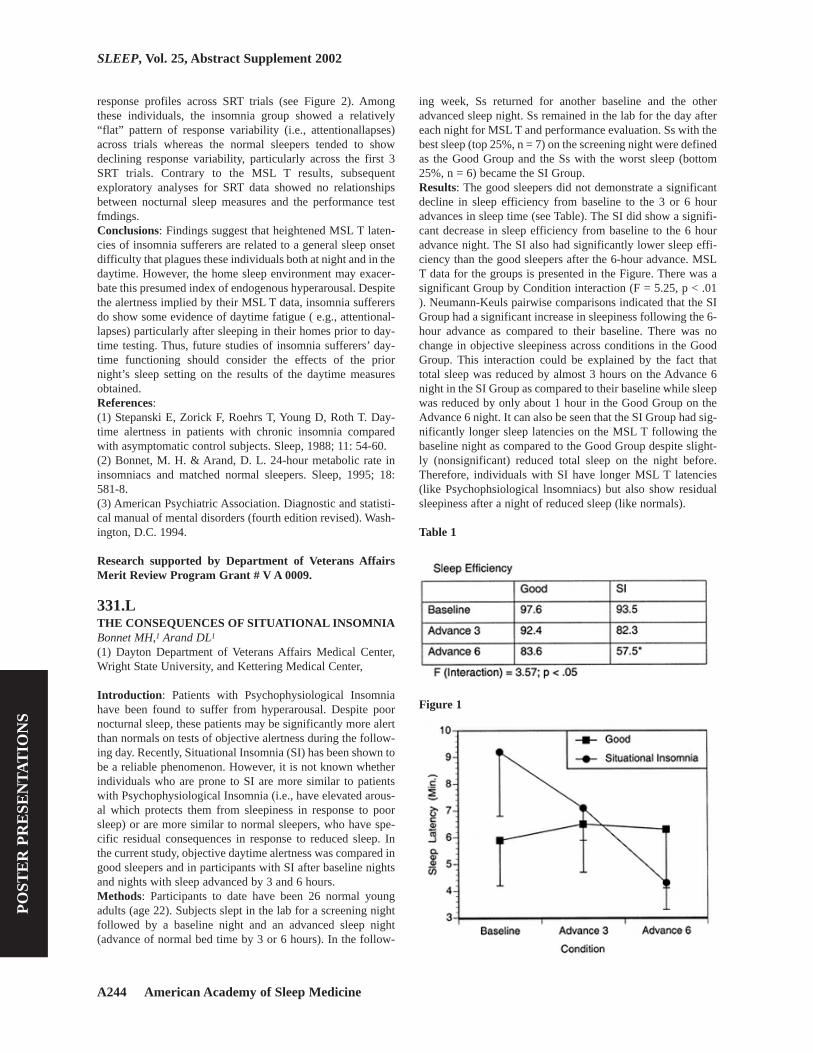
A244 American Academy of Sleep Medicine
PO
STE
R P
RE
SEN
TA
TIO
NS
SLEEP, Vol. 25, Abstract Supplement 2002
response profiles across SRT trials (see Figure 2). Amongthese individuals, the insomnia group showed a relatively“flat” pattern of response variability (i.e., attentionallapses)across trials whereas the normal sleepers tended to showdeclining response variability, particularly across the first 3SRT trials. Contrary to the MSL T results, subsequentexploratory analyses for SRT data showed no relationshipsbetween nocturnal sleep measures and the performance testfmdings.Conclusions: Findings suggest that heightened MSL T laten-cies of insomnia sufferers are related to a general sleep onsetdifficulty that plagues these individuals both at night and in thedaytime. However, the home sleep environment may exacer-bate this presumed index of endogenous hyperarousal. Despitethe alertness implied by their MSL T data, insomnia sufferersdo show some evidence of daytime fatigue ( e.g., attentional-lapses) particularly after sleeping in their homes prior to day-time testing. Thus, future studies of insomnia sufferers’ day-time functioning should consider the effects of the priornight’s sleep setting on the results of the daytime measuresobtained.References: (1) Stepanski E, Zorick F, Roehrs T, Young D, Roth T. Day-time alertness in patients with chronic insomnia comparedwith asymptomatic control subjects. Sleep, 1988; 11: 54-60. (2) Bonnet, M. H. & Arand, D. L. 24-hour metabolic rate ininsomniacs and matched normal sleepers. Sleep, 1995; 18:581-8. (3) American Psychiatric Association. Diagnostic and statisti-cal manual of mental disorders (fourth edition revised). Wash-ington, D.C. 1994.
Research supported by Department of Veterans AffairsMerit Review Program Grant # V A 0009.
331.LTHE CONSEQUENCES OF SITUATIONAL INSOMNIABonnet MH,1 Arand DL1
(1) Dayton Department of Veterans Affairs Medical Center,Wright State University, and Kettering Medical Center,
Introduction: Patients with Psychophysiological Insomniahave been found to suffer from hyperarousal. Despite poornocturnal sleep, these patients may be significantly more alertthan normals on tests of objective alertness during the follow-ing day. Recently, Situational Insomnia (SI) has been shown tobe a reliable phenomenon. However, it is not known whetherindividuals who are prone to SI are more similar to patientswith Psychophysiological Insomnia (i.e., have elevated arous-al which protects them from sleepiness in response to poorsleep) or are more similar to normal sleepers, who have spe-cific residual consequences in response to reduced sleep. Inthe current study, objective daytime alertness was compared ingood sleepers and in participants with SI after baseline nightsand nights with sleep advanced by 3 and 6 hours.Methods: Participants to date have been 26 normal youngadults (age 22). Subjects slept in the lab for a screening nightfollowed by a baseline night and an advanced sleep night(advance of normal bed time by 3 or 6 hours). In the follow-
ing week, Ss returned for another baseline and the otheradvanced sleep night. Ss remained in the lab for the day aftereach night for MSL T and performance evaluation. Ss with thebest sleep (top 25%, n = 7) on the screening night were definedas the Good Group and the Ss with the worst sleep (bottom25%, n = 6) became the SI Group. Results: The good sleepers did not demonstrate a significantdecline in sleep efficiency from baseline to the 3 or 6 houradvances in sleep time (see Table). The SI did show a signifi-cant decrease in sleep efficiency from baseline to the 6 houradvance night. The SI also had significantly lower sleep effi-ciency than the good sleepers after the 6-hour advance. MSLT data for the groups is presented in the Figure. There was asignificant Group by Condition interaction (F = 5.25, p < .01). Neumann-Keuls pairwise comparisons indicated that the SIGroup had a significant increase in sleepiness following the 6-hour advance as compared to their baseline. There was nochange in objective sleepiness across conditions in the GoodGroup. This interaction could be explained by the fact thattotal sleep was reduced by almost 3 hours on the Advance 6night in the SI Group as compared to their baseline while sleepwas reduced by only about 1 hour in the Good Group on theAdvance 6 night. It can also be seen that the SI Group had sig-nificantly longer sleep latencies on the MSL T following thebaseline night as compared to the Good Group despite slight-ly (nonsignificant) reduced total sleep on the night before.Therefore, individuals with SI have longer MSL T latencies(like Psychophsiological lnsomniacs) but also show residualsleepiness after a night of reduced sleep (like normals).
Table 1
Figure 1
Sleep Research Society A245
PO
STE
R P
RE
SEN
TA
TIO
NS
SLEEP, Vol. 25, Abstract Supplement 2002
Conclusions: These data indicate that individuals who have SImay display greatly reduced sleep time in a stressful situationthat can result in significantly increased sleepiness after as lit-tle as one night.
Research supported by a Merit Review Grant from theDepartment of Veterans Affairs, Wright State UniversitySchool of Medicine, and the Sleep-Wake DisordersResearch Institute.
332.LMENOPAUSAL METHYLTESTOSTERONE TREAT-MENT INDUCES CHANGES IN SUBJECTIVE SLEEPTHAT CORRELATE WITH CHANGES IN MOTORSPEED BUT NOT IN VIGILANCERegestein QR,1 Friebely J,1 Shifren JL,1 Schiff I2
(1) Brigham & Women’s Hospital, (2) Massachusetts GeneralHospital,
Introduction: Healthy menopausal women on hormonereplacement therapy (N = 34) were recruited to test the effectsof testosterone replacement on affective measures and cogni-tive function. During two consecutive, randomized, double-blind, 8-week treatment periods, subjects took identically-appearing pills daily. The pills contained esterified estrogen0.625 mg in both periods. During one of the periods, it alsocontained methyl-testo-sterone 1.25 mg. In a post-hoc analy-sis, we tested whether subjective sleep quality would correlatewith objective measures of daytime cognitive function.Methods: Subjective Sleep was measured by an index com-pounded of z-transformed scores for St. Mary’s Sleep Ques-tionnaire items (depth, satisfaction, how well one slept, clear-headedness in morning, number of awakenings, ease of fallingasleep and trouble with early awakening). Daytime cognitivemeasures were taken at the end of both treatment periods.These included measures of complex verbal and associationalfluency, motor speed (finger tapping), and vigilance (continu-ous performance response time, number of responses slowerthan normal [> 432 msec; “lapses”], and response steadiness[standard deviation]) (Neurobehavioral Systems, Atlanta).Analyses were directed at finding whether subjective sleepwas reflected in daytime cognitive measures. Stepwise regres-sion (SAS, Cady, NC) was used to model subjective sleepindex by cognitive measures during the estrogen alone condi-tion, when not on methyltestosterone. Subjective sleepchanges induced by added methyltestosterone were modeledby changes in cognitive measures between treatment condi-tions. To control for possible effects of independent co-vari-ants, oöphorectomy status, age and order of treatment wereentered into the analyses. Results: No significant relationship was found between self-reported sleep quality and cognitive variables in the estrogenalone condition. Testosterone replacement induced a trendtowards improved sleep quality (p = .06). Change in subjectivesleep index after added methyltestosterone treatment correlat-ed with change in cognitive measures (Model F = 7.2; p = .01).Of the three cognitive functions measured, motor speedchanges accounted for 18% of the variance in sleep qualitychanges, but vigilance changes did not significantly correlate
with subjective sleep index changes.Conclusions: We conclude that in menopausal women treatedwith estrogen alone, subjective sleep does not correlate withcognitive measures. However, methyltestosterone-inducedchanges in subjective sleep may correlate with motor speed.Hypothetically, this might occur because motor speed report-edly correlates with dopaminergic functions (1), which poten-tially affect sleep, and because dopamine may mediate somebehavioral effects of testosterone (2).References: (1) Association of dopamine transporter reduction with psy-chomotor impairment in methamphetamine abusers. VolkowND, Chang L, Wang GJ et al. Am J Psychiatry 2001; 158:377-382(2) Schroeder JP, Packard MG . Role of dopamine receptorsubtypes in the acquisition of a testosterone conditioned placepreference in rats. Neurosci Lett 2000; 282:17-20.
Research supported by an unrestricted grant from SolvayPharmaceuticals
333.LCOMPARISON OF HOME AND LABORATORYACTIGRAPHY IN YOUNG ADULTS WITH AND WITH-OUT PRIMARY INSOMNIAMeans MK,1 Edinger JD,1 Husain AM1,2
(1) VA Medical Center, Durham, NC, (2) Duke UniversityMedical Center, Durham, NC,
Introduction: Recent research indicates that the setting inwhich sleep is measured (e.g., home vs. laboratory) may affectinsomnia sufferers differently than normal sleepers (1) and isthus an important consideration in characterizing the sleep ofinsomnia sufferers. This study investigated setting differencesin a sample of young adult insomnia sufferers using wristactigraphy, an inexpensive and non-obtrusive objective meansof estimating sleep patterns.Methods: Participants were age and gender-matched samplesof young adults with insomnia (n = 22; 10 male, 12 female; Mage = 29) and normal sleepers (n = 31; 14 male, 17 female; Mage = 28). Insomnia sufferers met ICSD diagnostic criteria fora chronic (> 6 months) primary insomnia disorder. Normalsleepers had no history of sleep complaints and failed to meetcriteria for any sleep disorder. All participants were screenedfor the absence of medical and/or psychiatric difficulties thatcould compromise sleep. Participants underwent 3 consecu-tive nights of laboratory PSG and 3 consecutive nights ofhome PSG (in a counterbalanced order) on separate weeks, aspart of a larger research project investigating sleep-settingeffects. On these six study nights (3 laboratory, 3 home) par-ticipants were also monitored with Actiwatch-L (Mini-MitterCo., Inc., Bend OR) devices, the results of which are present-ed herein.Results: The following sleep variables were estimated fromactigraphy: total sleep time (TST), total wake time (TWT),sleep onset latency (SOL), sleep efficiency index (SEI), meansleep bout time (MSBT; average length of blocks of continu-ous sleep), and mean wake bout time (MWBT; average lengthof blocks of continuous wake). Differences were analyzed in 2
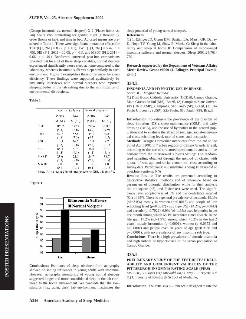
A246 American Academy of Sleep Medicine
PO
STE
R P
RE
SEN
TA
TIO
NS
SLEEP, Vol. 25, Abstract Supplement 2002
(Group: insomnia vs. normal sleepers) X 2 (Place: home vs.lab) ANCOVAs, controlling for gender, night (1 through 3),order (home or lab), and time in bed. Adjusted means are pre-sented in Table 1. There were significant interaction effects forTST (F[1, 261] = 8.77, p < .01), TWT (F[1, 261] = 5.47, p <.05), SEI (F[1, 261] = 10.05, p < .01), and MSBT (F[1, 261] =9.66, p < .01). Bonferroni-corrected post-hoc comparisonsrevealed that for all 4 of these sleep variables, normal sleepersexperienced significantly worse sleep at home compared to thelaboratory, whereas insomnia sufferers slept similarly in eachenvironment. Figure 1 exemplifies these differences for sleepefficiency. These findings were supported qualitatively bypost-study interviews with normal sleepers who reportedsleeping better in the lab setting due to the minimization ofenvironmental distractions.
Table 1
Figure 1
Conclusions: Estimates of sleep obtained from actigraphyshowed no setting influences in young adults with insomnia.However, actigraphy monitoring of young normal sleeperssuggested longer and more consolidated sleep in the lab com-pared to the home environment. We conclude that the low-stimulus (i.e., quiet, dark) lab environment maximizes the
sleep potential of young normal sleepers.References: (1) 1. Edinger JD, Glenn DM, Bastian LA, Marsh GR, DaileyD, Hope TV, Young M, Shaw E, Meeks G: Sleep in the labo-ratory and sleep at home II: Comparisons of middle-agedinsomnia sufferers and normal sleepers. Sleep 2001;24:761-770.
Research supported by the Department of Veterans AffairsMerit Review Grant #0009 (J. Edinger, Principal Investi-gator)
334.LINSOMNIA AND HYPNOTIC USE IN BRAZILSouza JC,1 Magna,1 Reimão2
(1) Dom Bosco Catholic University (UCDB), Campo Grande,Mato Grosso do Sul (MS), Brazil, (2) Campinas State Univer-sity (UNICAMP), Campinas, São Paulo (SP), Brazil, (3) SãoPaulo University (USP), São Paulo, São Paulo (SP), Brazil,
Introduction: To estimate the prevalence of the disorder ofsleep initiation (DSI), sleep maintenance (DSM), and earlyarousing (DEA), and the use of hypnotics in the general pop-ulation and to evaluate the effect of sex, age, social-economi-cal class, schooling level, marital status, and occupation.Methods: Design: Domiciliar interviews from the 3rd to the8th of April 2001 in 7 urban regions of Campo Grande, Brazil,according to the use of structured questionnaire and with theconsent from the interviewed subjects.Setting: The random-ized sampling obtained through the method of cluster withquotas of sex, age and social-economical class according tosurvey data. Participants: 408 inhabitants being 18 years old orover.Interventions: N/AResults: Results: The results are presented according todescriptive statistical methods and of inference based onparameters of binomial distribution, while for their analysisthe qui-square (c2), and Fisher test were used. The signifi-cance level adopted was of 5% and the confidence interval(CI) of 95%. There is a general prevalence of insomnia 19.1%(sd=2.0%) mostly in women (p=0.0015) and people of lowschooling level (p=0.0317) - sub type DSI (14.2%, p=0.0043)and chronic (p=0.7022). 6.9% (sd=1.3%) used hypnotics in thelast month among which 68.1% over three times a week. In thelife span 17.2% (sd=1.9%) among which 70.3% in the last 2years, mostly insomniac (p<0.0001), women (p=0.0372 andp<0.0001) and people over 30 years of age (p=0.0536 andp=0.0091), with no prevalence of any insomnia sub type.Conclusions: There is a high prevalence of chronic insomniaand high indices of hypnotic use in the urban population ofCampo Grande.
335.LPRELIMINARY STUDY OF THE TEST-RETEST RELI-ABILITY AND CONCURRENT VALIDITIES OF THEPITTSBURGH INSOMNIA RATING SCALE (PIRS)Moul DE,1 Pilkonis PA,1 Miewald JM,1 Carey TJ,1 Buysse DJ1
(1) University of Pittsburgh School of Medicine,
Introduction: The PIRS is a 65-item scale designed to rate the

Sleep Research Society A247
PO
STE
R P
RE
SEN
TA
TIO
NS
SLEEP, Vol. 25, Abstract Supplement 2002
severity of insomnia in clinical trials. Subjects rate items ask-ing about subjective distress (Part 1 - 46 items), subjectivesleep parameters (Part 2 - 10 items), and quality-of-life (Part 3- 9 items) in the past week. The global score sums the items,each scaled 0-3. This preliminary study evaluated PIRS test-retest reliability and its correlations with the Pittsburgh SleepQuality Index (PSQI) (1) and the Spielman Insomnia Symp-tom Questionnaire (SISQ) (2).Methods: Adults with a primary diagnosis of an insomnia,depression, anxiety, or sleep apnea were eligible to participate.Subjects completed the PIRS, PSQI, and SISQ at baseline.Some competed a retest PIRS that was mailed to them. Miss-ing data on the PIRS and the SISQ did not exceed 10% ofitems; average item values were imputed to missing items.PSQI scores were obtained using standard procedures (1).Pearson product-moment correlations were obtained betweenthe two time points for the global PIRS score, as well as sub-part scores. Correlations were also obtained between baselineglobal scores of the three questionnaires. Statistical testing ofcorrelations utilized t-tests (3).
Figure 1
Table 1
Results: To date, 20 subjects (5 males, 15 females) have pro-vided baseline data. The mean age was 43 (S.D. = 14). Therewas no missing baseline data for the PIRS or SISQ, but 4 sub-jects had missing data that prevented calculating a baselinePSQI score. Sixteen subjects provided retest completions, 13within a 2-week period and with sufficient data. Nineteen sub-jects had a primary diagnosis of Primary Insomnia (18 Psy-chophysiologic Insomnia and 1 Idiopathic Insomnia), and 1with sleep apnea. The global test-retest correlation ( r = 0.90)is shown in Figure 1. Correlations within the baseline PIRSand between the test and retest PIRS completions are given inTable 1. The PIRS:PSQI, PIRS:SISQ, and SISQ:PSQI correla-tions were r = 0.73(t = 4.00, df = 14, p = 0.0013), r = 0.71( t =4.27, df = 18, p = 0.00045), and r = 0.56 (t = 2.52, df = 14, p= 0.024), respectively.Conclusions: This preliminary psychometric study of thePIRS indicated that the PIRS has good test-retest reliability asa measure of insomnia severity in the past week. It does nothave ceiling or floor effects for measuring insomnia severity.Lower, but significant between-subscale correlations suggestseparate dimensions of insomnia are rated. It appears to havegood concurrent validity with the PSQI and the SISQ. Furtherevaluation of the PIRS concurrent (i.e. between question-naires) and discriminant (i.e. between clinical group) validitiesis needed. The PIRS may serve as a convenient, multidimen-sional global severity metric for insomnia symptoms in thepast week.References: (1) Buysse DJ, Reynolds CF, Monk TH, Berman SR, KupferDJ. The Pittsburgh Sleep Quality Index: a new instrument forpsychiatric practice and research. Psychiatry Res1989;28:193-213.(2) Spielman AJ, Saskin P, Thorpy MJ. Treatment of chronicinsomnia by restriction of time in bed. Sleep 1987;10:45-56.(3) Rosenthal R, Rosnow RL. Essentials of BehavioralResearch: Methods and Data Analysis. New York, NY:McGraw-Hill, 1984.
Research supported by AG15138, AG00972, MH30915,MH16804
336.MUNCONSCIOS MOTIVATION FOR SLEEP-RELATEDVIOLENCE: DREAMS AS EVIDENCECartwright, RD1
(1) Sleep Disorder Service and Research Center, Rush-Pres-byterian-St. Luke’s Medical Center, Chicago,
Introduction: Bonkalo’s(1) guidelines for evaluating “impul-sive behavior during the confusional state of a prolonged peri-od of arousal from sleep” include: “the act (is) senseless, thevictim anybody who happened to be present”, “amnesia for theevent”, as well as “perplexity on full awakening withoutattempt to escape or cover up”. These fit a recent case of astabbing death of a “beloved” wife.With no evidence of priorhostility toward the victim and no other explanation, “uncon-scious motivation” is often invoked. Freud suggested thatdreams are a source of information concerning the motivesunderlying dysfunctional behavior. Thus dreams may provide
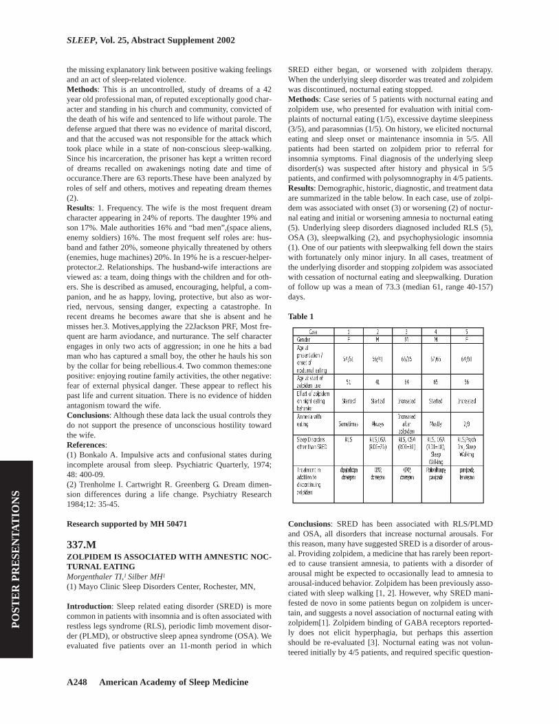
A248 American Academy of Sleep Medicine
PO
STE
R P
RE
SEN
TA
TIO
NS
SLEEP, Vol. 25, Abstract Supplement 2002
the missing explanatory link between positive waking feelingsand an act of sleep-related violence.Methods: This is an uncontrolled, study of dreams of a 42year old professional man, of reputed exceptionally good char-acter and standing in his church and community, convicted ofthe death of his wife and sentenced to life without parole. Thedefense argued that there was no evidence of marital discord,and that the accused was not responsible for the attack whichtook place while in a state of non-conscious sleep-walking.Since his incarceration, the prisoner has kept a written recordof dreams recalled on awakenings noting date and time ofoccurance.There are 63 reports.These have been analyzed byroles of self and others, motives and repeating dream themes(2).Results: 1. Frequency. The wife is the most frequent dreamcharacter appearing in 24% of reports. The daughter 19% andson 17%. Male authorities 16% and “bad men”,(space aliens,enemy soldiers) 16%. The most frequent self roles are: hus-band and father 20%, someone phyically threatened by others(enemies, huge machines) 20%. In 19% he is a rescuer-helper-protector.2. Relationships. The husband-wife interactions areviewed as: a team, doing things with the children and for oth-ers. She is described as amused, encouraging, helpful, a com-panion, and he as happy, loving, protective, but also as wor-ried, nervous, sensing danger, expecting a catastrophe. Inrecent dreams he becomes aware that she is absent and hemisses her.3. Motives,applying the 22Jackson PRF, Most fre-quent are harm aviodance, and nurturance. The self characterengages in only two acts of aggression; in one he hits a badman who has captured a small boy, the other he hauls his sonby the collar for being rebellious.4. Two common themes:onepositive: enjoying routine family activities, the other negative:fear of external physical danger. These appear to reflect hispast life and current situation. There is no evidence of hiddenantagonism toward the wife.Conclusions: Although these data lack the usual controls theydo not support the presence of unconscious hostility towardthe wife.References: (1) Bonkalo A. Impulsive acts and confusional states duringincomplete arousal from sleep. Psychiatric Quarterly, 1974;48: 400-09.(2) Trenholme I. Cartwright R. Greenberg G. Dream dimen-sion differences during a life change. Psychiatry Research1984;12: 35-45.
Research supported by MH 50471
337.MZOLPIDEM IS ASSOCIATED WITH AMNESTIC NOC-TURNAL EATINGMorgenthaler TI,1 Silber MH1
(1) Mayo Clinic Sleep Disorders Center, Rochester, MN,
Introduction: Sleep related eating disorder (SRED) is morecommon in patients with insomnia and is often associated withrestless legs syndrome (RLS), periodic limb movement disor-der (PLMD), or obstructive sleep apnea syndrome (OSA). Weevaluated five patients over an 11-month period in which
SRED either began, or worsened with zolpidem therapy.When the underlying sleep disorder was treated and zolpidemwas discontinued, nocturnal eating stopped.Methods: Case series of 5 patients with nocturnal eating andzolpidem use, who presented for evaluation with initial com-plaints of nocturnal eating (1/5), excessive daytime sleepiness(3/5), and parasomnias (1/5). On history, we elicited nocturnaleating and sleep onset or maintenance insomnia in 5/5. Allpatients had been started on zolpidem prior to referral forinsomnia symptoms. Final diagnosis of the underlying sleepdisorder(s) was suspected after history and physical in 5/5patients, and confirmed with polysomnography in 4/5 patients.Results: Demographic, historic, diagnostic, and treatment dataare summarized in the table below. In each case, use of zolpi-dem was associated with onset (3) or worsening (2) of noctur-nal eating and initial or worsening amnesia to nocturnal eating(5). Underlying sleep disorders diagnosed included RLS (5),OSA (3), sleepwalking (2), and psychophysiologic insomnia(1). One of our patients with sleepwalking fell down the stairswith fortunately only minor injury. In all cases, treatment ofthe underlying disorder and stopping zolpidem was associatedwith cessation of nocturnal eating and sleepwalking. Durationof follow up was a mean of 73.3 (median 61, range 40-157)days.
Table 1
Conclusions: SRED has been associated with RLS/PLMDand OSA, all disorders that increase nocturnal arousals. Forthis reason, many have suggested SRED is a disorder of arous-al. Providing zolpidem, a medicine that has rarely been report-ed to cause transient amnesia, to patients with a disorder ofarousal might be expected to occasionally lead to amnesia toarousal-induced behavior. Zolpidem has been previously asso-ciated with sleep walking [1, 2]. However, why SRED mani-fested de novo in some patients begun on zolpidem is uncer-tain, and suggests a novel association of nocturnal eating withzolpidem[1]. Zolpidem binding of GABA receptors reported-ly does not elicit hyperphagia, but perhaps this assertionshould be re-evaluated [3]. Nocturnal eating was not volun-teered initially by 4/5 patients, and required specific question-
Sleep Research Society A249
PO
STE
R P
RE
SEN
TA
TIO
NS
SLEEP, Vol. 25, Abstract Supplement 2002
ing to elicit this symptom. Nocturnal eating is more commonin those with insomnia than in the general population. Forpracticing sleep physicians, these cases highlight the impor-tance of inquiring about nocturnal eating behaviors, especiallyin those taking zolpidem, and of arriving at an accurate diag-nosis and appropriate treatment plan. Providing zolpidem forinsomnia without seeking specific diagnoses may lead tounusual and potentially harmful nocturnal behaviors.
References: (1) Harazin, J. and T.R. Berigan, Zolpidem tartrate and som-nambulism. Military Medicine, 1999. 164(9): p. 669-70(2) Mendelson, W.B., Sleepwalking associated with zolpidem.[see comments]. Journal of Clinical Psychopharmacology,1994. 14(2): p. 150.(3) Filizola, M., D.L. Harris, and G.H. Loew, Benzodiazepine-induced hyperphagia: development and assessment of a 3Dpharmacophore by computational methods. Journal of Bio-molecular Structure & Dynamics, 2000. 17(5): p. 769-78
338.MPOLYSOMNOGRAPHIC CHANGES IN PTSD ANDIDIOPATHIC NIGHTMARE PATIENTS FOLLOWINGIMAGERY REHEARSAL TREATMENTGermain A,1,2 Nielsen TA1,2
(1) Dream and Nightmare Laboratory, Hopital Sacre-Coeur deMontreal, Montreal, Quebec, Canada, (2) Dept of Psychology,University of Montreal, Montreal, Quebec, Canada, (3) Deptof Psychiatry, University of Montreal, Montreal, Quebec,Canada,
Introduction: Imagery Rehearsal (IR), a cognitive-behavioraltechnique for alleviating nightmares, is associated with sub-jective improvements in sleep quality (1-3). However, noindependent replication of this effect has been conducted, andsleep improvements have not yet been assessed usingpolysomnography (PSG). This study addresses these limita-tions and investigates whether IR differentially affects PTSDand idiopathic nightmare (I-NM) patients.Methods: Twelve nightmare patients completed prospectivedream logs, measures of psychological distress, and under-went polysomnographic (PSG) recordings both prior to and 6-10 weeks after receiving the IR treatment. Six patients suf-fered from posttraumatic stress disorder and frequent night-mares (P-NM) and six suffered from I-NM. IR was providedduring a single session in a small-group format. Pre-post treat-ment comparisons were made for dream logs, measures ofpsychological distress, and PSG recordings.Results: Post-treatment, significant reductions in retrospectivenightmare frequency (p = 0.007), in prospective bad dreamfrequency (p = 0.03), and in anxiety scores (p = 0.004) wereobserved. Apart from an increased micro-arousal index (p =0.04), no changes in sleep structure were remarkable. Further,post-treatment sleep profiles were different for the two sub-groups. Post-treatment, I-NM patients showed an increase in%S2 (p =0.05) and a reduction of periodic leg movements inREM sleep (p = 0.04), whereas P-NM patients exhibited ele-vated micro-arousal indices (p = 0.01).Conclusions: These results independently replicate the effica-
cy of IR treatment for alleviating nightmares and other symp-toms of psychological distress, but suggest that the effects mayvary as a function of nightmare pathology. It remains possiblethat the reduction in disturbed dreaming and psychologicaldistress precede the occurrence of significant objective sleepimprovements. Further, the use of prospective dream logs inthe present study indicates that bad dreams (unpleasant dreamsthat do not immediately awaken the sleeper), rather than night-mares (unpleasant dreams associated with awakenings), are amore sensitive measure of disturbed dreaming.References: (1) Kellner R, Neidhardt EJ, Krakow B, Pathak D. Changes inchronic nightmares after one session of desensitization orrehearsal instructions. Am J Psychiatry 1992;149:659-663.(2) Krakow B, Kellner R, Neidhardt J, Pathak D, Lambert L.Imagery rehearsal treatment for chronic nightmares: A thirtymonth follow-up. J Beh Ther Exp Psychiatry 1993;24:325-330.(3) Krakow B, Hollifield M, Schrader R, Koss M, Tandberg D,Lauriello J, McBride L, et al. A controlled study of imageryrehearsal of chronic nightmares in sexual assault survivorswith PTSD: A preliminary report. J Trauma Stress2000;13:589-609.
Research supported by Canadian Institute of HealthResearch and Fonds de la Recherche en Santé du Québec
339.MDOPAMINERGIC AND OPIATE THERAPY OF NOC-TURNAL SLEEP-RELATED EATING DISORDERASSOCIATED WITH SLEEPWALKING OR UNASSO-CIATED WITH ANOTHER NOCTURNAL DISORDERSchenck CH, Mahowald MW(1) Minnesota Regional Sleep Disorders Center, (2) HennepinCounty Medical Center (Depts of Psychiatry & Neurology),(3) Univ. of Minnesota Medical School (Depts of Psychiatry &Neurology),
Introduction: Nocturnal sleep-related eating disorder(NSRED)is a female-predominant disorder often associatedwith (or symptomatic of) another sleep disorder, esp. sleep-walking (SW), but also restless legs syndrome (RLS), period-ic limb movement disorder (PLMD), insomnia, obstructivesleep apnea (OSA), narcolepsy, and circadian rhythm distur-bances (including poor sleep hygiene). Various other condi-tions and their treatments/toxicity can be closely linked withNSRED. Nevertheless, NSRED can present as an idiopathiccondition, or at least as a condition unassociated with anothernocturnal disorder (“idiopathic NSRED”). Pharmacotherapy isa cornerstone of therapy, along with proper sleep-wakehygiene, daytime eating habits and stress-management skills.Whereas bedtime benzodiazepine therapy controls problemat-ic—and even violent—SW in >80% of reported cases, benzo-diazepine monotherapy is considerably less successful is con-trolling SW with problematic nocturnal eating (<50% ofreported cases). In contrast, dopaminergic therapy (at timescombined with opiate and/or clonazepam therapy) has beenreported to be effective in controlling problematic nocturnaleating in patients with SW. The rationale for this therapeutic
A250 American Academy of Sleep Medicine
PO
STE
R P
RE
SEN
TA
TIO
NS
SLEEP, Vol. 25, Abstract Supplement 2002
approach in SW/NSRED was based on the successful controlof problematic nocturnal eating in patients withRLS/PLMD/NSRED. Furthermore, this therapeutic approach(which is standard therapy for RLS/PLMD without nocturnaleating) may also be quite effective with idiopathic NSRED.We now report our center’s cumulative experience in treatingthese two sub-groups of NSRED with dopaminergic and opi-ate therapy.Methods: Over a 15 year period, 18 patients with NSREDassociated with SW or unassociated with another nocturnaldisorder (“idiopathic”) were treated, by one of the authors,with dopaminergic and/or opiate therapy. This group com-prised approximately 25% of all NSRED patients evaluatedand diagnosed at our center during that time period. The SWpatients with NSRED not included in this report had eitherresponded to benzodiazepine (or other) therapy, had receivedtreatment elsewhere, had refused therapy, or were lost to fol-low-up. All pts with SW had prior histories of SW involvingnon-eating behaviors, although at the time of presentation,most or all SW behaviors involved eating. All pts had under-gone extensive clinical evaluations, including hospital-basedpolysomnographic (PSG) and audio-visual monitoring, withexpanded EEG and EMG montages, utilizing standard meth-ods of recording and scoring.Results: All 18 pts had nightly eating episodes, with partialconsciousness, and without perceived hunger, during arousalsfrom sleep. PSG monitoring: 77.8% (14/18) had eatingepisodes from NREM sleep. No pt. had PLMD, clinical OSA,REM motor abnormalities or EEG epileptiform activity.72.2% (13/18) of pts reported 75-100% control of NSREDwith bedtime treatment (tabulated below).N=13 RESPON-DERS: mean age, 38.2 ±10.5 yrs; mean age, NSRED onset,26.0 ±9.9 yrs; females, 76.9% (10/13); mean duration, treat-ment follow-up, 2.5 ±3.0 yrs (2-6 months, n=3; 1-2 yrs, n=7;6-9 yrs, n=3). Currently, 69.2% (9/13) of responders are stillbeing followed at our center (including all 3 pts with a 2-6month follow-up interval).N=5 NON-RESPONDERS: meanage, 30.2 ± 16.6 yrs; mean age, NSRED onset, 18.6 ± 11.2 yrs;females, 60% (3/5); idiopathic NSRED, 60% (3/5);SW/NSRED, 40% (2/5). TREATMENT RESPON-DERS I) SW-NSRED SUB-GROUP: n=6 (females, n=4);mean age, 36.8 ± 7.2 yrs; mean age, NSRED onset, 19.0 ±7.8yrs.II) IDIOPATHIC SUB-GROUP: n=7 (females, n=6); meanage, 38.2 ±10.5 yrs; mean age, NSRED onset, 26.9 ±9.9 yrs.TABULATION OF TREATMENTS (N=13)N=2 (L-dopa orbromocriptine) N=2 (L-dopa & benzodiazepine) N=2 (L-dopa & codeine)N=1 (L-dopa & codeine & benzo) N=1 (L-dopa & trazodone) N=1 (bupropion & codeineN=2 (L-dopa& bupropion/trazodone)N=1 (L-dopa &bupropion/codeine)N=1 (codeine & benzo)(3 pts took com-bined carbidopa/L-dopa and CR-carbidopa/L-dopa hs)(Benzo-diazepines: clonazepam (n=2); alprazolam(n=1);estazolam(n=1)(L-dopa: Rxed as carbidopa/L-dopa orCR—controlled-release—tablets)(Bupropion:Rxed as regularor SR—sustained-release—tablets; dopaminergic anti-depres-sant(Codeine: Rxed in combination with acetaminophen: 325mg acetaminophen per 30 mg codeine)(All medications weregenerally well-tolerated, with infrequent side-effects)DOSE RANGESCarbidopa/L-dopa (20/200—100/400 mghs); carbidopa/L-dopa CR (25/100—100/400 mg hs)Bupropi-
on (n=1; 450 mg); Bupropion SR (n=3; 200-350 mg): taken as2-3 split daily dosesCodeine (30-60 mg hs); Trazodone (100-200 mg hs); Bromocriptine (2.5 mg hs)Clonazepam (0.5 mg;2.0 mg); Alprazolam (0.5 mg); Estazolam (3.0 mg)Conclusions: Our findings indicate that dopaminergic andopiate therapy of NSRED associated with SW or unassociatedwith another nocturnal disorder can be successful in the major-ity of treated patients. A benzodiazepine agent or trazodonemay need to be included in the treatment regimen to controlany persistent sleep disruption after control of nocturnal eatingis achieved; these agents may also contribute to control of noc-turnal eating. Our findings also call attention to dopaminergicand opioid mechanisms of control and dyscontrol of feedingbehavior across sleep and wakefulness.
340.NSLEEP STAGE SCORING METHODOLOGY WITHRHYTHMIC MASTICATORY MUSCLE ACTIVITY.Guitard F,1 Saber M,1 Rompré PH,1 Montplaisir JY,1 LavigneGJ2(1) Faculté de Médecine Dentaire, Université de Montréal, (2)Hôpital du Sacré-Coeur and Université de Montréal,
Introduction: Rhythmic Masticatory Muscle Activity(RMMA) has been reported in 60% of normal sleepers and inpatients with tooth-grinding during sleep, a condition knownas sleep bruxism (Lavigne et al., 2001). The present study wasconducted to describe the method that we developed to scoresleep stages associated with RMMA episodes and to charac-terize sleep stage shifts occurring in close temporal associationwith RMMA.Methods: In this study, 20 moderate to severe bruxers wereselected. Polysomnographic recordings were performed ontwo consecutive nights in a sleep laboratory. The first nightwas used for habituation to sleep laboratory conditions and torule out other sleep disorders. RMMA episodes were scored aspreviously described (Lavigne et al., 2001). Sleep was record-ed and first scored using the standard method of Rechtschaf-fen and Kales (1968, R&K) with 20-sec epochs. In the secondanalysis, all RMMA episodes scored wake with R&K weregiven the stage of the preceding epoch (modified scoring). Asa third analysis, the sleep stage during the 20 sec before (B20)the beginning of the episode was scored (event-based scoring).The sleep stage during the 20 sec after (A20) the end of theepisode, and during following 20 to 40 sec (A40) and 40 to 60sec (A60) were scored. RMMA episodes separated by lessthan 100 sec were considered as being part of the same globalepisode (cluster) and sleep stages before and after the clusterwere compared.Results: With the R&K sleep stage scoring, 24.6% of RMMAepisodes were scored as occurring in wake, while thisdecreased to 1.0% with the modified scoring (figure 1). ACohen Kappa of 0.65 was observed between both methods,which indicates substantial agreement. The distribution ofRMMA episodes (n=1107), over sleep stages, according to themodified scoring was 27.0% in stage 1, 58.0% in stage 2, 4.9%in 3+4, 9.1% in REM and 1.0% in wake. The distribution ofRMMA episodes according to the event-based sleep stagescoring was 20.8% in stage 1, 60.1% in stage 2, 5.0% in 3+4,
Sleep Research Society A251
PO
STE
R P
RE
SEN
TA
TIO
NS
SLEEP, Vol. 25, Abstract Supplement 2002
11.3% in REM and 2.9% in wake. A Cohen Kappa of 0.78 wasobserved, which indicates substantial agreement between bothscoring methods. A sleep stage shift occurred in 53.6% ofepisodes between B20 and A20. However, this decreases to39.0% and 31.2% after 40 and 60 sec respectively. Episodesoccurring in cluster were associated with sleep stage change in56.8% of cases compared to 50.4% in isolated episodes(p=0.14).
Figure 1
Conclusions: These results show that the modified scoringand the event-based scoring give similar results. Moreover,approximately half of the RMMA episodes are associated witha transient sleep stage change immediately after the episode.The sleep stage then returns to its previous value in mostepisodes (69%).
References: (1) 1. Lavigne GJ, Rompré PH, Poirier G, Huard H, Kato T,Montplaisir JY (2001). Rhythmic masticatory muscle activityduring sleep in humans. J Dent Res 80:443-448.(2) 2. Lavigne GJ, Rompré PH, Montplaisir JY (1996). Sleepbruxism: validity of clinical research diagnostic criteria in acontrolled polysomnographic study. J Dent Res 75:546-552.(3) 3. Rechtschaffen A, Kales A, editors (1968). A manual ofstandardized terminology, techniques and scoring techniquesfor sleep stages of human subjects. Los Angeles: BrainResearch Institute.
Research supported by Canadian Institutes of HealthResearch and Fonds de la recherche en santé du Québec
341.NAN OPEN-LABEL EXTENSION STUDY OFMODAFINIL FOR THE TREATMENT OF DAYTIMESLEEPINESS IN PATIENTS WITH PARKINSON’S DIS-EASEAdler CH,1 Caviness JN,1 Hentz JG,1 Lind M,1 Tiede J1
(1) Parkinson’s Disease and Movement Disorders Center,Mayo Clinic Scottsdale,
Introduction: Excessive daytime sleepiness (EDS) and sleep
disorders are common complaints in patients with Parkinson’sdisease (PD). More than 15% of patients with PD experienceEDS as compared with 1% of healthy elderly people. EDS andsleep disorders are among the most common causes of declin-ing health-related quality of life in patients with PD.Modafinil, a novel wake-promoting agent, is effective for thetreatment of EDS in narcolepsy1 and obstructive sleep apnea,2and is potentially useful for treating EDS in patients with PD.Recently, we reported that modafinil was effective for thetreatment of EDS in patients with PD who participated in adouble-blind, placebo-controlled, study.3 In this report, weextend our earlier observations to include results from a sub-sequent 4-week, open-label extension study.Methods: Following completion of a 7-week, double-blind,placebo-controlled, study of modafinil and a 1-week washoutperiod, 20 patients with idiopathic PD and EDS (ie, anEpworth Sleepiness Scale [ESS] score \scc10 at entry into thedouble-blind study) received modafinil 200 mg/d for 1 weekand modafinil 400 mg/d for 3 weeks in a 4-week, open-labelstudy. Efficacy was assessed using the ESS and patient- andphysician-rated Clinical Global Impression of Change (CGI-C) scores. Other evaluations included scales for assessingfatigue, severity of PD, and activities of daily living.
Table 1
Results: Mean (SD) ESS scores improved from 15.6 (4.8) atthe postwashout baseline of the open-label study to 12.2 (4.4)after 4 weeks of treatment with modafinil (p = 0.002).Improvements in wakefulness were also significant asassessed by patient- and physician-rated CGI-C scores (p =0.015 and p = 0.003, respectively). Mean scores on measuresof fatigue, severity of PD, and activities of daily livingremained relatively stable with respect to postwashout base-line values. Modafinil was well tolerated. Most adverse events(16 of 17; 94%) were mild to moderate in nature, and includ-ed two reports each of somnolence and abnormal dreaming.Four patients reported an increase in motor symptoms (ataxia,dyskinesia, hyperkinesia, or tremors), and two patients report-ed increased “OFF” time. There were no significant changes inmean diastolic or systolic blood pressure or mean pulse or res-piration rates.
A252 American Academy of Sleep Medicine
PO
STE
R P
RE
SEN
TA
TIO
NS
SLEEP, Vol. 25, Abstract Supplement 2002
Conclusions: Treatment with modafinil significantlyimproved daytime wakefulness in patients with PD and EDS,and modafinil was well tolerated. The improvements in wake-fulness observed in this open-label study were similar to thoseobtained in an earlier double-blind, placebo-controlled study.References: (1) US Modafinil in Narcolepsy Multicenter Study Group.Randomized trial of modafinil as a treatment for the excessivedaytime somnolence of narcolepsy. Neurology 2000; 54:1166-75. (2) Pack AI, Black JE, Schwartz JRL, Matheson JK, for the U.S. Modafinil in Obstructive Sleep Apnea Study Group.Modafinil as adjunct therapy for daytime sleepiness inobstructive sleep apnea. Am J Respir Crit Care Med. In press. (3) Adler CH, Caviness JN, Hentz JG, Lind M, Tiede J.Modafinil for the treatment of excessive daytime sleepiness inpatients with Parkinson’s disease. Sleep 2001; 24(Suppl):A368-A9
Research supported by Cephalon, Inc.
342.NPERIODIC LIMB MOVEMENTS DURING SLEEP INPATIENTS WITH HEART FAILURE Silva RS,1 Alves RS,1 Figueiredo AC,1 Prezotti S,1 Mady C,1
Lorenzi-Filho G1
(1) InCor Sleep Lab, São Paulo Medical School, University ofSão Paulo, SP, Brazil,
Introduction: Periodic limb movements (PLM) is prevalent inseveral medical disorders as uremia, chronic myelopathies andanemia. Some patients may have asymptomatic PLM but it isusually associated with recurrent arousals from sleep leadingto daytime sleepiness. Patients with congestive heart failure(CHF) usually have sleep complaints that can be related toboth PLM and sleep breathing disorders (SBD). The objectiveof this study was to determine the prevalence of PLM in CHFpatients with and without SBD.Methods: We studied 31 patients with severe CHF with stableclinical condition and had left ventricular ejection fraction(LVEF) /leq 45 %. The etiologies of CHF were idiophatic(64.5% of total), Chagas’ disease (19.4%), and ischemic(16.1%). Overnight polysomnography was performed usingEMBLA equipment and all polysomnograms were performedand scored based on the guidelines for sleep studies. Daytimesleepiness was assessed by a subjective rating (EpworthSleepiness Scale [ESS]). Significant differences betweengroups were analyzed by t test with p<0.05 considered signif-icant. Of the 31 patients, 11 (35.5%) were women and 20(64.5%) were men. The mean age was 60.8 (/pm 15.1) yearswith BMI of 20.5 (/pm 3.7) kg/m2 .The LVEF was 35.6 (/pm6.1)%.Results: The sleep efficiency in patients without PLM was85.9 (/pm 9.4) and with PLM was 71.7 (/pm 22.8). When justcompared the polysomnographic characteristics of patientswith SBD (Table 1), patients without PLM had higher TST,Sleep Efficiency, mean SaO2 (p<0.05), and had lower AHI(p<0.05), compared with those with PLM. There are not dif-ferences between either age, BMI or LVEF. The ESS score
was 9.8 (/pm 6.3) in patients without PLM and 10.35 (/pm 5)in patients with PLM. These scores did not reach statisticalsignificance.
Table 1
Conclusions: This study suggests a high prevalence of PLMin patients with CHF, independently of the associated SBD.References: (1) AASM Task Force – Sleep-Related Breathing Disorders inAdults: Recommendations for Syndrome Definition and Mea-surement Techniques in Clinical Research. Sleep, 1999,22(5):667-689.(2) Hanly, PJ & Zuberi-Khokhar, N: Periodic Limb Move-ments during Sleep in Patients with Congestive Heart Failure.Chest, 1996, 109:1497-1502.
343.NASSOCIATION OF RESTLESS LEGS SYNDROMEWITH PARKINSON’S DISEASE, ESSENTIALTREMOR, AND TOURETTE’S SYNDROMEWalters AS,1,2,3 Lebrocq C,1,2,3 Wagner M,1 Passi V,4 Patel S4
(1) New Jersey Neuroscience Institute at JFK Medical Center,(2) Seton Hall University School of Graduate Medical Educa-tion, (3) Dept of Neurology UMDNJ-Robert Wood JohnsonMedical School, (4) Dept of Pharmacy Practice—RutgersUniversity,
Introduction: Patients with Parkinson’s Disease (PD) have a19.5% prevalence of Restless Legs Syndrome (RLS) (1) andpatients with Tourette’s Syndrome (TS) have a 59% preva-lence of RLS (2). Because of this we did an opposite surveylooking at the prevalence of PD, TS, and Essential tremor (ET)in RLS.Methods: We looked at the prevalence of PD, TS, and ET in120 patients with RLS. To eliminate referral bias patients whowere referred for PD, TS or ET were excluded. Results werecompared with prevalence rates of these 3 conditions from theliterature using the Exact Binomial Test.Results: The prevalence of PD in the general population formen in their 70’s is .008. Four out of 14 men with RLS in their70’s had PD (P=.0001). For women in their 70’s and men intheir 80’s the P value was <.05 for PD. Only one patient out ofthe 120 RLS patients had TS (P=NS). The prevalence of ET inthe general population is .0042. Five out of our 120 RLSpatients had ET (P <.00016.)Conclusions: Putting together the results from the literature

Sleep Research Society A253
PO
STE
R P
RE
SEN
TA
TIO
NS
SLEEP, Vol. 25, Abstract Supplement 2002
and the current results we suggest that PD predisposes to RLSand RLS predisposes to PD. This may reflect the underlyingdopaminergic deficit postulated for both conditions. AlthoughTS and RLS are both responsive to dopaminergic drugs, TSappears to predispose to RLS whereas RLS does not appear topredispose to TS. The possible relationship of ET to RLS bearsfurther exploration.References: (1) (1) Atassi F, Voung KD, Jankovic J, Ondo WG. The preva-lence of Restless Legs Syndrome in patients with Parkinson’sDisease. Neurology 2001; 56 (Suppl 3); A21.(2) (2) Lipinski JF, Sallee FR, Jackson C, Sethuraman G.Dopamine agonist treatment of Tourette Disorder in children:Results of an open-label trial of pergolide. Movement Disor-ders 1997; 12: 402-407.
344.NAUGMENTATION WITH PRAMIPEXOLE IN THELONG-TERM TREATMENT OF RESTLESS LEGSSYNDROMEWinkelman JW,1 Bennett S1
(1) Brigham and Women’s Hospital; Harvard Medical School,(2) Simmons College,
Introduction: Dopaminergic agonists have become first-linetreatments for Restless Legs Syndrome (RLS). One of thegreat advantages of this class of agents appears to be the rela-tive lack of “augmentation” (daytime rebound of symptoms),which can appear in 80% of L-Dopa treated patients. Mont-plaisir (2001) followed 7 patients for a mean of 7.8 monthsand did not find augmentation with pramipexole (mean dose0.5 mg). Similarly, Ferini-Strambi et al. (2001) found that only5 of 60 patients (8%) had augmentation with pramipexole(mean dose ~.5 mg). In all patients, pramipexole was given asa single dose at bedtime. We now report our longterm experi-ence with use of pramipexole in patients with RLS.Methods: All patients seen in a clinical practice given a diag-nosis of RLS according to the IRLSSG criteria, who had beenmaintained on pramipexole for at least 6 months, were includ-ed. Augmentation was defined as an earlier appearance of RLSsymptoms, requiring a change in the time of administration ofpramipexole.Results: Thirty-six patients (61 % female) were included, witha mean age of 59.6 yrs. (range 37-90). All but four patientswere classified as primary RLS. Pramipexole had been takencontinuously for an average of 21.6 months (range 6-41).Roughly 25% of patients were taking .125-.25 mg per day ofpramipexole, 35% were taking .375-.5 mg per day, 20% weretaking .75mg, and 20% were using 1.0 mg or more per day.Half (18/36) of the patients were taking the first daily dose ofpramipexole at 6 pm or before. Nearly half (16/36) were tak-ing pramipexole at least BID.Augmentation developed in 39%of patients (14/36). The precise time to the development ofaugmentation was unclear in many patients, and for some, thisprocess continued to evolve over time. Augmentation did notnecessitate discontinuation of pramipexole in any patient, andwas generally managed with earlier dose administration or,occasionally, addition of other agents (gabapentin, opiates). Inthose for whom information was available, 88% (7/8) with
augmentation to pramipexole had previously developed aug-mentation to L-Dopa, whereas 58% (11/19) without augmen-tation to pramipexole had previous augmentation to L-Dopa.Most (25/36 or 69%) patients taking pramipexole were alsotaking one or more nocturnal sedative(s) to assist with sleepinitiation or maintenance (trazodone=11, gabapentin=8, ben-zodiazepines=4, others=6). There was no statistically signifi-cant difference in age between those patients with and withoutaugmentation, or in those receiving sedatives versus those notgetting such agents.Conclusions: We found much higher rates of augmentationthan have previously been reported with pramipexole. The fac-tors responsible for augmentation are unclear, and thus thecauses for the disparity between our data and previous reportsis unknown. However, two studies have noted augmentation inover one-quarter of RLS patients administered pergolide (Sil-ber et al., 1996; Stiasny et al., 2001). Although augmentationwas frequently observed in our patients, it was generally man-aged with earlier administration of pramipexole, or in unusualcases, addition of secondary agents. High rates of sedative-hypnotic use were found in our patients, suggesting thatpramipexole may frequently be inadequate as a sole treatmentfor the sleep disruption in RLS.References: (1) Ferini-Strambi et al.: Long-term treatment for restless legssyndrome with pramipexole: augmentation effect findings.Sleep 24: A362, 2001(2) Montplaisir et al.: Pramipexole in the treatment of restlesslegs syndrome: a follow-up study. Eur. J. Neurology 7(s1): 27-31, 2000. (3) Stiasny et al.: Long-term effects of pergolide in the treat-ment of restless legs syndrome. Neurology 56: 1399-1402,2001.
345.NTHE IMPACT OF RESTLESS LEGS SYNDROME (RLS)ON PATIENT QUALITY OF LIFE: RESULTS FROM ASURVEY OF RLS SUFFERERSAllen RP,1 Vallow SM,1 Abetz L,2 Washburn T,3 Earley CJ1
(1) John Hopkins Univ., Dept of Neurology, Baltimore MD,USA, (2) GlaxoSmithKline, Philadelphia PA, USA, (3) MapiValues, Cheshire, UK,
Introduction: RLS is a sleep-related movement disorder inwhich a person during the transition between sleeping andwaking experiences a strong urge to move usually associatedwith unpleasant sensations in the legs . This leads to severesleep disruption. Whilst much research has shown the delete-rious effects of sleep disorders on QoL (1), little research hasbeen conducted to measure empirically the humanistic burdenof RLS. The objectives of this study were to evaluate thevalidity of the new Restless Legs Syndrome Quality of Life(RLSQoL) questionnaire and to assess the impact of RLS onQoL among patients diagnosed with primary RLS.Methods: The RLSQoL is an 18-item patient-report question-naire that assesses the impact of RLS on daily life, sleep, emo-tional well-being, social life and work life. The RLSQoL wasadministered at baseline and 2 weeks later to 85 patients withprimary RLS. The severity of RLS symptoms was assessed

A254 American Academy of Sleep Medicine
PO
STE
R P
RE
SEN
TA
TIO
NS
SLEEP, Vol. 25, Abstract Supplement 2002
using a patient-reported version of the RLS Rating Scale(RLSRS). Patients were also asked to report whether theirsymptoms improved or deteriorated over the 2-week periodResults: The majority of the sample (64.5%) were women andthe mean age was 62.4 (SD, 14.0) years. The mean age atwhich RLS was first diagnosed was 36.6 (SD, 19.6) years.Four weeks prior to questionnaire completion, 67.1% ofpatients reported experiencing RLS symptoms almost daily.Patients not experiencing RLS symptoms daily had a meansymptom frequency of 7.54 (± 6.3) days per month. Fifty-eightpercent of patients reported that they experienced symptomsfor 3 or more hours a day. The RLSQoL questionnaire yieldeda single summary score of life impact that demonstratedacceptable internal consistency and test-retest reliability(alpha=0.94; intra-class correlation coefficient=0.75, respec-tively), satisfactory item-convergent and -discriminant validi-ty, and excellent known-groups validity.QoL was significantlypoorer for patients with more severe RLS symptoms, with atleast a 10-point difference in RLSQoL scores between mildand moderate and between moderate and severe symptomgroups. In addition, patients who reported that their symptomshad worsened over the 2-week period experienced a decreasein QoL, and those whose symptoms had improved showed animprovement in QoL. The effect sizes for the groups showingimprovement (0.44) and deterioration (0.49) indicated a mod-erate change.Conclusions: The RLSQoL questionnaire is a valid measurefor evaluating RLS patients’ QoL. The impact of RLS onpatient QoL correlates with the severity of the disease, sug-gesting that treatment strategies aimed at improving or pre-venting RLS would have a significant effect on patient QoL.Instruments such as the RLSQoL can be used to evaluate fur-ther the humanistic burden of RLS and the benefits of thera-peutic interventions on patient QoL.References: (1) Weaver TE, Laizner AM, Maislin G, Chugh DK, Lyon K,Smith PL, et al. An instrument to measure functional outcomesfor disorders of excessive sleepiness. Sleep 1997;20:835-843.
Research supported by GlaxoSmithKline Pharmaceuticals
346.NMULTIPLE BLOOD DONATIONS AS A CAUSE OFIRON DEFICIENCY IN RESTLESS LEGS SYNDROMESilber MH,1 Richardson JW1
(1) Sleep Disorders Center, Mayo Clinic, Rochester, Minneso-ta,
Introduction: Iron deficiency has long been recognized as apredisposing factor for restless legs syndrome (RLS). Serumferritin concentrations of <50 mcg/l correlate with increasedseverity of RLS [1]. Common causes of iron deficiencyinclude menorrhagia, gastrointestinal blood loss and malnutri-tion. A less well-known mechanism is iron depletion frommultiple blood donations. Serum ferritin concentration falls by17-26% per donation and subjects donating 5 or more times in2 years develop inevitable iron deficiency (ferritin <20 mcg/l)[2].Methods: We identified 8 patients seen at the Mayo Sleep Dis-
orders Center in 2001 with RLS (International RLS StudyGroup criteria) and iron deficiency who were regular blooddonors. Clinical and laboratory data was analyzed.Results: Four were men; 4 premenopausal women (one with ahysterectomy, 3 with normal menstrual periods). Mean agewas 47 years (range 40-59). RLS had been present for a yearor less in 3 patients, for 7 years in one patient, and for 15 yearsor more in 4 patients. Two had family histories of first-degreerelatives with RLS. No gastrointestinal causes of bleedingwere identified. Patients had donated blood between 2 and 6times a year (mean 4 times) for 3-25 years. RLS commencedafter or at about the same time as the start of blood donationsin 6/8 patients. Mean serum ferritin concentration was 8 mcg/l(range 3-15). Four patients were mildly anemic (hematocrits31.9-37.3%). Two patients were treated with iron alone and 6with iron and dopaminergic agents or opioids. Blood donationwas stopped. Available follow up in 5 patients, including onetreated only with iron and another who was able to discontin-ue pramipexole, revealed improvement in serum ferritin con-centrations and excellent control of RLS.Conclusions: We believe that iron deficiency from multipleblood donations either induced or helped perpetuate RLS inour patients. No other causes for iron deficiency were deter-mined. Physicians treating RLS should question and councilpatients regarding blood donation. Staff of donor sites shouldbe trained to question volunteers about RLS. Even if a screen-ing hemoglobin concentration is normal, donations should notbe accepted from RLS patients if their serum ferritin concen-tration is low. Donors should be prevented from sufferingadverse clinical consequences of their altruism.References: (1) Sun ER, Chen CA, Ho G, Earley CJ, Allen RP. Iron and therestless legs syndrome. Sleep 1998;371-7.(2) Boulton F, Collis D, Inskip H, Paes H, Garlick M. A studyof the iron and HFE status of blood donors, including a groupwho failed the initial screen for anemia. Br J Hem2000;108:434-439.
347.NBRIEF NOCTURNAL BEHAVIOR OBSERVATIONS OFMOTOR ACTIVITY DISTINGUISH PARKINSON’SDISEASE FROM ALZHEIMER’S DISEASE Rungruang BJ,1 Bliwise DL1
(1) Emory University Medical School, Dept of Neurology,Sleep Disorders Research, Atlanta, GA,
Introduction: The increased recognition of REM-dyscontrolin many neurodegenerative diseases such as Parkinson’s Dis-ease (PD) and Lewy Body Dementia and the development ofPD in over a third of idiopathic REM Behavior Disorder casesleads to the prediction that motor activity in sleep is neitherunexpected nor rare in PD. This study tested this hypothesis byconducting very brief observations (< 20 mins) of behaviorduring sleep in an unselected sample of inpatients with PD anda similarly hospitalized comparison group.Methods: All patients were hospitalized in a specialized, sub-acute, tertiary care facility dedicated to geriatric patients (Wes-ley Woods Geriatric Hospital). Patients were hospitalized for avariety of reasons, but most typically for infections, injury or

Sleep Research Society A255
PO
STE
R P
RE
SEN
TA
TIO
NS
SLEEP, Vol. 25, Abstract Supplement 2002
medication adjustment. A total of 30 patients were studied,consisting of PD (n = 15) and other diagnoses (n = 15), the lat-ter consisting of probable/possible Alzheimer’s Disease (AD)(n = 9), major depression (n = 4), and generalized anxiety dis-order (n = 2). Informed consent was obtained in allcases.Observations were made during behaviorally definedsleep between 0030 and 0500 and lasted between 7 and 35minutes (X = 17.7 mins). Filming was made with a low light,audio-capable, second-by-second digital video camera placedon a tripod. Quantification of real-time observations was madeby a second-by-second basis, with total seconds of movementobserved for lower limbs, upper limbs, and body trunk com-puted separately. A fourth category, vocalization, was alsorecorded. Movement intensity for each behavioral categorywas defined on an ordinal scale (1) and then multiplied by timeweights (5 12-sec intervals ranging from 0 - 60 sec). Thesummed score across categories was corrected for total # ofseconds of observation. Higher scores thus reflected both per-vasiveness and duration of movement, adjusted for duration ofobservation.Results: The summed motor activity score was significantlyhigher in PD relative to AD/other patients (891.1 vs 205.7,Mann-Whitney Z = 2.94,p < .01). All four types of motoractivity differentiated the groups, however there was somesuggestion that the strongest differences occurred for thelower limbs. Among the PD patients, daily levo-dopa dose waspositively correlated with motor activity score (rho = .53, p <.05), suggesting greater movement associated with moresevere disease.Conclusions: Use of these brief (but highly systematic)behavioral observations to detect motor activity in sleep in PDsuggests very large effects. Given caregiver reports of disrup-tive nocturnal behavior in PD (2) and higher levels ofovernight activity assessed with whole body actigraphy in PD(3), such effects cannot be considered altogether surprising.What is novel is their robustness based on the exceedinglyshort duration of such observations.References: (1) Bliwise & Lee, J Nurs Meas 1993;1:115-24(2) Bliwise et al, J Geriatr Psych Neurol 1995;8:107-10(3) van Hilten et al, Arch Neurol 1994;51:922-28
Research supported by AG-10642
348.OCLINICAL EXPERIENCE WITH VAGAL NERVESTIMULATION AND EXCESSIVE DAYTIME SLEEPI-NESS AS MEASURED BY THE EPWORTH SLEEPI-NESS SCALECochran JW1
(1) Drs. Cochran, Eberly & Howe, PC Alexandria, VirginiaUSA,
Introduction: Excessive daytime sleepiness is a commonproblem that may be associated with sleep loss (insomnia) orsleep fragmentation. Daytime sleepiness has been assessedusing a tool called the Epworth Sleepiness Scale (ESS). Thetest consists of eight items and the patient is asked to gauge thechance of dosing on a 0-3 scale with 0 being would never dose
and 3 being high chance of dosing. Mean scores in normalsubjects usually 5.9 with a range of 2-10.It was demonstratedby Zabara (2). Zabara demonstrated the anti-convulsant actionof vagal nerve stimulation (VNS) in experimental animals. Itwas hypothesized that VNS could prevent or control seizures.The technique of VNS involves the implantation of a device inthe left anterior chest wall. A subcutaneous generator sends anelectrical signal to the left vagus nerve. VNS with the NCPsystem (Cyberonics). VNS with the NCP system utilizes animplantable, programmable bipolar pulse generator that stim-ulates the left vagus nerve with a bipolar lead. With a pro-gramming wand and software and a personal computer thepulse generator can be activated, interrogated and the charac-ter of the stimulation changed. VNS has been shown to beaffective double blind studies and reduces seizure frequencyoverall a mean of 25% to 30% compared with baseline. Asmall sample open label study revealed that vagal nerve stim-ulation reduced the total in rapid eye movement sleep (3). Forthese reasons it was undertaken to have all patients treated forcomplex partial epilepsy with VNS in an outpatient Neurolo-gy/Sleep Medicine practice have serial EPS performed.Methods: In a prospective fashion, patients referred for treat-ment by vagal nerve stimulation who had the clinical diagno-sis of complex partial seizures were administered the EpworthSleepiness Scale (1) preoperatively, postoperatively and thenat intervals to see if there was any effect on daytime sleepinessin patients with epilepsy treated with vagal nerve stimulation.Fourteen patients have been enrolled. The results of theEpworth Sleepiness Scale given serially are presented graphi-cally. Fourteen such patients were evaluated. Data is availableon 10 patients. The study is ongoing.Results: The results are seen in the accompanying table. Sev-eral patients manifest a trend toward reduction in daytimesleepiness. Large scale controlled studies may be helping infurther elucidating whether VNS has a role in treating exces-sive daytime sleepiness. Confounding variables such as con-comitant medication and seizure frequency should be evaluat-ed.
Figure 1
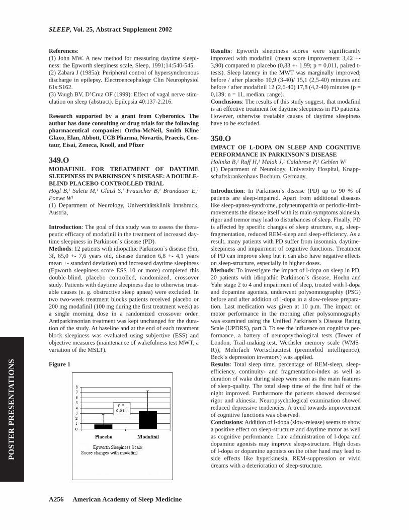
A256 American Academy of Sleep Medicine
PO
STE
R P
RE
SEN
TA
TIO
NS
SLEEP, Vol. 25, Abstract Supplement 2002
References: (1) John MW. A new method for measuring daytime sleepi-ness: the Epworth sleepiness scale, Sleep, 1991;14:540-545.(2) Zabara J (1985a): Peripheral control of hypersynchronousdischarge in epilepsy. Electroencephalogr Clin Neurophysiol61s:S162.(3) Vaugh BV, D’Cruz OF (1999): Effect of vagal nerve stim-ulation on sleep (abstract). Epilepsia 40:137-2.216.
Research supported by a grant from Cyberonics. Theauthor has done consulting or drug trials for the followingpharmaceutical companies: Ortho-McNeil, Smith KlineGlaxo, Elan, Abbott, UCB Pharma, Novartis, Praecis, Cen-taur, Eisai, Zeneca, Knoll, and Pfizer
349.OMODAFINIL FOR TREATMENT OF DAYTIMESLEEPINESS IN PARKINSON`S DISEASE: A DOUBLE-BLIND PLACEBO CONTROLLED TRIALHögl B,1 Saletu M,1 Glatzl S,1 Frauscher B,1 Brandauer E,1
Poewe W1
(1) Department of Neurology, Universitätsklinik Innsbruck,Austria,
Introduction: The goal of this study was to assess the thera-peutic efficacy of modafinil in the treatment of increased day-time sleepiness in Parkinson`s disease (PD).Methods: 12 patients with idiopathic Parkinson`s disease (9m,3f, 65,0 +- 7,6 years old, disease duration 6,8 +- 4,1 yearsmean +- standard deviation) and increased daytime sleepiness(Epworth sleepiness score ESS 10 or more) completed thisdouble-blind, placebo controlled, randomized, crossoverstudy. Patients with daytime sleepiness due to otherwise treat-able causes (e. g. obstructive sleep apnea) were excluded. Intwo two-week treatment blocks patients received placebo or200 mg modafinil (100 mg during the first treatment week) asa single morning dose in a randomized crossover order.Antiparkinsonian treatment was kept unchanged for the dura-tion of the study. At baseline and at the end of each treatmentblock sleepiness was evaluated using subjective (ESS) andobjective measures (maintenance of wakefulness test MWT, avariation of the MSLT).
Figure 1
Results: Epworth sleepiness scores were significantlyimproved with modafinil (mean score improvement 3,42 +-3,90) compared to placebo (0,83 +- 1,99; p = 0,011, paired t-tests). Sleep latency in the MWT was marginally improved;before / after placebo 10,9 (3-40)/ 15,1 (2,5-40) minutes andbefore / after modafinil 12 (2,6-40) 17,8 (4,2-40) minutes (p =0,139; n = 11, median, range).Conclusions: The results of this study suggest, that modafinilis an effective treatment for daytime sleepiness in PD patients.However, otherwise treatable causes of daytime sleepinesshave to be excluded.
350.OIMPACT OF L-DOPA ON SLEEP AND COGNITIVEPERFORMANCE IN PARKINSON´S DISEASEHolinka B,1 Ruff H,1 Malak J,1 Calabrese P,1 Gehlen W1
(1) Department of Neurology, University Hospital, Knapp-schaftskrankenhaus Bochum, Germany,
Introduction: In Parkinson`s disease (PD) up to 90 % ofpatients are sleep-impaired. Apart from additional diseaseslike sleep-apnea-syndrome, polyneuropathia or periodic-limb-movements the disease itself with its main symptoms akinesia,rigor and tremor may lead to disturbances of sleep. Finally, PDis affected by specific changes of sleep structure, e.g. sleep-fragmentation, reduced REM-sleep and sleep-efficiency. As aresult, many patients with PD suffer from insomnia, daytime-sleepiness and impairment of cognitive functions. Treatmentof PD can improve sleep but it can also have negative effectson sleep-structure, especially in higher doses.Methods: To investigate the impact of l-dopa on sleep in PD,20 patients with idiopathic Parkinson´s disease, Hoehn andYahr stage 2 to 4 and impairment of sleep, treated with l-dopaand dopamine agonists, underwent polysomnographiy (PSG)before and after addition of l-dopa in a slow-release prepara-tion. Last medication was given at 10 p.m. The impact onmotor performance in the morning after polysomnographywas examined using the Unified Parkinson`s Disease RatingScale (UPDRS), part 3. To see the influence on cognitive per-formance, a battery of neuropsychological tests (Tower ofLondon, Trail-making-test, Wechsler memory scale (WMS-R)), Mehrfach Wortschatztest (premorbid intelligence),Beck´s depression inventory) was applied.Results: Total sleep time, percentage of REM-sleep, sleep-efficiency, continuity- and fragmentation-index as well asduration of wake during sleep were seen as the main featuresof sleep-quality. The total sleep time of the first half of thenight improved. Furthermore the patients showed decreasedrigor and akinesia. Neuropsychological examination showedreduced depressive tendencies. A trend towards improvementof cognitive functions was observed.Conclusions: Addition of l-dopa (slow-release) seems to showa positive effect on sleep-structure and daytime motor as wellas cognitive performance. Late administration of l-dopa anddopamine agonists may improve sleep-structure. High dosesof l-dopa or dopamine agonists on the other hand may lead toside effects like hyperkinesia, REM-suppression or vividdreams with a deterioration of sleep-structure.
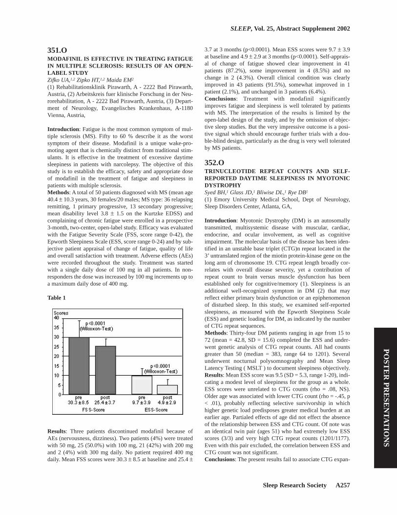
Sleep Research Society A257
PO
STE
R P
RE
SEN
TA
TIO
NS
SLEEP, Vol. 25, Abstract Supplement 2002
351.OMODAFINIL IS EFFECTIVE IN TREATING FATIGUEIN MULTIPLE SCLEROSIS: RESULTS OF AN OPEN-LABEL STUDYZifko UA,1,2 Zipko HT,1,2 Maida EM2
(1) Rehabilitationsklinik Pirawarth, A - 2222 Bad Pirawarth,Austria, (2) Arbeitskreis fuer klinische Forschung in der Neu-rorehabilitation, A - 2222 Bad Pirawarth, Austria, (3) Depart-ment of Neurology, Evangelisches Krankenhaus, A-1180Vienna, Austria,
Introduction: Fatigue is the most common symptom of mul-tiple sclerosis (MS). Fifty to 60 % describe it as the worstsymptom of their disease. Modafinil is a unique wake-pro-moting agent that is chemically distinct from traditional stim-ulants. It is effective in the treatment of excessive daytimesleepiness in patients with narcolepsy. The objective of thisstudy is to establish the efficacy, safety and appropriate doseof modafinil in the treatment of fatigue and sleepiness inpatients with multiple sclerosis.Methods: A total of 50 patients diagnosed with MS (mean age40.4 ± 10.3 years, 30 females/20 males; MS type: 36 relapsingremitting, 1 primary progressive, 13 secondary progressive;mean disability level 3.8 ± 1.5 on the Kurtzke EDSS) andcomplaining of chronic fatigue were enrolled in a prospective3-month, two-center, open-label study. Efficacy was evaluatedwith the Fatigue Severity Scale (FSS, score range 0-42), theEpworth Sleepiness Scale (ESS, score range 0-24) and by sub-jective patient appraisal of change of fatigue, quality of lifeand overall satisfaction with treatment. Adverse effects (AEs)were recorded throughout the study. Treatment was startedwith a single daily dose of 100 mg in all patients. In non-responders the dose was increased by 100 mg increments up toa maximum daily dose of 400 mg.
Table 1
Results: Three patients discontinued modafinil because ofAEs (nervousness, dizziness). Two patients (4%) were treatedwith 50 mg, 25 (50.0%) with 100 mg, 21 (42%) with 200 mgand 2 (4%) with 300 mg daily. No patient required 400 mgdaily. Mean FSS scores were 30.3 ± 8.5 at baseline and 25.4 ±
3.7 at 3 months (p<0.0001). Mean ESS scores were 9.7 ± 3.9at baseline and 4.9 ± 2.9 at 3 months (p<0.0001). Self-apprais-al of change of fatigue showed clear improvement in 41patients (87.2%), some improvement in 4 (8.5%) and nochange in 2 (4.3%). Overall clinical condition was clearlyimproved in 43 patients (91.5%), somewhat improved in 1patient (2.1%), and unchanged in 3 patients (6.4%).Conclusions: Treatment with modafinil significantlyimproves fatigue and sleepiness is well tolerated by patientswith MS. The interpretation of the results is limited by theopen-label design of the study, and by the omission of objec-tive sleep studies. But the very impressive outcome is a posi-tive signal which should encourage further trials with a dou-ble-blind design, particularly as the drug is very well toleratedby MS patients.
352.OTRINUCLEOTIDE REPEAT COUNTS AND SELF-REPORTED DAYTIME SLEEPINESS IN MYOTONICDYSTROPHYSyed BH,1 Glass JD,1 Bliwise DL,1 Rye DB1
(1) Emory University Medical School, Dept of Neurology,Sleep Disorders Center, Atlanta, GA,
Introduction: Myotonic Dystrophy (DM) is an autosomallytransmitted, multisystemic disease with muscular, cardiac,endocrine, and ocular involvement, as well as cognitiveimpairment. The molecular basis of the disease has been iden-tified in an unstable base triplet (CTG)n repeat located in the3’ untranslated region of the miotin protein-kinase gene on thelong arm of chromosome 19. CTG repeat length broadly cor-relates with overall disease severity, yet a contribution ofrepeat count to brain versus muscle dysfunction has beenestablished only for cognitive/memory (1). Sleepiness is anadditional well-recognized symptom in DM (2) that mayreflect either primary brain dysfunction or an epiphenomenonof disturbed sleep. In this study, we examined self-reportedsleepiness, as measured with the Epworth Sleepiness Scale(ESS) and genetic loading for DM, as indicated by the numberof CTG repeat sequences.Methods: Thirty-four DM patients ranging in age from 15 to72 (mean = 42.8, SD = 15.6) completed the ESS and under-went genetic analysis of CTG repeat counts. All had countsgreater than 50 (median = 383, range 64 to 1201). Severalunderwent nocturnal polysomnography and Mean SleepLatency Testing ( MSLT ) to document sleepiness objectively.Results: Mean ESS score was 9.5 (SD = 5.3, range 1-20), indi-cating a modest level of sleepiness for the group as a whole.ESS scores were unrelated to CTG counts (rho = .08, NS).Older age was associated with lower CTG count (rho = -.45, p< .01), probably reflecting selective survivorship in whichhigher genetic load predisposes greater medical burden at anearlier age. Partialed effects of age did not effect the absenceof the relationship between ESS and CTG count. Of note wasan identical twin pair (ages 51) who had extremely low ESSscores (3/3) and very high CTG repeat counts (1201/1177).Even with this pair excluded, the correlation between ESS andCTG count was not significant.Conclusions: The present results fail to associate CTG expan-
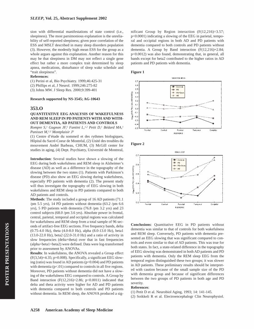
A258 American Academy of Sleep Medicine
PO
STE
R P
RE
SEN
TA
TIO
NS
SLEEP, Vol. 25, Abstract Supplement 2002
sion with differential manifestations of state control (i.e.,sleepiness). The most parsimonious explanation is the unrelia-bility of self-reported sleepiness; given poor correlation of theESS and MSLT described in many sleep disorders population(3). However, the modestly high mean ESS for the group as awhole argues against this explanation. Another reason for thismay be that sleepiness in DM may not reflect a single geneeffect but rather a more complex trait determined by sleepapnea, medications, disturbance of sleep wake schedule and“trait sleepiness”.References: (1) Perini et al, Bio Psychiatry. 1999;46:425-31(2) Phillips et al, J Neurol. 1999;246:275-82(3) Johns MW. J Sleep Res. 2000;9:399-401
Research supported by NS-3545; AG-10643
353.OQUANTITATIVE EEG ANALYSIS OF WAKEFULNESSAND REM SLEEP IN PD PATIENTS WITH AND WITH-OUT DEMENTIA, AD PATIENTS AND CONTROLSRompre S,1 Gagnon JF,1 Fantini L,1,2 Petit D,1 Bédard MA,1
Panisset M,1,2 Montplaisir J3
(1) Centre d’étude du sommeil et des rythmes biologiques,Hôpital du Sacré-Coeur de Montréal, (2) Unité des troubles dumouvement André Barbeau, CHUM, (3) McGill center forstudies in aging, (4) Dept. Psychiatry, Université de Montreal,
Introduction: Several studies have shown a slowing of theEEG during both wakefulness and REM sleep in Alzheimer’sdisease (AD) as well as a difference in the topography of theslowing between the two states (1). Patients with Parkinson’sdisease (PD) also show an EEG slowing during wakefulness,especially PD patients with dementia (2). The present studywill thus investigate the topography of EEG slowing in bothwakefulness and REM sleep in PD patients compared to bothAD patients and controls.Methods: The study included a group of 16 AD patients (71.1/pm 5.5 yrs), 14 PD patients without dementia (63.2 /pm 6.6yrs), 5 PD patients with dementia (76.8 /pm 3.2 yrs) and 23control subjects (68.0 /pm 3.6 yrs). Absolute power in frontal,central, parietal, temporal and occipital regions was calculatedfor wakefulness and REM sleep from a total sample of 96 sec-onds of artifact-free EEG sections. Five frequency bands, delta(0.75-4.0 Hz), theta (4.0-8.0 Hz), alpha (8.0-13.0 Hz), beta1(13.0-22.0 Hz), beta2 (22.0-31.0 Hz) and a ratio of activity inslow frequencies (delta+theta) over that in fast frequencies(alpha+beta1+beta2) were defined. Data were log-transformedprior to assessment by ANOVAs.Results: In wakefulness, the ANOVA revealed a Group effect(F(3,54)=4.35; p=0.008). Specifically, a significant EEG slow-ing (ratio) was found in AD patients (p<0.004) and PD patientswith dementia (p<.01) compared to controls in all five regions.Moreover, PD patients without dementia did not have a slow-ing of the wakefulness EEG compared to controls. A Group byBand interaction (F(12,216)=2.86; p<0.0011) indicated thatdelta and theta activity were higher for AD and PD patientswith dementia compared to both controls and PD patientswithout dementia. In REM sleep, the ANOVA produced a sig-
nificant Group by Region interaction (F(12,216)=3.57;p<0.0001) indicating a slowing of the EEG in parietal, tempo-ral and occipital regions in both AD and PD patients withdementia compared to both controls and PD patients withoutdementia. A Group by Band interaction (F(12,216)=2.84;p<0.0012) was also found, demonstrating that, in general, allbands except for beta2 contributed to the higher ratios in ADpatients and PD patients with dementia.
Figure 1
Figure 2
Conclusions: Quantitative EEG in PD patients withoutdementia was similar to that of controls for both wakefulnessand REM sleep. Conversely, PD patients with dementia pre-sented an EEG slowing that was significant compared to con-trols and even similar to that of AD patients. This was true forboth states. In fact, a state-related difference in the topographyof EEG slowing was demonstrated in both AD patients and PDpatients with dementia. Only the REM sleep EEG from thetemporal region distinguished these two groups; it was slowerin AD patients. These preliminary results should be interpret-ed with caution because of the small sample size of the PDwith dementia group and because of significant differencesbetween the two groups of PD patients in both age and PDseverity.References: (1) Petit D et al. Neurobiol Aging, 1993; 14: 141-145.(2) Soikkeli R et al. Electroencephalogr Clin Neurophysiol.
Sleep Research Society A259
PO
STE
R P
RE
SEN
TA
TIO
NS
SLEEP, Vol. 25, Abstract Supplement 2002
1991; 79: 159-165.
Research supported by the Canadian Institutes of HealthResearch and by Fonds de la Recherche en Santé duQuébec.
354.OTHE EFFECT OF MELATONIN TREATMENT ONSUBJECTIVE APPRAISAL OF NIGHTTIME SLEEPAND DAYTIME SLEEPINESS IN PATIENTS WITHPARKINSON’S DISEASEDowling GA,1 Mastick J,1 Colling E,1 Carter J,2 Singer C3
(1) Institute on Aging, Goldman Research Center, Universityof California, San Francisco Center on Aging, California, (2)Department of Psychiatry, Sleep and Mood Disorders Labora-tory, Oregon Health Sciences University, Portland, Oregon,(3) Department of Neurology, Oregon Health Sciences Uni-versity, Portland, Oregon, U.S.A.,
Introduction: Melatonin treatment in patients with age-relat-ed insomnia has been reported to improve a variety of objec-tively measured sleep parameters (e.g., sleep latency, sleepefficiency). Most investigations have not included subjectiveevaluations of nighttime sleep and daytime sleepiness. Thereis, however, at least one report in which objective improve-ments did not coincide with subjective self-reports (Hughs,Sack & Lewy, 1998). As part of a larger multicenter trial toexamine the effects of melatonin at two different doses (5mgand 50mg) versus placebo to treat sleep complaints in patientswith Parkinson’s disease (PD), participants provided their sub-jective appraisals on various sleep and sleepiness relatedparameters.Methods: The clinical design was a placebo-controlled, dou-ble cross-over trial. Subjects received each treatment (placebo,5mg, 50mg) for a 2-week time period with a 1-week washoutbetween treatments. The General Sleep Disturbance Scale(GSDS) (Lee, 1992) was used to assess participants’ subjec-tive sleep disturbances, daytime sleepiness and level of func-tion over the past 1-week time period. The GSDS consists of21 items related to difficulty getting to sleep, waking duringsleep, waking up too early from sleep, quality and quantity ofsleep, fatigue and sleepiness, and use of substances to helpinduce sleep. Subjects completed the GSDS for each of the 2treatment weeks and results were averaged to achieve a singlescore for each treatment period.Results: Thirty subjects (mean age=64 years, range=53-76)with PD (median Hoehn & Yahr scale score=2.5;mean=3.5+0.7) and without primary sleep pathology complet-ed the experimental protocol. Wilcoxon signed ranks analysesof the GSDS results (see table) revealed a significant improve-ment (decrease) (p<.005) in total score in the 5mg melatonintreatment condition compared to placebo. Further analyses ofGSDS subscales revealed that the improvement in total scorereflected significant improvements in reports of sleep quantity(p<.01), mid-sleep awakening (p<.04), early awakening(p<.007) and daytime sleepiness (p<.001). No significant dif-ferences were found in the use of sleep aids, sleep quality ordifficulty initiating sleep. There was also improvement(p<.04) in total GSDS score in the 5mg melatonin treatment
condition compared to the 50mg condition. This improvementreflects a significantly (p<.006) lower daytime sleepiness rat-ing in the 5mg condition compared to 50mg but not signifi-cantly different from placebo. No other significant differenceswere found in GSDS subscale scores. Subjects were also askedto identify during which treatment period they felt they slept“best.” Eighty-four percent of the subjects identified the mela-tonin treatment periods as “best” (42% 5mg, 42% 50mg) com-pared to only 16% for placebo.
Table 1
Conclusions: Compared to placebo, a 2-week treatment withmelatonin in this sample of patients with PD resulted inimproved subjective reports of sleep disturbances, daytimesleepiness and level of function. Future analyses will comparethese subjective reports to objectively measured parameters.References: (1) Hughes RJ, Sack RL, Lewy AJ. The role of melatonin andcircadian phase in age-related sleep-maintenance insomnia:Assessment in a clinical trial of melatonin replacement. Sleep1998;21:52-68.(2) Lee KA. Self-reported sleep disturbances in employedwomen. Sleep 1992;15:493-498.
Research supported by NIH/NINR R01 NR04774
355.PSLEEP PATTERNS IN PRADER WILLI PATIENTS ONAND OFF MEDICATIONSPrativa D,1 Chung SA,1 Kayumov L,1 Berall G,1 Shapiro CM1
(1) Sleep Research Unit and the Child and Adolescent HealthUnit, Toronto Western Hospital, University Health Network,Toronto, Canada,
Introduction: Prader Willi syndrome is a congenital disordercharacterized by neonatal hypotonia, life-threatening obesityfrom childhood and onward, hyperphagia, hypomentia,hypogonadism, characteristic dysmorphic facial features, andshort stature. Excessive daytime sleepiness (EDS) is a well-
A260 American Academy of Sleep Medicine
PO
STE
R P
RE
SEN
TA
TIO
NS
SLEEP, Vol. 25, Abstract Supplement 2002
known feature of Prader Willi syndrome. This study was con-ducted to determine if changes in sleep patterns could accountfor this EDS.Methods: Fourteen Prader Willi patients ranging in age from5 to 36 years were referred to The Sleep Research Unit withcomplaints of EDS. Six patients were on psychotropic and/orneuroleptic medications (fluoxetine, risperidone,methylphenidate, buspirone, benztropine, chlorpromazine,valproic acid, lithium and perphenazine) and 8 were medica-tion-free. Patients underwent two overnight polysomnograph-ic studies and daytime testing - Multiple Sleep Latency Test(MSLT) and Maintenance of Wakefulness Test (MWT) on suc-cessive days.Results: There were no differences in the sleep architecturebetween the non-medicated and medicated groups but as awhole the Prader Willi patients had a significantly reducedpercentage of REM sleep as compared to age-matched popu-lation normal values from the literature. The average REMpercentage in the medicated group was 12.2 ± 5% and in thenon-medicated group was 14.7 ± 5%, (not significantly differ-ent, p=0.4). A diagnosis of sleep apnea was made in onepatient (RDI = 42) from the medicated group, and twopatients, one from each group, had elevated RDIs during REMsleep. The medicated patients had more severe daytime sleepi-ness as measured by the MSLT (9.3 ± 6 mins versus 13.8 ± 3mins in the non-medicated group, p<0.05) and a greater occur-rence of REM intrusions on the MSLT.Conclusions: Our study found reduced amounts of REM sleepin Prader Willi patients that was independent of medicationeffects. We did not find a high frequency of sleep apnea orother sleep disturbances in our patients to justify the com-plaints of EDS, although patients on medications had moresevere EDS. The psychotropic and neuroleptic medicationscommonly used to treat the behavioural and mood distur-bances may particularly exacerbate sleepiness in this popula-tion. Further studies need to be conducted to determine if phar-macological intervention can help alleviate the EDS and ifthere are alternative medications that have less sedating actioncompared to the commonly used psychotropics and neurolep-tics.
356.PCHANGES IN SLEEP BELIEFS AND PRACTICESFOLLOWING SURGERYFloyd JA,1 Shreve WS,1 Medler SM,2 Wimbush FB3
(1) College of Nursing, Wayne State University, (2) St. Vin-cent Medical Center, Toledo, Ohio, (3) Department of Psy-chology, University of Florida,
Introduction: One purpose of this study was to explorewhether sleep beliefs and sleep promotion practices changeduring a stressful life event. Patients electing coronary arterybypass graft (CABG) surgery were selected for study becausethey are exposed to many of the factors known to generateshort-term insomnia including hospitalization for a frighteningand painful procedure. The surgical procedure itself has amajor effect on circadian rhythms, thus disturbing sleep-wakepatterns.Methods: Elective CABG patients (N=25) completed ques-
tionnaires related to their sleep beliefs and sleep promotionpractices pre-operatively and at 6-weeks post-operatively. Thesubjects were predominantly Caucasian men treated at a Mid-western USA medical center. Ages ranged from 47-82 years(M=64.5, SD=10.09). Sleep beliefs were measured by an 18-item Short-Form of the Floyd-Medler Sleep Belief Scaledesigned for use in clinical settings (1). It measures beliefsabout (a) Next-Day Consequences, (b) Health Consequences,(c) Sleep Need, (d) Sleep Regularity, and (e) Coping. TheSleep Hygiene Awareness and Practices Scale (SHAPS) wasused to measure sleep promotion practices (2). Two-tailedalpha was set at .05 for significance and .10 for trends.Results: Sleep belief scores increased for all five factors, butnone were significantly different from baseline. There weretrends for beliefs about Sleep Need (t(24) = 1.98) and Next-Day Consequences (t(24) = 1.75) to increase following sur-gery. Only three sleep promotion practices were used morethan 3 times/week and they did not change significantly in fre-quency of use pre- to post-operatively, see Table 1. Two relat-ed sleep hygiene practices changed following surgery: (a)There was a significant increase in worries while preparing forbed about being unable to sleep (t(24) = 2.31) and (b) a trendtoward increased worries during the day about being unable tosleep (t(24) = 1.72). Subjects with strong pre-operative beliefsabout Coping were significantly more likely following surgeryto worry while preparing for bed about being unable to sleep,r=.44. Subjects with strong pre-operative beliefs about SleepNeed and the negative Next-Day Consequences of poor sleepwere more likely to exercise during the day post-operatively,r=.45 and r=.72, respectively. Also, subjects with strong pre-operative beliefs about Next-Day Consequences were likely toreport post-operatively that they exercised vigorously withintwo hours of bedtime, r=.62.
Table 1
Conclusions: Although some practices adopted by this samplewere positive, others, such as worrying about their ability tosleep and doing strenuous exercise close to bedtime are coun-terproductive. Overall, subjects in this sample used few of thesleep hygiene practices believed to promote sleep. There ispartial support for the premise that adults’ sleep beliefs maychange during stressful life events and that beliefs about sleepmay be related to sleep promotion practices adopted at thosetimes. It remains to be demonstrated whether some beliefsabout sleep protect or predispose adults to develop and main-tain chronic insomnia during periods of stress.
Sleep Research Society A261
PO
STE
R P
RE
SEN
TA
TIO
NS
SLEEP, Vol. 25, Abstract Supplement 2002
References: (1) Floyd JA, Medler SM, Doorenbos AZ. Adults’ beliefsabout sleep: Further development of the Floyd-Medler SleepBelief Scale. Sleep 2001;24:A340.(2) Lacks P. Behavioral treatment for persistent insomnia.NY:Pergarnon, 1987
Research supported by Funding from the Center forHealth Research, Wayne State University
357.PREPORTED SLEEP-WAKE COMPLAINTS IN GENER-AL PRACTICE PATIENTS: AN EPWORTH SLEEPI-NESS SCALE AND ATHENS INSOMNIA SCALEBASED STUDYCluydts RJ,1 Hoffmann G,1 Heyde K,2 Kerkhofs M,3 de Bouyal-sky I,4 Viérin L5
(1) Dept. of Cognitive and Physiological Psychology, Univer-sity of Brussels, (2) Sleep Center, Brugmann Hospital, Brus-sels, (3) Sleep-Wake Disorders Center, University Hospital,Antwerp, (4) Sleep Laboratory, CHU Vesale, Charleroi, (5)Medical Dept., Sanofi-Synthélabo Belgium,
Introduction: The prevalence of sleep-wake complaints in thegeneral population and in psychiatric patients is well docu-mented. However within the health system as it is organized inBelgium, general practitioners are most likely the first to beconfronted with these complaints. As we do not exactly knowif physicians routinely question their patients about their sleepquality, we performed a one-day data collection on sleep-wakebehavior and quality in a large group of patients consultingtheir general physician regardless of the reason.Methods: 110 general practitioners agreed to participate inthis study. During one day (a Wednesday in spring 2001) theypresented following questionnaires to all successive patientsthat came for a consultation: The Epworth Sleepiness Scale(ESS), the Athens Insomnia Scale (AIS) and 10 short demo-graphic and life style questions. 1343 patients (56% female,44% male) agreed to participate.Results: ESS: 228 patients (or 17%) had a score = 10 which isconsidered a significant risk for falling asleep during the day.Of these 228 patients, 64% were female, so slightly more thenthe 56% in the whole sample. The age group most represented(28%) are the 30 to 40 year old females. Further analysis isneeded to find out if this finding is related to sleep disorders orto a possible sleep debt associated with life style andduties?AIS: A score = 4 on the first 5 AIS-items (questions onsleep) is considered as indicative of a sleep disturbance. Thelast 3 items of the AIS sample the quality of daytime func-tioning. 555 patients (or 41%) reached the cut-off of 4 on theAIS-5. Sixty two percent (343 patients) were females. There isa positive correlation between the 2 aspects assessed with theAIS. The higher the sleep scores (poorer sleep), the higher thecomplaints about poor daytime functioning. Regarding age,the highest prevalence of sleep complaints (39.3%) was foundin patients within the 40-60 years group.Finally, relating theAIS and ESS scores we find an increasing percentage ofpatients with an ESS score = 10 as the AIS scores increase, ascan be seen in the table below:
Table 1
Conclusions: Forty-one percent of the patients consultingtheir primary physician present with sleep-wake complaintsthat may need some attention and further investigation, where-as seventeen percent have significant problems with maintain-ing daytime alertness as evidenced by well-validated ques-tionnaires. Based on these findings, general practitionersshould be encouraged to inquire more routinely about thesleep of their patients.References: (1) Johns MW. A new method for measuring daytime sleepi-ness: the Epworth Sleepiness Scale. Sleep 1991;14:540-545(2) Soldatos CR, Dikeos DG, Paparrigopoulos TJ. AthensInsomnia Scale: validation of an instrument based on ICD-10criteria. J Psychosom Res 2000;48:555-560.
The authors would like to thank the “Société Scientifiquede Médecine Générale” for their collaboration
358.PQUALITY OF SLEEP FOLLOWING BURN INJURIES:A SIX MONTH FOLLOW-UP.Raymond I,1 Nielsen TA,1 Lavigne G,2 Choinière M2
(1) Burn Center, Hôtel-Dieu du Centre hospitalier de l’Uni-versité de Montréal, (2) Centre d’étude du sommeil, HôpitalSacré-Coeur de Montréal, Québec, Canada,
Introduction: Sleep disturbances are frequently reported inburn victims. Disturbances during hospitalization, such aspoor sleep quality and nightmares, are often reported, andhave been known to persist for months or years (1,2). The pur-pose of this study was to evaluate sleep quality six month fol-lowing hospitalization for severe burn injuries and to identifyvariables related to enduring sleep disturbances.Methods: Twenty-eight oriented and non-ventilated patients(24M, 4W), between the ages of 17 and 50 years (mean = 34.8± = 10) were interviewed during five consecutive days withinthe first week of hospitalization and again (N = 18) at 6-monthfollow-up. During morning interviews, patients reported per-ceived quality of sleep (10cm visual analogue scale (VAS),#hours, #awakenings, #nightmares) and pain intensity (0-10numerical scale). Patients also completed the Impact of EventScale (IES) to determine posttraumatic stress symptoms andthe Sleep Questionnaire for Adults to assess pre and post-hos-pitalization insomnia symptoms. Quality of sleep, pain inten-sity and medication ratings were averaged within subjects toproduce single scores. Paired samples t-tests and McNemartests were used to compare sleep measures at both times. Pear-son correlations were used to evaluate the relationship
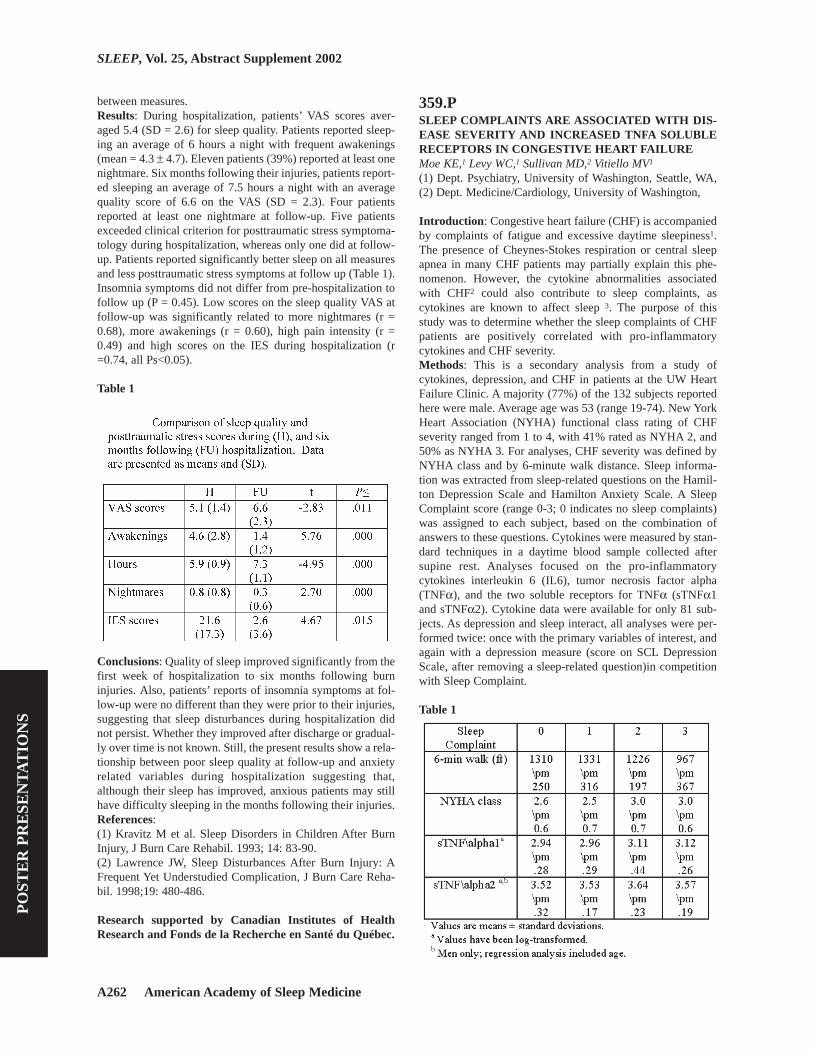
A262 American Academy of Sleep Medicine
PO
STE
R P
RE
SEN
TA
TIO
NS
SLEEP, Vol. 25, Abstract Supplement 2002
between measures.Results: During hospitalization, patients’ VAS scores aver-aged 5.4 (SD = 2.6) for sleep quality. Patients reported sleep-ing an average of 6 hours a night with frequent awakenings(mean = 4.3 ± 4.7). Eleven patients (39%) reported at least onenightmare. Six months following their injuries, patients report-ed sleeping an average of 7.5 hours a night with an averagequality score of 6.6 on the VAS (SD = 2.3). Four patientsreported at least one nightmare at follow-up. Five patientsexceeded clinical criterion for posttraumatic stress symptoma-tology during hospitalization, whereas only one did at follow-up. Patients reported significantly better sleep on all measuresand less posttraumatic stress symptoms at follow up (Table 1).Insomnia symptoms did not differ from pre-hospitalization tofollow up (P = 0.45). Low scores on the sleep quality VAS atfollow-up was significantly related to more nightmares (r =0.68), more awakenings (r = 0.60), high pain intensity (r =0.49) and high scores on the IES during hospitalization (r=0.74, all Ps<0.05).
Table 1
Conclusions: Quality of sleep improved significantly from thefirst week of hospitalization to six months following burninjuries. Also, patients’ reports of insomnia symptoms at fol-low-up were no different than they were prior to their injuries,suggesting that sleep disturbances during hospitalization didnot persist. Whether they improved after discharge or gradual-ly over time is not known. Still, the present results show a rela-tionship between poor sleep quality at follow-up and anxietyrelated variables during hospitalization suggesting that,although their sleep has improved, anxious patients may stillhave difficulty sleeping in the months following their injuries.References: (1) Kravitz M et al. Sleep Disorders in Children After BurnInjury, J Burn Care Rehabil. 1993; 14: 83-90.(2) Lawrence JW, Sleep Disturbances After Burn Injury: AFrequent Yet Understudied Complication, J Burn Care Reha-bil. 1998;19: 480-486.
Research supported by Canadian Institutes of HealthResearch and Fonds de la Recherche en Santé du Québec.
359.PSLEEP COMPLAINTS ARE ASSOCIATED WITH DIS-EASE SEVERITY AND INCREASED TNFA SOLUBLERECEPTORS IN CONGESTIVE HEART FAILUREMoe KE,1 Levy WC,1 Sullivan MD,2 Vitiello MV1
(1) Dept. Psychiatry, University of Washington, Seattle, WA,(2) Dept. Medicine/Cardiology, University of Washington,
Introduction: Congestive heart failure (CHF) is accompaniedby complaints of fatigue and excessive daytime sleepiness1.The presence of Cheynes-Stokes respiration or central sleepapnea in many CHF patients may partially explain this phe-nomenon. However, the cytokine abnormalities associatedwith CHF2 could also contribute to sleep complaints, ascytokines are known to affect sleep 3. The purpose of thisstudy was to determine whether the sleep complaints of CHFpatients are positively correlated with pro-inflammatorycytokines and CHF severity.Methods: This is a secondary analysis from a study ofcytokines, depression, and CHF in patients at the UW HeartFailure Clinic. A majority (77%) of the 132 subjects reportedhere were male. Average age was 53 (range 19-74). New YorkHeart Association (NYHA) functional class rating of CHFseverity ranged from 1 to 4, with 41% rated as NYHA 2, and50% as NYHA 3. For analyses, CHF severity was defined byNYHA class and by 6-minute walk distance. Sleep informa-tion was extracted from sleep-related questions on the Hamil-ton Depression Scale and Hamilton Anxiety Scale. A SleepComplaint score (range 0-3; 0 indicates no sleep complaints)was assigned to each subject, based on the combination ofanswers to these questions. Cytokines were measured by stan-dard techniques in a daytime blood sample collected aftersupine rest. Analyses focused on the pro-inflammatorycytokines interleukin 6 (IL6), tumor necrosis factor alpha(TNFα), and the two soluble receptors for TNFα (sTNFα1and sTNFα2). Cytokine data were available for only 81 sub-jects. As depression and sleep interact, all analyses were per-formed twice: once with the primary variables of interest, andagain with a depression measure (score on SCL DepressionScale, after removing a sleep-related question)in competitionwith Sleep Complaint.
Table 1
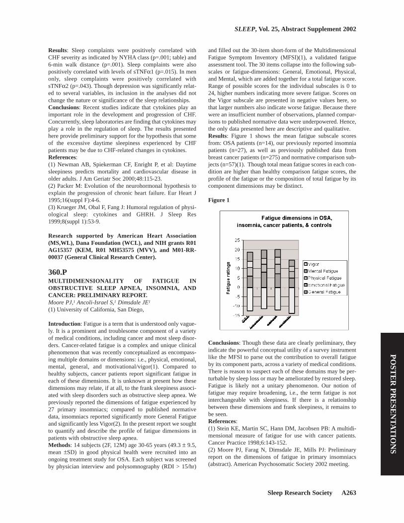
Sleep Research Society A263
PO
STE
R P
RE
SEN
TA
TIO
NS
SLEEP, Vol. 25, Abstract Supplement 2002
Results: Sleep complaints were positively correlated withCHF severity as indicated by NYHA class (p=.001; table) and6-min walk distance (p=.001). Sleep complaints were alsopositively correlated with levels of sTNFα1 (p=.015). In menonly, sleep complaints were positively correlated withsTNFα2 (p=.043). Though depression was significantly relat-ed to several variables, its inclusion in the analyses did notchange the nature or significance of the sleep relationships.Conclusions: Recent studies indicate that cytokines play animportant role in the development and progression of CHF.Concurrently, sleep laboratories are finding that cytokines mayplay a role in the regulation of sleep. The results presentedhere provide preliminary support for the hypothesis that someof the excessive daytime sleepiness experienced by CHFpatients may be due to CHF-related changes in cytokines.References: (1) Newman AB, Spiekerman CF, Enright P, et al: Daytimesleepiness predicts mortality and cardiovascular disease inolder adults. J Am Geriatr Soc 2000;48:115-23.(2) Packer M: Evolution of the neurohormonal hypothesis toexplain the progression of chronic heart failure. Eur Heart J1995;16(suppl F):4-6.(3) Krueger JM, Obal F, Fang J: Humoral regulation of physi-ological sleep: cytokines and GHRH. J Sleep Res1999;8(suppl 1):53-9.
Research supported by American Heart Association(MS,WL), Dana Foundation (WCL), and NIH grants R01AG15357 (KEM, R01 MH53575 (MVV), and M01-RR-00037 (General Clinical Research Center).
360.PMULTIDIMENSIONALITY OF FATIGUE INOBSTRUCTIVE SLEEP APNEA, INSOMNIA, ANDCANCER: PRELIMINARY REPORT.Moore PJ,1 Ancoli-Israel S,1 Dimsdale JE1
(1) University of California, San Diego,
Introduction: Fatigue is a term that is understood only vague-ly. It is a prominent and troublesome component of a varietyof medical conditions, including cancer and most sleep disor-ders. Cancer-related fatigue is a complex and unique clinicalphenomenon that was recently conceptualized as encompass-ing multiple domains or dimensions: i.e., physical, emotional,mental, general, and motivational/vigor(1). Compared tohealthy subjects, cancer patients report significant fatigue ineach of these dimensions. It is unknown at present how thesedimensions may relate, if at all, to the frank sleepiness associ-ated with sleep disorders such as obstructive sleep apnea. Wepreviously reported the dimensions of fatigue experienced by27 primary insomniacs; compared to published normativedata, insomniacs reported significantly more General Fatigueand significantly less Vigor(2). In the present report we soughtto quantify and describe the profile of fatigue dimensions inpatients with obstructive sleep apnea.Methods: 14 subjects (2F, 12M) age 30-65 years (49.3 ± 9.5,mean ±SD) in good physical health were recruited into anongoing treatment study for OSA. Each subject was screenedby physician interview and polysomnography (RDI > 15/hr)
and filled out the 30-item short-form of the MultidimensionalFatigue Symptom Inventory (MFSI)(1), a validated fatigueassessment tool. The 30 items collapse into the following sub-scales or fatigue-dimensions: General, Emotional, Physical,and Mental, which are added together for a total fatigue score.Range of possible scores for the individual subscales is 0 to24, higher numbers indicating more severe fatigue. Scores onthe Vigor subscale are presented in negative values here, sothat larger numbers also indicate worse fatigue. Because therewere an insufficient number of observations, planned compar-isons to published normative data were underpowered. Hence,the only data presented here are descriptive and qualitative.Results: Figure 1 shows the mean fatigue subscale scoresfrom: OSA patients (n=14), our previously reported insomniapatients (n=27), as well as previously published data frombreast cancer patients (n=275) and normative comparison sub-jects (n=57)(1). Though total mean fatigue scores in each con-dition are higher than healthy comparison fatigue scores, theprofile of the fatigue or the composition of total fatigue by itscomponent dimensions may be distinct.
Figure 1
Conclusions: Though these data are clearly preliminary, theyindicate the powerful conceptual utility of a survey instrumentlike the MFSI to parse out the contribution to overall fatigueby its component parts, across a variety of medical conditions.There is reason to suspect each of these domains may be per-turbable by sleep loss or may be ameliorated by restored sleep.Fatigue is likely not a unitary phenomenon. Our notion offatigue may require broadening, i.e., the term fatigue is notinterchangeable with sleepiness. If there is a relationshipbetween these dimensions and frank sleepiness, it remains tobe seen.References: (1) Stein KE, Martin SC, Hann DM, Jacobsen PB: A multidi-mensional measure of fatigue for use with cancer patients.Cancer Practice 1998;6:143-152.(2) Moore PJ, Farag N, Dimsdale JE, Mills PJ: Preliminaryreport on the dimensions of fatigue in primary insomniacs(abstract). American Psychosomatic Society 2002 meeting.
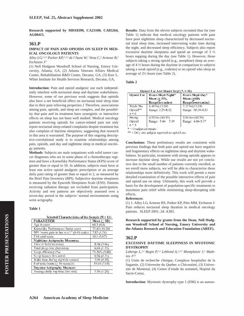
A264 American Academy of Sleep Medicine
PO
STE
R P
RE
SEN
TA
TIO
NS
SLEEP, Vol. 25, Abstract Supplement 2002
Research supported by MH18399, CA23100, CA85264,AG08415.
361.PIMPACT OF PAIN AND OPIOIDS ON SLEEP IN MED-ICAL ONCOLOGY PATIENTSAlley LG,1,2,3 Parker KP,1,2,3 de l’Aune W,1 Vena C,2 Armour B,1
Etchason J3
(1) Nell Hodgson Woodruff School of Nursing, Emory Uni-versity, Atlanta, GA, (2) Atlanta Veterans Affairs MedicalCenter, Rehabilitation R&D Center, Decatur, GA, (3) Kerr L.White Institute for Health Services Research, Decatur, GA,
Introduction: Pain and opioid analgesic use each independ-ently interfere with nocturnal sleep and daytime wakefulness.However, some of our previous work suggests that opioidsalso have a net beneficial effect on nocturnal total sleep timedue to their pain relieving properties.1 Therefore, associationsamong pain, opioids, and sleep are complex, and the possibil-ity that pain and its treatment have synergistic or interactiveeffects on sleep has not been well studied. Medical oncologypatients receiving opioids for cancer-related pain not onlyreport nocturnal sleep-related complaints despite treatment butalso complain of daytime sleepiness, suggesting that researchin this area is warranted. The purpose of this ongoing descrip-tive-correlational study is to examine relationships amongpain, opioids, and day and nighttime sleep in medical oncolo-gy patients.Methods: Subjects are male outpatients with solid tumor can-cer diagnoses who are in some phase of a chemotherapy regi-men and have a Karnofsky Performance Status (KPS) score ofgreater than or equal to 50. In addition, subjects must have atleast one active opioid analgesic prescription or an averagedaily pain rating of greater than or equal to 2, as measured bythe Brief Pain Inventory (BPI). Subjective daytime sleepinessis measured by the Epworth Sleepiness Scale (ESS). Patientsreceiving radiation therapy are excluded from participation.Activity and rest patterns are objectively assessed over aseven-day period in the subjects’ normal environments usingwrist actigraphy.
Table 1
Results: Data from the eleven subjects recruited thus far (seeTable 1) indicate that medical oncology patients with painhave poor nighttime sleep characterized by decreased noctur-nal total sleep time, increased intervening wake time duringthe night, and decreased sleep efficiency. Subjects also reportexcessive daytime sleepiness and spend an average of 3 ½hours napping during the day (see Table 1). However, thosesubjects taking a strong opioid (e.g., morphine) sleep an aver-age of 4 ½ hours during the daytime in comparison to subjectstaking a weak opioid (e.g., codeine) or no opioid who sleep anaverage of 2½ hours (see Table 2).
Table 2
Conclusions: These preliminary results are consistent withprevious findings that both pain and opioid use have negativeand fragmentary effects on nighttime sleep and daytime wake-fulness. In particular, treatment with strong opioids appears toincrease daytime sleep. While our results are not yet conclu-sive due to the small number of patients currently enrolled, aswe enroll more subjects, we will be able to characterize theserelationships more definitively. This work will permit a moredetailed examination of the possible interactive effects of painand opioid use on sleep. Ultimately, this work will provide abasis for the development of population-specific treatments tomaximize pain relief while minimizing sleep-disrupting sideeffects.References: (1) 1. Alley LG, Armour BS, Parker KP, Pitts MM, Etchason J:Pain reduces nocturnal sleep duration in medical oncologypatients. SLEEP 2001; 24: A383.
Research supported by grants from the Dean, Nell Hodg-son Woodruff School of Nursing, Emory University andthe Atlanta Research and Education Foundation (AREF).
362.PEXCESSIVE DAYTIME SLEEPINESS IN MYOTONICDYSTROPHYLaberge L,1,2 Begin P,1,2 Leblond A,1,2,3 Montplaisir J,1 Math-ieu J3,4
(1) Unite de recherche clinique, Complexe hospitalier de laSagamie, (2) Universite du Quebec a Chicoutimi, (3) Univer-site de Montreal, (4) Centre d’etude du sommeil, Hopital duSacre-Coeur,
Introduction: Myotonic dystrophy type 1 (DM) is an autoso-
Sleep Research Society A265
PO
STE
R P
RE
SEN
TA
TIO
NS
SLEEP, Vol. 25, Abstract Supplement 2002
mal dominant progressive disease characterized by muscularwasting, myotonia, endocrine dysfunction, and cataracts.Many patients affected by DM describe excessive daytimesleepiness (EDS) as the most incapacitating symptom of theircondition. EDS associated with DM show clinical and neuro-physiological traits similar to those seen in narcolepsy, name-ly disturbed nocturnal sleep, sleep-onset REM periods, fre-quent daytime napping and over representation of specificHLA DQ haplotypes.(1) The objectives of the present studywere to estimate the prevalence of the classic symptoms of thenarcolepsy tetrad in DM patients and to assess the relationshipbetween EDS and sleep disturbances.Methods: A sleep questionnaire based on the Sleep Question-naire and Assessment of Wakefulness (2) was completed by196 adult DM patients followed at the Neuromuscular Clinicof the Complexe Hospitalier de la Sagamie and by 26 controls.Mean age was 36.1 and 33.0 years, respectively (n.s.). Asleepiness scale was developed from 5 questions and scoresvaried between 0 and 15. The subjects indicated whether eachbrief descriptor always, often, sometimes or never applied.Sleep disturbances and other narcolepsy symptoms weredichotomized by the pairing of choices always/often andsometimes/never. Group differences in prevalence were ana-lyzed with Chi-square tests. Bonferroni corrections for multi-ple comparisons (n=21) were applied.
Table 1
Results: An acceptable level of internal consistency betweenthe sleepiness scale questions was found (alpha=.79), and fac-tor analysis revealed that they measure a common factor. DMpatients reported a higher sleepiness score than controls (6.14vs. 1.85, p<.001): they more often had sudden sleep attacksduring daytime, felt less healthy during daytime, had more dif-ficulty staying fully awake after meals, fell asleep more oftenwhile watching TV/or movies and took more naps. The 95thpercentile value of controls on the sleepiness scale was 6.65: ascore >= 7 was thus considered as indicative of EDS. EDS andcataplexy (CP) were more frequent in DM patients than incontrol subjects (43.1% vs. 3.8%, and 17.0% vs. 0%, respec-tively) while sleep paralysis (SP) and hypnagogic hallucina-tions (HH) did not differ between groups (Table 1). On theother hand, DM patients with EDS reported more frequent SP(10.3% vs. 0.9%) and HH (12.8% vs. 0.9%) than those with-out EDS (p<.002). Finally, DM patients with EDS reportedmore difficulty remaining still for long periods, more difficul-ty staying up late in the evening, more respiratory disturbancesduring sleep, more pain during nocturnal sleep and felt lessrefreshed and less receptive in the morning compared withthose without EDS (p<.003).Conclusions: This study reports a high prevalence of EDS ina large sample of DM patients. The presence of many clinical
traits indicative of hypersomnolence suggests a possible dys-function of central sleep regulation in this condition. SinceDM shares many physiological features of disordered arousalencountered in narcolepsy, the involvement of hypocretinsshould be assessed. (3)References: (1) Manni R, Zucca C, Martinetti M, Ottolini A, Lanzi G, Tar-tara A. Hypersomnia in dystrophia myotonica: a neurophysio-logical and immunogenetic study. Acta Neurologica Scandi-navica 1991;84:498-502.(2) Guilleminault C, ed. Sleeping and waking disorders: indi-cations and techniques. Menlo Park: Addison-Wesley Publish-ing Co., 1982.(3) Silber MH, Rye DB. Solving the mysteries of narcolepsy.Neurology 2001;56:1616-1618.
Research supported by the Canadian Institutes of HealthResearch (CIHR)
363.PENVIRONMENTAL, DEMOGRAPHIC, AND ILLNESS-RELATED CORRELATES OF SLEEP QUALITY INTHE ACUTE CARE HOSPITALRedeker NS,1 Olsen EM1
(1) Rutgers, The State University of New Jersey, Newark, NewJersey, (2) Children’s Hospital of Philadelphia, Philadelphia,PA,
Introduction: Acute care hospitalization is known to be asso-ciated with disturbed sleep. Past research has implicated char-acteristics of the patient care environment (noise, lighting,patient care interactions), patients’ demographic characteris-tics, and illness/treatment-related factors as correlates of sleepquality during hospitalization.(1) Yet, little is known about therelative contributions of these variables to sleep quality. Thepurpose of this study was to examine the relationshipsbetween environmental characteristics (noise, private vs. non-private room, hospital unit), demographics (age, gender,race),illness/treatment characteristics (type of diagnosis: medical,surgical, post-partum or antepartum and symptoms: pain,fatigue, anxiety) and self-reported sleep quality in hospitalizedadults.Methods: We surveyed 97 hospitalized patients (M age =57,SD = 18.6 years, 50% women) who had spent the previousnight in a 450-bed acute care hospital. The sample included53(55%) medical, 32 (33%)surgical,and 12 (12%)antepartumand post-partum patients. The sample was 80% white, 10%black, 4% Hispanic, 3% Native American, and 2% Asian.Study participants completed questionnaires that includeddemographic and illness/treatment and environmental charac-teristics, and numeric rating scales (0-10, 0 = lowest, 10 =highest) of sleep quality, anxiety, pain, fatigue, noise, annoy-ance due to noise, and satisfaction with care.Results: Descriptive statistics appear in the table. Overall lev-els of satisfaction with care were high. Participants reportedmore fatigue than anxiety or pain. There were no statisticallysignificant correlations between age, gender, type of patientroom (private vs. non-private), type of diagnosis (medical, sur-gical, maternity), pain, satisfaction with care, and sleep quali-
A266 American Academy of Sleep Medicine
PO
STE
R P
RE
SEN
TA
TIO
NS
SLEEP, Vol. 25, Abstract Supplement 2002
ty. Non-white patients reported better sleep quality (M =7.3,SD = 1.6) than white (M = 5.3,SD = 3.1) patients, t(80) =-2.02, p < .05). There were negative correlations between anx-iety (r = -.34, p = .001), fatigue (r = -.215, p < .05), perceptionsof noise during the night (r = -.21, p < .05), annoyance withnoise during the night (r = -.30, p < .01) and sleep quality.Regression analysis demonstrated that race, anxiety, andannoyance with noise at night explained 23% of the variancein sleep quality. Race explained 6% of the variance in sleepquality, and anxiety and annoyance with noise explained 17%.Inspection of the beta weights revealed that the most salientcorrelates of sleep quality were anxiety (beta = -.31), annoy-ance with noise (beta = -.29), and race (beta = -.22). Pain,fatigue, and satisfaction with care did not explain a significantportion of the variance in sleep quality when included in theregression equations with the other variables.
Table 1
Conclusions: These findings suggest that the most relevantcorrelates of sleep quality during hospitalization are affective:anxiety and annoyance with levels of noise. Environmentalcharacteristics were not correlated with sleep quality. Furtherstudy of the role of perception and affectivity as influences onsleep quality in the acute care hospital is needed. The findingof differences between white and non-white patients wasunexpected and warrants further study.References: (1) Redeker NS. Sleep in Acute Care Settings: An integrativereview. J Nurs Scholarsh 2000;32(1):31-8
Research supported by NR00102
364.QHEART RATE VARIABILITY DURING SLEEP INPATIENTS WITH PSYCHOGENIC ERECTILE DYS-FUNCTIONMann K,1 Pankok J,1 Connemann B1
(1) Department of Psychiatry, University of Mainz, Germany,
Introduction: Regarding the etiology of erectile dysfunction(ED), besides a number of known organic causes, psychologi-cal factors play an important role in many patients. However,the neurobiological mechanisms underlying the so-called“psychogenic” ED are not yet understood. On the assumptionthat psychogenic ED is based on distinct disturbances of thecentral nervous system, heart rate variability (HRV) was stud-ied with the aim to identify possible functional alterations of
the central autonomic activity as a neurobiological substrate ofpsychogenic ED. The study was carried out during differentsleep stages, where cognitive and emotional influences wereexcluded to a great extent.Methods: 24 male patients, 25-57 years old, with psychogenicED participated in the study. They had no lifetime diagnosis ofany other psychiatric disorder. Based on detailed case history,comprehensive clinical examination, laboratory parametersincluding hormones as well as assessment of nocturnal erec-tions, there was no evidence of organic factors relevant forsexual function. In addition, an age-matched control groupconsisting of 24 healthy male volunteers, 26-56 years old,without sexual dysfunctions was also studied. None of the sub-jects had sleep disturbances. Each subject spent three succes-sive nights in the sleep laboratory. After an adaptation night,two polysomnographies (EEG, EOG, EMG) were performedover 8 hours for each subject with registration of the electro-cardiogram (ECG), which was sampled by 400 Hz. Off-line,after automatic detection of the QRS complexes, HRV wascomputed from the distances between successive RR intervals.Sleep EEGs were scored visually according to the criteria ofRechtschaffen and Kales.Results: HRV was analyzed both in the time domain and thefrequency domain during different sleep stages (1). For that,time segments of 10 minutes duration were selected fromstage II, SWS and REM sleep, which were free of artifacts andmovements. For most parameters, significant differencesbetween sleep stages were found, pointing to different func-tional autonomic states during sleep. Particularly, SWS wascharacterized by a predominance of the parasympathetic overthe sympathetic tone, whereas during REM sleep the auto-nomic balance was shifted in favor of the sympathetic activi-ty. However, comparing patients and controls, no significantdifferences were found for all parameters.Conclusions: Although in psychogenic ED a disturbance ofthe central nervous system including autonomic pathways hasto be assumed, we could not prove any norm deviation ofHRV, which is an important parameter reflecting central auto-nomic control of the heart. However, this is consistent withanimal studies (2), which could demonstrate that autonomicactivations can reveal regionally very different patterns.Therefore, our results with unchanged HRV do not excludepossible functional disturbances in other subsystems of theautonomic nervous system.References: (1) Task Force of the European Society of Cardiology and theNorth American Society of Pacing and Electrophysiology:Heart rate variability. Circulation 1996; 93: 1043-1065.(2) Futuro-Neto HA, Coote JH: Changes in sympathetic activ-ity to heart and blood vessels during desynchronized sleep.Brain Res 1982; 252: 259-268.
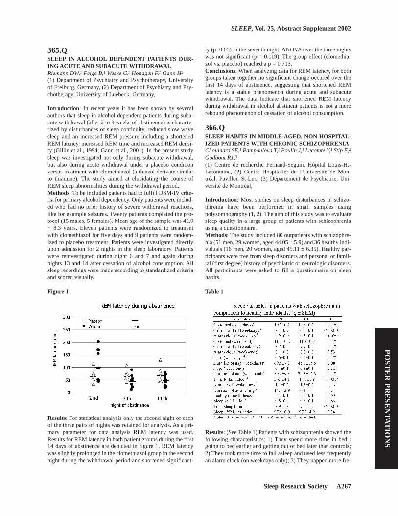
Sleep Research Society A267
PO
STE
R P
RE
SEN
TA
TIO
NS
SLEEP, Vol. 25, Abstract Supplement 2002
365.QSLEEP IN ALCOHOL DEPENDENT PATIENTS DUR-ING ACUTE AND SUBACUTE WITHDRAWALRiemann DW,1 Feige B,1 Weske G,1 Hohagen F,1 Gann H2
(1) Department of Psychiatry and Psychotherapy, Universityof Freiburg, Germany, (2) Department of Psychiatry and Psy-chotherapy, University of Luebeck, Germany,
Introduction: In recent years it has been shown by severalauthors that sleep in alcohol dependent patients during suba-cute withdrawal (after 2 to 3 weeks of abstinence) is characte-rized by disturbances of sleep continuity, reduced slow wavesleep and an increased REM pressure including a shortenedREM latency, increased REM time and increased REM densi-ty (Gillin et al., 1994; Gann et al., 2001). In the present studysleep was investigated not only during subacute withdrawal,but also during acute withdrawal under a placebo conditionversus treatment with clomethiazol (a thiazol derivate similarto thiamine). The study aimed at elucidating the course ofREM sleep abnormalities during the withdrawal period.Methods: To be included patients had to fulfill DSM-IV crite-ria for primary alcohol dependency. Only patients were includ-ed who had no prior history of severe withdrawal reactions,like for example seizures. Twenty patients completed the pro-tocol (15 males, 5 females). Mean age of the sample was 42.0+ 8.3 years. Eleven patients were randomized to treatmentwith clomethiazol for five days and 9 patients were random-ized to placebo treatment. Patients were investigated directlyupon admission for 2 nights in the sleep laboratory. Patientswere reinvestigated during night 6 and 7 and again duringnights 13 and 14 after cessation of alcohol consumption. Allsleep recordings were made according to standardized criteriaand scored visually.
Figure 1
Results: For statistical analysis only the second night of eachof the three pairs of nights was retained for analysis. As a pri-mary parameter for data analysis REM latency was used.Results for REM latency in both patient groups during the first14 days of abstinence are depicted in figure 1. REM latencywas slightly prolonged in the clomethiazol group in the secondnight during the withdrawal period and shortened significant-
ly (p=0.05) in the seventh night. ANOVA over the three nightswas not significant (p = 0.119). The group effect (clomethia-zol vs. placebo) reached a p = 0.713.Conclusions: When analyzing data for REM latency, for bothgroups taken together no significant change occured over thefirst 14 days of abstinence, suggesting that shortened REMlatency is a stable phenomenon during acute and subacutewithdrawal. The data indicate that shortened REM latencyduring withdrawal in alcohol abstinent patients is not a mererebound phenomenon of cessation of alcohol consumption.
366.QSLEEP HABITS IN MIDDLE-AGED, NON HOSPITAL-IZED PATIENTS WITH CHRONIC SCHIZOPHRENIAChouinard SE,1 Pampoulova T,1 Poulin J,1 Lecomte Y,1 Stip E,2
Godbout R1,3
(1) Centre de recherche Fernand-Seguin, Hôpital Louis-H.-Lafontaine, (2) Centre Hospitalier de l’Université de Mon-tréal, Pavillon St-Luc, (3) Département de Psychiatrie, Uni-versité de Montréal,
Introduction: Most studies on sleep disturbances in schizo-phrenia have been performed in small samples usingpolysomnography (1, 2). The aim of this study was to evaluatesleep quality in a large group of patients with schizophreniausing a questionnaire.Methods: The study included 80 outpatients with schizophre-nia (51 men, 29 women, aged 44.05 ± 5.9) and 36 healthy indi-viduals (16 men, 20 women, aged 45.11 ± 6.35). Healthy par-ticipants were free from sleep disorders and personal or famil-ial (first degree) history of psychiatric or neurologic disorders.All participants were asked to fill a questionnaire on sleephabits.
Table 1
Results: (See Table 1) Patients with schizophrenia showed thefollowing characteristics: 1) They spend more time in bed :going to bed earlier and getting out of bed later than controls;2) They took more time to fall asleep and used less frequentlyan alarm clock (on weekdays only); 3) They napped more fre-
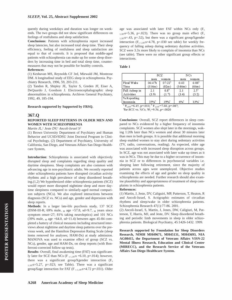
A268 American Academy of Sleep Medicine
PO
STE
R P
RE
SEN
TA
TIO
NS
SLEEP, Vol. 25, Abstract Supplement 2002
quently during weekdays and duration was longer on week-ends. The two groups did not show significant differences onfeelings of restfulness and sleep satisfaction.Conclusions: Patients with schizophrenia report increasedsleep latencies, but also increased total sleep time. Their sleepefficiency, feeling of restfulness and sleep satisfaction areequal to that of controls. It is proposed that middle-agedpatients with schizophrenia can make up for some sleep disor-ders by increasing time in bed and total sleep time, counter-measures that may not be possible for healthy controls.References: (1) Keshavan MS, Reynolds CF 3rd, Miewald JM, MontroseDM. A longitudinal study of EEG sleep in schizophrenia. Psy-chiatry Research, 1996, 59, 203-211.(2) Tandon R, Shipley JE, Taylor S, Greden JF, Eiser A,DeQuardo J, Goodson J. Electroencephalographic sleepabnormalities in schizophrenia. Archives General Psychiatry,1992, 49, 185-194.
Research supported by Supported by FRSQ.
367.QREPORTED SLEEP PATTERNS IN OLDER MEN ANDWOMEN WITH SCHIZOPHRENIAMartin JL,1 Jeste DV,1 Ancoli-Israel S2
(1) Brown University Department of Psychiatry and HumanBehavior and UCSD/SDSU Joint Doctoral Program in Clini-cal Psychology, (2) Department of Psychiatry, University ofCalifornia, San Diego, and Veterans Affairs San Diego Health-care System,
Introduction: Schizophrenia is associated with objectivelydisrupted sleep and complaints regarding sleep quality anddaytime sleepiness. Sleep complaints are also common withadvancing age in non-psychiatric adults. We recently reportedolder schizophrenia patients have disrupted circadian activityrhythms and a high prevalence of sleep disordered breath-ing.(1,2) We hypothesized older schizophrenia patients (SCZ)would report more disrupted nighttime sleep and more day-time sleepiness compared to similarly-aged normal compari-son subjects (NCs). We also explored interactions betweendiagnosis (SCZ vs. NCs) and age, gender and depression withsleep reports.Methods: In a larger late-life psychosis study, 137 SCZ(DSM-III-R; 69% male, M age =57.8, sd=9.7, M years sincesymptom onset=27; 81% taking neuroleptics) and 101 NCs(39% male, M age =64.0, sd=11.4) between ages 45-84 com-pleted a battery of clinical measures including structured inter-views about nighttime and daytime sleep patterns over the pre-vious week, and the Hamilton Depression Rating Scale (sleepitems removed for analyses; HAM-Ds) at study admission.MANOVA was used to examine effect of group (SCZ vs.NCs), gender, age and HAM-Ds, on sleep reports (with Bon-ferroni-corrected follow-up tests).Results: Overall, final awakening time (FAT) was significant-ly later for SCZ than NCs (F s 1,187s =6.10, p=.014); however,there was a significant groupXgender interaction (Fs
1,187s=5.27, p=.023; see Table). There was a significantgroupXage interaction for FAT (F s 1,187s=4.72 p=.031). Older
age was associated with later FAT within NCs only (Fs
1,187s=5.36, p=.023). There was no group main effect (Fs
1,187s=.43, p=.52), but there was a significant groupXgenderinteraction (Fs 1,187s=4.78, p=.030 see table) for weekly fre-quency of falling asleep during sedentary daytime activities.SCZ were 3.3x more likely to complain of insomnia than NCs(see table). There were no other significant group effects orinteractions.
Table 1
Conclusions: Overall, SCZ report differences in sleep com-pared to NCs evidenced by a higher frequency of insomniacomplaints. SCZ women also slept later in the mornings, wak-ing 1:20h later than NCs women and about 30 minutes laterthan men in both groups. It is possible that additional morningsleep enabled women to stay alert during sedentary activities(TV, radio, conversations, reading). As expected, older agewas associated with increased sleep disruption across groups.In SCZ, age was not associated with later wake up times as itwas in NCs. This may be due to a higher occurrence of insom-nia in SCZ or to differences in psychosocial variables i.e.sleeping later following retirement, since the majority ofpatients across ages were unemployed. Objective studiesexamining the effects of age and gender on sleep quality inschizophrenia are needed. Further research should also exam-ine plausibility and appropriateness of treatment of sleep com-plaints in schizophrenia patients.References: (1) Martin, J, Jeste, DV, Caliguiri, MP, Patterson, T, Heaton, Rand Ancoli-Israel, S. Actigraphic estimates of circadianrhythms and sleep/wake in older schizophrenia patients.Schizophrenia Research 47(1):77-86. 2001.(2) Ancoli-Israel, S, Martin, J, Jones, DW, Caligiuri, M, Pat-terson, T, Harris, MJ, and Jeste, DV. Sleep disordered breath-ing and periodic limb movements in sleep in older schizo-phrenia patients. Biological Psychiatry, 45:1426-1432. 1999.
Research supported by Foundation for Sleep DisordersResearch, NIMH MH49671, MH45131, MH43693, NIAAG08415, the Department of Veterans Affairs VISN-22Mental Illness Research, Education and Clinical Center(MIRECC), and the Research Service of the VeteransAffairs San Diego Healthcare System.
Sleep Research Society A269
PO
STE
R P
RE
SEN
TA
TIO
NS
SLEEP, Vol. 25, Abstract Supplement 2002
368.QSELF-REPORTED SLEEP DISTURBANCE ANDMOOD STATES IN EARLY ABSTINENT OPIATEADDICTSDavis HC,1 Lee KA,1 Smith D1
(1) University of California, San Francisco,
Introduction: Poor sleep in the general population has manydetrimental health outcomes, yet early reports suggest that theoutcomes in the addicted population contribute to the relapseprocess, as well as continued health and psychiatric problems.While many researchers have investigated sleep disturbance inother populations, few have researched this phenomenonamong drug-abusers. The primary purpose of this longitudinaldescriptive study was to characterize sleep/wake patterns inearly abstinence (10-90 drug-free days) of opiate-addictedmen and women in a therapeutic community treatment setting.Methods: A longitudinal design was employed to obtain base-line and repeated measures from a convenience sample of 22recently abstinent opiate-addicted men and women over a two-week period. Self-report of sleep was assessed with the Pitts-burgh Sleep Quality Index (PSQI) and the General Sleep Dis-turbance Scale (GSDS). The Beck Depression Inventory(BDI) was scored on each Monday as well as a 37-item Pro-file of Mood States (POMS) for four consecutive evenings.Visual Analogue Scales (VAS), with one item related to sleepquality were completed for each night for the entire two-weekperiod. A wrist actigraph was worn continuously for the com-plete study period of 14 days.
Table 1
Results: The results to date are presented for 13 men and 9women. The average age was 40.8 years (SD = 9.0) and edu-cation level ranged from eleventh grade to two years of col-lege. The length of sobriety averaged 49.7 (SD = 23.4). Theattrition rate was 14%. Cronbach’s alpha for the PSQI sleepdisturbance subscale was .71. The GSDS alpha coefficient was.70. The BDI’s alpha coefficient for time one and time two was.82 and .87 respectively. Preliminary findings in PSQI globalscores suggest opiate addicts in early abstinence period have ahigh degree of sleep disturbance (> 5) in this sample. GSDS
total scores ranged from 59.0 to 92.0, (scores > 60 indicatesleep disturbance). Women had a higher presence and severityof depression than men on the BDI initially, yet dropped attime 2 to a similar level to men. Sleep quality was rated on a100mm VAS anchored at 0 = “very poor” and 100 = “verygood”. The VAS results (Monday thru Friday) were highlycorrelated (r = .78) and not statically different from week oneto week two. The POMS fatigue-inertia correlated duringweek 1(r = .69 to .85). The vigor-activity subscale from time 1to time 4 was also correlated (r = .31 to .61).Conclusions: These data suggest a preponderance of dis-turbed sleep and depression as well as labile mood statesthroughout the early abstinence period by subjective meas-ures. Moreover, analysis of individual subscales may provideevidence to target specific interventions. Further analysis ofobjective measures of sleep/wake will provide a more detaileddescription of their sleep disturbance and its effect on daytimemood state.
Research supported by University of California, San Fran-cisco, Graduate Research Award and International NursesSociety of Addictions.
369.QA QUESTIONNAIRE-BASED STUDY OF SLEEP PROB-LEMS IN CHILDREN WITH AUTISM SPECTRUMDISORDERS.Giannotti F,1 Cortesi F,1 Cerquiglini A,1 Volzone A,1 Bernabei,P1
(1) Center of Pediatric Sleep Disorders - Dept. of Dev. Neurol& Psychiatry - University of Rome La Sapienza - Italy
Introduction: General high rates of sleep disturbances havebeen estimated for approximately 34 to 89% of children withAutism Spectrum Disorders (ASD). The majority of studieshave been carried out on heterogeneous groups and mostly ofthem on school-aged children. In order to making a longitudi-nal analysis of sleep behavior possible, we decided to investi-gate sleep on a sample of young children with ASD. As a partof longitudinal study on sleep problems in children with ASDand the long- term effect of behavioral and phamacologicaltreatment, we reported here preliminary baseline data. Methods: A sample of 25 children, (22 males, 3 females),aged 3.6-10.4 (mean age 4,3) with ASD was included in thestudy. Diagnosis was based on DSM IV criteria. Assessmentincluded: neurological and cognitive evaluation, ChildhoodAutism Rating Scale (CARS) and Vineland Adaptive Behav-ior Scale. To assess sleep problems, we used the Children’sSleep Habits Questionnaire (CSHQ, Owens, 2000) completedby parents. It yields both total and 8 subscales scores, reflect-ing key sleep domains. Higher score are indicative of moredisturbed sleep. Furthermore, a clinical interview inquiringprevious child’s sleep problems and current sleep patterns,including detailed information on bedtime routines and night-time behavior, was carried out. Moreover, parents were askedto complete a diary of child’s sleep. Baseline sleep variables,CSHQ scores were compared with sex- and age-matched con-trols.Results: Clinical interview comfirmed a high prevalence of
A270 American Academy of Sleep Medicine
PO
STE
R P
RE
SEN
TA
TIO
NS
SLEEP, Vol. 25, Abstract Supplement 2002
sleep problems with ASD, including bedtime difficulties(56%), nightwakings lasting more than 45 min (30%) or both(30%).No significant irregularity of sleep/wake pattern wasfound. Cosleeping, the practice of parents and children sleep-ing together in body contact for the whole night, was reportedby 42% of children with ASD compared to 8.3% of controls(chi square <.001). Parents of 67% of children with ASDreported sleep disturbances in the first two year of life com-pared to 17% of controls (chi square <.001). While in controlsonset of sleep disorders was in the first six months of life, inchildren with ASD a problematic sleep started usually fromthe second year, after a period of regular sleep/wake cycle.Children with ASD reported significant higher CSHQ scores(total 57 vs 37; bedtime resistance 12 vs 6; night wakings 11vs 8; parasomnias 5 vs 3, p<.001) . A significant negative cor-relation was found only between sleep duration and CARSscore (r = -.54; p<.01) . Conclusions: Consistent with other studies our childrenexhibit more dysomnias and parasomnias.Therefore, assesss-ment of sleep in children with ASD need particular attention,not only because of the high prevalence, persistence and sever-ity of the problem, but also for the additional distress that itmay place upon caretakers, and for the contribution that a dis-turbed sleep might be making to daytime difficulties andbehavior.References: (1) Owens JA et al. The Children’s Sleep Habits Question-naire: Psychometric Properties of A Survey Instrument forSchool-Aged Children. SLEEP 2000; 23, 8: 1043-1051(2) Richdale AL. Sleep problems in autism: prevalence, cause,and intervention. Dev Medicine Child Neurology, 1999,41:60-66
370.RTHE USE OF WRIST ACTIGRAPHY TO INCREASECOMPLIANCE TO SLEEP HYGIENE INSTRUCTIONSIN UNDERGRADUATESCarney CE,1 Lajos LE,1 Waters WF1
(1) Department of Psychology, Louisiana State University,
Introduction: Compliance is a term referring to adherance tomedical/treatment advice (Spilker, 1991). The interpretationof experiments involving human behavior can be confoundedwhen research participants fail to comply with a study’sinstructions. The use of wrist actigraphy has become a widelyused and inexpensive method of objective activity monitoringover 24-hour periods. Although applications to the field ofsleep disorders medicine are numerous (Ancoli-Israel, 2000),it is unclear if instructions regarding the actigraphy procedurecan significantly affect adherence. The present study com-pared compliance with sleep hygiene rules in subjects whowere told that compliance would be verified with actigraphy,to subjects who were not told the actigraph would be used toassess compliance.Methods: Sixty-eight undergraduate psychology students atLouisiana State University, age18-30 years old, were givencourse credit for their participation. Participants were screenedfor (1) DSM-IV Axis I Disorders known to affect sleep archi-tecture and/or sleep quality/quantity (e.g., anxiety, depression,
substance abuse/dependence); (2) self-reported signs of sleepdisorders using a Sleep Disorders Inventory, and; (3) extremesin circadian tendency using the Morningness/EveningnessQuestionnaire. There were 30 participants in the “instruction”group (IG) and 38 in the “no instruction” group (NIG). The IGwere given instructions that stressed that the actigraph wouldverify reported sleep diary data, while the NIG was told thatthe unit would simply monitor their activity.Results: A repeated measures MANOVA confirmed a maineffect of group (Wilks’ Lambda=.780, F[6,60]=2.82, p=.017),and follow-up ANOVAs confirmed a between group differ-ence on Actigraphy bedtime (F=14.181, p=.001) and Actigra-phy wake time (F=11.179, p=.001). Those in the NIG had laterbedtimes and later waketimes, relative to the IG, and relativeto the bedtime specified in the sleep hygiene instructions.Conclusions: Although both groups were instructed to go tobed at a specified time (e.g., between 9 PM and 11 PM) andawaken 8 hours later (e.g., between 6 AM and 8 AM), the sub-jects who was told the actigraph would be used to verifywhether they went to bed and woke at the specified times weremore compliant than those who were not told their compliancewould be verified. This has implications for studies requiringparticipants to follow sleep hygiene instructions, or in sleepdisorder treatment to help ensure compliance with therapeuticrecommendations.References: (1) Spilker, B. Guide to Clinical Trials. New York, NY: RavenPress, 1991.(2) Ancoli-Israel, S. Actigraphy. In MH Kryger, T Roth, WCDement (Eds.). Principles and Practice of Sleep Medicine(Third Edition). Philadelphia, WB Saunders, 2000.
371.RRECORDING MSLT IN THE HOMEMason WJ,1 Gehrman P,1 Greenfield D,1 Ancoli-Israel S1
(1) Department of Psychiatry, UCSD and VASDHS,
Introduction: Multiple sleep latency tests (MSLT) are alwaysconducted in the laboratory to determine sleep onset latencythereby estimating the level of daytime sleepiness. StandardMSLTs are conducted the morning after an all-nightpolysomnogram (PSG). However, there are some situationswhere it may not be possible to have the patient come into thelaboratory. For this reason, many research and clinical labora-tories now conduct PSGs in the home. As part of a larger studyon sleep in dementia, we have been conducting both PSGs andMSLTs in the home.Methods: Subjects todate include 4 men and 1 woman (meanage \equiv 71.4 years; range: 54 to 82 years. All had a diagno-sis of possible or probable Alzheimer’s disease (AD). Eachsubject had a home MSLT and two had two additional homeMSLTs, each three weeks apart. The first and third MSLT fol-lowed a home PSG. The second MSLT was done with no pre-vious night’s PSG. Embla recording system (Flaga MedicalDevices, Iceland) and the Somnologica scoring program wereused for all PSGs and a Dell Latitude laptop computer wasadded for the MSLT recordings. The recordings were doneaccording to published criteria.(1) The subjects were instruct-ed to lie down and try to sleep. The sleep recording was mon-
Sleep Research Society A271
PO
STE
R P
RE
SEN
TA
TIO
NS
SLEEP, Vol. 25, Abstract Supplement 2002
itored by the technician via the laptop computer. If the subjectdid not fall asleep within twenty minutes, or if three epochs ofstage 1 sleep or an epoch of any other stage of sleep was seen,the subject was awakened and allowed to go about his or hernormal daily activities. The technician periodically checkedthe subjects to be sure they had not fallen asleep during theinterval between the recorded naps. Oft times the subjectrequired encouragement to continue the study as they wouldsometimes forget about the full day commitment. Electrodeshad to be replaced about 45% of the time when the subjectsremoved them.Results: Environmental aspects could not be controlled aswell in the home as in the laboratory. The subject’s bedroomswere darkened as much as possible, using curtains and blinds.All windows and doors were shut. A sign was hung outside thebedroom door reminding other household members that astudy was in progress and requesting quiet and remindingthem not to enter. The figure shows the mean sleep onsetlatency (SOL) of the group.
Figure 1
Conclusions: Although home MSLTs do not fully conform tothe published criteria for the laboratory, we have shown that itis possible to gather information about daytime sleepiness inhome-bound populations, particularly in patients with demen-tia.References: (1) Carskadon MA, Dement WC, Mitler MM, Roth T, West-brook PR, Keenan S. Guidelines for the multiple sleep latencytest (MSLT): a standard measure of sleepiness. Sleep 1986;9:519-524.
Research supported by AG08415, MIRECC VISN 22,VASDHS research service
372.RSLEEP STAGE SHIFT INDEX AS AN INDICATOR FORTHE LEVEL OF DAYTIME SLEEPINESS AND ALERT-NESSTrajanovic N, Jovanovic D, Kayumov LY, Shapiro CM(1) Sleep and Alertness Clinic, Toronto, Canada, (2) Universi-ty of Toronto, Toronto, Canada, (3) TASC, Fort Erie, Canada
Introduction: Several sleep parameters have been identifiedas predictors for the level of daytime sleepiness and alertness.
Most robust indicators are sleep efficiency, sleep duration andarousal index. Our interest was to investigate the potency ofthe sleep stage shift index as such indicator.Methods: A retrospective study was carried out utilising largedatabase of eight hundred patients who underwent a sleepassessment in a tertiary unit during the 18-month period. Thiscohort served as a primary pool that provided the study sam-ple. A subject was included if following criteria was met: asuccessful overnight sleep study, Multiple Sleep Latency Test(MSLT) or Maintenance of Wakefulness Test (MWT), andeither of two – sleep stage shift index (SSSI) of >30 and arous-al index (AI) of <15, or SSSI <20 and AI of <15. The cut-offSSSI of 30 and AI of 15 were set arbitrarily, having in mindcommonly used normal values (15 for SSSI, and5 for AI). Four subgroups were formed with subjectmatched for sleep efficiency, age and AI, as tabulatedbelow(Group I- High SSSI (MSLT; Group II -Normal SSSI(MSLT); Group III- High SSSI (MWT); Group IV - NormalSSSI (MWT:Results: The subgroup selection and matching provided thatthe only major variable distinguishing respective subgroupswas the SSSI. The most obvious finding during the matchingwas that there were very few subjects who had relatively nor-mal AI and high SSSI at the same time. This dramaticallyreduced the numbers in all matched subgroups. The Indepen-dent samples’ Mann-Whitney U-test with Bonferroni correc-tion for p-values was used for statistical analysis of the results.This analysis suggests that the number of sleep stage shifts isan independent predictor of daytime sleepiness, but not ofimpaired daytime alertness. We were not able to explain thisdiscrepancy aside from the possibility that the subject match-ing was less effective in the later group.
Table 1
Conclusions: We are aware of the critical reduction of thesample size that came as a result the multi-variable matching.Nevertheless, we were able to show that the SSSI could beused as one of the predictors of excessive daytime sleepiness,as the observed trends indicate. This may prove to be helpfulin understanding the arousal mechanisms and their relationwith different (sub)cortical EEG generators. The analysis of alarger sample is merited to confirm the observed trend, andshould also help in understanding the absence of such trend inthe MWT group, as well as relative absence of REM periodsduring daytests. The results also confirm that MSLT and MWTmeasure different neuro-physiological domains.
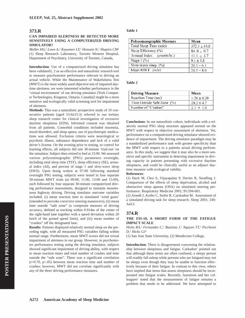
A272 American Academy of Sleep Medicine
PO
STE
R P
RE
SEN
TA
TIO
NS
SLEEP, Vol. 25, Abstract Supplement 2002
373.RCAN IMPAIRED ALERTNESS BE DETECTED MORESENSITIVELY USING A COMPUTERIZED DRIVINGSIMULATOR?Moller HG,1 Lowe A,1 Kayumov LY,1 Hossain N,1 Shapiro CM1
(1) Sleep Research Laboratory, Toronto Western Hospital,Department of Psychiatry, University of Toronto, Canada,
Introduction: Use of a computerized driving simulator hasbeen validated1, 2 as an effective and naturalistic research toolto measure psychomotor performance relevant to driving anactual vehicle. While the Maintenance of Wakefulness Test(MWT) is the most widely used objective test of impaired day-time alertness, we were interested whether performance in the‘virtual environment’ of our driving simulator (York Comput-er Technologies, Kingston, Ontario, Canada)2 might be a moresensitive and ecologically valid screening tool for impairmentof alertness.Methods: This was a naturalistic prospective study of 10 con-secutive patients (aged 53.6±15.3) referred to our tertiarysleep research center for clinical investigation of excessivedaytime sleepiness (EDS). Informed consent was obtainedfrom all patients. Comorbid conditions included insomnia,mood disorders, and sleep apnea; use of psychotropic medica-tions was allowed. Exclusion criteria were neurological orpsychotic illness, substance dependence and lack of a validdriver’s license. On the evening prior to testing, to control forlearning effects, all subjects did one 30-minute ‘trial-run’ onthe simulator. Subject then retired to bed at 23:00. We assessedvarious polysomnographic (PSG) parameters overnight,including total sleep time (TST), sleep efficiency (SE), arous-al index (AI), and percent of stage 1 and slow-wave sleep(SWS). Upon being woken at 07:00 following standardovernight PSG testing, subjects were tested in four separate30-minute MWT trials (at 09:00, 11:00, 13:00 and 15:00),each followed by four separate 30-minute computerized driv-ing performance assessments, designed to simulate monoto-nous highway driving. Driving simulator outcome variablesincluded: (i) mean reaction time to simulated ‘wind gusts’(intended to provoke corrective steering maneuvers), (ii) meantime outside “safe zone” (a composite measure of drivingaccuracy, defined as tracking within 0.914m of the center ofthe right-hand lane together with a speed deviation within 20km/h of the posted speed limit), and (iii) mean number of“crashes” off the designated lane.Results: Patients displayed relatively normal sleep on the pre-ceding night, with all measured PSG variables falling withinnormal range. Furthermore, mean MWT scores did not revealimpairment of alertness in our group. However, in psychomo-tor performance testing using the driving simulator, subjectsshowed significant impairment of driving ability, with respectto mean reaction times and total number of crashes and timeoutside the “safe zone”. There was a significant correlation(r=0.70, p<.05) between mean reaction time and number ofcrashes; however, MWT did not correlate significantly withany of the three driving performance measures.
Table 1
Table 2
Conclusions: In our naturalistic cohort, individuals with a rel-atively normal PSG sleep structure appeared normal on theMWT with respect to objective assessment of alertness. Yet,performance on a computerized driving simulator showed evi-dence of impairment. The driving simulator paradigm may bea standardized performance task with greater specificity thanthe MWT with respect to a patients actual driving perform-ance. In this study, we suggest that it may also be a more sen-sitive and specific instrument in detecting impairment in driv-ing capacity in patients presenting with excessive daytimesleepiness, and could be clinically useful as an adjunct day-time measure with ecological validity.References: (1) Hack M, Choi S, Vijayapalan P, Davies R, Stradling J.Comparison of the effects of sleep deprivation, alcohol andobstructive sleep apnoea (OSA) on simulated steering per-formance. Respiratory Medicine 2001; 95:594-601.(2) Arnedt J, Acebo C, Seifer R, Carskadon M. Assessment ofa simulated driving task for sleep research. Sleep 2001; 24S:A413.
374.RTHE FIS-10, A SHORT FORM OF THE FATIGUEIMPACT SCALEHicks RA,1 Fernandez C,1 Bautista J,1 Nguyen TT,1 PicchioniD,1 Hicks GJ1
(1) San Jose State University, (2) Mendocino College,
Introduction: There is disagreement concerning the relation-ship between sleepiness and fatigue. Carkadon1 pointed outthat although these terms are often confused, a sleepy personwill readily fall asleep while persons who are fatigued may notbe sleepy even though they may be unable to function effec-tively because of their fatigue. In contrast to this view, othershave implied that items that assess sleepiness should be incor-porated into fatigue scales. Recently, Aaronson and her col-leagues2 noted that the measurement of fatigue remains aproblem that needs to be addressed. We have attempted to
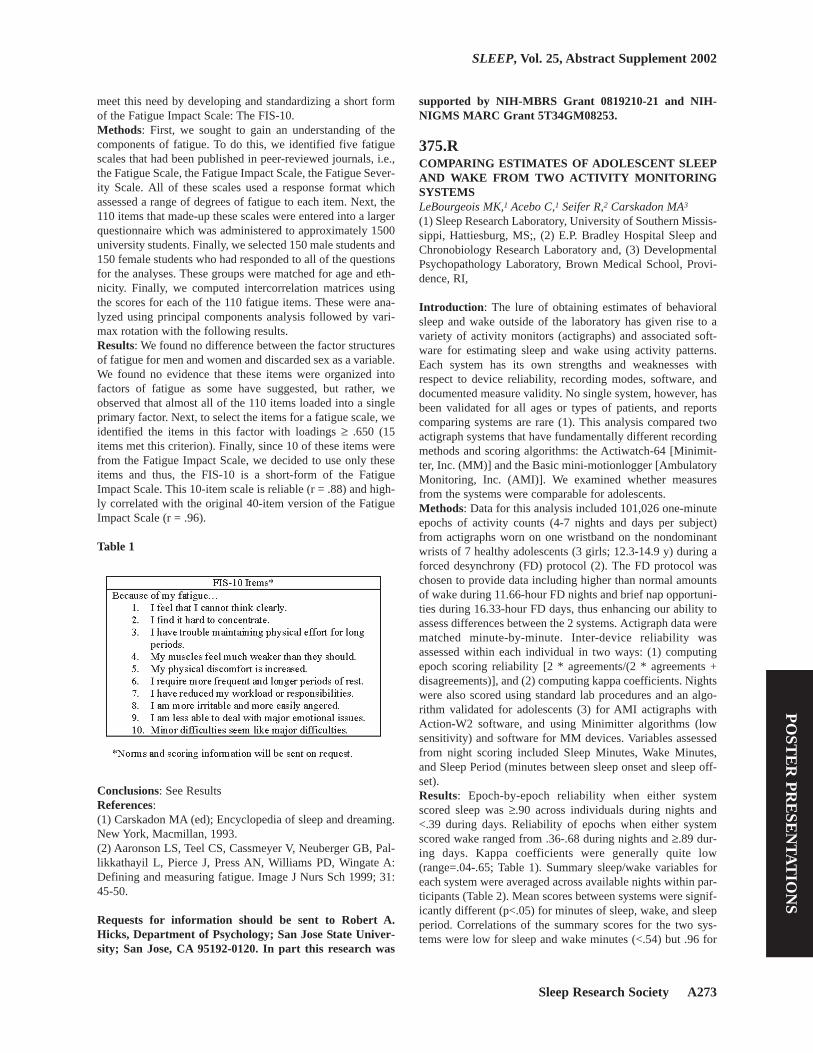
Sleep Research Society A273
PO
STE
R P
RE
SEN
TA
TIO
NS
SLEEP, Vol. 25, Abstract Supplement 2002
meet this need by developing and standardizing a short formof the Fatigue Impact Scale: The FIS-10.Methods: First, we sought to gain an understanding of thecomponents of fatigue. To do this, we identified five fatiguescales that had been published in peer-reviewed journals, i.e.,the Fatigue Scale, the Fatigue Impact Scale, the Fatigue Sever-ity Scale. All of these scales used a response format whichassessed a range of degrees of fatigue to each item. Next, the110 items that made-up these scales were entered into a largerquestionnaire which was administered to approximately 1500university students. Finally, we selected 150 male students and150 female students who had responded to all of the questionsfor the analyses. These groups were matched for age and eth-nicity. Finally, we computed intercorrelation matrices usingthe scores for each of the 110 fatigue items. These were ana-lyzed using principal components analysis followed by vari-max rotation with the following results.Results: We found no difference between the factor structuresof fatigue for men and women and discarded sex as a variable.We found no evidence that these items were organized intofactors of fatigue as some have suggested, but rather, weobserved that almost all of the 110 items loaded into a singleprimary factor. Next, to select the items for a fatigue scale, weidentified the items in this factor with loadings ≥ .650 (15items met this criterion). Finally, since 10 of these items werefrom the Fatigue Impact Scale, we decided to use only theseitems and thus, the FIS-10 is a short-form of the FatigueImpact Scale. This 10-item scale is reliable (r = .88) and high-ly correlated with the original 40-item version of the FatigueImpact Scale (r = .96).
Table 1
Conclusions: See ResultsReferences: (1) Carskadon MA (ed); Encyclopedia of sleep and dreaming.New York, Macmillan, 1993.(2) Aaronson LS, Teel CS, Cassmeyer V, Neuberger GB, Pal-likkathayil L, Pierce J, Press AN, Williams PD, Wingate A:Defining and measuring fatigue. Image J Nurs Sch 1999; 31:45-50.
Requests for information should be sent to Robert A.Hicks, Department of Psychology; San Jose State Univer-sity; San Jose, CA 95192-0120. In part this research was
supported by NIH-MBRS Grant 0819210-21 and NIH-NIGMS MARC Grant 5T34GM08253.
375.RCOMPARING ESTIMATES OF ADOLESCENT SLEEPAND WAKE FROM TWO ACTIVITY MONITORINGSYSTEMSLeBourgeois MK,1 Acebo C,1 Seifer R,2 Carskadon MA3
(1) Sleep Research Laboratory, University of Southern Missis-sippi, Hattiesburg, MS;, (2) E.P. Bradley Hospital Sleep andChronobiology Research Laboratory and, (3) DevelopmentalPsychopathology Laboratory, Brown Medical School, Provi-dence, RI,
Introduction: The lure of obtaining estimates of behavioralsleep and wake outside of the laboratory has given rise to avariety of activity monitors (actigraphs) and associated soft-ware for estimating sleep and wake using activity patterns.Each system has its own strengths and weaknesses withrespect to device reliability, recording modes, software, anddocumented measure validity. No single system, however, hasbeen validated for all ages or types of patients, and reportscomparing systems are rare (1). This analysis compared twoactigraph systems that have fundamentally different recordingmethods and scoring algorithms: the Actiwatch-64 [Minimit-ter, Inc. (MM)] and the Basic mini-motionlogger [AmbulatoryMonitoring, Inc. (AMI)]. We examined whether measuresfrom the systems were comparable for adolescents.Methods: Data for this analysis included 101,026 one-minuteepochs of activity counts (4-7 nights and days per subject)from actigraphs worn on one wristband on the nondominantwrists of 7 healthy adolescents (3 girls; 12.3-14.9 y) during aforced desynchrony (FD) protocol (2). The FD protocol waschosen to provide data including higher than normal amountsof wake during 11.66-hour FD nights and brief nap opportuni-ties during 16.33-hour FD days, thus enhancing our ability toassess differences between the 2 systems. Actigraph data werematched minute-by-minute. Inter-device reliability wasassessed within each individual in two ways: (1) computingepoch scoring reliability [2 * agreements/(2 * agreements +disagreements)], and (2) computing kappa coefficients. Nightswere also scored using standard lab procedures and an algo-rithm validated for adolescents (3) for AMI actigraphs withAction-W2 software, and using Minimitter algorithms (lowsensitivity) and software for MM devices. Variables assessedfrom night scoring included Sleep Minutes, Wake Minutes,and Sleep Period (minutes between sleep onset and sleep off-set).Results: Epoch-by-epoch reliability when either systemscored sleep was ≥.90 across individuals during nights and<.39 during days. Reliability of epochs when either systemscored wake ranged from .36-.68 during nights and ≥.89 dur-ing days. Kappa coefficients were generally quite low(range=.04-.65; Table 1). Summary sleep/wake variables foreach system were averaged across available nights within par-ticipants (Table 2). Mean scores between systems were signif-icantly different (p<.05) for minutes of sleep, wake, and sleepperiod. Correlations of the summary scores for the two sys-tems were low for sleep and wake minutes (<.54) but .96 for
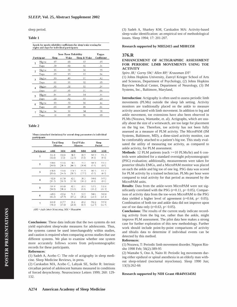
A274 American Academy of Sleep Medicine
PO
STE
R P
RE
SEN
TA
TIO
NS
SLEEP, Vol. 25, Abstract Supplement 2002
sleep period.
Table 1
Table 2
Conclusions: These data indicate that the two systems do notyield equivalent sleep/wake measures for adolescents. Thus,the systems cannot be used interchangeably within studies,and caution is required when comparing across studies that usedifferent systems. We plan to examine whether one systemmore accurately follows scores from polysomnographicrecords for these participants.References: (1) Sadeh A, Acebo C: The role of actigraphy in sleep medi-cine. Sleep Medicine Reviews, in press.(2) Carskadon MA, Acebo C, Labyak SE, Seifer R: Intrinsiccircadian period of adolescent humans measured in conditionsof forced desynchrony. Neuroscience Letters 1999; 260: 129-132.
(3) Sadeh A, Sharkey KM, Carskadon MA: Activity-basedsleep-wake identification: an empirical test of methodologicalissues. Sleep 1994; 17: 201-207.
Research supported by MH52415 and MH01358
376.RENHANCEMENT OF ACTIGRAPHIC ASSESSMENTFOR PERIODIC LIMB MOVEMENTS USING TOEACTIVITYSpiro JR,1 Gorny SW,1 Allen RP,1 Krausman DT2
(1) Johns Hopkins University, Zanvyl Krieger School of Artsand Sciences, Department of Psychology, (2) Johns HopkinsBayview Medical Center, Department of Neurology, (3) IMSystems, Inc., Baltimore, Maryland,
Introduction: Actigraphy is often used to assess periodic limbmovements (PLMs) outside the sleep lab setting. Activitymonitors are traditionally placed on the ankle to measureactivity associated with limb movement. In addition to leg andankle movement, toe extensions have also been observed inPLMs (Nozawa, Wantanbe, et. al). Actigraphs, which are usu-ally about the size of a wristwatch, are too large for placementon the big toe. Therefore, toe activity has not been fullyassessed as a measure of PLM activity. The MicroPAM (IMSystems, Baltimore, MD), a dime-sized activity monitor, canbe comfortably attached to a patient’s big toe. This study eval-uated the utility of measuring toe activity, as compared toankle activity, for PLM assessment.Methods: 12 PLM patients (each >=10 PLMs/hr) and 6 con-trols were admitted for a standard overnight polysomnograpic(PSG) evaluation; additionally, measurements were taken forposterior tibialis EMGs, and a MicroPAM monitor was placedon each the ankle and big toe of one leg. PSG data was scoredfor PLM activity by a trained technician. PLMs per hour werecompared to total activity for that period as measured by theMicroPAM units.Results: Data from the ankle-worn MicroPAM were not sig-nificantly correlated with the PSG (r=0.11, p> 0.05). Compar-ison of activity data from the toe-worn MicroPAM to the PSGdata yielded a higher level of agreement (r=0.64, p< 0.05).Combination of both toe and ankle data did not improve uponuse of toe data only (r=0.63, p< 0.05).Conclusions: The results of the current study indicate record-ing activity from the big toe, rather than the ankle, mightimprove PLM assessment. The pilot data here makes a strongcase for further exploration of this new methodology. Furtherwork should include point-by-point comparisons of activityand tibialis data to determine if individual events can bedetected by this method.References: (1) Nozawa, T: Periodic limb movement disorder. Nippon Rin-sho 1998 Feb; 56(2):389-95(2) Wantabe S, Ono A, Naito H: Periodic leg movements dur-ing either epidural or spinal anesthesia in an elderly man with-out sleep-related (nocturnal myoclonus). Sleep 1990 Jun;13(3):262-66
Research supported by NIH Grant #R44NS34592
Sleep Research Society A275
PO
STE
R P
RE
SEN
TA
TIO
NS
SLEEP, Vol. 25, Abstract Supplement 2002
377.RPILOT EVALUATION OF AN AMUBULATORY AIR-FLOW PRESSURE MONITOR FOR IMMEDIATEIDENTIFICATION OF SLEEP DISORDEREDBREATHING EVENTSSpiro JR,1 Gorny SW,1 Allen RP,1 Krausman DT2
(1) Johns Hopkins University, Zanvyl Krieger School of Artsand Sciences, Department of Psychology, (2) Johns HopkinsBayview Medical Center, Department of Neurology, (3) IMSystems, Inc., Baltimore, Maryland,
Introduction: There are several systems available for at-homerecording of Sleep Disordered Breathing Events (SDBEs).One type typically consists of a small tabletop recorder, withan array of tethered sensors, from which data must be down-loaded to a computer for analysis. A second type is self-con-tained, disposable unit that provides an on-line indication ofSDBE severity at the end of a sleep period, but can only beused for a single night. These are features not fully efficientfor use in a clinical setting. A new version of the ApneaCheck(IM Systems, Baltimore, MD) was designed to quantifySDBEs for multiple nights, in real-time, with no off-lineanalysis required; a cumulative count of detected SDBEs isdisplayed on an LCD display. The current pilot study evaluat-ed the efficacy and accuracy of this new ApneaCheck model.Methods: 6 participants with a range of SDBE severity, wereadmitted for a standard, overnight, polysomnographic (PSG)evaluation. Participants were also outfitted with theApneaCheck monitor, which is comprised of a small, self-con-tained recording unit that is clipped to the wearer’s shirt andattached to a disposable airflow pressure cannula. To facilitatecomparison with standard PSG measure, the ApneaCheckmonitor used in the current study was specially modified bythe manufacturer to allow two channels of information to besent from the monitor to the PSG: the air pressure signal and amarker (a pen deflection on the PSG tracing when theApneaCheck’s algorithm detected a SDBE). The PSG datawas scored by a trained technician for the identification ofSDBEs, defined as a reduction of 50% or greater in air pres-sure, lasting for a minimum of 10 seconds. A point-by-pointcomparison of the PSG and ApneaCheck estimations ofSDBEs was conducted. Each participant then wore the deviceat home for two consecutive nights to evaluate comfort andease of self application.Results: Point-by-point comparison of the PSG andApneaCheck data indicated a high level of agreement. Of the315 apnea events identified from the PSG data across allpatients, the ApneaCheck yielded an 82.3% sensitivity and89.9% specificity. Further analysis indicated that discrepan-cies between the two measures typically occurred when apotential SDBE had borderline characteristics for accept-ance/rejection as a candidate event, either in terms of durationpercentage or airflow pressure decrease. In the at-home trials,all subjects reported that the ApneaCheck monitor was easy touse and caused no significant sleep disturbance. The observa-tion of expected data for all subjects during the at-home trialsindicated that the monitors were properly self-applied.Conclusions: The results of this pilot study indicate that thelatest version of the ApneaCheck device provides a new and
unique means of identifying and recording SDBEs without theuse of traditional, multi-channel PSG recording. A larger scalestudy is now necessary to assess the technology more fully andto suggest possible enhancements to the overall accuracy ofthe system. The stand-alone, non-downloadable design of thesystem represents a significant technological advance in thedevelopment of ambulatory devices for measurement ofSDBEs.
Research supported by NIH Grant # 1R43HL65166
378.SUSE OF AN OBJECTIVE STANDARDIZED CLINICALEXAMINATION (OSCE) IN SLEEP MEDICINE TRAIN-INGWare J,1 Vorona RD,1 Matson CC,1 Ullian JA,1 Winn MP1
(1) Eastern Virginia Medical School,
Introduction: We used an Objective Standardized ClinicalExamination (OSCE) case to investigate whether a lecture tothird year medicine clerks could improve students’ physicalexams when obstructive sleep apnea syndrome (OSAS) wassuspected. For the case, a standardized patient presents withmorning headaches, and reveals on careful history symptomsof OSAS. Students have 20 minutes for a history and focusedphysical examination (PE). For the class graduating in 2001,most students correctly diagnosed the patient. However, thefocused PE often did not match their diagnosis, lackinginspection of the oral cavity and nose despite a diagnosis ofOSAS. Therefore, we studied whether a lecture to third yearstudents on the PE of suspected OSAS patients would improvesubsequent performance.Methods: One of the authors [RV] provided 45-minute lectureon the PE for apnea to 41 students taking Internal Medicineclerkships in the last half of their third year. Those taking theclerkship in the first half did not receive the lecture, and servedas the control group. The lecture covered the definition andepidemiology of OSAS, pertinent physical findings includingvital signs (specifically blood pressure), and body habitus. Itstressed the importance of the examination of the nose,oropharynx and facial features, including. the modified Mal-lampati classification of oropharyngeal anatomy as well asdental and jaw findings. Discussion of cardiopulmonary stig-mata of pulmonary hypertension and the overlap syndromefollowed, as did ocular findings such as floppy eyelid syn-drome, endocrine abnormalities including acromegaly andhypothyroidism, findings of end-stage renal disease, and Mar-fan’s syndrome. At the beginning of their fourth year, 100 stu-dents completed a nine-station OSCE. Student scores for theOSA case were based on a 78-item checklist (including PEitems) completed by the standardized patient and ten multiple-choice questions answered by the student. Those who admin-istered and scored the OSCE were blind to which students hadreceived the lecture.Results: We examined the PE scores on the eight non-apneacases and the OSAS case. Because of skewed PE data, we useda log transformation. The mean score for the lecture and con-trol groups on the non-OSA cases was 1.79 (t = 0.286; p =0.78, ns). For the OSA case, the mean score for the lecture

A276 American Academy of Sleep Medicine
PO
STE
R P
RE
SEN
TA
TIO
NS
SLEEP, Vol. 25, Abstract Supplement 2002
group was 1.45 versus 1.37 for the control group (t = 1.97; p= 0.051).Conclusions: The development and use of an OSCE OSAScase 1) resulted in an additional sleep medicine teachingopportunity, 2) revealed a deficiency in the PE performance,and 3) gave us information on the effectiveness of a lectureaddressing that deficiency. Although the probability that thisone lecture would affect OSCE performance several monthslater may be unlikely, we believe that students’ poor perform-ance on the previous year’s OSCE increased the likelihood forimprovement. However, coordination with sleep medicine andward attendings for subsequent demonstration of PE tech-niques with patients is probably necessary for long-termimprovement in students’ performance.
Research supported by NIH Award HL03652-01A1
379.SSLEEP APNEA, INSOMNIA AND DEPRESSION INTHE PRIMARY CARE SETTINGMaresky HS,1 Chung S,1 Hussain MG,1 Shapiro1
(1) Sleep Research Laboratory, Toronto Western Hospital,University Health Network,
Introduction: Sleep disorders are routinely under-diagnosedby primary care physicians. Education sessions aimed at thesephysicians are attempting to improve this problem but small-town primary care physicians are often not able to attend ses-sions, which are held in large urban centres. The aim of thispilot study was to examine if sleep disorders constitute animportant part of small-town primary practice by investigatingwhat proportion of small-town primary care patients screenpositive for sleep apnea, insomnia and depression. Methods: Waiting room patients for five primary care physi-cians in two small towns (Beamsville and Fort Erie) in South-ern Ontario, Canada, were administered questionnaires toscreen for sleep apnea (the Berlin Questionnaire), insomnia(the Athens Insomnia Scale) and depression (the CES-Dscale). Physicians were contacted in advance and permissionto approach their patients was obtained. The patients wereasked to complete the questionnaire battery before going in tosee their physician. The outcome variables were the scores onthe three questionnaires.Results: In total, 141 patients across the five practices agreedto be interviewed. Of those patients, 33% screened positive forsleep apnea, 38% screened positive for insomnia and 28%screened positive for depression. On further analysis of thetotal population, 8% screened positive for both insomnia andsleep apnea, 1% for sleep apnea and depression, 14% forinsomnia and depression and 6% screened positive for allthree disorders. None of these patients were currently diag-nosed with or were undergoing treatment for any of the threedisorders. As one purpose of this exercise was to help informphysicians an outline of our findings in these patients wasgiven to their physician.Conclusions: A very high proportion of small-town primarycare patients screen positive for sleep apnea, insomnia anddepression, as assessed by self-report questionnaires. The per-centages are well above the population norms. This study sup-
ports earlier findings that patients with sleep disorders are notdiagnosed as such and emphasizes that a substantial number ofsmall-town primary care patients may have undetected sleepapnea, insomnia and depression. The high frequency ofdepression could be in part secondary to the untreated sleepapnea and insomnia. The reason for the failed diagnosis of thesleep disorders cannot be attributed to an absence of facilitiesto which to refer patients since a sleep clinic is located in theimmediate area. A likely reason for the inability of primarycare physicians to identify these patients is a lack of knowl-edge about sleep disorders. The findings of this study supportour previous findings of a lack of primary care physicians’knowledge about sleep apnea and underscore the need formaking post-graduate education in sleep medicine available toprimary care physicians in non-urban areas.
380.UWORK SCHEDULES, STRESSFUL LIFE EVENTS,AND SLEEP QUALITY IN WOMENChaiarj S,1 Brown PS,1 Floyd JA,2 Watson NL2
(1) Faculty of Nursing, Chiang Mai University, (2) College ofNursing, Wayne State University,
Introduction: Over 25% of the US workforce is engaged insome form of shift work; nearly half are women. Monk 1 hassuggested that the quality of sleep for women shift workersmay be of special concern because of life stress. The majorquestions addressed in this study were: (1) Are women whosework schedules involve day shifts only significantly differentregarding life stress and sleep quality from women whosework schedules involve other shifts? (2) Is life stress related tosleep quality? (3) Are background characteristics (age, hor-mone replacement therapy, and employment in a health-careprofession) related to reports of stressful life events or sleepquality?Methods: The subjects for this project were 476 employedwomen aged 18-74 years old (M=43.3, SD=10.6). Other sam-ple characteristics are shown on Table 1. Sixty-four percent(n=303) of subjects worked day shifts only. Those whoworked days only were compared with all other shifts com-bined. Table 2 shows the frequency with which other shifts orcombinations of shifts were worked. Life stress was measuredby the Holms and Raye Life Events Checklist (LEC) 2 usingunit scoring. Sleep quality was measured by the Global Scoreon the Pittsburgh Sleep Quality Index (PSQI) 3 . Non-para-metric statistics were used whenever variables were not nor-mally distributed in the population. Alpha was set at .05.Results: The number of stressful life events ranged from 0-14(M=2.3, SD=2.3). The median was 2.0; the mode was 1.0. TheGlobal PSQI Score ranged from 0-16 (M=6.1, SD=3.1). Forthe total sample poor sleep quality was related to the numberof stressful life events, Kendall’s tau_b =.14, p = .000 butunrelated to age. Work schedules were not related to the num-ber of reported stressful life events, but dayshift-only workershad better sleep quality (M=5.9, SD=3.0) than those with otherwork schedules (M=6.5, SD=3.4), t (428) = 1.76, p = .039.Older subjects reported fewer stressful life events thanyounger subjects, Kendall’s tau_b = .10, p = .007. Neither hor-mone replacement therapy nor employment in a health-care
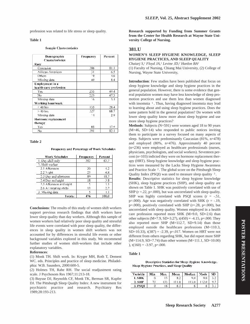
Sleep Research Society A277
PO
STE
R P
RE
SEN
TA
TIO
NS
SLEEP, Vol. 25, Abstract Supplement 2002
profession was related to life stress or sleep quality.
Table 1
Table 2
Conclusions: The results of this study of women shift workerssupport previous research findings that shift workers havelower sleep quality than day workers. Although this sample ofwomen workers had relatively poor sleep quality and stressfullife events were correlated with poor sleep quality, the differ-ences in sleep quality in women shift workers was notaccounted for by differences in stressful life events or otherbackground variables explored in this study. We recommendfurther studies of women shift-workers that include otherexplanatory variables. References: (1) Monk TH. Shift work. In: Kryger MH, Roth T, DementWC. eds. Principles and practice of sleep medicine. Philadel-phia: W.B. Saunders, 2000:600-5.(2) Holmes TH, Rahe RH. The social readjustment ratingscale. J Psychosom Res 1967;11:213-18.(3) Buysse DJ, Reynolds CF, Monk TK, Berman SR, KupferDJ. The Pittsburgh Sleep Quality Index: A new instrument forpsychiatric practice and research. Psychiatry Res1989;28:193-213.
Research supported by Funding from Summer Grantsfrom the Center for Health Research at Wayne State Uni-versity College of Nursing.
381.UWOMEN’S SLEEP HYGIENE KNOWLEDGE, SLEEPHYGIENE PRACTICES, AND SLEEP QUALITYChaiarj S,1 Floyd JA,1 Levine JD,2 Hanlon BA2
(1) Faculty of Nursing, Chiang Mai University, (2) College ofNursing, Wayne State University,
Introduction: Few studies have been published that focus onsleep hygiene knowledge and sleep hygiene practices in thegeneral population. However, there is some evidence that gen-eral population women may have less knowledge of sleep pro-motion practices and use them less than women diagnosedwith insomnia 1 . Thus, having diagnosed insomnia may leadto learning about and using sleep hygiene practices. Does thesame pattern hold in the general population? Do women withlower sleep quality know more about sleep hygiene and usemore sleep hygiene practices?Methods: Subjects (N=591) were women aged 18 to 90 years(M=46, SD=14) who responded to public notices invitingthem to participate in a survey focused on many aspects ofsleep. Subjects were predominantly Caucasian (81%, n=478)and employed (80%, n=476). Approximately 40 percent(n=236) were employed as healthcare professionals (nurses,physicians, psychologists, and social workers). Seventeen per-cent (n=103) indicted they were on hormone replacement ther-apy (HRT). Sleep hygiene knowledge and sleep hygiene prac-tices were measured by the Lacks Sleep Hygiene Awarenessand Practice Scale 2 . The global score on the Pittsburgh SleepQuality Index (PSQI) was used to measure sleep quality 3 .Results: Descriptive statistics for sleep hygiene knowledge(SHK), sleep hygiene practices (SHP), and sleep quality areshown on Table 1. SHK was positively correlated with use ofSHP (r =.22, p=.000), but was uncorrelated with sleep quality.SHP was highly correlated with PSQI scores (r = -.51,p=.000). Age was negatively correlated with SHK (r = -.19,p=.000), positively correlated with SHP (r=.28, p=.000), butuncorrelated with sleep quality. Women employed in a healthcare profession reported more SHK (M=9.0, SD=2.6) thanother subjects (M=7.9, SD=3.27), t(450) = -4.15, p=.000. Theyalso reported more SHP (M=112.7, SD=9.14) than thoseemployed outside the healthcare professions (M=110.3,SD=10.33), t(387) = -2.39, p=.017. Women on HRT were notdifferent from others regarding SHK, but did report more SHP(M=114.9, SD=7.74) than other women (M=111.1, SD=10.00)), t(160) = -3.97, p=.000.
Table 1

A278 American Academy of Sleep Medicine
PO
STE
R P
RE
SEN
TA
TIO
NS
SLEEP, Vol. 25, Abstract Supplement 2002
Conclusions: Unlike comparisons between insomniacs andnormal controls—where insomniacs report more knowledgeand sometimes more adoption of practices than controls—inthis sample of women from the general population, there wasno evidence that poorer sleep quality led to learning moreabout sleep hygiene or using more sleep hygiene practices.Rather, knowledge was not related to sleep quality and moreuse of sleep hygiene practices was associated with higherrather than lower sleep quality. Women’s adoption of goodsleep hygiene practices and avoidance of poor ones was onlypartially accounted for by accurate knowledge of the effects ofpractices on sleep. Additional research is needed to identifyfactors affecting adoption of positive sleep hygiene practices.References: (1) Cheek RE. The relationship between sleep hygiene prac-tices and nocturnal sleep for midlife women with and withoutinsomnia. University of Washington, DAI 1996; 57(12B):7447.(2) Lacks P. Behavioral treatment for persistent insomnia.New York: Pergamon Press, 1987.(3) Buysse DJ, Reynolds CF, Monk TK, Berman SR, KupferDJ. The Pittsburgh Sleep Quality Index: A new instrument forpsychiatric practice and research. Psychiatry Res1989;28:193-213.
Research supported by Funding from Summer Grantsfrom the Center for Health Research at Wayne State Uni-versity College of Nursing.
382.USLEEP, EXERCISE, AND WORK-RELATED CORRE-LATES OF CHRONIC FATIGUE IN CRITICAL CARENURSESRuggiero J(1) College of Staten Island, City University of New York,Staten Island, NY, (2) Rutgers, the State University of NewJersey, Newark, NJ,
Introduction: The national sleep debt in the U.S. populationis growing, particularly in shiftworkers, with dangerous con-sequences in terms of fatigue (Bonnet & Arand, 1995). Littleis known about the relationships between sleep, fatigue, healthpromoting behaviors, and work-related issues among nurses.The purpose of this study was to explore the relationshipsamong sleep quality, chronic shiftworker fatigue, exercisehabits, job satisfaction, mental workload, and emotional stressin a representative sample of critical care nurses.Methods: The random, nationwide sample included 142female registered nurses who were full-time providers (8, 10,or 12 hour shifts) of direct patient care in critical care units (Mage = 45 years; SD = 8.31). Of this sample, 67 nurses workedpermanent day shifts without night rotation, and 75 workedpermanent night shifts. Participants completed the PittsburghSleep Quality Index (PSQI), the Standard Shiftwork IndexChronic Fatigue and General Job Satisfaction Scales, one-itemratings of mental workload and emotional stress at work, andindicated the number of days per week that they engaged inexercise and napping behavior.Results: Means, standard deviations, and Pearson’s correla-
tions for the major study variables are shown on the table.Sixty-eight percent of the participants (n = 96) were poorsleepers (Global PSQI > 5) (Buysse et al., 1989). Global PSQIand sleep quality were moderately and positively correlatedwith chronic fatigue. Exercise had a small, negative relation-ship with sleep quality, disturbance, daytime dysfunction, andnapping behavior. Lower job satisfaction was associated withlower sleep efficiency and more sleep disturbance. Nappinghad a moderate, positive relationship with chronic fatigue anda small, negative relationship with exercise. PSQI sleep dura-tion, latency, and the use of sleep medication were not signif-icantly correlated with chronic fatigue, exercise, or job satis-faction. Additional findings indicated that poor sleep qualitywas positively correlated with perceptions of increased mentalworkload (r = .20; p < .05) and emotional stress at work (r =.20; p < .05).
Table 1
Conclusions: Poor sleep quality is common in critical carenurses and is impacted by chronic fatigue, exercise, mentalworkload, and stress. Decreased job satisfaction is associatedwith increased sleep disturbance and lower sleep efficiency.Future studies should examine the critical care work environ-ment and job satisfaction, and test interventions to improve thesleep quality of critical care nurses and encourage increasedexercise.References: (1) Bonnet MH, Arand DL. We are chronically sleep deprived.Sleep 1995; 18: 908-911. (2) 2. Buysse DJ, Reynolds CF, Monk TH, Berman SR,Kupfer, DJ. The Pittsburgh sleep quality index: Anew instrument for psychiatric practice and research. Psychia-try Research 1989; 28: 193-213.2. Buysse DJ, Reynolds CF, Monk TH, Berman SR, Kupfer,DJ. The Pittsburgh sleep quality index: A new
Research supported by Sigma Theta Tau International
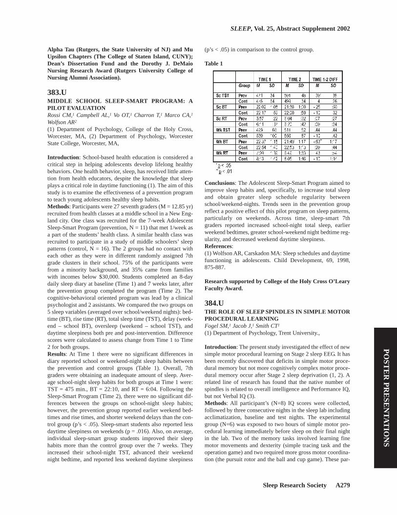
Sleep Research Society A279
PO
STE
R P
RE
SEN
TA
TIO
NS
SLEEP, Vol. 25, Abstract Supplement 2002
Alpha Tau (Rutgers, the State University of NJ) and MuUpsilon Chapters (The College of Staten Island, CUNY);Dean’s Dissertation Fund and the Dorothy J. DeMaioNursing Research Award (Rutgers University College ofNursing Alumni Association).
383.UMIDDLE SCHOOL SLEEP-SMART PROGRAM: APILOT EVALUATIONRossi CM,1 Campbell AL,1 Vo OT,1 Charron T,1 Marco CA,1
Wolfson AR2
(1) Department of Psychology, College of the Holy Cross,Worcester, MA, (2) Department of Psychology, WorcesterState College, Worcester, MA,
Introduction: School-based health education is considered acritical step in helping adolescents develop lifelong healthybehaviors. One health behavior, sleep, has received little atten-tion from health educators, despite the knowledge that sleepplays a critical role in daytime functioning (1). The aim of thisstudy is to examine the effectiveness of a prevention programto teach young adolescents healthy sleep habits.Methods: Participants were 27 seventh graders (M = 12.85 yr)recruited from health classes at a middle school in a New Eng-land city. One class was recruited for the 7-week AdolescentSleep-Smart Program (prevention, N = 11) that met 1/week asa part of the students’ health class. A similar health class wasrecruited to participate in a study of middle schoolers’ sleeppatterns (control, N = 16). The 2 groups had no contact witheach other as they were in different randomly assigned 7thgrade clusters in their school. 75% of the participants werefrom a minority background, and 35% came from familieswith incomes below $30,000. Students completed an 8-daydaily sleep diary at baseline (Time 1) and 7 weeks later, afterthe prevention group completed the program (Time 2). Thecognitive-behavioral oriented program was lead by a clinicalpsychologist and 2 assistants. We compared the two groups on5 sleep variables (averaged over school/weekend nights): bed-time (BT), rise time (RT), total sleep time (TST), delay (week-end – school BT), oversleep (weekend – school TST), anddaytime sleepiness both pre and post-intervention. Differencescores were calculated to assess change from Time 1 to Time2 for both groups.Results: At Time 1 there were no significant differences indiary reported school or weekend-night sleep habits betweenthe prevention and control groups (Table 1). Overall, 7thgraders were obtaining an inadequate amount of sleep. Aver-age school-night sleep habits for both groups at Time 1 were:TST = 475 min., BT = 22:10, and RT = 6:04. Following theSleep-Smart Program (Time 2), there were no significant dif-ferences between the groups on school-night sleep habits;however, the prevention group reported earlier weekend bed-times and rise times, and shorter weekend delays than the con-trol group (p’s < .05). Sleep-smart students also reported lessdaytime sleepiness on weekends (p = .016). Also, on average,individual sleep-smart group students improved their sleephabits more than the control group over the 7 weeks. Theyincreased their school-night TST, advanced their weekendnight bedtime, and reported less weekend daytime sleepiness
(p’s < .05) in comparison to the control group.
Table 1
Conclusions: The Adolescent Sleep-Smart Program aimed toimprove sleep habits and, specifically, to increase total sleepand obtain greater sleep schedule regularity betweenschool/weekend-nights. Trends seen in the prevention groupreflect a positive effect of this pilot program on sleep patterns,particularly on weekends. Across time, sleep-smart 7thgraders reported increased school-night total sleep, earlierweekend bedtimes, greater school-weekend night bedtime reg-ularity, and decreased weekend daytime sleepiness.References: (1) Wolfson AR, Carskadon MA: Sleep schedules and daytimefunctioning in adolescents. Child Development, 69, 1998,875-887.
Research supported by College of the Holy Cross O’LearyFaculty Award.
384.UTHE ROLE OF SLEEP SPINDLES IN SIMPLE MOTORPROCEDURAL LEARNINGFogel SM,1 Jacob J,1 Smith CT1
(1) Department of Psychology, Trent University.,
Introduction: The present study investigated the effect of newsimple motor procedural learning on Stage 2 sleep EEG. It hasbeen recently discovered that deficits in simple motor proce-dural memory but not more cognitively complex motor proce-dural memory occur after Stage 2 sleep deprivation (1, 2). Arelated line of research has found that the native number ofspindles is related to overall intelligence and Performance IQ,but not Verbal IQ (3).Methods: All participant’s (N=8) IQ scores were collected,followed by three consecutive nights in the sleep lab includingacclimatization, baseline and test nights. The experimentalgroup (N=6) was exposed to two hours of simple motor pro-cedural learning immediately before sleep on their final nightin the lab. Two of the memory tasks involved learning finemotor movements and dexterity (simple tracing task and theoperation game) and two required more gross motor coordina-tion (the pursuit rotor and the ball and cup game). These par-
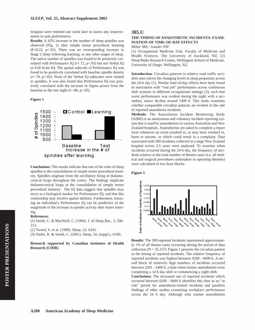
A280 American Academy of Sleep Medicine
PO
STE
R P
RE
SEN
TA
TIO
NS
SLEEP, Vol. 25, Abstract Supplement 2002
ticipants were retested one week later to assess any improve-ments in task performance.Results: A 42% increase in the number of sleep spindles wasobserved (Fig. 1) after simple motor procedural learning(F=8.22, p<.05). There was no corresponding increase inStage 2 sleep following learning, or any other stages of sleep.The native number of spindles was found to be positively cor-related with Performance IQ (r=.72, p<.05) but not Verbal IQor Full Scale IQ. The spatial subscale of Performance IQ wasfound to be positively correlated with baseline spindle density(r=.78, p<.05). None of the Verbal IQ subscales were relatedto spindles. It was also found that Performance IQ was posi-tively correlated with the increase in Sigma power from thebaseline to the test night (r=.88, p<.05).
Figure 1
Conclusions: The results indicate that one of the roles of sleepspindles is the consolidation of simple motor procedural mem-ory. Spindles originate from the oscillatory firing of thalamo-cortical loops throughout the cortex. The findings implicatethalamocortical loops in the consolidation of simple motorprocedural memory. The IQ data suggest that spindles mayserve as a biological marker for Performance IQ, and that thisrelationship may involve spatial abilities. Furthermore, know-ing an individual’s Performance IQ can be predictive of themagnitude of the increase in spindle activity after motor learn-ing.References: (1) Smith, C. & MacNeill, C. (1994). J. of Sleep Res., 3, 206-213.(2) Tweed, S. et al. (1999). Sleep, 22, S241.(3) Nader, R. & Smith, C. (2001). Sleep, 24, (suppl.), A160.
Research supported by Canadian Institutes of HealthResearch (CIHR)
385.UTHE TIMING OF ANAESTHETIC INCIDENTS: EXAM-INATION OF TIME-OF-DAY EFFECTS Millar MA,1 Gander PH1
(1) Occupational Medicine Unit, Faculty of Medicine andHealth Sciences, The University of Auckland, NZ, (2)Sleep/Wake Research Centre, Wellington School of Medicine,University of Otago, Wellington, NZ,
Introduction: Circadian patterns in relative road traffic acci-dent rates mirror the changing levels in sleep propensity acrossthe 24-h day [1]. Similar time-of-day effects have been foundin association with “real job” performance across continuousshift systems in different occupational settings [2], such thatworst performance was evident during the night with a sec-ondary, minor decline around 1400 h. This study examineswhether comparable circadian patterns are evident in the rateof reported anaesthesia incidents.Methods: The Australasian Incident Monitoring Study(AIMS) is an anonymous and voluntary incident reporting sys-tem that is used by anaesthetists in various Australian and NewZealand hospitals. Anaesthetists are asked to complete a reportform whenever an event resulted in, or may have resulted in,harm to anyone, or which could result in a complaint. Dataassociated with 389 incidents collected in a large New Zealandhospital across 2.5 years were analysed. To examine whenincidents occurred during the 24-h day, the frequency of inci-dents relative to the total number of theatre cases (i.e. all med-ical and surgical procedures undertaken in operating theatres)were calculated in two hour blocks.
Figure 1
Results: The 389 reported incidents represented approximate-ly 1% of all theatre cases occurring during the period of datacollection (N = 35,157). Figure 1 presents the circadian patternin the timing of reported incidents. The relative frequency ofreported incidents was highest between 0200 - 0600 h. A sec-ond block of relatively high numbers of incidents occurredbetween 2201 - 2400 h, a time when trainee anaesthetists werecompleting a 14 h day shift or commencing a night shift.Conclusions: The increased rate of reported incidents whichoccurred between 0200 - 0600 h identifies this time as an “atrisk” period for anaesthesia-related incidents and parallelsfindings of other studies examining workplace performanceacross the 24 h day. Although only trainee anaesthetists
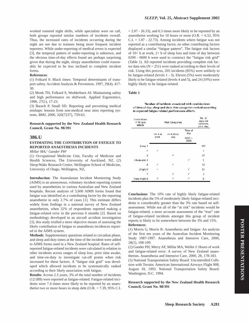
Sleep Research Society A281
PO
STE
R P
RE
SEN
TA
TIO
NS
SLEEP, Vol. 25, Abstract Supplement 2002
worked rostered night shifts, while specialists were on call,both groups reported similar numbers of incidents overall.Thus, the increased rates of incidents occurring during thenight are not due to trainees being more frequent incidentreporters. While under-reporting of medical errors is expected[3], the temporal pattern of under-reporting is unknown, andthe obvious time-of-day effects found are perhaps surprisinggiven that during the night, sleepy anaesthetists could reason-ably be expected to be less inclined to complete incidentforms.References: (1) Folkard S: Black times: Temporal determinants of trans-port safety. Accident Analysis & Prevention, 1997, 29(4), 417-30.(2) Monk TH, Folkard S, Wedderburn AI: Maintaining safetyand high performance on shiftwork. Applied Ergonomics,1996, 27(1), 17-23.(3) Barach P, Small SD: Reporting and preventing medicalmishaps: lessons from non-medical near miss reporting sys-tems. BMJ, 2000, 320(7237), 759-63.
Research supported by the New Zealand Health ResearchCouncil, Grant No. 98/391
386.UESTIMATING THE CONTRIBUTION OF FATIGUE TOREPORTED ANAESTHESIA INCIDENTSMillar MA,1 Gander PH1
(1) Occupational Medicine Unit, Faculty of Medicine andHealth Sciences, The University of Auckland, NZ, (2)Sleep/Wake Research Centre, Wellington School of Medicine,University of Otago, Wellington, NZ,
Introduction: The Australasian Incident Monitoring Study(AIMS) is an anonymous, voluntary incident reporting systemused by anaesthetists in various Australian and New Zealandhospitals. Recent analysis of 5,600 AIMS forms found thatfatigue was identified as a contributing factor by the reportinganaesthetist in only 2.7% of cases [1]. This estimate differswidely from findings in a national survey of New Zealandanaesthetists, when 32% of respondents reported making afatigue-related error in the previous 6 months [2]. Based onmethodology developed in an aircraft accident investigation[3], this study trialled a more objective means of assessing thelikely contribution of fatigue to anaesthesia incidences report-ed in the AIMS system.Methods: Supplementary questions related to circadian phase,and sleep and duty times at the time of the incident were addedto AIMS forms used in a New Zealand hospital. Rates of self-reported fatigue-related incidents were calculated in relation toother incidents across ranges of sleep loss, prior time awake,and time-on-duty to investigate cut-off points when riskincreased for these factors. A “fatigue risk grid” was devel-oped which allowed incidents to be systematically rankedaccording to their likely association with fatigue.Results: Across 2.5 years, 3% of the total number of incidents(12/389) were reported as fatigue-related. Fatigue-related inci-dents were 7.4 times more likely to be reported by an anaes-thetist two or more hours in sleep debt (O.R. = 7.39, 95% C.I.
= 2.07 - 26.33), and 6.5 times more likely to be reported by ananaesthetist working for 10 hours or more (O.R. = 6.52, 95%C.I. = 1.87 - 22.73). Among incidents where fatigue was notreported as a contributing factor, no other contributing factorsdisplayed a similar “fatigue pattern”. The fatigue risk factorsof 10+ h at work, 2+ h of sleep loss and time of day between0200 - 0600 h were used to construct the “fatigue risk grid”(Table 1). All reported incidents providing complete risk fac-tor data sets (N = 251) were ranked according to their levels ofrisk. Using this process, 205 incidents (85%) were unlikely tobe fatigue-related (levels 1 - 3). Eleven (5%) were moderatelylikely to be fatigue-related (levels 4 and 5), and 24 (10%) werehighly likely to be fatigue-related.
Table 1
Conclusions: The 10% rate of highly likely fatigue-relatedincidents plus the 5% of moderately likely fatigue-related inci-dents is considerably greater than the 3% rate based on self-assessment. While not all of the “at risk” incidents would befatigue-related, a more accurate assessment of the “true” rateof fatigue-related incidents amongst this group of incidentreports is likely to lie somewhere between the 3% and 15%.References: (1) Morris G, Morris R: Anaesthesia and fatigue: An analysisof the first ten years of the Australian Incident MonitoringStudy 1987-1997. Anaesthesia and Intensive Care, 2000,28(1), 108-109.(2) Gander PH, Merry AF, Millar MA, Weller J: Hours of workand fatigue-related error: A survey of New Zealand anaes-thetists. Anaesthesia and Intensive Care, 2000, 28, 178-183.(3) National Transportation Safety Board: Uncontrolled Colli-sion with Terrain: American International Airways Flight 808,August 18, 1993. National Transportation Safety Board:Washington, D.C. 1994.
Research supported by the New Zealand Health ResearchCouncil, Grant No. 98/391
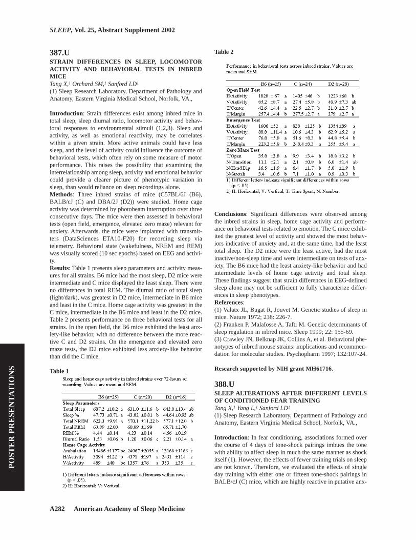
A282 American Academy of Sleep Medicine
PO
STE
R P
RE
SEN
TA
TIO
NS
SLEEP, Vol. 25, Abstract Supplement 2002
387.USTRAIN DIFFERENCES IN SLEEP, LOCOMOTORACTIVITY AND BEHAVIORAL TESTS IN INBREDMICETang X,1 Orchard SM,1 Sanford LD1
(1) Sleep Research Laboratory, Department of Pathology andAnatomy, Eastern Virginia Medical School, Norfolk, VA.,
Introduction: Strain differences exist among inbred mice intotal sleep, sleep diurnal ratio, locomotor activity and behav-ioral responses to environmental stimuli (1,2,3). Sleep andactivity, as well as emotional reactivity, may be correlateswithin a given strain. More active animals could have lesssleep, and the level of activity could influence the outcome ofbehavioral tests, which often rely on some measure of motorperformance. This raises the possibility that examining theinterrelationship among sleep, activity and emotional behaviorcould provide a clearer picture of phenotypic variation insleep, than would reliance on sleep recordings alone.Methods: Three inbred strains of mice (C57BL/6J (B6),BALB/cJ (C) and DBA/2J (D2)) were studied. Home cageactivity was determined by photobeam interruption over threeconsecutive days. The mice were then assessed in behavioraltests (open field, emergence, elevated zero maze) relevant foranxiety. Afterwards, the mice were implanted with transmit-ters (DataSciences ETA10-F20) for recording sleep viatelemetry. Behavioral state (wakefulness, NREM and REM)was visually scored (10 sec epochs) based on EEG and activi-ty.Results: Table 1 presents sleep parameters and activity meas-ures for all strains. B6 mice had the most sleep, D2 mice wereintermediate and C mice displayed the least sleep. There wereno differences in total REM. The diurnal ratio of total sleep(light/dark), was greatest in D2 mice, intermediate in B6 miceand least in the C mice. Home cage activity was greatest in theC mice, intermediate in the B6 mice and least in the D2 mice.Table 2 presents performance on three behavioral tests for allstrains. In the open field, the B6 mice exhibited the least anx-iety-like behavior, with no difference between the more reac-tive C and D2 strains. On the emergence and elevated zeromaze tests, the D2 mice exhibited less anxiety-like behaviorthan did the C mice.
Table 1
Table 2
Conclusions: Significant differences were observed amongthe inbred strains in sleep, home cage activity and perform-ance on behavioral tests related to emotion. The C mice exhib-ited the greatest level of activity and showed the most behav-iors indicative of anxiety and, at the same time, had the leasttotal sleep. The D2 mice were the least active, had the mostinactive/non-sleep time and were intermediate on tests of anx-iety. The B6 mice had the least anxiety-like behavior and hadintermediate levels of home cage activity and total sleep.These findings suggest that strain differences in EEG-definedsleep alone may not be sufficient to fully characterize differ-ences in sleep phenotypes.References: (1) Valatx JL, Bugat R, Jouvet M. Genetic studies of sleep inmice. Nature 1972; 238: 226-7.(2) Franken P, Malafosse A, Tafti M. Genetic determinants ofsleep regulation in inbred mice. Sleep 1999; 22: 155-69.(3) Crawley JN, Belknap JK, Collins A, et al. Behavioral phe-notypes of inbred mouse strains: implications and recommen-dation for molecular studies. Psychopharm 1997; 132:107-24.
Research supported by NIH grant MH61716.
388.USLEEP ALTERATIONS AFTER DIFFERENT LEVELSOF CONDITIONED FEAR TRAININGTang X,1 Yang L,1 Sanford LD1
(1) Sleep Research Laboratory, Department of Pathology andAnatomy, Eastern Virginia Medical School, Norfolk, VA.,
Introduction: In fear conditioning, associations formed overthe course of 4 days of tone-shock pairings imbues the tonewith ability to affect sleep in much the same manner as shockitself (1). However, the effects of fewer training trials on sleepare not known. Therefore, we evaluated the effects of singleday training with either one or fifteen tone-shock pairings inBALB/cJ (C) mice, which are highly reactive in putative anx-
Sleep Research Society A283
PO
STE
R P
RE
SEN
TA
TIO
NS
SLEEP, Vol. 25, Abstract Supplement 2002
iety provoking situations (1).Methods: Sixteen C mice were intraperitoneally implantedwith transmitters (DataSciences ETA10-F20) for sleep record-ing via telemetry. Behavioral state was visually scored basedon EEG and activity. After baseline sleep recording sessions,the mice were trained to associate a cue (tone) with footshock.One group (n=8) received a single cue-shock pairing and thesecond group (n=8) received 15 cue-shock pairings on a singleday. All training was conducted between 0800 to 0900 h. Fortesting, the mice were presented 15 cues alone in their homecage between 1045 and 1100 h on day 5, 12, 19, 26, 33, 40(cue 1 to 6) following shock training. Sleep was recorded from0900 (following shock) or 1100 (following cue) to 0700 in thenext day.Results: Single shock-training trials (SST) produced REMsuppression in the 4 h immediately after training whereas fol-lowing multiple (15) shock-training trials (MST) both totalNREM and REM were decreased (Table 1). On the day fol-lowing training, REM had returned to baseline levels in theSST mice, but was still suppressed in the MST mice. NREMwas increased in SST mice and at baseline levels in MST mice.In MST mice, cue presentations continued to produce signifi-cant suppressions of REM for up to 26 days after training,whereas in SST mice, presentations of the cue did not signifi-cantly alter REM (Figure 1). Cue presentation did not signifi-cantly alter NREM in either MST or SST mice.
Table 1
Figure 1
Conclusions: These data demonstrate that a single shock canalter sleep in reactive mice, and that the impact of shock onREM is greater and longer lasting suppression after MST. Therelative absence of effects on sleep with repeated presentationsof cues after SST compared to after MST demonstrate that thealterations in sleep are produced by the psychological value ofthe tones as reminders of the shock, and not by the soundalone. Differences in sleep after SST and MST suggest that itmay be possible to examine graded sleep responses to differ-ent levels of foot shock stress and shock-associated cues.References: (1) Sanford LD, Tang X, Ross RJ, Morrison AR. Influence ofanticipatory anxiety on sleep in “anxious” and “non-anxious”mice. Sleep 2001; 24: 53.
Research supported by NIH grant MH61716.
389.USTRAIN DIFFERENCES IN SLEEP ARCHITECTUREOF MICE AFTER REPEATED PRESENTATIONS OFFEAR CONDITIONED CUESLiu X,1 Tang X,1 Sanford LD1
(1) Sleep Research Laboratory, Department of Pathology andAnatomy,Eastern Virginia Medical School, Norfolk, VA.,
Introduction: Shock training and the presentation of auditorycues associated with shock produce alterations in sleep thatcan be long-lasting and can vary with mouse strain (1). Effectson sleep, particularly reduced REM, could last for up to sixhours in mice that were more reactive on behavioral tests ofanxiety (BALBc/J (C) and DBA/2J (D2)). Reductions in REMwere of shorter duration in less reactive mice (C57BL/6J(B6)). This raised the question of whether mice strains woulddifferentially respond to repeated presentations of “reminders”associated with shock.Methods: Three inbred strains (B6, n=10; C, n=10; D2, n=8)were used in this study. The mice were intraperitoneallyimplanted with transmitters (DataSciences ETA10-F20) forrecording sleep via telemetry. Behavioral state was visuallyscored based on EEG and activity. After baseline sleep record-ing sessions, the mice were trained to associate a cue (tone)with footshock (15 tone - shock pairing on 4 consecutive days,conducted between 0800 - 0900 h). The mice were presentedwith the cue alone in their home cage between 1045 to 1100 hon day 5, 12, 19 and 26 (cue 1 to 4) after the last shock-train-ing day. Sleep was recorded from 1100 following cue presen-tation to 0700 in the next day.Results: All mice showed an increase in total NREM duringthe light period, especially on later cue presentation days (Fig-ure 1). During the dark period total NREM was decreased in Cand D2 mice, whereas there was no change in the B6 mice.The effects on total REM varied among strains (Figure 2).During the light period, REM was suppressed in both C andD2 mice, with the suppression in C mice persisting through thefinal cue presentation. Light period REM was not significant-ly altered in the B6 mice. During the dark period, REM wassuppressed in all strains, though the effect lasted longer andwas greater in the C and D2 mice.
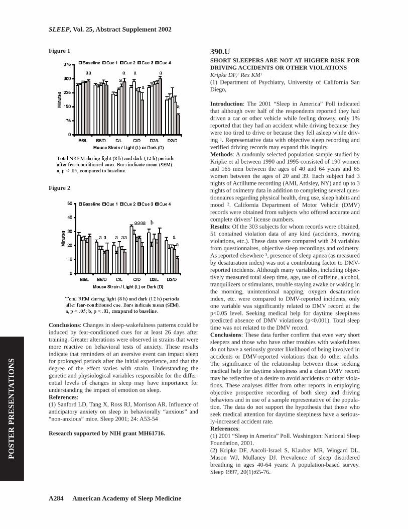
A284 American Academy of Sleep Medicine
PO
STE
R P
RE
SEN
TA
TIO
NS
SLEEP, Vol. 25, Abstract Supplement 2002
Figure 1
Figure 2
Conclusions: Changes in sleep-wakefulness patterns could beinduced by fear-conditioned cues for at least 26 days aftertraining. Greater alterations were observed in strains that weremore reactive on behavioral tests of anxiety. These resultsindicate that reminders of an aversive event can impact sleepfor prolonged periods after the initial experience, and that thedegree of the effect varies with strain. Understanding thegenetic and physiological variables responsible for the differ-ential levels of changes in sleep may have importance forunderstanding the impact of emotion on sleep.References: (1) Sanford LD, Tang X, Ross RJ, Morrison AR. Influence ofanticipatory anxiety on sleep in behaviorally “anxious” and“non-anxious” mice. Sleep 2001; 24: A53-54
Research supported by NIH grant MH61716.
390.USHORT SLEEPERS ARE NOT AT HIGHER RISK FORDRIVING ACCIDENTS OR OTHER VIOLATIONSKripke DF,1 Rex KM1
(1) Department of Psychiatry, University of California SanDiego,
Introduction: The 2001 “Sleep in America” Poll indicatedthat although over half of the respondents reported they haddriven a car or other vehicle while feeling drowsy, only 1%reported that they had an accident while driving because theywere too tired to drive or because they fell asleep while driv-ing 1. Representative data with objective sleep recording andverified driving records may expand this inquiry.Methods: A randomly selected population sample studied byKripke et al between 1990 and 1995 consisted of 190 womenand 165 men between the ages of 40 and 64 years and 65women between the ages of 20 and 39. Each subject had 3nights of Actillume recording (AMI, Ardsley, NY) and up to 3nights of oximetry data in addition to completing several ques-tionnaires regarding physical health, drug use, sleep habits andmood 2. California Department of Motor Vehicle (DMV)records were obtained from subjects who offered accurate andcomplete drivers’ license numbers.Results: Of the 303 subjects for whom records were obtained,51 contained violation data of any kind (accidents, movingviolations, etc.). These data were compared with 24 variablesfrom questionnaires, objective sleep recordings and oximetry.As reported elsewhere 3, presence of sleep apnea (as measuredby desaturation index) was not a contributing factor to DMV-reported incidents. Although many variables, including objec-tively measured total sleep time, age, use of caffeine, alcohol,tranquilizers or stimulants, trouble staying awake or waking inthe morning, unintentional napping, oxygen desaturationindex, etc. were compared to DMV-reported incidents, onlyone variable was significantly related to DMV record at thep<0.05 level. Seeking medical help for daytime sleepinesspredicted absence of DMV violations (p<0.001). Total sleeptime was not related to the DMV record. Conclusions: These data further confirm that even very shortsleepers and those who have other troubles with wakefulnessdo not have a seriously greater likelihood of being involved inaccidents or DMV-reported violations than do other adults.The significance of the relationship between those seekingmedical help for daytime sleepiness and a clean DMV recordmay be reflective of a desire to avoid accidents or other viola-tions. These analyses differ from other reports in employingobjective prospective recording of both sleep and drivingbehaviors and in use of a sample representative of the popula-tion. The data do not support the hypothesis that those whoseek medical attention for daytime sleepiness have a serious-ly-increased accident rate.References: (1) 2001 “Sleep in America” Poll. Washington: National SleepFoundation, 2001.(2) Kripke DF, Ancoli-Israel S, Klauber MR, Wingard DL,Mason WJ, Mullaney DJ. Prevalence of sleep disorderedbreathing in ages 40-64 years: A population-based survey.Sleep 1997, 20(1):65-76.
Sleep Research Society A285
PO
STE
R P
RE
SEN
TA
TIO
NS
SLEEP, Vol. 25, Abstract Supplement 2002
(3) Rex KM, Kripke DF, Klauber MR, Ancoli-Israel S. Sleep-disordered breathing in middle-aged adults predicts no signif-icantly higher rates of traffic violations. Chest2001,119(5):1623-1624.
Research supported by AG12364, AG15246, AG15763 andHL61280.
391.UTHE SLEEP BEHAVIOUR OF LOCOMOTIVE DRIV-ERS DURING RELAY OPERATIONS Lamond N,1 Darwent D,1 Dawson D1
(1) The Centre for Sleep Research, The University of SouthAustralia,
Introduction: Relay operations for the return trip from Ade-laide to Perth involve the use of two locomotive crews, onedriving and the other resting in a relay van, each workingalternating eight hour shifts. To date, there is no published dataconcerning the quality and quantity of rest attained by traincrew during relay operations, nor about the degree of work-related fatigue experienced by relay workers. This studysought to investigate the sleep behaviour of train drivers work-ing relay operations, and the impact that sleeping in relay vanshas on sleep quality and quantity.Methods: Twelve locomotive drivers volunteered for thestudy. A schematic representation of the relay work is dis-played in Figure 1. During each trip, drivers were asked to (1)provide detailed information about their sleep/work patterns,(2) wear a wrist actigraph, (3) provide saliva sample every 2-hours during periods of wakefulness, and (4) complete a 10-minute psychomotor vigilance task (PVT) at the beginningand end of each 8-hour work shift. Each driver participated inthree separate Adelaide-Perth relay trips (drivers typicallyhave a minimum of five days off between each relay trip),resulting in 36 data sets.
Figure 1
Results: Preliminary analysis of the data indicates that driversaveraged 3.8 (SD=1.98) sleep periods on each 5-day trip. Dur-
ing each sleep period (including the time spent in Perth), driv-ers obtained an average of only 3.86 (SD=1.8) hours sleep.Sleep periods that were initiated at night and early in the morn-ing (2100 to 0600 hr) were significantly (p<0.0001) longerthan sleep initiated during the day or the evening (0600 to2100 hr). Specifically, sleep periods initiated at night and dur-ing the early morning hours were 4.8 (SD= 1.7) and 4.6(SD=1.5) hours in length, respectively, while periods initiatedduring the day and evening were only 2.9 (SD=1.4) and 3.0(SD=1.6) hours, respectively.Conclusions: Although analysis of the data is still in progress,the findings thus far suggest that locomotive drivers do notobtain sufficient sleep during relay operations. However, itshould be noted that during the initial phase of data collectiontraining of drivers for accreditation was still in progress,resulting in unusual disruption in the usual roster. Further-more, new crew vans that are better equipped have recentlybeen introduced, which is expected to significantly impact onthe quality and quantity of sleep obtained.
Research supported by the Australian Rail Consortium:Shiftwork and Workload Study Phase II
Related Documents