Post-embryonic development and ultrastructural characteristics of the polycephalic larva of Taenia parva Baer, 1926 (Cyclophyllidea, Taeniidae) ** Zdzis³aw Œwiderski 1,2* , Jordi Miquel 3 , Daniel M³ocicki 1,4 , Boyko B. Georgiev 5,6 , Catarina Eira 3 , Barbara Grytner-Ziêcina 2 and Carlos Feliu 3 1 W. Stefaæski Institute of Parasitology, Polish Academy of Sciences, 51/55 Twarda Street, 00-818 Warsaw, Poland; 2 Department of General Biology and Parasitology, Medical University of Warsaw, 5 Cha‡ubiæskiego Street, 02-004 Warsaw, Poland; 3 Laboratori de Parasitologia, Departament de Microbiologia i Parasitologia SanitBries, Facultat de FarmBcia, Universitat de Barcelona, Av. Joan XXIII s/n, E-08028 Barcelona, Spain; 4 Department of Medical Biology, Medical University of Warsaw, 73 Nowogrodzka Street, 02-004 Warsaw, Poland; 5 Central Laboratory of General Ecology, Bulgarian Academy of Sciences, 2 Gagarin Street, 1113 Sofia, Bulgaria; 6 Department of Zoology, The Natural History Museum, Cromwell Road, London SW7 5BD, UK Abstract Post-embryonic development and fully-formed polycephalic larvae of Taenia parva Baer, 1926 were examined by light (LM) and transmission electron microscopy (TEM). Three developmental stages were recognised: (1) an early stage of exogenous budding at the surface of the central vesicle; (2) a stage of polycephalic cyst development accompanied by segmentation of the growing larval strobile and an obvious decrease in the size of the central vesicle; (3) fully-formed larval strobile and invagi- nated scoleces. In fully-developed encysted polycephalic larvae, there are usually 1424 segmented larval strobilae, each ter- minating with an invaginated scolex; larval strobilae arise from a common central vesicle and remain attached posterior to it during the entire development. The number of segments varies between 109 and 120 per larval strobila. The polycephalic lar- vae examined closely resemble the strobilocercus type of taeniid larvae. The structure of developing and fully-formed larvae was examined by TEM. The tegument, scolex, subtegumental musculature of the strobilar segments, protonephridial system, calcareous corpuscles and medullary parenchyma of larvae exhibit general similarity with the same structures in adults at both LM and TEM levels. The morphogenesis of the larva of T. parva is compared with that of the polycephalic larvae of other Taenia spp. (T. krepkogorski, T. twitchelli and T. endothoracica) and with other asexually-multiplying cestode larvae (mesocestoidids, hymenolepidids and dilepidids). Key words Cestoda, Taenia parva, polycephalic larva, post-embryonic development, ultrastructure * Corresponding author: [email protected] ** Dedicated to the memory of Professor Konstanty Janicki, on the occasion of the 75th anniversary of his death Introduction The post-embryonic developmental stages of taeniid cestodes deserve particular attention. This family comprises 44 species, including 5 of medical importance and about 20 of veterinary importance (Abuladze 1964, Verster 1969, Loos-Frank 2000). Because of this, some taeniids such as Echinococcus gra- nulosus (Batsch, 1786) and E. multilocularis (Leuckart, 1863) have already been studied extensively (see Smyth 1964; Morseth 1967; Sakamoto and Sigimura 1969, 1970; lais 1973; Mehlhorn et al. 1983; Thompson 1986; Eckert et al. 2002). Their larvae show the highest rate of asexual repro- duction among cestodes, resulting in the proliferation of thou- sands of scoleces from a single oncosphere. They cause, re- spectively, cystic and alveolar echinococcosis and are a major current problem of medical parasitology. However, the larvae of Echinococcus spp. are essentially different from those of other taeniids. Ultrastructural features of larval cestodes can provide use- ful characters for phylogenetic and evolutionary analyses. Skra Stefaæski DOI: 10.2478/s11686-007-0009-0 © 2007 W. Stefañski Institute of Parasitology, PAS Acta Parasitologica, 2007, 52(1), 31–50; ISSN 1230-2821

Welcome message from author
This document is posted to help you gain knowledge. Please leave a comment to let me know what you think about it! Share it to your friends and learn new things together.
Transcript

Post-embryonic development and ultrastructural characteristics of the polycephalic larva of Taenia parva Baer,
1926 (Cyclophyllidea, Taeniidae)**
Zdzis³aw Œwiderski1,2*, Jordi Miquel3, Daniel M³ocicki1,4, Boyko B. Georgiev5,6, Catarina Eira3,Barbara Grytner-Ziêcina2 and Carlos Feliu3
1W. Stefañski Institute of Parasitology, Polish Academy of Sciences, 51/55 Twarda Street, 00-818 Warsaw, Poland; 2Department of General Biology and Parasitology, Medical University of Warsaw, 5 Cha³ubiñskiego Street, 02-004 Warsaw, Poland;
3Laboratori de Parasitologia, Departament de Microbiologia i Parasitologia SanitBries, Facultat de FarmBcia, Universitat de Barcelona, Av.Joan XXIII s/n, E-08028 Barcelona, Spain; 4Department of Medical Biology, Medical University of Warsaw, 73 Nowogrodzka Street,
02-004 Warsaw, Poland; 5Central Laboratory of General Ecology, Bulgarian Academy of Sciences, 2 Gagarin Street, 1113 Sofia, Bulgaria;6Department of Zoology, The Natural History Museum, Cromwell Road, London SW7 5BD, UK
AbstractPost-embryonic development and fully-formed polycephalic larvae of Taenia parva Baer, 1926 were examined by light (LM)and transmission electron microscopy (TEM). Three developmental stages were recognised: (1) an early stage of exogenousbudding at the surface of the central vesicle; (2) a stage of polycephalic cyst development accompanied by segmentation ofthe growing larval strobile and an obvious decrease in the size of the central vesicle; (3) fully-formed larval strobile and invagi-nated scoleces. In fully-developed encysted polycephalic larvae, there are usually 14�24 segmented larval strobilae, each ter-minating with an invaginated scolex; larval strobilae arise from a common central vesicle and remain attached posterior to itduring the entire development. The number of segments varies between 109 and 120 per larval strobila. The polycephalic lar-vae examined closely resemble the strobilocercus type of taeniid larvae. The structure of developing and fully-formed larvaewas examined by TEM. The tegument, scolex, subtegumental musculature of the strobilar segments, protonephridial system,calcareous corpuscles and medullary parenchyma of larvae exhibit general similarity with the same structures in adults at bothLM and TEM levels. The morphogenesis of the larva of T. parva is compared with that of the polycephalic larvae of other Taeniaspp. (T. krepkogorski, T. twitchelli and T. endothoracica) and with other asexually-multiplying cestode larvae (mesocestoidids,hymenolepidids and dilepidids).
Key wordsCestoda, Taenia parva, polycephalic larva, post-embryonic development, ultrastructure
*Corresponding author: [email protected]
**Dedicated to the memory of Professor Konstanty Janicki, on the occasion of the 75th anniversary of his death
Introduction
The post-embryonic developmental stages of taeniid cestodesdeserve particular attention. This family comprises 44 species,including 5 of medical importance and about 20 of veterinaryimportance (Abuladze 1964, Verster 1969, Loos-Frank 2000).Because of this, some taeniids such as Echinococcus gra-nulosus (Batsch, 1786) and E. multilocularis (Leuckart, 1863)have already been studied extensively (see Smyth 1964;Morseth 1967; Sakamoto and Sigimura 1969, 1970; �lais
1973; Mehlhorn et al. 1983; Thompson 1986; Eckert et al.2002). Their larvae show the highest rate of asexual repro-duction among cestodes, resulting in the proliferation of thou-sands of scoleces from a single oncosphere. They cause, re-spectively, cystic and alveolar echinococcosis and are a majorcurrent problem of medical parasitology. However, the larvaeof Echinococcus spp. are essentially different from those ofother taeniids.
Ultrastructural features of larval cestodes can provide use-ful characters for phylogenetic and evolutionary analyses.
Skóra
Stefañski
DOI: 10.2478/s11686-007-0009-0© 2007 W. Stefañski Institute of Parasitology, PASActa Parasitologica, 2007, 52(1), 31–50; ISSN 1230-2821

Zdzis³aw �widerski et al.
Until now, however, they have been relatively little utilised(Beveridge 2001, Chervy 2002). Earlier reviews by Freeman(1973) and Jarecka (1975) on the post-embryonic develop-mental stages of cestodes provided important comparativedata for phylogenetic analyses. Studies on the histology andultrastructure of larval cestodes have been �largely restrictedto the classical �cysticerci�and �cysticercoids� limiting the pos-sibilities of making comparison across all taxa within the Cy-clophyllidea, let alone the wider array of cestode orders� (Bev-eridge 2001; p. 253). A far greater diversity of ultrastructuraldata on a variety of metacestodes is needed before we canapply them successfully in analyses of cestode phylogeny.
During the last 20�30 years, the ultrastructure of the post-embryonic developmental stages of Taenia spp. has been ne-glected. Apart from five rather old papers describing the finemorphology of the post-embryonic larvae of T. multicepsLeske, 1780 by Race et al. (1965), T. taeniaeformis (Batsch,1786) by Nieland and Weinbach (1968), T. crassiceps (Zeder,1800) by Baron (1968) and Mount (1970) and T. saginataGoeze, 1782 by �lais et al. (1972), there is apparently no otherultrastructural information on the taeniid larvae. In addition to these five papers, some details on the ultrastructure of theouter cytoplasmic layer of the metacestode tegument of Ano-plotaenia dasyuri Beddard, 1911 have been presented (Bev-eridge et al. 1975); however, the position of this species iscontroversial and it probably does not belong within the Tae-niidae (Khalil et al. 1994). Unfortunately, in many old papers,the ultrastructural preparations were made with the fixationand embedding procedures of 20�30 years ago which causedartifacts, including shrinkage of tissues, separation of organel-les and failure to preserve glycogen and lipids.
In other cestode groups, there are numerous more recentLM and TEM studies on larvae, mostly in other cyclophyl-lidean families; some have been reviewed by Ubelaker (1980,1983) and Jarecka et al. (1981). Other studies on various typesof larval cestodes (Caley 1974, 1976; Burt and Jarecka 1984;Jarecka and Burt 1984; Jarecka et al. 1984; Krasnoshchekovand Pluzhnikov 1984; Burt 1987; Conn 1986, 1988a, 1990,2005; Conn et al. 2002; Galán-Puchades et al. 2002; �wider-ski et al. 2002a; Georgiev et al. 2005) have yet to be recon-sidered in a broader comparative context.
No information on the ultrastructure of the polycephaliclarvae of taeniids is available. In order to obtain such data, weselected Taenia parva Baer, 1926 for the present study. Withthe single exception of the classification of Wardle and Mc-Leod (1952), who included it in Hydatigera Lamarck, 1816, ithas always been considered a species of Taenia L., 1758 (seeBaer 1926, Abuladze 1964, Verster 1969, Loos-Frank 2000).Adults of T. parva are frequent parasites of the Viverridae car-nivores in Africa. This parasite has also been found in theIberian Peninsula and is believed to be progressively expand-ing its range into Europe (Mas-Coma and Feliu 1977, Feliu1980, Alvarez et al. 1990, Casanova et al. 2000). The inter-mediate hosts include several species of African and Europeanrodents (Verster 1969, Loos-Frank 2000). Polycephalic larvaeidentified as T. parva were described by Campana-Rouget
(1950) and Bernard (1961, 1963) from the abdominal cavityof Apodemus sylvaticus L., 1758 (Rodentia, Muridae). Dollfusand Saint Girons (1958) reported behavioural modifications inwood mice infected with T. parva, which probably resultedfrom the increase of the parasite cyst volume. Our materialwas obtained from A. sylvaticus, the intermediate host ofT. parva in the Iberian Peninsula.
The aim of this study is to describe the development andultrastructural characteristics of the post-embryonic larva ofT. parva and to compare it with other polycephalic larvae ofTaenia spp. and other asexually-multiplying cestode larvae.
Materials and methods
Larvae of Taenia parva were obtained from the abdominal cavity of naturally-infected wood mice, Apodemus sylvaticus,captured in Quiaios, Portugal. Live specimens were placed in0.9% NaCl solution. For TEM examination, they were dis-sected and fixed in cold (4°C) 2.5% glutaraldehyde in 0.1 Msodium cacodylate buffer at pH 7.2 for 1 h, rinsed in 0.1 Msodium cacodylate buffer at pH 7.2, postfixed in cold (4°C)1% osmium tetroxide in the same buffer for 1 h, dehydrated in an ethanol series and propylene oxide and embedded inSpurr�s resin. Semithin sections stained with 1% methyleneblue in borax solution were used for LM examination. Ultra-thin sections were obtained using a Reichert-Jung Ultracut Eultramicrotome, placed on copper grids and double-stainedwith uranyl acetate and lead citrate. Ultrathin sections wereexamined using a Jeol 1010 transmission electron micro-scope.
Results
Development and general topography of the polycephaliclarva of T. parvaEncysted polycephalic larvae of T. parva were found in thebody cavity of the intermediate host (Fig. 1). Individual cystsreached ~3.5 cm in diameter. The number of cysts per hostindividual was 1�3; a single cyst per host was most frequent.Each cyst was covered with a thin, semitransparent envelope.The outer part of the cyst wall (tunica adventitia), originatingfrom the host connective tissue, had a network of small bloodvessels. In the early stage, an exogenously-budding centralvesicle was situated inside the cyst wall (Fig. 2A). Subsequentstages showed elongation of the buds and gradual transfor-mation into segmented strobilae, each terminating with aninvaginated scolex (Fig. 2B). In the developed encysted larvae(Fig. 2C), 14�24 larval strobile, arising from the central vesi-cle, were recorded. During their growth and development, thelarval strobile remained connected posteriorly to the centralvesicle (or bladder). The size of the latter gradually decreasedand it was gradually transformed into a vestigial structure thatdid not completely disappear. Each invaginated larval scolexof T. parva had four suckers and a rostellum with two alter-
32
�l¹ski

Post-embryonic development of Taenia parva
nating crowns of large and small rostellar hooks (Figs 3 and 4).Fully-formed strobile were 2.35�3.25 cm long and 2.00�2.15 mm wide. The number of segments ranged from 109�120. The scoleces examined were 569�619 µm wide at thelevel of the rostellar hooks and 1139�1423 µm wide at thelevel of the suckers. The average diameter of suckers was227�258 µm. The rostellum was armed with 32�44 µm ros-tellar hooks. The lengths of the large and small rostellar hookswere 340�356 and 213�231 µm, respectively. Each developedlarval strobila, except for the invaginated scolex, resembled aminiature adult cestode. However, no primordia of genitalorgans were observed.
The development and general topography of the larvaewere examined mainly by LM, and the fine morphology ofdeveloping (Fig. 2B) and fully-formed metacestodes (Fig. 2C)was studied by TEM.
Ultrastructure of the larval tegument
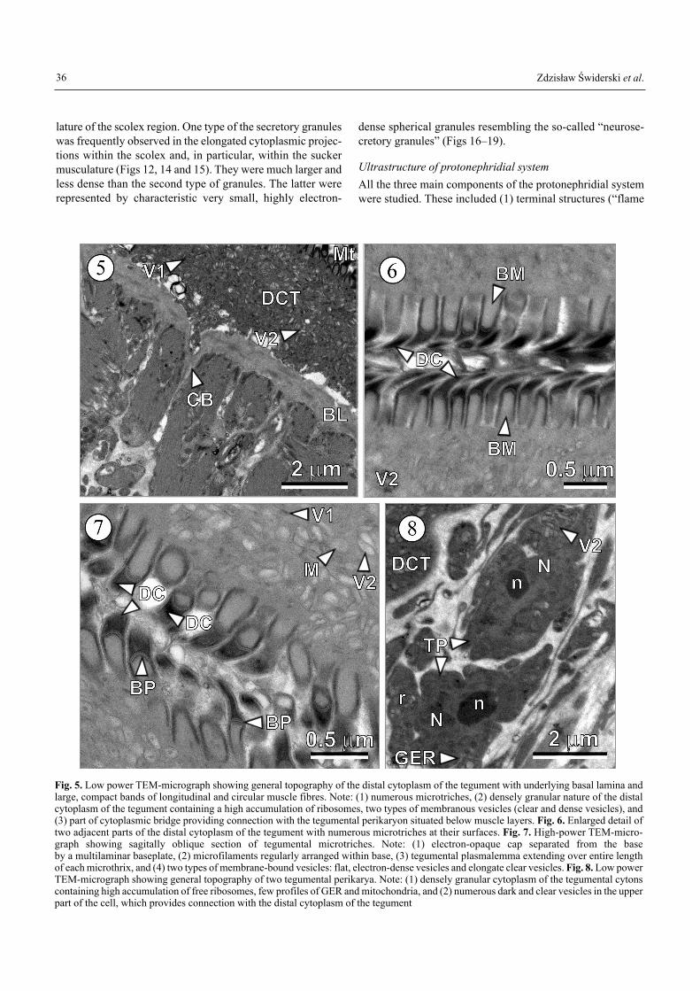
The electron micrographs of the surface of the larval strobilaand the scolex (Figs 5�7) showed that the larval tegumentresembled that of adult worms. The essential feature of theexternal surface was the brush border comprising microtrich-
es (Figs 6 and 7). Their tips were typically pointed and filledwith a keratinous, electron-opaque matrix of densely-packedfibrils or rods; their proximal shafts tended to be cylindrical,with a core of microfilaments (Fig. 7). The keratinous spikewas set off from the shaft by a multilaminate baseplate (Fig.7). Microtriches appeared polymorphic, as seen in differentanatomical regions of the same individual. The distal cyto-plasm of the tegument contained mitochondria and a substan-tial population of membrane-bound vesicles and granules ofvarious sizes, shapes and electron densities. The great major-ity of vesicles and granules appeared to have arisen from theGolgi apparatus of the cytons and transported to the distalcytoplasm via the cytoplasmic bridges uniting both parts ofthe tegument. The perinuclear cytoplasm of the cytons (Fig. 8)contained abundant free ribosomes, a few cisternae of granu-lar endoplasmic reticulum (GER) and numerous vesicles. Thedistal cytoplasmic layer of the tegument covering the suckerswas continuous with that of the strobila but somewhat thinnerin some regions.
Ultrastructure of muscle system
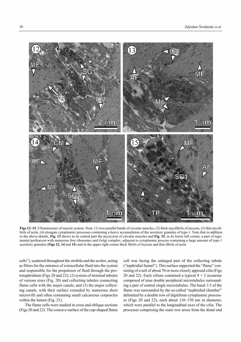
The musculature of the larvae studied consisted of subtegu-mental longitudinal and circular fibres, longitudinal medul-lary fibres, cortical transverse fibres and highly specialised ar-rangements of fibres associated with the suckers and rostel-lum. The strobilar myofibrils were smooth (Figs 9�14). Eachmuscle cell consisted of two major components: (1) the con-tractile myofibril, containing actin and myosin myofibrils(Figs 9�12) and (2) the myocyton, the non-contractile peri-karyon (Fig. 10). The myocytons were typically lateral to andat some distance from the myofibrils (Fig. 10), with cytoplas-mic continuity established via tendrillar processes. Lackingintact Z-discs, there was no division of the myofibril into sar-comeres. Nevertheless, both thick (myosin) and thin (actin)myofilaments were present (Figs 9�14); their arrangementwas not ordered to an extent showing clear demarcation of Aand I bands.
The sucker musculature was characterised by a very com-pact arrangement of fibres (Fig. 9). The cavity of each sucker(Fig. 9) was lined by a thin layer of tegument that was contin-uous with that of the scolex proper. The intrinsic sucker mus-culature (Fig. 9) consisted of circular, radial and longitudinalfibres, with the radial fibres predominating. The circular fibres(Fig. 9) were generally oriented around the rim of the sucker,as a purse string regulating its aperture. The strongly devel-oped radial muscles extended from the tegument of the base ofthe sucker. The highly-branched transverse muscle fibrils sur-rounded the myocytons and tegumental perikarya (Fig. 10).The extrinsic sucker musculature was inserted at the lateralmargins and interconnected with adjacent suckers. The pre-dominant extrinsic muscles at the base of each sucker con-sisted of transverse fibres (Figs 10 and 12�15) and undoubt-edly were responsible for most of the mobility of the sucker.
Two types of secretory granules were observed withinlong cytoplasmic processes associated with extensive muscu-
33
Stanis³a
Zdzis³aw
Fig. 1. Dissected Apodemus sylvaticus with a very large (~3 cm indiameter) encysted larva of Taenia parva of strobilocercus-type inthe abdominal cavity. The larva is surrounded by a thin layer of hostconnective tissue (tunica adventitia) with several small blood vessels

Zdzis³aw �widerski et al.34
Roborzyñski rosb��æv fjad kadsææ¿æ
Fig. 2A-C. Three consecutive stages of development of a polycephalic cyst of T. parva: A � early stage of exogenous budding at the surfaceof the central vesicle; B � stage of polycephalic cyst maturation, accompanied by (1) invagination of scoleces, (2) segmentation of growingstrobilocerci, and (3) decrease in size of the central vesicle; C � fully-formed, mature strobilocerci with invaginated scoleces. Note presenceof much elongated strobile with obvious segmentation and the residual central vesicle serving as an attachment point for the posterior endsof all larval strobile, which always remain united. Abbreviations to all figures: AD � amorphous diaphragms between external and inter-nal rods of the nephridial chamber, AF � actin filaments, B � basal region of the hook blade, BD � buds or primordia of scoleces, BL � basallamina, BM � bases of microtriches, BP � baseplates of microtriches, BV � blood vessels in cyst wall of host origin, C � core of hook, CB � cytoplasmic bridges, CC � calcareous corpuscles, CM � circular muscles, CS � surface of calcareous corpuscle, CT � connective tis-sue fibrils, CV � central vesicle or bladder, Cx � cortex of rostellar hook, D � desmosomes, DC � dense caps of microtriches, DCT � distalcytoplasm of tegument, EL � encysted polycephalic larva, ER � external rods of the nephridial chamber, F � �flame� or bundle of cilia, G � Golgi complex, GER � granular endoplasmic reticulum, Gl � glycogen-like rosettes of α-glycogen or individual particles of β-glyco-gen, HDZ � hook differentiation zone, IR � internal rods of the nephridial chamber, IS � concentric rings of inorganic substances in calcare-ous corpuscles, L � lipid droplet, LCD � large collecting duct, LH � large hooks, LM � longitudinal muscles, Lt � leptotriches, M � mito-chondria, Mc � myocytons, MF � myosin filaments, Mt � microtriches, Mx � matrix of calcareous corpucle, Mv � microvilli, N � nucleus,n � nucleolus, NCh � nephridial chamber, NF � nephridial funnel, R � rostellum, r � ribosomes, S � sucker, SC � strobilocerci, SG1 � secre-tory granules of type-1, SG2 � secretory granules of type-2, SH � small hooks, TC � tegumental collars surrounding rostellar hooks, TD �terminal collecting duct, TM � transverse muscles of sucker, TP � tegumetal perikaryons, V � vesicles, V1 � dense, flattened vesicles, V2 �clear, spherical or elongated vesicles

Post-embryonic development of Taenia parva 35
Figs 3 and 4. Semithin sections showing cross-sectioned apical regions of the invaginated scoleces of T. parva. Note two alternating crownsof large and small rostellar hooks and rostellar tissue in the centre. Fig. 4. Slightly enlarged lateral part of the above micrograph showing,in addition, parts of sucker at the right side
Zdzis³aw �widerski et al.
lature of the scolex region. One type of the secretory granuleswas frequently observed in the elongated cytoplasmic projec-tions within the scolex and, in particular, within the suckermusculature (Figs 12, 14 and 15). They were much larger andless dense than the second type of granules. The latter wererepresented by characteristic very small, highly electron-
dense spherical granules resembling the so-called �neurose-cretory granules� (Figs 16�19).
Ultrastructure of protonephridial system
All the three main components of the protonephridial systemwere studied. These included (1) terminal structures (�flame
36
Fig. 5. Low power TEM-micrograph showing general topography of the distal cytoplasm of the tegument with underlying basal lamina andlarge, compact bands of longitudinal and circular muscle fibres. Note: (1) numerous microtriches, (2) densely granular nature of the distalcytoplasm of the tegument containing a high accumulation of ribosomes, two types of membranous vesicles (clear and dense vesicles), and(3) part of cytoplasmic bridge providing connection with the tegumental perikaryon situated below muscle layers. Fig. 6. Enlarged detail oftwo adjacent parts of the distal cytoplasm of the tegument with numerous microtriches at their surfaces. Fig. 7. High-power TEM-micro-graph showing sagitally oblique section of tegumental microtriches. Note: (1) electron-opaque cap separated from the base by a multilaminar baseplate, (2) microfilaments regularly arranged within base, (3) tegumental plasmalemma extending over entire lengthof each microthrix, and (4) two types of membrane-bound vesicles: flat, electron-dense vesicles and elongate clear vesicles. Fig. 8. Low powerTEM-micrograph showing general topography of two tegumental perikarya. Note: (1) densely granular cytoplasm of the tegumental cytonscontaining high accumulation of free ribosomes, few profiles of GER and mitochondria, and (2) numerous dark and clear vesicles in the upperpart of the cell, which provides connection with the distal cytoplasm of the tegument

Post-embryonic development of Taenia parva 37
Fig. 9. Low power TEM micrograph showing general topography of sucker ultrastructure. Note: (1) thin layers of the distal cytoplasm ofthe tegument covered by microtriches, composed of granular, moderately electron-dense cytoplasm; (2) infolded basal lamina consisting ofconnective tissue fibres; and (3) a compact arrangement of the sucker musculature composed of large bands of longitudinal, transverse, radi-al and circular muscle fibres. Fig. 10. Characteristic crossing pattern of longitudinal and transverse muscle fibres and their myocytons. Notethat each muscle cell consists of two major components: (1) the contractile myofibril, containing the actin and myosin myofibrils; and (2)the myocyton, the noncontractile perikaryon. Fig. 11. Details of myocyton ultrastructure: Note: (1) large, irregularly-shaped nuclei with elon-gate, electron-dense nucleoli and granular cytoplasm rich in free ribosomes; (2) myocytons are typically positioned lateral to the longitudi-nal muscles and localised at some distance from the myofibrils, with cytoplasmic continuity established via tendrillar processes which con-tain mitochondria; (3) cytoplasmic processes with characteristic type-1 dense secretory granules are situated on both sides of myocytons
Zdzis³aw �widerski et al.
cells�), scattered throughout the strobila and the scolex, actingas filters for the entrance of extracellular fluid into the systemand responsible for the propulsion of fluid through the pro-tonephridium (Figs 20 and 22); (2) system of terminal tubulesof various sizes (Fig. 20) and collecting tubules connectingflame cells with the major canals; and (3) the major collect-ing canals, with their surface extended by numerous shortmicrovilli and often containing small calcareous corpuscleswithin the lumen (Fig. 21).
The flame cells were studied in cross and oblique sections(Figs 20 and 22). The concave surface of the cup-shaped flame
cell was facing the enlarged part of the collecting tubule(�nephridial funnel�). This surface supported the �flame� con-sisting of a tuft of about 70 or more closely apposed cilia (Figs20 and 22). Each cilium contained a typical 9 + 2 axonemecomposed of nine double peripheral microtubules surround-ing a pair of central single microtubules. The basal 1/3 of theflame was surrounded by the so-called �nephridial chamber�delimited by a double row of digitiform cytoplasmic process-es (Figs 20 and 22), each about 110�150 nm in diameter,which were parallel to the longitudinal axes of the cilia. Theprocesses comprising the outer row arose from the distal end
38
Figs 12�15. Ultrastructure of muscle system. Note: (1) two parallel bands of circular muscles, (2) thick myofibrils of myosin, (3) thin myofi-brils of actin, (4) elongate cytoplasmic processes containing a heavy accumulation of the secretory granules of type-1. Note that in additionto the above details, Fig. 13 shows in its central part the myocyton of circular muscles and Fig. 15, in its lower left corner, a part of tegu-mental perikaryon with numerous free ribosomes and Golgi complex, adjacent to cytoplasmic process containing a large amount of type-1secretory granules (Figs 12, 14 and 15) and in the upper right corner thick fibrils of myosin and thin fibrils of actin

Post-embryonic development of Taenia parva
of the enlarged collecting tubule or so-called �nephridial fun-nel�, but those of the inner row arose from the leading edgeof the flame cell (Figs 20 and 22). These overlapping process-es were separated from one another by about 40 nm but wereinterconnected by a thin, continuous, amorphous diaphragm(Fig. 20). Circularly-arranged, minute pores (�nephrostomes�)were evident between the endings of the outer row of digiti-form processes arising from the nephridial funnel and the sur-face of the leading edge of the flame cell, providing directcommunication between the nephridial chamber and intercel-lular spaces of the medullary parenchyma.
Ultrastructure of medullary parenchyma
Interstitial material was rather abundant in the parenchyma ofthe body segments. Loosely-arranged muscle fibres were sur-rounded by fibrous zones separating them from tegumentalperikarya and loosely-packed parenchymal cells rich in α-rosettes and β-particles of glycogen. They represented asyncytium of anastomosing cells, which formed a meshworkaround muscle fibres and/or fluid-filled intercellular spaces.The medullary parenchyma was visualised as the major sitefor polysaccharide and lipid deposition. The ultrastructural-ly-detectable components of extracellular connective tissue
39
Figs 16�19. Ultrastructural details of strobilocercus musculature. Note: (1) several accumulations of large, moderately electron-dense gran-ules of type-1 in the cytoplasmic processes situated usually between muscle bifurcations (see Figs 16 and 17), (2) different type of cell process-es containing heavy accumulations of α-glycogen rosettes or lipid droplets, (3) very characteristic cell processes with mitochondria and lessgranular cytoplasm containing the second type of secretory granules represented by very small, typical highly electron-dense spherical gran-ules, resembling the so-called �neurosecretory granules� (see Fig. 16 for their localization and Fig. 19 for their ultrastructural details)

Zdzis³aw �widerski et al.
were fine fibrils supporting the tegumentary basal lamina(Fig. 5) and muscle fasciculi (Figs 12�19); they resembled theprotein elastin. Most of the central part of larval segments wasoccupied by medullary parenchyma and the so-called �inter-stitial matrix�. The medullary parenchymal interstitium of lar-val strobile was voluminous (Fig. 25). Close associations andinterconnections between parenchymal cells, myofibrils and
myocytons, supporting the interpretation of cestode parenchy-ma as musculo-parenchymal tissue, were frequently observed.
Ultrastructure of calcareous corpuscles
In the polycephalic larvae of T. parva, the calcareous corpusclesoccurred in the central vesicle, the larval strobilae and the scole-
40
Figs 20�22. Ultrastructure of the protonephridial system. Figs 20 and 22. Cross-sections of the terminal organs, at the levels of the nephridi-al chambers showing interdigitations of flame cells and cytoplasmic digitiform projections of the nephridial funnel. Internal finger-likeprocesses from flame cells and external processes of the nephridial funnel interconnected by dense, amorphous material. Note: (1) leptotrichesfrom flame cells extending into lumen of terminal organ, and (2) about 70 cilia of flame tightly packed within the central part of the lumenof the terminal organ. Fig. 21. Note oblique section through the longitudinall collecting duct with numerous short microvilli at it surface andsmall calcareous corpuscles in its lumen

Post-embryonic development of Taenia parva 41
Figs 23�25. Ultrastructure of calcareous corpuscles of T. parva larvae. Note: (1) five fully-formed, elongate calcareous corpuscles embed-ded in the larval parenchyma (Fig. 23), and (2) a single, irregularly-shaped calcareous corpuscle showing several concentric layers of inor-ganic substances (Fig. 24). Fig. 25. Ultrastructure of the medullary parenchyma. Note loosely arranged muscular fibres and elongate cellprocesses embedded in an electron-lucent, fine-fibrillar connective tissue and large intercellular spaces (i.e., extracellular matrices)
Zdzis³aw �widerski et al.
ces (Fig. 23). They first appeared in the cytoplasm of spe-cialised corpuscle-forming cells situated among various othercell types of the parenchyma. Each corpuscle-forming cell con-tained a single, peripherally situated nucleus, mitochondria,ribosomes, few profiles of GER and Golgi complexes. Thesecells were enlarged by the accretion of organic homogeneousmatrix and of granular inorganic materials in concentric layers,
with progressive compression of the surrounding cytoplasm(Fig. 24). They were usually spherical, oval or irregularly-shaped and, when fully-developed, ranged 10�35 µm in diam-eter (Figs 23 and 24). In fully-developed corpuscles, theirground substance showed several concentric rings visible atboth LM and TEM levels. TEM examination revealed that theelectron-dense concentric rings were composed of opaque gran-
42
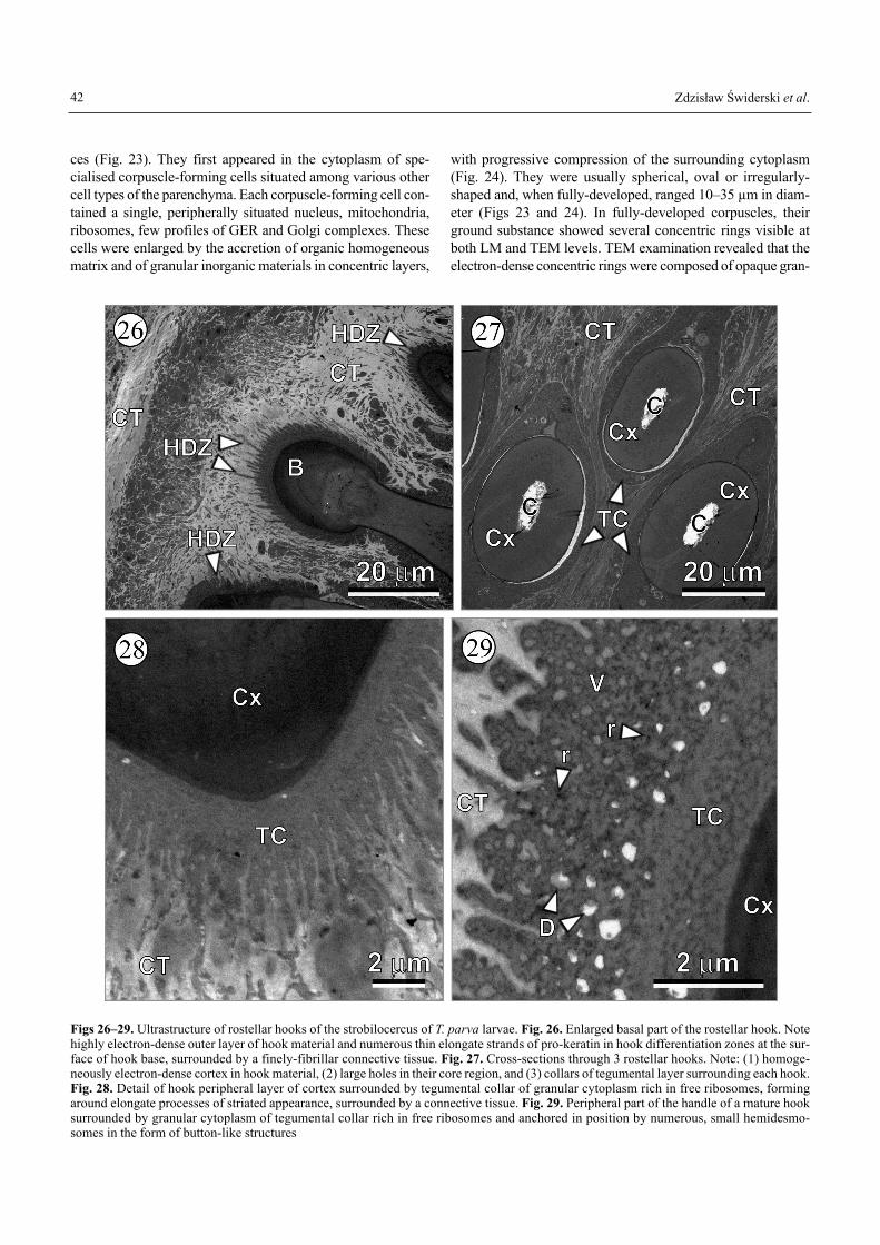
Figs 26�29. Ultrastructure of rostellar hooks of the strobilocercus of T. parva larvae. Fig. 26. Enlarged basal part of the rostellar hook. Notehighly electron-dense outer layer of hook material and numerous thin elongate strands of pro-keratin in hook differentiation zones at the sur-face of hook base, surrounded by a finely-fibrillar connective tissue. Fig. 27. Cross-sections through 3 rostellar hooks. Note: (1) homoge-neously electron-dense cortex in hook material, (2) large holes in their core region, and (3) collars of tegumental layer surrounding each hook.Fig. 28. Detail of hook peripheral layer of cortex surrounded by tegumental collar of granular cytoplasm rich in free ribosomes, formingaround elongate processes of striated appearance, surrounded by a connective tissue. Fig. 29. Peripheral part of the handle of a mature hooksurrounded by granular cytoplasm of tegumental collar rich in free ribosomes and anchored in position by numerous, small hemidesmo-somes in the form of button-like structures
Post-embryonic development of Taenia parva
ular material, but the remaining ground substance was homo-geneous (Fig. 24). As the corpuscle continued to enlarge, it dis-placed the nucleus and cytoplasm to the cell periphery.
All organelles of the corpuscle-forming cells degeneratedat the final stage of corpuscle development (Figs 23 and 24).
Origins and ultrastructure of the rostellar hooks
Ultrastructure of rostellar of T. parva larvae was examined oncross and oblique sections (Figs 26�29). The enlarged basalpart of the rostellar hook shows an electron-dense outer layerof hook material and numerous thin elongate strands of pro-keratin in hook differentiation zones at the surface of hookbase, surrounded by a finely-fibrillar connective tissue (Fig.26). On the cross-sections through rostellar hooks there aredistinct: (1) homogeneous electron-dense cortex in hookmaterial, (2) large holes in their core region, and (3) collars oftegumental layer surrounding each hook (Fig. 27). The hookperipheral layer of cortex is surrounded by a tegumental col-lar of granular cytoplasm, rich in free ribosomes, which formsaround elongate processes of striated appearance, surroundedby a connective tissue (Fig. 28). The handle of mature hooksis anchored in a correct position by numerous, small hemi-desmosomes in the form of button-like structures (Fig. 29).All the developmental stages of the rostellar hook morpho-genesis were not examined here in detail and they will be asubject of a separate paper.
Nervous system
The work on the differentiation and ultrastructure of the nerv-ous system of post-embryonic larvae of T. parva is still inprogress and will be published elsewhere.
Discussion
Developmental pattern
The oncosphere carries the germinative cells, which are theprimordium of the second larval stage, post-embryonic larvaor metacestode (�widerski 1972, 1981, 1983; �widerski et al.2002b; M³ocicki et al. 2006). However, most of the larvalattributes of the oncosphere are lost during development, andstructures such as the somatic musculature, the hook muscu-lature and the penetration glands do not persist in the metaces-tode. Our study on the development of the larvae of T. parvaconfirmed that all the metacestode structures are formed denovo and none of them is inherited from the oncosphere.Therefore, the formation of the metacestode is a true meta-morphosis (for detailed discussions see Stunkard 1962, Conn2005).
Asexual multiplication
Larval asexual multiplication is not common among cestodes.It is restricted to cyclophyllideans (some members of the fam-ilies Taeniidae, Hymenolepididae and Dilepididae), a fewunidentified pseudophyllideans (Nobrega-Lee et al. 2007) and
to the Mesocestoididae. The position of the latter family in theorder Cyclophyllidea has been questioned by proposing thedivision of the latter order into two groups: �mesocestoidatans+ cyclophyllideans� (Mariaux 1998). However, it appears thatthe status of the mesocestoidids as a distinct order or as thebasal member of the Cyclophyllidea may require further ex-amination (Mariaux 1998; Miquel et al. 1999, 2007; Hoberget al. 1999, 2001).
The unique feature of the asexual multiplication in somespecies of the Mesocestoididae, which distinguishes themfrom all other cestodes, is their capability of asexual repro-duction not only in the intermediate host but also in the gut of the definitive host. According to Eckert et al. (1969), asex-ual reproduction in adult Mesocestoides corti Hoeppli, 1925should be interpreted as the completion of larval reproductionwhile in the definitive host. A recent study on metacestodesof Mesocestoides sp. described a new type of endogenous asex-ual proliferation, in addition to two other previously describ-ed types, longitudinal fission and exogenous budding (Ga-lán-Puchades et al. 2002). However, the fact that most Meso-cestoides spp. are not known to reproduce asexually remainsan enigmatic feature of this group (Conn 1986, 1988a, 1990;Conn et al. 2002).
Of all the cestodes, larval asexual reproduction is mostcommon in the family Taeniidae, many species of which havethe capacity for exogenous and/or endogenous budding. Thegreatly-increased rate of asexual reproduction in Echinococ-cus larval cysts has potential unsurpassed by any other ces-tode. For details and references on the larvae of E. granulosus,see Smyth (1964), Morseth (1967) and Sakamoto and Sigi-mura (1969, 1970); for reviews, see �lais (1973), Thompson(1986) and Eckert et al. (2002); for details and references onthe larvae of E. multilocularis, see Mehlhorn et al. (1983),Thompson (1986) and Eckert et al. (2002).
In the genus Taenia, asexually multiplying larvae of thecysticercus type occur in eight species [T. crassiceps, T. endo-thoracica (Kirschenblatt, 1948), T. martis (Zeder, 1803), T. mul-ticeps, T. mustelae Gmelin, 1790, T. polyacantha Leuckart,1856, T. serialis (Gervais, 1847) and T. twitchelli Schwartz,1924] and polycephalic larvae of the strobilocercus type occurin three species [T. krepkogorski (Schulz et Landa, 1934),T. selousi Mettrick, 1962 and T. parva]. For a brief character-istic of the metacestodes of the eleven above-mentioned tae-niid species, see Loos-Frank (2000).
Polycephalic larvae developing in the abdominal and tho-racic cavities of small mammals, mainly rodents, greatly re-semble larvae of T. parva. Rausch (1952, 1959, 1977) exam-ined experimentally the life cycle and post-embryonic devel-opment of T. twitchelli, including the differentiation of theintermediate stages. The exogenous budding in T. crassicepswas studied by Freeman (1962). He also examined T. muste-lae, in which a single oncosphere may give rise to more thanone metacestode by the formation of multiple scoleces(Freeman 1956). Gvozdev and Agapova (1963) studied exper-imentally the life cycle of T. endothoracica, a species withmetacestodes resembling greatly those of T. parva. Like the
43

Zdzis³aw �widerski et al.
post-embryonic larva of T. parva, those of T. endothoracica,T. twitchelli, T. crassiceps and T. mustelae have scoleces thatare invaginated. In contrast, evaginated scoleces have beenreported in two species of taeniids with polycephalic larvae(Loos-Frank 2000), T. krepkogorski and T. selousi (see belowfor discussion on the significance of the scolex retraction orinvagination).
In the Hymenolepididae, asexual reproduction of metaces-todes was described in Urocystis prolifer Villot, 1880 (seeJoyeux 1922, Kisielewska 1960) and Staphylocystis pistillum(Dujardin, 1845) (see Joyeux and Baer 1936). Intermediatehosts of both species are glomerid diplopods (Glomeris spp.).The oncosphere of U. prolifer gives rise to an irregular cel-lular mass in the body cavity of the intermediate hosts; it pro-duces numerous cysticercoids by budding (Kisielewska 1960).The larval stage of S. pistillum is a polycephalic larva withindividual cysticercoids in the form of a bunch of grapes, forwhich the name Staphylocystis has been proposed (Villot1877). In an attempt to unify the cestode larval names, Chervy(2002) proposed for these two types of larvae the terms �uro-cysticercoid� and �staphylocysticercoid�, respectively.
In the Dilepididae, asexual reproduction leading to forma-tion of polycephalic larvae was found in earthworms and de-scribed either as Polycercus lumbrici Villot, 1883 or Paricte-rotaenia paradoxa (Rudolphi, 1802) (= Polycercus paradoxa,Sacciuterina paradoxa). The development of the larvae (�poly-cercus cysts�) was examined by LM (Metchnikov 1869, Scott1965, Gulyaev 2000) and by TEM (Crowe et al. 1974). Theirultrastructure shows a great similarity to the hydatid larvae ofE. granulosus with respect to asexual multiplication and thepresence of a PAS-positive layer of surrounding tissue. It isclearly different from the polycephalic larva of T. parva.Therefore, our ultrastructural results confirm that the asexualmultiplication of metacestodes does not reflect close phylo-genetic relationships among the species (Moore and Brooks1987, Hoberg et al. 2000, Trouve et al. 2003).
Types of larvae occurring in the genus Taenia and their ter-minology
Two general terms are currently used for the post-embryonic,post-oncospheral larvae of cestodes in the intermediate host:�metacestode� (see Wardle and McLeod 1952, Freeman 1973)and �cercoid� (see Jarecka 1970a,b, 1975). Both Freeman(1973) and Jarecka (1975) have commented on coexisting ter-minology and their criticisms are instructive. Conn (2005)pointed out that the true metamorphosis of oncospheres, intostages that are ultrastructurally like immature adults, makesthe metacestode stage of cestodes ontogenetically like the�juveniles� of other animal phyla (see Conn 2000). Althoughthe term �metacestode� seems to be controversial and etymo-logically may appear inappropriate (Jarecka 1975, Pojmanska1986, Chervy 2002), it remains still the most commonly-usedterm and it is impossible to avoid it. Whenever possible, how-ever, because of the above-mentioned differences of opinion,neutral terms such as �post-embryonic larvae� and �post-embryonic development� are used here.
The polycephalic larvae of T. parva are characterised by asegmented body and, therefore, they are very similar to thestrobilocercus type. The similar polycephalic and segmentedlarva of T. krepkogorski has been described as �coenurostro-bilocercus� (Agapova 1950). However, according to Abula-dze (1964) and Ryzhikov et al. (1978), there are no groundsfor introducing this term because (1) Agapova (1950) did notobserve the stages of coenurus development and (2) her obser-vations require confirmation as �she has not described her bio-logical experiment� (Abuladze 1964; p. 236). According toHoberg et al. (2000), the strobilocercus is considered to bederived from the cysticercus and is postulated as independentof other larval forms of Taenia spp.; it is defined as a strobilatemetacestode with well-developed scolex and prominent seg-mentation, which is present in life cycles of T. taeniaeformisand T. parva. The �coenurostrobilocercus� of T. parva hasbeen considered homologous with strobilocercus (Murai et al.1989, Hoberg et al. 2000).
The following six types of taeniid cestode larvae can be dis-tinguished (Hoberg et al. 2000, Chervy 2002, Georgiev et al.2006): (1) cysticercus, the most common type among Tae-nia spp., e.g. T. crassiceps, T. pisiformis (Bloch, 1780); (2) stro-bilocercus (= �strobilated cercoid�), e.g. T. taeniaeformis; (3)fimbriocercus (= armatetrathyridium), with an elongated unseg-mented body, e.g. T. martis; (4) coenurus, forming a large blad-der full of liquid, lined by a germinative membrane budding offmultiple scoleces, e.g. T. serialis; (5) hydatid, structurallyresembling a coenurus but producing daughter cysts, which canalso form both daughter cysts and scoleces; and (6) poly-cephalic, with numerous scoleces on elongated, segmented ornon-segmented strobile, arising by exogenous budding from acentral vesicle, which later degenerates, e.g. T. twitchelli.
The post-embryonic larva of T. parva possesses multiplescoleces and has consequently to be referred to as a poly-cephalic larval form. On the other hand, it closely resembles thestrobilocercus type of taeniid larvae by having a segmented lar-val body. However, the term �strobilocercus� has been pro-posed to be used for monocephalic larvae only (Chervy 2002)and, therefore, no existing term explicitly describes the larvaltraits of the species studied. This contradiction indicates thenecessity of further improvement of the unified terminology oflarval cestodes proposed by Chervy (2002). Hoberg et al.(2000) identified the larva of T. parva as a �polycephalic stro-bilocercus�, which more effectively reflects its morphology.
Loos-Frank (2000), in her valuable taxonomic survey ofTaenia, subdivided all multiplying larvae into two types, cys-ticerci and strobilocerci. In our opinion, it is not entirely clearwhy the larvae of T. endothoracica (�superficially annulatedanteriorly�) and T. twitchelli (�with pseudosegmentation at theanterior end�) are not included as belonging to the strobilo-cercus type. The metacestodes of these two species exhibit agreat resemblance to the larva of T. parva.
Significance of scolex invagination or retraction
As summarised by Beveridge (2001), the larval scolex oflower cestodes develops externally, whilst the scolex is either
44

Post-embryonic development of Taenia parva
inverted or withdrawn during post-embryonic development inhigher cestodes. The significance of evagination and invagi-nation of the scolex remains unclear. In the classical defini-tions of cysticercoids and cysticerci, it has always been anessential characteristic whether fully-developed scoleces wereinvaginated or retracted, although these terms were used inter-changeably by Wardle and McLeod (1952). In their reviewson cestode post-embryonic development, Voge (1967) and�lais (1973) considered scolex retraction or invagination asprominent, key developmental features. Freeman (1973), onthe other hand, did not include this character in his phyloge-netic analysis because he considered the scolex as a labilestructure, stating that �there is some evidence that the scolexof the metacestode may evert and retract as need be��(Freeman 1973; p. 502).
This character of scolex retraction or invagination is of great-est potential use in the Dilepididae. Hoberg et al. (1999) usedthis metacestode character in their analysis and supported thedivision of dilepidids into Dilepididae sensu stricto and Grypo-rhynchidae, with dilepidids having a withdrawn scolex and gry-porhynchids with an invaginated scolex (see Beveridge 2001).
Apparently, all taeniids are characterised by scolex mor-phogenesis in an invaginated condition. They differ fromhigher cyclophyllideans such as the hymenolepidids and dile-pidids, in which scolex morphogenesis takes place in anevaginated condition. As the results of the present study alsoindicate, the scoleces of T. parva remain invaginated duringthe morphogenesis of the post-embryonic larva. A similar con-dition has been described for two other taeniids with poly-cephalic larvae, T. twitchelli (see Rausch 1959) and T. endo-thoracica (see Gvozdev and Agapova 1963). However, in thepolycephalic larvae of T. krepkogorski, as described by Aga-pova (1950), when the early buds reach a certain size, theirscoleces evaginate and remain so during the entire further de-velopment (including the formation of the larval strobila).These observations of Agapova (1950) may still require con-firmation because, according to Abuladze (1964), her datawere incomplete. In T. selousi, the larva is composed of sev-eral larval strobila arising from a common bladder, each ofthem with ~50 segments and having an evaginated scolex (seeLoos-Frank 2000). Unfortunately, we have been unable to ver-ify this detail.
As summarised by Chervy (2002; p. 19), �careful exami-nation of the fully-developed larval cestode must reveal whether or not the scolex is invaginated or withdrawn anddevelopmental stages are not required to demonstrate this fea-ture�. We concur with Beveridge (2001; p. 253) that �furtherclarification of this character may assist in phylogeneticanalysis within the Cyclophyllidea, since, if the proteocepha-lideans are used as an outgroup, the withdrawn scolex wouldrepresent a synapomorphy linking the hymenolepidids, ano-plocephalids, davaineids and dilepidids�.
Larval strobilation
The polycephalic larva of T. parva shows distinct strobilation.However, as in T. taeniaeformis strobilocerci (Rees 1951;
Georgiev, unpublished information), no primordia of genitalorgans were observed in the larval strobila, even in stainedspecimens. It seems that it may be a general rule for the taeni-ids having �larval strobila� (e.g. T. taeniaeformis, T. krepko-gorski, T. parva) that genital systems never develop in the lar-val segments. For this reason, Loos-Frank (2000) generallyused the term �pseudostrobila� for the polycephalic larvae ofTaenia spp., including T. parva.
In Dioecocestus asper (Mehlis, 1831) (Cyclophyllidea,Dioecocestidae), the strobilate metacestodes show the pres-ence of a cirrus sac in the male specimens and the absence ofterminal genitalia in the females. However, when these tape-worms become mature in the intestine of the final host, the�larval proglottides� do not mature. The strobilar portion con-sisting of �larval proglottides� remains at the end of the grow-ing strobila as a sterile series before the beginning of apoly-sis (data of Yogis presented by Ryzhikov and Tolkacheva1981). On the basis of these studies and our observations onthe lack of genital primordia in the strobilate larva of T. parva,it may be concluded that the �larval strobila� of the cyclo-phyllideans is not an adaptation for rapid maturation. Appar-ently, it is rather an adaptation for a better feeding of the juve-nile cestodes during the first days after infecting the final host,when the larval strobila can provide a larger absorptive sur-face. Thus, the strobilate larval cestodes in cyclophyllideansare basically different from those of pseudophyllideans [e.g.Ligula intestinalis (L., 1758), Digramma interrupta (Rudol-phi, 1810) and Schistocephalus solidus (Mueller, 1776)],where the plerocercoid consists of proglottides at a veryadvanced stage, which mature rapidly and start egg produc-tion within 2�5 days after infecting the final host (Dubinina1966, Smyth and McManus 1989). It is worth mentioning thatin Lateriporus sp. (Cyclophyllidea, Dilepididae) there is asegmented cysticercoid termed strobilocysticercoid by Free-man (1973).
Tegument
The tegument of the larvae of T. parva resembles that of otheradult (Threadgold 1962, Lumsden 1975, M³ocicki et al. 2004)and larval tapeworms (Morseth 1967; Baron 1968, 1971;Crowe et al. 1974; Jarecka et al. 1981, 1984; Burt and Jarecka1984; Jarecka and Burt 1984; �widerski et al. 2002a). It iscomposed of an anucleate layer of peripheral cytoplasm cov-ered by a great number of microtriches and connected by cyto-plasmic projections with tegumental perikarya situated belowthe muscle layers. Our studies on T. parva corroborate thosementioned above and support the concept that the microtricheborders of both adult and larval cestodes should be regardedas a digestive-absorptive surface with specialised functionalcomponents (Threadgold 1962, Lumsden 1975).
Parenchyma
The nature of cestode parenchyma (and even its existence)remains unresolved. For example, a major question for a longtime has been whether the �parenchymal space� represents anintra- or intercellular space. It has been shown that much of
45

Zdzis³aw �widerski et al.
the volume of parenchymal tissue is made up of both volumi-nous lipid- and/or glycogen-rich cytoplasm and extracellularmatrices (ECM) in cyclophyllideans (Conn and Etges 1984)and proteocephalideans (Conn and Rocco 1989), and that thedynamic interaction between parenchymal cells and ECM isinvolved in histogenesis in some cestodes (Conn 1988b). Theclose association of parenchymal cells with myocytes andmuscle fibres has led to the conclusion that most of the �pa-renchymal cells� of cestodes are in fact myocytons (reviewedby Conn 1993). Later study on cyclophyllidean parenchymasimilarly reported two types of �parenchymal cells�: �typicalparenchymal cells� having numerous large outgrowths andexhibiting great storage capacity for lipids and glycogen, and�muscular cells� (Kornakova 1994), thus characterising thecestode parenchyma as �musculo-parenchymal� tissue. If thisinterpretation is confirmed by further studies, then the mus-culo-parenchymal tissue could be considered a kind of syncy-tial structure. Our study also confirms classical works (Lums-den 1965, 1966) demonstrating that the medullary parenchy-ma is the major site for polysaccharide and lipid deposition.
Muscle system
In general, the fine structure of cestode muscles is fairly uni-form, except for the highly-specialised ones associated withthe suckers and the rostellum (Lumsden and Hildreth 1983).Our observations are in agreement with the conclusion of Lums-den and Byram (1967) that cestode muscles are of a non-stri-ated type and are rather a variety of smooth muscles.
Functional correlates: possible role of regulatory peptides inneuromuscular interactions
The cytoplasmic projections with characteristic large, elec-tron-dense, spherical granules (type-1) observed between sco-lex muscles and, in particular, within the sucker musculatureof T. parva (see Figs 12, 14�19) resemble those described inthe proximity of the acetabular myocytons of Hymenolepisdiminuta (Rudolphi, 1819) (see Specian et al. 1979, Lumsdenand Specian 1980); they were believed to originate from themultipolar cells filled with the same type of electron-o-paque secretory granules found in perinuclear regions. Initially,these cells were interpreted as unicellular endocrine glands(Specian et al. 1979), probably because of their epithelial ori-gin; however, at the ultrastructural level, these glands wereconsidered as exhibiting many �non-neural� characteristics.According to Specian et al. (1979) and Lumsden and Specian(1980), the activity of these cells was of a neuroendocrine-type that had the possibility of establishing a direct commu-nication with the adjacent muscular tissue via a gap junction.The secreted material might function as a long-acting modu-lator of acetabular muscle activity (Specian et al. 1979). Therecent rapid development of research in this field has shownthat the borderline between the platyhelminth nervous systemand endocrine system has become indistinct and that the twoparts should now be considered under the name neuroendo-crine system. The last 16 years have witnessed, in addition tocholinergic and aminergic elements, the identification of a
third element to the nervous system of helminths, namely thepeptidergic component (Halton et al. 1990, 1994, 1999; Hal-ton and Gustafsson 1996). The three types of nerve fibres(cholinergic, aminergic and peptidergic) are sometimes reg-istered in close association as in the innervation of the rostellummusculature of davaineid cestodes (Stoitsova et al. 2001).Neuropeptides are ubiquitous intercellular signalling mole-cules in all Metazoa. There is increasing evidence, derivedmainly from light and electron microscope immunocyto-chemistry, that regulatory peptides and classical transmitters(catecholamines, 5-HT and acetylcholine) coexist in certainpopulations of neurons and endocrine cells. The peptidergicneuron is a multifunctional cell which probably has an impor-tant integrative function in platyhelminths since they do nothave true endocrine organs.
The second type of very small, highly electron-dense se-cretory granules (type-2), embedded in less granular cyto-plasm with mitochondria and observed within adjacent cyto-plasmic processes among muscle fibres of scolex and neckregion of T. parva larvae, resemble typical neurosecretorygranules (�widerski 1997, �widerski and Tkach 2002).
Origins and ultrastructure of the rostellar hooks
The general characteristics of rostellar histogenesis in taeniidshave been described at LM (Crusz 1947, 1948; Bilqees andFreeman 1969) and TEM level (Mount 1970). Initial disa-greement concerning the origin of the primary rostellar hook-lets was resolved by the convincing visual evidence providedby Mount (1970); he showed that the primordia of the rostel-lar hooks originate through enlargement of specialised micro-triches. Subsequently, hook protein is deposited along the edg-es of these microtriches to form the blade of the definitivehook. The handle and guard are formed as a secondary thick-ening from the deposition of hook protein along the blade. Thelarval tegument of T. parva, which surrounds the developinghooks, is modified by the presence of large accumulations ofribosomes and polyribosomal clusters as described for T. cras-siceps (see Mount 1970). The abundant rod-like bodies in thetegument forming the hook sheath in T. parva and T. crassi-ceps may represent the same hook-forming material describedin Polycercus paradoxa (see Crowe et al. 1974).
Calcareous corpuscles
The calcareous corpuscles (CC) observed in the larvae ofT. parva are characteristic for many cestode species, in par-ticular for their post-embryonic larvae (Conn 1993, Conn etal. 2002). They are of cellular origin and are formed in spe-cialised parenchymal cells, the so-called �CC-forming cells�(Nieland and Von Brand 1969, �widerski et al. 1970). Eachcalcareous corpuscle is composed of concentric lamellae andis made up of an organic basis together with inorganic mate-rial. Initially, they remain and grow in the cytoplasm of the�CC-forming cell� but, after reaching a certain size, they de-stroy the cells and remain as a large accumulation in the inter-cellular space of the parenchyma. The major inorganic com-ponents of calcareous corpuscles are calcium, magnesium,
46

Post-embryonic development of Taenia parva
carbonate and phosphorus, with small traces of other ele-ments; these constituents can vary considerably in relation tometabolic conditions and cestode species (Etges and Marina-kis 1991). Their role is still not entirely understood. It has beensuggested that they buffer anaerobically-produced acids orhelp to buffer gastric hydrochloric acid during larval passagethrough the host stomach, or they may also have an immuno-logical role. Other hypotheses are that they play an importantrole in: (1) the metabolism of early developing intestinalworms; (2) lipid metabolism; (3) tissue repair and osmoticbalance; and finally (4) may act as rudimentary skeletal struc-tures (Chung et al. 2003). Calcareous corpuscles have beenthe subject of numerous studies (see Von Brand et al. 1969,Smyth and McManus 1989) but their formation, compositionand function are still not completely clear.
Protonephridial system
The protonephridial system of T. parva larvae resembles, to agreat extent, that described from adult (�widerski et al. 1973,Lumsden and Specian 1980, Lumsden and Hildreth 1983) andlarval cestodes (Xylander 1987, 1992; �widerski et al. 2002a;�widerski and Mackiewicz 2004). Circularly-arranged, min-ute pores or �nephrostomes�, as described from three speciesof cyclophyllideans (�widerski et al. 1973), are between theendings of the outer row of digitiform processes arising fromthe nephridial funnel and the surface of the leading edge of theflame cell. These pores provide a direct communication be-tween the nephridial chamber and intercellular spaces of themedullary parenchyma and, therefore, play an important rolein the so-called �filtration apparatus�.
Concluding remarks
Both asexual reproduction and formation of polycephalic lar-vae, as observed in T. parva and in other taeniids as well as inthree other cestode families, are considered here as the resultsof multiple and independent origins and should be regarded asa good example of convergent evolution. Asexual prolifera-tion plays an important role in effective group infestation ofthe final hosts and undoubtedly increases the chances of com-pleting the parasite life cycle.
As concluded by Beveridge (2001) in his review on theapplication of life-cycle characters in cestode phylogeny, newLM and TEM data on a great variety of cestode species are ur-gently needed to provide novel characters to examine in seek-ing a better understanding of cestode evolution. In his opinion,histology and the ultrastructure of metacestodes offer �con-siderable opportunities and appear to be a field which has, un-til now, not been exploited in a systematic fashion� Beveridge(2001; p. 256), but which offers considerable scope for futureanalysis. Unfortunately, such studies are not common, despitethe fact that they can clearly provide direct evidence thatmight help to resolve many disputed questions concerninginterrelationships between cestode taxa and major evolution-ary lineages. Nevertheless, ultrastructural and histological stud-ies on metacestodes appear to have considerable promise forproviding new characters for phylogenetic analysis. As Bev-
eridge (2001; p. 254) has concluded, �until more comprehen-sive studies become available, the current data are not ame-nable to analysis�.
Acknowledgements. We are grateful to A. Jones, J.S. Mackie-wicz, I. Beveridge and D.B. Conn for kindly commenting on an ear-lier version of the manuscript. We also thank �Serveis Científics iTPcnics� of the University of Barcelona for their support in thepreparation of samples. This study was financially supported by theSpanish grant 2005-SGR-00576 from the �DURSI (Generalitat deCatalunya)�. The completion of results, analysis of TEM-micro-graphs, and preparation of the manuscript of this paper for publica-tion was supported by a Sabbatical Grant (SAB2005-0068) from the�Secretaría de Estado de Universidades e Investigación, Ministeriode Educación y Ciencia� of Spain for Z.S.
References
Abuladze K.I. 1964. Taeniata � tapeworms of animals and man, anddiseases caused by them. In: Osnovy tsestodologii. Vol. 4 (Ed.K.I. Skryabin). Nauka, Moscow (In Russian). (English trans-lation, 1970, Israel Program for Scientific Translations, Jeru-salem).
Agapova A.I. 1950. A new type of larval cestode structure. IzvestiyaAkademii Nauk Kazakhskoy SSR, Seriya Parazitologiche-skaya, 8, 242�245 (In Russian).
Alvarez F., Iglesias R., Bos J., Tojo M., Sanmartín M.L. 1990. Newfindings of the helminth fauna of the common European genet(Genetta genetta L.): first record of Toxocara genettae War-ren, 1972 (Ascarididae) in Europe. Annales de ParasitologieHumaine et Comparée, 65, 244�248.
Baer J.G. 1926. Contributions to the helminth fauna of South Africa.11 & 12 Reports on Veterinary Research. Union of SouthAfrica, Pretoria, 61�136.
Baron P.J. 1968. On the histology and ultrastructure of Cysticercuslongicollis, the cysticercus of Taenia crassiceps Zeder, 1800(Cestoda, Cyclophyllidea). Parasitology, 58, 497�513.
Baron P.J. 1971. On the histology, histochemistry and ultrastructureof the cysticercoid of Raillietina cesticillus (Molin, 1858)Fuhrmann, 1920 (Cestoda, Cyclophyllidea). Parasitology, 62,233�245.
Bernard J. 1961. Quelques espPces d�helminthes de micromam-mifPres récoltés en France et en Espagne. Vie et Milieu, 12,125�149.
Bernard J. 1963. Cysticerques polycéphales chez le mulot. Archivesde l�Institut Pasteur, Tunis, 40, 269�277.
Beveridge I. 2001. The use of life-cycle characters in studies of theevolution of cestodes. In: Interrelationships of the Platyhel-minthes (Eds. D.T.J. Littlewood and R.A. Bray). Taylor andFrancis, London, 250�256.
Beveridge I., Richard M.D., Gregory G.G., Munday B.L. 1975.Studies on Anoplotaenia dasyuri Beddard, 1911 (Cestoda:Taeniidae), a parasite of Tasmanian devil. Observations on theegg and metacestode. International Journal for Parasitology,5, 257�267.
Bilqees F.M., Freeman R.S. 1969. Histogenesis of the rostellum ofTaenia crassiceps (Zeder, 1800) (Cestoda), with special ref-erence to hook development. Canadian Journal of Zoology,47, 251�261.
Burt M.D.B. 1987. Early morphogenesis in the Platyhelminthes withspecial reference to egg development and development ofcestode larvae. International Journal for Parasitology, 17,241�253.
Burt M.D.B., Jarecka L. 1984. Studies on the life cycle of Hyme-nolepis ductilis Linton, 1927 and on the ultrastructure of its
47

Zdzis³aw �widerski et al.
cysticercoid tegument. Acta Parasitologica Polonica, 39,35�42.
Caley J. 1974. The functional significance of scolex retraction andsubsequent cyst formation in the cysticercoid larva of Hyme-nolepis microstoma. Parasitology, 68, 207�227.
Caley J. 1976. Ultrastructural studies of the cysticercoid of Monieziaexpansa (Anoplocephalidae) with special reference to the de-velopment of the cyst. Zeitschrift für Parasitenkunde, 48, 251�262.
Campana-Rouget Y. 1950. Un cysticerque polycéphale chez lemulot, Apodemus sylvaticus L. Vie et Milieu, 1, 202�206.
Casanova J.C., Feliu C., Torres J., Miquel J., �pakulová M. 2000.Faunistic and ecological trends on the helminthic communi-ty of Genetta genetta Linnaeus, 1758 (Carnivora: Viverridae)in the Iberian Peninsula. Helminthologia, 37, 223�228.
Chervy L. 2002. The terminology of larval cestodes or metacestodes.Systematic Parasitology, 52, 1�33.
Chung Y., Kong Y., Cho S., Yang H. 2003. Purification and localiza-tion of a 10 kDa calcareous corpuscle binding protein ofSpirometra mansoni plerocercoid. Parasitology Research, 89,235�237.
Conn D.B. 1986. Comparative fine structure of Mesocestoides spp.tetrathyridia. In: Handbook of the 6th International Congressof Parasitology (Ed. M.J. Howell). Australian Academy ofScience, Canberra, 114.
Conn D.B. 1988a. Fine structure of the tegument of Mesocestoideslineatus tetrathyridia (Cestoda: Cyclophyllidea). Internation-al Journal for Parasitology, 18, 133�135.
Conn D.B. 1988b. The role of cellular parenchyma and extracellularmatrix in the histogenesis of the paruterine organ of Meso-cestoides lineatus (Platyhelminthes: Cestoda). Journal of Mor-phology, 197, 303�314.
Conn D.B. 1990. The rarity of asexual reproduction among Meso-cestoides tetrathyridia (Cestoda). Journal of Parasitology, 76,453�455.
Conn D.B. 1993. The biology of flatworms (Platyhelminthes): Pa-renchyma cells and extracellular matrices. Transactions of theAmerican Microscopical Society, 112, 241�261.
Conn D.B. 2000. Atlas of Invertebrate Reproduction and Devel-opment. John Wiley & Sons, Inc., New York.
Conn D.B. 2005. Comparative aspects of postembryonic develop-ment of cestodes (Platyhelminthes) and other animal taxa. In:Multidisciplinarity for Parasites, Vectors and Parasitic Dis-eases. Proceedings of the 9th European Multicolloquium ofParasitology. Vol. 1. Articles of Keynote Speakers (Ed. S. Mas-Coma). Monduzzi Editore, Bologna, 319�325.
Conn D.B., Etges F.J. 1984. Fine structure and histochemistry of theparenchyma and uterine egg capsules of Oochoristica anolis(Cestoda: Linstowiidae). Zeitschrift für Parasitenkunde, 70,769�779.
Conn D.B., Galán-Puchades M.T., Fuentes M. 2002. Ultrastructureof cells and extracellular matrices in the hindbody of non-pro-liferative tetrathyridia of Mesocestoides lineatus (Cestoda:Cyclophyllidea). In: Proceedings of the 10th InternationalCongress of Parasitology. Monduzzi Editore, Bologna, 535�538.
Conn D.B., Rocco L.J. 1989. Fine structure of the cellular parenchy-ma and extracellular matrix of Ophiotaenia loennbergii (Ces-toda: Proteocephalidea). Acta Zoologica (Stockholm), 70,105�110.
Crowe D.G., Burt M.D.B., Scott J.S. 1974. On the ultrastructure ofthe polycercus larvae of Paricterotaenia paradoxa (Cestoda:Cyclophyllidea). Canadian Journal of Zoology, 52, 1397�1411.
Crusz H. 1947. The early development of the rostellum of Cys-ticercus fasciolaris Rud., and the chemical nature of its hooks.Journal of Parasitology, 33, 87�98.
Crusz H. 1948. Further studies on the development of Cysticercusfasciolaris and Cysticercus pisiformis, with special referenceto the growth and sclerotization of the rostellar hooks. Jour-nal of Helminthology, 22, 179�198.
Dollfus R.-P., Saint Girons M.C. 1958. Modification du comporte-ment d�un Apodemus parasité par de cystiques polycéphales,en relation avec la croissance de ceux-ci. Vie et Milieu, 9,116�123.
Dubinina M.N. 1966. Ligulids (Cestoda: Ligulidae) of the fauna ofthe USSR. Nauka, Moscow (In Russian).
Eckert J., Von Brand T., Voge M. 1969. Asexual multiplication ofMesocestoides corti (Cestoda) in the intestine of dogs. Jour-nal of Parasitology, 55, 241�249.
Eckert J., Gemmell M.A., Meslin F.-X., Paw³owski Z.S. (Eds.).2002. WHO/OIE Manual on Echinococcosis in Humans andAnimals: a Public Health Problem of Global Concern. WorldOrganisation for Animal Health (Office International desEpizooties) and World Health Organization, Paris.
Etges F.J., Marinakis V. 1991. Formation and excretion of calcareousbodies by the metacestode (tetrathyridium) of Mesocestoidesvogae. Journal of Parasitology, 77, 595�602.
Feliu C. 1980. Contribución al conocimento de la helmintofauna demicromammíferos ibéricos. Helmintos de Gliridae y Muridae(Rodentia). PhD Thesis, University of Barcelona.
Freeman R.S. 1956. Life history studies on Taenia mustelae Gmelin,1790 and the taxonomy of certain taenioid cestodes fromMustelidae. Canadian Journal of Zoology, 34, 219�242.
Freeman R.S. 1962. Studies on the biology of Taenia crassiceps(Zeder, 1800) Rudolphi, 1810 (Cestoda). Canadian Journalof Zoology, 40, 969�990.
Freeman R.S. 1973. Ontogeny of cestodes and its bearing on theirphylogeny and systematics. Advances in Parasitology, 11,481�556.
Galán-Puchades M.T., Fuentes M., Conn D.B. 2002. A new type ofendogenous asexual proliferation in cyclophyllidean meta-cestodes. Acta Parasitologica, 47, 288�293.
Georgiev B.B., Bray R.A., Littlewood D.T.J. 2006. Cestodes of smallmammals: taxonomy and life cycles. In: Micromammals andmacroparasites. From evolutionary ecology to management(Eds. S. Morand, B.R. Krasnov, R. Poulin). Springer Verlag,Tokyo, 29�62.
Georgiev B.B., Sánchez M.I., Green A.J., Nikolov P.N., Vasileva G.P.Mavrodieva R.S. 2005. Cestodes from Artemia parthenoge-netica (Crustacea, Branchiopoda) in the Odiel Marshes, Spain:a systematic survey of cysticercoids. Acta Parasitologica, 50,105�117.
Gulyaev V.D. 2000. On gemmation of Polycercus paradoxa (Ces-toda, Cyclophyllidea) metacestode generations. Zoologiche-skiy Zhurnal, 79, 1103�1110 (In Russian).
Gvozdev E.V., Agapova A.I. 1963. Life cycle of Multiceps endotho-racicus Kirschenblatt, 1948. Trudy Instituta Zoologii, Alma-Ata, 19, 46�48 (In Russian).
Halton D.W., Fairweather I., Shaw C., Johnston C.F. 1990. Regu-latory peptides in parasitic platyhelminths. Parasitology To-day, 6, 284�290.
Halton D.W., Gustafsson M.K.S. 1996. Functional morphology of theplatyhelminth nervous system. Parasitology, 113, S47�S72.
Halton D.W., Maule A.G., Marks N.J., Followes R.A., Shaw C. 1999.FMRF amide-related peptides (FaRPs) in helminth parasites.Acta Parasitologica, 44, 11�18.
Halton D.W., Shaw C., Maule A.G., Smart D. 1994. Regulatory pep-tides in helminth parasites. Advances in Parasitology, 34, 163�227.
Hoberg E.P., Jones A., Bray R.A. 1999. Phylogenetic analysis amongthe families of the Cyclophyllidea (Eucestoda) based on com-parative morphology, with new hypotheses for co-evolutionin vertebrates. Systematic Parasitology, 42, 51�73.
48

Post-embryonic development of Taenia parva
Hoberg E.P., Jones A., Rausch R.L., Eom K.S., Gardner S.L. 2000. Aphylogenetic hypothesis for species of the genus Taenia.Journal of Parasitology, 86, 89�98.
Hoberg E.P., Mariaux J., Brooks D.R. 2001. Phylogeny among theorders of the Eucestoda (Cercomeromorphae): integratingmorphology, molecules and total evidence. In: Interrelation-ships of the Platyhelminthes (Eds. D.T.J. Littlewood and R.A.Bray). Taylor and Francis, London, 112�126.
Jarecka L. 1970a. Phylogeny and evolution of life cycles of Cestodafrom fresh water and terrestrial vertebrates. Journal of Para-sitology, 56 (sect. II, pt. 1), 169�170.
Jarecka L. 1970b. Life cycle of Valipora campylancristrota (Wedl,1855) Baer and Bona 1958�1960 (Cestoda � Dilepididae) andthe description of cercoscolex � a new type of cestode larva.Bulletin de l�Academie Polonaise des Sciences, Classe II,Series des Sciences Biologiques, 18, 99�102.
Jarecka L. 1975. Ontogeny and evolution of cestodes. Acta Para-sitologica Polonica, 23, 93�114.
Jarecka L., Bance G.N., Burt M.D.B. 1984. On the life cycle ofAnomotaenia micracantha dominicana (Railliet et Henry,1912), with ultrastructural evidence supporting the definitionof cercoscolex for dilepidid larvae (Cestoda, Dilepididae).Acta Parasitologica Polonica, 29, 27�34.
Jarecka L., Burt M.D.B. 1984. The cercoid larvae of Pseudantho-bothrium hanseni Baer, 1956 and Pseudanthobothrium sp.(Cestoda, Tetraphyllidea) from experimentally infected har-pacticoid copepods. Acta Parasitologica Polonica, 29, 23�26.
Jarecka L., Michajlow W., Burt M.D.B. 1981. Comparative ultra-structure of cestode larvae and Janicki�s cercomer theory.Acta Parasitologica Polonica, 28, 65�72.
Joyeux C. 1922. Recherches sur l�Urocystis prolifer Villot. Note pre-liminaire. Bulletin de Société Zoologique de France, 47, 53�58.
Joyeux C., Baer J.G. 1936. Quelques helminthes nouveaux et peuconnus de la musaraigne, Crocidura rusula Herm. RevueSuisse de Zoologie, 43, 25�50.
Khalil L.F., Jones A., Bray R.A. 1994. Keys to the cestode parasitesof vertebrates. CAB International, Wallingford.
Kisielewska K. 1960. Life cycle of the tapeworm Pseudodiorchisprolifer (Villot, 1890) comb. nova (= Pseudodiorchis multi-spinosa Zarnowski, 1955). Acta Parasitologica Polonica, 8,199�203.
Kornakova E.E. 1994. Ultrastructure of parenchyma and the extra-cellular matrix of Passerilepis crenata (Cestoda: Cyclophyl-lidea). Parazitologiya, 28, 119�123 (In Russian).
Krasnoshchekov G.P., Pluzhnikov L.T. 1984. Ultrastructure of thecysticercoid of Fimbraria fasciolaris (Hymenolepididae). Parazitologiya, 18, 47�52 (In Russian).
Loos-Frank B. 2000. An up-date of Verster�s (1969) �Taxonomicrevision of the genus Taenia Linnaeus� (Cestoda) in table for-mat. Systematic Parasitology, 45, 155�183.
Lumsden R.D. 1965. Macromolecular structure of glycogen in somecyclophyllidean and trypanorhynch cestodes. Journal of Parasitology, 51, 501�515.
Lumsden R.D. 1966. Fine structure of the medullary parenchymalcells of a trypanorhynch cestode, Lacistorhynchus tenuis (VanBeneden, 1858), with emphasis on specializations for glyco-gen metabolism. Journal of Parasitology, 52, 417�427.
Lumsden R.D. 1975. Surface ultrastructure and cytochemistry ofparasitic helminths. Experimental Parasitology, 37, 267�339.
Lumsden R.D., Byram J.E. III 1967. The ultrastructure of cestodemuscle. Journal of Parasitology, 53, 326�342.
Lumsden R.D., Hildreth M.B. 1983. The fine structure of adult tape-worms. In: Biology of the Eucestoda. Vol. 1 (Eds. C. Armeand P.W. Pappas). Academic Press, London, 177�233.
Lumsden R.D., Specian R.D. 1980. The morphology, histology andfine structure of the adult stage of the cyclophyllidean tape-
worm Hymenolepis diminuta. In: Biology of the tapewormHymenolepis diminuta (Ed. H.P. Arai). Academic Press, NewYork and London, 157�280.
Mariaux J. 1998. A molecular phylogeny of the Eucestoda. Journalof Parasitology, 84, 114�124.
Mas-Coma S., Feliu C. 1977. Contribución al conocimento de lahelmintofauna de micromamíferos ibéricos. IV. Parásitos deApodemus sylvaticus Linnaeus, 1758 (Rodentia: Muridae).Revista Ibérica de Parasitología, 37, 301�317.
Mehlhorn H., Eckert J., Thompson R.C.A. 1983. Proliferation andmetastases formation of larval Echinococcus multilocularis.II. Ultrastructural investigations. Zeitschrift für Parasiten-kunde, 69, 749�763.
Metchnikov E. 1869. Entwicklungsgeschichtliche Beiträge. Bulle-tin de l�Académie Impériale de Saint-Pétersburg, 13, 284�300.
Miquel J., Eira C., �widerski Z., Conn D.B. 2007. Mesocestoides line-atus (Goeze, 1782) (Mesocestoididae): new data on spermultrastructure. Journal of Parasitology, 93, in press.
Miquel J., Feliu C., Marchand B. 1999. Ultrastructure of spermio-genesis and the spermatozoon of Mesocestoides litteratus(Cestoda, Mesocestoididae). International Journal for Para-sitology, 29, 499�510.
M³ocicki D., Eira C., ¯ebrowska J., Miquel J., �widerski Z. 2004.Ultrastructure of the tegument of the anoplocephalid cestodeMosgovoyia ctenoides (Railliet, 1890) Beveridge, 1978. Wia-domo�ci Parazytologiczne, 50, 579�585.
M³ocicki D., �widerski Z., Miquel J., Eira C., Conn DB. 2006. Cel-lular organisation of the oncosphere of Mosgovoyia ctenoi-
des (Cestoda, Anoplocephalidae). Journal of Parasitology,92, 953�961.
Moore J., Brooks D.R. 1987. Asexual reproduction in cestodes (Cy-clophyllidea: Taeniidae): ecological and phylogenetic influ-ences. Evolution, 41, 882�891.
Morseth D.J. 1967. Fine structure of the hydatid cyst and protoscolexof Echinococcus granulosus. Journal of Parasitology, 53,312�325.
Mount P. 1970. Histogenesis of the rostellar hooks of Taenia crassi-ceps (Zeder, 1800) (Cestoda). Journal of Parasitology, 56,947�961.
Murai E., Tenora F., Stanek M. 1989. Atypical strobilocercus (Ces-toda: Taeniidae) � a parasite in experimental stocks of On-datra zibethicus (Rodentia). Miscellanea Zoologica Hunga-rica, 5, 21�27.
Nieland M.L., Von Brand T. 1969. Electron microscopy of cestodecalcareous corpuscle formation. Experimental Parasitology,24, 279�289.
Nieland M.L., Weinbach E.C. 1968. The bladder of Cysticercus fas-ciolaris: electron microscopy and carbohydrate content. Par-asitology, 58, 489�496.
Nobrega-Lee M., Hubbard G., Gardiner C.H., LoVerde P., Carvalho-Queiroz C., Conn D.B., Rohde K., Dick Jr. E.J., NathanielszP., Martin D., Siler-Khodr T., Schlabritz-Loutsevitch N. 2007.Sparganosis in wild-caught baboons (Papio cynocephalusanubis). Journal of Medical Primatology, 36, 47�54.
Pojmanska T. 1986. Points in question: terminology in parasitology.International Journal for Parasitology, 16, 433�434.
Race G.J., Larsh J.E. Jr., Esch G.W., Martin J.H. 1965. A study of thelarval stage of Multiceps serialis by electron microscopy.Journal of Parasitology, 51, 364�369.
Rausch R. 1952. Studies on the helminthofauna of Alaska. XI.Helminth parasites of microtine rodents � taxonomic consid-erations. Journal of Parasitology, 38, 415�444.
Rausch R. 1959. Studies on the helminthofauna of Alaska. XXXVI.Parasites of the wolverine, Gulo gulo L., with observations onthe biology of Taenia twitchelli Schwartz 1924. Journal ofParasitology, 45, 465�484.
49

Zdzis³aw �widerski et al.
Rausch R. 1977. The specific distinction of Taenia twitchelli Schwartz1924 from T. martis (Zeder, 1803) (Cestoda: Taeniidae). Excer-ta Parasitológica en Memoria del Doctor Eduardo Caballeroy Caballero. Instituto de Biología, Publicaciones Especiales,4, 357�366.
Rees G. 1951. The anatomy of cysticercus of Taenia taeniaeformis(Batsch, 1786) (Cysticercus fasciolaris Rud., 1808) from theliver of Rattus norvegicus (Erx.), including an account of spi-ral torsion in the species and some minor abnormalities instructure. Parasitology, 41, 46�59.
Ryzhikov K.M, Gvozdev E.V., Tokobaev M.M., Shaldybin L.S.,Matsaberidze G.V., Merkusheva I.V., Nadtochiy E.V., Kho-khlova I.G., Sharpilo L.D. 1978. Key to the helminth parasitesof rodents from the fauna of the USSR. Cestodes and trema-todes. Nauka, Moscow (In Russian).
Ryzhikov K.M., Tolkacheva L.M. 1981. Acoleata � tapeworms ofbirds. In: Osnovy tsestodologii. Vol. 10 (Ed. V.I. Freze.). Nau-ka, Moscow (In Russian).
Sakamoto T., Sigimura M. 1969. Studies on echinococcosis. XXI.Electron microscopical observations on general structure oflarval tissue of multilocular echinococcosis. Japanese Jour-nal of Veterinary Research, 17, 67�80.
Sakamoto T., Sigimura M. 1970. Studies on echinococcosis. XXIII.Electron microscopical observations on histogenesis of larvalEchinococcus multilocularis. Japanese Journal of VeterinaryResearch, 18, 131�144.
Scott J.S. 1965. The development and morphology of Polycercus lum-brici (Cestoda: Cyclophyllidea). Parasitology, 55, 127�143.
�lais J. 1973. Functional morphology of cestode larvae. Advances inParasitology, 11, 395�480.
�lais J., Serbus C., Schramlová J. 1972. The fine structure of the con-tractile elements in the bladder wall of Cysticercus bovis.Folia Parasitologica, 19, 165�168.
Smyth J.D. 1964. The biology of the hydatid organisms. Advancesin Parasitology, 2, 169�219.
Smyth J.D., McManus D.P. 1989. The physiology and biochemistryof cestodes. Cambridge University Press, Cambridge.
Specian R.D., Lumsden R.D., Ubelaker J.E., Allison V.E. 1979. Aunicellular endocrine gland in cestodes. Journal of Parasit-ology, 65, 569�578.
Stoitsova S.R., Georgiev B.B., Vasileva G.P., Jordanova M. 2001.Rostellar apparatus of Fernandezia spinosissima (von Lin-stow, 1894) (Cestoda, Cyclophyllidea, Davaineidae): micro-anatomy and fine structure. Acta Zoologica (Stockholm), 82,241�250.
Stunkard H.W. 1962. The organization, ontogeny, and orientation ofthe Cestoda. Quarterly Review of Biology, 37, 23�34.
�widerski Z. 1972. La structure fine de l�oncosphPre du cestodeCatenotaenia pusilla (Goeze, 1782) (Cyclophyllidea, Cateno-taeniidae). La Cellule, 69, 205�237.
�widerski Z. 1981. Reproductive and developmental biology of thecestodes. In: Advances in invertebrate reproduction (Eds.W.H. Clark, Jr. and T.S. Adams). Elsevier/North Holland,New York, Amsterdam, Oxford.
�widerski Z. 1983. Echinococcus granulosus: hook-muscle systemand cellular organisation of the infective oncospheres. In-ternational Journal for Parasitology, 13, 289�299.
�widerski Z. 1997. Ultrastructure of the oncospheral nerve cells inEchinococcus granulosus and E. multilocularis. In: ArchivosInternacionales de la Hidatidosis. Vol. 32. XVIII InternationalCongress of Hydatidology, Lisboa, Portugal, 126�128.
�widerski Z., Chomicz L., Grytner-Ziêcina B., Sereda M.J. 2002b.Ultrastructural characteristic of the oncospheral germinativecells in Echinococcus multilocularis and E. granulosus. In:
(Accepted January 3, 2007)
Proceedings of the 10th International Congress of Parasit-ology � ICOPA X: Symposia, Workshops and ContributedPapers, Vancouver, Canada, August 4�9, 2002, 551�554.
�widerski Z., Euzet L., Schönenberger N. 1973. Ultrastructures dusystPme nephridien des cestodes cyclophyllides Inermicap-sifer madagascariensis, Catenotaenia pusilla et Hymenolepismicrostoma. La Cellule, 71, 7�18.
�widerski Z., Huggel H., Schönenberger N. 1970. Electron micros-copy of calcareous corpuscle formation and their ultrastruc-ture in the cestode Inermicapsifer madagascariensis. In: Pro-ceedings of the 7th International Congress of Electron Mi-croscopy (Ed. J. André), 6�12 September, 1970, Grenoble,France, 821�822.
�widerski Z., Mackiewicz J.S. 2004. Ultrastructural studies on thecellular organisation of the coracidium of the cestodeBothriocephalus clavibothrium Ariola, 1899 (Pseudophylli-dea, Bothriocephalidae). Acta Parasitologica, 49, 116�139.
�widerski Z., Sereda M.J., Vakarenko E., Kornyushin V.V., SalamatinR.V. 2002a. Fine structure of the cysticercoid of the cestodeSobolevitaenia verulamii (Cyclophyllidea, Dilepididae). In:Proceedings of the 15th International Congress on ElectronMicroscopy, 1�6 September, 2002, Durban, South Africa. Vol.2. Biology and Medicine (Ed. T. Sewell), 489�490.
�widerski Z., Tkach V.V. 2002. Ultrastructure of embryonic devel-opment of Inermicapsifer madagascariensis (Cestoda, Ano-plocephalidae) with emphasis on the cellular organisation ofthe infective eggs. Acta Parasitologica, 47, 105�120.
Thompson R.C.A. (Ed.). 1986. The biology of Echinococcus andhydatid disease. George Allen & Unwin Publishers Ltd.,London, Boston & Sidney.
Threadgold L. 1962. An electron microscope study of the tegumentand associated structures of Dipylidium caninum. QuarterlyJournal of Microscopical Sciences, 103, 135�140.
Trouvé S., Morand S., Gabrion C. 2003. Asexual multiplication oflarval parasitic worms: a predictor of adult life-history traitsin Taeniidae? Parasitology Research, 89, 81�88.
Ubelaker J.E. 1980. Structure and ultrastructure of the larvae andmetacestodes of Hymenolepis diminuta. In: Biology of thetapeworm Hymenolepis diminuta (Ed. H.P. Arai). AcademicPress, New York and London, 59�156.
Ubelaker J.E. 1983. Metacestodes: morphology and development.In: Biology of the Eucestoda. Vol. 1 (Eds. C. Arme and P.W.Pappas). Academic Press, London & New York, 139�175.
Verster A. 1969. A taxonomic revision of the genus Taenia Linnaeus1758. Onderstepoort Journal of Veterinary Research, 36,3�58.
Villot F.C.A. 1877. Migrations et métamorphoses des ténias desmusaraignes. Annales des Sciences Naturelles, Zoologie, 6PmeSérie, 8, 1�19.
Voge M. 1967. The post-embryonic developmental stages of ces-todes. Advances in Parasitology, 11, 707�730.
Von Brand T., Nylen M.U., Martin G.N., Churchwell F.K., Sites E.1969. Cestode calcareous corpuscles: phosphate relation-ships, crystalization patterns, and variations in size and shape.Experimental Parasitology, 25, 291�310.
Wardle R.A., McLeod J.A. 1952. The zoology of tapewoms. TheUniversity of Minnesota Press, Minneapolis.
Xylander W.E.R. 1987. Ultrastructure of the lycophora larvae ofGyrocotyle urna (Cestoda, Gyrocotylidea). II. The proto-nephridial system. Zoomorphology, 107, 88�95.
Xylander W.E.R. 1992. Investigations of the protonephridial systemof postlarval Gyrocotyle urna and Amphilina foliacea (Ces-toda). International Journal for Parasitology, 22, 287�300.
50
Related Documents