NDT Plus (2010) 3: 405–406 doi: 10.1093/ndtplus/sfq043 Advance Access publication 8 April 2010 Images in Nephrology (Section Editor: G. H. Neild) Post-ESWL fragments as core of new kidney stones Maria Luigia Giannossi and Vito Summa Laboratory of Environmental and Medical Geology, IMAA-CNR—C.da Santa Loja, Tito Scalo (Pz), Italy Correspondence and offprint requests to: Maria Luigia Giannossi; E-mail: [email protected] Keywords: calcium oxalate kidney stones; post-ESWL fragments; regrowth The use of extracorporeal shockwave lithotripsy (ESWL) to treat kidney stones gives excellent fragmentation results. However, the retention of post-ESWL fragments within the kidney remains a serious health problem. A study carried out on calcium stone patients showed that only 32% of these were found to be stone free 12 months after ESWL [1]. Therefore, the persistence and the growth of fragments seem to be common after ESWL [2,3]. After ESWL, stone-free rates are closely related to stone location, size, number and composition, so a complete stone analysis of the first ESWL residual available must be carried out in order to perform the following treatments correctly so as to prevent recidivisms. Kidney stones collected from two patients previously treated with ESWL were chosen for this study (Figures 1 and 2). The main component of these kidney stones is calcium oxalate monohydrate (whewellite, CaC 2 O 4 ·H 2 O) mixed with uric acid (C 5 H 4 N 4 O 3 ), identified by X-ray diffrac- tion analysis. A microscopic examination was made on petrographic thin section with a stereomicroscope cou- pled to polarizing microscope in order to provide infor- mation on the internal structure, location and type of core. The results show that these kidney stones were due to a regrowth on post-ESWL fragments. In both cases, the ESWL treatment has not reduced the previous stones in fragments of dimensions easy to be expelled or eliminated with an appropriated pharmaco- logical treatment [4]. In the latter case (Figure 2), the re- sidual fragment is very small (1 mm), not easily visible and recognizable only by a careful analysis, while the former stone (Figure 1) is made of a fragment of remark- able dimensions. Obviously, stone fragment regrowth is an unfavourable factor for spontaneous passage, and consequently an ad- ditional serious complication of post-ESWL residual stones. Conflict of interest statement. None declared. Fig. 2. Case 2—mixed kidney stone (whewellite and uric acid) with the core formed by a post-ESWL fragment identifiable by polarizing microscopy on petrographic thin section. Fig. 1. Case 1—image obtained by a stereomicroscopy of a post-ESWL fragment of a mixed kidney stone (whewellite and uric acid layers) with in vivo regrowth. © The Author 2010. Published by Oxford University Press on behalf of ERA-EDTA. All rights reserved. For permissions, please e-mail: [email protected] Downloaded from https://academic.oup.com/ckj/article-abstract/3/4/405/557203 by guest on 09 April 2019

Welcome message from author
This document is posted to help you gain knowledge. Please leave a comment to let me know what you think about it! Share it to your friends and learn new things together.
Transcript
NDT Plus (2010) 3: 405–406doi: 10.1093/ndtplus/sfq043Advance Access publication 8 April 2010
Images in Nephrology(Section Editor: G. H. Neild)
Post-ESWL fragments as core of new kidney stones
Maria Luigia Giannossi and Vito Summa
Laboratory of Environmental and Medical Geology, IMAA-CNR—C.da Santa Loja, Tito Scalo (Pz), Italy
Correspondence and offprint requests to: Maria Luigia Giannossi; E-mail: [email protected]
Keywords: calcium oxalate kidney stones; post-ESWL fragments;regrowth
The use of extracorporeal shockwave lithotripsy (ESWL)to treat kidney stones gives excellent fragmentation results.However, the retention of post-ESWL fragments within thekidney remains a serious health problem. A study carriedout on calcium stone patients showed that only 32% ofthese were found to be stone free 12 months after ESWL[1]. Therefore, the persistence and the growth of fragmentsseem to be common after ESWL [2,3].
After ESWL, stone-free rates are closely related to stonelocation, size, number and composition, so a completestone analysis of the first ESWL residual available mustbe carried out in order to perform the following treatmentscorrectly so as to prevent recidivisms.
Kidney stones collected from two patients previouslytreated with ESWL were chosen for this study (Figures1 and 2).
The main component of these kidney stones is calciumoxalate monohydrate (whewellite, CaC2O4·H2O) mixed
with uric acid (C5H4N4O3), identified by X-ray diffrac-tion analysis. A microscopic examination was made onpetrographic thin section with a stereomicroscope cou-pled to polarizing microscope in order to provide infor-mation on the internal structure, location and type ofcore.
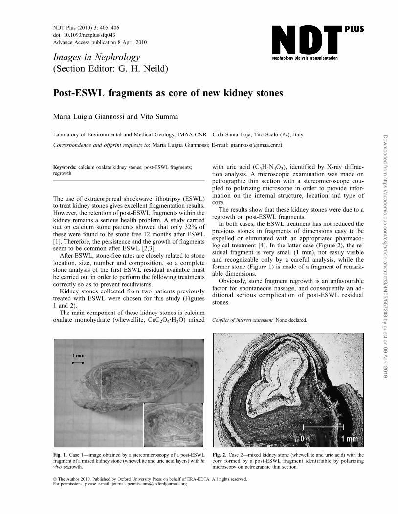
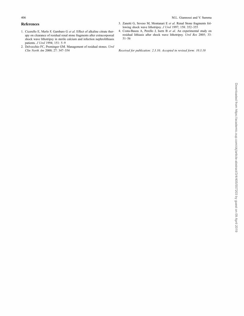
The results show that these kidney stones were due to aregrowth on post-ESWL fragments.
In both cases, the ESWL treatment has not reduced theprevious stones in fragments of dimensions easy to beexpelled or eliminated with an appropriated pharmaco-logical treatment [4]. In the latter case (Figure 2), the re-sidual fragment is very small (1 mm), not easily visibleand recognizable only by a careful analysis, while theformer stone (Figure 1) is made of a fragment of remark-able dimensions.
Obviously, stone fragment regrowth is an unfavourablefactor for spontaneous passage, and consequently an ad-ditional serious complication of post-ESWL residualstones.
Conflict of interest statement. None declared.
Fig. 2. Case 2—mixed kidney stone (whewellite and uric acid) with thecore formed by a post-ESWL fragment identif iable by polarizingmicroscopy on petrographic thin section.
Fig. 1. Case 1—image obtained by a stereomicroscopy of a post-ESWLfragment of a mixed kidney stone (whewellite and uric acid layers) with invivo regrowth.
© The Author 2010. Published by Oxford University Press on behalf of ERA-EDTA. All rights reserved.For permissions, please e-mail: [email protected]
Dow
nloaded from https://academ
ic.oup.com/ckj/article-abstract/3/4/405/557203 by guest on 09 April 2019
References
1. Cicerello E, Merlo F, Gambaro G et al. Effect of alkaline citrate ther-apy on clearance of residual renal stone fragments after extracorporealshock wave lithotripsy in sterile calcium and infection nephrolithiasispatients. J Urol 1994; 151: 5–9
2. Delvecchio FC, Preminger GM. Management of residual stones. UrolClin North Am 2000; 27: 347–354
3. Zanetti G, Seveso M, Montanari E et al. Renal Stone fragments fol-lowing shock wave lithotripsy. J Urol 1997; 158: 352–355
4. Costa-Bauza A, Perello J, Isern B et al. An experimental study onresidual lithiasis after shock wave lithotripsy. Urol Res 2005; 33:51–56
Received for publication: 2.3.10; Accepted in revised form: 10.3.10
406 M.L. Giannossi and V. Summa
Dow
nloaded from https://academ
ic.oup.com/ckj/article-abstract/3/4/405/557203 by guest on 09 April 2019
Related Documents


