Post-Conditioning Reduces Infarct Size and Edema in Patients With ST-Segment Elevation Myocardial Infarction Franck Thuny, MD, PHD,* Olivier Lairez, MD, PHD,† François Roubille, MD, PHD,‡§ Nathan Mewton, MD, PHD,¶ Gilles Rioufol, MD, PHD, Catherine Sportouch, MD,†§ Ingrid Sanchez, MD,*¶ Cyrille Bergerot, MD,¶ Hélène Thibault, MD, PHD,* Thien Tri Cung, MD,‡§ Gérard Finet, MD, PHD, Laurent Argaud, MD, PHD,# Didier Revel, MD, PHD,* Geneviève Derumeaux, MD, PHD,¶ Eric Bonnefoy-Cudraz, MD, PHD,¶ Meier Elbaz, MD, PHD,† Christophe Piot, MD, PHD,‡§ Michel Ovize, MD, PHD,¶ Pierre Croisille, MD, PHD* ** Lyon, Toulouse, Montpellier, and Saint Etienne, France Objectives This study aimed to determine whether post-conditioning at the time of percutaneous coronary intervention could reduce reperfusion-induced myocardial edema in patients with acute ST-segment elevation myocardial infarction (STEMI). Background Myocardial edema is a reperfusion injury with potentially severe consequences. Post-conditioning is a cardiopro- tective therapy that reduces infarct size after reperfusion, but no previous studies have analyzed the impact of this strategy on reperfusion-induced myocardial edema in humans. Methods Fifty patients with STEMI were randomly assigned to either a control or post-conditioned group. Cardiac magnetic res- onance imaging was performed within 48 to 72 h after admission. Myocardial edema was measured by T2-weighted sequences, and infarct size was determined by late gadolinium enhancement sequences and creatine kinase release. Results The post-conditioned and control groups were similar with respect to ischemia time, the size of the area at risk, and the ejection fraction before percutaneous coronary intervention. As expected, post-conditioning was associated with smaller infarct size (13 7 g/m 2 vs. 21 14 g/m 2 ;p 0.01) and creatine kinase peak serum level (median [interquartile range]: 1,695 [1,118 to 3,692] IU/l vs. 3,505 [2,307 to 4,929] IU/l; p 0.003). At reperfusion, the ex- tent of myocardial edema was significantly reduced in the post-conditioned group as compared with the control group (23 16 g/m 2 vs. 34 18 g/m 2 ;p 0.03); the relative increase in T2W signal intensity was also significantly lower (p 0.02). This protective effect was confirmed after adjustment for the size of the area at risk. Conclusions This randomized study demonstrated that post-conditioning reduced infarct size and edema in patients with rep- erfused STEMI. (Post Cond No Reflow; NCT01208727) (J Am Coll Cardiol 2012;59:2175–81) © 2012 by the American College of Cardiology Foundation Reperfusion therapy of jeopardized myocardium is the most effective method for reducing infarct size and improving the outcome in patients with ST-segment elevation myocardial infarction (STEMI). However, the restoration of coronary blood flow can paradoxically induce additional myocardial damage, making reperfusion therapy a “double-edged sword” (1). Reperfusion injury is a complex phenomenon mediated by several factors, including oxidative stress, intracellular calcium accumulation, rapid restoration of pH, and inflammation, and involves, at least partly, opening of the so-called mitochondrial permeability transition pore (2). Clinically identified features of this reperfusion injury may be reversible and transient, such as From *CREATIS, CNRS UMR 5220, INSERM U1044, Université de Lyon, Lyon, France; †Service de Cardiologie, Hôpital Rangueil, Université Paul Sabatier, Tou- louse, France; ‡INSERM U661, Montpellier, France; §Hôpital Arnaud de Ville- neuve, Université de Montpellier I and II, Montpellier, France; INSERM U1060 (CARMEN, Cardioprotection Team) et CIC de Lyon, Lyon, France; ¶Service d’Exploration Fonctionnelles Cardiovasculaires, Hospices Civils de Lyon, Lyon, France; #Service de Reanimation Médicale, Hospices Civils de Lyon, Lyon, France; and the **Service de Radiologie, Centre Hospitalier Universitaire de Saint Etienne, Université Jean-Monnet, Saint Etienne, France. This clinical trial was supported by a grant from the Actions Incitatives from Hospices Civils de Lyon. Dr. Thuny was the recipient of a grant from the Assistance Publique Hôpitaux de Marseille. Drs. Mewton and Thibault were recipients of a grant from the Fédération Française de Cardiologie and the Société Française de Cardiologie. Dr. Derumeaux has received research grants from AstraZeneca, Toshiba, and Philips. Dr. Bonnefoy-Cudraz is a consultant with ThermoFisher; and a lecturer with AstraZeneca, Eli Lilly, and Daiichi Sankyo. Dr. Croisille is a lecturer with Siemens, Guerbert, and Novartis. All other authors have reported that they have no relationships to disclose relevant to the contents of this paper to disclose. Manuscript received December 12, 2011; revised manuscript received March 7, 2012, accepted March 7, 2012. Journal of the American College of Cardiology Vol. 59, No. 24, 2012 © 2012 by the American College of Cardiology Foundation ISSN 0735-1097/$36.00 Published by Elsevier Inc. doi:10.1016/j.jacc.2012.03.026

Welcome message from author
This document is posted to help you gain knowledge. Please leave a comment to let me know what you think about it! Share it to your friends and learn new things together.
Transcript
Post-Conditioning Reduces Infarct Size and Edemain Patients With ST-Segment Elevation Myocardial Infarction
Franck Thuny, MD, PHD,* Olivier Lairez, MD, PHD,† François Roubille, MD, PHD,‡§Nathan Mewton, MD, PHD,!¶ Gilles Rioufol, MD, PHD,! Catherine Sportouch, MD,†§Ingrid Sanchez, MD,*¶ Cyrille Bergerot, MD,¶ Hélène Thibault, MD, PHD,*! Thien Tri Cung, MD,‡§Gérard Finet, MD, PHD,! Laurent Argaud, MD, PHD,# Didier Revel, MD, PHD,*Geneviève Derumeaux, MD, PHD,!¶ Eric Bonnefoy-Cudraz, MD, PHD,¶ Meier Elbaz, MD, PHD,†Christophe Piot, MD, PHD,‡§ Michel Ovize, MD, PHD,!¶ Pierre Croisille, MD, PHD* **
Lyon, Toulouse, Montpellier, and Saint Etienne, France
Objectives This study aimed to determine whether post-conditioning at the time of percutaneous coronary interventioncould reduce reperfusion-induced myocardial edema in patients with acute ST-segment elevation myocardialinfarction (STEMI).
Background Myocardial edema is a reperfusion injury with potentially severe consequences. Post-conditioning is a cardiopro-tective therapy that reduces infarct size after reperfusion, but no previous studies have analyzed the impact ofthis strategy on reperfusion-induced myocardial edema in humans.
Methods Fifty patients with STEMI were randomly assigned to either a control or post-conditioned group. Cardiac magnetic res-onance imaging was performed within 48 to 72 h after admission. Myocardial edema was measured by T2-weightedsequences, and infarct size was determined by late gadolinium enhancement sequences and creatine kinase release.
Results The post-conditioned and control groups were similar with respect to ischemia time, the size of the area at risk,and the ejection fraction before percutaneous coronary intervention. As expected, post-conditioning was associatedwith smaller infarct size (13 ! 7 g/m2 vs. 21 ! 14 g/m2; p " 0.01) and creatine kinase peak serum level (median[interquartile range]: 1,695 [1,118 to 3,692] IU/l vs. 3,505 [2,307 to 4,929] IU/l; p " 0.003). At reperfusion, the ex-tent of myocardial edema was significantly reduced in the post-conditioned group as compared with the control group(23 ! 16 g/m2 vs. 34 ! 18 g/m2; p " 0.03); the relative increase in T2W signal intensity was also significantly lower(p " 0.02). This protective effect was confirmed after adjustment for the size of the area at risk.
Conclusions This randomized study demonstrated that post-conditioning reduced infarct size and edema in patients with rep-erfused STEMI. (Post Cond No Reflow; NCT01208727) (J Am Coll Cardiol 2012;59:2175–81) © 2012 by theAmerican College of Cardiology Foundation
Reperfusion therapy of jeopardized myocardium is the mosteffective method for reducing infarct size and improving theoutcome in patients with ST-segment elevation myocardialinfarction (STEMI). However, the restoration of coronaryblood flow can paradoxically induce additional myocardialdamage, making reperfusion therapy a “double-edged sword”
(1). Reperfusion injury is a complex phenomenon mediated byseveral factors, including oxidative stress, intracellular calciumaccumulation, rapid restoration of pH, and inflammation, andinvolves, at least partly, opening of the so-called mitochondrialpermeability transition pore (2). Clinically identified features ofthis reperfusion injury may be reversible and transient, such as
From *CREATIS, CNRS UMR 5220, INSERM U1044, Université de Lyon, Lyon,France; †Service de Cardiologie, Hôpital Rangueil, Université Paul Sabatier, Tou-louse, France; ‡INSERM U661, Montpellier, France; §Hôpital Arnaud de Ville-neuve, Université de Montpellier I and II, Montpellier, France; !INSERM U1060(CARMEN, Cardioprotection Team) et CIC de Lyon, Lyon, France; ¶Serviced’Exploration Fonctionnelles Cardiovasculaires, Hospices Civils de Lyon, Lyon,France; #Service de Reanimation Médicale, Hospices Civils de Lyon, Lyon, France;and the **Service de Radiologie, Centre Hospitalier Universitaire de Saint Etienne,Université Jean-Monnet, Saint Etienne, France. This clinical trial was supportedby a grant from the Actions Incitatives from Hospices Civils de Lyon.
Dr. Thuny was the recipient of a grant from the Assistance Publique Hôpitaux deMarseille. Drs. Mewton and Thibault were recipients of a grant from the FédérationFrançaise de Cardiologie and the Société Française de Cardiologie. Dr. Derumeaux hasreceived research grants from AstraZeneca, Toshiba, and Philips. Dr. Bonnefoy-Cudrazis a consultant with ThermoFisher; and a lecturer with AstraZeneca, Eli Lilly, and DaiichiSankyo. Dr. Croisille is a lecturer with Siemens, Guerbert, and Novartis. All otherauthors have reported that they have no relationships to disclose relevant to thecontents of this paper to disclose.
Manuscript received December 12, 2011; revised manuscript received March 7,2012, accepted March 7, 2012.
Journal of the American College of Cardiology Vol. 59, No. 24, 2012© 2012 by the American College of Cardiology Foundation ISSN 0735-1097/$36.00Published by Elsevier Inc. doi:10.1016/j.jacc.2012.03.026
arrhythmias or myocardial stun-ning, or irreversible, such as myo-cardial infarction or microvascularobstruction (1).
Myocardial edema begins dur-ing the ischemic phase butabruptly expands during the firstminutes of reperfusion when thegradient between the hyperos-motic extravascular fluid and thenormo-osmotic blood rapidlygrows (1). By increasing the hy-drostatic pressure within the in-terstitial space, this edema can con-tribute to capillary compression andaggravation of cell damage (3).Myocardial edema is thus a conse-quence, but through a vicious cycle,is also a mechanism of reperfusioninjury (4). One may therefore ques-tion whether measuring myocardialedema might be interesting for esti-
mation of reperfusion injury and evaluation of protectiveinterventions.
Cardiac magnetic resonance (CMR) imaging is an ap-propriate method for measuring myocardial edema in vivo,which is depicted as the area of hypersignal on T2-weighted(T2W) images (5). Indeed, increases in the transversecomponent of the proton relaxation time (T2) and T2Wsignal intensity (SI) have been shown to correlate well withthe water content in a model of ischemia reperfusion in dogs(6). This property has been used in various clinical settings,including the assessment of acute ischemic damage (5) andthe quantification of myocardial salvage (7–9). However, toour knowledge, it has never been used to evaluate reperfu-sion injury per se.
Ischemic post-conditioning (brief repeated periods ofischemia applied at the onset of reperfusion) is a cardiopro-tective strategy that has been proven to specifically attenuatelethal reperfusion injury in animal models and in patientswith STEMI (10–12). Post-conditioning has been shownto reduce myocardial edema after reperfusion in experimen-tal preparations (13). However, no previous studies haveanalyzed the impact of this protective intervention onpost-reperfusion myocardial edema in patients withSTEMI. The objective of the present study was to deter-mine whether post-conditioning could reduce post-reperfusion myocardial edema, as assessed by CMR imag-ing, in patients with ongoing STEMI.
Methods
Study population. From May 2008 to October 2010, menand women of age !18 years who presented within 12 hafter the onset of chest pain, who had ST-segment elevationof #0.1 mV in 2 contiguous leads, and for whom the
clinical decision was made to treat with percutaneouscoronary intervention (PCI) were considered for inclusionin the study. Patients were eligible whether they wereundergoing primary PCI or rescue PCI. Occlusion of theculprit coronary artery (thrombolysis in myocardial infarc-tion flow grade 0 to 1) at the time of admission was also acriterion for inclusion (14). Patients with cardiac arrest,ventricular fibrillation, cardiogenic shock, stent thrombosis,previous acute myocardial infarction, angina within 48 hbefore infarction, and contraindications to CMR imagingwere not included in the study. Patients with occlusion ofthe left circumflex coronary artery were included only in thecase of left circulation dominance. Patients with occlusionof the left main or with evidence of coronary collaterals(Rentrop grade !1) to the region at risk on initial coronaryangiography (at the time of admission) were excluded (15).This study was approved by the Ethics Committee of Lyon,France (IRB 123406519). All patients gave written in-formed consent for participation.Angiography and PCI. Left ventricular (LV) and coronaryangiography was performed using standard techniques justbefore revascularization. The size of the area at risk wasestimated for each patient by measuring the circumferentialextent of abnormally contracting segments (ACS) accordingto the method of Feild et al. (16) and Lapeyre et al. (17), asperformed in previous randomized trials (11,18). Briefly, thelength of the end-diastolic ventricular endocardial perimeter(circumference) and the length of the ACS of the end-diastolic perimeter were determined by computerized pla-nimetry (Image J 1.38$ software). ACS were expressed as:ACS (%) " (abnormally contracting length of end-diastoliccircumference/total end-diastolic circumference) $ 100.Measurement of the ACS was performed in a blindedmanner by an experienced investigator. Revascularizationwas performed with the use of direct stenting.Experimental protocol. Patients were randomly allocatedto either the control or the post-conditioned group. Coro-nary angiography was performed using standard techniquesjust before revascularization. Randomization was performedwith the use of a computer-generated randomization se-quence. Numbered sealed envelopes that contained thestudy group assignment were distributed to each catheter-ization laboratory and were opened after informed consenthad been obtained. Revascularization was performed withthe use of direct stenting as previously reported (11,12). Inthe control group, no additional intervention was performedduring the first 8 min of reperfusion. In the post-conditioned group, within 1 min of reflow after directstenting, the angioplasty balloon was reinflated 4 times for1 min with low-pressure (4 to 6 atmospheres) inflations,each separated by 1 min of reflow. After the eighth minuteof reperfusion, PCI was completed according to the physi-cian’s judgment with respect to patient status.Myocardial edema and infarct size. All CMR imagingstudies were performed on a 1.5-T MAGNETOM Avantototal imaging matrix system (Siemens Healthcare, Erlan-
Abbreviationsand Acronyms
ACS ! abnormallycontracting segments
CK ! creatine kinase
CMR ! cardiac magneticresonance
LGE ! late gadoliniumenhancement
LV ! leftventricle/ventricular
PCI ! percutaneouscoronary intervention
SI ! signal intensity
STEMI ! ST-segmentelevation myocardialinfarction
STIR ! short tau inversionrecovery
T2W ! T2-weighted
2176 Thuny et al. JACC Vol. 59, No. 24, 2012Myocardial Edema in Post-Conditioning June 12, 2012:2175–81

gen, Germany) 48 to 72 h after admission with vectocar-diogram monitoring and a phased-array cardiac receivercoil. The standard acute myocardial infarction protocolcomplied with the recommendations of the Society forCardiac Magnetic Resonance (19). LV volumes and massmeasurements were taken from the balanced steady-statefree precession cine sequences with dedicated software(Argus, Siemens Medical Solutions, Malvern, Pennsylva-nia). T2W imaging was based on breath-hold T2W–shorttau inversion recovery (T2W-STIR) sequences (matrix118 $ 256, voxel size 2.3 $ 1.3 $ 7 mm, flip angle 90/180°,effective echo time 47 ms, bandwidth 235 Hz/pixel) thatcovered the whole LV. The extent of myocardial edema wasquantified using VPT software (Siemens Corporate Re-search, Erlangen, Germany), as previously reported (20).After manual tracing of the epicardial and endocardialcontours, the T2W hyperintense area was quantified on theT2W images using semiautomatic detection with the fullwidth at half-maximum approach (21). If present, a centralcore of hypointense signal within the area of increased SI(hemorrhagic infarction) was included in the T2W-CMRhypersignal area. Increased SI from the blood pool adjacentto the endocardium due to slow flow was excluded. For eachslice level, T2W images were matched with cine-MR imagesfor the same corresponding time trigger to provide anatomiclandmarks and differentiate between slow flowing blood andincreased myocardial intensities. The myocardial edema areaand the total LV slice area were quantified in each short-axisslice. Eventually, the extent of myocardial edema was expressed asthe indexed mass of the myocardial edema (in grams of tissue)according to the following formula: % (hyperenhanced area [cm2]$ slice thickness [cm] $ myocardial-specific density [1.05g/cm3]/body surface area [m2]). Regions of interest of the samesize were manually drawn within the hypersignal area (excludingthe central core of the hyposignal) and the remote myocardium ofthe median LV slice to measure the mean T2W SI peak valuefrom the distribution scatter. Then, the percent signal enhance-ment between the myocardial edema area and remote myocar-dium was calculated as follows: %SE " (SIE&SIR)/SIR (6,22).
Infarct size was assessed by 2 different techniques: 1) thepeak of serum creatine kinase (CK) release determined fromblood samples obtained at admission and repeatedly over thenext 3 days (every 4 h on day 1 and every 6 h on days 2 and3); and 2) the area of late gadolinium enhancement (LGE)on the CMR images. LGE was evaluated 10 min after theinjection of gadolinium (0.2 mmol/kg at 3 ml/s; DOTAREM,Guerbet, France) with the use of a 3-dimensional inversion-recovery gradient-echo sequence. The images were analyzed inthe same T2W-CMR–matched short-axis slices by delinea-tion of the areas of LGE using semiautomatic detection withthe full width at half-maximum approach (9,23). The area ofLGE was expressed as the indexed mass of the infarctedmyocardium. In cases of a dark subendocardial zone in the areaof the hyperenhancement (microvascular obstruction), thezone was included in the infarct area.
All CMR images were analyzed by 2 senior investigatorsblinded to the study arm and any other clinical or imagingdata. Consensus between the 2 observers was used forsubsequent analysis. For all of these analyses, manualadjustments were performed when the computer algorithmwas considered to be obviously wrong.Statistical analysis. The target sample size was calculatedto assess the effect of post-conditioning on the myocardialedema measured by CMR imaging. The expected effect wasa 30% reduction in the extent of edema (comparable to theinfarct size reduction observed in our previous studies), witha statistical power of 80% and a probability of a type I errorof 0.05 with a 2-sided test (11,12).
Continuous data were reported as mean ! SD or medianand interquartile range (IQR) for values without normaldistribution. Categorical data were reported as frequenciesand percentages. The data were analyzed according to theintention-to-treat principle. The Fisher exact test was usedto compare categorical data. Either the unpaired Student ttest or the Mann-Whitney test was used to comparecontinuous variables between the 2 independent groups.Simple linear regression analysis was used to assess thecorrelation between continuous variables. We performed ananalysis of covariance to compare the treatment effect oninfarct size, serum CK release, and myocardial edema afteradjustment on the size of the area at risk. An analysis wasalso performed to adjust the effect of treatment on myocar-dial edema for the effect on the infarct size.
This statistical analysis was conducted using SPSS forWindows, version 16.0 (SPSS Inc., Chicago, Illinois). Alltests were 2-sided. A p value of ' 0.05 was consideredstatistically significant.
Results
Study population. Seventy-six patients were considered tobe eligible for the present study. Of these, 14 were notincluded for the following reasons: previous myocardialinfarction in the same territory (n " 4), Thrombolysis InMyocardial Infarction flow grade of #1 at admission (n "3), or evidence of coronary collaterals to the area at risk onadmission coronary angiography (n " 7). Twelve additionalpatients were excluded because CMR imaging could not beperformed or was not interpretable. Data are thus presentedfor 50 patients (25 in the control group and 25 in thepost-conditioned group). Baseline characteristics of thestudy population are presented in Table 1, and no signifi-cant differences in these characteristics were observed be-tween the 2 groups. No significant differences were observedin LV volumes, ejection fraction, or mass between thegroups (Table 2).
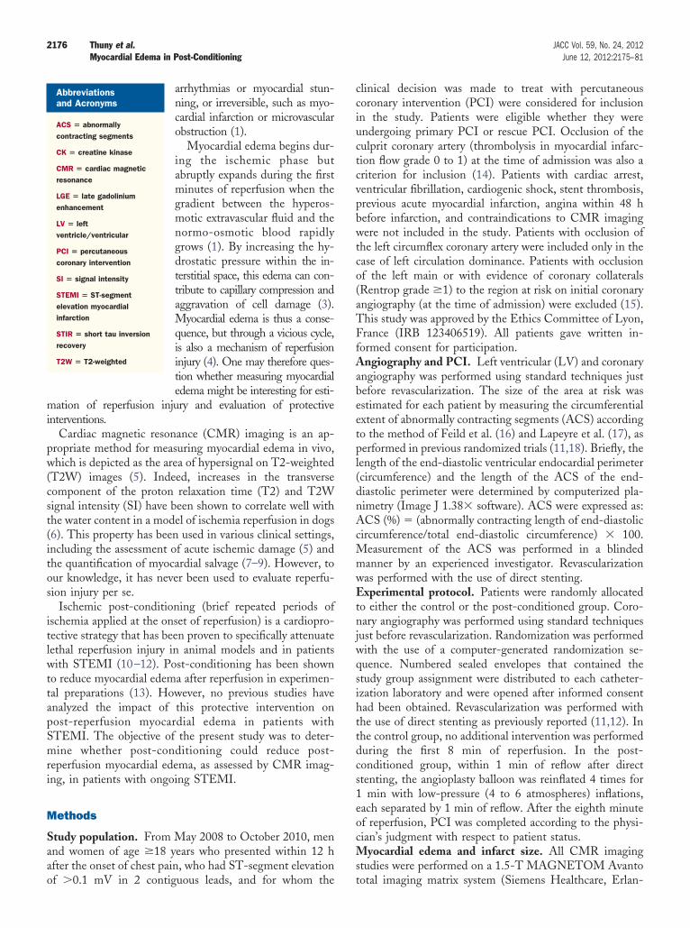
As expected, the infarct size, as measured by myocardialLGE extent, in the post-conditioned group was 38% lessthan that in the control group, averaging 13 ! 7 g/m2 versus21 ! 14 g/m2, respectively (p " 0.01) (Fig. 1A). The infarctsize reduction by post-conditioning was also confirmed
2177JACC Vol. 59, No. 24, 2012 Thuny et al.June 12, 2012:2175–81 Myocardial Edema in Post-Conditioning

when the size of the area at risk was taken into account(p " 0.0005 by covariance analysis). Finally, the peak forserum CK release was significantly lower in the post-conditioned group versus that in the control patients (me-dian [IQR]: 1,695 [1,118 to 3,692] UI/l vs. 3,505 [2,307 to4,929] UI/l; p " 0.003) (Fig. 1B) for all sizes of the area atrisk (p " 0.02 by covariance analysis). Similar infarct sizereduction was obtained when the area under the curve ofCK release was considered (not shown).Myocardial edema and post-conditioning. The meanarea at risk size as measured by ACS was comparable for thepost-conditioned and control groups (39 ! 14% vs. 36 !14% of LV circumference; p " 0.41).
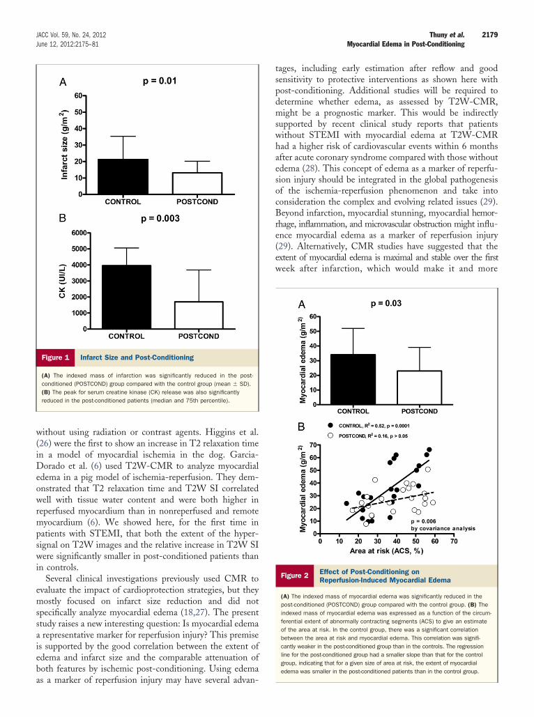
The mean extent of myocardial edema in the post-conditioned group was 32% less than that in the controlgroup, averaging 23 ! 16 g/m2 versus 34 ! 18 g/m2,respectively (p " 0.03) (Fig. 2A). In the control group,there was a significant correlation between the extent ofedema and size of the area at risk (p " 0.0001). Importantly,the regression line for the post-conditioned group had asmaller slope than the regression line for the control group,indicating that for any given size of area at risk, the extentof myocardial edema was smaller in the post-conditionedpatients. This difference in the slope was significant byanalysis of covariance (p " 0.006) (Fig. 2B). Moreover, thenormalized T2W signal difference between the edema area
and remote area was significantly lower in the post-conditioned group than in the controls (median [IQR]: 58%[58% to 169%] vs. 82% [62% to 115%]; p " 0.02).
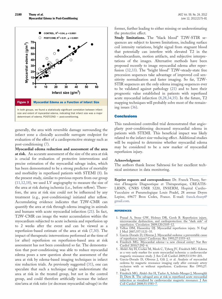
The effect of post-conditioning on myocardial edema wasdependent on the infarct size (p " 0.35 after correction forthe infarct size). Indeed, in both groups, we found astatistically significant correlation between infarct size andextent of myocardial edema, indicating that infarct size wasa major determinant of edema. As shown in Figure 3, thelarger the infarct, the larger the edema.
Discussion
In the present study, we found that the extent of myocardialedema after reperfusion therapy in patients with STEMIcan be attenuated by angioplasty post-conditioning.Impact of post-conditioning on myocardial edema. Inthe dog model, Zhao et al. (10) demonstrated that ischemicpost-conditioning significantly reduced myocardial edema,as assessed by the ex vivo measurement of water content ina tissue sample of the area at risk. Other reports in rat ormouse models confirmed that ischemic post-conditioningcould reduce edema in other organs, including the skeletalmuscle and brain (24,25). Our study is the first to show thatischemic post-conditioning could attenuate myocardialedema in the clinical setting of STEMI. The mechanism ofthe reduction of myocardial edema by post-conditioning isunclear. Our data strongly suggest that it is most likelyrelated to the infarct size–reducing effect because we founda good correlation between infarct size and myocardial edematogether with a comparable impact of post-conditioning oninfarct size and myocardial edema. Reduction of cell deathwith subsequent reduced release of water content into theextracellular space probably contributed to the limiting ofedema by post-conditioning. The negative modulation ofcoronary perfusion pressure would potentially reduce theStarling forces, favoring the movement of intravascularwater toward the interstitial space. Eventually, the limitingof both the inflammation process and the generation ofreactive oxygen species by post-conditioning, as reported inexperimental models, might prevent endothelial injury andsubsequent increase in capillary permeability (1).T2W-CMR for the assessment of reperfusion injury.T2W-CMR is a water-sensitive modality that offers aunique means for assessment of myocardial edema in vivo
Baseline Characteristics of the Study PopulationTable 1 Baseline Characteristics of the Study Population
Control Group(n ! 25)
Post-ConditionedGroup (n ! 25) p Value
Age, yrs 57 ! 12 57 ! 13 0.88
Male/female 18/7 19/6 0.78
BMI, kg/m2 26 ! 4 26 ! 4 1.0
Hypertension 12 (48) 10 (40) 0.78
Smoking 16 (64) 17 (68) 1.0
Dyslipidemia 12 (48) 9 (36) 0.57
Diabetes 4 (14) 5 (20) 1.0
Angiographic findings
Infarct-related artery 0.33
LAD 14 (56) 14 (56)
RCA 9 (36) 11 (44)
Cx 2 (8) 0 (0)
Area at risk, % LV 36 ! 12 39 ! 14 0.41
PCI
Duration of ischemia, min 215 ! 20 289 ! 31 0.08
Post-PCI TIMI flow grade 2.9 ! 0.3 2.9 ! 0.6 0.94
Treatment before PCI
Intravenous nitrates 7 (28) 6 (24) 1.0
Morphine 13 (52) 19 (76) 0.14
Thrombolytic agents 4 (16) 3 (12) 1.0
Treatment at time of PCI
Heparin 25 (100) 25 (100) 1.0
Aspirin and/or clopidogrel 25 (100) 25 (100) 1.0
Glycoprotein IIb/IIIa inhibitor 18 (72) 19 (76) 1.0
Values are mean ! SD, n/n, or n (%).BMI " body mass index; Cx " circumflex coronary artery; LAD " left anterior descending
coronary artery; LV " left ventricle; PCI " percutaneous coronary intervention; RCA " rightcoronary artery; TIMI " Thrombolysis In Myocardial Infarction.
LV Volumes and MassAccording to the Reperfusion StrategyTable 2 LV Volumes and MassAccording to the Reperfusion Strategy
Control Group(n ! 25)
Post-Conditioned Group(n ! 25) p Value
LVEDV, ml/m2 79 ! 17 73 ! 23 0.07
LVESV, ml/m2 40 ! 15 35 ! 11 0.24
LVEF, % 50 ! 12 52 ! 8 0.66
LV mass, g/m2 74 ! 15 73 ! 12 0.69
Values are mean ! SD.LV " left ventricular; LVEDV " left ventricular end-diastolic volume; LVEF " left ventricular
ejection fraction; LVESV " left ventricular end-systolic volume.
2178 Thuny et al. JACC Vol. 59, No. 24, 2012Myocardial Edema in Post-Conditioning June 12, 2012:2175–81
without using radiation or contrast agents. Higgins et al.(26) were the first to show an increase in T2 relaxation timein a model of myocardial ischemia in the dog. Garcia-Dorado et al. (6) used T2W-CMR to analyze myocardialedema in a pig model of ischemia-reperfusion. They dem-onstrated that T2 relaxation time and T2W SI correlatedwell with tissue water content and were both higher inreperfused myocardium than in nonreperfused and remotemyocardium (6). We showed here, for the first time inpatients with STEMI, that both the extent of the hyper-signal on T2W images and the relative increase in T2W SIwere significantly smaller in post-conditioned patients thanin controls.
Several clinical investigations previously used CMR toevaluate the impact of cardioprotection strategies, but theymostly focused on infarct size reduction and did notspecifically analyze myocardial edema (18,27). The presentstudy raises a new interesting question: Is myocardial edemaa representative marker for reperfusion injury? This premiseis supported by the good correlation between the extent ofedema and infarct size and the comparable attenuation ofboth features by ischemic post-conditioning. Using edemaas a marker of reperfusion injury may have several advan-
tages, including early estimation after reflow and goodsensitivity to protective interventions as shown here withpost-conditioning. Additional studies will be required todetermine whether edema, as assessed by T2W-CMR,might be a prognostic marker. This would be indirectlysupported by recent clinical study reports that patientswithout STEMI with myocardial edema at T2W-CMRhad a higher risk of cardiovascular events within 6 monthsafter acute coronary syndrome compared with those withoutedema (28). This concept of edema as a marker of reperfu-sion injury should be integrated in the global pathogenesisof the ischemia-reperfusion phenomenon and take intoconsideration the complex and evolving related issues (29).Beyond infarction, myocardial stunning, myocardial hemor-rhage, inflammation, and microvascular obstruction might influ-ence myocardial edema as a marker of reperfusion injury(29). Alternatively, CMR studies have suggested that theextent of myocardial edema is maximal and stable over the firstweek after infarction, which would make it and more
Figure 1 Infarct Size and Post-Conditioning
(A) The indexed mass of infarction was significantly reduced in the post-conditioned (POSTCOND) group compared with the control group (mean ! SD).(B) The peak for serum creatine kinase (CK) release was also significantlyreduced in the post-conditioned patients (median and 75th percentile).
Figure 2 Effect of Post-Conditioning onReperfusion-Induced Myocardial Edema
(A) The indexed mass of myocardial edema was significantly reduced in thepost-conditioned (POSTCOND) group compared with the control group. (B) Theindexed mass of myocardial edema was expressed as a function of the circum-ferential extent of abnormally contracting segments (ACS) to give an estimateof the area at risk. In the control group, there was a significant correlationbetween the area at risk and myocardial edema. This correlation was signifi-cantly weaker in the post-conditioned group than in the controls. The regressionline for the post-conditioned group had a smaller slope than that for the controlgroup, indicating that for a given size of area at risk, the extent of myocardialedema was smaller in the post-conditioned patients than in the control group.
2179JACC Vol. 59, No. 24, 2012 Thuny et al.June 12, 2012:2175–81 Myocardial Edema in Post-Conditioning
generally, the area with reversible damage surrounding theinfarct zone a clinically accessible surrogate endpoint forevaluation of the effect of a cardioprotective strategy such aspost-conditioning (7).Myocardial edema reduction and assessment of the areaat risk. An accurate assessment of the size of the area at riskis crucial for evaluation of protective interventions andprecise estimation of the myocardial salvage index, whichhas been demonstrated to be a strong predictor of mortalityand morbidity in reperfused patients with STEMI (8). Inthe present study, similar to previous reports from our group(11,12,18), we used LV angiography to measure the size ofthe area at risk during ischemia (i.e., before reflow). There-fore, the area at risk size could not be influenced by anytreatment (e.g., post-conditioning) initiated after reflow.Accumulating evidence indicates that T2W-CMR canquantify the area at risk through edema imaging in animalsand humans with acute myocardial infarction (21). In fact,T2W-CMR can image the water accumulation within themyocardium subjected to acute ischemia and reperfusion upto 2 weeks after the event and can be viewed as areperfusion-based estimate of the area at risk (7,30). Theimpact of therapeutic interventions performed at the time of(or after) reperfusion on reperfusion-based area at riskassessment has not been considered so far. The demonstra-tion that post-conditioning was able to reduce myocardialedema poses a new question about the assessment of thearea at risk by edema-based imaging techniques in infarctsize reduction trials. As previously suggested (31), one canspeculate that such a technique might underestimate thearea at risk in the treated group, but not in the controlgroup, and could therefore artificially increase the infarctsize/area at risk ratio (or decrease myocardial salvage) in the
former, further leading to either missing or underestimatingthe protective effect.Study limitations. The “black blood” T2W-STIR se-quences are subject to known limitations, including surfacecoil intensity variations, bright signal from stagnant bloodthat potentially can interfere with elevated T2 in thesubendocardium, motion artifacts, and subjective interpre-tations of the images. Alternative methods have beenproposed recently to image myocardial edema after reper-fusion (32,33). The “bright blood” T2W–steady-state freeprecession sequences take advantage of improved coil sen-sitivity normalization and faster imaging. So far, T2W-STIR sequences are the only edema imaging sequences everto be validated against pathology (21) and to have theirprognostic value established in patients with reperfusedacute myocardial infarction (8,28,34,35). In the future, T2mapping techniques will probably solve most of the remain-ing issues (36).
Conclusions
This randomized controlled trial demonstrated that angio-plasty post-conditioning decreased myocardial edema inpatients with STEMI. This beneficial impact was likelyrelated to the infarct size–reducing effect. Additional studieswill be required to determine whether myocardial edemamay be considered to be a new marker of myocardialreperfusion injury.
AcknowledgmentThe authors thank Inesse Sahraoui for her excellent tech-nical assistance in data monitoring.
Reprint requests and correspondence: Dr. Franck Thuny, Ser-vice d’Imagerie Diagnostique et Thérapeutique, CREATIS-LRMN, CNRS UMR 5220, INSERM, Hopital Cardio-Vasculaire et Pneumologique Louis Pradel, 28 avenue DoyenLepine, 69677 Bron Cedex, France. E-mail: [email protected].
REFERENCES
1. Prasad A, Stone GW, Holmes DR, Gersh B. Reperfusion injury,microvascular dysfunction, and cardioprotection: the “dark side” ofreperfusion. Circulation 2009;120:2105–12.
2. Yellon DM, Hausenloy DJ. Myocardial reperfusion injury. N EnglJ Med 2007;357:1121–35.
3. Garcia-Dorado D, Oliveras J. Myocardial oedema: a preventable causeof reperfusion injury? Cardiovasc Res 1993;27:1555–63.
4. Friedrich MG. Myocardial edema—a new clinical entity? Nat RevCardiol 2010;7:292–6.
5. Abdel-Aty H, Cocker M, Meek C, Tyberg JV, Friedrich MG. Edemaas a very early marker for acute myocardial ischemia: a cardiovascularmagnetic resonance study. J Am Coll Cardiol 2009;53:1194–201.
6. Garcia-Dorado D, Oliveras J, Gili J, et al. Analysis of myocardialoedema by magnetic resonance imaging early after coronary arteryocclusion with or without reperfusion. Cardiovasc Res 1993;27:1462–9.
7. Friedrich MG, Abdel-Aty H, Taylor A, Schulz-Menger J, MessroghliD, Dietz R. The salvaged area at risk in reperfused acute myocardialinfarction as visualized by cardiovascular magnetic resonance. J AmColl Cardiol 2008;51:1581–7.
Figure 3 Myocardial Edema as a Function of Infarct Size
In both groups, we found a statistically significant correlation between infarctsize and extent of myocardial edema, indicating that infarct size was a majordeterminant of edema. POSTCOND " post-conditioning.
2180 Thuny et al. JACC Vol. 59, No. 24, 2012Myocardial Edema in Post-Conditioning June 12, 2012:2175–81

8. Eitel I, Desch S, Fuernau G, et al. Prognostic significance anddeterminants of myocardial salvage assessed by cardiovascular mag-netic resonance in acute reperfused myocardial infarction. J Am CollCardiol 2010;55:2470–9.
9. Larose E, Rodes-Cabau J, Pibarot P, et al. Predicting late myocardialrecovery and outcomes in the early hours of ST-segment elevationmyocardial infarction traditional measures compared with micro-vascular obstruction, salvaged myocardium, and necrosis character-istics by cardiovascular magnetic resonance. J Am Coll Cardiol2010;55:2459 – 69.
10. Zhao M, Sonnenblick EH, Zhang H, Eng C. Increase in myofilamentseparation in the “stunned” myocardium. J Mol Cell Cardiol 1992;24:269–76.
11. Staat P, Rioufol G, Piot C, et al. Postconditioning the human heart.Circulation 2005;112:2143–8.
12. Thibault H, Piot C, Staat P, et al. Long-term benefit of postcondi-tioning. Circulation 2008;117:1037–44.
13. Zhao ZQ, Corvera JS, Halkos ME, et al. Inhibition of myocardialinjury by ischemic postconditioning during reperfusion: comparisonwith ischemic preconditioning. Am J Physiol Heart Circ Physiol2003;285:H579–88.
14. Chesebro JH, Knatterud G, Roberts R, et al. Thrombolysis inMyocardial Infarction (TIMI) trial, phase I: a comparison betweenintravenous tissue plasminogen activator and intravenous streptoki-nase. Clinical findings through hospital discharge. Circulation 1987;76:142–54.
15. Rentrop KP, Cohen M, Blanke H, Phillips RA. Changes in collateralchannel filling immediately after controlled coronary artery occlusionby an angioplasty balloon in human subjects. J Am Coll Cardiol1985;5:587–92.
16. Feild BJ, Russell RO Jr, Dowling JT, Rackley CE. Regional leftventricular performance in the year following myocardial infarction.Circulation 1972;46:679–89.
17. Lapeyre AC 3rd, St. Gibson W, Bashore TM, Gibbons RJ. Quanti-tative regional wall motion analysis with early contrast ventriculogra-phy for the assessment of myocardium at risk in acute myocardialinfarction. Am Heart J 2003;145:1051–7.
18. Piot C, Croisille P, Staat P, et al. Effect of cyclosporine on reperfusioninjury in acute myocardial infarction. N Engl J Med 2008;359:473–81.
19. Kramer CM, Barkhausen J, Flamm SD, Kim RJ, Nagel E. Standard-ized cardiovascular magnetic resonance imaging (CMR) protocols,Society for Cardiovascular Magnetic Resonance: Board of TrusteesTask Force on Standardized Protocols. J Cardiovasc Magn Reson2008;10:35.
20. Ibanez B, Prat-Gonzalez S, Speidl WS, et al. Early metoprololadministration before coronary reperfusion results in increased myo-cardial salvage: analysis of ischemic myocardium at risk using cardiacmagnetic resonance. Circulation 2007;115:2909–16.
21. Aletras AH, Tilak GS, Natanzon A, et al. Retrospective determinationof the area at risk for reperfused acute myocardial infarction withT2-weighted cardiac magnetic resonance imaging: histopathologicaland displacement encoding with stimulated echoes (DENSE) func-tional validations. Circulation 2006;113:1865–70.
22. Simonetti OP, Finn JP, White RD, Laub G, Henry DA. “Blackblood” T2-weighted inversion-recovery MR imaging of the heart.Radiology 1996;199:49–57.
23. Amado LC, Gerber BL, Gupta SN, et al. Accurate and objectiveinfarct sizing by contrast-enhanced magnetic resonance imaging in acanine myocardial infarction model. J Am Coll Cardiol 2004;44:2383–9.
24. Yan H, Zhang F, Kochevar AJ, Akdemir O, Gao W, Angel M. Theeffect of postconditioning on the muscle flap survival after ischemia-reperfusion injury in rats. J Invest Surg 2010;23:249–56.
25. Wang JK, Yu LN, Zhang FJ, et al. Postconditioning with sevofluraneprotects against focal cerebral ischemia and reperfusion injury viaPI3K/Akt pathway. Brain Res 2010;1357:142–51.
26. Higgins CB, Herfkens R, Lipton MJ, et al. Nuclear magneticresonance imaging of acute myocardial infarction in dogs: alterationsin magnetic relaxation times. Am J Cardiol 1983;52:184–8.
27. Lonborg J, Kelbaek H, Vejlstrup N, et al. Cardioprotective effects ofischemic postconditioning in patients treated with primary percutane-ous coronary intervention, evaluated by magnetic resonance. CircCardiovasc Interv 2010;3:34–41.
28. Raman SV, Simonetti OP, Winner MW 3rd, et al. Cardiac magneticresonance with edema imaging identifies myocardium at risk andpredicts worse outcome in patients with non-ST-segment elevationacute coronary syndrome. J Am Coll Cardiol 2010;55:2480–8.
29. Ghugre NR, Ramanan V, Pop M, et al. Quantitative tracking ofedema, hemorrhage, and microvascular obstruction in subacute myo-cardial infarction in a porcine model by MRI. Magn Reson Med2011;66:1129–41.
30. Carlsson M, Ubachs JF, Hedstrom E, Heiberg E, Jovinge S, ArhedenH. Myocardium at risk after acute infarction in humans on cardiacmagnetic resonance: quantitative assessment during follow-up andvalidation with single-photon emission computed tomography. JACCCardiovasc Imaging 2009;2:569–76.
31. Hausenloy DJ, Baxter G, Bell R, et al. Translating novel strategies forcardioprotection: the Hatter Workshop Recommendations. Basic ResCardiol 2010;105:677–86.
32. Berry C, Kellman P, Mancini C, et al. Magnetic resonance imagingdelineates the ischemic area at risk and myocardial salvage in patientswith acute myocardial infarction. Circ Cardiovasc Imaging 2010;3:527–35.
33. Payne AR, Casey M, McClure J, et al. Bright-blood T2-weightedMRI has higher diagnostic accuracy than dark-blood short tauinversion recovery MRI for detection of acute myocardial infarctionand for assessment of the ischemic area at risk and myocardial salvage.Circ Cardiovasc Imaging 2011;4:210–9.
34. Fuernau G, Eitel I, Franke V, et al. Myocardium at risk in ST-segment elevation myocardial infarction comparison of T(2)-weightededema imaging with the MR-assessed endocardial surface area andvalidation against angiographic scoring. JACC Cardiovasc Imaging2011;4:967–76.
35. Eitel I, Kubusch K, Strohm O, et al. Prognostic value and determi-nants of a hypointense infarct core in T2-weighted cardiac magneticresonance in acute reperfused ST-elevation-myocardial infarction.Circ Cardiovasc Imaging 2011;4:354–62.
36. Verhaert D, Thavendiranathan P, Giri S, et al. Direct T2 quantifica-tion of myocardial edema in acute ischemic injury. JACC CardiovascImaging 2011;4:269–78.
Key Words: ischemia y myocardial edema y myocardial infarction ypost-conditioning y reperfusion.
2181JACC Vol. 59, No. 24, 2012 Thuny et al.June 12, 2012:2175–81 Myocardial Edema in Post-Conditioning
Related Documents