Possible Involvement of Myofibroblasts in Cellular Recovery of Uranyl Acetate-Induced Acute Renal Failure in Rats Di Fei Sun,* Yoshihide Fujigaki,* Taiki Fujimoto,* Katsuhiko Yonemura, † and Akira Hishida* From The First Department of Medicine * and the Hemodialysis Unit, † Hamamatsu University School of Medicine, Hamamatsu, Shizuoka, Japan Cellular recovery in acute renal failure is a form of wound healing. Fibroblast-like cells or myofibro- blasts are involved in wound healing. We examined the serial changes in tubular damage and origin and kinetics of regenerating cells in uranyl acetate-in- duced acute renal failure, with a special emphasis on interstitial myofibroblasts. Acute renal failure was induced in rats by intravenous injection of uranyl acetate (5 mg/kg). All rats received bromodeoxyuri- dine intraperitoneally 1 hour before sacrifice. Serial changes in the distribution of tubular necrosis and bromodeoxyuridine-incorporated or vimentin-posi- tive regenerating cells , and their spatial and temporal relation to -smooth muscle actin-positive myofibro- blasts as well as ED 1-positive monocytes/macro- phages were examined. Necrotic tubules initially ap- peared around the corticomedullary junction after uranyl acetate injection , then spread both down- stream and upstream of proximal tubules. Peritubular -smooth muscle actin-positive myofibroblasts ap- peared and extended along the denuded tubular base- ment membrane , establishing network formation throughout the cortex and the outer stripe of outer medulla at days 4 to 5. Tubular regeneration origi- nated in nonlethally injured cells in the distal end of S3 segments, which was confirmed by lectin and im- munohistochemical staining using markers for tubu- lar segment. Subsequently , upstream proliferation was noted along the tubular basement membrane firmly attached by myofibroblasts. During cellular re- covery , no entry of myofibroblasts into the tubular lumen across the tubular basement membrane was noted and only a few myofibroblasts showed bro- modeoxyuridine positivity. The fractional area of -smooth muscle actin-positive interstitium reached a peak level at day 7 in the cortex and outer stripe of outer medulla , then gradually disappeared by day 15 and re- mained only around dilated tubules and in the ex- panded interstitium at day 21. ED 1-positive monocytes/ macrophages were transiently infiltrated mainly into the region of injury. They did not show specific associ- ation with initially necrotic tubules , but some of them located in close proximity to regenerating tubules. Non- lethally injured cells at the distal end of proximal tu- bules are likely to be the main source of tubular regen- eration , and the transient appearance of interstitial myofibroblasts attached to the tubular basement mem- brane immediately after tubular necrosis might play a role in promoting cellular recovery in possible associa- tion with monocytes/macrophages in uranyl acetate- induced acute renal failure. (Am J Pathol 2000, 157:1321–1335) Renal interstitial myofibroblasts have often been re- garded rather harmful in clinical and experimental set- tings as they are considered as indicators of interstitial fibrosis. 1–3 However, fibroblastic cells contribute to wound healing through the formation of granulation tissue and are responsible for wound contraction. Fibroblastic cells present in granulation tissue are mesenchymal cells known to exhibit features of smooth-muscle cell differen- tiation and hence have occasionally been named myofi- broblasts. Cellular recovery after acute renal failure (ARF) is a unique form of wound healing or tissue remodeling, and complete regenerative repair can occur. It is possible that interstitial myofibroblasts also participate in the re- generative repair in ARF based on their potential role in wound healing. In this regard, embryonic differentiation of the kidney requires interaction between the mesen- chyme and epithelium. 4 During nephrogenesis, a part of the renal mesenchyme that responds to a signal from the ureteral epithelium is converted into a new epithelium, kidney tubules. 4 The source of regenerating cells in ARF is thought to be tubular epithelial cells at both ends of the necrotic (damaged) segment of the nephron, as well as other isolated surviving cells along the denuded surface of the tubular basement membrane (TBM). 5 However, the exact origin of regenerating cells and the sequence of cellular recovery after acute tubular damage are not well known. Therefore, it is important to understand the role of Accepted for publication July 1, 2000. Address reprint requests to Yoshihide Fujigaki, M.D., The First Depart- ment of Medicine, Hamamatsu University School of Medicine, Handa-cho 3600 431-3192 Hamamatsu, Japan. E-mail: [email protected]. American Journal of Pathology, Vol. 157, No. 4, October 2000 Copyright © American Society for Investigative Pathology 1321

Welcome message from author
This document is posted to help you gain knowledge. Please leave a comment to let me know what you think about it! Share it to your friends and learn new things together.
Transcript
Possible Involvement of Myofibroblasts in CellularRecovery of Uranyl Acetate-Induced Acute RenalFailure in Rats
Di Fei Sun,* Yoshihide Fujigaki,* Taiki Fujimoto,*Katsuhiko Yonemura,† and Akira Hishida*From The First Department of Medicine * and the Hemodialysis
Unit,† Hamamatsu University School of Medicine, Hamamatsu,
Shizuoka, Japan
Cellular recovery in acute renal failure is a form ofwound healing. Fibroblast-like cells or myofibro-blasts are involved in wound healing. We examinedthe serial changes in tubular damage and origin andkinetics of regenerating cells in uranyl acetate-in-duced acute renal failure, with a special emphasis oninterstitial myofibroblasts. Acute renal failure wasinduced in rats by intravenous injection of uranylacetate (5 mg/kg). All rats received bromodeoxyuri-dine intraperitoneally 1 hour before sacrifice. Serialchanges in the distribution of tubular necrosis andbromodeoxyuridine-incorporated or vimentin-posi-tive regenerating cells, and their spatial and temporalrelation to �-smooth muscle actin-positive myofibro-blasts as well as ED 1-positive monocytes/macro-phages were examined. Necrotic tubules initially ap-peared around the corticomedullary junction afteruranyl acetate injection, then spread both down-stream and upstream of proximal tubules. Peritubular�-smooth muscle actin-positive myofibroblasts ap-peared and extended along the denuded tubular base-ment membrane, establishing network formationthroughout the cortex and the outer stripe of outermedulla at days 4 to 5. Tubular regeneration origi-nated in nonlethally injured cells in the distal end ofS3 segments, which was confirmed by lectin and im-munohistochemical staining using markers for tubu-lar segment. Subsequently, upstream proliferationwas noted along the tubular basement membranefirmly attached by myofibroblasts. During cellular re-covery, no entry of myofibroblasts into the tubularlumen across the tubular basement membrane wasnoted and only a few myofibroblasts showed bro-modeoxyuridine positivity. The fractional area of�-smooth muscle actin-positive interstitium reached apeak level at day 7 in the cortex and outer stripe of outermedulla, then gradually disappeared by day 15 and re-mained only around dilated tubules and in the ex-panded interstitium at day 21. ED 1-positive monocytes/macrophages were transiently infiltrated mainly into
the region of injury. They did not show specific associ-ation with initially necrotic tubules, but some of themlocated in close proximity to regenerating tubules. Non-lethally injured cells at the distal end of proximal tu-bules are likely to be the main source of tubular regen-eration, and the transient appearance of interstitialmyofibroblasts attached to the tubular basement mem-brane immediately after tubular necrosis might play arole in promoting cellular recovery in possible associa-tion with monocytes/macrophages in uranyl acetate-induced acute renal failure. (Am J Pathol 2000,157:1321–1335)
Renal interstitial myofibroblasts have often been re-garded rather harmful in clinical and experimental set-tings as they are considered as indicators of interstitialfibrosis.1–3 However, fibroblastic cells contribute towound healing through the formation of granulation tissueand are responsible for wound contraction. Fibroblasticcells present in granulation tissue are mesenchymal cellsknown to exhibit features of smooth-muscle cell differen-tiation and hence have occasionally been named myofi-broblasts.
Cellular recovery after acute renal failure (ARF) is aunique form of wound healing or tissue remodeling, andcomplete regenerative repair can occur. It is possiblethat interstitial myofibroblasts also participate in the re-generative repair in ARF based on their potential role inwound healing. In this regard, embryonic differentiationof the kidney requires interaction between the mesen-chyme and epithelium.4 During nephrogenesis, a part ofthe renal mesenchyme that responds to a signal from theureteral epithelium is converted into a new epithelium,kidney tubules.4 The source of regenerating cells in ARFis thought to be tubular epithelial cells at both ends of thenecrotic (damaged) segment of the nephron, as well asother isolated surviving cells along the denuded surfaceof the tubular basement membrane (TBM).5 However, theexact origin of regenerating cells and the sequence ofcellular recovery after acute tubular damage are not wellknown. Therefore, it is important to understand the role of
Accepted for publication July 1, 2000.
Address reprint requests to Yoshihide Fujigaki, M.D., The First Depart-ment of Medicine, Hamamatsu University School of Medicine, Handa-cho3600 431-3192 Hamamatsu, Japan. E-mail: [email protected].
American Journal of Pathology, Vol. 157, No. 4, October 2000
Copyright © American Society for Investigative Pathology
1321

renal interstitial myofibroblasts in the cellular recovery inARF and the pathway involved in such process.
To investigate the above issues, we used in thepresent study a rat model of ARF induced by injection ofuranyl acetate (UA). Specifically, we investigated the se-rial changes in the distribution of tubular necrosis andregenerating cells, ie, bromodeoxyuridine (BrdU)-incor-porating cells or vimentin-expressing cells, and their tem-poral and spatial relationship to interstitial cells express-ing �-smooth muscle actin (�-SMA; myofibroblasts) aswell as interstitial monocytes/macrophages. In addition,we determined the origin of the initial regenerating cellsusing lectins and immunohistochemical markers fornephron segments. Our results indicate that tubular re-generation commences in nonlethally injured cells in thedistal end of S3 segment of the proximal tubule (PT) andthat transient appearance of interstitial myofibroblastsmight play a role in cellular recovery in UA-induced ARF.
Materials and Methods
Preparation of UA-Induced ARF
A total of 50 male Sprague-Dawley rats weighing 230 to300 g (SLC Co., Shizuoka, Japan) were used in thepresent study. Rats had free access to standard rat chowand drinking water. They received a single intravenousinjection of 5 mg/kg of UA via the dorsal penile vein, andfive rats were sacrificed before and at 2, 2.5, 3, 4, 5, 7, 9,15, and 21 days after UA injection. To label cells thatactively synthesize DNA, all rats were injected intraperi-toneally with 40 mg/kg BrdU (Sigma Chemical Co., St.Louis, MO) 1 hour before sacrifice.6 Rats were anesthe-tized with intraperitoneal pentobarbital sodium (30 mg/kg), and a blood sample was collected through the ab-dominal aorta, then both kidneys were removed afterflushing with phosphate-buffered saline (PBS). Serumcreatinine level was measured by the enzymatic method(Mizuho Med., Saga, Japan).
Immunohistochemistry and LectinHistochemistry
The kidneys were bisected through longitudinal axis andwere fixed with either 4% paraformaldehyde or methacarn
solution and embedded in paraffin. For histological ex-amination of renal tissues, 4-�m sections were stainedwith periodic acid-Schiff (PAS). A direct or indirect per-oxidase or alkaline phosphatase method was used todetect a variety of antigens and lectins. This included theanalysis of BrdU, �-SMA, vimentin, phaseolus vulgariserythroagglutinin (PHA-E),7 aquaporin-1 (AQP-1),8 Tamm-Horsfall protein (THP),9 and ED 1monocytes/macrophages.
To detect BrdU, 4% paraformaldehyde-fixed sectionswere deparaffinized and endogenous peroxidase wasblocked by treatment with 3% H2O2 for 30 minutes, thenthe sections were incubated with normal rabbit serum for20 minutes, followed by incubation with mouse monoclo-nal antibody against BrdU (Amersham International,Poole, UK) overnight at 37°C. After washing in PBS, sec-tions were incubated with peroxidase-conjugated rabbitanti-mouse IgG (1:50; Chemicon International Inc., Te-mecula, CA) for 60 minutes at room temperature. Thereaction products were visualized by incubation with dia-minobenzidine.
To detect PHA-E lectin binding sites, methacarn-fixedsections were deparaffinized and rehydrated then incu-bated with biotinylated PHA-E lectins (1:800; Sigma) for60 minutes. After washing in PBS, sections were incu-bated with Vectastain ABC-AP reagent, an avidin-biotin-alkaline phosphatase complex (Vector Laboratories, Bur-lingame, CA) for 30 minutes. Finally, the Vector Red
Figure 1. Serial changes in serum creatinine (Scr) after induction of UA-induced ARF. Data represent the mean � SEM values of five animals. *, P� 0.05versus before induction of ARF; #, P � 0.001 versus before induction of ARF.
Table 1. Antibodies Used to Detect Specific Antigens in Paraffin-Embedded Kidney Sections
Primary antibodies(suppliers)
Biotinylated secondary antibodies(suppliers) Labeled enzyme
Rabbit polyclonal antibody against aquaporin-1 (Chemicon)
Donkey polyclonal antibody against rabbitIgG (Chemicon)
Alkaline phosphatase
Sheep polyclonal antibody against Tamm-Horsfall glycoprotein (Chemicon)
Rabbit polyclonal antibody against sheepIgG (Vector Laboratories)
Alkaline phosphatase
Mouse monoclonal antibody against human�-smooth muscle actin (DAKO)
Donkey polyclonal antibody against mouseIgG (Chemicon)
Peroxidase
Mouse monoclonal antibody against vimentin(clone V9) (Sigma)
Donkey polyclonal antibody against mouseIgG (Chemicon)
Peroxidase
Mouse monoclonal antibody against ratmonocytes/macrophages (ED1) (Serotec,Oxford, UK)
Donkey polyclonal antibody against mouseIgG (Chemicon)
Peroxidase
1322 Sun et alAJP October 2000, Vol. 157, No. 4
Alkaline Phosphatase Substrate Kit I (Vector Laborato-ries) was used as chromogen in which 1 mmol/L levami-sole (Sigma) was added to block endogenous alkalinephosphatase. All incubations were performed at roomtemperature.
To detect other antigens, a standardized ABC tech-nique was performed by using the antibodies listed inTable 1. Briefly, sections were incubated with the primaryantibody and reacted with the biotinylated secondaryantibody for 30 minutes at room temperature. Then,streptavidin-conjugated peroxidase (Nichirei, Tokyo, Ja-pan) or Vectastain ABC-AP reagent was added for 30minutes. Visualization was performed after BrdU or lectinstaining.
To determine the exact location of the initial regener-ating cells, double-immunostaining for BrdU and AQP-1,or BrdU and THP was performed. For this purpose, tissuesections were first stained for BrdU using the diamino-benzidine detection system followed by detection ofAQP-1 or THP using the Vectastain ABC-AP kit with Vec-tor Red. As for BrdU and AQP-1 staining, biotin-conju-gated donkey anti-mouse IgG (1:50; Chemicon) wasused as a secondary antibody for BrdU, whereas alkalinephosphatase-conjugated donkey anti-rabbit IgG (1:60;Chemicon) was used for AQP-1. To determine whetherinterstitial myofibroblasts show DNA synthesis, double-immunostaining for �-SMA and BrdU was performed onthe sections at day 5. Sections were first stained for
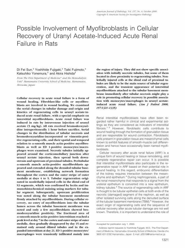
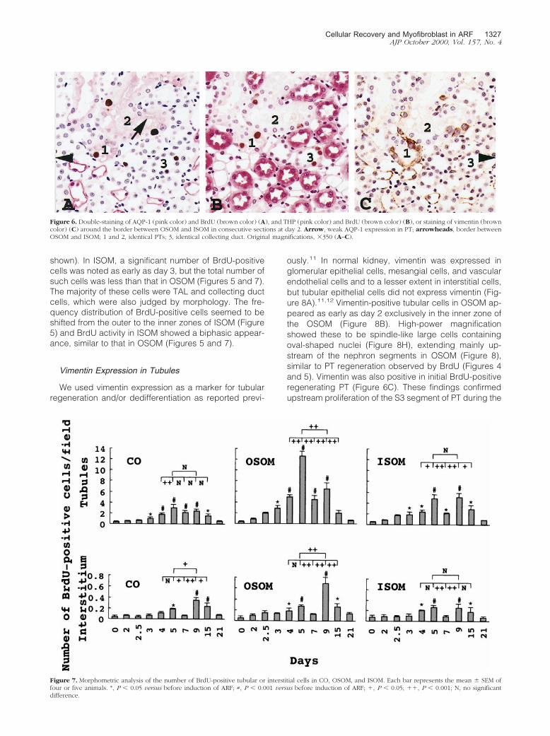
Figure 2. Photomicrographs of periodic acid-Schiff-stained sections, showing PT injury in OSOM before induction of ARF (A); day 2 (B); day 3 (C); day 5 (D);day 7 (E and H); and day 9 (F and I). G: Enlarged photomicrograph of the inset in B; arrows, border between CO and OSOM; arrowheads, border betweenOSOM and ISOM. PT necrosis initially occurred around the corticomedullary junction where denuded TBM was evident at day 2 (B and G), then spread upstreamand downstream of the nephron, reaching a peak level around day 5 (D). At day 7 (E) the TBM was almost covered with flattened, regenerated tubular cells (H,arrowheads). Hyperplastic tubules (I, arrow) were evident at day 9. Original magnifications, �100 (A–F); �300 (G); �650 (H and I).
Cellular Recovery and Myofibroblast in ARF 1323AJP October 2000, Vol. 157, No. 4
�-SMA using the Vectastain ABC-AP kit with Vector Red,then stained for BrdU using the diaminobenzidine detec-tion system. Consecutive sections were also used forcomparison of different staining patterns. Counterstain-ing, if necessary, was performed by using hematoxylin ormethyl green.
For control sections, the first antibodies were omittedor replaced by the normal serum of corresponding animaland signals in both control and experimental sectionswere negative or negligible.
Immunoelectron Microscopy for Detection of�-SMA
Cortical tissues fixed with 4% paraformaldehyde weredehydrated in a graded ethanol series and embedded inUnicryl (British BioCell, Cardiff, UK) at �30°C accordingto the instructions provided by the manufacturer. Ultrathinsections were incubated with mouse anti-human �-SMA(DAKO, Carpinteria, CA) diluted 1:100 in PBS, and sub-sequently, with rabbit anti-mouse IgG (Chemicon) diluted
1:500 in PBS, and goat anti-rabbit IgG conjugated with 10nm gold (Ultra Biosols, Liverpool, UK) diluted 1:10 in 0.02mol/L Tris-HCl buffer (pH 7.0). Sections were stained withUA then examined by a JEM-1220 electron microscope(JEOL, Tokyo, Japan).
Morphometric Analysis of Location of TubularNecrosis and of BrdU-Positive Tubular Cells
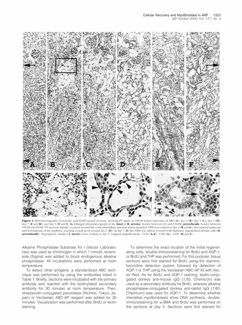
To examine the serial changes in the distribution of PTnecrosis, areas of PAS-stained longitudinal sections ofthe kidney obtained at days 2, 2.5, 3, 4, 5, and 7 wereprojected onto a TV monitor. The viewed area of eachsection was �6.4 mm2 in each rat. The cross-section ofPT with severe damage (representing desquamation oftubular epithelial cells associated with focal granulovacu-olar epithelial cell degeneration amounting to 75 to 100%of the section) was counted as necrotic tubules. Thelocation of targeted PT in the cortex (CO) or the outerstripe of outer medulla (OSOM) was expressed as A/B (0to 1.0) using a computer-assisted image analyzer, whereA represented the distance between the center of PT andthe border of CO-OSOM or OSOM, the inner stripe ofouter medulla (ISOM), respectively, and B the thicknessof each kidney layer at the same site. The number ofsevere necrotic PT in each 20 arbitrarily divided zones ineach kidney layer was displayed in histograms.
The method for locating BrdU-positive cells in tubulesat days 2, 2.5, 3, 4, 5, 7, 9, 15, and 21 was similar to thatused for PT necrosis described above. Areas of BrdU-stained longitudinal sections were displayed onto themonitor. The viewed area of each section was �10.0mm2 in each rat. BrdU-positive cells in CO, OSOM, orISOM were expressed as A/B, where A represents thedistance between a BrdU-positive cell and the border ofCO-OSOM, OSOM-ISOM, or ISOM-the inner medulla, re-spectively, and B the thickness of each kidney layer atthe same site. The number of BrdU-positive cells in each20 arbitrarily divided zones in each kidney layer wasdisplayed in histograms.
Morphometric Analysis of the Number of BrdU-Positive Cells and of ED 1-Positive InterstitialMonocytes/Macrophages
For semiquantitative analysis, the number of BrdU-posi-tive cells in both tubules and the interstitium or the num-ber of ED 1-positive cells in the interstitium in CO, OSOM,or ISOM at days 2, 2.5 (only for BrdU), 3, 4, 5, 7, 9, 15,and 21 were counted in 50 randomly selected fields of eachlayer in each kidney at �400 magnification. The mean num-ber at each time point was displayed in histograms.
Morphometric Analysis of �-SMA/BrdU Double-Positive Interstitial Cells
The number of �-SMA/BrdU double-positive cells inBrdU-positive cells or in �-SMA-positive cells in the inter-stitium was counted in 50 randomly selected fields of
Figure 3. Morphometric analysis of serial changes in the distribution of PTnecrosis in CO and OSOM. S, surface of the kidney; B1, border between COand OSOM; B2, border between OSOM and ISOM. Data represent the meanvalues of three or four animals.
1324 Sun et alAJP October 2000, Vol. 157, No. 4
OSOM at day 5 at �400 magnification, and the meanpercentage of �-SMA/BrdU double-positive cells wascalculated.
Morphometric Analysis of �-SMA-Positive Cells
For semiquantification of �-SMA-positive area in CO,OSOM, and ISOM at days 2, 3, 4, 5, 7, 9, 15, and 21, pointcounting was performed using a routine establishedmethod.10 A total of 20 fields at �400 magnification persection were counted in each animal on a 1-cm2 eye-piece graticule with 10 equidistant grid lines. Each fieldwas 0.0625 mm2, and the total area counted per slidewas 1.25 mm2. The percentage of fractional area (per-centage of positive area per total area counted of thesection) was calculated using the following formula: Per-centage of fractional area equals number of grid inter-sections with positive staining/total number of grid inter-sections multiplied by 100.
Statistical Analysis
Data are expressed as the mean � SEM. Differencesbetween data sets were examined for statistical signifi-cance using one-way analysis of variance followed by
Fisher’s t-test. A P level �0.05 was accepted as statisti-cally significant.
Results
Serum Creatinine (Scr)
Scr increased significantly as early as day 3 after UAinjection (P � 0.05 versus level before induction of ARF),and reached a peak value at day 7 (P � 0.001 versuslevel before induction of ARF), then returned to normallevel by day 15 (Figure 1).
Damage of PTs Observed by PAS Staining
Although slight morphological changes could be seenthroughout the PT in PAS-stained tissues, the initial le-sions of severe necrosis were predominantly locatedaround the corticomedullary junction where the denudedTBM was evident as early as day 2 (Figure 2, B and G).PT damage with severe necrosis spread progressively toboth downstream and upstream of PT (Figures 2 and 3).Between days 4 and 5, tubular necrosis was almost max-imally distributed in OSOM and CO (Figure 3) with cellu-
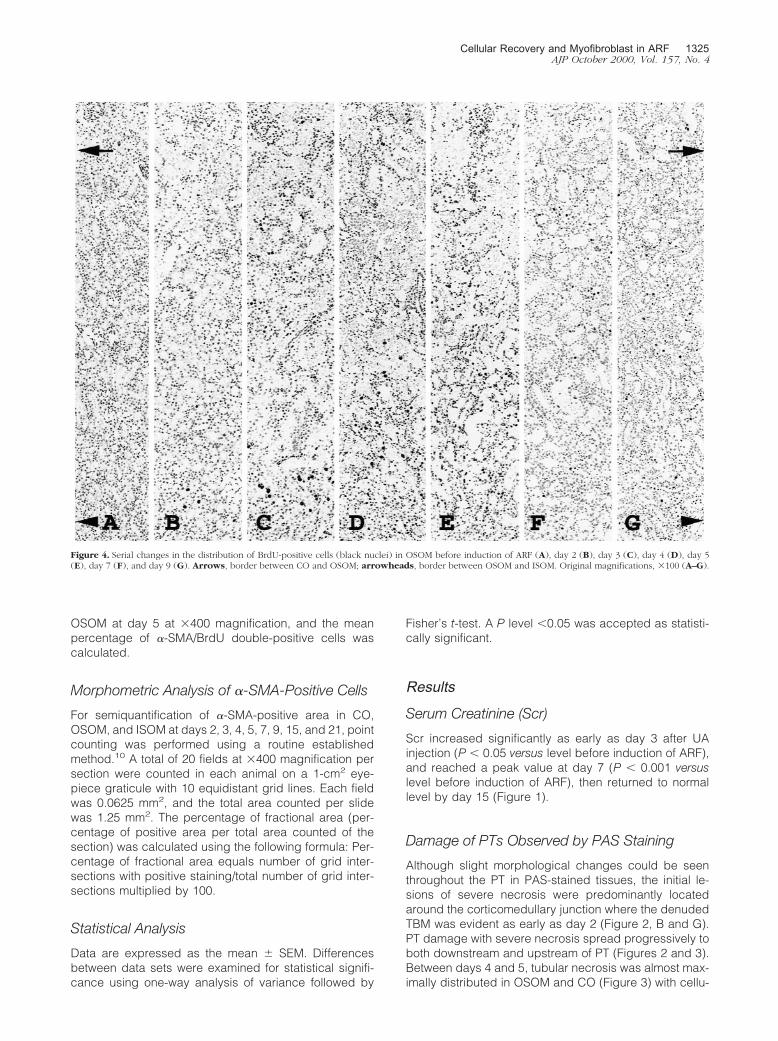
Figure 4. Serial changes in the distribution of BrdU-positive cells (black nuclei) in OSOM before induction of ARF (A), day 2 (B), day 3 (C), day 4 (D), day 5(E), day 7 (F), and day 9 (G). Arrows, border between CO and OSOM; arrowheads, border between OSOM and ISOM. Original magnifications, �100 (A–G).
Cellular Recovery and Myofibroblast in ARF 1325AJP October 2000, Vol. 157, No. 4
lar debris in the tubular lumen (Figure 2D). By day 7, themajority of necrotic tubules disappeared and denudedTBM were almost covered with regenerating cells withflattened cytoplasm (Figure 2, E and H), but there werestill few clusters of severely damaged tubules (Figure2E). After day 7, stratification of hyperplastic PT with focal
papillary projections could be seen in OSOM (Figure 2, Eand I). By day 21, desquamation of epithelial cells inpapillary clusters was noted in the tubular lumen, andhyperproliferation was almost remodeled with occasionaltubular dilation (Figure 2F).
Recovery of Tubular Cell Damage
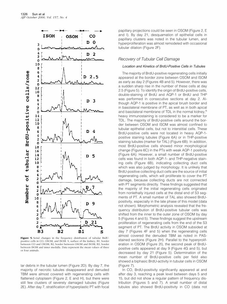
Location and Kinetics of BrdU-Positive Cells in Tubules
The majority of BrdU-positive regenerating cells initiallyappeared at the border zone between OSOM and ISOMas early as day 2 (Figures 4B and 5). However, there wasa sudden sharp rise in the number of these cells at day2.5 (Figure 5). To identify the origin of BrdU-positive cells,double-staining of BrdU and AQP-1 or BrdU and THPwas performed in consecutive sections at day 2. Al-though AQP-1 is positive in the apical brush border andin basolateral membrane of PT, as well as in both apicaland basolateral membrane of TDL in the normal kidney,8
heavy immunostaining is considered to be a marker forTDL. The majority of BrdU-positive cells around the bor-der between OSOM and ISOM was almost confined totubular epithelial cells, but not to interstitial cells. TheseBrdU-positive cells were not located in heavy AQP-1-positive staining tubules (Figure 6A) or in THP-positivestaining tubules (marker for TAL) (Figure 6B). In addition,most BrdU-positive cells showed minor morphologicalchange (Figure 6C) in the PTs with weak AQP-1 positivity(Figure 6A). However, a small number of BrdU-positivecells was found in both AQP-1- and THP-negative stain-ing cells (Figure 6B), indicating collecting duct cellswhich was also judged by morphology. It is unlikely thatBrdU-positive collecting duct cells are the source of initialregenerating cells, which will proliferate to cover the PTdamage, because collecting ducts are not connectedwith PT segments directly. These findings suggested thatthe majority of the initial regenerating cells originatedfrom nonlethally injured cells at the distal end of S3 seg-ments of PT. A small number of TAL also showed BrdU-positivity, especially in the late phase of this model (datanot shown). Morphometric analysis revealed that the fre-quency distribution of BrdU-positive tubular cells wasshifted from the inner to the outer zone of OSOM by day5 (Figures 4 and 5). These findings suggest the upstreamproliferation of regenerating cells from the end of the S3segment of PT. The BrdU activity in OSOM subsided atday 7 (Figures 4F and 5) when the regenerating cellsalmost covered the denuded TBM as noted in PAS-stained sections (Figure 2H). Parallel to the hyperprolif-eration in OSOM (Figure 2I), the second peak of BrdU-positive cells appeared at day 9 (Figure 4G and 5), butdecreased by day 21 (Figure 5). Determination of themean number of BrdU-positive cells per field alsoshowed a biphasic BrdU-activity in tubular cells in OSOM(Figure 7).
In CO, BrdU-positivity significantly appeared at andafter day 3, reaching a peak level between days 5 and15, but did not show a regular pattern of frequency dis-tribution (Figures 5 and 7). A small number of distaltubules also showed BrdU-positivity in CO (data not
Figure 5. Serial changes in the frequency distribution of tubular BrdU-positive cells in CO, OSOM, and ISOM. S, surface of the kidney; B1, borderbetween CO and OSOM; B2, border between OSOM and ISOM; B3, borderbetween ISOM and inner medulla. Data represent the mean values of threeor four animals.
1326 Sun et alAJP October 2000, Vol. 157, No. 4
shown). In ISOM, a significant number of BrdU-positivecells was noted as early as day 3, but the total number ofsuch cells was less than that in OSOM (Figures 5 and 7).The majority of these cells were TAL and collecting ductcells, which were also judged by morphology. The fre-quency distribution of BrdU-positive cells seemed to beshifted from the outer to the inner zones of ISOM (Figure5) and BrdU activity in ISOM showed a biphasic appear-ance, similar to that in OSOM (Figures 5 and 7).
Vimentin Expression in Tubules
We used vimentin expression as a marker for tubularregeneration and/or dedifferentiation as reported previ-
ously.11 In normal kidney, vimentin was expressed inglomerular epithelial cells, mesangial cells, and vascularendothelial cells and to a lesser extent in interstitial cells,but tubular epithelial cells did not express vimentin (Fig-ure 8A).11,12 Vimentin-positive tubular cells in OSOM ap-peared as early as day 2 exclusively in the inner zone ofthe OSOM (Figure 8B). High-power magnificationshowed these to be spindle-like large cells containingoval-shaped nuclei (Figure 8H), extending mainly up-stream of the nephron segments in OSOM (Figure 8),similar to PT regeneration observed by BrdU (Figures 4and 5). Vimentin was also positive in initial BrdU-positiveregenerating PT (Figure 6C). These findings confirmedupstream proliferation of the S3 segment of PT during the
Figure 6. Double-staining of AQP-1 (pink color) and BrdU (brown color) (A), and THP (pink color) and BrdU (brown color) (B), or staining of vimentin (browncolor) (C) around the border between OSOM and ISOM in consecutive sections at day 2. Arrow, weak AQP-1 expression in PT; arrowheads, border betweenOSOM and ISOM; 1 and 2, identical PTs; 3, identical collecting duct. Original magnifications, �350 (A–C).
Figure 7. Morphometric analysis of the number of BrdU-positive tubular or interstitial cells in CO, OSOM, and ISOM. Each bar represents the mean � SEM offour or five animals. *, P � 0.05 versus before induction of ARF; #, P � 0.001 versus before induction of ARF; �, P � 0.05; ��, P � 0.001; N, no significantdifference.
Cellular Recovery and Myofibroblast in ARF 1327AJP October 2000, Vol. 157, No. 4
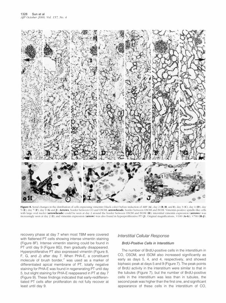
recovery phase at day 7 when most TBM were coveredwith flattened PT cells showing intense vimentin staining(Figure 8F). Intense vimentin staining could be found inPT until day 9 (Figure 8G), then gradually disappeared.Hyperproliferative PT also expressed vimentin (Figure 8,F, G, and J) after day 7. When PHA-E, a constituentmolecule of brush border,7 was used as a marker ofdifferentiated apical membrane of PT, totally negativestaining for PHA-E was found in regenerating PT until day5, but slight staining for PHA-E reappeared in PT at day 7(Figure 9). These findings indicated that early-redifferen-tiated PT cells after proliferation do not fully recover atleast until day 9.
Interstitial Cellular Response
BrdU-Positive Cells in Interstitium
The number of BrdU-positive cells in the interstitium inCO, OSOM, and ISOM also increased significantly asearly as days 5, 4, and 4, respectively, and showedbiphasic peak at days 5 and 9 (Figure 7). The peak pointsof BrdU activity in the interstitium were similar to that inthe tubules (Figure 7), but the number of BrdU-positivecells in the interstitium was less than in tubules, thesecond peak was higher than the first one, and significantappearance of these cells in the interstitium of CO,
Figure 8. Serial changes in the distribution of cells expressing vimentin (black color) before induction of ARF (A), day 2 (B, H, and I), day 3 (C), day 4 (D), day5 (E), day 7 (F), day 9 (G and J). Arrows, border between CO and OSOM; arrowheads, border between OSOM and ISOM. Vimentin-positive spindle-like cellswith large oval nuclei (arrowheads) could be seen at day 2 around the border between OSOM and ISOM (H); interstitial vimentin expression (arrows) wasincreasingly seen at day 2 (I); and vimentin expression (arrow) was also found in hyperproliferative PT (J). Original magnifications, �100 (A–G); �700 (H–J).
1328 Sun et alAJP October 2000, Vol. 157, No. 4

OSOM, and ISOM did not precede that in tubules (Figure7). Moreover, the distribution of BrdU-positive cells in theinterstitium in OSOM did not show a regular pattern andtemporal association with regenerating tubules.
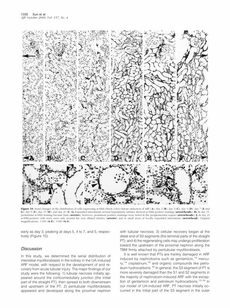
Appearance of Interstitial Myofibroblasts
In normal rats, �-SMA protein was detected in vascularsmooth muscle cells but not in glomeruli, tubules, orinterstitial cells (Figure 10A). �-SMA-positive interstitialcells, indicating myofibroblasts, were visible in OSOM asearly as day 2 (Figure 10B). Initially they appeared ex-clusively around damaged tubules with denuded TBMwhich were located mainly in the outer zone of the OSOM(Figure 10B) and extended along with the progression ofsevere tubular damage (Figures 2 and 10). Between days4 and 5, myofibroblasts formed a network around ne-crotic tubules throughout the cortex and medulla as ifthey structurally supported the frame of the denudedTBM (Figures 10D and 11A). The exact location of myo-fibroblasts when they entered the tubular lumen was notclear under light microscopic examination (Figure 11A).Immunoelectron microscopy revealed that the cell pro-cesses labeled with gold particles, indicating �-SMA,were firmly attached to the outer surface of the denudedTBM and formed a network appearance by day 5 (Figure12). Entry of myofibroblasts into tubular lumen acrossTBM was not evident at the ultrastructural level (Figure12). We performed �-SMA/BrdU double-immunostainingin OSOM at day 5 (Figure 13) when interstitial myofibro-blasts are developing network formation. Among BrdU-positive interstitial cells, only 2.5 � 0.5% showed �-SMA-positivity, and among �-SMA-positive cells, only 1.5 �0.3% expressed BrdU positivity, suggesting that mostBrdU-positive interstitial cells were not myofibroblastsand that myofibroblasts seemed not to undergo cell pro-liferation actively. Most peritubular �-SMA expressionwas transient in OSOM and CO and began to decreasefrom day 9 (Figure 10F), became faint by day 15 (Figure10H), and almost negative by day 21 (Figure 10I). How-ever, �-SMA staining in focally expanded interstitium,around hyperplastic tubules (Figure 10G) and dilated
tubules (Figure 10I), was seen at day 7 and thereafter. Byday 21, �-SMA-positive cells were present only arounddilated tubules (Figure 10I) and in small areas of focallyexpanded interstitium (Figure 10I). Periglomerular �-SMAstaining was noted at day 7 and became most prominentat day 15 (Figure 10H) but diminished rapidly by day 21.�-SMA-positive staining in the expanded interstitial areaand periglomerular area was confirmed by increasedfractional areas of �-SMA in the later phase of this model(Figure 14).
In ISOM the interstitial expression of �-SMA extendedfrom the OSOM, and significantly increased in the outerzone of ISOM at day 5, then extended to the whole ISOM,reaching a peak level at day 9 (Figure 14).
In normal rats, only a faint vimentin staining could beseen in some renal interstitial cells in OSOM. After induc-tion of ARF, the number of faint vimentin-positive peritu-bular interstitial cells increased throughout OSOM asearly as day 2 (Figure 8I). This was followed by furtherincreases in intensity of staining (Figure 11B). After day 4,the serial changes in vimentin-staining in the interstitiumwere similar to those of �-SMA staining (Figure 11, A andB). These findings suggest that interstitial cells may firstlyexpress vimentin then acquire �-SMA.
Interstitial ED 1-Positive Monocytes/Macrophages
ED 1-positive monocytes/macrophages were foundonly occasionally in control kidneys. With the develop-ment of PT damage after induction of ARF, interstitial ED1-positive cells increased in the region of injury, thengradually disappeared after tubules have regenerated(data not shown). Specific accumulation of ED 1-positivecells closely around the initially necrotic PT with the de-nuded TBM was not evident, and the distribution of ED1-positive cells did not show a regular pattern in CO,OSOM, and ISOM. However, some ED 1-positive cellslocated in close proximity to myofibroblasts surroundingregenerating tubules (Figure 11C) after day 3. Morpho-metric analysis revealed that interstitial ED 1-positivecells in CO, OSOM, and ISOM significantly increased as
Figure 9. Location of PHA-E binding sites in normal rats (A) and in experimental rats at days 5 (B) and 7 (C). Staining of brush-border was quite strong (arrows)in PTs of normal rats (A), but totally negative in regenerating PTs at day 5 (B). A faint staining could be seen on the reappeared brush-border (arrowheads) atday 7 (C). Original magnifications, �750 (A–C).
Cellular Recovery and Myofibroblast in ARF 1329AJP October 2000, Vol. 157, No. 4
early as day 3, peaking at days 5, 4 to 7, and 5, respec-tively (Figure 15).
DiscussionIn this study, we determined the serial distribution ofinterstitial myofibroblasts in the kidney in the UA-inducedARF model, with respect to the development of and re-covery from acute tubular injury. The major findings of ourstudy were the following: 1) tubular necrosis initially ap-peared around the corticomedullary junction (the initialpart of the straight PT), then spread to both downstreamand upstream of the PT; 2) peritubular myofibroblastsappeared and developed along the proximal nephron
with tubular necrosis; 3) cellular recovery began at thedistal end of S3 segments (the terminal parts of the straightPT); and 4) the regenerating cells may undergo proliferationtoward the upstream of the proximal nephron along theTBM firmly attached by peritubular myofibroblasts.
It is well known that PTs are frankly damaged in ARFinduced by nephrotoxins such as gentamicin,13 mercu-ry,14 cisplatinum,15 and organic compounds like petro-leum hydrocarbons.16 In general, the S3 segment of PT ismore severely damaged than the S1 and S2 segments inthe majority of nephrotoxin-induced ARF with the excep-tion of gentamicin and petroleum hydrocarbons.13,16 Inour model of UA-induced ARF, PT necrosis initially oc-curred in the initial part of the S3 segment in the outer
Figure 10. Serial changes in the distribution of cells expressing �-SMA (black color) before induction of ARF (A), day 2 (B), day 3 (C), day 5 (D), day 7 (E andG), day 9 (F), day 15 (H), and day 21 (I). G: Expanded interstitium around hyperplastic tubules showed �-SMA-positive staining (arrowheads). H: At day 15,peritubular �-SMA staining became faint (arrows), however, prominent positive stainings were noted in the periglomerular region (arrowheads). I: At day 21�-SMA-positive cells were seen only around the very dilated tubules (arrows) and in small areas of focally expanded interstitium (arrowhead). Originalmagnifications, �100 (A–F); �300 (G–I).
1330 Sun et alAJP October 2000, Vol. 157, No. 4
OSOM and slight morphological changes were alreadypresent throughout the whole S3 segment as early as day2. Necrotic tubules progressively spread from the corti-comedullary junction toward both downstream and up-stream of PT, and were maximally distributed almostthroughout the whole S3 segment by days 4 to 5. To ourknowledge, detailed description of the progression of PT
necrosis has not been previously described in nephro-toxin- and ischemia-induced ARF.
In the present study, we provided additional evidenceregarding the origin of regenerating cells and the modeof cellular recovery. The initial regenerating tubular cells(BrdU- or vimentin-positive cells) were found at the distalend of the S3 segment as early as day 2. This wasconfirmed by using markers for nephron segments. Thefirst group of regenerating cells showed partial cellulardamage. Subsequently, regenerating cells proliferatedtoward upstream of proximal nephron in OSOM and al-most covered the denuded TBM by day 7. Based onmesenchymal-epithelial cell transdifferentiation duringnephrogenesis,4 it is possible that interstitial multipotentstem cells could trans-differentiate into tubular epithelialcells under pathological conditions. In this study, how-ever, during cellular recovery, electron microscopic ex-amination revealed the lack of invasion of myofibroblastsacross the TBM into tubular lumen around the distal endsof S3 segments and even throughout the proximalnephron in OSOM. These findings suggest that the non-lethally injured PT cells themselves at the distal end of theS3 segment may be the main source of cellular recovery.In most nephrotoxin- and ischemia-induced ARF, cellularrecovery was reported to originate from surviving cells inthe necrotic zone or viable cells adjacent to the necroticzone.4,13,17 It is conceivable that isolated surviving cellsare responsible for cellular recovery after tubular damagewhen the degree of the latter was low and many isolated
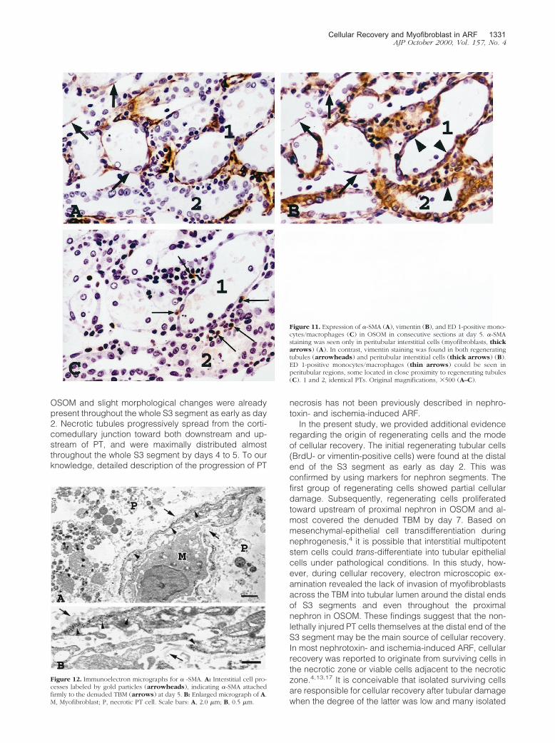
Figure 11. Expression of �-SMA (A), vimentin (B), and ED 1-positive mono-cytes/macrophages (C) in OSOM in consecutive sections at day 5. �-SMAstaining was seen only in peritubular interstitial cells (myofibroblasts, thickarrows) (A). In contrast, vimentin staining was found in both regeneratingtubules (arrowheads) and peritubular interstitial cells (thick arrows) (B).ED 1-positive monocytes/macrophages (thin arrows) could be seen inperitubular regions, some located in close proximity to regenerating tubules(C). 1 and 2, identical PTs. Original magnifications, �500 (A–C).
Figure 12. Immunoelectron micrographs for � -SMA. A: Interstitial cell pro-cesses labeled by gold particles (arrowheads), indicating �-SMA attachedfirmly to the denuded TBM (arrows) at day 5. B: Enlarged micrograph of A.M, Myofibroblast; P, necrotic PT cell. Scale bars: A, 2.0 �m; B, 0.5 �m.
Cellular Recovery and Myofibroblast in ARF 1331AJP October 2000, Vol. 157, No. 4
surviving cells were still present in these areas. Thisseems not to be the case in our model. Only Haagsma etal18 reported that regeneration commenced at both endsof damaged PT in HgCl2-induced ARF. However, they didnot elaborate on the mode of progression of cellularrecovery. In the present model, BrdU-positive tubularcells in CO appeared later and in a sporadic and irregularpattern than in OSOM, suggesting that the isolated sur-viving cells mainly participate in cellular recovery in CO.However, it is difficult to assess this issue because the S1� S2 segments of PT do not run straightly from thejunction of the S3 to S2 segment to the glomerulus.19 Itremains unclear at this stage whether the cells at thedistal end of PT are characteristically resistant to UA-insult or are proliferatively potent. Furthermore, the exactstimulus that initiates the proliferative response remainsto be determined.
A particularly noteworthy finding in the present studywas that the myofibroblasts appeared and surroundedthe initial necrotic PT, developed along with the progres-sion of PT necrosis, attaching to the TBM, and formednetwork throughout the CO and OSOM before accelera-tion of cellular recovery. The origin of �-SMA-expressing
interstitial cells was not clear, but their immediate appear-ance adjacent to TBM strongly suggests that residentstromal cells located adjacent to the TBM, which initiallydo not express features of smooth muscle cell differenti-ation (identified by �-SMA), later differentiated to �-SMAexpressing cells. The mechanisms responsible for suchphenotypic modulations are unknown, but recent studiessuggest that transforming growth factor-�, platelet-de-rived growth factor, and extracellular matrix could trans-form fibroblasts to myofibroblasts.20 In the presentmodel, injured tubular cells might secrete cytokines suchas platelet-derived growth factor and transforminggrowth factor-�,21,22 resulting in the activation and prolif-eration of myofibroblasts. However, in the present study,analysis of �-SMA/BrdU double-immunostaining re-vealed that most myofibroblasts did not undergo cellproliferation actively. Interstitial infiltrating cells are otherpossible candidates for cytokine production. Diamond etal23 reported that renal cortical transforming growth fac-tor-�1, derived from infiltrating macrophages, in part,contribute to foster the modulation of fibroblasts to myo-fibroblasts within the renal cortex after ureteral obstruc-tion in rats. In fact, several studies have reported the
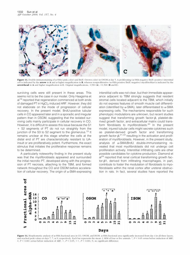
Figure 13. Double-immunostaining of �-SMA (pink color) and BrdU (brown color) in OSOM at day 5. A proliferating (�-SMA-negative/BrdU-positive) interstitialcell is indicated by the arrow in A and at higher magnification in B, whereas nonproliferative (�-SMA-positive/BrdU-negative) myofibroblast is indicated by thearrowhead in A and higher magnification in C. Original magnifications, �350 (A); �1,500 (B and C).
Figure 14. Morphometric analysis of �-SMA fractional area in CO, OSOM, and ISOM. �-SMA fractional area significantly increased from day 4 in all three layers,and reached peak values at days 7, 7, or 9, respectively. Each bar represents the mean � SEM of four or five animals. *, P � 0.05 versus before induction of ARF;#, P � 0.001 versus before induction of ARF; �, P � 0.05; ��, P � 0.001; N, no significant difference.
1332 Sun et alAJP October 2000, Vol. 157, No. 4
presence of infiltrating cells in the interstitium after acuterenal injury.24 In our model, ED 1-positive monocytes/macrophages did infiltrate transiently in the interstitium.Specific association of them with initially necrotic tubulessurrounded by myofibroblasts, however, was not evident.In addition, alterations in components of TBM similar tothe extracellular matrix were reported in ARF.25,26 More-over, back-leak of filtrate through the denuded TBM27
and/or relatively high stretching force acting on the inter-stitium through the TBM because of loss of tubular cellsand/or tubular obstruction by cell debris, might activateinterstitial cells to express �-SMA.
Interestingly, Nagle et al28 observed that interstitialfibroblasts underwent morphological transformation tosmooth muscle-like interstitial cells in a rabbit model ofcomplete ureteral obstruction and demonstrated that un-der these conditions the kidney may demonstrate in-creased whole-organ contractility.29 In the present study,immunoelectron microscopic examination revealed thatmyofibroblasts with possible stress fibers closely cov-ered the external side of TBM as if they provided con-tractility and resisted the stretching force. Myofibroblastshave been described in clinical disorders related to injuryand repair phenomena, associated with prominent tissueretraction and remodeling.20 During wound healing, fi-broblastic cells modulate to a phenotype characterizedby ultrastructural (stress fibers) and biochemical (�-SMA)features typical of smooth muscle cells, indicating thatthis change is instrumental in activities such as woundcontraction.20 This might also be the case for myofibro-blasts during cellular recovery in ARF. Interstitial accu-mulation of myofibroblasts is one of the best predictiveindicators of renal fibrosis in progressive renal dis-ease.1–3 In the present acute tubular injury model, themajority of peritubular myofibroblasts gradually disap-peared after regeneration of PT, suggesting that theymight revert to a quiescent form or undergo apopto-sis.30,31
Although no direct evidence for a role of interstitialcells in ARF is provided in the present study, we postulate
that peritubular myofibroblasts play a mediating role incellular recovery in our model because the regeneratingPT underwent proliferation toward upstream of PT alongthe denuded TBM firmly attached by myofibroblasts. Inaddition to the possible function of supporting the TBM,myofibroblasts might furnish extracellular matrix compo-nents to condition PT proliferation, because fibroblastsplay an essential role in the synthesis and regulation ofextracellular matrix components in wound healing.20
Moreover, myofibroblasts can provide cytokines as para-crine mitogens, which might promote cellular recoveryfrom ARF. At present, fibroblast growth factors-1 and -7and hepatocyte growth factor may be possible candi-dates for cytokines secreted by myofibroblasts in renal in-terstitium.32–35 Transient interstitial infiltrating cells24,32,36 aswell as tubular cells21,22,37–39 are also a possible source ofcytokines, which can promote regeneration of the dam-aged tubules. Indeed, in the present study transient infil-tration of interstitial monocytes/macrophages could befound in some association with regenerating tubules.
Regenerative repair of BrdU-positive PT was almostcompleted by day 7. After day 7, hyperproliferation of PTbecame prominent and was followed by remodeling ofhyperplastic tubules with a resolution of hypercellularityprobably through apoptosis, as reported previously.6,40
This proliferation pattern seemed to be reflected by thefindings that the biphasic waves of BrdU activities inOSOM reached peak values at days 5 and 9. However,BrdU-positive staining could be also seen in cells stainedpositive for THP, a marker for the thick ascending limb,especially in the late phase of this model (data notshown). Moreover, although no visible signs of tubularnecrosis could be seen in ISOM, a significant number ofBrdU-positive cells were also seen as early as day 3.Thus, BrdU-positive cells probably include loop cellsand/or collecting duct cells. Although PT is the main site ofrenal damage in ARF, recent studies indicate that the distalnephron may also react extensively to ischemic41,42 andnephrotoxic13,33 insults, suggesting that the distalnephron might function as part of an adaptive response
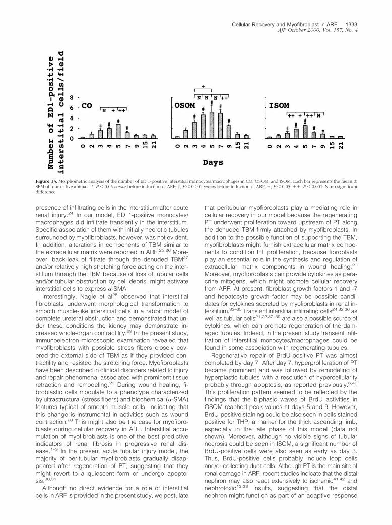
Figure 15. Morphometric analysis of the number of ED 1-positive interstitial monocytes/macrophages in CO, OSOM, and ISOM. Each bar represents the mean �SEM of four or five animals. *, P � 0.05 versus before induction of ARF; #, P � 0.001 versus before induction of ARF; �, P � 0.05; ��, P � 0.001; N, no significantdifference.
Cellular Recovery and Myofibroblast in ARF 1333AJP October 2000, Vol. 157, No. 4
to the loss of PT function. The significance of BrdU-positive cells in the distal nephron remains to be eluci-dated. On the other hand, the early changes in vimentinstaining in tubules corresponded to those of BrdU, butvimentin stained-cells were noted throughout a long pe-riod in PT tubules with or without hyperproliferation atleast until day 9, and at day 21, vimentin was still positivein dilated tubules. These findings indicate that vimentinwould be a marker for tubular dedifferentiation or injuryrather than for regeneration itself as reported previous-ly.11 Although regenerated tubular cells at day 7 showeda weakly positive staining for PHA-E, a constituent mole-cule of the brush border and a marker of differentiatedapical PT membrane, the majority of PT did not show thefinal differentiation at least until day 9 with respect tovimentin expression.
In summary, we have demonstrated in the presentstudy that nonlethally injured cells at the distal end of S3segments of PT are likely to be responsible for the cellularrecovery in UA-induced ARF. Based on the serial changesin the distribution of tubular damage and peritubular myo-fibroblasts, as well as their relationship to cellular recovery,we propose that the transient appearance of peritubularmyofibroblasts immediately after PT necrosis plays an im-portant role in promoting the regenerative repair.
References
1. Alpers CE, Hudkins KL, Floege J, Johnson RJ: Human renal corticalinterstitial cells with some features of smooth muscle cells participatein tubulointerstitial and crescentic glomerular injury. J Am Soc Neph-rol 1994, 5:201–209
2. Hewitson TD, Becker GJ: Interstitial myofibroblasts in IgA glomerulo-nephritis. Am J Nephrol 1995, 15:111–117
3. Zhang G, Moorhead PJ, el Nahas AM: Myofibroblasts and the pro-gression of experimental glomerulonephritis. Exp Nephrol 1995,3:308–318
4. Hay ED, Zuk A: Transformations between epithelium and mesenchyme:normal, pathological, and experimentally induced. Am J Kidney Dis1995, 26:678–690
5. Toback FG: Regeneration after acute tubular necrosis. Kidney Int1992, 41:226–246
6. Sano K, Fujigaki Y, Miyaji T, Ikegaya N, Ohishi K, Yonemura K,Hishida A: Role of apoptosis in uranyl acetate-induced acute renalfailure and acquired resistance to uranyl acetate. Kidney Int 2000,57:1560–1570
7. Ivanyi B, Olsen TS: Immunohistochemical identification of tubularsegments in percutaneous renal biopsies. Histochemistry 1991, 95:351–356
8. Nielsen S, Smith BL, Christensen EI, Knepper MA, Agre P: CHIP28water channels are localized in constitutively water-permeable seg-ments of the nephron. J Cell Biol 1993, 120:371–383
9. Kitamura H, Yamauchi A, Sugiura T, Matsuoka T, Horio M, TohyamaM, Shimada S, Imai E, Hori M: Inhibition of myo-inositol transportcauses acute renal failure with selective medullary injury in the rats.Kidney Int 1998, 53:146–153
10. Hewitson TD, Wu HL, Becker GJ: Interstitial myofibroblasts in exper-imental renal infection and scarring. Am J Nephrol 1995, 15:411–417
11. Grone HJ, Weber K, Grone E, Helmchen U, Osborn M: Coexpressionof keratin and vimentin in damaged and regenerating tubular epithe-lia of the kidney. Am J Pathol 1987, 129:1–8
12. Holthofer H, Miettinen A, Lehto VP, Lehtonen E, Virtanen I: Expressionof vimentin and cytokeratin types of intermediate filament proteins indeveloping and adult human kidneys. Lab Invest 1984, 50:552–559
13. Nouwen EJ, Verstrepen WA, Buyssens N, Zhu MQ, De Bore ME:Hyperplasia, hypertrophy, and phenotypic alterations in the distalnephron after acute proximal tubular injury in the rat. Lab Invest 1994,70:479–493
14. Cuppage FE, Cunningham N, Tate A: Nucleic acid synthesis in theregenerating nephron following injury with mercuric chloride. LabInvest 1969, 21:449–457
15. Nonclercq D, Toubeau G, Laurent G, Tulkens PM, Heuson-StiennonJA: Tissue injury and repair in the rat kidney after exposure to cispla-tin or carboplatin. Exp Mol Pathol 1989, 51:123–140
16. Short BG, Burnett VL, Cox MG, Bus JS, Swenberg JA: Site-specificrenal cytotoxicity and cell proliferation in male rats exposed to petro-leum hydrocarbons. Lab Invest 1987, 57:564–577
17. Peterson DR, Carone FA: Renal regeneration following d-serine in-duced acute tubular necrosis. Anat Rec 1979, 193:383–388
18. Haagsma BH, Pound AW: Mercuric chloride-induced tubulonecrosisin the rat kidney: the recovery phase. Br J Exp Pathol 1980, 61:229–241
19. Charles Jennette J, Olson JL, Schwartz MM, Silva FG: Anatomy.Heptinstall’s Pathology of the Kidney. Edited by C Jennette, JL Olson,MM Schwartz, FG Silva. Philadelphia, Lippincott-Raven, 1998, pp3–66
20. Sappino AP, Schurch W, Gabbiani G: Differentiation repertoire offibroblastic cells: expression of cytoskeletal proteins as marker ofphenotypic modulations. Lab Invest 1990, 63:144–161
21. Nakagawa T, Sasahara M, Haneda M, Kataoka H, Nakagawa H, YagiM, Kikkawa R, Hazama F: Role of PDGF B-chain and PDGF receptorsin rat tubular regeneration after acute injury. Am J Pathol 1999,155:1689–1699
22. Basile DP, Rovak JM, Martin DR, Hammerman MR: Increased trans-forming growth factor-beta 1 expression in regenerating rat renaltubules following ischemic injury. Am J Physiol 1996, 270:F500–F509
23. Diamond JR, van Goor H, Ding G, Engelmyer E: Myofibroblasts inexperimental hydronephrosis. Am J Pathol 1995, 146:121–129
24. Ghielli M, Verstrepen WA, Nouwen E, De Broe ME: Regenerationprocesses in the kidney after acute injury: role of infiltrating cells. ExpNephrol 1998, 6:502–507
25. Walker PD: Alterations in renal tubular extracellular matrix compo-nents after ischemia-reperfusion injury to the kidney. Lab Invest 1994,70:339–346
26. Basile DP, Martin DR, Hammerman MR: Extracellular matrix-relatedgenes in kidney after ischemic injury: potential role for TGF-� inrepair. Am J Physiol 1998, 275:F894–F903
27. Stein JH, Gottschall J, Osgood RW, Ferris TF: Pathophysiology of anephrotoxic model of acute renal failure. Kidney Int 1975, 8:27–41
28. Nagle RB, Kneiser MR, Bulger RE, Benditt EP: Induction of smoothmuscle characteristics in renal interstitial fibroblasts during obstruc-tive nephropathy. Lab Invest 1973, 29:422–427
29. Nagle RB, Evans LW, Reynolds DG: Contractility of renal cortexfollowing complete ureteral obstruction. Proc Soc Exp Biol Med 1975,148:611–614
30. Darby I, Skalli O, Gabbiani G: �-smooth muscle actin is transientlyexpressed by myofibroblasts during experimental wound healing.Lab Invest 1990, 63:21–29
31. Clark RA: Regulation of fibroplasia in cutaneous wound repair. Am JMed Sci 1993, 306:42–48
32. Ichimura T, Maier JA, Maciag T, Zhang G, Stevens JL: FGF-1 innormal and regenerating kidney: expression in mononuclear, intersti-tial, and regenerating epithelial cells. Am J Physiol 1995, 269:F653–F662
33. Ichimura T, Finch PW, Zhang G, Kan M, Stevens JL: Induction ofFGF-7 after kidney damage: a possible paracrine mechanism fortubule repair. Am J Physiol 1996, 271:F967–F976
34. Finch PW, Rubin JS, Miki T, Ron D, Aaronson SA: Human KGF isFGF-related with properties of a paracrine effector of epithelial cellgrowth. Science 1989, 245:752–755
35. Igawa T, Matsumoto K, Kanda S, Saito Y, Nakamura T: Hepatocytegrowth factor may function as a renotropic factor for regeneration inrats with acute renal injury. Am J Physiol 1993, 265:F61–F69
36. Verstrepen WA, Nouwen EJ, Yue XS, BeBroe ME: Altered growthfactor expression during toxic proximal tubular necrosis and regen-eration. Kidney Int 1993, 43:1267–1279
37. Matejka GL, Jennische E: IGF-I binding and IGF-I mRNA expression
1334 Sun et alAJP October 2000, Vol. 157, No. 4

in the post-ischemic regenerating rat kidney. Kidney Int 1992, 42:1113–1123
38. Tsao T, Wang J, Fervenza FC, Vu TH, Jin IH, Hoffman AR, Rabkin R:Renal growth hormone-insulin-like growth factor-I system in acuterenal failure. Kidney Int 1995, 47:1658–1668
39. Homma T, Sakai M, Cheng HF, Yasuda T, Coffey RJ Jr, Harris RC:Induction of heparin-binding epidermal growth factor-like growth factormRNA in rat kidney after acute injury. J Clin Invest 1995, 96:1018–1025
40. Shimizu A, Yamanaka N: Apoptosis and cell desquamation in repair
process of ischemic tubular necrosis. Virchows Arch (Cell Pathol)1993, 64:171–180
41. Fernandez-Llama P, Andrews P, Turner R, Saggi S, Dimari J, KwonTH, Nielsen S, Safirstein R, Knepper MA: Decreased abundance ofcollecting duct aquaporins in post-ischemic renal failure in rats. J AmSoc Nephrol 1999, 10:1658–1668
42. Safirstein R, Megyesi J, Saggi SJ, Price PM, Poon M, Rollins BJ,Taubman MB: Expression of cytokine-like genes JE and KC is in-creased during renal ischemia. Am J Physiol 1991, 261:F1095–F1101
Cellular Recovery and Myofibroblast in ARF 1335AJP October 2000, Vol. 157, No. 4
Related Documents


