medicina Review Porcine Xenograft and Epidermal Fully Synthetic Skin Substitutes in the Treatment of Partial-Thickness Burns: A Literature Review Herbert L. Haller 1, * , Sigrid E. Blome-Eberwein 2 , Ludwik K. Branski 3 , Joshua S. Carson 4 , Roselle E. Crombie 5 , William L. Hickerson 6 , Lars Peter Kamolz 7 , Booker T. King 8 , Sebastian P. Nischwitz 7 , Daniel Popp 7 , Jeffrey W. Shupp 9 and Steven E. Wolf 2 Citation: Haller, H.L.; Blome-Eberwein, S.E.; Branski, L.K.; Carson, J.S.; Crombie, R.E.; Hickerson, W.L.; Kamolz, L.P.; King, B.T.; Nischwitz, S.P.; Popp, D.; et al. Porcine Xenograft and Epidermal Fully Synthetic Skin Substitutes in the Treatment of Partial-Thickness Burns: A Literature Review. Medicina 2021, 57, 432. https://doi.org/10.3390/ medicina57050432 Academic Editor: Rytis Rimdeika Received: 30 March 2021 Accepted: 22 April 2021 Published: 30 April 2021 Publisher’s Note: MDPI stays neutral with regard to jurisdictional claims in published maps and institutional affil- iations. Copyright: © 2021 by the authors. Licensee MDPI, Basel, Switzerland. This article is an open access article distributed under the terms and conditions of the Creative Commons Attribution (CC BY) license (https:// creativecommons.org/licenses/by/ 4.0/). 1 HLMedConsult, Zehetlandweg 7, 4060 Leonding, Austria 2 Lehigh Valley Health Network 1200 S. Cedar Crest Blvd. Kasych 3000, Allentown, PA 18103, USA; [email protected] (S.E.B.-E.); [email protected] (S.E.W.) 3 Department of Surgery—Burn Surgery, The University of Texas Medical Branch and Shriners Hospitals for Children, 301 University BLVD, Galveston, TX 77555, USA; [email protected] 4 Department of Surgery, UF Health Shands Burn Center, University of Florida, 1600 SW Archer Rd, Gainesville, FL 32610, USA; [email protected]fl.edu 5 Connecticut Burn Center, Yale New Haven Heal System, 267 Grant St, Bridgeport, CT 06610, USA; [email protected] 6 Memphis Medical Center Burn Center, 890 Madison Avenue, Suite TG032, Memphis, TN 38103, USA; [email protected] 7 Division of Plastic, Aesthetic and Reconstructive Surgery, Department of Surgery, Medical University, 8053 Graz, Austria; [email protected] (L.P.K.); [email protected] (S.P.N.); [email protected] (D.P.) 8 Division of Burn Surgery, Department of Surgery, 101 Manning Drive CB #7206, Chapel Hill, NC 27599, USA; [email protected] 9 The Burn Center, Department of Surgery, MedStar Washington Hospital Center, 110 Irving St NW, Washington, DC 20010, USA; [email protected] * Correspondence: [email protected] Abstract: Background and Objectives: Porcine xenografts have been used successfully in partial thickness burn treatment for many years. Their disappearance from the market led to the search for effective and efficient alternatives. In this article, we examine the synthetic epidermal skin substitute Suprathel ® as a substitute in the treatment of partial thickness burns. Materials and Methods: A systematic review following the PRISMA guidelines has been performed. Sixteen Suprathel ® and 12 porcine xenograft studies could be included. Advantages and disadvantages between the treatments and the studies’ primary endpoints have been investigated qualitatively and quantitatively. Results: Although Suprathel had a nearly six times larger TBSA in their studies (p < 0.001), it showed a significantly lower necessity for skin grafts (p < 0.001), and we found a significantly lower infection rate (p < 0.001) than in Porcine Xenografts. Nonetheless, no significant differences in the healing time (p = 0.67) and the number of dressing changes until complete wound healing (p = 0.139) could be found. Both products reduced pain to various degrees with the impression of a better performance of Suprathel ® on a qualitative level. Porcine xenograft was not recommended for donor sites or coverage of sheet-transplanted keratinocytes, while Suprathel ® was used successfully in both indications. Conclusion: The investigated parameters indicate that Suprathel ® to be an effective replacement for porcine xenografts with even lower subsequent treatment rates. Suprathel ® appears to be usable in an extended range of indications compared to porcine xenograft. Data heterogeneity limited conclusions from the results. Keywords: dressing changes; epidermal skin substitute; grafting; healing time; infection rate; partial thickness burns; porcine xenograft; resorbable; suprathel; synthetic; workload Medicina 2021, 57, 432. https://doi.org/10.3390/medicina57050432 https://www.mdpi.com/journal/medicina

Welcome message from author
This document is posted to help you gain knowledge. Please leave a comment to let me know what you think about it! Share it to your friends and learn new things together.
Transcript
medicina
Review
Porcine Xenograft and Epidermal Fully Synthetic SkinSubstitutes in the Treatment of Partial-Thickness Burns: ALiterature Review
Herbert L. Haller 1,* , Sigrid E. Blome-Eberwein 2, Ludwik K. Branski 3, Joshua S. Carson 4,Roselle E. Crombie 5 , William L. Hickerson 6, Lars Peter Kamolz 7, Booker T. King 8, Sebastian P. Nischwitz 7 ,Daniel Popp 7, Jeffrey W. Shupp 9 and Steven E. Wolf 2
�����������������
Citation: Haller, H.L.;
Blome-Eberwein, S.E.; Branski, L.K.;
Carson, J.S.; Crombie, R.E.; Hickerson,
W.L.; Kamolz, L.P.; King, B.T.;
Nischwitz, S.P.; Popp, D.; et al.
Porcine Xenograft and Epidermal
Fully Synthetic Skin Substitutes in the
Treatment of Partial-Thickness Burns:
A Literature Review. Medicina 2021,
57, 432. https://doi.org/10.3390/
medicina57050432
Academic Editor: Rytis Rimdeika
Received: 30 March 2021
Accepted: 22 April 2021
Published: 30 April 2021
Publisher’s Note: MDPI stays neutral
with regard to jurisdictional claims in
published maps and institutional affil-
iations.
Copyright: © 2021 by the authors.
Licensee MDPI, Basel, Switzerland.
This article is an open access article
distributed under the terms and
conditions of the Creative Commons
Attribution (CC BY) license (https://
creativecommons.org/licenses/by/
4.0/).
1 HLMedConsult, Zehetlandweg 7, 4060 Leonding, Austria2 Lehigh Valley Health Network 1200 S. Cedar Crest Blvd. Kasych 3000, Allentown, PA 18103, USA;
[email protected] (S.E.B.-E.); [email protected] (S.E.W.)3 Department of Surgery—Burn Surgery, The University of Texas Medical Branch and Shriners Hospitals for
Children, 301 University BLVD, Galveston, TX 77555, USA; [email protected] Department of Surgery, UF Health Shands Burn Center, University of Florida, 1600 SW Archer Rd,
Gainesville, FL 32610, USA; [email protected] Connecticut Burn Center, Yale New Haven Heal System, 267 Grant St, Bridgeport, CT 06610, USA;
[email protected] Memphis Medical Center Burn Center, 890 Madison Avenue, Suite TG032, Memphis, TN 38103, USA;
[email protected] Division of Plastic, Aesthetic and Reconstructive Surgery, Department of Surgery, Medical University,
8053 Graz, Austria; [email protected] (L.P.K.); [email protected] (S.P.N.);[email protected] (D.P.)
8 Division of Burn Surgery, Department of Surgery, 101 Manning Drive CB #7206, Chapel Hill, NC 27599, USA;[email protected]
9 The Burn Center, Department of Surgery, MedStar Washington Hospital Center, 110 Irving St NW,Washington, DC 20010, USA; [email protected]
* Correspondence: [email protected]
Abstract: Background and Objectives: Porcine xenografts have been used successfully in partialthickness burn treatment for many years. Their disappearance from the market led to the searchfor effective and efficient alternatives. In this article, we examine the synthetic epidermal skinsubstitute Suprathel® as a substitute in the treatment of partial thickness burns. Materials and Methods:A systematic review following the PRISMA guidelines has been performed. Sixteen Suprathel®
and 12 porcine xenograft studies could be included. Advantages and disadvantages between thetreatments and the studies’ primary endpoints have been investigated qualitatively and quantitatively.Results: Although Suprathel had a nearly six times larger TBSA in their studies (p < 0.001), it showeda significantly lower necessity for skin grafts (p < 0.001), and we found a significantly lower infectionrate (p < 0.001) than in Porcine Xenografts. Nonetheless, no significant differences in the healing time(p = 0.67) and the number of dressing changes until complete wound healing (p = 0.139) could befound. Both products reduced pain to various degrees with the impression of a better performance ofSuprathel® on a qualitative level. Porcine xenograft was not recommended for donor sites or coverageof sheet-transplanted keratinocytes, while Suprathel® was used successfully in both indications.Conclusion: The investigated parameters indicate that Suprathel® to be an effective replacement forporcine xenografts with even lower subsequent treatment rates. Suprathel® appears to be usablein an extended range of indications compared to porcine xenograft. Data heterogeneity limitedconclusions from the results.
Keywords: dressing changes; epidermal skin substitute; grafting; healing time; infection rate; partialthickness burns; porcine xenograft; resorbable; suprathel; synthetic; workload
Medicina 2021, 57, 432. https://doi.org/10.3390/medicina57050432 https://www.mdpi.com/journal/medicina
Medicina 2021, 57, 432 2 of 24
1. Introduction
Contemporary burn care aims at rapid closure of open wounds, either temporarilyor permanently. Wound closure reduces infectious complications and downregulatesinflammation and other detrimental systemic responses. Moreover, it curbs the hyperme-tabolic response and supports re-establishment of undisturbed energy expenditure in themitochondria [1,2].
Porcine xenograft (PX) (Mölnlyke, Peachtree Corners, GA, USA) and biosynthetic andsynthetic dressings, such as human skin allografts, amniotic membrane, Biobrane® (DowHickman/Bertek Pharmaceuticals, Sugarland, TX, USA), Dermagraft™ (Organogenesis,Canton, MA, USA), Appligraf® (Organogenesis, Canton, MA, USA), OrCel® (ORTECint. Inc., New York, NY, USA), Hyalomatrix® (Medline Industries, Northfield, IL, USA),Transcyte® (Takeda Pharmaceutical Co. Ltd., Tokyo, Japan), and Suprathel® (ST) (Poly-medics Innovations GmbH, Denkendorf, Germany) as epidermal skin substitutes, havebeen used for the closure of partial-thickness wounds. The requirements of these productsinclude safety, ease of application, a short healing time, effectiveness, hypo- allergenicity,and non-oncogenicity, while being able to be stored easily and cost-effective. The PX EZDerm® was used with numerous indications but is not available on the market anymore,yielding the need for finding the optimal replacement and delivering the motivation forthis review.
This paper compares the biological pig skin-derived skin substitute (EZ Derm) toa fully synthetic and biodegradable epidermal substitute (ST) based on the publishedliterature. After describing general product characteristics, we conducted a modifiedsystematic review of the literature to evaluate the suitability or advantages of productsother than PX.
2. Materials and Methods
Given the absence of studies directly comparing PX and ST® treatment in burns, weextracted data from studies comparing either PX or ST® to other treatment modalities.
2.1. Data Retrieval
PubMed®, Science Direct®, and Google Scholar® were searched. The primary strategywas to find studies describing the results of the different products in partial thickness burns.
2.2. Study Selection
Studies were selected according to the PRISMA guidelines. We selected articlespublished in peer-reviewed journals or reviewed and published abstracts of an internationalmeeting on burns.
2.3. Exclusions
Studies on the treatment of mainly or exclusively deep partial-thickness burns werenot described. We excluded studies on donor site areas, porcine small intestine submucosa,genetically modified pigskin, and full-thickness burns. We excluded in vitro studies andstudies that were not relevant, mentioning one treatment method without numerical data.Non-English articles or articles without full-text have been excluded as well.
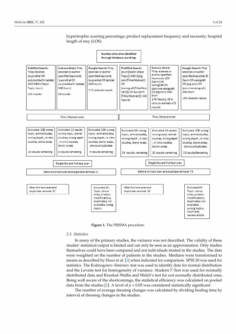
2.4. Search Method and Search Results Based on the PRISMA Flow Chart
Figure 1 shows the Prisma procedure.The following data were retrieved from the studies: study type (prospective, retro-
spective, randomized, non-randomized, descriptive); study population (pediatric, adult,or mixed); sex distribution (male, female); age; cause of burn (scald, flame, contact, flash);timing of epidermal substitute application; description of use in donor sites (Yes/No);information on detailed burn depth (partial superficial, partial deep, or full-thicknessburn); technique of dressing application; wound ground preparation; dressing methodand dressing change frequency; healing time; information and percentage of infections;
Medicina 2021, 57, 432 3 of 24
hypertrophic scarring percentage; product replacement frequency and necessity; hospitallength of stay (LOS).
Figure 1. The PRISMA procedure.
2.5. Statistics
In many of the primary studies, the variance was not described. The validity of thesestudies’ statistical output is limited and can only be seen as an approximation. Only studiesthemselves could have been compared and not individuals treated in the studies. The datawere weighted on the number of patients in the studies. Medians were transformed tomeans as described by Hozo et al. [1] when indicated for comparison. SPSS 20 was used forstatistics. The Kolmogorov–Smirnov test was used to identify data for normal distributionand the Levene test for homogeneity of variance. Student T-Test was used for normallydistributed data and Kruskal–Wallis and Welch’s test for not normally distributed ones.Being well aware of the shortcomings, the statistical efficiency was calculated on pooleddata from the studies [2]. A level of p < 0.05 was considered statistically significant.
The number of average dressing changes was calculated by dividing healing time byinterval of dressing changes in the studies.
Medicina 2021, 57, 432 4 of 24
2.6. Effect Size of Treatment Modalities
The effect size calculated can only be a rough estimate due to the heterogeneity ofstudies. The standard effect size was calculated using the SPSS T-Test and Two-SampleT-Test Calculator from statistics Kingdom for unknown unequal standard deviation [3].The effect size interpretation was made with no effect when dCohen was <0.1, a small effectwith a dCohen of 0.2 to 0.4, a medium effect with dCohen of 0.4–0.6, and a large effect dCohenof >0.6.
3. Results
In total, 29 studies have been found with two of them describing pediatric and adultsseparately and where counted separately. There was no special evaluation of mixedpopulations (pediatric and adult). After exclusion of non-relevant studies (see above), 17and 16 studies have been included dealing with ST and PX, respectively.
3.1. Quality of Studies
In the ST studies, nine out of 17 studies were done prospectively. Five of the studieswere randomized.
In the PX studies, six out of 16 studies were prospective and four of them wererandomized. Details are given in Tables 1 and 2.
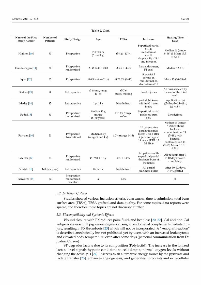
Table 1. Calculation of the effect size for healing time, percentage of infections, the percentage to begrafted, and dressing changes with weighted data. Stdev = standard deviation.
Healing Time Infected % Grafted % Dressing Changes
Mean ST 13.59 3.83 2.50 4.38
Mean PX 7.03 7.04 8.36 4.79
Stdev ST 1.86 6.44 4.05 1.83
Stdev PX 2.09 15.62 13.14 4.28
Sample size ST 371 625 681 398
Sample size PX 143 1124 1136 286
StandardizedEffectsize at 95%
ConfidenceIntervall
0.19 0.2 0.52 0.13
Table 2. Healing time in ST studies.
Name of the FirstStudy Author
Number ofPatients Study Design Age TBSA Inclusion Healing Time
Days
Blome Eberwein [4] 229 Retro P (Pediatric): 138,a (adults): 91
Ø 8.61–60.5
Superficial anddeep second
degree
Mean 13.7 d (days)p: 11.9 d A: 14.7 d
Everett [5] 17 Retro P, Ø 33 m(months) Ø 5% Superficial and
partial thickness Mean 9.4 (5–24) d
Fischer [6] 1 Case report A, 81 a 51% Partial thickness 14 d
Glat [7] 12 Prospective Ø 3.6 y (years) Ø 5.5% Superficial andmixed Mean 8.4 d
Glik [8] 24Retrospective
unblinded paircontrol
Ø 48 y(21–86 y) Ø 23.8
Burns of bothhands tominimize
differences
From Figure 1:complete healing d
20
Gürünlüoglu [9] 20 Prospectiverandomized 4.9 ± 3.8 y Ø 31.95 ± 4.43%
Acute burns,1–60 y, 20–50%including deepburns 5–10%
Median 13.5 d(range 9–21 d)
Mean 14.25 ±3.46 d
Medicina 2021, 57, 432 5 of 24
Table 2. Cont.
Name of the FirstStudy Author
Number ofPatients Study Design Age TBSA Inclusion Healing Time
Days
Highton [10] 33 Prospective P: Ø 29 m(5 m–11 y) Ø 4 (1–13)%
Superficial partialn = 24
mid-dermal:n = 19
deep n = 10, >21 dand infection
Median 16 (range9–38) d; Mean 19.5
± 8.4 d
Hundeshagen [11] 30 Prospectiverandomized A: Ø 24.0 ± 23.0 Ø 5.5 ± 4.6% Partial thickness,
FT excl. Median 12.0 d,
Iqbal [12] 65 Prospective Ø 4.9 y (4 m–11 y) Ø 23.6% (8–45)
Superficialdermal 16,
mid-dermal 34,deep-dermal 15
Mean 15 (10–35) d
Kukko [13] 8 Retrospective Ø 18 mo, range10–39
Ø 7.6Stdev. missing Scald injuries
All burns healed bythe end of the third
week.
Madry [14] 15 Retrospective 1 p, 14 a Not definedpartial thicknesswithin 96 h after
injury
Application: (a)≤24 hs; (b) 24–48 h;
(c) >48 h
Radu [15] 30 Prospectiverandomized
Median 42 y,(range
18–80 years)
Ø 18% (range6–36)
Superficial partialthickness burn
>3%Not defined
Rashaan [16] 21 Prospectiveobservational
Median 2.4 y(range 5 m–14 y) 4.0% (range 1–18)
All consecutivepartial thicknessburns < 48 h afterinjury and age <
18 years SPTB: 12DPTB: 9
Median 13 (range7–29); without
bacterialcontamination: 13
(7–18); withbacterial
contamination 15(9–29) Mean: 15.5 ±
6.36 d
Schiefer [17] 24 Prospectiverandomized Ø 39.8 ± 18 y 0.5 ± 3.0%
All patients withsuperficial partialthickness burn of
the hands
All patients after 7to 10 days healed
completely
Schriek [18] 149 (last year) Retrospective Pediatric Not defined All partialthickness burns
After 10–12 days,7–9% grafted
Schwarze [19] 30Prospective,randomized
bicentrica 1.5% 0
3.2. Inclusion Criteria
Studies showed various inclusion criteria, burn causes, time to admission, total burnsurface area (TBSA), TBSA grafted, and data quality. For some topics, data reports weresparse, and therefore these topics are not discussed further.
3.3. Biocompatibility and Systemic Effects
Wound closure with PX reduces pain, fluid, and heat loss [20–22]. Gal and non-Galantigens are essential pig xenoantigens, causing an endothelial complement-mediated in-jury, resulting in PX thrombosis [23] which will not be incorporated. A “xenograft reaction”is described anechoically but not published yet by users with an increased leukocytosisand elevated body temperature, even after some days (personal communication from Dr.Joshua Carson).
ST degrades lactate due to its composition (Polylactid). The increase in the ionizedlactate level signals hypoxic conditions to cells despite normal oxygen levels withoutchanging the actual pH [24]. It serves as an alternative energy source by the pyruvate andlactate transfer [25], enhances angiogenesis, and generates fibroblasts and extracellular
Medicina 2021, 57, 432 6 of 24
matrix [26,27]. Groussard et al., and, recently, Gürünlüoglu et al., demonstrated lactate’sability to act as a scavenger of free radicals demonstrating the influence on the inflammatoryresponse [9,28]. A positive effect on wound healing was demonstrated compared toHydrofiber Ag, showing increased keratinocyte generation and faster healing [29,30].
3.4. Wound Preparation3.4.1. Wound Bed Preparation
Preparation of the wounds before applying the epidermal templates can be consid-ered similar in both products. After cleaning, debridement, and necrectomy [12], bothproducts were usually applied under general anesthesia [20,21,31,32] or moderate-to-deepsedation [22], primarily due to the patient’s stress after the injury. The wound bed prepa-ration technique varies among the studies: abrasion was performed using scratchpadsor other metallic sponges, brushes, dermabrasion, Versajet, or dermatomes [21,32]. Gen-erally, wound bed preparation was done similarly, depending on the burn depth, andnecrosectomy was sometimes performed to induce punctate bleeding [33].
3.4.2. Template Fixation
For template fixation, most authors used staples for mechanical fixation ofPX [31,32,34–36] and in some cases fibrin glue [20,22] cyanoacrylate glue [20] topicalskin adhesives [36], or sutures [21]. Alternatively, xenograft fixation on superficial partial-thickness burns was achieved by 1-day compression [32] and additional dressing changeson day 1 in PX studies to drain blood or serum retention and control the substitutes’ ad-herence. Frequently, splints were used during the first days to reduce the mobilization ofextremities.
However, ST was not mechanically fixated in most studies [10,19,37] with only asecondary dressing holding it in place (see below).
3.4.3. Separation Layer
A separation layer was applied between the product and an absorptive protectivedressing in both groups with different dressings, such as antibiotic-loaded agents, silicone,fatty gauze, or nylon dressings being used.
Troy et al. used external dressings with a separation layer until the first dressingchange on postoperative day 1, and the PX was exposed to air [32].
3.5. Healing Time3.5.1. Healing Time in Partial Thickness Burns
The comparison was impeded by a missing or inconsistent description of the heal-ing status.
Effects of grafting or conservative treatment were not specified. Therefore, the healingtime was considered in uncomplicated wounds without infections or transplantations.
The two treatment groups had a significantly different TBSA with ST mean 11.36 ± 7.37%and PXs with 4.79 ± 5.78 (p-value of 0.035) or as weighted data 11.72 ± 7.37 and 1.58 ± 3.44(p < 0.001). Nevertheless, the healing time was not significantly different (p = 0.067).
3.5.2. ST Studies
Data were derived from 16 ST studies with 676 patients (See details in Table 2). Eightwere excluded as no data at all or no sufficient data on healing time were provided. Theremaining nine studies weighted on the number of patients: a mean healing time of 13.59 days with a mean TBSA of 11.73% ± 7.37% can be reported. The study populations werecomposed of children, adults, or both. Rashaan et al. found the healing time range’s uppervalues to be 38 days and 29 days, respectively.
3.5.3. PX Studies
Thirteen PX studies included 1136 patients (see details in Table 3), and seven of them
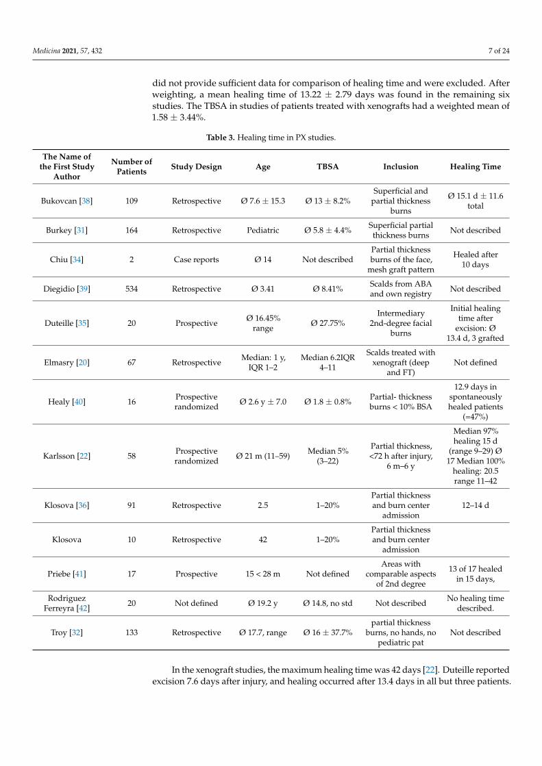
Medicina 2021, 57, 432 7 of 24
did not provide sufficient data for comparison of healing time and were excluded. Afterweighting, a mean healing time of 13.22 ± 2.79 days was found in the remaining sixstudies. The TBSA in studies of patients treated with xenografts had a weighted mean of1.58 ± 3.44%.
Table 3. Healing time in PX studies.
The Name ofthe First Study
Author
Number ofPatients Study Design Age TBSA Inclusion Healing Time
Bukovcan [38] 109 Retrospective Ø 7.6 ± 15.3 Ø 13 ± 8.2%Superficial and
partial thicknessburns
Ø 15.1 d ± 11.6total
Burkey [31] 164 Retrospective Pediatric Ø 5.8 ± 4.4% Superficial partialthickness burns Not described
Chiu [34] 2 Case reports Ø 14 Not describedPartial thicknessburns of the face,
mesh graft pattern
Healed after10 days
Diegidio [39] 534 Retrospective Ø 3.41 Ø 8.41% Scalds from ABAand own registry Not described
Duteille [35] 20 Prospective Ø 16.45%range Ø 27.75%
Intermediary2nd-degree facial
burns
Initial healingtime after
excision: Ø13.4 d, 3 grafted
Elmasry [20] 67 Retrospective Median: 1 y,IQR 1–2
Median 6.2IQR4–11
Scalds treated withxenograft (deep
and FT)Not defined
Healy [40] 16 Prospectiverandomized Ø 2.6 y ± 7.0 Ø 1.8 ± 0.8% Partial- thickness
burns < 10% BSA
12.9 days inspontaneouslyhealed patients
(=47%)
Karlsson [22] 58 Prospectiverandomized Ø 21 m (11–59) Median 5%
(3–22)
Partial thickness,<72 h after injury,
6 m–6 y
Median 97%healing 15 d
(range 9–29) Ø17 Median 100%
healing: 20.5range 11–42
Klosova [36] 91 Retrospective 2.5 1–20%Partial thicknessand burn center
admission12–14 d
Klosova 10 Retrospective 42 1–20%Partial thicknessand burn center
admission
Priebe [41] 17 Prospective 15 < 28 m Not definedAreas with
comparable aspectsof 2nd degree
13 of 17 healedin 15 days,
RodriguezFerreyra [42] 20 Not defined Ø 19.2 y Ø 14.8, no std Not described No healing time
described.
Troy [32] 133 Retrospective Ø 17.7, range Ø 16 ± 37.7%partial thickness
burns, no hands, nopediatric pat
Not described
In the xenograft studies, the maximum healing time was 42 days [22]. Duteille reportedexcision 7.6 days after injury, and healing occurred after 13.4 days in all but three patients.
Medicina 2021, 57, 432 8 of 24
3.6. Change of the Templates or Discontinuation of Treatment
Troy et al. described adhesion loss in their PX studies in 6.8% of patients [32]. Klosovaet al., using XE Derma, found adhesion loss in 16% of patients and at least partial disinte-gration of xenografts in an additional 12% of patients [36]. Out of eight xenograft studiesdescribing unexpected or not defined autografting, adhesion loss was found in five studies,and xenograft change was done between daily and every third day in three studies.
Early detachment or poor wound healing was mentioned in three of the ST® studies.In all these wounds, conservative treatment until wound closure was performed due to theresidual defects’ small size. Two studies described at least a partial removal of ST®. In onestudy, early detachment occurred in 33% of the patients [16], attributed to the method ofdebridement or dressing. In the other study, in three of 15 patients, a dressing removal wasnecessary without a reason given [14] (Table 4).
Table 4. Change or discontinuation of Suprathel or PX treatment.
ST® Xenograft
First Author ST® Comment First Author Xenografts Changeor Diss. Comment
Blome Eberwein [4] No change and noautografts.
In 5.2% failure orprogression to fullthickness, residual
defects treatedconservatively
Burkey [31]
11% of 164 notanticipated
autografting +prolonged topical
wound care in 6 pat.(3.7%) not anticipated
and 22 (14%)anticipated
14.7% (in a total ofnot anticipatedautografting or
prolonged woundcare)
Everett [5] No change Burleson [43] cited byChiu [34]
Change every twodays
Partial-thicknessporcine split skin
Fischer [6] No change Duteille [35]
EZ derm in placeafter surgery for
three days, followedby grafting or topical
wound care
Grafting in 3 patients,
Gürünüloglu [9] No change Elmasry [20] 20% needed anoperation No use in hands
Klosova [36] 19% (81% no signs ofdissolution) XE derma
Hundeshagen [11] No change Priebe [41] EZ Derm replacedevery third day
Madry [14] No change
One dressing removalnecessary when ST
applied at 24–48 h; 2removals necessary,applied >48 h afterinjury (reasons nor
specified).
Rappaport [44] citedby Chiu [34]
Daily change ofxenograft Deep Frozen pigskin
Rashaan [16]
No change, earlydetachment in 43%
treatedconservatively
33% contaminationbefore ST®, detachmentis linked to the method
of debridement andtopical wound care
when detached.
Troy [32]6.8% with prematuregraft separation, 15%
lost for follow-up
After separation,local wound care
Schiefer [17] No change
Schwarze [19] No change
3.7. Auto-Grafting as Indicator for Burn Wound Conversion
Sufficient data on grafting rates were mentioned in 13 and 17 studies in the PX and STgroups, respectively.
Medicina 2021, 57, 432 9 of 24
In PX studies, Troy et al. described excision and autografting in 4.5% of patients in a“no variable burn depth group with only partial-thickness burns” [32]. In their retrospective,unselected study, Elmasry et al. [20] had a grafting rate of 30% due to non-closure aftertwo weeks. Details are shown in Table 5. Only clearly defined grafting procedures wereincluded in the table. The time to evaluate the necessity of the use of autografting varied.Blome-Eberwein evaluated skin grafting after three weeks, while Schriek and Sinnig didtheir evaluation after 11 to 14 days.
Table 5. Grafting rates in partial thickness burns.
ST® PX
Study n= %Grafted n= Type of
Burn Study n= % Grafted Number ofGrafted Type of Burn
BlomeEberwein [4] 229 0% 0
2nd degreeburns
superficialand partial
Bukovcan [38] 109 3.7% 4superficial
partial scaldburns
Everett [5] 17 0% 0Partial
thicknesswithin 6 h
Burkey [31] 167 5.5% unex-pected 3 + 21
Superficialpartial-
thicknessinclusion
Fischer [6] 1 0% 0 Partialthickness Duteille [35] 20 15% 3 Intermediate
face burns
Gürünlüoglu [9] 20 0% 0
Superficialand deep
partialthickness
burns
Elmasry [20] 67 30% 20
Onlysuperficial
partial-thickness
burns
Hundeshagen [11] 30 6.6% 1Partial
thicknessburns
Healy [40] 327 out of 16EZ Derm
44%7 Partial, no
hands or faces
Iqbal [12] 65 0% 0Partial-
thicknessburns
Karlsson [22] 29 13% 6 No palms,soles, or faces
Madry [14] 15 26% 2Children,
Flame andscald burns
Klosova [36] 91children 30% 27
Partialthickness
burns and fullthickness
Rashaan [16] 21 14% 3
Superficial.and deep
partial,7% of allpatients
colonizationbefore ST®
Klosova [36] 10 adults 90% 9
Partialthickness
burns and fullthickness
Schulz [17] 24 0% 0 Partialthickness Priebe [41] 15 13% 2 Scald burns,
children
Schriek andSinnig [18] 149 9%
11 lastyear oftable
Superficialand partialdeep burns
RodriguezFerreyra [42] 20 0% 0 superficial
Schwarze [19] 30 0%
Superficialor middermalburns
Troy [32] 157 8.6%
6.8 + 4.5 + 2.2Partial, nohands, no
faces
According to the studies analyzed, treatment resulted in a mean grafted rate of2.50% ± 4.05% per ST and 8.63% ± 13.14% per PX study (p < 0.0001) as weighted values.
The same effect could be verified by evaluating the statistical effect size of 0.58,demonstrating a medium effect of ST to reduce grafting.
Medicina 2021, 57, 432 10 of 24
3.8. Infection Rates in Partial Thickness Burns
Infection rates were described in 11 studies on PX and 14 on ST. Infection was eval-uated only where explicitly described as “infection” (Table 6). Reasons for autograftingmight overlap these results, as they were not distinguished to prolonged healing timeor infection.
Table 6. Infection rates in partial thickness burn studies (p = pediatric, a = adult).
ST Xenograft
First Author N= Infections Infect. % HealingTime First Author N= Infections Infect. % Healing
Time
BlomeEberwein [4]
138 p91 a
08
08.8%
13.914.70 Bukovcan [38] 109 p 4 4% 15.10
Everett [5] 17 p 0 0 9.40 Burkey [31] 167 p 4 2% insuff.Data
Glat [7] 12 p 0 0 8.40 Diegidio [39] 534 p 3 0.01% Insuff.Data
Hundeshagen [11] 31 a 1 6.45% Duteille [35] 20 a 3 15% insuff.Data
Iqbal [12] 65 p 13 20% 15.00 Elmasry [20] 20 p 7 35% insuff.Data
Rashaan [16] 21 p 1 4.76% 15.56 Healy [40] 16 a 7 43% insuff.Data
Schwarze [19] 30 a 0 0 10.20 Karlsson [22] 58 p 9 16% 17.00
Klosova [36] 101 p + a 5 5% Nd
Priebe [41] 15 p Nd (Notdefined) Nd
RodriguezFerreyra [42] 20 p + a 0 0% insuff.
Data
Troy [32] 15 a 2 13% insuff.Data
Average per study 3.83% ±6.34 Average perstudies 7.039 15.62
3.8.1. Infection Rate ST
Weighted infection rates in the ST studies were 3.83 ± 6.34 in the ST studies. In 24of 631 (3.8%) participants, a wound infection was described in the ST studies with nodifference between pediatric and adult patients.
3.8.2. Infection Rate PX
Weighted infection rates in the studies was 3.83 ± 6.34 in the ST, and 7.04 ± 15.62in the PX studies. No difference could be found between pediatric and adult patients(p = 0.10).
3.9. Pain Reduction
Both products were found to reduce pain.In the ST group, Everett et al. demonstrated a significantly reduced need for intra-
venous narcotics after ST application [5]. A direct comparison was not possible due to theuse of different scales used to investigate pain.
VAS with different ranges were used by Schwarze et al. [19], Blome Eberwein et al. [4],and Hundeshagen et al. [11], showing pain reduction by the ST dressings, partly significantin comparison to other dressings. Wong–Baker and Comfort B scores used by Glat et al. [7]and Rashaan et al. [16] showed values between no pain and minimal pain after ST treatment.Glik et al. [8] showed OASIS superior only on day four without statistical significance.
Medicina 2021, 57, 432 11 of 24
In the PX group, medication use was evaluated by Burkey et al. [31], finding reducednarcotic doses in 32.4% of the patients and 6.1% needing sedation who did not need itbefore. Karlsson et al. [22] used Parents Postoperative Pain Measure (PPPM) scores andfound no difference in opioid and analgesics use compared to the use of silver foam.Routine use of analgesics was described by Zajicek et al. [45]. Elmasry et al. [20] used theFLACC score, showing a reduction after two days to minimal pain values (3 of 10). Otherauthors experienced, discussed, or claimed pain reduction without detailed information.
3.10. Frequency of the Secondary Dressing Changes
In the study by Fischer et al., the hospital length-of-stay was 69 days, during whichnine dressing changes were performed, even though the wounds were closed after 14 days [6].In five studies, dressing changes were performed every 1–10 days (Table 7).
Table 7. The frequency of outer dressing changes.
ST Studies, FirstAuthor
Outer DcEvery Day
Approx.Healing Time
Total Numberof DC
PX Studies,First Author
Outer DcEvery Day
Approx.Healing
Time
Total Numberof DC
Blome Eberwein [4] 1–4 (2.5) 14.2 5.68 Burkey [31] Average DC1.6
Healingtime not
described1.6
Everett [5] 5–7 (6) 9.5 1.59 Bukovcan [38] 2 15.1 7.6
Hundeshagen [11] 3–5 (4) 12 3 Elmasry [20] * 1 12.2 12
Iqbal [12] 4–5 (4.5) 15 3.33 Duteille [35]3 days thenmoistened
gauze3 * * excluded
Rashaan [16] 3 15 5 Karlsson [22]
3 regularly, upto three times aweek, Numberof DC: 5 (−9),time for DC 20
min (10–50)
Time to 95%healing15 days
5
Priebe [41] 3 15 5
The averagenumber of dressing
changes duringHealing time and
13.613.43 ± 1.46
Median 3.165Range 4.09
14.337.4 ± 2.86Median 5
Range 10.4
* The study of Duteille et al. was excluded, as no exact healing time and dressing changes were provided.
Often the frequency was described as an interval of dressing changes. Calculatingthe number of dressing changes, the weighted healing time given in the respective studieswas divided by the interval of dressing changes. The number of dressing changes in theST group was on average 4.38 ± 1.83 dressing changes during the healing period and4.79 ± 4.29 in the PX studies (p = 0.139).
3.11. Outpatient Visits and Hospital Length of Stay
Hospital length of stay (LOS) was described in 11 and eight of the PXs and ST® studies,respectively, in different non-comparable modalities. The number of outpatient visits andhospital length of stay depends on the frequency of dressing changes, the burn unit’s policy,and the study design. Burn severity might also influence hospital LOS, which could not beconsidered due to insufficient data. In prospective ST and PX studies, hospital LOS rangedfrom 0 [5] to 23.3 days [9] and 2 to approximately 40 days [8], respectively.
Medicina 2021, 57, 432 12 of 24
3.12. Results of the Literature Review on Other Indications for Epidermal Templates inBurns Treatment
When covering freshly harvested keratinocytes after seeding and culturing or precul-tured keratinocytes, PX did not adhere to the keratinocytes and, therefore, did not survivethe first week [46].
In a prospective study of 19 patients, ST was successfully used to cover sprayedkeratinocytes in deep dermal burns of the face, with excellent cosmetic outcomes [47].Moreover, similar results were found in a retrospective study of 103 patients with ker-atinocytes applied to deep partial-thickness burns and covered with ST [48]. The studiesmentioned above showed a mean healing time of 8.04 days, which was shorter than that inthe literature wherein other dressings were used [49–51]. Neither other wound-associatedinfections nor patient age influenced the duration of wound healing.
In the sandwich technique, both ST® and PX can be used over a meek graft or a widelymeshed autograft to reduce the risk of infection and fluid loss [52].
3.13. Results from the Literature on Oxidative Stress during Burns Treatment
Karlsson et al. compared C-reactive protein (CRP) levels during treatment with asilver foam dressing and found lower levels in the PX group without significant intergroupdifferences [22]. Feng et al. [53] used PX and found a significantly decreased CRP levelthan in the use of betadine gauze [53]. Iwase et al. could demonstrate that an IL-6antagonist could reduce the inflammatory response on pig derived transplants, but not onD-dimer [54].
ST decreases total oxidant capacity, increases total antioxidant capacity [29], restorestelomere length [9], reduces IL-6 and TNF α activity, and increases TGF-β generation [55]over two weeks in comparison to a silver-containing Hydrofiber product, possibly mediatedby the radical scavenging ability of lactate released during degradation accompanied by ashorter healing time [29,55].
4. Discussion
PX’s disappearance from the United States market raises several fundamental chal-lenges for burn treatment and the question of the best available replacement.
4.1. General Aspects4.1.1. Viral and Prion Safety
Concerns about the safety of biological products are accompanying the use, at leastas a theoretical consideration. In Internet-based research by Wurzer et al. [56] with 111burn specialists over 36 countries in 2016, the participants rated the risk associated withxenografts as essential in only 32%, which may have changed during the current pandemicsituation. The approximately hypothetical risk has been well-known over time [34]; how-ever, epidermal skin replacement’s urgent need supported the application. Unique methodsnowadays even might allow for the use of virus-free animals, at least for transplantationtrials with pervasive and expensive means so that they are not in general use.
A fully synthetic and biocompatible epidermal skin substitute makes a biological riskassessment needless, as it poses no viral or prion or (probably) even nowadays unknownpathogens risk.
4.1.2. Biocompatibility
Not decellularized PX’s lack of biocompatibility is caused by endothelial membrane-bound Gal and non-Gal antigens. Besides, human monocytes can also recognize porcineendothelial cells [57] causing thrombosis in the template and hindering PX incorporationin the dermal scaffold. The decellularization procedure might reduce thrombosis andincrease viral safety to a more theoretical aspect, cross-linking of collagen by aldehydetreatment reduced antigenicity, and rejection and inflammation but could not eliminateit [58–61]. Even when PX does not vascularize, it remains a biological cover, thereby
Medicina 2021, 57, 432 13 of 24
increasing inflammation as described by Salisbury and Vanstraelen [62,63]. Moreover, thelack of vascularization led to frequent dressing changes in many studies [41], a high rate ofunexpected autografting [31,36], prolonged topical wound care after dissolution [36], andthe generation of granulation tissue in long term use [21].
Biogenetically reengineered PX could avoid these unwanted effects; nonetheless, it isnot yet clinically used [64,65]. Troy et al. [32] discussed rejection and stated a “self-limitingeffect by host epidermis reconstitution under the dressing” in partial thickness burns.
The observed, but until now unpublished “xenograft reaction” with leukocytosis andfever might be provoked by this.
Although no actual trans-species viral transmissions are reported in the PX, a potentialrisk remains [66]. Hume et al. described mitigating factors in viral inactivation such assample volume and protein content and underscored the necessity to evaluate inactiva-tion protocols of BSL-4 pathogens (viruses) using “worst-case scenarios” [67]. Risks areeliminated with the non-availability of PXs are no more available. Other potential risksof biological replacement products like prions were unknown until the first cases withCreutzfeldt Jacobs Disease remain.
Karlsson et al. compared C-reactive protein (CRP) levels during treatment with asilver foam dressing and found lower levels in the PX group without significant intergroupdifferences [22]. Feng et al. described a lower CRP level to controls in the early and latetreatment phases and hypothesized a positive effect on SIRS by PXs [68] but Iwase et al.demonstrated evidence of a sustained systemic inflammatory response [54].
ST® is biocompatible, fully resorbed without a foreign body reaction, and does notcause rejection as tested in CE and FDA 510 k clearance. Shelf-life discussions are irrelevantin a non-available product. Other similar products are not the topic of this paper.
4.1.3. Ethical and Religious Considerations for a Replacement Decision
Non-availability of PXs eliminates, at least in the US, Deliberations linked to the use.In the areas of the world with pigskin production like XE-Derma [45], the aspects
as described by Eriksson et al. [69] are still relevant: Sunni and Shiite Muslims whoreject porcine-derived products, whereas, for Hindus and Sikhs, these are acceptable if noalternative product is available and if the treatment is considered life-prolonging. In Iran,lyophilized PX has been legalized [21]. Therefore, PX use requires the patient’s informedconsent or its legal deputy [70]. For ST®, no ethical, cultural, or religious limitations aredescribed as a fully synthetic product.
4.2. Usability4.2.1. The Usability in Donor Areas
The safe and effective treatment of donor areas is of concern, as these artificially createdwounds are of partial thickness, and nonhealing donor areas may prolong morbidity.
The use in donor areas was seen differently. Although PX is described as indicatedfor donor site closure, many authors disagreed with this because it might trigger local siteinflammation [22,62,63,71]. ST® is widely used to cover donor sites [7,72–74], and manyauthors described a positive impact on wound healing, pain control, patient comfort, andease of use [5,7,72,73,75–77].
4.2.2. Covering Keratinocytes
When used as a cover for cultured keratinocytes, PX did not adhere to the wounds,and the keratinocytes did not survive the first week [46] no matter whether preculturedor not-precultured keratinocytes were used. In a prospective study of 19 patients, ST wassuccessfully used to cover sprayed keratinocytes in deep dermal burns of the face, withreasonable cosmetic outcomes [47]. Moreover, similar results were found in a retrospectivestudy of 103 patients with keratinocytes applied to deep partial-thickness burns andcovered with ST [48]. The studies’ results revealed a mean healing time of 7.34 ± 2.84 daysafter application, which was shorter than that in the literature wherein other dressings
Medicina 2021, 57, 432 14 of 24
were used [49–51]. Neither wound-associated infections nor patient age influenced theduration of wound healing in this case-series.
4.2.3. The Use as a Sandwich Technique
Using a sandwich technique, both PX and ST® have been used successfully over Meekgrafts or widely meshed autograft to reduce the risk of infection and fluid loss [52,78]. Thepotent pain-reducing abilities of ST® and the reduced number of dressing changes may beadvantageous in this indication.
4.2.4. The Use for Preparation of the Wound Bed by Xenografts
Xenografts can be used to prepare the wound bed before grafting, thereby creatinggranulation tissue in deeper parts [21], and ST can be used to prepare the wound bed aswell [79] and to induce tissue neoformation and is reported to reduce the sizes of areas tobe grafted and therefore donor areas [37].
4.3. The Use of the Products to Provide Undisturbed Wound Healing
Healing time, the frequency of dressing changes, the rate of infections, dissolutionof the epidermal skin substitute, grafting rates, and pain during treatment and dressingchanges might be indicators for undisturbedness.
4.3.1. Healing Time
Data are presented in Tables 1 and 2. Healing time only seems to be an easy parameterfor undisturbed wound healing. The number of dressing changes, infection rates, andgrafting rates is other parameters. The healing time evaluated in this paper was the timeof uncomplicated healing in wounds without transplantations. When evaluating healingtime, the number of patients grafted has to be considered, as must be considered, as theindication for grafting might be a predictable prolonged healing time. It also has to beconsidered that the wounds covered with ST were nearly six times as large as those coveredwith PXs.
Healing Time in Partial Thickness Burns
With similar inclusion and exclusion criteria, the healing time in uncomplicatedwound healing was in the ST Ø 13.59 ± 1.86 days and the PX group Ø 13.22 ± 2.1 daysafter weighting the data.
Comparison of weighted data showed a healing time in the ST studies, with a statisti-cally not significant difference of p = 0.067. The difference might influence this in weightedTBSA, which was about seven times as high in the ST group (11.36 ± 7.37%, compared to1.58 ± 344%), a significantly higher infection rate (3.85 ± 6.35 versus 7.03 ± 15.65). Earlygrafting based on the evaluation that no spontaneous healing was expected within threeweeks and early infections may have classified patients as drop-out for wound healingtime evaluation and shortened by this the PX average healing time. The impact on thestandardized effect size of mean wound healing days was small (0.19).
No study provided data with a healing time without infections and grafting as signsof undisturbed healing in the xenograft group.
In the ST group, undisturbed wound healing was reported in six studies with218 patients.
In the ST® studies, 96.8% of the patients healed without transplants, while 91.7% inthe PX studies. Infections without transplantation prolonged the healing time from aboutten days to 16 days; the healing time after transplantations remains unclear.
Mixed and Deep Partial Thickness Burns
The treatment of mixed and deep partial-thickness burns is of high interest, as thestandard procedure suggested for this condition is grafting [37]; treatment with an epider-mal skin substitute may reduce the area grafted, thereby reducing donor sites. Grafting in
Medicina 2021, 57, 432 15 of 24
partial-thickness burns has cosmetic consequences, especially with mesh grafts [37], wherea graft pattern and graft margins may remain visible. Healing time® in mixed burns is anessential parameter for the choice of conservative or operative treatment and ranged from8.4 [9] to >38 days, indicating the presence of minor full thickness burns or the influence ofinfections on the healing process.
Healing time in mixed burns in the xenograft group was described by Bukovcan et al.,who reported a correlation with TBSA. Patients with a TBSA < 10% and >20% had healingtimes of 13.6 ± 11.1 days and 24.6 ± 12.7 days, respectively. The mean healing time notregarding TBSA was 13.47 days in PX treated children and in adults, the mean healing timewas 15 days in their study. Highton et al. [10] described a median healing time in theirsuperficial and deep dermal wounds of 16 days.
Therefore, no conclusions can be drawn. When looking at the results, most studieswith xenografts only described healing in parts of the patients after thirty days.
Other components like clinical practice might influence the results: Elmasry had agrafting rate of 30%. Nevertheless, in TBSA and burn depth analysis, superficial second-degree burns in his study had a mean TBSA of 5%, and deep second- and third-degreeburns only had a TBSA range from 0 to 0.1%, so the depth of wounds could not be thereason for the higher grafting rate.
The healing time in deep partial-thickness burns with completed healing within218 patients. 30 days as demonstrated by Keck et al. with ST® compared to that of PX, asreported by Hosseini et al. [21] revealed that after one week, stage four granulation tissuewas found in 13% of the PX patients (see Table 7). The results are lacking statistical validity.
4.3.2. Burn Wound Progression
In some studies, wounds were covered in mixed and deep burns until definitivehealing or grafting [4,18,37,80]. As shown in longitudinal and comparative ST® studies, atemporary covering predisposes to partial spontaneous healing and limits the areas thatmust be grafted.
ST® is possibly causing less irritation and positive healing effects [29,55]. Both STand PXs trigger faster epithelialization than does silver sulfadiazine and povidone-iodinecream [21,53]. Healey et al. described no significant difference in healing time between PXand paraffin gauze [40]. The reduced grafting rate in ST studies might indicate a reductionof burn wound conversion.
The reduction of oxidative stress is an essential prerequisite in ongoing wound healing.Dressings can have systemic effects, as demonstrated by occlusive dressings [81]. Karlssonet al. found lower CRP levels, indicating reduced oxidative stress when comparing PXefficacy with that of silver foam in partial-thickness burns; however, PX will trigger animmune response in wounds.
Ogawa found chronic inflammation as an essential trigger of hypertrophic scar-ring [82]. Gürünlüoglu et al. demonstrated that polylactide epidermal substitutes exertpositive systemic effects on oxidative stress in burns’ pathophysiology [29,30,55]. Thesepositive effects were explained with a new understanding of lactate’s role in energy dis-tribution, utilization, and radical scavenging. The rate of hypertrophic scarring was notinvestigated in a direct comparison of PXs, and therefore only personal impressions abouta better scar outcome in ST® treated are reported [4,29,83].
4.3.3. Temporary Cover of Full Thickness Burns
Both products have been used for the temporary closure of full-thickness burns.Middelkoop, Grigg et al., and others described the use of PX for this indication [80,84].However, they provide no information about the maximum duration of the temporaryclosure. Heimbach et al. described PX use as limited to 7 days due to a reduced resistanceagainst infection [85,86]. Saffle concluded that PX was less effective than allograft in excisedburn wounds [87].
Medicina 2021, 57, 432 16 of 24
Chiu et al. did not include full-thickness burns as an indication for PX in theirreview [34]; nevertheless, it is used with frequent material changes. Notwithstanding, aprevious study reported partial healing of full thickness wounds in very young pigs afterapplying freshly harvested PX only [88].
Small full-thickness areas can be covered with ST® until complete wound heal-ing [75]. Case reports describe the temporary closure of excised burn wounds for up to218 patients. 3 weeks [89,90] under the same surgical conditions as temporary dermaltemplates. So far, ST® has been used as a temporization product, although with insuffi-cient evidence.
4.3.4. Use as a Dermal Template in Supporting Tissue Replacement and to Bridge Time toAvailability of Donor Skin or CEA
In deep dermal burns, where there is limited availability of donor areas, mono- andbilayer dermal regeneration templates [91–93] of biological or biosynthetic or fully syntheticorigin [94] can help bridge the time until skin grafts or cultured epithelial autografts ordermal–epidermal substitutes [8,95–98] are available again. Other methods use pathogen-free human keratinocyte progenitor cells to replace autologous epidermal cells [99] andcan be used immediately, as demonstrated in traumatic wounds [100].
Dermal templates can help to improve the stability of the new dermo-epidermalconstructs and the cosmetical outcome [92]. The use of Suprathel as a dermal template orin covering full thickness wounds temporarily has been demonstrated in single cases butnot described in studies [89,101].
Polylactic membranes might even have a positive effect on osteogenicity [102] andmight be helpful to support techniques like the “induced membrane technique” for replace-ment of bone loss [103] or in maxillofacial surgery, porcine bone xenografts were tested ina non-inferiority study to bovine-derived xenografts in rat calvaria with good results.
4.4. Pain Reduction
Reduced pain and workload are essential features during wound healing and enableearly mobilization and early weaning from the ventilator with reduced stress for patientsand staff. Pain reduction might even help to reduce opioid dependency after burnstreatment. Both products were shown to reduce pain [7,31,41,73]. The only direct studycomparing ST® and PX efficacies on pain control was conducted on TENS and not on burns.Lindford [104], in a case report, found no pain in the ST®- and xenograft treated areas;however, the allograft-treated areas were painful during movement.
In the xenograft studies, Burkey et al. [31] evaluated the effect of PX on pain usingthe need for intravenous narcotics and moderate sedation in each patient. They found lessuse of intravenous narcotics in 32%, unchanged in 61%, and increased by 6.7%. Therefore,positive effects on pain could be seen in 32% and no or adverse effects in the rest. Thesedation reduction effect was more pronounced, as only 35% did not show a positive effect.Sixty-four percent of patients no longer received sedation. In 29.9% of patients, no changein use was found, and 6.1% of patients who did not receive preoperative sedation receivedit postoperatively.
Elmasry found a significant reduction in the Face, Legs, Activity, Cry, and Consolability(FLACC) scores, initially ranging from 3 to 7 and decreased after day 3 to <3, which couldbe interpreted as mild discomfort [20]. Karlsson et al. found no difference in pain atany time when comparing the efficacies of xenografts and silver foam [22]. However,the dressing was applied with Safetac, which might reduce pain by itself [105]. Dressingchanges were conducted under ketamine and midazolam, propofol and fentanyl, and, insome cases, even under sevoflurane [22]. Zajicek needed analgesics in 90% of his pediatricpatients and 100% of his adult patients during the first seven days of dressing changes [45].Bukovcan et al. [38], Hobby et al. [106], Priebe et al. [41], and Troy et al. [32] found a positiveeffect on pain reduction.
In the ST® group, Everet et al. [5] reported delivery of intravenous narcotic doses with1.5 before ST® and 0.1 shortly after ST® application. The average pain score at the first
Medicina 2021, 57, 432 17 of 24
follow-up visit was 1.2/10, comparable to Blome-Eberwein et al., who reported an averagepain scale score of 1.9/10, both without describing variance interpreted as a moderate painthe study in partial-thickness burns over the entire period [4]. Glat et al. [7] used the Wong–Baker face pain scale score and calculated a pain score of 1.2/10 shortly after debridementand ST® application. Schwarze et al. [72] reported a median pain VAS score of 0.9/10,compared to that using Omiderm of 1.59. Hundeshagen et al. [11] showed a significantreduction in pain during the first 20 days compared to Mepilex Ag®, especially in children.Rashaan et al. [16], using Comfort B scores, described only minimal background pain andprocedural pain changes. Fischer et al. [6] reported positive side effects: the avoidance ofsecondary pain killers and sedative drugs during dressing changes contributed to stability.Only Glik et al. [8] found inferiority in pain reduction measured by VAS on day 5 withST® than with Oasis, without statistical significance; however, all studies comparing painreduction seemed to show a more substantial ST® effect, where no statistical comparisonscould be made.
4.5. Infection Rates
Infections are serious adverse effects in burns treatment. Infections, premature de-tachment, wound colonization, and possibly unexpected grafting are critical irritations inwound healing, which are only partially described. Infection rates seemed to be higher indeeper wounds, extensive burns, and burns treated later after injury.
Infections and the number of early dissolutions of ST® and PX might be reflected inthe number of external dressing changes. Infections prolonged the healing time with ST®.
In weighted cases, a statistical difference between the treatment groups could beidentified with a p-value of <0.001. Nevertheless, efficiency measured by Cohen’s d onlyshowed a small effect on infection reduction of ST compared to PXs.
A higher infection rate indicated deeper burns or necrotic tissue persistence. Closurewith an epidermal template might influence the infection rate. Iqbal et al., who initiallywashed and debrided the wound from dead tissue in superficial, mid-dermal, and deepdermal burns, had 20 patients (31%) with healing >21 days and a strong association oflonger healing time with infections. Similarly, Rashaan et al. found that only patients withwound infection had prolonged wound healing.
Xenografts are described as limiting bacterial growth [52,107], whereas ST® forms abacterial tight barrier [108]. Karlsson found no differences in C-reactive protein or coretemperature between PX and silver foam use [22] as indicators for reduced inflammatoryresponse. ST® has the feature of bacterial impermeability and reducing systemic oxidativestress compared to a silver product [29].
4.6. Grafting Rates in Partial Thickness Burns
One of the indications of skin substitutes in burns is the intention to reduce burnwound conversion. Some have different definitions of burn wound conversion; therefore,it is a pragmatic approach to evaluating the unexpected grafting rate in partial thicknessburns after a specific time. Grafting should generally be performed within three weeks inorder to avoid hypertrophic scarring [109].
The studies’ different grafting frequency demonstrates varying evaluation modalitiesof the grafting necessity and reflects different patient inclusion criteria and different waysof classifying partial-thickness burns. Wounds not entirely healed with minimal residualdefects after detachment of ST® or PX were treated conservatively in both groups untilhealing was attained.
In PX studies, Burkey et al. [31] (superficial partial-thickness as inclusion criterion) re-ported that 14% of patients needed unexpected autografting, Duteille et al., (undeterminedface burns as inclusion criterion) reported this in 3/20 patients [35], Elmasry et al. [20](superficial and deep partial-thickness as inclusion criterion) needed an operation in 20%of patients. However, his study contained nearly no full thickness burns. Klosova et al. [36](partial-thickness as inclusion criterion) reported early dissolution in 19% of patients.
Medicina 2021, 57, 432 18 of 24
Troy et al. [32] (charge codes as inclusion criteria) reported premature graft separation in6.8% of patients.
Grafting after application in the ST® studies in partial-thickness burns was 0% inthe Everett et al. study (n = 17); Blome-Eberwein et al. (n = 227) found no areas to begrafted, 2.4% were treated topically due to minimal size of residual defects. Patients inthe Hundeshagen et al. study (n = 31), in 3%, needed grafting; Schwarze et al., (n = 30)excluded patients with Abbreviated Burn Severity Index >10 and showed a skin graftingrate of 0%. Rashaan et al., (n = 21) found problems with ST® adherence attributed toinsufficient debridement with a grafting rate of 14%.
The average grafting rates derived from single studies were 2.5 ± 4.06 and8.63 ± 13.14 demonstrating the difference, supporting the calculated efficiency of 0.52with a p-value < 0. 001 and a power of 0.99.
4.7. The Frequency of Outer Dressing Changes
The frequency of outer dressing changes might be a summative effect of undisturbedwound healing, as it reflects infections, unexpected dissolution of the epidermal skinsubstitute, and unwanted effects derived from dressings, and the number of controlsestimated as necessary. It also reflects the workload for the staff.
It was calculated as the number of dressing changes until the wounds were healed.On average, the ST® treated patients had 4.38 ± 1.83 dressing changes, and the PX treatedpatients 4.79 ± 4.28. However, the difference is not significant (p = 0.139 Wilcoxon Test). Asthe data might be derived on study schedules, this limits the meaning. Nevertheless, thedifference might mean fewer unwanted situations and a lower workload in the ST® group.
Elmasry et al. performed daily dressing changes [20]; this frequency seemed predeter-mined by the study protocol. In the study by Karlsson et al. [22], up to three outpatientvisits and external dressing controls were performed weekly. Troy et al. [32] performedweekly wound surveillance. Duteille et al. [35] scheduled follow-up visits on day 14 afterthe facial treatment. Hosseini et al. reported a mean hospital LOS after PX of 4.69 days anda mean number of dressing changes of 1.5 after PX application. Patients were dischargedafter ST® Treatment the same day or the next day by Glat et al. [7].
4.8. Hospital LOS
Depending on the burn severity, the procedures applied in the different burn units,and complications, and the number of outpatients visits heretofore may reflect the studyprotocol. The average patient hospital LOS ranged from one day to 16 days in the PXstudies and 0 to 23 days. Two studies were excluded from this report: an 81-year-oldpatient with a 51% TBSA burn and 55 days LOS [8] and a 40 days average in a comparisonstudy with OASIS in the ST® [39] studies. It has to be considered that LOS can be reducedsubstantially when the outpatient treatment infrastructure is adapted to the needs.
4.9. Use of Both Product Categories in Other Fields of Trauma
In other indications as mechanical trauma, partial thickness wounds, donor areas forskin grafting, and temporary cover of skin defects might indicate both products. To reducethe consequences of surgical trauma, Suprathel also was used successfully as a peritonealadhesion barrier in abdominal surgery [110] and as a pericardial adhesion barrier in cardiacsurgery [111].
Many other products are in use for superficial and partial thickness burns and donorsites, but a comparison to Suprathel was not the paper’s topic.
5. Conclusions
ST has a broad range of indications and has become the dressing of choice in manyburn centers to treat partial thickness burns and donor areas, and it can be used successfullyto cover sprayed keratinocytes. It appears to enable undisturbed wound healing at asubstantially higher rate than PX. With an equal healing time, fewer infections, and a
Medicina 2021, 57, 432 19 of 24
significantly lower transplantation rate, a lower number of dressing changes that werenot statistically significant and may be based on study protocols during treatment ofpartial thickness burns supports wound healing even in more extensive burns. It reducesburn wound progression better than PX. Although no direct comparison was possible,there are strong indicators of more significant pain reduction and increased treatmentcomfort for patients and the team under ST treatment, as visible in the comparison ofeffectiveness data.
Although limitations exist regarding comparability, ST® treatment appears to be theright choice for PX replacement in the above-outlined indications. The fully synthetic andbiocompatible off-the-shelf product is safe and cannot transmit viral or bacterial diseases,unlike other biological products. We hope to evaluate the ongoing results as ST® entirelymoves to replace PXs. We suspect ST® will be superior to PXs, but this will need to berigorously studied.
6. Limitations
In nearly all the studies, the diagnosis of partial thickness burn was solely based onclinical assessment. No study has objectively evaluated burn depth, for example, by laserDoppler imaging. Therefore, the differentiation of superficial partial-thickness and deeppartial-thickness burns or partial full-thickness burns remains somewhat questionable.Many PX studies were retrospective investigations based on current procedural codes; thus,the primary indications may have differed.
The studies were based on an average TBSA in the groups, which were approximatelyonly one-sixth of the ST studies in the PX studies.
A definitive treatment intention or a diagnostic evaluation of wound healing potentialmight have been the indication for PX use; however, this was not defined in the studies.The same applies to some ST® studies, where the progress of wound healing up to a specificday was observed to minimize the grafted area. The low rate of PX studies with a defini-tive time of healing reduced the comparability and the incompleteness of the description.The study misses result on parameters, as pliability of the skin and functional impair-ment, and a long-time outcome that was not described sufficiently and in the numbers tobe comparable.
This comparison was based on partial thickness burns and wounds, as ST® was mainlyused for this purpose. In a few cases, however, ST® was placed on small full-thickness areas.Although some centers have successfully used ST® to temporize excised full-thicknessburns, there are no studies on this topic. Therefore, this review’s level of evidence isreduced by the small number of studies and non-standardized methods.
To date, there is no side-by-side comparison of ST to Xenograft, and likely will notbe one given one as PX is no longer available. Nonetheless, this manuscript describes theadvantages of utilizing a safe, allogenic alternative for burn care as PX’s old technologyphases out. Data quality limited the statistical evaluation, and the results should be seenwith caution.
Author Contributions: Conceptualization and methodology and reviewing: H.L.H., S.E.B.-E., L.P.K.,S.E.W. and W.L.H.; writing—original draft preparation, review and editing: H.L.H., L.K.B., J.S.C.,R.E.C., B.T.K., S.P.N., J.W.S. and D.P. All authors have read and agreed to the published version ofthe manuscript.
Funding: Editing costs were paid by Polymedics Innovations GmbH, Denkendorf. The companyhad no role in the design of the study; in the collection, analyses, or interpretation of data; in thewriting of the manuscript, and in the decision to publish the results.
Institutional Review Board Statement: Not applicable.
Informed Consent Statement: Not applicable.
Data Availability Statement: Data are publicly available, as cited in the references.
Medicina 2021, 57, 432 20 of 24
Conflicts of Interest: Herbert Haller is a consultant for Polymedics Innovations GmbH for trainingand teaching, and other companies not dealing with the paper’s topic. The other authors declare noconflicts of interest.
Abbreviations
ST: Suprathel®
PX: Porcine xenograftTNF-α: Tumor Necrosis Factor AlphaTEN: Toxic Epidermal NecrolysisLOS: Length of stayVAS: Visual Analogue ScalePPPM: Parents Postoperative Pain Measurementns: not significant
References1. Hozo, S.P.; Djulbegovic, B.; Hozo, I. Estimating the mean and variance from the median, range, and the size of a sample. BMC
Med. Res. Methodol. 2005, 5, 13. [CrossRef] [PubMed]2. Bravata, D.M.; Olkin, I. Simple pooling versus combining in meta-analysis. Eval. Health Prof. 2001, 24, 218–230.3. Two Sample T-Test (Welch’s T-test) n.d. Available online: https://www.statskingdom.com/150MeanT2uneq.html (accessed on
15 March 2021).4. Blome-Eberwein, S.A.A.; Amani, H.; Lozano, D.D.D.; Gogal, C.; Boorse, D.; Pagella, P. A bio-degradable synthetic membrane to
treat superficial and deep second degree burn wounds in adults and children—4 year experience. Burns 2020, 46, 1571–1584.[CrossRef] [PubMed]
5. Everett, M.; Massand, S.; Davis, W.; Burkey, B.; Glat, P. Use of a copolymer dressing on superficial and partial-thickness burns in apaediatric population. J. Wound Care 2015, 24, S4–S8. [CrossRef]
6. Fischer, S.; Kremer, T.; Horter, J.; Schaefer, A.; Ziegler, B.; Kneser, U.; Hirche, C. Suprathel® for severe burns in the elderly: Casereport and review of the literature. Burns 2016, 42, e86–e92. [CrossRef]
7. Glat, P.M.; Burkey, B.; Davis, W. The use of Suprathel in the treatment of pediatric burns: Retrospective review of first pilot trial ina burn unit in the United States. J. Burn Care Res. 2014, 35, S159.
8. Glik, J.; Kawecki, M.; Kitala, D.; Klama-Baryła, A.; Łabus, W.; Grabowski, M.; Durdzinska, A.; Nowak, M.; Misiuga, M.;Kasperczyk, A. A new option for definitive burn wound closure-pair matching type of retrospective case-control study of handburns in the hospitalised patients group in the Dr Stanislaw Sakiel Centre for Burn Treatment between 2009 and 2015. Int. WoundJ. 2017, 14, 849–855. [CrossRef]
9. Gürünlüoglu, K.; Demircan, M.; Koç, A.; Koçbıyık, A.; Tasçi, A.; Durmus, K.; Gürünlüoglu, S.; Bag, H.G.; Tasçı, A.; Koçbiyik, A.The effects of different burn dressings on length of telomere and expression of telomerase in children with thermal burns. J. BurnCare Res. 2019, 40, 302–311. [CrossRef]
10. Highton, L.; Wallace, C.; Shah, M. Use of Suprathel® for partial thickness burns in children. Burns 2012, 39, 2–7. [CrossRef]11. Hundeshagen, G.; Collins, V.N.; Wurzer, P.; Sherman, W.; Voigt, C.D.; Cambiaso-Daniel, J.; Nunez-Lopez, O.; Sheaffer, J.; Herndon,
D.N.; Finnerty, C.C.; et al. A prospective, randomized, controlled trial comparing the outpatient treatment of pediatric and adultpartial-thickness burns with Suprathel or Mepilex Ag. J. Burn Care Res. 2017, 39, 261–267. [CrossRef]
12. Iqbal, T.; Ali, U.; Iqbal, Z.; Fatima, Z.J.; Rehan, M.; Khan, M.S. Role of Suprathel in dermal burns in children. Emerg. Med. Investig.2018, 6, 2–5. [CrossRef]
13. Kukko, H.; Kosola, S.; Pyorala, S.; Vuola, J. Suprathel® in treatment of children’s scald injuries. Burns 2009, 35, S22. [CrossRef]14. Madry, R.; Struzyna, J.; Stachura-Kułach, A.; Drozdz, Ł.; Bugaj, M. Effectiveness of Suprathel® application in partial thickness
burns, frostbites and Lyell syndrome treatment. Pol. J. Surg. 2011, 83, 541–548. [CrossRef]15. Radu, C.; Gazyakan, E.; Germann, G.; Riedel, K.; Reichenberger, M.; Ryssel, H. Optimizing Suprathel®—Therapy by the use of
Octenidine-Gel®. Burns 2011, 37, 294–298. [CrossRef]16. Rashaan, Z.M.; Krijnen, P.; Allema, J.H.; Vloemans, A.F.; Schipper, I.B.; Breederveld, R.S. Usability and effectiveness of Suprathel
in partial thickness burns in children. Eur. J. Trauma Emerg. Surg. 2017, 43, 1–8. [CrossRef]17. Schulz, A.; Perbix, W.; Shoham, Y.; Daali, S.; Charalampaki, C.; Fuchs, P.; Schiefer, J. Our initial learning curve in the enzymatic de-
bridement of severely burned hands—Management and pit falls of initial treatments and our development of a post debridementwound treatment algorithm. Burns 2017, 43, 326–336. [CrossRef]
18. Schriek, K.S.; Sinnig, M.M. 473 The use of caprolacton dressings in pediatric burns—A gold standard? J. Burn Care Res. 2018, 39,S209. [CrossRef]
19. Schwarze, H.; Küntscher, M.; Uhlig, C.; Hierlemann, H.; Prantl, L.; Ottomann, C.; Hartmann, B. Suprathel, a new skin substitute,in the management of partial-thickness burn wounds. Ann. Plast. Surg. 2008, 60, 181–185. [CrossRef]
20. Elmasry, M.; Steinvall, I.; Thorfinn, J.; Abbas, A.H.; Abdelrahman, I.; Adly, O.A.; Sjöberg, F. Treatment of children with scalds byxenografts. J. Burn Care Res. 2016, 37, e586–e591. [CrossRef]
Medicina 2021, 57, 432 21 of 24
21. Hosseini, S.N.; Mousavinasab, S.N.; Fallahnezhat, M. Xenoderm dressing in the treatment of second degree burns. Burns 2007, 33,776–781. [CrossRef]
22. Karlsson, M.; Elmasry, M.; Steinvall, I.; Sjöberg, F.; Olofsson, P.; Thorfinn, J. Superiority of silver-foam over porcine xenograftdressings for treatment of scalds in children: A prospective randomised controlled trial. Burns 2019, 45, 1401–1409. [CrossRef][PubMed]
23. Hunt, T.K.; Conolly, W.B.; Aronson, S.B.; Goldstein, P. Anaerobic metabolism and wound healing: An hypothesis for the initiationand cessation of collagen synthesis in wounds. Am. J. Surg. 1978, 135, 328–332. [CrossRef]
24. Wahl, P.; Bloch, W.; Mester, J. Moderne Betrachtungsweisen des Laktats: Laktat ein überschätztes und zugleich unter-schätztesMolekül. Schweiz. Z. Sportmed. Sporttraumatol. 2009, 57, 100–107.
25. Philp, A.; Macdonald, A.L.; Watt, P.W. Lactate—A signal coordinating cell and systemic function. J. Exp. Biol. 2005, 208, 4561–4575.[CrossRef]
26. Milovanova, T.N.; Bhopale, V.M.; Sorokina, E.M.; Moore, J.S.; Hunt, T.K.; Hauer-Jensen, M.; Velazquez, O.C.; Thom, S.R.Lactate Stimulates Vasculogenic Stem Cells via the Thioredoxin System and Engages an Autocrine Activation Loop InvolvingHypoxia-Inducible Factor 1. Mol. Cell. Biol. 2008, 28, 6248–6261. [CrossRef]
27. Cruz, R.S.D.O.; De Aguiar, R.A.; Turnes, T.; Penteado Dos Santos, R.; Fernandes Mendes De Oliveira, M.; Caputo, F. Intracellularshuttle: The lactate aerobic metabolism. Sci. World J. 2012, 2012. [CrossRef]
28. Groussard, C.; Morel, I.; Chevanne, M.; Monnier, M.; Cillard, J.; Delamarche, A. Free radical scavenging and antioxidant effects oflactate ion: An in vitro study. J. Appl. Physiol. 2000, 89, 169–175. [CrossRef]
29. Gürünlüoglu, K.; Demircan, M.; Tasçı, A.; Üremis, M.M.; Türköz, Y.; Bag, H.G.; Ercan, B. The effects of two different burndressings on serum oxidative stress indicators in children with partial burn. J. Burn Care Res. 2019, 40, 444–450. [CrossRef]
30. Demircan, M.; Gürünlüoglu, K.; Bayrakçı, E.; Tasçı, A. Effects of Suprathel®, Aquacel® Ag or auto-grafting on human telomerasereverse transcriptase expression in the healing skin in children with partial thickness burn. Ann. Burn. Fire Disasters 2017, 48, 49.
31. Burkey, B.; Davis, W.; Glat, P.M. Porcine xenograft treatment of superficial partial-thickness burns in paediatric patients. J. WoundCare 2016, 25, 10–15. [CrossRef]
32. Troy, J.; Karlnoski, R.; Downes, K.; Brown, K.S.; Cruse, C.W.; Smith, D.J.; Payne, W.G. The use of EZ Derm® in partial-thicknessburns: An institutional review of 157 patients. Eplasty 2013, 13, 14.
33. Fabia, R.; Groner, J.I. Advances in the care of children with burns. Adv. Pediatr. 2009, 56, 219–248. [CrossRef]34. Chiu, T.; Burd, A. “Xenograft” dressing in the treatment of burns. Clin. Dermatol. 2005, 23, 419–423. [CrossRef]35. Duteille, F.; Perrot, P. Management of 2nd-degree facial burns using the Versajet® hydrosurgery system and xenograft: A
prospective evaluation of 20 cases. Burns 2012, 38, 724–729. [CrossRef]36. Klosová, H.; Klein, L.; Bláha, J. Analysis of a retrospective double-centre data-collection for the treatment of burns using biological
cover xe-derma. Ann. Burn. Fire Disasters 2014, 27, 171–174.37. Keck, M.; Selig, H.; Lumenta, D.; Kamolz, L.; Mittlbock, M.; Frey, M. The use of Suprathel® in deep dermal burns: First results of
a prospective study. Burns 2012, 38, 388–395. [CrossRef]38. Bukovcan, P.; Koller, J. Treatment of partial-thickness scalds by skin xenografts—A retrospective study of 109 cases in a three-year
period. Acta Chir. Plast. 2010, 52, 7–12. [CrossRef]39. Diegidio, P.; Hermiz, S.J.; Ortiz-Pujols, S.; Jones, S.W.; Van Duin, D.; Weber, D.J.; Cairns, B.A.; Hultman, C.S. Even better than the
real thing? Xenografting in pediatric patients with scald injury. Clin. Plast. Surg. 2017, 44, 651–656. [CrossRef]40. Healy, C.; Boorman, J. Comparison of E-Z Derm and Jelonet dressings for partial skin thickness burns. Burns 1989, 15, 52–54.
[CrossRef]41. Priebe, C.; Friedman, R.; Noble, G.; Martucci, G.; Driessnack, M.; Soroff, H. Treatment of second-degree burns with porcine
xenografts versus silver sulfadiazine cream: A study of pain and wound healing. J. Pediatr. Surg. 1992, 27, 390–391. [CrossRef]42. Rodriguez-Ferreyra, P. The use of xenograft to manage extensive but superficial burns. Burns 2007, 33, S91. [CrossRef]43. Burleson, R.; Eiseman, B. Nature of the bond between partial-thickness skin and wound granulations. Plast. Reconstr. Surg. 1973,
51, 353. [CrossRef]44. Rappaport, I.; Pepino, A.; Dietrick, W. Early use of xenografts as a biologic dressing in burn trauma. Am. J. Surg. 1970, 120,
144–148. [CrossRef]45. Zajicek, R.; Matouskova, E.; Broz, L.; Kubok, R.; Waldauf, P.; Königova, R. New biological temporary skin cover Xe-Derma® in
the treatment of superficial scald burns in children. Burns 2011, 37, 333–337. [CrossRef]46. Esteban-Vives, R.; Young, M.T.; Ziembicki, J.; Corcos, A.; Gerlach, J.C. Effects of wound dressings on cultured primary ker-
atinocytes. Burns 2016, 42, 81–90. [CrossRef]47. Hartmann, B.; Ekkernkamp, A.; Johnen, C.; Gerlach, J.C.; Belfekroun, C.; Küntscher, M.V. Sprayed cultured epithelial autografts
for deep dermal burns of the face and neck. Ann. Plast. Surg. 2007, 58, 70–73. [CrossRef]48. Sander, F.; Haller, H.; Belfekroun, C.; Hartmann, B. Suprathel und gesprühte Keratinozyten—Eine retrospektive Qual-
itätssicherungsstudie. Ger. Med. Sci. 2019, 36–37. [CrossRef]49. Wood, F.; Martin, L.; Lewis, D.; Rawlins, J.; McWilliams, T.; Burrows, S.; Rea, S. A prospective randomised clinical pilot study
to compare the effectiveness of Biobrane® synthetic wound dressing, with or without autologous cell suspension, to the localstandard treatment regimen in paediatric scald injuries. Burns 2012, 38, 830–839. [CrossRef]
Medicina 2021, 57, 432 22 of 24
50. Wood, F.; Kolybaba, M.; Allen, P. The use of cultured epithelial autograft in the treatment of major burn wounds: Eleven years ofclinical experience. Burns 2006, 32, 538–544. [CrossRef]
51. Tan, A.; Whybro, N.; Frew, Q.; Barnes, D.; Philp, B.; Dziewulski, P. The use of Recell® in a regional burn service. Ann. Burn. FireDisasters 2015, 28, 1–2.
52. Rapp, M.; Schappacher, R.; Liener, U. Zweizeitige Deckung von Spalthaut-Meek-Inseln nach 7–10 Tagen mit einer Polylactid-Membran (Suprathel). In Proceedings of the DAV 2020, Schladming, Austria, 15–18 January 2020. [CrossRef]
53. Feng, X.S.; Pan, Y.G.; Tan, J.J.; Wu, Q.H.; Shen, R.; Ruan, S.B.; Chen, X.D.; Zhang, F.G.; Lin, Z.P.; Du, Y.L. Treatment of deep partialthickness burns by a single dressing of porcine acellular dermal matrix. Chin. J. Surg. 2006, 44, 467–470. [PubMed]
54. Iwase, H.; Ekser, B.; Zhou, H.; Liu, H.; Satyananda, V.; Humar, R.; Humar, P.; Hara, H.; Long, C.; Bhama, J.K.; et al. Furtherevidence for sustained systemic inflammation in xenograft recipients (SIXR). Xenotransplantation 2015, 22, 399–405. [CrossRef][PubMed]
55. Demircan, M.; Gürünlüoglu, K.; Bag, H.G.G.; Koçbıyık, A.; Gül, M.; Uremis, N.; Gul, S.; Gürünlüoglu, S.; Türköz, Y.; Tasçı,A. Impaction of the polylactic membrane or hydrofiber with silver dressings on the Interleukin-6, Tumor necrosis factor-α,Transforming growth factor-3 levels in the blood and tissues of pediatric patients with burns. Turk. J. Trauma Emerg. Surg. 2021,27, 122–131. [CrossRef]
56. Wurzer, P.; Keil, H.; Branski, L.K.; Parvizi, D.; Clayton, R.P.; Finnerty, C.C.; Herndon, D.N.; Kamolz, L.P. The use of skin substitutesand burn care—A survey. J. Surg. Res. 2016, 201, 293–298. [CrossRef]
57. Jin, R.; Greenwald, A.; Peterson, M.D.; Waddell, T.K. Human Monocytes Recognize Porcine Endothelium via the Interaction ofGalectin 3 and α-GAL. J Immunol. 2006, 177, 1289–1295. [CrossRef]
58. Schechter, I. Prolonged retention of glutaraldehyde-treated skin allografts and xenografts. Ann. Surg. 1975, 182, 699–704.[CrossRef]
59. Cooke, A.; Oliver, R.F.; Edward, M. An in vitro cytotoxicity study of aldehyde-treated pig dermal collagen. Br. J. Exp. Pathol. 1983,64, 172–176.
60. El-Khatib, H.A.; Hammouda, A.; Al-Ghol, A.; Habib, B. Aldehyde-treated porcine skin versus biobrane as biosynthetic skinsubstitutes for excised burn wounds: Case series and review of the literature. Ann. Burn. Fire Disasters 2007, 20, 78–82.
61. Yamamoto, T.; Iwase, H.; King, T.W.; Hara, H.; Cooper, D.K. Skin xenotransplantation: Historical review and clinical potential.Burns 2018, 44, 1738–1749. [CrossRef]
62. Salisbury, R.E.; Wilmore, D.W.; Silverstein, P.; Pruitt, B.A. Biological dressings for skin graft donor sites. Arch. Surg. 1973, 106,705–706. [CrossRef]
63. Vanstraelen, P. Comparison of calcium sodium alginate (KALTOSTAT) and porcine xenograft (E-Z DERM) in the healing ofsplit-thickness skin graft donor sites. Burns 1992, 18, 145–148. [CrossRef]
64. Barone, A.A.L.; Mastroianni, M.; Farkash, E.A.; Mallard, C.; Albritton, A.; Torabi, R.; Leonard, D.A.; Kurtz, J.M.; Sachs, D.H.;Cetrulo, C.L., Jr. Genetically modified porcine split-thickness skin grafts as an alternative to allograft for provision of temporarywound coverage: Preliminary characterization. Burns 2015, 41, 565–574. [CrossRef]
65. Leonard, D.; Mallard, C.; Albritton, A.; Torabi, R.; Mastroianni, M.; Sachs, D.; Kurtz, J.; Cetrulo, C. Skin grafts from geneticallymodified α-1,3-galactosyltransferase knockout miniature swine: A functional equivalent to allografts. Burns 2017, 43, 1717–1724.[CrossRef]
66. Janich, E.J. Safety of Xenotransplantation: Development of Screening Methods and Testing for Porcine Viruses. Ph.D. Thesis,Freie Universität, Berlin, Germany, 2017.
67. Hume, A.J.; Ames, J.; Rennick, L.J.; Duprex, W.P.; Marzi, A.; Tonkiss, J.; Mühlberger, E. Inactivation of RNA viruses by gammairradiation: A study on mitigating factors. Viruses 2016, 8, 204. [CrossRef]
68. Feng, X.; Shen, R.; Tan, J.; Chen, X.; Pan, Y.; Ruan, S.; Zhang, F.; Lin, Z.; Zeng, Y.; Wang, X.; et al. The study of inhibiting systematicinflammatory response syndrome by applying xenogenic (porcine) acellular dermal matrix on second-degree burns. Burns 2007,33, 477–479. [CrossRef]
69. Eriksson, A.; Burcharth, J.; Rosenberg, J. Animal derived products may conflict with religious patients’ beliefs. BMC Med. Ethics2013, 14, 48. [CrossRef]
70. Jenkins, E.D.; Yip, M.; Melman, L.; Frisella, M.M.; Matthews, B.D. Informed consent: Cultural and religious issues associated withthe use of allogeneic and xenogeneic mesh products. J. Am. Coll. Surg. 2010, 210, 402–410. [CrossRef]
71. Brady, S.C.; Snelling, C.F.T.; Chow, G. Comparison of donor site dressings. Ann. Plast. Surg. 1980, 5, 238–245. [CrossRef]72. Schwarze, H.; Küntscher, M.; Uhlig, C.; Hierlemann, H.; Prantl, L.; Noack, N.; Hartmann, B. Suprathel®, a new skin substitute,
in the management of donor sites of split-thickness skin grafts: Results of a clinical study. Burns 2007, 33, 850–854. [CrossRef][PubMed]
73. Kaartinen, I.S.; Kuokkanen, O.H. Suprathel® causes less bleeding and scarring than Mepilex® Transfer in the treatment of donorsites of split-thickness skin grafts. J. Plast. Surg. Hand Surg. 2011, 45, 200–203. [CrossRef] [PubMed]
74. Grigg, M.; Brown, J.T.C. Donor Site Dressings: How Much Do They Affect Pain? EJCB 2020, 1, 88. Available online: www.mendeley.com/reference-manager/reader/8ea85f41-059c-3096-ab92-ee76df2b04e4/1e556e1f-5932-1173-6f10-287641fb2542/ (accessed on15 May 2020).
75. Uhlig, C.; Rapp, M.; Hartmann, B.; Hierlemann, H.; Planck, H.; Dittel, K.-K. Suprathel®—An innovative, resorbable skin substitutefor the treatment of burn victims. Burns 2007, 33, 221–229. [CrossRef]
Medicina 2021, 57, 432 23 of 24
76. Markl, P.; Prantl, L.; Schreml, S.; Babilas, P.; Landthaler, M.; Schwarze, H. Management of split-thickness donor sites withsynthetic wound dressings. Ann. Plast. Surg. 2010, 65, 490–496. [CrossRef]
77. Griggs, C.; Goverman, J.; Bittner, E.A.; Levi, B. Sedation and Pain Management in Burn Patients. Clin. Plast. Surg. 2017, 44,535–540. [CrossRef]
78. Lamy, J.; Yassine, A.-H.; Gourari, A.; Forme, N.; Zakine, G. Place des substituts cutanés dans le traitement chirurgical des grandsbrûlés sur plus de 60% de la Surface corporelle. Revue de patients sur 11 ans dans le centre des brûlés adultes du CHRU de Tours.Ann. Chir. Plast. Esthét. 2015, 60, 131–139. [CrossRef]
79. Shubitidze, D.K. Prospektive Multicenterstudie für Eine Neue Lokaltherapie des Ulcus Cruris Mit Einer Resorbierbaren Wundab-deckung (Suprathel®). Ph.D. Thesis, Eberhard Karls Universität, Tubingen, Germany, 2016.
80. Uhlig, C.; Rapp, M.; Dittel, K.-K. Neue Strategien zur Behandlung thermisch geschädigter Hände unter Berücksichtigung desEpithelersatzes Suprathel®. Handchir. Mikrochir. Plast. Chir. 2007, 39, 314–319. [CrossRef]
81. Kloeters, O.; Schierle, C.; Tandara, A.; Mustoe, T.A. The use of a semiocclusive dressing reduces epidermal inflammatory cytokineexpression and mitigates dermal proliferation and inflammation in a rat incisional model. Wound Repair Regen. 2008, 16, 568–575.[CrossRef]
82. Ogawa, R. Keloid and hypertrophic scars are the result of chronic inflammation in the reticular dermis. Int. J. Mol. Sci. 2017, 18,606. [CrossRef]
83. Ziegler, B.; Hundeshagen, G.; Cordts, T.; Kneser, U.; Hirche, C. State of the art in enzymatic debridement. Plast. Aesthet. Res. 2018,5, 33. [CrossRef]
84. Blome-Eberwein, S.; Pagella, P.; Boorse, D.; Amani, H. Results from Application of an Absorbable Synthetic Membrane toSuperficial and Deep Second Degree Burn Wounds. In Proceedings of the 7th World Congress on Pediatric Burns, Boston, MA,USA, 29 August–1 September 2014; Available online: http://www.silon.com/wp-content/uploads/2014/09/ECPB2014-Results-from-Application-of-an-Absorbable-Synthetic-Membrane.pdf (accessed on 20 December 2020).
85. Mu, X.X. Treatment of full-thickness burn by planned replacement using glutaraldehyde porcine skin with an autograft. Chin. J.Plast. Surg. Burn. 1989, 5, 193–196.
86. Middelkoop, E.; Sheridan, R.L. Skin substitutes and ‘the next level’. In Total Burn Care, 5th ed.; Elsevier: Amsterdam, TheNetherlands, 2018.
87. Saffle, J.R. Closure of the Excised Burn Wound: Temporary Skin Substitutes. Clin. Plast. Surg. 2009, 36, 627–641. [CrossRef]88. Zuo, H.; Song, G.; Shi, W.; Jia, J.; Zhang, Y. Observation of viable alloskin vs. xenoskin grafted onto subcutaneous tissue wounds
after tangential excision in massive burns. Burn. Trauma 2016, 4, 23. [CrossRef]89. Reumuth, G.; Schulz, T.; Reichelt, B.; Corterier, C.; Siemers, F. Die Temporäre Anwendung Eines Alloplastischen Hautersatzes—
SUPRATHEL®—Bei Drittgradiger Verbrennung Fallbericht. In Proceedings of the DAV 2019, Schladming, Austria, 9–12 January2019; Available online: https://www.egms.de/static/en/meetings/dav2019/19dav51.shtml (accessed on 4 December 2020).
90. Haller, L.H.; Hafner, R.; Giretzlehner, M.; Thumfart, S.; Ottomann, C.; Hartmann, B.; Rapp, M.; Sander, F.; Lumenta, D.;Katzensteiner, K. Suprathel More Than a Dressing?! Ann. Burn. Fire Disasters 2015, 28, 173.
91. Schneider, J.; Biedermann, T.; Widmer, D.; Montano, I.; Meuli, M.; Reichmann, E.; Schiest, C. Matriderm® versus Integra®: Acomparative experimental study. Burns 2009, 35, 51–57. [CrossRef]
92. Phillips, G.S.A.; Nizamoglu, M.; Wakure, A.; Barnes, D.; El-Muttardi, N.; Dziewulski, P. The use of dermal regeneration templatesfor primary burns surgery in a UK regional burns centre. Ann. Burn. Fire Disasters 2020, 33, 245–252.
93. Baur, J.O.; Rahmanian-Schwarz, A.; Held, M.; Schiefer, J.; Daigeler, A.; Eisler, W. Evaluation of a cross-linked versus non-cross-linked collagen matrix in full-thickness skin defects. Burns 2021, 47, 150–156. [CrossRef]
94. Greenwood, J.E.; Wagstaff, M.J. Changing Practice in the Surgical Management of Major Burns—Delayed Definitive Closure. J.Burn Care Res. 2018, 39 (Suppl. S1), S231–S232. [CrossRef]
95. SkinTE For Providers: PolarityTE, Inc. (PTE): PolarityTE, Inc. (PTE) n.d. Available online: https://www.polarityte.com/products/skinte-providers (accessed on 13 May 2020).
96. ClinicalTrials.gov. Study with an Autologous Dermo-epidermal Skin Substitute for the Treatment of Full-Thickness Skin Defectsin Adults and Children—Full Text View—ClinicalTrials.gov n.d. Available online: https://clinicaltrials.gov/ct2/show/NCT03394612?term=denovoSkin&draw=2&rank=2 (accessed on 13 May 2020).
97. Martinson, M.; Martinson, N. A comparative analysis of skin substitutes used in the management of diabetic foot ulcers. J. WoundCare 2016, 25, S8–S17. [CrossRef]
98. Boyce, S.T.; Simpson, P.S.; Rieman, M.T.; Warner, P.M.; Yakuboff, K.P.; Bailey, J.K.; Nelson, J.K.; Fowler, L.A.; Kagan, R.J.Randomized, Paired-Site Comparison of Autologous Engineered Skin Substitutes and Split-Thickness Skin Graft for Closure ofExtensive, Full-Thickness Burns. J. Burn Care Res. 2017, 38, 61–70. [CrossRef] [PubMed]
99. Roy, M.; King, T.W. Epidermal growth factor regulates NIKS keratinocyte proliferation through Notch signaling. J. Surg. Res.2013, 185, 6–11. [CrossRef] [PubMed]
100. Centanni, J.M.; Straseski, J.A.; Wicks, A.; Hank, J.A.; Rasmussen, C.A.; Lokuta, M.A.; Schurr, M.J.; Foster, K.N.; Faucher, L.D.;Caruso, D.M.; et al. Stratagraft skin substitute is well-tolerated and is not acutely immunogenic in patients with traumaticwounds: Results from a prospective, randomized, controlled dose escalation trial. Ann. Surg. 2011, 253, 672–683. [CrossRef][PubMed]
Medicina 2021, 57, 432 24 of 24
101. Rapp, M.; Al-Shukur, F.-F.; Liener, U.C. Der Einsatz des Alloplastischen Epithelersatzes Suprathel®bei Großflächigen Gemischt2.-Gradigen Verbrennungen Über 25% KOF—Das Stuttgarter Konzept. (DAV 2010). 2010. Available online: https://www.egms.de/static/en/meetings/dav2010/10dav18.shtml (accessed on 27 November 2020).
102. Tsuchiya, S.; Ohmori, M.; Hara, K.; Fujio, M.; Ikeno, M.; Hibi, H.; Ueda, M. An Experimental Study on Guided Bone RegenerationUsing a Polylactide-co-glycolide Membrane–Immobilized Conditioned Medium. Int. J. Oral Maxillofac. Implant. 2015, 30,1175–1186. [CrossRef]
103. Ronga, M.; Cherubino, M.; Corona, K.; Fagetti, A.; Bertani, B.; Valdatta, L.; Mora, R.; Cherubino, P. Induced membrane techniquefor the treatment of severe acute tibial bone loss: Preliminary experience at medium-term follow-up. Int. Orthop. 2019, 43,209–215. [CrossRef]
104. Lindford, A.J.; Kaartinen, I.S.; Virolainen, S.; Vuola, J. Comparison of Suprathel® and allograft skin in the treatment of a severecase of toxic epidermal necrolysis. Burns 2011, 37, e67–e72. [CrossRef]
105. Morris, C.; Emsley, P.; Meuleneire, F.; White, R.; Marland, E. Use of wound dressings with soft silicone adhesive technology.Paediatr. Nurs. 2009, 21, 38–43. [CrossRef]
106. Hobby, J.A.; Levick, P.L. Clinical evaluation of porcine xenograft dressings. Burns 1978, 4, 188–192. [CrossRef]107. Sun, T.; Han, Y.; Chai, J.; Yang, H. Transplantation of microskin autografts with overlaid selectively decellularized split-thickness
porcine skin in the repair of deep burn wounds. J. Burn Care Res. 2011, 32, e67–e73. [CrossRef]108. Haller, H.; Held-Föhn, E. 654 Investigation of germ patency of a polylactic acid-based membrane for the treatment of burns. J.
Burn Care Res. 2020, 41, S173. [CrossRef]109. Deitch, E.A.; Wheelahan, T.M.; Rose, M.P.; Clothier, J.; Cotter, J. Hypertrophic burn scars. J. Trauma Inj. Infect. Crit. Care 1983, 23,
895–898. [CrossRef]110. Kraemer, B.; Wallwiener, M.; Brochhausen, C.; Planck, C.; Hierlemann, H.; Isaacson, K.B.; Rajab, T.K.; Wallwiener, C. A Pilot
Study of Laparoscopic Adhesion Prophylaxis after Myomectomy with a Copolymer Designed for Endoscopic Application. J.Minim. Invasive Gynecol. 2010, 17, 222–227. [CrossRef]
111. Al-Saidi, A.A.M. Myokardiale Adhäsionsprophylaxe am Schweinemodell Mittels Antiadhäsiver Bioresorbierbarer Polymerfolien.Ph.D. Thesis, Eberhard Karls Universität zu Tübingen, Tuebingen, Germany, 2018.
Related Documents