Polysarcosine as an Alternative to PEG for Therapeutic Protein Conjugation Yali Hu, †,‡ Yingqin Hou, † Hao Wang, † and Hua Lu* ,† † Beijing National Laboratory for Molecular Sciences, Center for Soft Matter Science and Engineering, Key Laboratory of Polymer Chemistry and Physics of Ministry of Education, College of Chemistry and Molecular Engineering and ‡ Peking-Tsinghua Center for Life Sciences, Academy for Advanced Interdisciplinary Studies, Peking University, Beijing 100871, China * S Supporting Information ABSTRACT: The performance of many therapeutic proteins, including human interferon-α2b (IFN), is often impeded by their intrinsic instability to protease, poor pharmacokinetics, and strong immunity. Although PEGylation has been an effective approach to improve the pharmacokinetics of many proteins, a few noticeable limitations have aroused vast research efforts in seeking alternatives to PEG for bioconjugation. Herein, we report our investigation on the use of polysarcosine (PSar), a nonionic and hydrophilic polypeptoid, for IFN modification. The site-specific conjugate PSar-IFN, generated by native chemical ligation in high yield, is systematically compared with a similarly produced PEG−interferon conjugate (PEG-IFN) to evaluate the in vitro and in vivo behaviors. PSar is found to show comparable ability in stabilizing IFN from protease digestion in vitro and prolonging the circulation half-life in vivo. Interestingly, PSar-IFN retains more activity in vitro and accumulates more in the tumor sites upon systemic administration than PEG-IFN. Most importantly, PSar-IFN is significantly more potent in inhibiting tumor growth and elicits considerably less anti-IFN antibodies in mouse than PEG-IFN. Together, our results demonstrate for the first time that PSar is an outstanding candidate for therapeutic protein conjugation. Considering the low toxicity, biodegradability, and excellent stealth effect of PSar, this study suggests that such polypeptoids hold enormous potential for many biomedical applications including protein delivery, colloidal stabilization, and nanomedicine. ■ INTRODUCTION Protein−polymer conjugates are biohybrids in which the bioactive proteins are covalently modified with synthetic polymers. Presumably, the polymer conjugation can stabilize the therapeutic proteins from protease digestion, improve their pharmacokinetics in vivo, and sometimes enhance their activity. 1−3 Currently, all therapeutic protein−polymer con- jugates on the market are based on poly(ethylene glycol) (PEG), the so-called PEGylation. Albeit effective, there is a growing awareness of a few intrinsic limitations of PEG including its nonbiodegradable backbone, reported patient hypersensitivity, and accelerated blood clearance (ABC effect). 4 This has stimulated vast research efforts in seeking alternatives to PEG for protein conjugation including poly(2-oxazoline)s, 5 polyglycerol, 6 zwitterionic polymers, 7 OEGylated poly(meth)- acrylates, 8,9 and poly(amino acid)s. 10 Recently, polysarcosine (PSar), a polypeptoid based on the endogenous but non- proteinogenic amino acid sarcosine, has been considered an emerging “stealth” polymer for many biomedical applica- tions. 11−14 In one sense, the nonionic and highly hydrophilic PSar can afford a large hydrodynamic volume and thus impart the desirable nonfouling character. 15 More interestingly, there has been accumulated evidence suggesting that the biodegrad- able PSar is more biocompatible and less immunogenic than PEG. For instance, PSar has been used to coat the surface of inorganic particles such as quantum dots and gold nanorods, which incurred less nonspecific interactions and systemic toxicity, preserved higher colloidal stability, and gave longer circulation time in vivo than the PEGylated ones. 16−18 PSar- conjugated Indocyanine Green (ICG), a small molecular photosensitizer for photoacoustic tumor imaging, has shown enhanced tumor accumulation and a higher signal/noise ratio in vivo as compared with a PEG-modified ICG analogue. 19 Copolymers containing PSar designed for peptide or protein encapsulation have preserved more activity than PEG did. 20,21 In light of these advances, we hypothesize that PSar is a promising material for therapeutic protein conjugation. Notably, although the conjugation of PSar to peptides has been nicely done, 22 the generation and systematic pharmaco- logical evaluation of PSar-protein conjugate remains absent. ■ RESULTS AND DISCUSSION Synthesis and Characterization. To test the hypothesis, we synthesized PSar-IFN, a N-terminal specific PSar-modified human interferon-α2b (IFN). IFN was selected because it was a therapeutic cytokine requiring PEGylation for longer circulating Received: April 3, 2018 Revised: May 25, 2018 Published: June 4, 2018 Article pubs.acs.org/bc Cite This: Bioconjugate Chem. 2018, 29, 2232-2238 © 2018 American Chemical Society 2232 DOI: 10.1021/acs.bioconjchem.8b00237 Bioconjugate Chem. 2018, 29, 2232−2238 Downloaded via PEKING UNIV on July 19, 2018 at 06:14:37 (UTC). See https://pubs.acs.org/sharingguidelines for options on how to legitimately share published articles.

Welcome message from author
This document is posted to help you gain knowledge. Please leave a comment to let me know what you think about it! Share it to your friends and learn new things together.
Transcript

Polysarcosine as an Alternative to PEG for Therapeutic ProteinConjugationYali Hu,†,‡ Yingqin Hou,† Hao Wang,† and Hua Lu*,†
†Beijing National Laboratory for Molecular Sciences, Center for Soft Matter Science and Engineering, Key Laboratory of PolymerChemistry and Physics of Ministry of Education, College of Chemistry and Molecular Engineering and ‡Peking-Tsinghua Center forLife Sciences, Academy for Advanced Interdisciplinary Studies, Peking University, Beijing 100871, China
*S Supporting Information
ABSTRACT: The performance of many therapeutic proteins,including human interferon-α2b (IFN), is often impeded by theirintrinsic instability to protease, poor pharmacokinetics, and strongimmunity. Although PEGylation has been an effective approach toimprove the pharmacokinetics of many proteins, a few noticeablelimitations have aroused vast research efforts in seeking alternatives toPEG for bioconjugation. Herein, we report our investigation on the useof polysarcosine (PSar), a nonionic and hydrophilic polypeptoid, forIFN modification. The site-specific conjugate PSar-IFN, generated bynative chemical ligation in high yield, is systematically compared with asimilarly produced PEG−interferon conjugate (PEG-IFN) to evaluate the in vitro and in vivo behaviors. PSar is found to showcomparable ability in stabilizing IFN from protease digestion in vitro and prolonging the circulation half-life in vivo. Interestingly,PSar-IFN retains more activity in vitro and accumulates more in the tumor sites upon systemic administration than PEG-IFN.Most importantly, PSar-IFN is significantly more potent in inhibiting tumor growth and elicits considerably less anti-IFNantibodies in mouse than PEG-IFN. Together, our results demonstrate for the first time that PSar is an outstanding candidate fortherapeutic protein conjugation. Considering the low toxicity, biodegradability, and excellent stealth effect of PSar, this studysuggests that such polypeptoids hold enormous potential for many biomedical applications including protein delivery, colloidalstabilization, and nanomedicine.
■ INTRODUCTION
Protein−polymer conjugates are biohybrids in which thebioactive proteins are covalently modified with syntheticpolymers. Presumably, the polymer conjugation can stabilizethe therapeutic proteins from protease digestion, improve theirpharmacokinetics in vivo, and sometimes enhance theiractivity.1−3 Currently, all therapeutic protein−polymer con-jugates on the market are based on poly(ethylene glycol)(PEG), the so-called PEGylation. Albeit effective, there is agrowing awareness of a few intrinsic limitations of PEGincluding its nonbiodegradable backbone, reported patienthypersensitivity, and accelerated blood clearance (ABC effect).4
This has stimulated vast research efforts in seeking alternativesto PEG for protein conjugation including poly(2-oxazoline)s,5
polyglycerol,6 zwitterionic polymers,7 OEGylated poly(meth)-acrylates,8,9 and poly(amino acid)s.10 Recently, polysarcosine(PSar), a polypeptoid based on the endogenous but non-proteinogenic amino acid sarcosine, has been considered anemerging “stealth” polymer for many biomedical applica-tions.11−14 In one sense, the nonionic and highly hydrophilicPSar can afford a large hydrodynamic volume and thus impartthe desirable nonfouling character.15 More interestingly, therehas been accumulated evidence suggesting that the biodegrad-able PSar is more biocompatible and less immunogenic thanPEG. For instance, PSar has been used to coat the surface of
inorganic particles such as quantum dots and gold nanorods,which incurred less nonspecific interactions and systemictoxicity, preserved higher colloidal stability, and gave longercirculation time in vivo than the PEGylated ones.16−18 PSar-conjugated Indocyanine Green (ICG), a small molecularphotosensitizer for photoacoustic tumor imaging, has shownenhanced tumor accumulation and a higher signal/noise ratioin vivo as compared with a PEG-modified ICG analogue.19
Copolymers containing PSar designed for peptide or proteinencapsulation have preserved more activity than PEG did.20,21
In light of these advances, we hypothesize that PSar is apromising material for therapeutic protein conjugation.Notably, although the conjugation of PSar to peptides hasbeen nicely done,22 the generation and systematic pharmaco-logical evaluation of PSar-protein conjugate remains absent.
■ RESULTS AND DISCUSSION
Synthesis and Characterization. To test the hypothesis,we synthesized PSar-IFN, a N-terminal specific PSar-modifiedhuman interferon-α2b (IFN). IFN was selected because it was atherapeutic cytokine requiring PEGylation for longer circulating
Received: April 3, 2018Revised: May 25, 2018Published: June 4, 2018
Article
pubs.acs.org/bcCite This: Bioconjugate Chem. 2018, 29, 2232−2238
© 2018 American Chemical Society 2232 DOI: 10.1021/acs.bioconjchem.8b00237Bioconjugate Chem. 2018, 29, 2232−2238
Dow
nloa
ded
via
PEK
ING
UN
IV o
n Ju
ly 1
9, 2
018
at 0
6:14
:37
(UT
C).
Se
e ht
tps:
//pub
s.ac
s.or
g/sh
arin
ggui
delin
es f
or o
ptio
ns o
n ho
w to
legi
timat
ely
shar
e pu
blis
hed
artic
les.
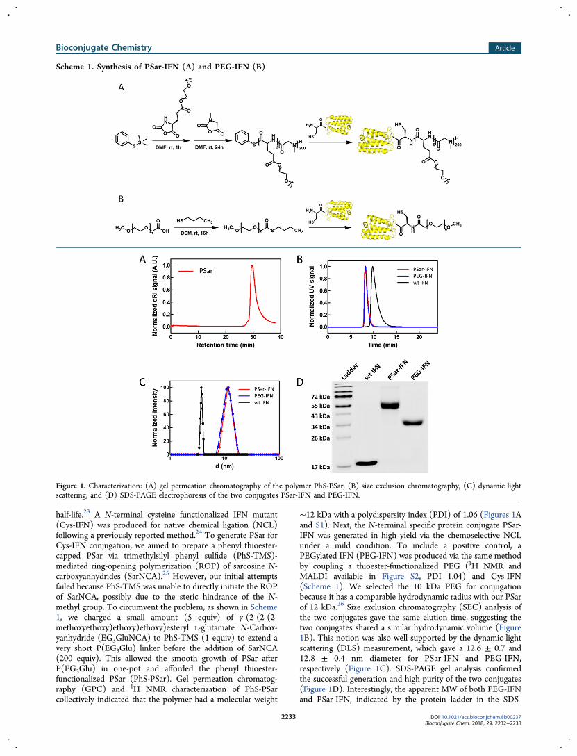
half-life.23 A N-terminal cysteine functionalized IFN mutant(Cys-IFN) was produced for native chemical ligation (NCL)following a previously reported method.24 To generate PSar forCys-IFN conjugation, we aimed to prepare a phenyl thioester-capped PSar via trimethylsilyl phenyl sulfide (PhS-TMS)-mediated ring-opening polymerization (ROP) of sarcosine N-carboxyanhydrides (SarNCA).25 However, our initial attemptsfailed because PhS-TMS was unable to directly initiate the ROPof SarNCA, possibly due to the steric hindrance of the N-methyl group. To circumvent the problem, as shown in Scheme1, we charged a small amount (5 equiv) of γ-(2-(2-(2-methoxyethoxy)ethoxy)ethoxy)esteryl L-glutamate N-Carbox-yanhydride (EG3GluNCA) to PhS-TMS (1 equiv) to extend avery short P(EG3Glu) linker before the addition of SarNCA(200 equiv). This allowed the smooth growth of PSar afterP(EG3Glu) in one-pot and afforded the phenyl thioester-functionalized PSar (PhS-PSar). Gel permeation chromatog-raphy (GPC) and 1H NMR characterization of PhS-PSarcollectively indicated that the polymer had a molecular weight
∼12 kDa with a polydispersity index (PDI) of 1.06 (Figures 1Aand S1). Next, the N-terminal specific protein conjugate PSar-IFN was generated in high yield via the chemoselective NCLunder a mild condition. To include a positive control, aPEGylated IFN (PEG-IFN) was produced via the same methodby coupling a thioester-functionalized PEG (1H NMR andMALDI available in Figure S2, PDI 1.04) and Cys-IFN(Scheme 1). We selected the 10 kDa PEG for conjugationbecause it has a comparable hydrodynamic radius with our PSarof 12 kDa.26 Size exclusion chromatography (SEC) analysis ofthe two conjugates gave the same elution time, suggesting thetwo conjugates shared a similar hydrodynamic volume (Figure1B). This notion was also well supported by the dynamic lightscattering (DLS) measurement, which gave a 12.6 ± 0.7 and12.8 ± 0.4 nm diameter for PSar-IFN and PEG-IFN,respectively (Figure 1C). SDS-PAGE gel analysis confirmedthe successful generation and high purity of the two conjugates(Figure 1D). Interestingly, the apparent MW of both PEG-IFNand PSar-IFN, indicated by the protein ladder in the SDS-
Scheme 1. Synthesis of PSar-IFN (A) and PEG-IFN (B)
Figure 1. Characterization: (A) gel permeation chromatography of the polymer PhS-PSar, (B) size exclusion chromatography, (C) dynamic lightscattering, and (D) SDS-PAGE electrophoresis of the two conjugates PSar-IFN and PEG-IFN.
Bioconjugate Chemistry Article
DOI: 10.1021/acs.bioconjchem.8b00237Bioconjugate Chem. 2018, 29, 2232−2238
2233
PAGE gel, did not agree well with their calculated sizes. Ofnote, such discrepancies were also frequently observed in theSDS-PAGE of other protein−polymer conjugates.27 Apart fromthe MW, many other parameters including the chemicalstructure, polarity, and conformation of the polymers mayalter their electrophoresis behaviors. Here, it was hypothesizedthat the rigidity difference of PEG and PSar (Kuhn length was1.5 nm for PSar while 1.1 nm for PEG)28 may affect themigration of the corresponding conjugates in the PAGE gel.In Vitro Activity. To investigate the protease resistance and
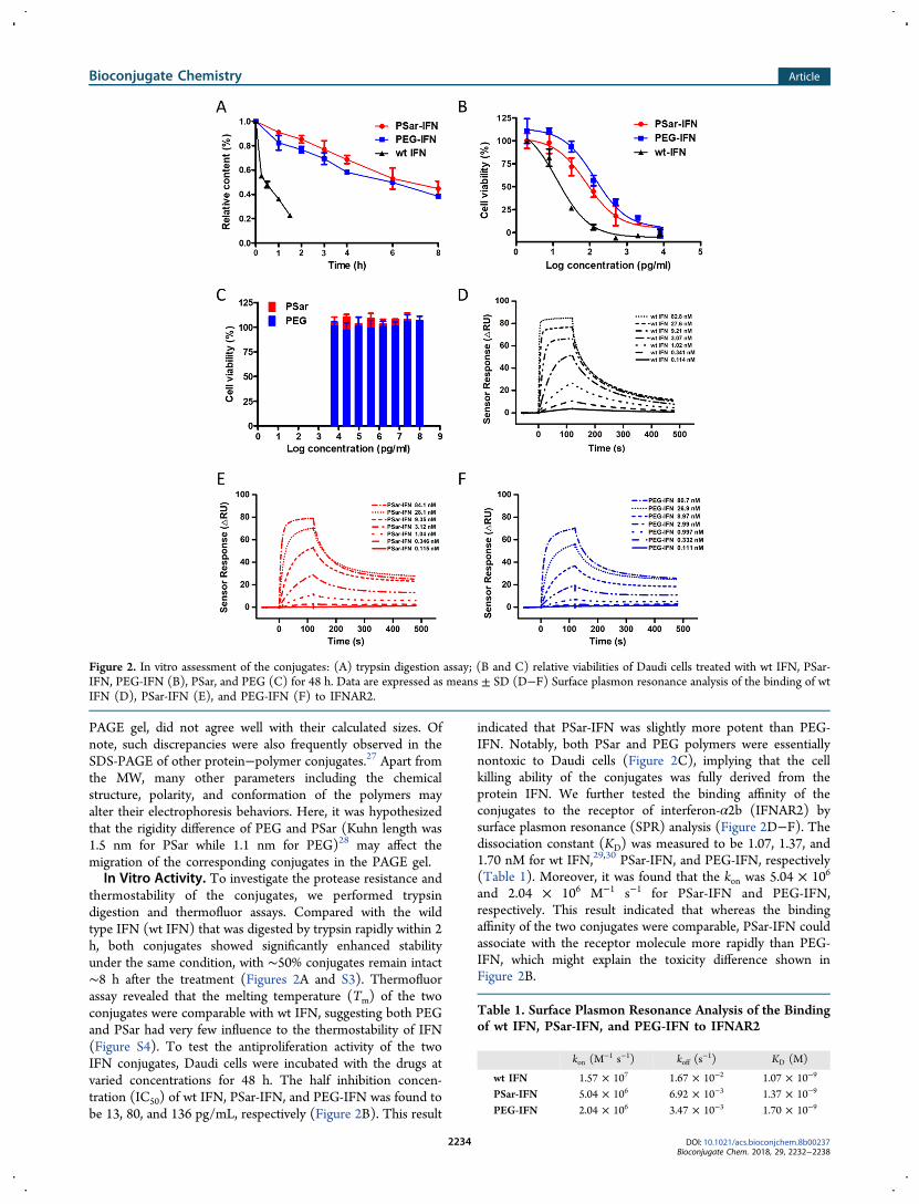
thermostability of the conjugates, we performed trypsindigestion and thermofluor assays. Compared with the wildtype IFN (wt IFN) that was digested by trypsin rapidly within 2h, both conjugates showed significantly enhanced stabilityunder the same condition, with ∼50% conjugates remain intact∼8 h after the treatment (Figures 2A and S3). Thermofluorassay revealed that the melting temperature (Tm) of the twoconjugates were comparable with wt IFN, suggesting both PEGand PSar had very few influence to the thermostability of IFN(Figure S4). To test the antiproliferation activity of the twoIFN conjugates, Daudi cells were incubated with the drugs atvaried concentrations for 48 h. The half inhibition concen-tration (IC50) of wt IFN, PSar-IFN, and PEG-IFN was found tobe 13, 80, and 136 pg/mL, respectively (Figure 2B). This result
indicated that PSar-IFN was slightly more potent than PEG-IFN. Notably, both PSar and PEG polymers were essentiallynontoxic to Daudi cells (Figure 2C), implying that the cellkilling ability of the conjugates was fully derived from theprotein IFN. We further tested the binding affinity of theconjugates to the receptor of interferon-α2b (IFNAR2) bysurface plasmon resonance (SPR) analysis (Figure 2D−F). Thedissociation constant (KD) was measured to be 1.07, 1.37, and1.70 nM for wt IFN,29,30 PSar-IFN, and PEG-IFN, respectively(Table 1). Moreover, it was found that the kon was 5.04 × 10⁶
and 2.04 × 10⁶ M−1 s−1 for PSar-IFN and PEG-IFN,respectively. This result indicated that whereas the bindingaffinity of the two conjugates were comparable, PSar-IFN couldassociate with the receptor molecule more rapidly than PEG-IFN, which might explain the toxicity difference shown inFigure 2B.
Figure 2. In vitro assessment of the conjugates: (A) trypsin digestion assay; (B and C) relative viabilities of Daudi cells treated with wt IFN, PSar-IFN, PEG-IFN (B), PSar, and PEG (C) for 48 h. Data are expressed as means ± SD (D−F) Surface plasmon resonance analysis of the binding of wtIFN (D), PSar-IFN (E), and PEG-IFN (F) to IFNAR2.
Table 1. Surface Plasmon Resonance Analysis of the Bindingof wt IFN, PSar-IFN, and PEG-IFN to IFNAR2
kon (M−1 s−1) koff (s
−1) KD (M)
wt IFN 1.57 × 10⁷ 1.67 × 10⁻² 1.07 × 10⁻⁹
PSar-IFN 5.04 × 10⁶ 6.92 × 10⁻³ 1.37 × 10⁻⁹
PEG-IFN 2.04 × 10⁶ 3.47 × 10⁻³ 1.70 × 10⁻⁹
Bioconjugate Chemistry Article
DOI: 10.1021/acs.bioconjchem.8b00237Bioconjugate Chem. 2018, 29, 2232−2238
2234

Pharmacokinetics, Biodistribution, and Immunoge-nicity. Next, we examined the performance of the conjugates inlive animals. To analyze the pharmacokinetics (PK), wt IFNand the two conjugates were intravenously injected intoSprague−Dawley (SD) rats and the plasma IFN levels atdifferent time points were measured by enzyme-linkedimmunosorbent assay (ELISA). The measured eliminationhalf-life of wt IFN was almost equally extended from ∼0.8 h to∼4.8 and 4.6 h for PSar-IFN and PEG-IFN, respectively(Figure 3A). To explore the biodistribution of differentconjugates, all IFN variants were labeled with Cy5 (FigureS5) and injected intravenously into BALB/c nude mice bearingOVCAR3 tumors. The major organs and tumors were extracted24 h after administration and analyzed with an in vivo imagingequipment. It was found that kidney was the brightest organ forall IFN variants, suggesting that renal clearance was the majorelimination route (Figure 3B). This was not surprisingconsidering the size of the conjugates shown in DLS (Figure1C). Apart from the kidney, liver and tumor also displayedconsiderably higher fluorescence intensities than other organsincluding the heart, spleen, and lung. Compared with PEG-
IFN, interestingly, PSar-IFN was shown to accumulate more inthe tumor (p value <0.05) and have less exposure in the liver (pvalue <0.01) (Figure 3B and C). To evaluate the ability of thepolymers in shielding IFN from immune recognition, PSar-IFNand PEG-IFN were intravenously administrated to immunecompetent SD rats weekly (n = 4). ELISA analysis indicatedthat, upon repetitive administration, PSar-IFN elicited signifi-cantly less anti-IFN IgG in week 3 than those produced byPEG-IFN (Figure 3D). Overall, the results suggested that PSarwas an excellent stealth polymer for protein modification.
Antitumor Efficacy. Finally, we evaluated the antitumorefficacy of the conjugates on OVCAR3 tumor-bearing BALB/cnude mice. On day 0, the mice with a mean tumor volume at 50mm3 were randomly assigned to four groups (n = 5−7). PSar-IFN, PEG-IFN, wt IFN, and PBS were infused at the same IFNdosage every 5 days. On day 25, the mean volume of tumor inthe PBS group and the wt IFN group increased to over 1000mm3. In contrast, both PSar-IFN and PEG-IFN treatmentsshowed conspicuously better tumor growth inhibition. Mostimportantly, mice in the PSar-IFN group were found to haveappreciably smaller tumors than those in the PEG-IFN group
Figure 3. In vivo pharmacological evolution of the conjugates: (A) plasma IFN concentration at varied time points; (B) biodistribution at 24 hestimated by the fluorescence intensity of the extracted tissues; (C) fluorescent images of tumors at 24 h; (D) development of anti-IFN IgG atdifferent times after weekly administration of the conjugates. Data are expressed as means ± SD: *p < 0.05, **p < 0.01, ***p < 0.001.
Figure 4. Antitumor efficacy of the conjugates on OVCAR3 tumor-bearing mice. (A) Tumor growth inhibition curve. Data are expressed as means ±SD, *** p < 0.001. (B) Images of the extracted tumors on day 25.
Bioconjugate Chemistry Article
DOI: 10.1021/acs.bioconjchem.8b00237Bioconjugate Chem. 2018, 29, 2232−2238
2235

(Figure 4). This result clearly supported the argument ofutilizing PSar as a conjugation partner for enhanced therapeuticefficacy of protein. Notably, no significant loss in weight wasobserved in the PSar-IFN group, suggesting the conjugate waswell-tolerated at the current dose (Figure S6). The excellentbiosafety profile of PSar-IFN was also illustrated by thehistological examination of the dissected organ sections, whichshowed no major damage in the heart, liver, spleen, lung, andkidney (Figure S7).
■ CONCLUSIONS
In summary, we synthesized PSar-IFN, an N-terminal specificpolysarcosine−interferon conjugate, and compared its activityin parallel with a PEG-modified IFN. Our results indicated thatthe similarly sized PSar-IFN and PEG-IFN possessedcomparable protease resistance to trypsin digestion. Moreover,the two conjugates also exhibited a similarly improvedcirculation half-life in plasma. Interestingly, PSar-IFN wasslightly more potent than PEG-IFN in inhibiting tumor cellproliferation in vitro, and accumulated more in tumor sites aftersystemic administration than PEG-IFN. Remarkably, PSar-IFNincurred less anti-IFN IgG in plasma after multiple admin-istrations. The superior in vivo antitumor efficacy of PSar-IFNover PEG-IFN was confirmed by the tumor growth inhibitionstudy. Taken together, our results demonstrated for the firsttime that PSar was an outstanding candidate for therapeuticprotein conjugation. Considering the low toxicity, biodegrad-ability, and excellent stealth effect of PSar, the present worksuggested that such polypeptoids had enormous potential formany biomedical applications including protein delivery,colloidal stabilization, and nanomedicine.
■ EXPERIMENTAL SECTION
Materials. Sarcosine was purchased from Aladdin (Shang-hai, China). Phenyl trimethylsilyl sulfide (PhS-TMS) waspurchased from Sigma-Aldrich (St. Louis, USA). Methoxy PEGCarboxyl (mPEG-COOH, MW 10 kDa) was purchased fromJenKem Technology Co., Ltd. (Beijing, China). Butanethiolwas purchased from Energy Chemical (Shanghai, China). Cy5was purchased from ApexBio (Houston, USA). CellTiter-Bluewas purchased from Promega (Madison, USA). IFNAR2 waspurchased from Sino Biological Inc. (Beijing, China). IFN alphahuman matched antibody pair was purchased from eBiosciense(California, USA). Wild type and mutant IFNs, TEV proteasewere produced according to protocols reported elsewhere.31 γ-(2-(2-(2-Methoxyethoxy)ethoxy)ethoxy)esteryl L-glutamate N-Carboxyanhydride (EG3GluNCA)
32 and Sarcosine N-Carbox-yanhydride (SarNCA) were obtained in accordance withestablished methods.33
Instrument. Gel permeation chromatography (GPC)characterization was performed on tandem columns (500 Å,103 Å, 104 Å Phenogel columns, 5 μm, 7.8 × 300 mm2,Phenomenex, Torrance, CA) at 50 °C using DMF with 0.1 MLiBr as the mobile phase. The molecular weight (MW) of PSarwas calculated based on a dn/dc value reported previously.34
NMR spectra were analyzed on ARX400 (Bruker Co.,Germany). Mass spectrum was recorded on a MALDI-TOF(AB Sciex TOF 5800, CA). Protein purification was performedon AKTA pure (GE, USA) using Mono S 5/50 GL column orSuperdex 75 10/300 GL column. Dynamic light scattering(DLS) examinations were recorded on a Nanobrook Omni at25 °C (Brookhaven Instrument Corp. USA). SDS-PAGE gel
was imaged on typhoon FLA 9500 (GE, USA). Proteinconcentration was determined by NanoPhotometer P330(Implen, Germany). The viability assay and ELISA wererecorded on multimode plate reader (PerkinElmer, USA).Thermofluor assay was performed on LightCycler 96 (Roche,Switzerland). Surface plasmon resonance (SPR) analysis wasperformed on Biacore T200 (GE, USA). Biodistributions of theconjugates were imaged by FX Pro (Kodak, USA). Thehistologic sections were imaged on Vectra (Caliper, USA).
Cell Lines and Animals. Human cell line Daudi, obtainedfrom China Infrastructure of Cell Line Resource, was grown inRPMI 1640 with L-glutamine (Cellgro, USA) with 20% FBS(Gibco, USA), 100 IU penicillin, and 100 μg/mL streptomycin(Cellgro, USA). Human ovarian carcinoma OVCAR3 wascultured in RPMI 1640 with L-glutamine supplemented with20% FBS (PAN, Germany), 100 IU penicillin, 100 μg/mLstreptomycin (Cellgro, USA), and 0.01 mg/mL insulin frombovine (SIGMA, USA). Female SD rats and female BALB/cnude mice were purchased from Vital River Laboratories(Beijing, China). All the in vivo experiments were carried outwith the permission of the experimental animal ethicscommittee.
Synthesis of PhS-PSar. In a glovebox, EG3GluNCA (6.9mg, 0.022 mmol, 5.0 equiv) in anhydrous DMF (60 μL) wasadded to PhS-TMS (8.7 μL × 0.5 M, 1.0 equiv) and stirred for1 h at room temperature. SarNCA (100 mg, 0.87 mmol, 200.0equiv) was added to the mixture and stirred for another 48 h atroom temperature. The polymer was precipitated in anhydrousether (100 mL), centrifuged at 4000 g for 5 min, and thesediment was redissolved in ultrapure H2O and further purifiedby a PD 10 desalting column (GE, USA). The collectedpolymer solution was freeze-dried to yield a fluffy powder (52mg, 85%).
Synthesis of the Thioester Capped PEG. mPEG-COOH(200 mg, MW 10 kDa) was dissolved in CH2Cl2 (2 mL), towhich was added DCC (74 mg) and butanethiol (80 μL). Thereaction was stirred overnight at room temperature. Thepolymer was precipitated in anhydrous ether (100 mL),centrifuged at 4000 g for 5 min, and the sediment wasredissolved in ultrapure H2O and further purified by a PD 10desalting column (GE, USA). The collected polymer solutionwas freeze-dried to yield a fluffy powder (165 mg, yield 82%).
General Protocol for the Synthesis of PSar-IFN andPEG-IFN. Cys-IFN was produced by TEV protease digestionand concentrated to ∼5 mg/mL. Typically, Cys-IFN (4.5 mg/mL × 1 mL, 1.0 equiv) in Tris-HCl buffer was added PhS-PSarpowder (6.8 mg, 3.0 equiv). The mixture was incubated atroom temperature for ∼8 h, during which the smell ofphenylthiol could be noticed. Purification was performed bypassing the diluted mixture through a PD 10 desalting columnto remove all small molecular impurities, and followed by aMono S column on FPLC (Buffer A: 50 mM CH3COONa, pH4.5; Buffer B: 50 mM CH3COONa with 2 M NaCl, pH 4.5).Cy5 labeling of the conjugate was performed by following thesame protocol reported elsewhere.24 Endotoxin was removedby passing the conjugate solution through an endotoxin affinitycolumn before animal studies. PEG-IFN was synthesized andpurified by following the same protocol.
Trypsin Digestion Assay. wt IFN, PEG-IFN, and PSar-IFN were diluted to 10 μM in Tris-HCl buffer, mixed withisometric 0.1 μM trypsin, and then incubated at 37 °C. Atselected time points, each sample in triplicate was boiled at 98°C for 10 min to terminate the digestion. All trypsin-digested
Bioconjugate Chemistry Article
DOI: 10.1021/acs.bioconjchem.8b00237Bioconjugate Chem. 2018, 29, 2232−2238
2236

samples at different time points were analyzed on one SDS-PAGE gel together. Degradation of the conjugates at each timepoint was determined by the relative coomassie blue stainingsignal intensity and quantitatively analyzed through typhoon.Thermofluor Assay. The thermostability of all samples
were performed on LightCycler 96 (Roche, Switzerland).Briefly, 5 μM conjugates and 20 × Sypro orange protein stain(both are final concentrations) in PBS were added into anopaque white 96-well plate with temperature varied from 37 to98 °C and detected the change of fluorescence intensities. Tmwas analyzed by the LightCycler 96 SW 1.1.Surface Plasmon Resonance (SPR) Analysis. Binding
affinities to receptor were performed on Biacore T200 usingCM5 sensor at 25 °C. The IFNAR2 was diluted into 10 mMNaOAc buffer pH 5.0 at a concentration of 50 ug/mL andcovalently attached to the surface of the sensor via NHS-aminechemistry. Analyses of the interactions between the IFNvariants with IFNAR2 were performed in HBS-EP+ buffer (10mM HEPES, 150 mM NaCl, 3 mM EDTA, 0.05% v/vsurfactant P20, pH 7.4) at a flow rate of 30 μL/min. Sevendifferent concentrations were measured for each sample.Between measurements, 10 mM glycine-HCl (pH 3.0) wasused for chip regeneration. The results were analyzed by BIAevaluation software and fitted with one to one kinetic model.Cytotoxicity Assay. PhS-PSar, PEG-thioester or wt IFN,
PSar-IFN, PEG-IFN were added into a black 96-well plate atgradient concentrations which seeded 5000 Daudi cells per welland incubated for 48 h (n = 3). The relative viabilities weredetected by CellTiter-Blue (Promega, USA).Pharmacokinetics Assay. Female SD rats weighing ∼250
g were randomly assigned to three groups (n = 2 or 3). wt IFN,PSar-IFN, or PEG-IFN was intravenously injected to the rats ata 0.2 mg IFN/kg dose. At predetermined time points, theplasma were acquired from orbit followed by centrifugation.The concentration of IFN in each plasma sample was evaluatedby ELISA and data were processed by GraphPad Prism 5.0.In Vivo Biodistribution. OVCAR3 cells (1.0 × 10⁷)
suspended in RPMI 1640 were mixed with isometric matrigeland subcutaneously inoculated into 6-weeks-old BALB/c nudemice. The mice were randomly assigned to three groups (n = 2)while the tumors grew to ∼250 mm3 and were injected withCy5-marked PSar-IFN, PEG-IFN, or wt IFN at 20 μg IFN/mouse via the tail vein. The mice were anaesthetized by chloralhydrate to extract major organs and tumors 24 h after the druginfusion. The organs and tumors were recorded on FX Pro(Kodak, USA) and the biodistribution was assessed based onthe fluorescent intensity.Immunogenicity Assay. Female SD rats were randomly
assigned to two groups (n = 4) on day 1 and infused with PSar-IFN or PEG-IFN (100 ng IFN each) through tail vein underanesthetic conditions. The same administration was repeatedon days 8 and 15. Before each injection, blood were collectedon day 1, 8, 15, and 22. The anti-IFN IgG levels in the serawere then evaluated by ELISA.In Vivo Antitumor Efficacy. OVCAR3 cells (1.0 × 10⁷)
suspended in RPMI 1640 were mixed with isometric matrigeland subcutaneously inoculated into 6-week-old BALB/c nudemice. The mice were randomly assigned to four groups (n = 5−7) while the tumors grew to ∼50 mm3, and received PBS, wtIFN, PEG-IFN, or PSar-IFN treatment at a 10 μg IFN/mousedose via the tail vein route once every 5 days. The volume oftumor was acquired through the following formula: V = L ×
W2/2. Experimental results were processed using GraphPadPrism 5.0.
Histopathology Evaluation. On day 25, the mice wereexecuted to extract the tumors and major organs such as heart,liver, spleen, lung, and kidney for cut into slices and stainedwith hematoxylin and eosin (H&E), which were then imagedon Vectra.
■ ASSOCIATED CONTENT*S Supporting InformationThe Supporting Information is available free of charge on theACS Publications website at DOI: 10.1021/acs.bioconj-chem.8b00237.
1H NMR, MALDI, SDS-PAGE, thermal denaturationcurves, relative weight of mice, histopathology evaluation(PDF)
■ AUTHOR INFORMATIONCorresponding Author*E-mail: [email protected] Lu: 0000-0003-2180-3091NotesThe authors declare no competing financial interest.
■ ACKNOWLEDGMENTSThis work was financially supported by National Key Researchand Development Program of China (2016YFA0201400). Wethank the grants from National Natural Science Foundation ofChina (21722401, 21474004, and 21434008). H.L. thanks thestartup funding from Youth Thousand-Talents Program ofChina. We thank the protein interaction facility of School ofLife Sciences, Peking University, and Dr. Hui Li for help withSPR analysis.
■ REFERENCES(1) Ríhova, B., Jelínkova, M., Strohalm, J., Subr, V., Plocova, D.,Hovorka, O., Novak, M., Plundrova, D., Germano, Y., and Ulbrich, K.(2000) Polymeric drugs based on conjugates of synthetic and naturalmacromolecules.: II. Anti-cancer activity of antibody or (Fab′)2-targeted conjugates and combined therapy with immunomodulators. J.Controlled Release 64, 241−261.(2) Pasut, G., and Veronese, F. M. (2012) State of the art inPEGylation: the great versatility achieved after forty years of research.J. Controlled Release 161, 461−472.(3) Hu, J., Wang, G., Zhao, W., and Gao, W. (2016) In situ growth ofa C-terminal interferon-alpha conjugate of a phospholipid polymerthat outperforms PEGASYS in cancer therapy. J. Controlled Release237, 71−77.(4) Pelegri-O’Day, E. M., Lin, E.-W., and Maynard, H. D. (2014)Therapeutic Protein−Polymer Conjugates: Advancing Beyond PEGy-lation. J. Am. Chem. Soc. 136, 14323−14332.(5) Mero, A., Fang, Z., Pasut, G., Veronese, F. M., and Viegas, T. X.(2012) Selective conjugation of poly(2-ethyl 2-oxazoline) togranulocyte colony stimulating factor. J. Controlled Release 159, 353−361.(6) Thomas, A., Muller, S. S., and Frey, H. (2014) BeyondPoly(ethylene glycol): Linear Polyglycerol as a MultifunctionalPolyether for Biomedical and Pharmaceutical Applications. Biomacro-molecules 15, 1935−1954.(7) Keefe, A. J., and Jiang, S. (2012) Poly(zwitterionic)proteinconjugates offer increased stability without sacrificing binding affinityor bioactivity. Nat. Chem. 4, 59−63.
Bioconjugate Chemistry Article
DOI: 10.1021/acs.bioconjchem.8b00237Bioconjugate Chem. 2018, 29, 2232−2238
2237

(8) Hu, J., Wang, G., Zhao, W., Liu, X., Zhang, L., and Gao, W.(2016) Site-specific in situ growth of an interferon-polymer conjugatethat outperforms PEGASYS in cancer therapy. Biomaterials 96, 84−92.(9) Nguyen, T. H., Kim, S.-H., Decker, C. G., Wong, D. Y., Loo, J. A.,and Maynard, H. D. (2013) A heparin-mimicking polymer conjugatestabilizes basic fibroblast growth factor. Nat. Chem. 5, 221−227.(10) Lu, H., Wang, J., Song, Z., Yin, L., Zhang, Y., Tang, H., Tu, C.,Lin, Y., and Cheng, J. (2014) Recent advances in amino acid N-carboxyanhydrides and synthetic polypeptides: chemistry, self-assembly and biological applications. Chem. Commun. 50, 139−155.(11) Lau, K. H. A., Ren, C., Sileika, T. S., Park, S. H., Szleifer, I., andMessersmith, P. B. (2012) Surface-Grafted Polysarcosine as a PeptoidAntifouling Polymer Brush. Langmuir 28, 16099−16107.(12) Chan, B. A., Xuan, S., Li, A., Simpson, J. M., Sternhagen, G. L.,Yu, T., Darvish, O. A., Jiang, N., and Zhang, D. (2018) Polypeptoidpolymers: Synthesis, characterization, and properties. Biopolymers 109,e23070.(13) Birke, A., Ling, J., and Barz, M. (2018) Polysarcosine-containingcopolymers: Synthesis, characterization, self-assembly, and applica-tions. Prog. Polym. Sci. 81, 163−208.(14) Sela, M., and Arnon, R. (1960) Studies on the chemical basis ofthe antigenicity of proteins. 3. The role of rigidity in the antigenicity ofpolypeptidyl gelatins. Biochem. J. 77, 394−399.(15) Tao, X., Deng, C., and Ling, J. (2014) PEG-Amine-InitiatedPolymerization of Sarcosine N-Thiocarboxyanhydrides Toward NovelDouble-Hydrophilic PEG-b-Polysarcosine Diblock Copolymers. Mac-romol. Rapid Commun. 35, 875−881.(16) Zhu, H., Chen, Y., Yan, F.-J., Chen, J., Tao, X.-F., Ling, J., Yang,B., He, Q.-J., and Mao, Z.-W. (2017) Polysarcosine brush stabilizedgold nanorods for in vivo near-infrared photothermal tumor therapy.Acta Biomater. 50, 534−545.(17) Fokina, A., Klinker, K., Braun, L., Jeong, B. G., Bae, W. K., Barz,M., and Zentel, R. (2016) Multidentate Polysarcosine-Based Ligandsfor Water-Soluble Quantum Dots. Macromolecules 49, 3663−3671.(18) Chen, Y., Xu, Z., Zhu, D., Tao, X., Gao, Y., Zhu, H., Mao, Z.,and Ling, J. (2016) Gold nanoparticles coated with polysarcosinebrushes to enhance their colloidal stability and circulation time in vivo.J. Colloid Interface Sci. 483, 201−210.(19) Sano, K., Ohashi, M., Kanazaki, K., Makino, A., Ding, N.,Deguchi, J., Kanada, Y., Ono, M., and Saji, H. (2017) IndocyanineGreen -Labeled Polysarcosine for in Vivo Photoacoustic TumorImaging. Bioconjugate Chem. 28, 1024−1030.(20) Weber, B., Kappel, C., Scherer, M., Helm, M., Bros, M., Grabbe,S., and Barz, M. (2017) PeptoSomes for Vaccination: CombiningAntigen and Adjuvant in Polypept(o)ide-Based Polymersomes.Macromol. Biosci. 17, 1700061.(21) Hsiao, L. W., Lai, Y. D., Lai, J. T., Hsu, C. C., Wang, N. Y.,Wang, S. S., and Jan, J. S. (2017) Cross-linked polypeptide-based gelparticles by emulsion for efficient protein encapsulation. Polymer 115,261−272.(22) Klinker, K., Holm, R., Heller, P., and Barz, M. (2015) Evaluatingchemical ligation techniques for the synthesis of block copolypeptides,polypeptoids and block copolypept(o)ides: a comparative study.Polym. Chem. 6, 4612−4623.(23) Chawla-Sarkar, M., Lindner, D. J., Liu, Y.-F., Williams, B. R.,Sen, G. C., Silverman, R. H., and Borden, E. C. (2003) Apoptosis andinterferons: Role of interferon-stimulated genes as mediators ofapoptosis. Apoptosis 8, 237−249.(24) Hou, Y. Q., Yuan, J. S., Zhou, Y., Yu, J., and Lu, H. (2016) AConcise Approach to Site-Specific Topological Protein-Poly(aminoacid) Conjugates Enabled by in Situ-Generated Functionalities. J. Am.Chem. Soc. 138, 10995−11000.(25) Yuan, J., Sun, Y., Wang, J., and Lu, H. (2016) PhenylTrimethylsilyl Sulfide-Mediated Controlled Ring-Opening Polymer-ization of α-Amino Acid N-Carboxyanhydrides. Biomacromolecules 17,891−896.(26) Huesmann, D., Sevenich, A., Weber, B., and Barz, M. (2015) Ahead-to-head comparison of poly(sarcosine) and poly(ethylene glycol)in peptidic, amphiphilic block copolymers. Polymer 67, 240−248.
(27) Yun, Q., Xing, W., Ma, G., and Su, Z. (2006) Preparation andcharacterization of mono-PEGylated consensus interferon by a novelpolyethylene glycol derivative. J. Chem. Technol. Biotechnol. 81, 776−781.(28) Weber, B., Birke, A., Fischer, K., Schmidt, M., and Barz, M.(2018) Solution Properties of Polysarcosine: From Absolute andRelative Molar Mass Determinations to Complement Activation.Macromolecules 51, 2653−2661.(29) Zhang, B., Xu, H., Chen, J., Zheng, Y., Wu, Y., Si, L., Wu, L.,Zhang, C., Xia, G., Zhang, L., and Zhou, D. (2015) Development ofnext generation of therapeutic IFN-α2b via genetic code expansion.Acta Biomater. 19, 100−111.(30) Schmeisser, H., Gorshkova, I., Brown, P. H., Kontsek, P.,Schuck, P., and Zoon, K. C. (2007) Two Interferons Alpha InfluenceEach Other during Their Interaction with the Extracellular Domain ofHuman Type Interferon Receptor Subunit 2. Biochemistry 46, 14638−14649.(31) Hou, Y., Zhou, Y., Wang, H., Wang, R., Yuan, J., Hu, Y., Sheng,K., Feng, J., Yang, S., and Lu, H. (2018) Macrocyclization ofInterferon−Poly(α-amino acid) Conjugates Significantly Improves theTumor Retention, Penetration, and Antitumor Efficacy. J. Am. Chem.Soc. 140, 1170−1178.(32) Chen, C., Wang, Z., and Li, Z. (2011) ThermoresponsivePolypeptides from Pegylated Poly-l-glutamates. Biomacromolecules 12,2859−2863.(33) Fetsch, C., Grossmann, A., Holz, L., Nawroth, J. F., andLuxenhofer, R. (2011) Polypeptoids from N-Substituted Glycine N-Carboxyanhydrides: Hydrophilic, Hydrophobic, and AmphiphilicPolymers with Poisson Distribution. Macromolecules 44, 6746−6758.(34) Cui, S., Pan, X., Gebru, H., Wang, X., Liu, J., Liu, J., Li, Z., andGuo, K. (2017) Amphiphilic star-shaped poly(sarcosine)-block-poly(ε-caprolactone) diblock copolymers: one-pot synthesis, characterization,and solution properties. J. Mater. Chem. B 5, 679−690.
Bioconjugate Chemistry Article
DOI: 10.1021/acs.bioconjchem.8b00237Bioconjugate Chem. 2018, 29, 2232−2238
2238
Related Documents