Polysaccharides from green and black teas and their protective effect against murine sepsis Camila T. Scoparo a , Lauro M. de Souza a, ⁎, Yanna D. Rattmann a, b , Nessana Dartora a , Simone M.M. Paiva a , Guilherme L. Sassaki a , Philip A.J. Gorin a , Marcello Iacomini a, ⁎ a Departamento de Bioquímica e Biologia Molecular, Universidade Federal do Paraná, CP 19046, CEP 81531-980, Curitiba-PR, Brazil b Departamento de Saúde Comunitária, Universidade Federal do Paraná, Rua Padre Camargo, 280, CEP: 80060-240, Alto da Glória, Curitiba-PR, Brazil abstract article info Article history: Received 23 August 2012 Received in revised form 1 November 2012 Accepted 2 November 2012 Keywords: Green tea Black tea Rhamnogalacturonan Cecal ligation and puncture Murine sepsis Polysaccharides from green and black teas were obtained via hot aqueous extraction, and after fractionation, chemical analysis showed that they consisted mainly of a rhamnogalacturonan containing a long sequence of →4)-6-O-Me-α-D-GalpA-(1→, interrupted by α-L-Rhap residues, which were substituted by type II arabinogalactan chains. The polysaccharides were similar, except that black tea contained lesser galacturonic acid residues (35%), when compared with green tea (65%). Starch was present in both teas. The polysaccha- rides were tested, via oral administration in mice against induced-polymicrobial sepsis, at doses of 30, 50 and 100 mg/kg. Green and black tea polysaccharides were capable of reducing mortality rate by 40% and 25% re- spectively, compared to sham-controls. The influx of neutrophils was also affected, decreasing its accumula- tion in lungs and tissue injury. The difference between the green and black tea polysaccharides seems to result from the processing that the leaves have undergone and the different abilities to prevent mice death can be related to the different uronic acid contents. © 2012 Elsevier Ltd. All rights reserved. 1. Introduction After water, teas from Camellia sinensis L. (mainly green and black teas) are considered to be the most consumed beverage worldwide. The main difference among the teas is based on the process of auto-oxidation catalyzed by the enzymes polyphenol oxidase (PPO) and peroxidase. Basically, oxidation is a process to which the leaves of C. sinensis interact with oxygen, after being processed (e.g. being minced, crushed or dried) that results in darkening. Those from C. sinensis are thus classified as white, green, oolong, black and Pu'erh teas which undergo unfermented, semi-fermented, and fermented processes (Engelhardt, 2010; Ho, Lin, & Shahidi, 2008). Products of secondary metabolism, especially flavonoids from C. sinensis, have been widely studied for their chemical composition and biological effects (Scoparo et al., 2012; Sharangi, 2009), although the polysaccharides have received less attention. However, in infu- sions, many compound classes are extracted from the leaves, including polysaccharides which are then ingested. It is known that many plants used in folk medicine contain polysaccharides with a recognized variety of properties, including immunological, anti-radiation, anticoagulation, anti-cancer, anti-HIV, hypoglycemic and gastric protection activities (Cipriani et al., 2006; Wang, Wang, Li, & Zhao, 2001; Xie & Nie, 2006; Zhou, Xie, & Fu, 2001). Recent studies have shown that the structures of polysaccharides are closely related to their biological activities (Cooper, Morré, & Morré, 2005; Harold & Graham, 1992), so elucidation of their structures is fundamental for understanding structure–activity relationships. Some structural studies have been carried out on tea polysaccharides, being described a polysaccharide composed of rhamnose, glucose, galactose, arabinose and xylose (Zhou, Xie, & Nie, 2004). Also, other poly- saccharides were identified as neutral (1 →4)-β-galactan and pectin-type polysaccharide formed by blocks of (1 →4)-α-D-galactopyranosyluronic acids interconnected by (1 →2)-linked rhamnose residues (Wang, Wei, & Jin, 2009a, 2009b). Several pharmacological properties have been attributed to poly- saccharides from different sources, although few investigations have correlated their structure with sepsis treatment. Sepsis is a consider- able health problem and a leading cause of morbidity and mortality in many intensive care units. It represents a state of overproduction of pro-inflammatory mediators which frequently occurs after various noxious injuries, especially bacterial infection arising from abdominal surgery, appendicitis, perforated ulcers, or an ischemic bowel (Angus, Linde-Zwirble, Lidicker, & Clermont, 2001; Cohen, 2002). We now Food Research International 53 (2013) 780–785 Abbreviations: AcOH, acetic acid; Ac 2 O, acetic anhydride; CLP, cecal ligation and punc- ture; EtOH, ethanol; CHCL 3 , chloroform; HPSEC, high-performance size-exclusion chro- matography; GC–MS, gas chromatography–mass spectrometry; MPO, myeloperoxidase; TMSP-d 4 , 2,2,3,3-tetradeuterium-3-trimethylilsilylpropionate; GT, green tea; BT, black tea; GSP, green soluble polysaccharide; BSP, black soluble polysaccharide. ⁎ Corresponding authors. Tel.: +55 41 3361 1577; fax: +55 41 3266 2042. E-mail addresses: [email protected] (L.M. de Souza), [email protected] (M. Iacomini). 0963-9969/$ – see front matter © 2012 Elsevier Ltd. All rights reserved. http://dx.doi.org/10.1016/j.foodres.2012.11.006 Contents lists available at ScienceDirect Food Research International journal homepage: www.elsevier.com/locate/foodres

Welcome message from author
This document is posted to help you gain knowledge. Please leave a comment to let me know what you think about it! Share it to your friends and learn new things together.
Transcript

Food Research International 53 (2013) 780–785
Contents lists available at ScienceDirect
Food Research International
j ourna l homepage: www.e lsev ie r .com/ locate / foodres
Polysaccharides from green and black teas and their protective effectagainst murine sepsis
Camila T. Scoparo a, Lauro M. de Souza a,⁎, Yanna D. Rattmann a,b, Nessana Dartora a, Simone M.M. Paiva a,Guilherme L. Sassaki a, Philip A.J. Gorin a, Marcello Iacomini a,⁎a Departamento de Bioquímica e Biologia Molecular, Universidade Federal do Paraná, CP 19046, CEP 81531-980, Curitiba-PR, Brazilb Departamento de Saúde Comunitária, Universidade Federal do Paraná, Rua Padre Camargo, 280, CEP: 80060-240, Alto da Glória, Curitiba-PR, Brazil
Abbreviations: AcOH, acetic acid; Ac2O, acetic anhydriture; EtOH, ethanol; CHCL3, chloroform; HPSEC, high-pematography; GC–MS, gas chromatography–mass spectroTMSP-d4, 2,2,3,3-tetradeuterium-3-trimethylilsilylpropiotea; GSP, green soluble polysaccharide; BSP, black soluble⁎ Corresponding authors. Tel.: +55 41 3361 1577; fa
E-mail addresses: [email protected] (L.M. de(M. Iacomini).
0963-9969/$ – see front matter © 2012 Elsevier Ltd. Allhttp://dx.doi.org/10.1016/j.foodres.2012.11.006
a b s t r a c t
a r t i c l e i n f oArticle history:Received 23 August 2012Received in revised form 1 November 2012Accepted 2 November 2012
Keywords:Green teaBlack teaRhamnogalacturonanCecal ligation and punctureMurine sepsis
Polysaccharides from green and black teas were obtained via hot aqueous extraction, and after fractionation,chemical analysis showed that they consisted mainly of a rhamnogalacturonan containing a long sequence of→4)-6-O-Me-α-D-GalpA-(1→, interrupted by α-L-Rhap residues, which were substituted by type IIarabinogalactan chains. The polysaccharides were similar, except that black tea contained lesser galacturonicacid residues (35%), when compared with green tea (65%). Starch was present in both teas. The polysaccha-rides were tested, via oral administration in mice against induced-polymicrobial sepsis, at doses of 30, 50 and100 mg/kg. Green and black tea polysaccharides were capable of reducing mortality rate by 40% and 25% re-spectively, compared to sham-controls. The influx of neutrophils was also affected, decreasing its accumula-tion in lungs and tissue injury. The difference between the green and black tea polysaccharides seems toresult from the processing that the leaves have undergone and the different abilities to prevent mice deathcan be related to the different uronic acid contents.
© 2012 Elsevier Ltd. All rights reserved.
1. Introduction
After water, teas from Camellia sinensis L. (mainly green and blackteas) are considered to be the most consumed beverage worldwide.The main difference among the teas is based on the process ofauto-oxidation catalyzed by the enzymes polyphenol oxidase (PPO)and peroxidase. Basically, oxidation is a process to which the leavesof C. sinensis interact with oxygen, after being processed (e.g. beingminced, crushed or dried) that results in darkening. Those fromC. sinensis are thus classified as white, green, oolong, black and Pu'erhteas which undergo unfermented, semi-fermented, and fermentedprocesses (Engelhardt, 2010; Ho, Lin, & Shahidi, 2008).
Products of secondary metabolism, especially flavonoids fromC. sinensis, have been widely studied for their chemical compositionand biological effects (Scoparo et al., 2012; Sharangi, 2009), althoughthe polysaccharides have received less attention. However, in infu-sions, many compound classes are extracted from the leaves, including
de; CLP, cecal ligation and punc-rformance size-exclusion chro-metry; MPO, myeloperoxidase;nate; GT, green tea; BT, blackpolysaccharide.x: +55 41 3266 2042.Souza), [email protected]
rights reserved.
polysaccharides which are then ingested. It is known that many plantsused in folkmedicine contain polysaccharideswith a recognized varietyof properties, including immunological, anti-radiation, anticoagulation,anti-cancer, anti-HIV, hypoglycemic and gastric protection activities(Cipriani et al., 2006; Wang, Wang, Li, & Zhao, 2001; Xie & Nie, 2006;Zhou, Xie, & Fu, 2001).
Recent studies have shown that the structures of polysaccharidesare closely related to their biological activities (Cooper, Morré, &Morré, 2005; Harold & Graham, 1992), so elucidation of their structuresis fundamental for understanding structure–activity relationships.Some structural studies have been carried out on tea polysaccharides,being described a polysaccharide composed of rhamnose, glucose,galactose, arabinose and xylose (Zhou, Xie, & Nie, 2004). Also, other poly-saccharideswere identified as neutral (1→4)-β-galactan andpectin-typepolysaccharide formed by blocks of (1→4)-α-D-galactopyranosyluronicacids interconnected by (1→2)-linked rhamnose residues (Wang, Wei,& Jin, 2009a, 2009b).
Several pharmacological properties have been attributed to poly-saccharides from different sources, although few investigations havecorrelated their structure with sepsis treatment. Sepsis is a consider-able health problem and a leading cause of morbidity and mortality inmany intensive care units. It represents a state of overproduction ofpro-inflammatory mediators which frequently occurs after variousnoxious injuries, especially bacterial infection arising from abdominalsurgery, appendicitis, perforated ulcers, or an ischemic bowel (Angus,Linde-Zwirble, Lidicker, & Clermont, 2001; Cohen, 2002). We now

781C.T. Scoparo et al. / Food Research International 53 (2013) 780–785
evaluate differences in the chemical structure of the polysaccharidesisolated from both green and black teas and their protection againstsepsis, in a murine model of cecal ligation and puncture (CLP), andtheir effects on neutrophil migration.
2. Experimental methods
2.1. Plant material
Green and black teas were purchased in a local market (Curitiba,State of Paraná, Brazil), as commercial processed leaves.
2.2. Polysaccharide extraction and fractionation
Green and black teas (50 g of each) were submitted to aqueousextraction (100 °C, 500 mL, ×3). The extracts were combined andevaporated to 250 mL. High molecular weight components, mainlypolysaccharides, were precipitated by addition of cold EtOH (3 vol.),and centrifuged (8,000 rpm at 4 °C, 20 min). The sediment wasthen dissolved in H2O and dialyzed, in 12,000 Da cutoff membrane,against tap water for 24 h to remove the remaining low-molecularweight compounds, giving rise to crude polysaccharide fractions.These were frozen and then allowed to thaw at room temperature(Gorin & Iacomini, 1984), resulting in green (GSP) and black solublepolysaccharide (BSP) fractions and insoluble fractions which wereseparated by centrifugation as described above. The insoluble frac-tions were not analyzed.
2.3. Monosaccharide analysis
GSP and BSP (1 mg) were each hydrolyzed with 0.5 mL of 2 M TFAat 100 °C for 8 h, then the solution was evaporated, and the residuewas dissolved in water (1 mL). The resulting monosaccharides weresubmitted to thin layer chromatography (TLC) on silica gel-60(Merck), the eluant being ethyl acetate:acetic acid:n-propanol:water(4:2:2:1 v/v), followed by development with orcinol-sulfuric acid at100 °C (Skipski, 1975). The monosaccharides were reduced with2 mg NaBH4 yielding alditols, which were acetylated in Ac2O-pyridine(1:1 v/v, 0.5 mL) at room temperature for 12 h (Wolfrom &Thompson, 1963a, 1963b). The resulting alditol acetateswere extractedwith CHCl3, and analyzed by GC–MS (Varian, Saturn 2000R-3800 gaschromatograph coupled to a Varian Ion-Trap 2000Rmass spectrometer),using a DB-225-MS column (30 m×0.25 mm×0.25 μm) programmedfrom 50 to 220 °C at 40 °C/min, with He as carrier gas. Monosaccharideswere identified by their relative retention times and typical electronionization spectra (EI — 70 eV). The uronic acid contents of the solublefractions were determined using the colorimetric m-hydroxybiphenylmethod of Filisetti-Cozzi and Carpita (1991).
GSP and BSP (10 mg) were each submitted to carboxy-reductionthrough the carbodiimide method (Taylor & Conrad, 1972), usingNaBH4 as the reducing agent, giving the reduced products (GSP-CR,BSP-CR), and releasing their neutral equivalent monosaccharide.
2.4. Methylation analysis
The fractions GSP, BSP, GSP-CR and BSP-CR (5 mg) were eachper-O-methylated in DMSO (0.5 mL), powdered NaOH (30 mg) andiodomethane (0.5 mL), adapted from the method of Ciucanu andKerek (1984). The suspension was neutralized with acetic acid anddialyzed (12,000 Da cutoff) to eliminate reagents. The samples werelyophilized and alkylated polysaccharides were then hydrolyzedwith 45% aq. v/v formic acid (1 mL) for 12 h at 100 °C, filtered andevaporated to dryness. The partially O-methylated monosaccharideswere reduced with NaBD4 and acetylated as described above, givingrise to a mixture of partially O-methylated alditol acetates, whichwere analyzed by GC–MS similar to what was described above, except
that the temperature program was 50 to 210 °C in 30 min. Identifica-tion of the partially O-methylated alditol acetates was based on them/zspectra (EI — 70 eV), and by comparison with the standard library(Sassaki, Gorin, Souza, & Czelusniak, 2005), the results being expressedas a relative percentage of each component.
2.5. Nuclear magnetic resonance (NMR) analysis
Mono- (13C, 1H and DEPT-135) and bi-dimensional 1H/13C (HSQC)NMR spectra were obtained by employing a Bruker Avance III400 MHz spectrometer. Samples were dissolved in D2O at ~40 mgin 400 μL and analysis was carried out at 70 °C. The chemical shiftswere expressed in δ (ppm), standardized with TMSP-d4 (sodium2,2,3,3-tetradeuterio-3-trimethylsilyl-propionate) δ=0 for 13C and 1H.
2.6. Animals
Male albino Swiss mice (3 months old, weighing 25–30 g), fromthe University of Paraná colony, were used for biological tests. Theywere maintained under standard laboratory conditions, with a con-stant 12 h light/dark cycle and controlled temperature (22±2 °C),and standard pellet food (Nuvital®, Curitiba/PR, Brazil) and waterwere available ad libitum. All experimental procedures were previouslyapproved by the Institutional Ethics Committee of the university(authorization number 430).
2.7. Sepsis induction by cecal ligation and puncture (CLP)
Mice were randomly grouped into five clusters of 10 mice:sham-operation, CLP plus vehicle (water p.o.), and CLP plus GSP orBSP (30, 50 and 100 mg/kg, p.o.). According to the body weight(~30 g), they were administered with 50 μL of each tea polysaccha-ride solution. Ketamine (80 mg/kg) and xylazine (20 mg/kg) wereinjected intraperitoneally to anesthetize the mice prior to surgicalprocedures. Polymicrobial sepsis was induced by CLP as previouslydescribed (Rittirsch, Huber-Lang, Flierl, & Ward, 2009). A midline in-cision of ~1.5 cm was performed on the abdomen and the cecum wascarefully exposed and 50% of the distal portion was ligated. Thececumwas then punctured three times with a sterile 16-gauge needleand squeezed to extrude fecal material from the wounds. The cecumwas replaced and the abdomen was stitched surgically. Sham-controlanimals were treated identically, but no cecal ligation or puncturewas carried out. Each mouse received subcutaneous sterile saline in-jection (1 mL) for fluid resuscitation after surgery. The mice werethen kept on a heating pad (35 °C) until they recovered from the an-esthesia. Food and water, ad libitum, were provided throughout theexperiment. The survival rate was monitored for 7 days, 12 h each.During this period, vehicle (water) and treatments were orally ad-ministered daily.
In another set of experiment, 1 h prior to the surgery, mice wereorally treated with vehicle, GSP, or BSP (30 and 50 mg/kg, p.o.).After 6 h post-operation, mice were sacrificed. Their lungs were col-lected and frozen for further use to determine the myeloperoxidase(MPO) activity.
2.8. Lung MPO activity
MPO activity was measured in order to determine neutrophilinflux, according to established protocols (Bradley, Priebat,Christensen, & Rothstein, 1982). Briefly, the lung tissue was homoge-nized in 0.5 mL of 50 mM potassium buffer pH 6.0 with 0.5%hexadecyltrimethylammonium bromide, sonicated on ice, and thencentrifuged at 14,000 rpm for 15 min at 4 °C. Supernatants werethen assayed at a 1:20 dilution in reaction buffer (9.6 mM 3,3,5,5-tetramethylbenzine, 150 nM H2O2 in 50 mM potassium phosphate

782 C.T. Scoparo et al. / Food Research International 53 (2013) 780–785
buffer), and read at 620 nm. Results were expressed as change in op-tical density per milligram of protein (measured by Bradford assay).
2.9. Statistical analysis
Data were expressed as means±SEM of, at least, 10 mice examinedin each group. Statistical error was determined by one-way ANOVA;the post hoc test was Bonferroni's. Calculations were performed withGraphpad Prism5.0, and the p valuesb0.05were considered significant.
3. Results and discussion
3.1. Structural characterization of green and black tea polysaccharides
Currently, polysaccharides have received special attention due totheir pharmacological activities, so that structural analysis is requiredfor understanding their actions. Thus, like in hot infusions, green andblack teas were extracted with water at 100 °C. Their polysaccharideswere obtained by precipitation with excess of ethanol, being recoveredby centrifugation and dialysis, yielding crude polysaccharide fractions,for the green (12%) and black teas (10%), which were freeze-dried,then fractionated by a freeze–thawing procedure, resulting in coldwater-soluble fractions, for green (GSP) and black tea (BSP), at 83%and 64% respectively, and the insoluble fractions being discarded.
The monosaccharide composition of GSP and BSP was obtained byGC–MS analysis and a colorimetric method was employed for theuronic acids, resulting in galacturonic acid (65%), arabinose (19%),galactose (7%), glucose (7%) and rhamnose (2%) for green tea (GSP);and galacturonic acid (35%), arabinose (30%), galactose (16%), rham-nose (3%) and glucose (16%) for black tea (BSP). The galacturonicacid content was confirmed by TLC (data not shown) and carboxy-reduction and further analysis of neutral monosaccharide by GC–MS.The polysaccharide from green tea had a greater concentration ofgalacturonic acid when compared to black tea which, in contrast, hadhigher amounts of arabinose, galactose and rhamnose, suggestingthat the processing that the leaves undergo in black tea preparationmay be interfering with its monosaccharide composition.
3.1.1. NMR analysisThe 1H/13C HSQC spectra of GSP and BSP (Fig. 1A and B, respec-
tively) suggest the presence of a similar pectic structure, with unitsof α-D-galactopyranosyluronic acid, with signals at δ 100.6/4.9 attrib-uted to C-1/H-1 of GalpA units. The signal at δ 79.2/4.4 correspondedto C4/H4 from (1→4)-linked α-D-galacturonic acid residues, andthose at δ 71.0/5.0, 69.3/3.94 and 68.4/3.84 to C5/H5, C-3/H-3 andC-2/H2 of GalpA units, respectively (Cipriani, Mellinger, Gorin, &Iacomini, 2004; Ovodova, Golovchenko, Popov, & Popova, 2009;Popov, Ovodova, Golovchenko, & Popova, 2011). The 13C signals at δ171.4, and δ 171.2 were from C-6, confirming the presence of GalpAresidues. The chemical shifts of C-6 from GalpA residues are usuallyclose to δ 176, although many of the carboxyl groups from GalpAare substituted by methyl ester, and these signals would shift to ahigher magnetic field. Thus, the signals at δ~171 suggest the occur-rence of methyl esterification in GalpA residues. This was confirmedby the HSQC-signals at δ 53.4/3.75, from the methyl ester group(Wang et al., 2009a, 2009b).
Type I rhamnogalacturonan (RGI) is a family of pectic polysac-charides that contain a backbone with repeating disaccharide(1→4)-α-D-GalpA-(1→2)-α-L-Rhap (Lau, McNeil, Darvill, &Albersheim, 1985), but the rhamnosyl units may also interrupt longruns of polygalacturonic acid (Jarvis, 1984). Side chains of arabinans,galactans, and highly branched arabinogalactans (AGs) with manyconfigurations and sizes can be attached to the O-4 of many of therhamnosyl residues (Carpita & Gibeaut, 1993). The HSQC signal at δ100.3/5.2 (C1/H1), those at δ 76.1/3.9 (C2/H2) and δ 82.8/4.13 (C4/H4)
were attributed to linked α-L-Rhap units. HQSC-signals at δ 17.4/1.28and 17.28/1.24 (Fig. 1A, B upper insertions), confirmed the presenceof rhamnose, (1→2)- and (1→2, 1→4)-linked, as expected from arhamnogalacturonan (Renard, Lahaye, Mutter, Voragen, & Thibault,1998).
Type II arabinogalactans are usually found in association withrhamnogalacturonans, being classified according to their main chainof (1→3)-β-D-Galp, substituted by (1→6)-β-D-Galp side-chains,differing from type I arabinogalactans which are (1→4)-β-D-Galplinked (Renard et al., 1998). The HSQC spectra of GSP and BSP(Fig. 1A and B) contained signals consistent with (1→3) and(1→6)-linked β-D-Galp units, appearing at δ 104.8/4.55 and 103.4/4.4 (C1/H1), 82.2/4.19 (C3/H3) and δ 68.6/3.68 (C6/H6). Arabinosewas found attached to C-3 of the β-D-Galp units, forming (1→3)and (1→5)-linked side chains. The HSQC spectra from both GSPand BSP contained typical signals of (1→3)- and (1→5)-linkedα-L-Araf units, appearing at δ 109.6/5.2 and δ 108.1/5.01 (C1/H1), inaddition to signals at δ 74.0/3.87 (C3/H3) and δ 63.9/3.61 (C5/H5)of the substituted α-L-Araf (Delgobo, Gorin, Jones, & Iacomini, 1998;Wang et al., 2009a, 2009b).
The presence of starch was also identified for both teas, in theanomeric region (Fig. 1A, B) that signals at δ 100.3/5.27 correspondingto C1/H1 of the α-D-Glcp units, and those at δ 75.4/3.64 (C-4/H-4)from the O-substituted α-D-Glcp units of amylose and amylopectin,and δ 69.7/3.85 (C-6/H-6) from the O-substituted α-D-Glcp units ofamylopectin. Also present was a signal at δ 60.3/3.69 which is consis-tent with the C6/H6 non-substituted units. Starch is themain storagecarbohydrate of higher plants, and after cellulose is the most abun-dant polysaccharide. It is commonly found in leaves, seeds, fruits,roots, rhizomes, stems and tubers, accumulated in the form of gran-ules in the plant cell cytoplasm (Agrawal, 1992; BeMiller, 2001).
3.1.2. Methylation analysisMethylation analysis of native and carboxy-reduced GSP and BSP
(Table 1) showed that both were similar. For green tea, its GSPcontained 2-O- (1%) and 2,4-di-O-substituted Rhap units (0.5%).Also present were nonreducing end-units of Araf (7%), and Galp(3.1%). The galactopyranosyl units were mainly 3-O- (5%), 6-O-(1.2%) and 3,6-di-O-substituted (0.5%). Arabinose appeared as 3-O-(3.0%), 5-O- (3.5%), and 3,5-di-O-substituted (1%) units. Methylationmakes the differences between green (GSP) and black (BSP) tea poly-saccharides more evident, considering that, although same deriva-tives were found, their ratios were quite different. In BSP, the Rhapunits were also 2-O-substituted (1.5%) and 2,4-di-O-substituted(1%), with greater amounts of nonreducing end-units of Araf (11%)and Galp (7.2%). In the side chains, the Galp units are substituted inO-3, O-6 or both, thus giving the partially methylated derivatives of2,4,6-Me3-Gal (10.6%), 2,3,4-Me3-Gal (3.0%) and 2,4-Me2-Gal (1.2%).The partially methylated derivatives from arabinose residues con-firmed linkages at 5-O- (5.8%), 3-O- (6.1%) and 3,5-di-O- (1.2%) posi-tions. The percentages of each methyl derivative are presentedrelative to neutral monosaccharide contents, i.e. 35% in green teaand 65% in black tea.
In order to confirm the polygalacturonic acid chain structure, bothGSP and BSP were carboxy-reduced prior to methylation analysis. Asexpected, this gave an increase in 2,3,6-Me3-galactitol acetate for GSP(62%) and BSP (33%), indicating that (1→4)-linked galacturonic acidresidues were present in the native GSP and BSP (Table 1).
In regard to starch, the presence of 2,3,4,6-Me4-, 2,3,6-Me3-, and2,3-Me2-glucitol acetate was also found for green tea (GSP), the amountbeing 3.7%, 3.3%, and 0.3%, and for black tea, 5.9%, 6.0%, and 1.0% respec-tively corresponding to terminal, (1→4)- and (1→4,→6)-linkedα-D-Glcp.
Based on methylation and NMR analysis of GSP and BSP, it waspossible to infer the presence of a type I rhamnogalacturonan
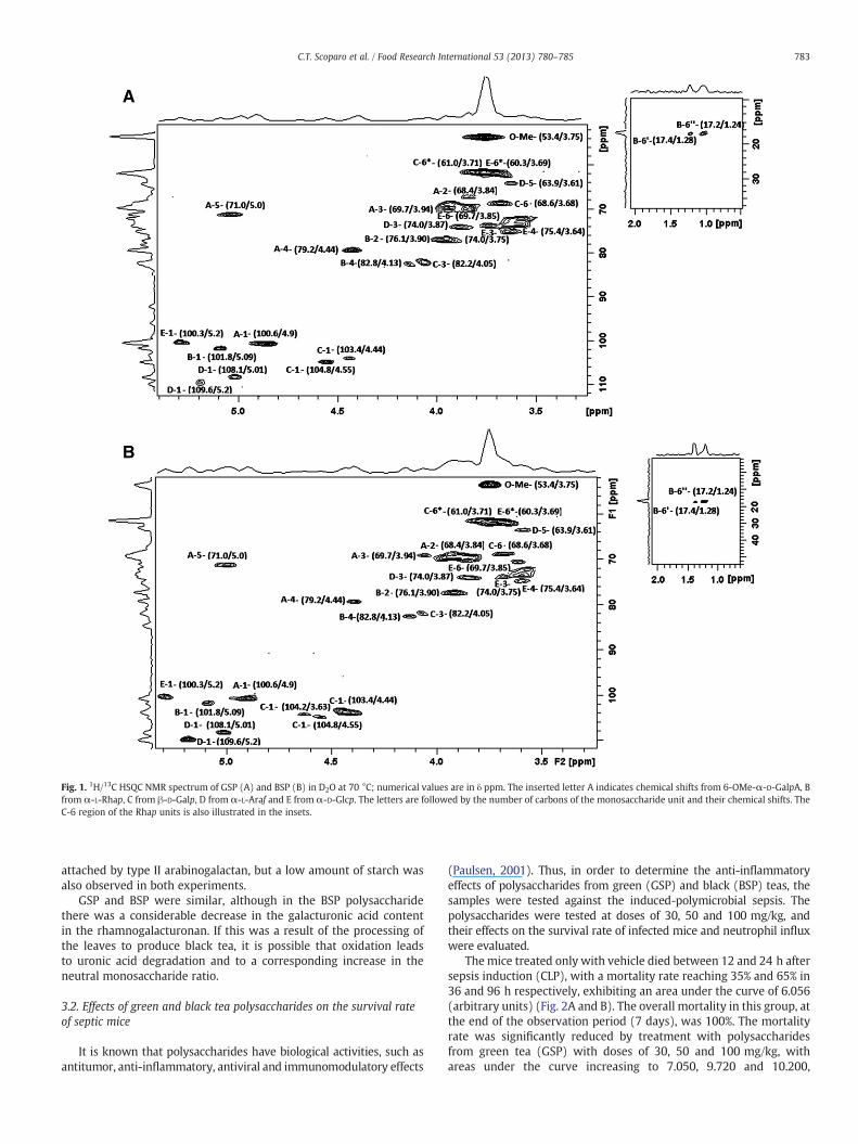
Fig. 1. 1H/13C HSQC NMR spectrum of GSP (A) and BSP (B) in D2O at 70 °C; numerical values are in δ ppm. The inserted letter A indicates chemical shifts from 6-OMe-α-D-GalpA, Bfrom α-L-Rhap, C from β-D-Galp, D from α-L-Araf and E from α-D-Glcp. The letters are followed by the number of carbons of the monosaccharide unit and their chemical shifts. TheC-6 region of the Rhap units is also illustrated in the insets.
783C.T. Scoparo et al. / Food Research International 53 (2013) 780–785
attached by type II arabinogalactan, but a low amount of starch wasalso observed in both experiments.
GSP and BSP were similar, although in the BSP polysaccharidethere was a considerable decrease in the galacturonic acid contentin the rhamnogalacturonan. If this was a result of the processing ofthe leaves to produce black tea, it is possible that oxidation leadsto uronic acid degradation and to a corresponding increase in theneutral monosaccharide ratio.
3.2. Effects of green and black tea polysaccharides on the survival rateof septic mice
It is known that polysaccharides have biological activities, such asantitumor, anti-inflammatory, antiviral and immunomodulatory effects
(Paulsen, 2001). Thus, in order to determine the anti-inflammatoryeffects of polysaccharides from green (GSP) and black (BSP) teas, thesamples were tested against the induced-polymicrobial sepsis. Thepolysaccharides were tested at doses of 30, 50 and 100 mg/kg, andtheir effects on the survival rate of infected mice and neutrophil influxwere evaluated.
The mice treated only with vehicle died between 12 and 24 h aftersepsis induction (CLP), with a mortality rate reaching 35% and 65% in36 and 96 h respectively, exhibiting an area under the curve of 6.056(arbitrary units) (Fig. 2A and B). The overall mortality in this group, atthe end of the observation period (7 days), was 100%. The mortalityrate was significantly reduced by treatment with polysaccharidesfrom green tea (GSP) with doses of 30, 50 and 100 mg/kg, withareas under the curve increasing to 7.050, 9.720 and 10.200,

Table 1Partially O-methylated alditol acetates (PMAA) of the GSP, BSP, GSP-CR and BSP-CRpolysaccharides.
PMAA Tra GSPb
%BSPc
%GSP-CRd
%BSP-CRe
%Structure
2,3,5-Me3-Ara 0.805 7.0 11 7.3 11.5 Araf-(1→2,5-Me2-Ara 0.860 3.0 6.1 3.1 6.3 →3)-Araf-(1→3,4-Me2-Rha 0.957 1.0 1.5 1.3 1.9 →2)-Rhap-(1→2,3,4,6-Me4-Glc 1 3.7 5.9 3.9 5.8 Glcp-(1→2,3-Me2Ara 1.023 3.5 5.8 3.7 6.1 →5)-Araf-(1→2,3,4,6-Me4-Gal 1.044 3.1 7.2 3.3 7.5 Galp-(1→3-Me-Rha 1.192 0.5 1.0 0.7 1.2 →2,4)-Rhap-(1→2-Me-Ara 1.240 1.0 1.2 1.2 1.4 →3,5)-Araf-(1→2,3,4-Me3-Glc 1.272 1.0 2.0 1.2 1.9 →6)-Glcp-(1→2,3,6-Me3-Gal 1.312 0.5 0.9 62 33 →4)-Galp-(1→2,4,6-Me3-Gal 1.360 5.0 10.6 5.5 11 →3-Galp-(1→2,3,6-Me3Glc 1.366 3.3 6.0 3.5 6.1 →4)-Glcp-(1→2,3,4-Me3-Gal 1.504 1.2 3.0 1.5 3.2 →6)-Galp-(1→2,3-Me2-Glc 1.759 0.3 1.0 0.6 0.9 →4,6)-Glcp-(1→2,3-Me2-Gal 1.865 0.4 0.6 0.5 0.8 →4,6)-Galp-(1→2,4-Me2-Gal 2.026 0.5 1.2 0.7 1.4 →3,6)-Galp-(1→
a Tr = retention time relative to 2,3,4,6-Me4Glc acetate.b Galacturonic acid content of GSP was 65%.c Galacturonic acid content of BSP was 35%.d Carboxy-reduced GSP.e Carboxy-reduced BSP.
784 C.T. Scoparo et al. / Food Research International 53 (2013) 780–785
respectively. At the end of the observation, the survival of thesegroups corresponded to 25% at a dose of 30 mg/kg and 45% for bothdoses of 50 and 100 mg/kg (Fig. 2A).
Similar results, but with less protection against lethality, werefound in the groups treated with black tea polysaccharide (BSP)(Fig. 2B). At doses of 30, 50 and 100 mg/kg their areas under thecurve increased to 7.200, 8.550 and 7.680, respectively, comparedwith the control (6.056). At the end of the observation period, the rateof survival corresponded to 20%, 25% and 20%, relative to the dosesabove. Considering that no deaths were observed in the sham-operatedgroup, with an area under the lethality curve corresponding to 16.800
Fig. 2. Effect of polysaccharides GSP (A) and BSP (B) on survival rate in sepsis. GSPand BSP protect against sepsis-induced lethality. Mice (10 animals/group) were orallyadministered various doses of GSP (30, 50 or 100 mg/kg), vehicle (water) and dexa-methasone (0.5 mg/kg s.c). Survival analyses were compared by a log-rank test.These calculations were performed with Graphpad Prism 5.0. The null hypothesiswas rejected when pb0.05. ***pb0.001 versus vehicle.
(arbitrary units), this confirms that surgical intervention, without per-foration, was not sufficient to cause death.
3.3. Neutrophil influx is significantly inhibited by GSP and BSP
Sepsis is associated to an aberrant inflammatory response, causingcell injury and organ dysfunction. During sepsis, the generation of in-flammatory mediators induced recruitment of a large quantity of im-mune cells. Neutrophil infiltration is an important pathophysiologicalalteration associated with the severity of sepsis, which amplifies thesystemic inflammatory response and causes multiple organ failure(Landry & Oliver, 2001; Mainous, Ertel, Chaudry, & Deitch, 1995).
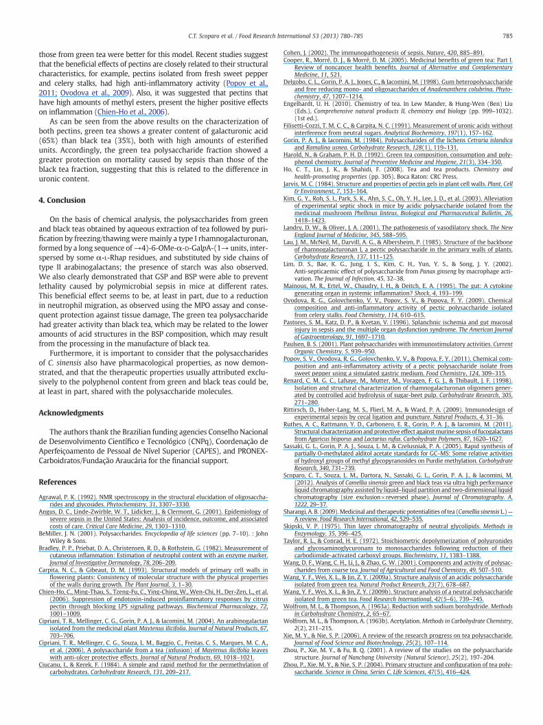
When compared with the sham group, CLP surgery markedlyincreased lung infiltration of neutrophils (50%), indirectly assessedby MPO activity (Fig. 3). It confirms that the neutrophil migrationoccurs after the experimental sepsis outcome. This increase was sig-nificantly prevented by both polysaccharides from green and blackteas (GSP and BSP) at a dose of 50 mg/kg, corresponding to 33% and28% inhibition respectively (Fig. 3). At a dose of 30 mg/kg, therewas no significant difference in comparison to the control group(CLP). Dexamethasone, used as an anti-inflammatory control, alsoinhibited infiltration of neutrophils (45%). GSP and BSP prevented el-evation of MPO activity, indirectly indicating reduction in neutrophilrecruitment to the lung and oxidative tissue damage, as well. This isof particular relevance because oxidative stress is known as a proba-ble mechanism for organ dysfunction during sepsis, amplifying andperpetuating the initial systemic inflammatory responses (Pastores,Katz, & Kvetan, 1996).
Recently, polysaccharides have demonstrated beneficial effects insepsis treatment, as reported for an acidic polysaccharide isolatedfrom Phellinus linteuswhich as able to decrease the septic shock inducedby high dose lipopolysaccharide injection in mice (Kim et al., 2003).Fucogalactans from the mushrooms Agaricus bisporus (RFP-Ab) andLactarius rufus (RFP-Lr) had a potent anti-inflammatory action in asepsis model (Ruthes, Rattmann, Carbonero, Gorin, & Iacomini, 2011).Also, a polysaccharide obtained from Panax ginseng had demonstrateda potent anti-septicemic activity, via macrophage activation, acting asan immunomodulator against sepsis induced by Staphylococcus aureus(Lim et al., 2002).
Based on the results now obtained, the polysaccharide fractions fromgreen and black teas showed a significant protective effect on sepsis, but
Fig. 3. Effect of polysaccharides on MPO activity in sepsis. Mice subjected to sepsis byCLP were treated with saline, GSP and BSP (30 and 50 mg/kg) or dexamethasone(0.5 mg/kg s.c). Myeloperoxidase activity (MPO, indicator of neutrophil accumulation)was measured spectrophotometrically in lung samples harvested 18 h after inductionof CLP. Results are expressed as change in optical density per milligram of protein.Values represent means±SEM. ###pb0.001 versus the sham group; ***pb0.001,**pb0.01 and *pb0.05 indicated values versus the vehicle group. ANOVA followed byBonferroni's test.
785C.T. Scoparo et al. / Food Research International 53 (2013) 780–785
those from green tea were better for this model. Recent studies suggestthat the beneficial effects of pectins are closely related to their structuralcharacteristics, for example, pectins isolated from fresh sweet pepperand celery stalks, had high anti-inflammatory activity (Popov et al.,2011; Ovodova et al., 2009). Also, it was suggested that pectins thathave high amounts of methyl esters, present the higher positive effectson inflammation (Chien-Ho et al., 2006).
As can be seen from the above results on the characterization ofboth pectins, green tea shows a greater content of galacturonic acid(65%) than black tea (35%), both with high amounts of esterifiedunits. Accordingly, the green tea polysaccharide fraction showed agreater protection on mortality caused by sepsis than those of theblack tea fraction, suggesting that this is related to the difference inuronic content.
4. Conclusion
On the basis of chemical analysis, the polysaccharides from greenand black teas obtained by aqueous extraction of tea followed by puri-fication by freezing/thawingweremainly a type I rhamnogalacturonan,formed by a long sequence of→4)-6-OMe-α-D-GalpA-(1→ units, inter-spersed by some α-L-Rhap residues, and substituted by side chains oftype II arabinogalactans; the presence of starch was also observed.We also clearly demonstrated that GSP and BSP were able to preventlethality caused by polymicrobial sepsis in mice at different rates.This beneficial effect seems to be, at least in part, due to a reductionin neutrophil migration, as observed using the MPO assay and conse-quent protection against tissue damage, The green tea polysaccharidehad greater activity than black tea, which may be related to the loweramounts of acid structures in the BSP composition, which may resultfrom the processing in the manufacture of black tea.
Furthermore, it is important to consider that the polysaccharidesof C. sinensis also have pharmacological properties, as now demon-strated, and that the therapeutic properties usually attributed exclu-sively to the polyphenol content from green and black teas could be,at least in part, shared with the polysaccharide molecules.
Acknowledgments
The authors thank the Brazilian funding agencies Conselho Nacionalde Desenvolvimento Científico e Tecnológico (CNPq), Coordenação deAperfeiçoamento de Pessoal de Nível Superior (CAPES), and PRONEX-Carboidratos/Fundação Araucária for the financial support.
References
Agrawal, P. K. (1992). NMR spectroscopy in the structural elucidation of oligosaccha-rides and glycosides. Phytochemistry, 31, 3307–3330.
Angus, D. C., Linde-Zwirble, W. T., Lidicker, J., & Clermont, G. (2001). Epidemiology ofsevere sepsis in the United States: Analysis of incidence, outcome, and associatedcosts of care. Critical Care Medicine, 29, 1303–1310.
BeMiller, J. N. (2001). Polysaccharides. Encyclopedia of life sciences (pp. 7–10). : JohnWiley & Sons.
Bradley, P. P., Priebat, D. A., Christensen, R. D., & Rothstein, G. (1982). Measurement ofcutaneous inflammation: Estimation of neutrophil content with an enzyme marker.Journal of Investigative Dermatology, 78, 206–209.
Carpita, N. C., & Gibeaut, D. M. (1993). Structural models of primary cell walls inflowering plants: Consistency of molecular structure with the physical propertiesof the walls during growth. The Plant Journal, 3, 1–30.
Chien-Ho, C., Ming-Thau, S., Tzeng-Fu, C., Ying-Ching, W., Wen-Chi, H., Der-Zen, L., et al.(2006). Suppression of endotoxin-induced proinflammatory responses by citruspectin through blocking LPS signaling pathways. Biochemical Pharmacology, 72,1001–1009.
Cipriani, T. R., Mellinger, C. G., Gorin, P. A. J., & Iacomini, M. (2004). An arabinogalactanisolated from the medicinal plant Maytenus ilicifolia. Journal of Natural Products, 67,703–706.
Cipriani, T. R., Mellinger, C. G., Souza, L. M., Baggio, C., Freitas, C. S., Marques, M. C. A.,et al. (2006). A polysaccharide from a tea (infusion) of Maytenus ilicifolia leaveswith anti-ulcer protective effects. Journal of Natural Products, 69, 1018–1021.
Ciucanu, I., & Kerek, F. (1984). A simple and rapid method for the permethylation ofcarbohydrates. Carbohydrate Research, 131, 209–217.
Cohen, J. (2002). The immunopathogenesis of sepsis. Nature, 420, 885–891.Cooper, R., Morré, D. J., & Morré, D. M. (2005). Medicinal benefits of green tea: Part I.
Review of noncancer health benefits. Journal of Alternative and ComplementaryMedicine, 11, 521.
Delgobo, C. L., Gorin, P. A. J., Jones, C., & Iacomini, M. (1998). Gum heteropolysaccharideand free reducing mono- and oligosaccharides of Anadenanthera colubrina. Phyto-chemistry, 47, 1207–1214.
Engelhardt, U. H. (2010). Chemistry of tea. In Lew Mander, & Hung-Wen (Ben) Liu(Eds.), Comprehensive natural products II, chemistry and biology (pp. 999–1032).(1st ed.).
Filisetti-Cozzi, T. M. C. C., & Carpita, N. C. (1991). Measurement of uronic acids withoutinterference from neutral sugars. Analytical Biochemistry, 197(1), 157–162.
Gorin, P. A. J., & Iacomini, M. (1984). Polysaccharides of the lichens Cetraria islandicaand Ramalina usnea. Carbohydrate Research, 128(1), 119–131.
Harold, N., & Graham, P. H. D. (1992). Green tea composition, consumption and poly-phenol chemistry. Journal of Preventive Medicine and Hygiene, 21(3), 334–350.
Ho, C. T., Lin, J. K., & Shahidi, F. (2008). Tea and tea products. Chemistry andhealth-promoting properties (pp. 305). Boca Raton: CRC Press.
Jarvis, M. C. (1984). Structure and properties of pectin gels in plant cell walls. Plant, Cell& Environment, 7, 153–164.
Kim, G. Y., Roh, S. I., Park, S. K., Ahn, S. C., Oh, Y. H., Lee, J. D., et al. (2003). Alleviationof experimental septic shock in mice by acidic polysaccharide isolated from themedicinal mushroom Phellinus linteus. Biological and Pharmaceutical Bulletin, 26,1418–1423.
Landry, D. W., & Oliver, J. A. (2001). The pathogenesis of vasodilatory shock. The NewEngland Journal of Medicine, 345, 588–595.
Lau, J. M., McNeil, M., Darvill, A. G., & Albersheim, P. (1985). Structure of the backboneof rhamnogalacturonan I, a pectic polysaccharide in the primary walls of plants.Carbohydrate Research, 137, 111–125.
Lim, D. S., Bae, K. G., Jung, I. S., Kim, C. H., Yun, Y. S., & Song, J. Y. (2002).Anti-septicaemic effect of polysaccharide from Panax ginseng by macrophage acti-vation. The Journal of Infection, 45, 32–38.
Mainous, M. R., Ertel, W., Chaudry, I. H., & Deitch, E. A. (1995). The gut: A cytokinegenerating organ in systemic inflammation? Shock, 4, 193–199.
Ovodova, R. G., Golovchenko, V. V., Popov, S. V., & Popova, F. Y. (2009). Chemicalcomposition and anti-inflammatory activity of pectic polysaccharide isolatedfrom celery stalks. Food Chemistry, 114, 610–615.
Pastores, S. M., Katz, D. P., & Kvetan, V. (1996). Splanchnic ischemia and gut mucosalinjury in sepsis and the multiple organ dysfunction syndrome. The American Journalof Gastroenterology, 91, 1697–1710.
Paulsen, B. S. (2001). Plant polysaccharides with immunostimulatory activities. CurrentOrganic Chemistry, 5, 939–950.
Popov, S. V., Ovodova, R. G., Golovchenko, V. V., & Popova, F. Y. (2011). Chemical com-position and anti-inflammatory activity of a pectic polysaccharide isolate fromsweet pepper using a simulated gastric medium. Food Chemistry, 124, 309–315.
Renard, C. M. G. C., Lahaye, M., Mutter, M., Voragen, F. G. J., & Thibault, J. F. (1998).Isolation and structural characterization of rhamnogalacturonan oligomers gener-ated by controlled acid hydrolysis of sugar-beet pulp. Carbohydrate Research, 305,271–280.
Rittirsch, D., Huber-Lang, M. S., Flierl, M. A., & Ward, P. A. (2009). Immunodesign ofexperimental sepsis by cecal ligation and puncture. Natural Products, 4, 31–36.
Ruthes, A. C., Rattmann, Y. D., Carbonero, E. R., Gorin, P. A. J., & Iacomini, M. (2011).Structural characterization and protective effect againstmurine sepsis of fucogalactansfrom Agaricus bisporus and Lactarius rufus. Carbohydrate Polymers, 87, 1620–1627.
Sassaki, G. L., Gorin, P. A. J., Souza, L. M., & Czelusniak, P. A. (2005). Rapid synthesis ofpartially O-methylated alditol acetate standards for GC–MS: Some relative activitiesof hydroxyl groups of methyl glycopyranosides on Purdie methylation. CarbohydrateResearch, 340, 731–739.
Scoparo, C. T., Souza, L. M., Dartora, N., Sassaki, G. L., Gorin, P. A. J., & Iacomini, M.(2012). Analysis of Camellia sinensis green and black teas via ultra high performanceliquid chromatography assisted by liquid–liquid partition and two-dimensional liquidchromatography (size exclusion×reversed phase). Journal of Chromatography. A,1222, 29–37.
Sharangi, A. B. (2009).Medicinal and therapeutic potentialities of tea (Camellia sinensis L.)—A review. Food Research International, 42, 529–535.
Skipski, V. P. (1975). Thin layer chromatography of neutral glycolipids. Methods inEnzymology, 35, 396–425.
Taylor, R. L., & Conrad, H. E. (1972). Stoichiometric depolymerization of polyuronidesand glycosaminoglycuronans to monosaccharides following reduction of theircarbodiimide-activated carboxyl groups. Biochemistry, 11, 1383–1388.
Wang, D. F., Wang, C. H., Li, J., & Zhao, G. W. (2001). Components and activity of polysac-charides from coarse tea. Journal of Agricultural and Food Chemistry, 49, 507–510.
Wang, Y. F., Wei, X. L., & Jin, Z. Y. (2009a). Structure analysis of an acidic polysaccharideisolated from green tea. Natural Product Research, 23(7), 678–687.
Wang, Y. F., Wei, X. L., & Jin, Z. Y. (2009b). Structure analysis of a neutral polysaccharideisolated from green tea. Food Research International, 42(5–6), 739–745.
Wolfrom, M. L., & Thompson, A. (1963a). Reduction with sodium borohydride.Methodsin Carbohydrate Chemistry, 2, 65–67.
Wolfrom, M. L., & Thompson, A. (1963b). Acetylation.Methods in Carbohydrate Chemistry,2(2), 211–215.
Xie, M. Y., & Nie, S. P. (2006). A review of the research progress on tea polysaccharide.Journal of Food Science and Biotechnology, 25(2), 107–114.
Zhou, P., Xie, M. Y., & Fu, B. Q. (2001). A review of the studies on the polysaccharidestructure. Journal of Nanchang University (Natural Science), 25(2), 197–204.
Zhou, P., Xie, M. Y., & Nie, S. P. (2004). Primary structure and configuration of tea poly-saccharide. Science in China. Series C, Life Sciences, 47(5), 416–424.
Related Documents