Polyphosphate kinase is involved in stress-induced mprAB-sigE-rel signalling in mycobacteria Kamakshi Sureka, 1 Supratim Dey, 1 Pratik Datta, 1 Anil Kumar Singh, 1† Arunava Dasgupta, 1‡ Sébastien Rodrigue, 2 Joyoti Basu 1 and Manikuntala Kundu 1 * 1 Department of Chemistry, Bose Institute, 93/1 Acharya Prafulla Chandra Road, Calcutta 700009, India. 2 Department of Civil and Environmental Engineering, Massachusetts, Institute of Technology, 15 Vassar Street, Cambridge, MA 02139, USA. Summary Polyphosphate kinase 1 (PPK1) helps bacteria to survive under stress. The ppk1 gene of Mycobacte- rium tuberculosis was overexpressed in Escherichia coli and characterized. Residues R230 and F176, predicted to be present in the head domain of PPK1, were identified as residues critical for polyphos- phate (polyP)-synthesizing ability and dimerization of PPK1. A ppk1 knockout mutant of Mycobacterium smegmatis was compromised in its ability to survive under long-term hypoxia. The transcription of the rel gene and the synthesis of the stringent response regulator ppGpp were impaired in the mutant and restored after complementation with ppk1 of M. tu- berculosis, providing evidence that PPK1 is required for the stringent response. We present evidence that PPK1 is likely required for mprAB-sigE-rel signal- ling. s E regulates the transcription of rel, and we hypothesize that under conditions of stress polyP acts as a preferred donor for MprB-mediated phos- phorylation of MprA facilitating transcription of the sigE gene thereby leading finally to the enhance- ment of the transcription of rel in M. smegmatis and M. tuberculosis. Downregulation of ppk1 led to impaired survival of M. tuberculosis in macroph- ages. PolyP plays a central role in the stress response of mycobacteria. Introduction Inorganic polyphosphate (polyP) is a polymer of tens or hundreds of phosphate residues linked by high-energy phosphoanhydride bonds. PolyP is ubiquitous (Kornberg et al., 1999). The enzyme that is primarily responsible for reversible polyP synthesis in Escherichia coli is polyphos- phate kinase 1 (PPK1) which catalyses the polymerization of the g-phosphate of ATP into a polyP chain (Ahn and Kornberg, 1990; Akiyama et al., 1992). PPK1 is highly conserved in many bacterial species including some of the major pathogens, but it is absent in eukaryotes (Tzeng and Kornberg, 2000), making it an attractive target for drug development. Studies in E. coli and Pseudomonas aeruginosa have shown that ppk1 plays an important role in bacterial survival under conditions of stress such as in the stationary phase, under nutrient limitation and under oxidative stress (Rao and Kornberg, 1996; Kuroda et al., 1999). In P. aeruginosa, decreased levels of polyP have been linked to defect in biofilm formation and quorum sensing, with associated attenuation of virulence (Rashid et al., 2000a,b). The possible physiological roles of polyP have been elegantly reviewed by Brown and Kornberg (2004). A third of the world’s population is infected with Mycobacterium tuberculosis, the causative agent of tuberculosis. New drugs are needed to shorten the course of therapy. At the same time, there is an urgent need to develop an effective vaccine. In order to meet these goals it is imperative to understand the biology of the tubercle bacillus, to identify gene products that are essential for mycobacterial growth in vivo, and those that when inactivated would lead to an attenuated phenotype. M. tuberculosis is an intracellular pathogen which sur- vives and replicates within macrophages. Persistent mycobacteria in lung lesions encounter hypoxia and nutri- tional deprivation and these mycobacterial cells regain acid-fastness and start growing even 2 years after removal of the starvation-induced stress (Nyka, 1974). Overpowering the persistent bacteria, therefore, poses a challenge. Drug-induced inhibition of translation is asso- ciated with the downregulation of ppk1, the polyphos- phate kinase 1 (PPK1) of M. tuberculosis (Boshoff et al., 2004), suggesting a role of polyP metabolism in the response to translational inhibition, a situation encoun- tered during starvation. In spite of the widening interest in Accepted 29 May, 2007. *For correspondence. E-mail manikuntala@ vsnl.net; Tel. (+91) 33 23506619; Fax (+91) 33 23506790. Present address: † GroupeAvenir, Inserm U570, Université Descartes- Paris V, Faculté de Médecine, Site Necker, 156 rue de Vaugirard, Paris, France; ‡ Institut Pasteur, Unite de Genetique Mycobacterienne, 25 rue du Dr Roux, Paris, France. Molecular Microbiology (2007) 65(2), 261–276 doi:10.1111/j.1365-2958.2007.05814.x © 2007 The Authors Journal compilation © 2007 Blackwell Publishing Ltd

Welcome message from author
This document is posted to help you gain knowledge. Please leave a comment to let me know what you think about it! Share it to your friends and learn new things together.
Transcript
Polyphosphate kinase is involved in stress-inducedmprAB-sigE-rel signalling in mycobacteria
Kamakshi Sureka,1 Supratim Dey,1 Pratik Datta,1
Anil Kumar Singh,1† Arunava Dasgupta,1‡
Sébastien Rodrigue,2 Joyoti Basu1 andManikuntala Kundu1*1Department of Chemistry, Bose Institute, 93/1 AcharyaPrafulla Chandra Road, Calcutta 700009, India.2Department of Civil and Environmental Engineering,Massachusetts, Institute of Technology, 15 VassarStreet, Cambridge, MA 02139, USA.
Summary
Polyphosphate kinase 1 (PPK1) helps bacteria tosurvive under stress. The ppk1 gene of Mycobacte-rium tuberculosis was overexpressed in Escherichiacoli and characterized. Residues R230 and F176,predicted to be present in the head domain of PPK1,were identified as residues critical for polyphos-phate (polyP)-synthesizing ability and dimerizationof PPK1. A ppk1 knockout mutant of Mycobacteriumsmegmatis was compromised in its ability to surviveunder long-term hypoxia. The transcription of the relgene and the synthesis of the stringent responseregulator ppGpp were impaired in the mutant andrestored after complementation with ppk1 of M. tu-berculosis, providing evidence that PPK1 is requiredfor the stringent response. We present evidence thatPPK1 is likely required for mprAB-sigE-rel signal-ling. sE regulates the transcription of rel, and wehypothesize that under conditions of stress polyPacts as a preferred donor for MprB-mediated phos-phorylation of MprA facilitating transcription of thesigE gene thereby leading finally to the enhance-ment of the transcription of rel in M. smegmatis andM. tuberculosis. Downregulation of ppk1 led toimpaired survival of M. tuberculosis in macroph-ages. PolyP plays a central role in the stressresponse of mycobacteria.
Introduction
Inorganic polyphosphate (polyP) is a polymer of tens orhundreds of phosphate residues linked by high-energyphosphoanhydride bonds. PolyP is ubiquitous (Kornberget al., 1999). The enzyme that is primarily responsible forreversible polyP synthesis in Escherichia coli is polyphos-phate kinase 1 (PPK1) which catalyses the polymerizationof the g-phosphate of ATP into a polyP chain (Ahn andKornberg, 1990; Akiyama et al., 1992). PPK1 is highlyconserved in many bacterial species including some ofthe major pathogens, but it is absent in eukaryotes (Tzengand Kornberg, 2000), making it an attractive target fordrug development. Studies in E. coli and Pseudomonasaeruginosa have shown that ppk1 plays an important rolein bacterial survival under conditions of stress such as inthe stationary phase, under nutrient limitation and underoxidative stress (Rao and Kornberg, 1996; Kuroda et al.,1999). In P. aeruginosa, decreased levels of polyP havebeen linked to defect in biofilm formation and quorumsensing, with associated attenuation of virulence (Rashidet al., 2000a,b). The possible physiological roles of polyPhave been elegantly reviewed by Brown and Kornberg(2004).
A third of the world’s population is infected withMycobacterium tuberculosis, the causative agent oftuberculosis. New drugs are needed to shorten the courseof therapy. At the same time, there is an urgent need todevelop an effective vaccine. In order to meet these goalsit is imperative to understand the biology of the tuberclebacillus, to identify gene products that are essentialfor mycobacterial growth in vivo, and those that wheninactivated would lead to an attenuated phenotype.M. tuberculosis is an intracellular pathogen which sur-vives and replicates within macrophages. Persistentmycobacteria in lung lesions encounter hypoxia and nutri-tional deprivation and these mycobacterial cells regainacid-fastness and start growing even 2 years afterremoval of the starvation-induced stress (Nyka, 1974).Overpowering the persistent bacteria, therefore, poses achallenge. Drug-induced inhibition of translation is asso-ciated with the downregulation of ppk1, the polyphos-phate kinase 1 (PPK1) of M. tuberculosis (Boshoff et al.,2004), suggesting a role of polyP metabolism in theresponse to translational inhibition, a situation encoun-tered during starvation. In spite of the widening interest in
Accepted 29 May, 2007. *For correspondence. E-mail [email protected]; Tel. (+91) 33 23506619; Fax (+91) 33 23506790. Presentaddress: †Groupe Avenir, Inserm U570, Université Descartes- Paris V,Faculté de Médecine, Site Necker, 156 rue de Vaugirard, Paris,France; ‡Institut Pasteur, Unite de Genetique Mycobacterienne, 25rue du Dr Roux, Paris, France.
Molecular Microbiology (2007) 65(2), 261–276 doi:10.1111/j.1365-2958.2007.05814.x
© 2007 The AuthorsJournal compilation © 2007 Blackwell Publishing Ltd
polyP metabolism, there has been no detailed investiga-tion on polyP metabolism in the genus Mycobacterium.Genes that likely encode counterparts of ppk1 have beenidentified both in M. tuberculosis and in its fast-growingnon-pathogenic counterpart Mycobacterium smegmatis,widely used as a model system to study mycobacterialphysiology. In this study, the putative ppk1 genes ofM. tuberculosis and M. smegmatis have been cloned andexpressed in E. coli, and the corresponding proteinproducts have been characterized. The ppk1 gene ofM. smegmatis has been inactivated in order to study therole of ppk1 in survival of the bacterium under conditionsof stress. We report that ppk1 is involved in survival of thebacterium under conditions of hypoxia, detergent stressand oxidative stress. We have uncovered a novel regula-tory signalling cascade in which ppk1 is required for thetranscription of the two-component signal transductionsystem mprAB, which in turn regulates the expression ofsigE, a stress-regulated s-factor. We show for the firsttime that sE regulates the transcription of the stringentresponse regulator rel. Delineation of this pathwayexplains how inactivation of ppk1 downregulates rel andthe alarmone ppGpp. Downregulation of ppk1 ofM. tuberculosis is associated with a loss in the ability ofthe bacterium to survive in macrophages supporting theview that ppk1 plays an important role in the intracellularlife of the bacterium.
Results
Expression of ppk1 of M. tuberculosis andM. smegmatis and activity assays
The genes encoding putative counterparts of PPK1 havebeen identified in M. tuberculosis H37Rv (Rv2984) andM. smegmatis mc2155 (MSMEG2390). The putativePPK1 of M. tuberculosis and M. smegmatis were of 742and 736 amino acid residues respectively (Fig. S1).M. tuberculosis and M. smegmatis were 85% similar and79% identical. PPK1 of M. tuberculosis was 74%, 54%and 56% similar to PPK1 of Streptomyces coelicolor,E. coli and P. aeruginosa, respectively; and 59%, 33%and 38% identical to the respective proteins. The geneproducts were expressed as N-terminal His-tagged pro-teins in E. coli and purified by affinity chromatography onNi2+-NTA agarose (Fig. S2A).
Antibody against PPK1 of M. tuberculosis was raised inrabbits. In E. coli, PPK1 is present in the membrane(Kuroda and Kornberg, 1997). The Kyte–Doolittle hydro-phobicity plot suggested the presence of severalstretches of hydrophobic amino acids in PPK1 ofM. tuberculosis (Fig. S2B). In harmony with this, immuno-blotting of the membrane fraction of M. tuberculosis withanti-PPK1 showed a prominent band of 81 kDa corre-
sponding to the position of recombinant PPK1. The anti-body also cross-reacted with a band of similar size inmembrane fractions of M. smegmatis (Fig. S2C).
His-PPK1 of M. tuberculosis and M. smegmatis wereboth capable of converting [g-32P]-ATP to acid-insolublepolyP in vitro. The recombinant M. tuberculosis andM. smegmatis PPK1 catalysed polyP synthesis with spe-cific activities of 4.5 � 0.16 and 4.20 � 0.23 mmol min-1
per mg of protein respectively. PPK1 was immunoprecipi-tated from membrane extracts of M. tuberculosis in order toassay specifically for PPK1 activity. The immunoprecipi-tated enzyme showed a specific activity of 0.05 mmol min-1
per mg of total membrane protein. The supernatant afterimmunoprecipitation was subjected to immunoblotting withanti-PPK1 antibody. No cross-reactive band was detected,suggesting that no residual PPK1 was left in the superna-tant after immunoprecipitation.
Similar to other PPKs, M. tuberculosis PPK1 trans-ferred the g-phosphate of ATP processively to generatepolyP chains of lengths 200–800 residues (data notshown). Addition of unlabelled ATP in the reaction mixturecould inhibit (> 90%) incorporation of radiolabel into polyP,whereas unlabelled GTP was less effective (< 15%).M. smegmatis PPK1 also showed similar characteristics(data not shown). Both proteins could also synthesize ATPby breaking down polyP isolated from mycobacteria, withactivities of 0.67 and 0.7 mmol min-1 per mg of proteinfor the M. smegmatis and M. tuberculosis proteinsrespectively. These results validated the functions of theidentified proteins as PPKs in mycobacteria.
Analysis of some PPK1 point mutations
Directed mutagenesis of E. coli PPK1 (Tzeng and Korn-berg, 2000) has helped to identify critical residuesrequired for activity, within the 300-residue carboxy-terminus (which is highly conserved among species).However, little is known about the less conserved regionwhich encompasses the head domain (Zhu et al., 2004).In this study, we therefore focused on this less conservedregion (spanning residues 155–376 of M. tuberculosisPPK1) for mutational analysis in order to gain insight intothe critical residues in this region that are required forPPK1 function. Based on sequence similarity, seven con-served residues within this region were mutated individu-ally to alanines. PPK activity was assayed in all thesemutants. Five of the mutants exhibited PPK activitiessimilar to that of the wild type. Two mutants, F176A andR230A, showed significant reduction in the ability to syn-thesize polyP as well as to regenerate ATP by the break-down of polyP (Table 1). The mutation R230K did notcause the marked decrease in activity observed with theR230A mutant. The F176Y mutation also showed a farlesser inhibitory effect than the F176A mutation. PPK1 is
262 K. Sureka et al.
© 2007 The AuthorsJournal compilation © 2007 Blackwell Publishing Ltd, Molecular Microbiology, 65, 261–276
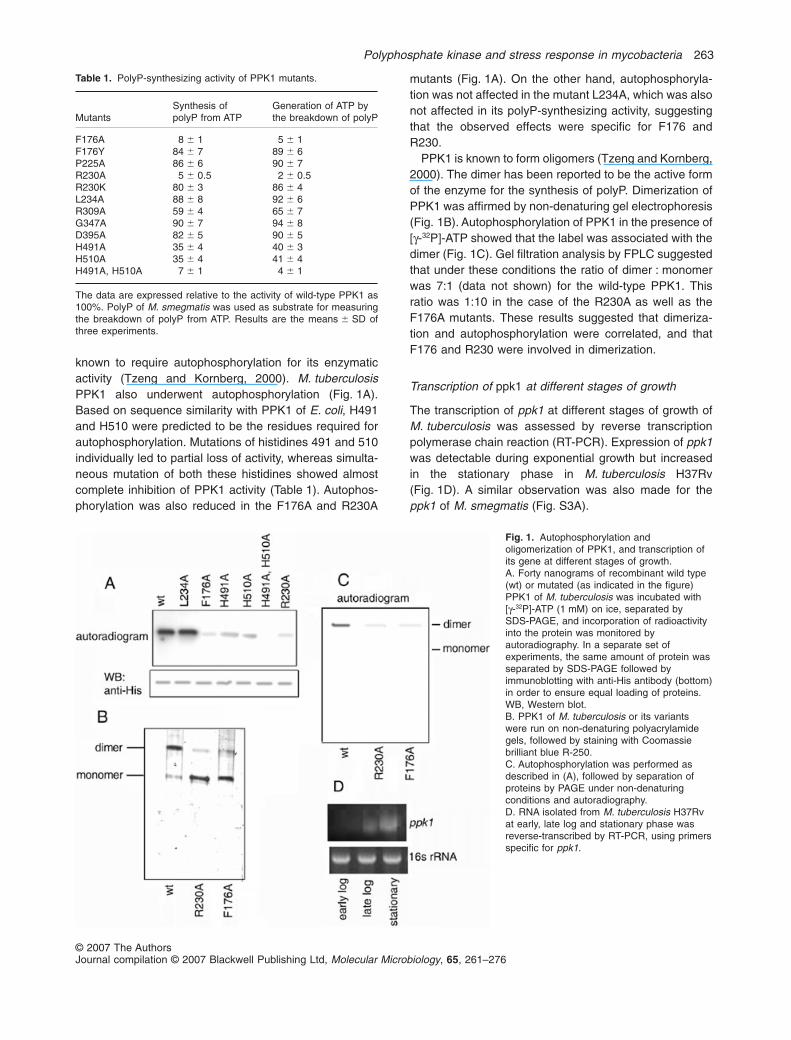
known to require autophosphorylation for its enzymaticactivity (Tzeng and Kornberg, 2000). M. tuberculosisPPK1 also underwent autophosphorylation (Fig. 1A).Based on sequence similarity with PPK1 of E. coli, H491and H510 were predicted to be the residues required forautophosphorylation. Mutations of histidines 491 and 510individually led to partial loss of activity, whereas simulta-neous mutation of both these histidines showed almostcomplete inhibition of PPK1 activity (Table 1). Autophos-phorylation was also reduced in the F176A and R230A
mutants (Fig. 1A). On the other hand, autophosphoryla-tion was not affected in the mutant L234A, which was alsonot affected in its polyP-synthesizing activity, suggestingthat the observed effects were specific for F176 andR230.
PPK1 is known to form oligomers (Tzeng and Kornberg,2000). The dimer has been reported to be the active formof the enzyme for the synthesis of polyP. Dimerization ofPPK1 was affirmed by non-denaturing gel electrophoresis(Fig. 1B). Autophosphorylation of PPK1 in the presence of[g-32P]-ATP showed that the label was associated with thedimer (Fig. 1C). Gel filtration analysis by FPLC suggestedthat under these conditions the ratio of dimer : monomerwas 7:1 (data not shown) for the wild-type PPK1. Thisratio was 1:10 in the case of the R230A as well as theF176A mutants. These results suggested that dimeriza-tion and autophosphorylation were correlated, and thatF176 and R230 were involved in dimerization.
Transcription of ppk1 at different stages of growth
The transcription of ppk1 at different stages of growth ofM. tuberculosis was assessed by reverse transcriptionpolymerase chain reaction (RT-PCR). Expression of ppk1was detectable during exponential growth but increasedin the stationary phase in M. tuberculosis H37Rv(Fig. 1D). A similar observation was also made for theppk1 of M. smegmatis (Fig. S3A).
Table 1. PolyP-synthesizing activity of PPK1 mutants.
MutantsSynthesis ofpolyP from ATP
Generation of ATP bythe breakdown of polyP
F176A 8 � 1 5 � 1F176Y 84 � 7 89 � 6P225A 86 � 6 90 � 7R230A 5 � 0.5 2 � 0.5R230K 80 � 3 86 � 4L234A 88 � 8 92 � 6R309A 59 � 4 65 � 7G347A 90 � 7 94 � 8D395A 82 � 5 90 � 5H491A 35 � 4 40 � 3H510A 35 � 4 41 � 4H491A, H510A 7 � 1 4 � 1
The data are expressed relative to the activity of wild-type PPK1 as100%. PolyP of M. smegmatis was used as substrate for measuringthe breakdown of polyP from ATP. Results are the means � SD ofthree experiments.
Fig. 1. Autophosphorylation andoligomerization of PPK1, and transcription ofits gene at different stages of growth.A. Forty nanograms of recombinant wild type(wt) or mutated (as indicated in the figure)PPK1 of M. tuberculosis was incubated with[g-32P]-ATP (1 mM) on ice, separated bySDS-PAGE, and incorporation of radioactivityinto the protein was monitored byautoradiography. In a separate set ofexperiments, the same amount of protein wasseparated by SDS-PAGE followed byimmunoblotting with anti-His antibody (bottom)in order to ensure equal loading of proteins.WB, Western blot.B. PPK1 of M. tuberculosis or its variantswere run on non-denaturing polyacrylamidegels, followed by staining with Coomassiebrilliant blue R-250.C. Autophosphorylation was performed asdescribed in (A), followed by separation ofproteins by PAGE under non-denaturingconditions and autoradiography.D. RNA isolated from M. tuberculosis H37Rvat early, late log and stationary phase wasreverse-transcribed by RT-PCR, using primersspecific for ppk1.
Polyphosphate kinase and stress response in mycobacteria 263
© 2007 The AuthorsJournal compilation © 2007 Blackwell Publishing Ltd, Molecular Microbiology, 65, 261–276
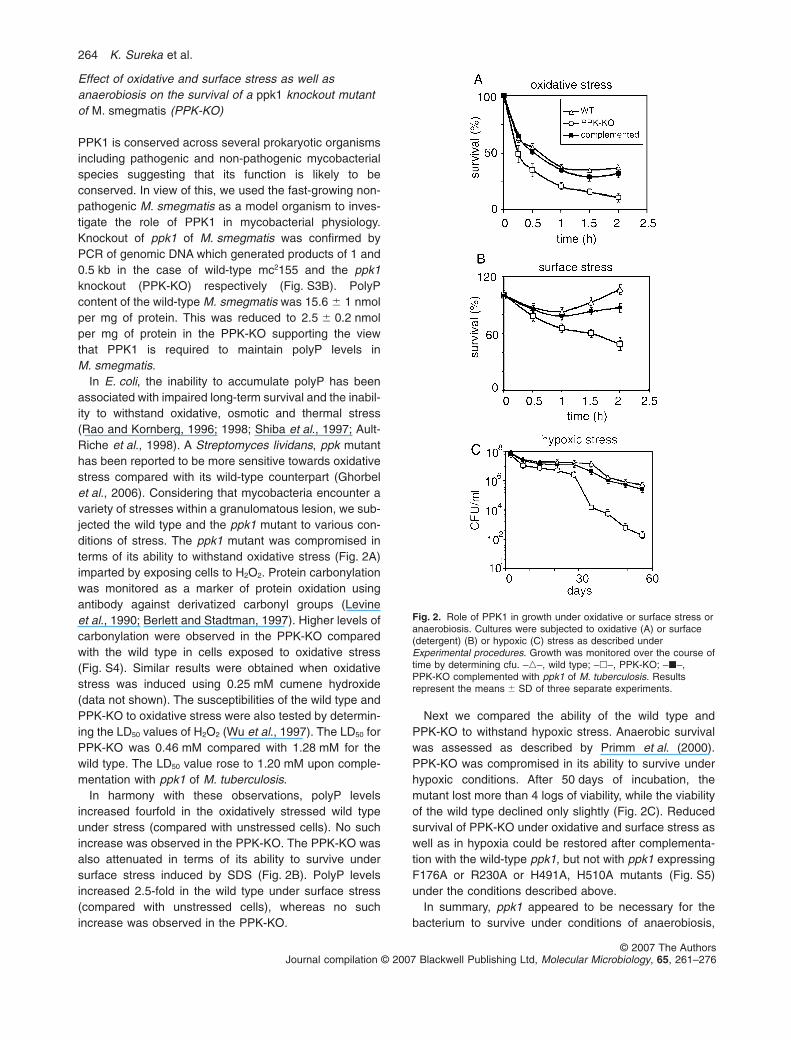
Effect of oxidative and surface stress as well asanaerobiosis on the survival of a ppk1 knockout mutantof M. smegmatis (PPK-KO)
PPK1 is conserved across several prokaryotic organismsincluding pathogenic and non-pathogenic mycobacterialspecies suggesting that its function is likely to beconserved. In view of this, we used the fast-growing non-pathogenic M. smegmatis as a model organism to inves-tigate the role of PPK1 in mycobacterial physiology.Knockout of ppk1 of M. smegmatis was confirmed byPCR of genomic DNA which generated products of 1 and0.5 kb in the case of wild-type mc2155 and the ppk1knockout (PPK-KO) respectively (Fig. S3B). PolyPcontent of the wild-type M. smegmatis was 15.6 � 1 nmolper mg of protein. This was reduced to 2.5 � 0.2 nmolper mg of protein in the PPK-KO supporting the viewthat PPK1 is required to maintain polyP levels inM. smegmatis.
In E. coli, the inability to accumulate polyP has beenassociated with impaired long-term survival and the inabil-ity to withstand oxidative, osmotic and thermal stress(Rao and Kornberg, 1996; 1998; Shiba et al., 1997; Ault-Riche et al., 1998). A Streptomyces lividans, ppk mutanthas been reported to be more sensitive towards oxidativestress compared with its wild-type counterpart (Ghorbelet al., 2006). Considering that mycobacteria encounter avariety of stresses within a granulomatous lesion, we sub-jected the wild type and the ppk1 mutant to various con-ditions of stress. The ppk1 mutant was compromised interms of its ability to withstand oxidative stress (Fig. 2A)imparted by exposing cells to H2O2. Protein carbonylationwas monitored as a marker of protein oxidation usingantibody against derivatized carbonyl groups (Levineet al., 1990; Berlett and Stadtman, 1997). Higher levels ofcarbonylation were observed in the PPK-KO comparedwith the wild type in cells exposed to oxidative stress(Fig. S4). Similar results were obtained when oxidativestress was induced using 0.25 mM cumene hydroxide(data not shown). The susceptibilities of the wild type andPPK-KO to oxidative stress were also tested by determin-ing the LD50 values of H2O2 (Wu et al., 1997). The LD50 forPPK-KO was 0.46 mM compared with 1.28 mM for thewild type. The LD50 value rose to 1.20 mM upon comple-mentation with ppk1 of M. tuberculosis.
In harmony with these observations, polyP levelsincreased fourfold in the oxidatively stressed wild typeunder stress (compared with unstressed cells). No suchincrease was observed in the PPK-KO. The PPK-KO wasalso attenuated in terms of its ability to survive undersurface stress induced by SDS (Fig. 2B). PolyP levelsincreased 2.5-fold in the wild type under surface stress(compared with unstressed cells), whereas no suchincrease was observed in the PPK-KO.
Next we compared the ability of the wild type andPPK-KO to withstand hypoxic stress. Anaerobic survivalwas assessed as described by Primm et al. (2000).PPK-KO was compromised in its ability to survive underhypoxic conditions. After 50 days of incubation, themutant lost more than 4 logs of viability, while the viabilityof the wild type declined only slightly (Fig. 2C). Reducedsurvival of PPK-KO under oxidative and surface stress aswell as in hypoxia could be restored after complementa-tion with the wild-type ppk1, but not with ppk1 expressingF176A or R230A or H491A, H510A mutants (Fig. S5)under the conditions described above.
In summary, ppk1 appeared to be necessary for thebacterium to survive under conditions of anaerobiosis,
Fig. 2. Role of PPK1 in growth under oxidative or surface stress oranaerobiosis. Cultures were subjected to oxidative (A) or surface(detergent) (B) or hypoxic (C) stress as described underExperimental procedures. Growth was monitored over the course oftime by determining cfu. –�–, wild type; –�–, PPK-KO; –�–,PPK-KO complemented with ppk1 of M. tuberculosis. Resultsrepresent the means � SD of three separate experiments.
264 K. Sureka et al.
© 2007 The AuthorsJournal compilation © 2007 Blackwell Publishing Ltd, Molecular Microbiology, 65, 261–276
oxidative stress and surface stress. The slight differencesobserved between the survival of the wild type and thecomplemented strain under surface stress (Fig. 2B) couldpossibly be due to the fact that the exogenously suppliedppk1 was not expressed from its endogenous promoter.
Characterization of the mprAB-sigE-rel signallingcascade in M. smegmatis
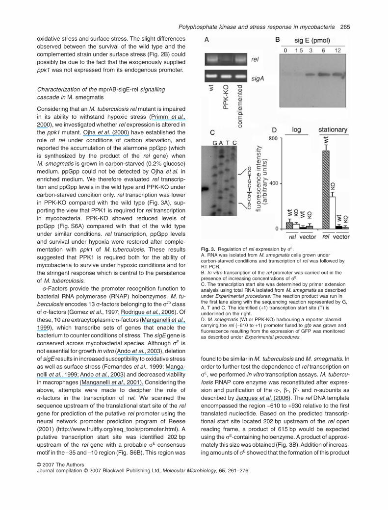
Considering that an M. tuberculosis rel mutant is impairedin its ability to withstand hypoxic stress (Primm et al.,2000), we investigated whether rel expression is altered inthe ppk1 mutant. Ojha et al. (2000) have established therole of rel under conditions of carbon starvation, andreported the accumulation of the alarmone ppGpp (whichis synthesized by the product of the rel gene) whenM. smegmatis is grown in carbon-starved (0.2% glucose)medium. ppGpp could not be detected by Ojha et al. inenriched medium. We therefore evaluated rel transcrip-tion and ppGpp levels in the wild type and PPK-KO undercarbon-starved condition only. rel transcription was lowerin PPK-KO compared with the wild type (Fig. 3A), sup-porting the view that PPK1 is required for rel transcriptionin mycobacteria. PPK-KO showed reduced levels ofppGpp (Fig. S6A) compared with that of the wild typeunder similar conditions. rel transcription, ppGpp levelsand survival under hypoxia were restored after comple-mentation with ppk1 of M. tuberculosis. These resultssuggested that PPK1 is required both for the ability ofmycobacteria to survive under hypoxic conditions and forthe stringent response which is central to the persistenceof M. tuberculosis.
s-Factors provide the promoter recognition function tobacterial RNA polymerase (RNAP) holoenzymes. M. tu-berculosis encodes 13 s-factors belonging to the s70 classof s-factors (Gomez et al., 1997; Rodrigue et al., 2006). Ofthese, 10 are extracytoplasmic s-factors (Manganelli et al.,1999), which transcribe sets of genes that enable thebacterium to counter conditions of stress. The sigE gene isconserved across mycobacterial species. Although sE isnot essential for growth in vitro (Ando et al., 2003), deletionof sigE results in increased susceptibility to oxidative stressas well as surface stress (Fernandes et al., 1999; Manga-nelli et al., 1999; Ando et al., 2003) and decreased viabilityin macrophages (Manganelli et al., 2001). Considering theabove, attempts were made to decipher the role ofs-factors in the transcription of rel. We scanned thesequence upstream of the translational start site of the relgene for prediction of the putative rel promoter using theneural network promoter prediction program of Reese(2001) (http://www.fruitfly.org/seq_tools/promoter.html). Aputative transcription start site was identified 202 bpupstream of the rel gene with a probable sE consensusmotif in the -35 and -10 region (Fig. S6B). This region was
found to be similar in M. tuberculosis and M. smegmatis. Inorder to further test the dependence of rel transcription onsE, we performed in vitro transcription assays. M. tubercu-losis RNAP core enzyme was reconstituted after expres-sion and purification of the a-, b-, b′- and s-subunits asdescribed by Jacques et al. (2006). The rel DNA templateencompassed the region -610 to +930 relative to the firsttranslated nucleotide. Based on the predicted transcrip-tional start site located 202 bp upstream of the rel openreading frame, a product of 615 bp would be expectedusing the sE-containing holoenzyme. A product of approxi-mately this size was obtained (Fig. 3B).Addition of increas-ing amounts of sE showed that the formation of this product
Fig. 3. Regulation of rel expression by sE.A. RNA was isolated from M. smegmatis cells grown undercarbon-starved conditions and transcription of rel was followed byRT-PCR.B. In vitro transcription of the rel promoter was carried out in thepresence of increasing concentrations of sE.C. The transcription start site was determined by primer extensionanalysis using total RNA isolated from M. smegmatis as describedunder Experimental procedures. The reaction product was run inthe first lane along with the sequencing reaction represented by G,A, T and C. The identified (+1) transcription start site (T) isunderlined on the right.D. M. smegmatis (Wt or PPK-KO) harbouring a reporter plasmidcarrying the rel (-610 to +1) promoter fused to gfp was grown andfluorescence resulting from the expression of GFP was monitoredas described under Experimental procedures.
Polyphosphate kinase and stress response in mycobacteria 265
© 2007 The AuthorsJournal compilation © 2007 Blackwell Publishing Ltd, Molecular Microbiology, 65, 261–276
was dependent on the concentration of sE (Fig. 3B), sug-gesting the specificity of the reaction. In addition, analysisof the DNA sequence in the region of the transcriptionalstart site confirmed the sE-dependent transcription of rel(Fig. 3C). No product was found using either the sB- orsF-containing holoenzymes (Fig. S7A). As positive control(to show that the reconstituted RNAP was functional),transcription potential of the sF holoenzyme was testedusing the usfX template (Beaucher et al., 2002), and thedesired product was obtained (Fig. S7A). The role of sE
was further confirmed by performing primer extensionanalysis on the RNA isolated from wild-type M. smegmatisand an isogenic sigE mutant. A product of expected sizewas obtained in the case of the wild type, whereas noproduct was obtained in the case of the sigE mutant (a giftfrom Professor Robert Husson, Children’s Hospital,Harvard Medical School, Boston, MA) (Fig. S7B). In orderto confirm that ppk1 was regulating the rel promoter, relpromoter-driven GFP expression was studied by insertingthe putative rel promoter (starting 610 bp upstream of thetranslational start site of the rel gene and ending at the startsite) upstream of the gfp gene in order to drive the expres-sion of GFP. The levels of expression of GFP were com-pared at different stages of growth. In the stationary phase,GFP expression (although induced) was consistently lowerin PPK-KO compared with the wild type (Fig. 3D). Thisstrengthened the view that the expression of rel was linkedto ppk1.
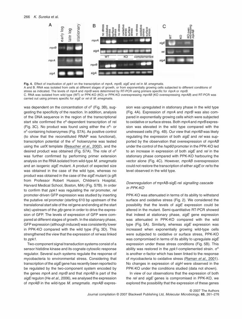
Two-component signal transduction systems consist of asensor histidine kinase and its cognate cytosolic responseregulator. Several such systems regulate the response ofmycobacteria to environmental stress. Considering thattranscription of the sigE gene has recently been reported tobe regulated by the two-component system encoded bythe genes mprA and mprB and that mprAB is part of thesigE regulon (He et al., 2006), we analysed the expressionof mprAB in the wild-type M. smegmatis. mprAB expres-
sion was upregulated in stationary phase in the wild type(Fig. 4A). Expression of mprA and mprB was also com-pared in exponentially growing cells which were subjectedto oxidative or surface stress. Both mprA and mprB expres-sion was elevated in the wild type compared with theunstressed cells (Fig. 4B). Our view that mprAB was likelyregulating the expression of both sigE and rel was sup-ported by the observation that overexpression of mprABunder the control of the hsp60 promoter in the PPK-KO ledto an increase in expression of both sigE and rel in thestationary phase compared with PPK-KO harbouring thevector alone (Fig. 4C). However, mprAB overexpressioncould not restore the transcription of either sigE or rel to thelevel observed in the wild type.
Downregulation of mprAB-sigE-rel signalling cascadein PPK-KO
PPK-KO was attenuated in terms of its ability to withstandsurface and oxidative stress (Fig. 2). We considered thepossibility that the levels of sigE expression could bealtered in the mutant. Semi-quantitative RT-PCR showedthat indeed at stationary phase, sigE gene expressionwas attenuated in PPK-KO compared with the wildtype (Fig. 5A). Similarly, whereas sigE expression wasincreased when exponentially growing wild-type cellswere subjected to oxidative or surface stress, PPK-KOwas compromised in terms of its ability to upregulate sigEexpression under these stress conditions (Fig. 5B). Thisability was restored in the ppk1-complemented strain. sH
is another s-factor which has been linked to the responseof mycobacteria to oxidative stress (Raman et al., 2001).No changes in expression of sigH were observed in thePPK-KO under the conditions studied (data not shown).
In view of our observations that the expression of boththe rel and sigE genes is compromised in PPK-KO, weexplored the possibility that the expression of these genes
Fig. 4. Effect of inactivation of ppk1 on the transcription of mprA, mprB, sigE and rel in M. smegmatis.A and B. RNA was isolated from cells at different stages of growth, or from exponentially growing cells subjected to different conditions ofstress as indicated. The levels of mprA and mprB were determined by RT-PCR using primers specific for mprA or mprB.C. RNA was isolated from wild type (WT) or PPK-KO (KO) or PPK-KO overexpressing mprAB (KO overexpressing mprAB) and RT-PCR wascarried out using primers specific for sigE or rel of M. smegmatis.
266 K. Sureka et al.
© 2007 The AuthorsJournal compilation © 2007 Blackwell Publishing Ltd, Molecular Microbiology, 65, 261–276
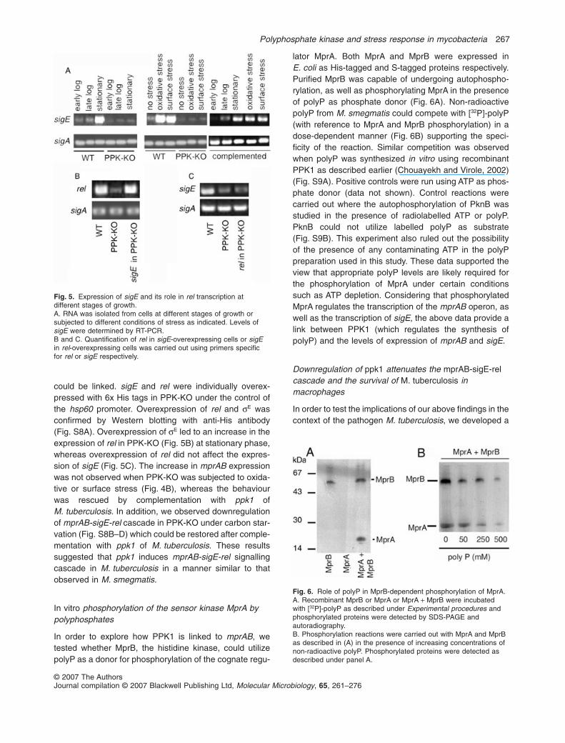
could be linked. sigE and rel were individually overex-pressed with 6x His tags in PPK-KO under the control ofthe hsp60 promoter. Overexpression of rel and sE wasconfirmed by Western blotting with anti-His antibody(Fig. S8A). Overexpression of sE led to an increase in theexpression of rel in PPK-KO (Fig. 5B) at stationary phase,whereas overexpression of rel did not affect the expres-sion of sigE (Fig. 5C). The increase in mprAB expressionwas not observed when PPK-KO was subjected to oxida-tive or surface stress (Fig. 4B), whereas the behaviourwas rescued by complementation with ppk1 ofM. tuberculosis. In addition, we observed downregulationof mprAB-sigE-rel cascade in PPK-KO under carbon star-vation (Fig. S8B–D) which could be restored after comple-mentation with ppk1 of M. tuberculosis. These resultssuggested that ppk1 induces mprAB-sigE-rel signallingcascade in M. tuberculosis in a manner similar to thatobserved in M. smegmatis.
In vitro phosphorylation of the sensor kinase MprA bypolyphosphates
In order to explore how PPK1 is linked to mprAB, wetested whether MprB, the histidine kinase, could utilizepolyP as a donor for phosphorylation of the cognate regu-
lator MprA. Both MprA and MprB were expressed inE. coli as His-tagged and S-tagged proteins respectively.Purified MprB was capable of undergoing autophospho-rylation, as well as phosphorylating MprA in the presenceof polyP as phosphate donor (Fig. 6A). Non-radioactivepolyP from M. smegmatis could compete with [32P]-polyP(with reference to MprA and MprB phosphorylation) in adose-dependent manner (Fig. 6B) supporting the speci-ficity of the reaction. Similar competition was observedwhen polyP was synthesized in vitro using recombinantPPK1 as described earlier (Chouayekh and Virole, 2002)(Fig. S9A). Positive controls were run using ATP as phos-phate donor (data not shown). Control reactions werecarried out where the autophosphorylation of PknB wasstudied in the presence of radiolabelled ATP or polyP.PknB could not utilize labelled polyP as substrate(Fig. S9B). This experiment also ruled out the possibilityof the presence of any contaminating ATP in the polyPpreparation used in this study. These data supported theview that appropriate polyP levels are likely required forthe phosphorylation of MprA under certain conditionssuch as ATP depletion. Considering that phosphorylatedMprA regulates the transcription of the mprAB operon, aswell as the transcription of sigE, the above data provide alink between PPK1 (which regulates the synthesis ofpolyP) and the levels of expression of mprAB and sigE.
Downregulation of ppk1 attenuates the mprAB-sigE-relcascade and the survival of M. tuberculosis inmacrophages
In order to test the implications of our above findings in thecontext of the pathogen M. tuberculosis, we developed a
Fig. 5. Expression of sigE and its role in rel transcription atdifferent stages of growth.A. RNA was isolated from cells at different stages of growth orsubjected to different conditions of stress as indicated. Levels ofsigE were determined by RT-PCR.B and C. Quantification of rel in sigE-overexpressing cells or sigEin rel-overexpressing cells was carried out using primers specificfor rel or sigE respectively.
Fig. 6. Role of polyP in MprB-dependent phosphorylation of MprA.A. Recombinant MprB or MprA or MprA + MprB were incubatedwith [32P]-polyP as described under Experimental procedures andphosphorylated proteins were detected by SDS-PAGE andautoradiography.B. Phosphorylation reactions were carried out with MprA and MprBas described in (A) in the presence of increasing concentrations ofnon-radioactive polyP. Phosphorylated proteins were detected asdescribed under panel A.
Polyphosphate kinase and stress response in mycobacteria 267
© 2007 The AuthorsJournal compilation © 2007 Blackwell Publishing Ltd, Molecular Microbiology, 65, 261–276
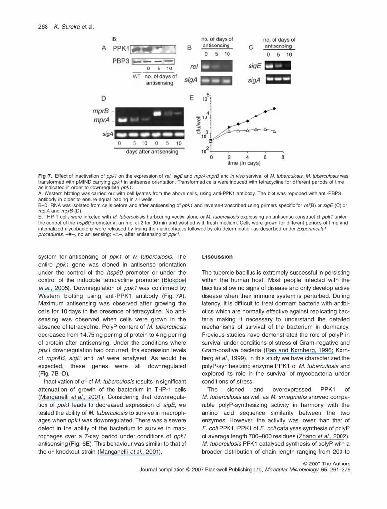
system for antisensing of ppk1 of M. tuberculosis. Theentire ppk1 gene was cloned in antisense orientationunder the control of the hsp60 promoter or under thecontrol of the inducible tetracycline promoter (Blokpoelet al., 2005). Downregulation of ppk1 was confirmed byWestern blotting using anti-PPK1 antibody (Fig. 7A).Maximum antisensing was observed after growing thecells for 10 days in the presence of tetracycline. No anti-sensing was observed when cells were grown in theabsence of tetracycline. PolyP content of M. tuberculosisdecreased from 14.75 ng per mg of protein to 4 ng per mgof protein after antisensing. Under the conditions whereppk1 downregulation had occurred, the expression levelsof mprAB, sigE and rel were analysed. As would beexpected, these genes were all downregulated(Fig. 7B–D).
Inactivation of sE of M. tuberculosis results in significantattenuation of growth of the bacterium in THP-1 cells(Manganelli et al., 2001). Considering that downregula-tion of ppk1 leads to decreased expression of sigE, wetested the ability of M. tuberculosis to survive in macroph-ages when ppk1 was downregulated. There was a severedefect in the ability of the bacterium to survive in mac-rophages over a 7-day period under conditions of ppk1antisensing (Fig. 6E). This behaviour was similar to that ofthe sE knockout strain (Manganelli et al., 2001).
Discussion
The tubercle bacillus is extremely successful in persistingwithin the human host. Most people infected with thebacillus show no signs of disease and only develop activedisease when their immune system is perturbed. Duringlatency, it is difficult to treat dormant bacteria with antibi-otics which are normally effective against replicating bac-teria making it necessary to understand the detailedmechanisms of survival of the bacterium in dormancy.Previous studies have demonstrated the role of polyP insurvival under conditions of stress of Gram-negative andGram-positive bacteria (Rao and Kornberg, 1996; Korn-berg et al., 1999). In this study we have characterized thepolyP-synthesizing enzyme PPK1 of M. tuberculosis andexplored its role in the survival of mycobacteria underconditions of stress.
The cloned and overexpressed PPK1 ofM. tuberculosis as well as M. smegmatis showed compa-rable polyP-synthesizing activity in harmony with theamino acid sequence similarity between the twoenzymes. However, the activity was lower than that ofE. coli PPK1. PPK1 of E. coli catalyses synthesis of polyPof average length 700–800 residues (Zhang et al., 2002).M. tuberculosis PPK1 catalysed synthesis of polyP with abroader distribution of chain length ranging from 200 to
Fig. 7. Effect of inactivation of ppk1 on the expression of rel, sigE and mprA-mprB and in vivo survival of M. tuberculosis. M. tuberculosis wastransformed with pMIND carrying ppk1 in antisense orientation. Transformed cells were induced with tetracycline for different periods of timeas indicated in order to downregulate ppk1.A. Western blotting was carried out with cell lysates from the above cells, using anti-PPK1 antibody. The blot was reprobed with anti-PBP3antibody in order to ensure equal loading in all wells.B–D. RNA was isolated from cells before and after antisensing of ppk1 and reverse-transcribed using primers specific for rel(B) or sigE (C) ormprA and mprB (D).E. THP-1 cells were infected with M. tuberculosis harbouring vector alone or M. tuberculosis expressing an antisense construct of ppk1 underthe control of the hsp60 promoter at an moi of 2 for 90 min and washed with fresh medium. Cells were grown for different periods of time andinternalized mycobacteria were released by lysing the macrophages followed by cfu determination as described under Experimentalprocedures. –�–, no antisensing; –�–, after antisensing of ppk1.
268 K. Sureka et al.
© 2007 The AuthorsJournal compilation © 2007 Blackwell Publishing Ltd, Molecular Microbiology, 65, 261–276
800 (data not shown). PPK1 could also catalyse thereverse reaction, i.e. generation of ATP from polyP (iso-lated from mycobacteria as well from E. coli ) like theE. coli enzyme (Kumble et al., 1996). By analogy withE. coli PPK1, the phosphate group appeared likely tohave been transferred on to the conserved histidine resi-dues at position 491 and 510 of M. tuberculosis PPK1.The importance of these residues was supported byobservations that the H491A, H510A mutation causedcomplete inhibition of autophosphorylation as well aspolyP-synthesizing ability (Table 1). The recently solvedcrystal structure of E. coli PPK1 suggests that PPK1forms an interlocked dimer and that its active site islocated in a tunnel which contains a unique ATP-bindingpocket (Zhu et al., 2004). The dimerization of PPK1 ofE. coli is required for its polyP-synthesizing activity.Dimerization and autophosphorylation were correlated inthe M. tuberculosis PPK1 Homology modelling using theE. coli PPK1 as template suggested that F176 and R230are located in the head domain which likely contributes toone dimer-forming interface (Fig. S10). Our resultssuggest that a positively charged residue at position 230and a hydrophobic phenyl ring at position 176 are likely tobe involved in dimerization activity.
In order to explore the role of ppk1 in the stressresponse of mycobacteria, M. smegmatis was chosen asthe model system. M. tuberculosis encounters differentconditions of stress both when it grows within macroph-ages and when the bacterium resides within a caseousgranuloma. The bacterium must have the ability to adaptto both these conditions. In vitro, M. tuberculosis canpersist for years in the absence of detectable replicationafter nutrient deprivation, whereas replenishing nutrientsrestores active metabolism and growth (Loebel et al.,1933). PPK-KO showed significant reduction in polyPcontent suggesting the role of PPK1 in the synthesis ofpolyP in M. smegmatis. We tested the ability of PPK-KOto withstand different conditions of stress. PPK-KO wascompromised in its ability to survive under conditions ofhypoxia. Considering that complementation of ppk1 ofM. tuberculosis restored the growth defect, it appearedunlikely that the observed phenotypic changes in PPK-KOwere due to polar effects attributable to gene(s) down-stream of ppk1. Mutants of ppk1 could not reverse thegrowth defect (Fig. S6) in harmony with the results of invitro enzymatic assays (Table 1) suggesting the crucialrole of the identified amino acid residues in PPK1function. When microorganisms encounter a nutrient-limited environment they slow their growth rate dramati-cally and reduce levels of RNA and protein synthesis. Thisoverall alteration in metabolism is known as the stringentresponse. Using the mouse model, several persistence-defective mutants deficient in persisting in the face of theimmunological onslaught of the host have been charac-
terized (Hingley-Wilson et al., 2003). In M. tuberculosis,the stringent response is reported to be mediated by(p)ppGpp (hyperphosphorylated guanine nucleotides)which are synthesized by the RelMTB enzyme (Avarbocket al., 2000). Microarray analysis has revealed thatH37Rv DrelMTB suffers from a generalized alteration of thetranscriptional apparatus as well as specific changes inexpression of virulence factors, secreted antigens, heatshock protein and cell wall-synthesizing enzymes (Dahlet al., 2003). The fact that rel has been identified as agene that is required for long-term survival under hypoxicconditions (Primm et al., 2000) prompted us to explorewhether this role was linked to, or independent of thestringent response. The level of the alarmone, ppGpp,was compared between the wild type and PPK-KO. Underconditions of carbon starvation, rel expression was mark-edly impaired in the mutant compared with the wild type,implicating ppk1 in the regulation of rel transcription. Thisfinding assumes particular significance considering thedocumented role of rel in the adaptation to stationaryphase and in long-term persistence of M. tuberculosis inmice. This is the first report linking polyP metabolism tothe stringent response in mycobacteria. The situation inmycobacteria is obviously different from an earlier reportwhere a ppk1-inactivated mutant of S. lividans did notshow any alteration in rel expression or ppGpp biosynthe-sis (Chouayekh and Virole, 2002).
Pathogenic mycobacteria encounter oxidative stress intheir intracellular milieu. The PPK-KO was more suscep-tible to oxidative stress as well as surface stress. Consid-ering that a large number of stress-regulated s-factorshave been linked to the stress response of this bacterium,we focused our attention on the expression of some ofthese extracytoplasmic s-factors in the absence of PPK1.sE and sB have been reported to be upregulated underconditions of oxidative stress, surface stress and lowaeration (Wu et al., 1997; Manganelli et al., 1999). Inharmony with these observations, sE was downregulatedin PPK-KO. We also observed reduced expression of sB inPPK-KO, although not to the extent of sE (unpublishedobservations). Considering that the downregulation of sE
was more pronounced than the downregulation of sB, wefocused our attention on exploring the link between sigEand rel. In this report we demonstrate that the transcrip-tion of the rel gene is regulated by sE, establishing animportant link between the s-factor-dependent stressresponse and the stringent response. We observed anincrease in the rel promoter-driven GFP expression in thestationary phase in both the wild type and PPK-KO.However, the increase observed was consistently lower inthe mutant compared with the wild type (Fig. 3D) suggest-ing that the presence of ppk1 potentiates the efficiency ofrel transcription in mycobacteria. We also observed thatoverexpression of sE from an hsp60 promoter could over-
Polyphosphate kinase and stress response in mycobacteria 269
© 2007 The AuthorsJournal compilation © 2007 Blackwell Publishing Ltd, Molecular Microbiology, 65, 261–276
come the rel expression deficiency associated with inac-tivation of ppk1, suggesting that sE is an importantregulator of rel expression. We hypothesized that optimalpolyP levels (regulated by PPK1) are probably requiredfor phosphorylation reactions under certain conditionssuch as ATP depletion, and therefore searched for thepossible regulation of sE by phosphorylation-dependentbinding of transcription factors. A recent report has dem-onstrated that both sB and sE are regulated by the two-component system comprising the histidine kinase MprBand its cognate regulator MprA which shows enhancedbinding to its target sequences after MprB-dependentphosphorylation. Interestingly, mprAB expression wascompromised in the PPK-KO strain. Overexpression ofmprAB in the PPK-KO was associated with increasedlevels of sigE and rel expression in the stationary phase.It therefore appeared likely that decreased levels of reland sigE in the PPK-KO were linked to attenuation ofmprAB expression. Considering that MprA regulates tran-scription of the mprAB operon as well as sigE and sigBtranscription, it appeared possible that MprB was capableof phosphorylating MprA using polyP as the phosphatedonor. In vitro kinase reactions using recombinant MprAand MprB affirmed our view that MprB could use polyP asa phosphate donor and phosphorylate MprA. In compari-son with the wild type, we observed low but detectablelevels of transcription of the genes of the mprAB-sigE-relcascade in PPK-KO, by semi-quantitative RT-PCR (Figs 4and 5). This could be due to the availability of otherphysiological substrates like ATP or aceyl phosphate(Zahrt et al., 2003) which possibly serve as donors forphosphorylation with a low efficiency in PPK-KO. Thecontribution of other, as yet uncharacterized, polyP-synthesizing enzyme(s) also cannot be ruled out. Thesepossibilities would explain the observation that mprABoverexpression in the PPK-KO led to an increase in reland sigE expression compared with PPK-KO itself. Wespeculate that polyP is a preferred substrate under stressand that PPK1 assumes a critical role under such condi-tions providing a polyP pool for efficient utilization byMprAB. It remains to be tested whether other histidinekinases of two-component systems also utilize polyP asphosphate donor. The mechanism of phosphate donationfrom polyP is open to further investigation.
PolyP metabolism remains unexplored in the context ofmycobacterial physiology and infection. Our studies haveshed light on the polyP-synthesizing enzyme PPK1 ofmycobacteria. We have uncovered a novel regulatorynetwork linking polyP to the optimal response of a two-component system which controls expression of thestress-regulated s-factor sE, which in turn controls theexpression of the stringent response regulator rel.
The observation that ppk1 attenuation compromisesthe ability of M. tuberculosis to survive in macrophages,
increases the importance of this gene in the context of theintracellular life of the bacterium. However, data obtainedby infecting macrophages with any particular mutant ofM. tuberculosis do not necessarily correlate to survivalcharacteristics of the mutant in its host. In fact, theimpaired survival of the M. tuberculosis sigE mutant withinmacrophages reported by Manganelli et al. (2001) doesnot appear to extend to a survival defect in lung andspleen tissues in C3H/HeJ mice (Ando et al., 2003). Nev-ertheless it would be of considerable interest to test therole of ppk1 in persistence, considering that the mprABsignal transduction system has been reported to berequired for persistent infection in the lung (Zahrt andDeretic, 2001).
Experimental procedures
Bacterial strains and growth conditions
The bacterial strains used in this study were E. coli DH5a,E. coli BL21(DE3) and E. coli Top 10. E. coli strains weregrown in Luria–Bertani (LB) Miller media. M. smegmatismc2155 was grown in Middle Brook (MB) 7H9 broth supple-mented with 0.05% Tween 80 or Lemco medium supple-mented with 0.05% Tween 80. Early log, late log andstationary phases refer to 16, 28 and 44 h of growth respec-tively (Dasgupta et al., 2006).
Electroporation in mycobacteria
This was carried out using a Bio-Rad Gene Pulser accordingto the protocol of Snapper et al. (1990).
Construction of expression plasmids for PPK1
ppk1 (Rv2984) of M. tuberculosis H37Rv and M. smegmatismc2155 (MSMEG239) were amplified from cosmid MTCY349(gift from Professor Stewart Cole, Institut Pasteur, Paris) andfrom the genomic DNA of M. smegmatis mc2155, respec-tively, followed by cloning between the NdeI and EcoRI sitesof the expression vector pET 28a (Novagen), or between theKpnI and HindIII sites of pBAD-His B (Invitrogen). Theprimers used are given in Table S1. Mutants of ppk1 weregenerated by overlap extension PCR. The primers used aregiven in Table S2 with restriction sites in bold. The integrity ofall constructs was checked by sequencing.
Expression and purification of PPKs
Recombinant plasmids derived from pET 28a were trans-formed in E. coli BL21(DE3). Cells were grown to an OD600 of0.6. IPTG was added to a final concentration of 0.1 mM andgrowth was continued at 37°C with shaking for 2 h. Recom-binant plasmids derived from pBAD-His B were transformedinto E. coli TOP10 and induced with 0.02% arabinose at 37°Cfor 4 h. Cells were harvested, re-suspended in 10 mM Tris-HCl (pH 7.4), 1 mM MgCl2, 1 mM PMSF, 20 mg ml-1 leupep-
270 K. Sureka et al.
© 2007 The AuthorsJournal compilation © 2007 Blackwell Publishing Ltd, Molecular Microbiology, 65, 261–276
tin, 10 mg ml-1 pepstatin and 10 mg ml-1 aprotinin anddisrupted by sonication. Recombinant His-tagged proteinswere purified from lysates by chromatography on Ni2+-NTAagarose.
Isolation of polyP
Polyphosphate was extracted by the method of Ault-Richeet al. (1998) with minor modifications. E. coli orM. smegmatis cell pellet was dissolved in 5 M guanidineisothiocyanate (GITC) and cells were lysed by heat, SDS andsonication. After addition of ethanol, DNA-binding resin wasadded and the resin was washed with 5 mM Tris-HCl (pH 7.5), 50 mM NaCl, 5 mM EDTA, 50% ethanol. After nucleasetreatment, polyP was eluted with water of pH 8 at 95°C. Sizesof polyP were determined after separation by 20% PAGEcontaining 7 M urea followed by autoradiography (Tzengand Kornberg, 2000). For isolation of 32P-labelled polyP,M. smegmatis was grown in low phosphate (0.15 mM)-containing Hartmans-de-Bont medium (Hartmans and DeBont, 1992) to an OD600 of 0.2 followed by the addition of[g-32P]-H3PO4 (BRIT, Hyderabad, India) to a final concentra-tion of 100 mCi ml-1. Cells were grown and [32P]-polyP wasisolated as described above.
Autophosphorylation and assay for enzyme activities
Polyphosphate kinase autophosphorylation was assayed byincubating purified enzyme in 50 mM HEPES-KOH (pH 7),40 mM (NH4)2SO4, 10 mM MgCl2 and 5 mM [g-32P]-ATP at37°C for 5 min (Tzeng and Kornberg, 2000). The reactionwas terminated by adding 40 mM EDTA and samples wereelectrophoresed on a 10% gel followed by autoradiography.The PPK1-catalysed production of acid-insoluble [32P]-polyPfrom ATP was quantified as described by Ahn and Kornberg(1990). PolyP breakdown catalysed by PPK was quantifiedusing the non-radioactive technique of Ault-Riche et al.(1998). The membrane fraction of M. tuberculosis H37Rvwas kindly provided by Dr John Belisle, Colorado State Uni-versity, Fort Collins, CO through NIAID, National Institutes ofHealth Contract NO1-AI-40091. PPK1 of M. tuberculosis wasimmunoprecipitated from the solubilized membranes withrabbit polyclonal antibody raised against recombinant PPK1of M. tuberculosis (at Imgenex, Bhubaneswar, India), fol-lowed by measurement of the enzymatic assay with theimmunoprecipitate.
Non-denaturing PAGE
This was performed using a discontinuous buffer system(Davis, 1964). SDS and b-mercaptoethanol were omittedfrom samples and buffers. Acrylamide concentrations rangingfrom 5% to 12% were used for Ferguson plots (Ferguson,1964).
Construction of suicidal delivery vector
The unmarked deletion mutant of ppk1 of M. smegmatismc2155 was constructed using the allelic replacementmethod of Parish and Stoker (2000). Briefly, the ppk1 gene of
M. smegmatis along with its upstream sequence was PCRamplified using the primer pairs indicated in Table S1 andgenomic DNA of wild-type mc2155 as template. The ampliconwas cloned between the KpnI and Hind III sites of the vectorpUC19 to generate pPPK101. A 450 bp was deleted from theppk1 gene by digesting pPPK101 with BsaI and religating thevector to give rise to pPPK102. The 2.1 kb insert carryingthe disrupted ppk1 gene was excised with KpnI and PacI andcloned between the same sites of p2NIL to generatepPPK103. The final delivery vector pPPK104 was generatedby cloning the PacI cassette (hyg, Pag85-lac Z, Phsp60-sac B)excised from the vector pGOAL 19 at the PacI site ofpPPK103.
Generation of a ppk1 mutant of M. smegmatis
pPPK104 was electroporated into electrocompetent cells ofM. smegmatis mc2155 and plated on Lemco agar platessupplemented with hygromycin B (100 mg ml-1), kanamycin(25 mg ml-1) and X-gal (50 mg ml-1). Blue colonies werereplated onto Lemco agar; a loopful of cells wasre-suspended in Lemco broth, serially diluted and plated ontoLemco agar supplemented with 2% sucrose and X-gal(50 mg ml-1). White kanS, hygS and sucR colonies were iden-tified and isolated.
Genotype analysis
Genomic DNA was prepared from the wild type and the kans,hygs and sucR colonies according to standard procedures.The genotype was checked by PCR using mc2155 genomicDNA and the genomic DNA isolated from kans, hygs and sucR
colonies as templates and the primer pair 5′-TGT GGG CCCCGC CAC GGC ACA GGC CAT-3′ and 5′-ATG TCG TCGGAA ACT TC-3′ which flanked the deletion site. The PCRproducts were sequenced.
Complementation of ppk1 of M. tuberculosis in theM. smegmatis strain inactivated in the ppk1 gene usingan integrating vector
Complementation of ppk1 was achieved by constructing anintegrating vector containing a hygromycin-resistance cas-sette along with a positive-selection L5 integrase cassette.ppk1 of M. tuberculosis was first cloned in pOLYG (O’Gaoraet al., 1997), under the control of the hsp60 promoter andhsp60–ppk1 was excised and cloned between the same sitesin pUC19 to generate pPPK105. A 3.7 kb Hyg-integrase cas-sette from pUC-HY-INT (Mahenthiralingam et al., 1998), wascloned at the single HindIII site of pPPK105 to generate theintegrating vector pPPK106. pPPK106 was electroporatedinto electrocompetent cells of the PPK-KO strain in order tocomplement the mutant strain with a single copy of the wild-type ppk1 of M. tuberculosis. The presence of the ppk1 genewas confirmed by PCR with ppk1-specific primers.
Isolation and detection of ppGpp
Mycobacterium smegmatis was grown in low phosphate(0.15 mM)-containing medium (Hartmans and De Bont,1992) to an OD600 of 0.2 followed by the addition of [g-32P]-
Polyphosphate kinase and stress response in mycobacteria 271
© 2007 The AuthorsJournal compilation © 2007 Blackwell Publishing Ltd, Molecular Microbiology, 65, 261–276
H3PO4 (BRIT, Hyderabad, India) to a final concentration of100 mCi ml-1. The labelled cells grown up to an OD600 of 0.8were harvested, washed, treated with 1 mg of lysozyme perml (in 10 mM Tris, pH 8.0 on ice for 20 min), and lysed with1% SDS. ppGpp was extracted with an equal volume of 2 Mformic acid. After centrifugation at 4°C, 5 ml of the supernatantwas loaded on a polyethyleneimine (PEI)-coated thin-layerchromatography (TLC) plate (Sigma) (Ojha et al., 2000). Theplate was developed in 1.5 M KH2PO4 (pH 3.5) in one dimen-sion, air-dried and exposed to X-ray film. Labelled ppGppisolated from E. coli strain CF3120, overexpressing theppGpp synthase gene, rel (gift from Professor DipankarChatterji, Indian Institute of Science, Bangalore), was usedas standard on TLC plates.
RNA preparation and RT-PCR
Cells were lysed by agitation with glass beads (0.1 mm) in aMini bead beater apparatus (BioSpec Products, Bartlesville,OK) (Stahl et al., 2001). RNA was isolated from cell lysatesusing the RNeasy kit according to the manufacturer’s proto-col (Qiagen GmbH, Germany). Purified RNA was eluted withRNase-free water and treated with RNase-free DNase I for30 min at 37°C. RNA (100 ng) was reverse-transcribed usingthe Titanium one-step RT-PCR kit (Clontech). The primerpairs used for RT-PCR are given in Table S3. Relative quan-tities of cDNA were determined from the products generatedby the amplification of genomic DNA after serial dilutions,using the appropriate primers, and were normalized foramounts of sigA or 16s rRNA as their expression remainsunchanged during stress or at different growth phases. TheDNA-free RNA preparation was checked by DNA-PCR (–RT)of the housekeeping gene. RT was performed only on RNApreparations confirmed to be free of contaminating DNAaccording to the manufacturer’s instructions using annealingtemperatures of 55°C for rel, sigA, 16s rRNA, mprA andmprB; 60°C for sigE; and 50°C for ppk1. All samples weresubjected to identical reactions lacking reverse transcriptasein order to confirm the absence of any genomic DNA.
PPK1 structural modelling
PPK1 (SWISSPROT Accession No. P65768) fromM. tuberculosis was modelled by using the First ApproachMode at the Swiss-Model protein structure homology model-ling server (http://ca.expasy.org) using the structural templateof E. coli PPK1. Amino acids 48–742 of M. tuberculosis PPK1were included in the model. The graphics were produced byusing Swiss Pdb viewer (Peitsch, 1996; Guex and Peitsch,1997).
Anaerobic survival assays
These assays were performed as described by Primm et al.(2000). Briefly, wild-type M. smegmatis and the ppk1 mutantwere grown to an OD600 of approximately 0.2–0.3 in 7H9medium supplemented with 2% glucose and 0.05% Tween 80(7H9-glucose-Tween 80). For anaerobic survival assays,bacteria were diluted to an OD of 0.05 in fresh 7H9-glucose-Tween 80 and were aliquoted into 1.5 ml screw-cap tubes
with rubber septa with no headspace and incubated station-ary at 37°C. Control tubes with methylene blue indicator dyedemonstrated that all oxygen was consumed after 4–5 days.For all experiments, the number of colony-forming units (cfu)per millilitre was determined by plating in triplicate onto 7H11plates.
Survival assays under surface or oxidative stress
For determination of survival following detergent or hydrogenperoxide exposure, cultures of wild-type M. smegmatis orPPK-KO were grown overnight in 7H9-glucose-Tween 80 at30°C to an OD600 of 0.3–0.4. Cultures were then diluted infresh medium to an OD600 of 0.2, and exposed to 0.01% SDS(detergent stress). Cultures were incubated with shaking at30°C and aliquots were removed from each culture andplated at the indicated time points. Colonies were countedafter 3–4 days with duplicate or triplicate plates counted ateach of two dilutions. Experiments were performed at leastthrice for each stress condition. For oxidative stress, cellswere exposed to 50 mM hydrogen peroxide for 1 h, followedby addition of hydrogen peroxide to 5 mM, followed by platingof dilutions as described above.
Detection of oxidized proteins
Oxidized proteins containing carbonyl groups were detectedusing the Oxyblot kit (Chemicon) as described (Lee et al.,2005).
Analysis of the activity of the rel promoter
The rel promoter (-610 to +1 relative to the translational startsite) was amplified from the genomic DNA of M. smegmatisusing primers depicted in Table S4 and cloned into thepromoter-less replicative gfp vector pFPV27 (Valdivia et al.,1996) using asymmetric KpnI and BamHI sites. The resultingplasmid was electroporated into wild-type M. smegmatismc2155, the isogenic PPK-KO and the ppk1-complementedstrain. Cells were grown in 7H9 broth containing kanamycin(25 mg ml-1) at 37°C. The cells were harvested by centrifuga-tion, washed once in PBS/0.05% Tween 80 and re-suspended in the same buffer to an OD600 of approximately 1.Fluorescence was measured with a spectrofluorimeter usingexcitation and emission wavelengths of 480 and 510 nmrespectively (Blokpoel et al., 2003).
Overexpression of mprAB, sigE and rel in PPK-KO
The mprAB, sigE and rel genes were amplified from thegenomic DNA of M. smegmatis using primers shown inTable S4, and the genes were cloned between the NdeI (orEcoRI) and HindIII sites of a shuttle vector under the controlof the hsp60 promoter (Choudhuri et al., 2002).
In vitro transcription assays and analysis of thetranscriptional start site
Recombinant RNAP subunits were purified and reconstitutedas described by Jacques et al. (2006). Recombinants-factors were purified as described by Jacques et al.
272 K. Sureka et al.
© 2007 The AuthorsJournal compilation © 2007 Blackwell Publishing Ltd, Molecular Microbiology, 65, 261–276
The rel promoter template for in vitro transcription assayswas a 1540 bp PCR product amplified with the primers5′-TAAGGTACCGAAGTTGTCGAAATGACGT-3′ (sense)and 5′-TTAAAGCTTTTGACGATCATCTTCTGGTAG-3′(antisense). The other template used was a 269 bp NheI–HindIII fragment from pYZ99 for the usfXP1 promoter(Jacques et al., 2006). The in vitro transcription protocol usedin this study was adapted from that used by Jacques et al.(2006). Briefly, reactions were carried out in a final volume of40 ml containing 45 mM Tris-HCl (pH 7.9), 5 mM MgCl2,75 mM KCl, 1 mM dithiothreitol, 250 mg ml-1 bovine serinealbumin (BSA) and 13% glycerol. Core RNAP (22.5 pmol)and s-factor (3 pmol) were mixed and incubated for 30 min at37°C in the transcription reaction buffer. Transcription reac-tions were initiated by adding 2 ml of NTPs. The final ATP,UTP, GTP concentration were 0.15 mM and the final CTPconcentration was 0.4 mM. Immediately after transcription,2 ml of a solution containing 6 mg of heparin, along with0.15 mM of CTP, was added. Reactions were left for another10 min at 37°C. Transcription reactions were stopped byadding 2 ml of 7.5 M ammonium acetate, 1 ml of 5 mg ml-1
bacterial tRNA and 100 ml of ice-cold ethanol. RNAs werethen precipitated at -80°C and washed with 70% ethanol.The pellets were re-suspended in 10 ml of water. Template-specific oligonucleotides were radiolabelled using [g-32P]-ATPand T4 polynucleotide kinase and used as probes for detec-tion by primer extension using the Rel- and UsfX-specificoligonucleotides 5′-AGCAGCGCGGCCACCAGCGTGGTGGTGT-3′, 5′-TCTGGTCTTCGAGCTGGTCGGTCATGGTC-3′, respectively, according to standard procedures. Primerextension assays were performed using template-specific oli-gonucleotides radiolabelled with [g-32P]-ATP and T4 poly-nucleotide kinase as described by Beaucher et al. (2002).Sequencing reactions were performed using the same primeras that used for primer extension, and run in adjacent lanesof 6% acrylamide-urea sequencing gel to determine the startsite of the transcripts.
Construction of a vector for antisensing of ppk1
The M. tuberculosis ppk1 coding region was amplified byPCR using the oligonucleotide primer pair 5′-TAGGATCCTCAGGGGCTGCGGTGC-3′ and 5′-TATCATATGATGAGCAATGATCGC-3′, followed by cloning of the PCRproduct in reverse orientation in the vector pMIND (Blokpoelet al., 2005) between the BamHI and NdeI sites (in bold) togenerate the plasmid pPPK103. pPPK103 was electropo-rated in M. tuberculosis in order to obtain a strain expressingthe antisense mRNA of ppk1. Transformants were grown upto mid log phase and induced with tetracycline (20 ng ml-1) fordifferent periods of time up to 15 days. The expression levelof PPK1 was determined by immunoblotting. In experimentswhere the survival of M. tuberculosis was assessed in mac-rophages, ppk1 was downmodulated by expression of thegene in antisense orientation under the control of the hsp60promoter (Choudhuri et al., 2002).
Expression and purification of MprA and MprB
The mprA and mprB genes of M. smegmatis were amplifiedusing primers depicted in Table S1 and genomic DNA
from M. smegmatis, and cloned in pET28a and pET29arespectively. Recombinant plasmids were transformed inE. coli BL21(DE3) pLysS. After induction with 0.1 mM IPTG,His-MprA and S tag-MprB were purified from the cell lysatesusing Ni2+-NTA agarose and S-protein agarose, respectively,according to the manufacturers’ protocol.
Phosphorylation of MprA and MprB
The soluble fractions of the cell lysate from E. coli cellsexpressing S-tagged MprB were adsorbed on S-proteinagarose pre-equilibriated with 20 mM Tris-HCl (pH 7.5),150 mM NaCl and 0.1% Triton X-100. The bound protein waspelleted, first washed with the same buffer and then withkinase buffer containing 50 mM Tris-HCl (pH 7.6), 50 mM KCland 50 mM MgCl2, and re-suspended in the same buffer. Thereaction was started with the addition of the substrate MprAand 50 mM [32P]-polyP and continued for 30 min at 37°C.Control phosphorylation reactions were carried out in thepresence of [g-32P]-ATP. Reactions were stopped by additionof Laemmli SDS-PAGE denaturation buffer and sampleswere electrophoresed on a 12.5% gel followed byautoradiography. Phosphorylation in the presence of [32P]-polyP was chased by increasing concentrations of non-radioactive polyP isolated either from M. smegmatis or polyPsynthesized in vitro. As a control, PknB of M. tuberculosiswas purified (Dasgupta et al., 2006) and phosphorylation wascarried out with [g-32P]-ATP.
Infection of THP-1 with M. tuberculosis
THP-1 cells (obtained from the National Centre for CellScience, Pune, India) were maintained in RPMI1640 supple-mented with 10% fetal bovine serum. Cells were treated withphorbol 12-myristate 13-acetate (20 nM) to induce matura-tion to macrophages (Bhattacharyya et al., 2002). Cells wereinfected with M. tuberculosis H37Rv at a multiplicity of infec-tion (moi) of 2 for 1.5 h at 37°C. Wells were washed toremove extracellular bacteria and incubated in the presenceof fresh medium at 37°C for the indicated periods of time.Intracellular bacteria were released by lysing the macroph-ages with 500 ml of 0.05% SDS, diluted in 7H9 broth andplated to determine the viable counts. The media removedfrom the wells were also plated to determine the number ofextracellular bacteria.
Acknowledgements
This work was supported by a grant from the Department ofBiotechnology, Government of India to M.K. The authorswould like to thank all those who gifted reagents used in thisstudy.
References
Ahn, K., and Kornberg, A. (1990) Polyphosphate kinase fromEscherichia coli. Purification and demonstration of aphosphoenzyme intermediate. J Biol Chem 265: 11734–11739.
Polyphosphate kinase and stress response in mycobacteria 273
© 2007 The AuthorsJournal compilation © 2007 Blackwell Publishing Ltd, Molecular Microbiology, 65, 261–276
Akiyama, M., Crooke, E., and Kornberg, A. (1992) The poly-phosphate kinase gene of Escherichia coli. Isolation andsequence of the ppk gene and membrane location of theprotein. J Biol Chem 267: 22556–22561.
Ando, M., Yoshimatsu, T., Ko, C., Converse, P.J., and Bishai,W.R. (2003) Deletion of Mycobacterium tuberculosis sigmafactor E results in delayed time to death with bacterialpersistence in the lungs of aerosol-infected mice. InfectImmun 71: 7170–7172.
Ault-Riche, D., Fraley, C.D., Tzeng, C.M., and Kornberg, A.(1998) Novel assay reveals multiple pathways regulatingstress-induced accumulation of inorganic polyphosphate inEscherichia coli. J Bacteriol 180: 1841–1847.
Avarbock, D., Avarbock, A., and Rubin, H. (2000) Differentialregulation of opposing RelMtb activities by the aminoacy-lation state of a tRNA ribosome.mRNA.RelMtb complex.Biochemistry 39: 11640–11648.
Beaucher, J., Rodrigue, S., Jacques, P.-E., Smith, I., Brzez-inski, R., and Gaudreau, L. (2002) Novel Mycobacteriumtuberculosis anti-s factor antagonists control sF activity bydistinct mechanisms. Mol Microbiol 45: 1527–1540.
Berlett, B.S., and Stadtman, E.R. (1997) Protein oxidation inaging, disease, and oxidative stress. J Biol Chem 272:20313–20316.
Bhattacharyya, A., Pathak, S., Datta, S., Chattopadhyay, S.,Basu, J., and Kundu, M. (2002) Mitogen-activated proteinkinases and nuclear factor-kB regulate Helicobacter pylori-mediated interleukin-8 release from macrophages.Biochem J 368: 121–129.
Blokpoel, M.C., Murphy, H.N., O’Toole, R., Wiles, S., Runn,E.S., Stewart, G.R., et al. (2005) Tetracycline-induciblegene regulation in mycobacteria. Nucleic Acids Res 33:e22.
Blokpoel, M.C.J., O’Toole, R., Smeulders, M.J., and Williams,H.D. (2003) Development and application of unstableGFP variants to kinetic studies of mycobacterial geneexpression. J Microbiol Methods 54: 203–211.
Boshoff, H.I.M., Myers, T.G., Copp, B.R., McNeil, M.R.,Wilson, M.A., and Barry, C.E., III (2004) The transcriptionalresponses of Mycobacterium tuberculosis to inhibitors ofmetabolism: novel insights into drug mechanisms of action.J Biol Chem 279: 40174–40184.
Brown, M.R., and Kornberg, A. (2004) Inorganic polyphos-phate in the origin and survival of species. Proc Natl AcadSci USA 101: 16085–16087.
Chouayekh, H., and Virole, M.J. (2002) The polyphosphatekinase plays a negative role in the control of antibioticproduction in Streptomyces lividans. Mol Microbiol 43:919–930.
Choudhuri, B.S., Bhakta, S., Barik, R., Basu, J., Kundu, M.,and Chakrabarti, P. (2002) Overexpression and functionalcharacterization of an ABC (ATP-binding cassette) trans-porter encoded by the genes drrA and drrB of Mycobacte-rium tuberculosis. Biochem J 367: 279–285.
Dahl, J.L., Kraus, C.N., Boshoff, H.I., Doan, B., Foley, K.,Avarbock, D., et al. (2003) The role of RelMtb-mediatedadaptation to stationary phase in long-term persistence ofMycobacterium tuberculosis in mice. Proc Natl Acad SciUSA 100: 10026–10031.
Dasgupta, A., Datta, P., Kundu, M., and Basu, J. (2006) Theserine/threonine kinase PknB of Mycobacterium tuberculo-
sis phosphorylates PBPA, a penicillin-binding proteinrequired for cell division. Microbiology 152: 493–504.
Davis, B.J. (1964) Disc electrophoresis. II. Method and appli-cation to human serum proteins. Ann NY Acad Sci 121:404–427.
Ferguson, K.A. (1964) Starch-gel electrophoresis-applicationto the classification of pituitary proteins and polypeptides.Metabolism 13: 985–1002.
Fernandes, N.D., Wu, Q.L., Kong, D., Puyang, X., Garg, S.,and Husson, R.N. (1999) A mycobacterial extracytoplasmicsigma factor involved in survival following heat shock andoxidative stress. J Bacteriol 181: 4266–4274.
Ghorbel, S., Kormanec, J., Artus, A., and Virolle, M. (2006)Transcriptional studies and regulatory interactions betweenthe phoR-phoP operon and the phoU, mtpA, and ppkgenes of Streptomyces lividans TK24. J Biol Chem 188:677–686.
Gomez, J.E., Chen, J.M., and Bishai, W.R. (1997) Sigmafactors of Mycobacterium tuberculosis. Tuber Lung Dis 78:175–183.
Guex, N., and Peitsch, M.C. (1997) SWISS-MODEL and theSwiss-PdbViewer: an environment for comparative proteinmodeling. Electrophoresis 18: 2714–2723.
Hartmans, S., and De Bont, J.A.M. (1992) The genusMycobacterium-nonmedical. In The Prokaryotes, a Hand-book on the Biology of Bacteria: Ecophysiology, Isolation,Identification, Application. Balows, A., Truper, H.G.,Dworkin, M., Harder, W., and Schleifer, K.H. (eds). NewYork: Springer-Verlag, pp. 1215–1237.
He, H., Hovery, R., Kane, J., Singh, V., and Zahrt, T.C. (2006)MprAB is a stress-responsive two-component system thatdirectly regulates expression of sigma factors SigB andSigE in Mycobacterium tuberculosis. J Bacteriol 188:2143–2143.
Hingley-Wilson, S.M., Sambandamurthy, V.K., and Jacobs,W.R. (2003) Survival perspectives from the world’s mostsuccessful pathogen, Mycobacterium tuberculosis. NatImmunol 4: 949–955.
Jacques, J.-F., Rodrigue, S., Brzezinski, R., and Gaudreau, L.(2006) A recombinant Mycobacterium tuberculosis in vitrotranscription system. FEMS Microbiol Lett 255: 140–147.
Kornberg, A., Rao, N.N., and Ault-Riché, D. (1999) Inorganicpolyphosphate: a molecule of many functions. Annu RevBiochem 68: 89–125.
Kumble, K.D., Ahn, K., and Kornberg, A. (1996) Phosphohis-tidyl active sites in polyphosphate kinase of Escherichiacoli. Proc Natl Acad Sci USA 93: 14391–14395.
Kuroda, A., and Kornberg, A. (1997) Polyphosphate kinaseas a nucleoside diphosphate kinase in Escherichia coli andPseudomonas aeruginosa. Proc Natl Acad Sci USA 94:439–442.
Kuroda, A., Tanaka, S., Ikeda, T., Kato, J., Takiguchi, N., andOhtaki, H. (1999) Inorganic polyphosphate kinase isrequired to stimulate protein degradation and for adapta-tion to amino acid starvation in Escherichia coli. Proc NatlAcad Sci USA 96: 14264–14269.
Lee, E.-J., Karoonuthaisiri, N., Kim, H.-S., Park, J.-H., Cha,C.-J., Kao, C.M., and Roe, J.-H. (2005) A master regulatorsB governs osmotic and oxidative response as well asdifferentiation via a network of sigma factors in Streptomy-ces coelicolor. Mol Microbiol 57: 1252–1264.
274 K. Sureka et al.
© 2007 The AuthorsJournal compilation © 2007 Blackwell Publishing Ltd, Molecular Microbiology, 65, 261–276
Levine, R.L., Garland, D., Oliver, C.N., Amici, A., Climent, I.,Lenz, A.G., et al. (1990) Determination of carbonyl contentin oxidatively modified proteins. Methods Enzymol 186:446–478.
Loebel, R.O., Shorr, E., and Richardson, H.B. (1933) Theinfluence of adverse conditions upon the respiratorymetabolism and growth of human tubercle bacilli.J Bacteriol 26: 167–200.
Mahenthiralingam, E., Marklund, B.I., Brooks, L.A., Smith,D.A., Bancroft, G.J., and Stokes, R.W. (1998) Site-directedmutagenesis of the 19-kilodalton lipoprotein antigenreveals no essential role for the protein in the growth andvirulence of Mycobacterium intracellulare. Infect Immun66: 3626–3634.
Manganelli, R., Dubnau, E., Tyagi, S., Kramer, F.R., andSmith, I. (1999) Differential expression of 10 sigma factorgenes in Mycobacterium tuberculosis. Mol Microbiol 31:715–724.
Manganelli, R., Voskuil, M.I., Schoolnik, G.K., and Smith, I.(2001) The Mycobacterium tuberculosis ECF sigma factorsE: role in global gene expression and survival inmacrophages. Mol Microbiol 41: 423–437.
Nyka, W. (1974) Studies on the effect of starvation onmycobacteria. Infect Immun 5: 843–850.
O’Gaora, P., Bernini, S., Hayward, C., Filley, E., Rook, G.,Young, D., and Thole, J. (1997) Mycobacteria as immuno-gens: development of expression vectors in multiple myco-bacterial species. Med Princ Pract 6: 91–96.
Ojha, A.K., Mukherjee, T.K., and Chatterji, D. (2000) Highintracellular level of guanosine tetraphosphate in Mycobac-terium smegmatis changes the morphology of thebacterium. Infect Immun 68: 4084–4091.
Parish, T., and Stoker, N.G. (2000) Use of a flexible cassettemethod to generate a double unmarked Mycobacteriumtuberculosis tlyA plcABC mutant by gene replacement.Microbiology 146: 1969–1975.
Peitsch, M.C. (1996) ProMod and Swiss-Model: Internet-based tools for automated comparative protein modelling.Biochem Soc Trans 24: 274–279.
Primm, T.P., Andersen, S.J., Mizrahi, V., Avarbock, D.,Rubin, H., and Barry, C.E. (2000) The stringent responseof Mycobacterium tuberculosis is required for long-termsurvival. J Bacteriol 182: 4889–4898.
Raman, S., Song, T., Puyang, X., Bardarov, S., Jacobs,W.R., and Husson, R.N. (2001) The alternative sigmafactor SigH regulates major components of oxidative andheat stress responses in Mycobacterium tuberculosis.J Bacteriol 183: 6119–6125.
Rao, N.N., and Kornberg, A. (1996) Inorganic polyphosphatesupports resistance and survival of stationary-phaseEscherichia coli. J Bacteriol 178: 1394–1400.
Rao, N.N., and Kornberg, A. (1998) Inorganic polyphosphateregulates responses of Escherichia coli to nutritional strin-gencies, environmental stresses and survival in the station-ary phase. Prog Mol Subcell Biol 23: 183–195.
Rashid, M.H., Rao, N.N., and Kornberg, A. (2000a) Inorganicpolyphosphate is required for motility of bacterialpathogens. J Bacteriol 182: 225–227.
Rashid, H., Rumbaugh, K., Pousador, L., Davin, D.G.,Hamood, A.N., Iglewski, B.H., and Kornberg, A. (2000b)Polyphosphate kinase is essential for biofilm development,
quorum sensing, and virulence of Pseudomonasaeruginosa. Proc Natl Acad Sci USA 97: 9636–9641.
Reese, M.G. (2001) Application of a time-delay neuralnetwork to promoter annotation in the Drosophila melano-gaster genome. Comput Chem 26: 51–56.
Rodrigue, S., Provvedi, R., Jacques, P.-E., Gaudreau, L., andManganelli, R. (2006) The sigma factors of Mycobacteriumtuberculosis. FEMS Microbiol Rev 30: 926–931.
Shiba, T., Tsutsumi, K., Yano, H., Ihara, Y., Kameda, A.,Tanaka, K., et al. (1997) Inorganic polyphosphate and theinduction of rpoS expression. Proc Natl Acad Sci USA 14:11210–11215.
Snapper, S.B., Melton, R.E., Mustafa, S., Kieser, T., andJacobs, W.R., Jr (1990) Isolation and characterization ofefficient plasmid transformation mutants of Mycobacteriumsmegmatis. Mol Microbiol 4: 1911–1919.
Stahl, C., Kubletzko, S., Kaps, I., Seeber, S., Engelhard, H.,and Niederweis, M. (2001) MspA provides the main hydro-philic pathway through the cell wall of Mycobacteriumsmegmatis. Mol Microbiol 40: 451–464.
Tzeng, C.M., and Kornberg, A. (2000) The multiple activitiesof polyphosphate kinase of Escherichia coli and theirsubunit structure determined by radiation target analysis.J Biol Chem 275: 3977–3983.
Valdivia, R.H., Hromockyj, A.E., Monack, D., Ramakrishnan,L., and Falkow, S. (1996) Applications for the greenfluorescent protein (GFP) in the study of host–pathogeninteractions. Gene 173: 47–52.
Wu, Q.L., Kong, D., Lam, K., and Husson, R.N. (1997) Amycobacterial extracytoplasmic function sigma factorinvolved in survival following stress. J Bacteriol 179: 2922–2929.
Zahrt, T.C., and Deretic, V. (2001) Mycobacterium tubercu-losis signal transduction system required for persistentinfections. Proc Natl Acad Sci USA 98: 12706–12711.
Zahrt, T.C., Wozniak, C., Jones, D., and Trevett, A. (2003)Functional analysis of the Mycobacterium tuberculosisMprAB two-component signal transduction system. InfectImmun 71: 6962–6970.
Zhang, H., Ishige, K., and Kornberg, A. (2002) A polyphos-phate kinase (PPK2) widely conserved in bacteria. ProcNatl Acad Sci USA 99: 16678–16683.
Zhu, Y., Hang, W., Lee, S.S.K., and Xu, W. (2004) Crystalstructure of a polyphosphate kinase and its implications forpolyphosphate synthesis. EMBO Rep 6: 681–687.
Supplementary material
The following supplementary material is available for thisarticle:Fig. S1. Alignment of the deduced amino acid sequencesof the PPK1 orthologues of M. tuberculosis (M.tb), andM. smegmatis (M.smeg) using CLUSTAL W.Fig. S2. Overexpression, purification and immunologicaldetection of PPK1 of mycobacteria and hydropathy plot.Fig. S3. Expression of ppk1 at different stages of growth andPCR analysis of the ppk1 knockout mutant.A. RNA isolated from M. smegmatis at early log, late log andstationary phase was reverse-transcribed by RT-PCR.B. Chromosomal DNA from wild-type M. smegmatis mc2155(wt) or PPK-KO was used as template for PCR analysis. The
Polyphosphate kinase and stress response in mycobacteria 275
© 2007 The AuthorsJournal compilation © 2007 Blackwell Publishing Ltd, Molecular Microbiology, 65, 261–276
sizes of the PCR products were analysed by agarose gelelectrophoresis. The first lane is the 1 kb ladder.Fig. S4. Role of PPK1 in growth under oxidative or surfacestress. Cultures growing logarithmically were subjected tooxidative stress. The oxidized (carbonylated) proteins in wildtype (WT), PPK-KO and the ppk1-complemented PPK-KO(complemented) strain were detected using the OxyBlot kit.Samples prepared from equal numbers of cells were analy-sed for carbonylated proteins by Western blotting after SDS-PAGE. The results shown are representative of three differentexperiments.Fig. S5. Role of PPK1 mutants in growth under oxidative orsurface stress or anaerobiosis. Logarithmically growingPPK-KO transformed with full-length wild-type (wt)M. tuberculosis ppk1 or its mutants (as indicated) were sub-jected to oxidative (top) or surface (detergent) (middle) orhypoxic (bottom) stress. Growth was monitored over thecourse of time by determining cfu. Results represent themeans � SD of three separate experiments.Fig. S6. Synthesis of ppGpp and the sequence of the puta-tive rel promoter.A. Cells were grown in the presence of [g-32P]-H3PO4 andppGpp present in the formic acid extract was separated byTLC followed by autoradiography.B. The sequence of the putative rel promoter region ofM. smegmatis. The arrow indicates the start of the gene.C. Alignment of rel promoter region from M. smegmatis andM. tuberculosis.Fig. S7. In vitro transcription assays and primer extensionanalysis of rel.A. In vitro transcription of the usfX promoter was carried outin the presence of sF (first lane, positive control) and that ofthe rel promoter was carried out in the presence of sB, or sF
or sE as indicated (second to fourth lanes).B. Primer extension analysis for the rel promoter was carriedout with RNA isolated from M. smegmatis (lane 2) or from thesE-inactivated mutant (lane 3) and the size of the product wascompared with the product obtained by in vitro transcription ofthe rel promoter as described under Fig. 3B (lane 1).Fig. S8. Western blot analysis of sE or rel expression andRT-PCR analysis for sE, mprA and mprB.A. Western blots was performed using anti-His antibody onlysates of the PPK-KO strain harbouring (a) vector alone orvector expressing either (b) His-Rel or (c) His-sE.
B. RNA was isolated from stationary-phase cells of the wildtype (WT) or PPK-KO (KO), or PPK-KO overexpressingppk1(com).Quantification of sigE (B) or mprA (C) or mprB (D) wascarried out by RT-PCR.Fig. S9. Phosphorylation of mprA and mprB by polyP.A. Role of polyP in MprB and MprA phosphorylation. Recom-binant MprA and MprB were incubated with [g-32P]-polyP(synthesized in vitro using PPK1 and [g-32P]-ATP), and phos-phorylated proteins were detected by SDS-PAGE andautoradiography.B. Phosphorylation reactions were carried out with recombi-nant PknB of M. tuberculosis using [g-32P]-ATP alone (top,first lane) or in the presence of either increasing concentra-tions of non-radioactive polyP (top) or non-radioactive ATP(bottom, second to fifth lanes). Phosphorylated proteins weredetected as described above. The first lane of the bottompanel shows the result of a phosphorylation reaction carriedout with PknB and [g-32P]-labelled polyP (first lane of thebottom panel).Fig. S10. Structural model of M. tuberculosis PPK1. PPK1 ofM. tuberculosis was modelled using the structural template ofE. coli PPK1. The head domain is shown in purple whereasthe rest of the protein is depicted in blue. The residues F176(PHE176) and R230 (ARG230) which are involved in polyP-synthesizing activity are indicated.Table S1. Primers for cloning the ppk1 genes ofM. tuberculosis and M. smegmatis, for generating the suicidevector (pPPK101), for inactivation of ppk1 of M. smegmatis,and for cloning the mprA and mprB genes of M. smegmatis.Table S2. Primers used for mutation of ppk1.Table S3. Primers used for RT-PCR.Table S4. Primers used for cloning constructs of rel andsigE.
This material is available as part of the online article from:http://www.blackwell-synergy.com/doi/abs/10.1111/j.1365-2958.2007.05814.x(This link will take you to the article abstract).
Please note: Blackwell Publishing is not responsible for thecontent or functionality of any supplementary materialssupplied by the authors. Any queries (other than missingmaterial) should be directed to the corresponding author forthe article.
276 K. Sureka et al.
© 2007 The AuthorsJournal compilation © 2007 Blackwell Publishing Ltd, Molecular Microbiology, 65, 261–276
Related Documents