Molecules 2013, 18, 13435-13445; doi:10.3390/molecules181113435 molecules ISSN 1420-3049 www.mdpi.com/journal/molecules Article Polyphenolic Extracts of Edible Flowers Incorporated onto Atelocollagen Matrices and Their Effect on Cell Viability Jorge López-García 1 , Zdenka Kuceková 1,2 , Petr Humpolíček 1,2, *, Jiři Mlček 3 and Petr Sáha 1 1 Centre of Polymer Systems, Tomas Bata University in Zlín, nám. T.G.Masaryka-5555, Zlín 76001, Czech Republic; E-Mails: [email protected] (J.L.-G.); [email protected] (Z.K.); [email protected] (P.S.) 2 Polymer Centre, Faculty of Technology, Tomas Bata University in Zlin, T.G.M. sq. 275, Zlin 76272, Czech Republic 3 Department of Food Analysis and Chemistry, Faculty of Technology, Tomas Bata University in Zlin, nám. T.G.Masaryka-5555, Zlin 76272, Czech Republic; E-Mail: [email protected] * Author to whom any correspondence should be addressed; E-Mail: [email protected]; Tel.: +420-73-479-2298; Fax: +420-57-603-1444. Received: 30 August 2013; in revised form: 3 October 2013 / Accepted: 23 October 2013 / Published: 30 October 2013 Abstract: The phenolic extract of chives flowers (Allium schoenoprasum, Liliaceae), introduced Sage (Salvia pratensis, Lamiaceae), European elderberry (Sambucus nigra, Caprifoliaceae) and common dandelion (Taraxacum officinale, Asteraceae) were characterised by High Performance Liquid Chromatography and incorporated in different concentrations onto atelocollagen thin films. In order to assess the biological impact of these phenolic compounds on cell viability, human immortalised non-tumorigenic keratinocyte cell line was seeded on the thin films and cell proliferation was determined by using an MTT assay. In addition, their antimicrobial activity was estimated by using an agar diffusion test. Data indicated the concomitance between cell viability and concentration of polyphenols. These findings suggest that these phenolic-endowed atelocollagen films might be suitable for tissue engineering applications, on account of the combined activity of polyphenols and collagen. Keywords: edible flowers; polyphenolic extracts; atelocollagen thin films; cell viability; tissue engineering OPEN ACCESS

Welcome message from author
This document is posted to help you gain knowledge. Please leave a comment to let me know what you think about it! Share it to your friends and learn new things together.
Transcript
Molecules 2013, 18, 13435-13445; doi:10.3390/molecules181113435
molecules ISSN 1420-3049
www.mdpi.com/journal/molecules
Article
Polyphenolic Extracts of Edible Flowers Incorporated onto Atelocollagen Matrices and Their Effect on Cell Viability
Jorge López-García 1, Zdenka Kuceková 1,2, Petr Humpolíček 1,2,*, Jiři Mlček 3 and Petr Sáha 1
1 Centre of Polymer Systems, Tomas Bata University in Zlín, nám. T.G.Masaryka-5555, Zlín 76001,
Czech Republic; E-Mails: [email protected] (J.L.-G.); [email protected] (Z.K.);
[email protected] (P.S.) 2 Polymer Centre, Faculty of Technology, Tomas Bata University in Zlin, T.G.M. sq. 275,
Zlin 76272, Czech Republic 3 Department of Food Analysis and Chemistry, Faculty of Technology, Tomas Bata University in Zlin,
nám. T.G.Masaryka-5555, Zlin 76272, Czech Republic; E-Mail: [email protected]
* Author to whom any correspondence should be addressed; E-Mail: [email protected];
Tel.: +420-73-479-2298; Fax: +420-57-603-1444.
Received: 30 August 2013; in revised form: 3 October 2013 / Accepted: 23 October 2013 /
Published: 30 October 2013
Abstract: The phenolic extract of chives flowers (Allium schoenoprasum, Liliaceae),
introduced Sage (Salvia pratensis, Lamiaceae), European elderberry (Sambucus nigra,
Caprifoliaceae) and common dandelion (Taraxacum officinale, Asteraceae) were
characterised by High Performance Liquid Chromatography and incorporated in different
concentrations onto atelocollagen thin films. In order to assess the biological impact of
these phenolic compounds on cell viability, human immortalised non-tumorigenic
keratinocyte cell line was seeded on the thin films and cell proliferation was determined by
using an MTT assay. In addition, their antimicrobial activity was estimated by using an
agar diffusion test. Data indicated the concomitance between cell viability and
concentration of polyphenols. These findings suggest that these phenolic-endowed
atelocollagen films might be suitable for tissue engineering applications, on account of the
combined activity of polyphenols and collagen.
Keywords: edible flowers; polyphenolic extracts; atelocollagen thin films; cell viability;
tissue engineering
OPEN ACCESS

Molecules 2013, 18 13436
1. Introduction
The overriding function of tissue engineering is to design biological substitutes for skin/organ
replacement. Within this framework, collagen is an abundant mammalian protein which is involved in
many important biological functions, such as tissue formation, cell attachment and proliferation. This
protein is employed in diverse fields, including medicines, foods, cosmetics and tissue engineering. It is
deemed as a primary source in biomaterial applications and one of the most useful biomaterials [1–4].
The current medical applications of collagen as a biodegradable material are associated with its natural
properties, including low immune response and the ability to promote cell growth [5].
Skin comprises essentially three types of cell: keratinocytes, melanocytes and fibroblasts. It is
foreseen through wound healing, transplantation and cell culture studies that HaCaT cells may be used
as an in vitro model for highly proliferative epidermis in tissue engineering. HaCaT cell line is
spontaneously transformed into human keratinocytes which have the traits of basal epidermal
keratinocytes. Hence, this cell line may be exploited as an in vitro model for highly proliferative epidermis.
In fact, several wounds have been successfully re-surfaced by culturing autogenic keratinocytes cells [6–8].
The extensive uses of chives (Allium schoenoprasum), introduced sage (Salvia pratensis,
Lamiaceae), European elderberry (Sambucus nigra, Caprifoliaceae) and common dandelion
(Taraxacum officinale) range from culinary for flavouring dishes to medical purposes, such as antitussives,
antiseptics, antifungals, antispasmodics, and anti-inflammatories, amongst other properties [9–12].
These pharmaceutical uses are associated with the rich amount of polyphenols which are found in these
plants [13,14]. It is well established that polyphenols have antitumoral properties by virtue of their
antioxidant activities, which have been studied for many years [15–18]. The polyphenols content and
concentration vary according to each plant, time of the year, and age of the plant, amongst other kinds of
factors [19–23].
Acute care is a serious concern in tissue replacement. Several implants have to be removed by their
poor performances. Indeed, infections, mycoses and inflammations are common causes of biomaterial
implant failure in medicine and by extension, a threat to patients’ lives and a source of high costs [24,25].
Therefore, the main purpose of this contribution is aimed at the addition of the abovementioned
edible flowers extracts onto collagen matrices and to evaluate the effect of their polyphenolic compounds
on keratinocyte cell response and their antibacterial properties by means of cell viability and antimicrobial
studies. In terms of novelty, to the best of our knowledge, it is the first time that those extracts are
incorporated onto any kind of collagen matrix. The findings of this research seek to shed more light on
fields, such as anti-infective biopolymers, phytochemistry, human cell growth and tissue engineering.
2. Results and Discussion
Table 1 summarises the compounds identified in the examined edible flowers along with their
concentrations as detected by HPLC. Ferulic, caffeic and sinapic acid were found as the major
constituents of the methanolic extracts of chives flowers, European elderberry and common dandelion
respectively. Likewise, gallic acid was found in all the extracts.

Molecules 2013, 18 13437
Table 1. Phenolic constituents identified by HPLC. The content of the detected phenolic
compounds is expressed as μg of compound/g of dry matter.
Name (compound number) Wild Chive Introduced Sage European Elderberry Common Dandelion
A. schoenoprasum S. pratensis S. nigra T. officinale
Gallic acid (1) 201.76 22.67 176.61 441.40
Coumaric acid (2) 207.29 / / /
Ferulic acid (3) 887.44 / / /
Rutin (4) 20.26 / / 18.66
Resveratrol (50 / / / 274.92
Vanillic acid (6) / / 299.38 82.88
Sinapic acid (7) / / / 593.04
Catechin (8) / 37.56 / /
Caffeic acid (9) / / 913.19 /
Total phenolic content (mg/g) 17.50 17.10 18.40 17.20
From these compounds, four (ferulic, sinapic coumaric and caffeic acid) belong to the
hydroxycinnamic acids family, two (rutin and catechin) are types of flavonoids, one (vanillic acid) is a
dihydroxybenzoic acid, one (gallic) a trihydroxybenzoic acid, and resveratrol is a stilbenoid. All of these
phenols come from the shikimate and phenylpropanoids metabolic pathways. Indeed, phenylpropanoids
are the largest group of secondary metabolites produced by plants mainly to counteract biotic or abiotic
stresses, where the protective action is related to their antioxidant and free radical scavenging
properties. For instance, the found flavonoids have shown a diversity of biological activities including
antioxidant, antimicrobial, anti-inflammatory, antithrombosis, antihyper-cholesterolemic, anticancer
activities, and spermatozoa activating bioactivity [26,27]. Ferulic acid has anti-inflammatory and
antitumoral properties [28]. Gallic acid induces apoptosis in a series of cancer cell lines, and produces
selective cytotoxicity against tumour cells with higher sensitivity than normal cells [29]. There is a
plethora of information about the health benefits of consuming moderate amounts of red wine, which
contains significant amounts of resveratrol [30–32]. Surprisingly, the flavonol quercetin, which is one
of the most common flavonoids in plants, was not detected in any of these methanolic extracts.
However, rutin, the glycoside formed between quercetin and the disaccharide rutinose was found [33].
It is important to emphasise that this study only examined 10 types of polyphenols and assuredly, there
is a sort of undetected components in the extracts. For instance, volatiles, terpenoids, coumarins,
chalcones, anthocyanins, that may be detected by gas chromatography, by using other HPLC column
with a different polarity or a different solvent gradient [34–38].
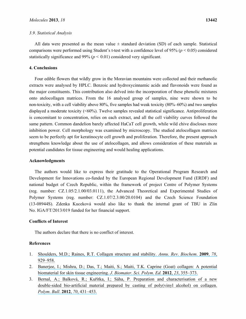
The effect of distinct concentrations of the above studied phenolic extracts on keratinocytes
proliferation is depicted in Figure 1. The dependence of cell viability on concentration is clearly
noticed. In general all the plots have the same pattern, where it shows a fall in cell viability by rising
concentration, and the antiproliferative activity depends upon each particular herb.
Common dandelion (Taraxacum officinale) has a minor effect on cell viability with the highest cell
proliferation rates. This curve exhibits a small drop followed by a plateau towards the end, where cell
viability is around 95%. In contrast, chives flowers (Allium schoenoprasum) show the lowest rates (highest
antiproliferative activity 72%–57%) describing a downwards trend. Introduced sage (Salvia pratensis) and
European elderberry (Sambucus nigra) display similar behaviours and their graphs evince a slight

Molecules 2013, 18 13438
decrease at 10 and 25 μg/mL, followed by a steep decline at the higher concentrations (50 and
100 μg/mL). According to ISO 10993-5, percentages of cell viability above 80% are considered as
non-cytotoxic; within 80%–60% weak; 60%–40% moderate and below 40% strong cytotoxicity,
respectively [39]. Thus, all the studied concentration of Allium schoenoprasum, 50 and 100 μg/mL of
Salvia pratensis, and the highest concentration of Sambucus nigra possess weak toxicity to the
keratinocytes cell line cultivated on the prepared atelocollagen films. The rest of the systems are
innoxious to HaCaT cells. Pristine atelocollagen films were taken as the ones with 100% viability.
Figure 1. Cell viability curves with respect to concentration of the phenolic extracts
incorporated onto the atelocollagen films. They illustrate the connexion between cell
viability and amount of agent applied (the error bars signify standard deviation).
The antiproliferative activity of each extract may be related to the polyphenolic content differences,
along with synergistic effects. For example, it has been substantiated that flavonoids have roughly
higher reactivity than phenolic acids. Likewise, the combination of different subclasses of phytochemicals
usually shows greater influence as a group than as individual entities. This might explain why the
phenolic extract of Salvia pratensis, with only two detected compounds (the flavanol catechin and
gallic acid) and Allium schoenoprasum with 20.26 μg/g of rutin and two hydroxycinnamic compounds,
couramic and ferulic acid have the highest antiproliferative rates. In fact coumaric and ferulic acid
together are efficient modulators of NF-κB activity compared with their effect separately [40–42].
Catechin and gallic acid individually have more reactivity than the rest of the phenolic counterparts
identified here [43]. Gallic acid has three hydroxyls on its phenyl ring and catechin two hydroxyls on
the B ring of its flavonoid backbone. Generally, for benzoic and phenylpropanoids, an increase in the
number of hydroxyl groups results in a higher antioxidant activity. Compounds with two or three
hydroxyl groups on the phenyl ring of phenolic acids or on the B ring of flavonoids present high
antioxidant activity. The loss of one hydroxyl group represents a slight decrease of their activity, but
the loss of two hydroxyl groups significantly diminishes it [44].
Molecules 2013, 18 13439
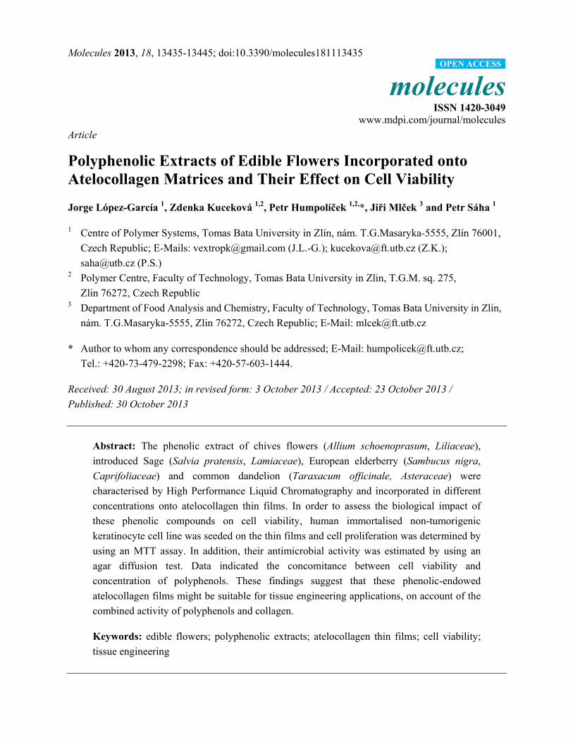
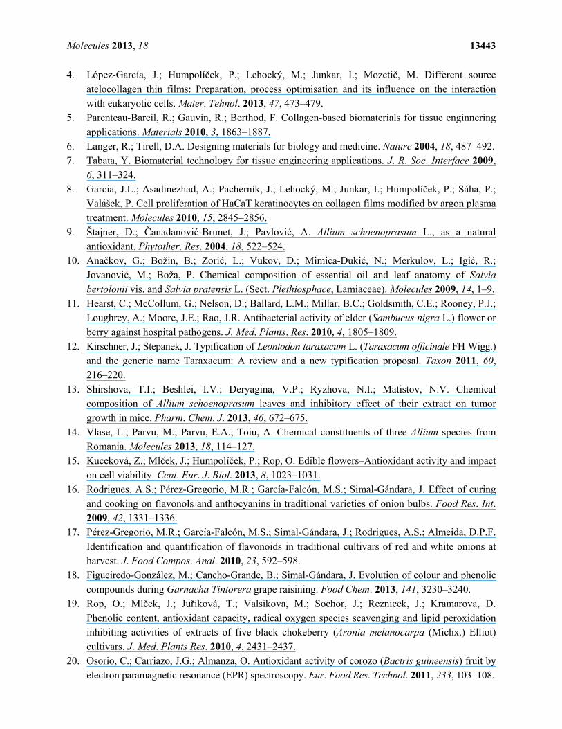
As far as cell morphology is concerned, Figure 2 reveals the HaCaT cell growth on atelocollagen
films with and without phenolic extracts, which shows cell aggregates in form of ripple-like areas
adhered on the film surfaces. It may be also observed that some group of cells do not have the typical
keratinocyte cell shapes, an anomaly that is probably a consequence of cell damage caused by the high
concentration of phenols in the atelocollagen matrix [45,46].
Figure 2. Light micrographs of human skin HaCaT keratinocytes in culture on
atelocollagen thin films with 100 μg/mL phenolic extract of: (A) Allium schoenoprasum;
(B) Salvia pratensis; (C) Sambucus nigra and (D) Taraxacum officinale.
Cell stability is a pivotal issue in cell culture. In that respect, it has been demonstrated that after
four days in culture, a proper concentration of these polyphenolic extracts does not negatively
influence cell viability.
Statistically speaking, the selected concentrations of Allium schoenoprasum as well as 50 and
100 μg/mL of Salvia pratensis and Sambucus nigra were found very statistically significant (99%,
p < 0.01), whilst in the rest of the samples, the differences are not statistically significant compared
with pristine atelocollagen thin films.

Molecules 2013, 18 13440
None of the added phenolic extracts evidenced any antimicrobial activity against pathogenic
Gram-negative or Gram-positive bacterial strains. Nevertheless, it has been comprehensively
demosntrated that polyphenols, such as catechin, gallic acid, ferulic acid, coumaric acid and resveratrol
may act either alone or in mixtures as long-term anti-inflammatory, antifungal and antineoplastic
agents, which are three of the most serious concerns in current medicine [47–49].
3. Experimental
3.1. Materials
Collagen gel from bovine splits (pH 5.2), which contains 16.2% of atelocollagen was supplied by
Vipo A.S, Partizánske, Slovakia. Acetic acid 99% was obtained from Penta, Prague, Czech Republic.
Tissue culture dishes of 40 mm diameter and individual wells of 96-well were commercially acquired
from TPP, Trasadingen, Switzerland. A Vybrant® MTT cell proliferation Assay kit V-13154 was
purchased from Invitrogen Corporation (Carlsbad, CA, USA).
3.2. Extraction Conditions
Polyphenols were extracted from the following flowers: chives (Allium schoenoprasum, Liliaceae),
introduced sage (Salvia pratensis, Lamiaceae), European elderberry (Sambucus nigra, Caprifoliaceae)
and common dandelion (Taraxacum officinale). All flowers were botanically identified and picked from
the White Carpathian Mountains, Zlin Region, Czech Republic and sent to the laboratory in the city of Zlin.
Immediately after cutting the flowers, those were frozen and stored at −40 °C. The frozen herbs were
homogenised in 90% methanol extracted at 4 °C for 30 min and subsequently centrifugated at 1990 rpm
for 10 min to separate the supernatant. Sediments were subjected to a new extraction, which was repeated
thrice. The extracts were carefully concentrated by using a Laborota 4011 digital rotary evaporator
(Heidolph, Schwabach, Germany). The final concentration of each extract was adjusted to 1,000 mg/mL.
3.3. Determination of Polyphenols
The quantification of the total flower phenolic content was ascertained by using the Folin-Ciocalteu
Assay. Extract (1 mL) was added to a 25 mL volumetric flask containing deionised water (20 mL) and
later on Folin-Ciocalteu’s phenol reagent (1 mL, Sigma-Aldrich, St. Louis, MO, USA), was
incorporated to the mixture. Three minutes after that, 20% Na2CO3 (5 mL) was added into the flask.
The aqueous solution was mixed and completed to a final volume of 50 mL. After 30 min, the colour
intensity was measured by using an UV-Mini 1240 spectrophotometer (Shimadzu, Kyoto, Japan) at
700 nm and compared to a no-tannin control sample. All samples were analysed in duplicate.
3.4. High Performance Liquid Chromatography (HPLC)
The determination of each polyphenol was carried out by using a Dionex UltiMate 3000
High-performance Liquid Chromatography system (Dionex, Sunnyvale, CA, USA). The HPLC was
equipped with a Supelcosil LC-18-DB column (25 cm × 4.6 mm I.D. S-5 μm) employing a binary
gradient of (A) 5% (v/v) acetonitrile, 0.035% (v/v) trifluoroacetic acid and (B) 50% (v/v) acetonitrile,
Molecules 2013, 18 13441
0.025% (v/v) trifluoroacetic acid. The flow-rate was set at 1.0 mL/min. The gradient elution profile
began with A–B (90:10), and then B was gradually increased to 20% at 10 min, to 40% at 16 min, to
50% at 20 min and back to 40% from 25 to 27 min.
3.5. Atelocollagen Thin Films Preparation
The atelocollagen was solubilised in 0.1 M acetic acid to prepare a 0.1% w/w solution using an IKA
RCT stirring machine (IKA® works, Inc., Staufen, Germany) for 1 h at 1000 rpm. Then, 2 mL of this
solution was casted on tissue culture dishes. The methanolic extracts were diluted to obtain final
concentrations of 100, 50, 25 and 10 μg/mL and incorporated into the casted solutions. The solvents
(acetic acid and methanol) were evaporated at ambient conditions for three days. Thin films of pristine
atelocollagen were prepared and set as experimental blanks.
3.6. HaCaT Cell Incubation
Human immortalised non-tumorigenic keratinocyte cell line HaCaT, (skin tissue, Caucasian
ethnicity; 62 years of age, male gender) was supplied by CLS Cell Lines Service, Eppelheim, Germany.
Dulbecco’s modified eagle medium, contains 4.5 g/L D-glucose, L-glutamine, and 110 mg/L sodium
pyruvate (DMEM; Invitrogen) supplemented with 2 mM L-glutamine, 10% foetal bovine serum (FBS)
and penicillin-streptomycin (100 U/mL–0.1 mg/mL) was used as a culture medium (Biotech Inc.,
Carlsbad, CA, USA). Cells were incubated at 37 °C for 24 h with 5% CO2 in humidified air.
3.7. Cell Viability
All cells in exponential growth phase were seeded in a concentration of 1 × 10⁵ cells/mL onto the
atelocollagen films with distinct concentrations of polyphenols. Cell viability was determined after
4 days in culture by MTT assay (Invitrogen Corporation). A volume of 12 mM MTT (10 μL) was
taken for cell incubation performed at 37 °C for 4 h in the darkness. Thereupon, the media were
decanted and washed with phosphate-buffered saline solution (PBS). The produced formazan salts were
dissolved with dimethylsulphoxide (DMSO) and its concentration was measured in a spectrophotometer
at 570 nm [50]. Absorbances were recorded utilising an infinite M200PRO multimode reader at 570
nm (Tecan Group, Männedorf, Switzerland), and all determinations were performed in quadruplicate.
Cell morphology was qualitatively appraised every 24 h after cultivation by using an inverted phase-
contrast microscope Olympus CKX 41 (Olympus, Hamburg, Germany) with an optical zoom of 100×.
3.8. Antimicrobial Assay in Vitro
The antimicrobial performance of the specimens against two pathogenic microorganisms, the
gram-positive S. aureus (ATCC 6538) and the gram-negative E. coli (ATCC 8739) strains (purchased
from the Czech Collection of Microorganisms, Brno, Czech Republic) was explored by agar diffusion
test. Circular samples of 8 mm diameter were put into nutrient agar which was inoculated with bacteria
(≈108 CFU mL−1). After 24 h incubation at 37 °C, the diameter of the inhibition zone was measured in
five directions and averaged value was calculated to evaluate the inhibition zone area. Four replicates
of each substrate were tested to maintain the statistical accountability.
Molecules 2013, 18 13442
3.9. Statistical Analysis
All data were presented as the mean value ± standard deviation (SD) of each sample. Statistical
comparisons were performed using Student’s t-test with a confidence level of 95% (p < 0.05) considered
statistically significance and 99% (p < 0.01) considered very significant.
4. Conclusions
Four edible flowers that wildly grow in the Moravian mountains were collected and their methanolic
extracts were analysed by HPLC. Benzoic and hydroxycinnamic acids and flavonoids were found as
the major constituents. This contribution also delved into the incorporation of these phenolic mixtures
onto atelocollagen matrices. From the 16 analysed group of samples, nine were shown to be
non-toxicity, with a cell viability above 80%, five samples had weak toxicity (80%–60%) and two samples
displayed a moderate toxicity (<60%). Twelve samples revealed statistical significance. Antiproliferation
is concomitant to concentration, relies on each extract, and all the cell viability curves followed the
same pattern. Common dandelion barely affected HaCaT cell growth, while wild chive discloses more
inhibition power. Cell morphology was examined by microscopy. The studied atelocollagen matrices
seem to be perfectly apt for keratinocyte cell growth and proliferation. Therefore, the present approach
strengthens knowledge about the use of atelocollagen, and allows consideration of these materials as
potential candidates for tissue engineering and would healing applications.
Acknowledgments
The authors would like to express their gratitude to the Operational Program Research and
Development for Innovations co-funded by the European Regional Development Fund (ERDF) and
national budget of Czech Republic, within the framework of project Centre of Polymer Systems
(reg. number: CZ.1.05/2.1.00/03.0111), the Advanced Theoretical and Experimental Studies of
Polymer Systems (reg. number: CZ.1.07/2.3.00/20.0104) and the Czech Science Foundation
(13-08944S). Zdenka Kuceková would also like to thank the internal grant of TBU in Zlin
No. IGA/FT/2013/019 funded for her financial support.
Conflicts of Interest
The authors declare that there is no conflict of interest.
References
1. Shoulders, M.D.; Raines, R.T. Collagen structure and stability. Annu. Rev. Biochem. 2009, 78, 929–958.
2. Banerjee, I.; Mishra, D.; Das, T.; Maiti, S.; Maiti, T.K. Caprine (Goat) collagen: A potential biomaterial for skin tissue engineering. J. Biomater. Sci. Polym. Ed. 2012, 23, 355–373.
3. Bernal, A.; Balková, R.; Kuřítka, I.; Sáha, P. Preparation and characterisation of a new double-sided bio-artificial material prepared by casting of poly(vinyl alcohol) on collagen. Polym. Bull. 2012, 70, 431–453.
Molecules 2013, 18 13443
4. López-García, J.; Humpolíček, P.; Lehocký, M.; Junkar, I.; Mozetič, M. Different source atelocollagen thin films: Preparation, process optimisation and its influence on the interaction with eukaryotic cells. Mater. Tehnol. 2013, 47, 473–479.
5. Parenteau-Bareil, R.; Gauvin, R.; Berthod, F. Collagen-based biomaterials for tissue enginnering applications. Materials 2010, 3, 1863–1887.
6. Langer, R.; Tirell, D.A. Designing materials for biology and medicine. Nature 2004, 18, 487–492. 7. Tabata, Y. Biomaterial technology for tissue engineering applications. J. R. Soc. Interface 2009,
6, 311–324. 8. Garcia, J.L.; Asadinezhad, A.; Pacherník, J.; Lehocký, M.; Junkar, I.; Humpolíček, P.; Sáha, P.;
Valášek, P. Cell proliferation of HaCaT keratinocytes on collagen films modified by argon plasma treatment. Molecules 2010, 15, 2845–2856.
9. Štajner, D.; Čanadanović-Brunet, J.; Pavlović, A. Allium schoenoprasum L., as a natural antioxidant. Phytother. Res. 2004, 18, 522–524.
10. Anačkov, G.; Božin, B.; Zorić, L.; Vukov, D.; Mimica-Dukić, N.; Merkulov, L.; Igić, R.; Jovanović, M.; Boža, P. Chemical composition of essential oil and leaf anatomy of Salvia bertolonii vis. and Salvia pratensis L. (Sect. Plethiosphace, Lamiaceae). Molecules 2009, 14, 1–9.
11. Hearst, C.; McCollum, G.; Nelson, D.; Ballard, L.M.; Millar, B.C.; Goldsmith, C.E.; Rooney, P.J.; Loughrey, A.; Moore, J.E.; Rao, J.R. Antibacterial activity of elder (Sambucus nigra L.) flower or berry against hospital pathogens. J. Med. Plants. Res. 2010, 4, 1805–1809.
12. Kirschner, J.; Stepanek, J. Typification of Leontodon taraxacum L. (Taraxacum officinale FH Wigg.) and the generic name Taraxacum: A review and a new typification proposal. Taxon 2011, 60, 216–220.
13. Shirshova, T.I.; Beshlei, I.V.; Deryagina, V.P.; Ryzhova, N.I.; Matistov, N.V. Chemical composition of Allium schoenoprasum leaves and inhibitory effect of their extract on tumor growth in mice. Pharm. Chem. J. 2013, 46, 672–675.
14. Vlase, L.; Parvu, M.; Parvu, E.A.; Toiu, A. Chemical constituents of three Allium species from Romania. Molecules 2013, 18, 114–127.
15. Kuceková, Z.; Mlček, J.; Humpolíček, P.; Rop, O. Edible flowers–Antioxidant activity and impact on cell viability. Cent. Eur. J. Biol. 2013, 8, 1023–1031.
16. Rodrigues, A.S.; Pérez-Gregorio, M.R.; García-Falcón, M.S.; Simal-Gándara, J. Effect of curing and cooking on flavonols and anthocyanins in traditional varieties of onion bulbs. Food Res. Int. 2009, 42, 1331–1336.
17. Pérez-Gregorio, M.R.; García-Falcón, M.S.; Simal-Gándara, J.; Rodrigues, A.S.; Almeida, D.P.F. Identification and quantification of flavonoids in traditional cultivars of red and white onions at harvest. J. Food Compos. Anal. 2010, 23, 592–598.
18. Figueiredo-González, M.; Cancho-Grande, B.; Simal-Gándara, J. Evolution of colour and phenolic compounds during Garnacha Tintorera grape raisining. Food Chem. 2013, 141, 3230–3240.
19. Rop, O.; Mlček, J.; Juřiková, T.; Valsikova, M.; Sochor, J.; Reznicek, J.; Kramarova, D. Phenolic content, antioxidant capacity, radical oxygen species scavenging and lipid peroxidation inhibiting activities of extracts of five black chokeberry (Aronia melanocarpa (Michx.) Elliot) cultivars. J. Med. Plants Res. 2010, 4, 2431–2437.
20. Osorio, C.; Carriazo, J.G.; Almanza, O. Antioxidant activity of corozo (Bactris guineensis) fruit by electron paramagnetic resonance (EPR) spectroscopy. Eur. Food Res. Technol. 2011, 233, 103–108.

Molecules 2013, 18 13444
21. Mlček, J.; Rop, O. Fresh edible flowers of ornamental plants–A new source of nutraceutical foods. Trends Food Sci. Tech. 2011, 22, 561–569.
22. Rodrigues, A.S.; Pérez-Gregorio, M.R.; García-Falcón, M.S.; Simal-Gándara, J.; Almeida, D.P.F. Effect of post-harvest practices on flavonoid content of red and white onion cultivars. Food Control 2010, 21, 878–884.
23. Pérez-Gregorio, M.R.; Regueiro, J.; González-Barreiro, C.; Rial-Otero, R.; Simal-Gándara, J. Changes in antioxidant flavonoids during freeze-drying of red onions and subsequent storage. Food Control 2011, 22, 1108–1113.
24. Hetrick, E.M.; Schoenfisch, M.H. Reducing implant-related infections: Active release strategies. Chem. Soc. Rev. 2006, 35, 780–789.
25. Kenawy, E.R.; Worley, S.D.; Broughton, R. The chemistry and applications of antimicrobial polymers: A state-of-the-art review. Biomacromolecules 2007, 8, 1359–1384.
26. Ferrer, J.L.; Austin, M.B.; Stewart, C.; Noel, J.P. Structure and function of enzymes involved in the biosynthesis of phenylpropanoids. Plant Physiol. Bioch. 2008, 46, 356–370.
27. Figueiredo-González, M.; Simal-Gándara, J.; Boso, S.; Martínez, M.C.; Santiago, J.L.; Cancho-Grande, B. Flavonoids in Gran Negro berries collected from shoulders and tips within the cluster, and comparison with Brancellao and Mouratón varieties. Food Chem. 2012, 133, 806–815.
28. Graf, E. Antioxidant potential of ferulic acid. Free Radic. Biol. Med. 1992, 13, 435–448. 29. Kuceková, Z.; Mlček, J.; Humpolíček, P.; Rop, O.; Valášek, P.; Sáha, P. Phenolic compounds
from Allium schoenoprasum, Tragopogon pratensis and Rumex acetosa and their antiproliferative effects. Molecules 2011, 16, 9207–9217.
30. Araim, O.; Ballantyne, J.; Waterhouse, A.L.; Sumpio, B.E. Inhibition of vascular smooth muscle cell proliferation with red wine and red wine polyphenols. J. Vasc. Surg. 2002, 35, 1226–1232.
31. Figueiredo-González, M.; Simal-Gándara, J.; Boso, S.; Martínez, M.C.; Santiago, J.L.; Cancho-Grande, B. Anthocyanins and flavonols berries from Vitis vinifera L. cv. Brancellao separately collected from two different positions within the cluster. Food Chem. 2012, 135, 47–56.
32. Quijada-Morín, N.; Regueiro, J.; Simal-Gándara, J.; Tomás, E.; Rivas-Gonzalo, J.C.; Escribano-Bailón, T. Relationship between the sensory-determined astringency and the flavanolic composition of red wines. J. Agric. Food Chem. 2012, 60, 12355–12361.
33. Katalinic, V.; Mozina, S.S.; Generalic, I.; Skroza, D.; Ljubenkov, I.; Klancnik, A. Phenolic profile, antioxidant capacity and antimicrobial activity of leaf extracts from six Vitis Viniferea L. varieties. Int. J. Food Prop. 2013, 16, 45–60.
34. Hashimoto, S.; Miyazawa, M.; Kameoka, H. Volatile flavour component of chive Allium Schoenprasum. J. Food Sci. 1983, 48, 1858–1863.
35. Veličković, D.T.; Randelović, N.V.; Ristić, M.S.; Šmelcerović, A.A.; Veličković, A.S. Chemical, composition and antimicrobial action of the ethanol extracts of Salvia pratensis L., Salvia glutinosa L. and Salvia aethiopis L. J. Serb. Chem. Soc. 2002, 67, 639–646.
36. Schmitzer, V.; Veberic, R.; Slatnar, A.; Stampar, F. Elderberry (Sambucus nigra L.) wine: A product rich in health promoting compounds. J. Agric. Food Chem. 2010, 58, 10143–10146.
37. Alonso-García, A.; Cancho-Grande, B.; Simal-Gándara, J. Development of a rapid method based on solid-phase extraction and liquid chromatography with ultraviolet detection for the determination of polyphenols in alcohol-free beers. J. Chromatogr. A 2004, 1054, 175–180.

Molecules 2013, 18 13445
38. Figueiredo-González, M.; Cancho-Grande, B.; Simal-Gándara, J. Garnacha Tintorera-based sweet wines: Chromatic properties and global phenolic composition by means of UV-Vis spectrophotometry. Food Chem. 2013, 140, 217–224.
39. International Organization for Standardization. ISO 10993 5: 2009. Biological evaluation of medical devices, Part 5: Tests for in vitro cytotoxicity. International Organization for Standardization: Geneva, Switzerland, 2009.
40. Campbell, J.K.; King, J.L.; Harmston, M.; Lila, M.A.; Erdman, J.W. Synergistic effects of flavonoids on cell proliferation in Hepa-1c1c7 and LNCaP cancer cell lines. J. Food Sci. 2006, 71, 358–363.
41. Kowalczyk, M.C.; Kowalczyk, P.; Tolstykh, O.; Hanausek, M.; Walaszek, Z.; Slaga, T.J. Synergistic effects of combined phytochemicals and skin cancer prevention in SENCAR mice. Cancer Prev. Res. 2010, 3, 170–178.
42. Hole, A.; Grimmer, S.; Jensen, M.R.; Sahlstrøm, S. Synergistic and suppressive effects of dietary phenolic acids and other phytochemicals from cereal extracts on nuclear factor kappa B activity. Food Chem. 2012, 133, 969–977.
43. Yilmaz, Y.; Toledo, R.T. Major flavonoids in grape seeds and skins: antioxidant capacity of catechin, epicatechin, and gallic acid. J. Agric. Food Chem. 2004, 52, 255–260.
44. Fukumoto, L.R.; Mazza, G. Assessing antioxidant and prooxidant activities of phenolic compounds. J. Agric. Food Chem. 2000, 48, 3597–3604.
45. García, J.L.; Pacherník, J.; Lehocký, M.; Junkar, I.; Humpolíček, P.; Sáha, P. Enhanced keratinocyte cell attachment to atelocollagen thin films through air and nitrogen plasma treatment. Prog. Colloid Polym. Sci. 2011, 138, 89–94.
46. Huang, C.-C.; Wu, W.-B.; Fang, J.-Y.; Chiang, H.-S.; Chen, S.-K.; Chen, B.-H.; Chen, Y.-T.; Hung, C.-F. (−)-Epicatechin-3-gallate, a green tea polyphenol is a potent agent against UVB-induced damage in HaCaT Keratinocytes. Molecules 2007, 12, 1845–1858.
47. Matić, I.; Žižak, Ž.; Simonović, M.; Simonović, B.; Godevadc, D.; Šavikin, K.; Juranić, Z. Cytotoxic effect of wine polyphenolic extracts and resveratrol against human carcinoma cells and normal peripheral blood mononuclear cells. J. Med. Food 2010, 13, 851–862.
48. Moravčíková, D.; Kuceková, Z.; Mlček, J.; Rop, O.; Humpolíček, P. Compositions of polyphenols in wild chive, meadow salsify, garden sorrel and agyoncha and their anti-proliferative effect. Acta Univ. Agric. Silvic. Mendel. Brun. 2012, 60, 125–132.
49. Gollucke, A.P.B.; Aguiar, O.; Barbisan, L.F.; Ribeiro, D.A. Use of grape polyphenols against carcinogenesis: Putative molecular mechanisms of action using in vitro and in vivo test systems. J. Med. Food 2013, 16, 199–205.
50. Boukamp, P.; Petrussevska, R.T.; Breitkreutz, D.; Hornung, J.; Markham, A. Normal keratinization in a spontaneously immortalized aneuploid keratinocyte cell line. J. Cell Biol. 1998, 106, 761–771.
Sample Availability: Not Available.
© 2013 by the authors; licensee MDPI, Basel, Switzerland. This article is an open access article
distributed under the terms and conditions of the Creative Commons Attribution license
(http://creativecommons.org/licenses/by/3.0/).
Related Documents