Polymyxins Facilitate Entry into Mammalian Cells Kristina M. Hamill, Lisa S. McCoy, Ezequiel Wexselblatt, Jeffrey D. Esko, and Yitzhak Tor* Table of Contents Materials and Instrumentation .................................................................................... S1 Synthesis of bGPMB and bPMB ................................................................................ S2 Synthesis of bArg8 ..................................................................................................... S10 Synthesis of bTob ........................................................................................................ S10 Purification of PMB and Synthesis of GPMB .............................................................. S11 Supporting Figures Figure S1: Cellular Uptake in Various Cell Lines ............................................ S12 Figure S2: Cell Viability ................................................................................... S12 Figure S3: Cellular Uptake ST-PE-Cy5 and PMB or GPMB ............................. S13 Figure S4: Mechanism(s) of GTob Uptake ...................................................... S13 Figure S5: Cellular Uptake of ST-Cy5 ............................................................. S14 Figure S6: Saporin Uptake Control Experiments and in pgsA Cells ............... S14 Table S1: Size, PDI, and Zeta Potential of Liposomes ................................... S15 NMR and HPLC Spectra Figures S7 and S8. 1 H and 13 C NMR of bPMB (10a) ...................................... S16 Figures S9 and S10. 1 H and 13 C NMR of bGPMB (10b) .................................. S17 Figure S11. HPLC trace of bArg8 ..................................................................... S18 Figure S12. HPLC trace of PMB ....................................................................... S18 Figure S13. HPLC trace of GPMB ................................................................... S18 Figure S14. 1 H NMR of GPMB ....................................................................... S19 Supporting References ............................................................................................... S19 Electronic Supplementary Material (ESI) for Chemical Science. This journal is © The Royal Society of Chemistry 2016

Welcome message from author
This document is posted to help you gain knowledge. Please leave a comment to let me know what you think about it! Share it to your friends and learn new things together.
Transcript
Polymyxins Facilitate Entry into Mammalian Cells Kristina M. Hamill, Lisa S. McCoy, Ezequiel Wexselblatt, Jeffrey D. Esko, and Yitzhak Tor*
Table of Contents Materials and Instrumentation .................................................................................... S1
Synthesis of bGPMB and bPMB ................................................................................ S2
Synthesis of bArg8 ..................................................................................................... S10
Synthesis of bTob ........................................................................................................ S10
Purification of PMB and Synthesis of GPMB .............................................................. S11
Supporting Figures
Figure S1: Cellular Uptake in Various Cell Lines ............................................ S12
Figure S2: Cell Viability ................................................................................... S12
Figure S3: Cellular Uptake ST-PE-Cy5 and PMB or GPMB ............................. S13
Figure S4: Mechanism(s) of GTob Uptake ...................................................... S13
Figure S5: Cellular Uptake of ST-Cy5 ............................................................. S14
Figure S6: Saporin Uptake Control Experiments and in pgsA Cells ............... S14
Table S1: Size, PDI, and Zeta Potential of Liposomes ................................... S15
NMR and HPLC Spectra
Figures S7 and S8. 1H and 13C NMR of bPMB (10a) ...................................... S16
Figures S9 and S10. 1H and 13C NMR of bGPMB (10b) .................................. S17
Figure S11. HPLC trace of bArg8 ..................................................................... S18
Figure S12. HPLC trace of PMB ....................................................................... S18
Figure S13. HPLC trace of GPMB ................................................................... S18
Figure S14. 1H NMR of GPMB ....................................................................... S19
Supporting References ............................................................................................... S19
Electronic Supplementary Material (ESI) for Chemical Science.This journal is © The Royal Society of Chemistry 2016

S1
Materials
Materials obtained from commercial suppliers were used without further purification. Pol-
ymyxin B and tobramycin were purchased from TCI America. H-Dab(Boc)-OMe was pur-
chased from Chem Impex International. Ficin was purchased from MP Biomedicals.
Deuterated NMR solvents were purchased from Cambridge Isotope Laboratories. Ami-
loride, sucrose, and genistein were purchased from Sigma-Aldrich. Nystatin and
EDTA/Trypsin were purchased from VWR and chlorpromazine was purchased from
Fisher. Steptavidin-PE-Cy5 was purchased from Biolegend. PBS, F-12 media, Versene,
streptavidin Cy-5, LysoTracker Green, and Hoescht dye were purchased from Life Tech-
nologies. Streptavidin saporin was purchased from Advanced Targeting Systems. 35 mm
glass bottom culture dishes were purchased from MatTek. CellTiter-Blue was purchased
from Promega. DOPC (1,2-dioleoyl-snglycero-3-phosphocholine), DOPE (1,2-dioleoyl-
sn-glycero-3-phospho-ethanolamine), and cholesterol were purchased from Avanti Polar
Lipids.
Instrumentation
NMR spectra were recorded on a Varian VX 500 MHz spectrometer or a Varian 400 MHz
spectrometer. Mass spectra were recorded at UCSD Chemistry and Biochemistry Mass
Spectrometry Facility utilizing an Agilent 6230 HR-ESI-TOF mass spectrometer. Re-
verse-phase HPLC purification (CLIPEUS, C18, 5µm, 10x250 mm, Higgins analytical)
and analysis (Eclipse, XDB-C18, 5µm, 4.6x150 mm) were carried out on an Agilent 1200
series instrument or Beckman Coulter System Gold 127P Solvent Module. Flow cytom-
etry studies were performed on a BD FACSCalibur. Confocal laser scanning microscopy
was performed using a Nikon A1R inverted fluorescence microscope with z-stepping mo-
tor. Particle size, polydispersity, and surface charge of the lipid vesicles were measured
by dynamic light scattering on a Zetasizer Nano ZS (model ZEN3600 from Malvern In-
struments).

S2
Scheme S1. Synthesis of Boc protected PMBN and Boc guanidinylated PMBN.
PMBN (1)
PMB (2.0 g, 1.4 mmol) was dissolved in 50 mL of H2O. Then 42 mg of dithiothreitrol and 450 mg of ficin (320-500 milk clotting units/mg) were added and the reaction was heated to 37 °C and stirred overnight. When all PMB was consumed and only PMBN was de-tected by HPLC, the reaction was heated to reflux to denature the enzyme. After cooling, the precipitate was filtered off and the mother liqueur was evaporated under reduced pressure. The product was purified by automated flash chromatography Teledyne Isco Redisep Rf C18 30g gold column using a gradient of 0 – 15% ACN (0.1 % TFA) in H2O (0.1% TFA), resulting in the TFA salt of 1 as a beige solid (1.5 g, 1.0 mmol, 72% yield). 1H NMR (400 MHz, D2O) δ 7.41 – 7.28 (m, 3H), 7.24 (d, J = 7.6 Hz, 2H), 4.58 – 4.45 (m, 3H), 4.32 – 4.12 (m, 7H), 3.95 (d, J = 5.5 Hz, 1H), 3.38 – 3.25 (m, 1H), 3.21 – 2.96 (m, 9H), 2.93 – 2.74 (m, 2H), 2.29 – 1.77 (m, 10H), 1.50 – 1.23 (m, 5H), 1.17 (d, J = 6.3 Hz, 3H), 0.77 – 0.59 (m, 7H). 13C NMR (126 MHz, D2O) δ 174.99, 173.33, 173.30, 172.72, 172.68, 171.94, 171.59, 171.39, 168.13, 163.29, 163.01, 162.73, 162.44, 135.41, 128.93, 127.37, 119.73, 117.41, 115.09, 112.77, 66.18, 66.01, 59.44, 58.16, 55.76, 52.85, 51.84, 51.72, 51.67, 51.19, 50.38, 39.04, 36.78, 36.38, 36.33, 36.08, 35.85, 30.51, 29.65, 28.61,
ficin, H2O, 37 °C
O
R2HN
NH
HN
NH
NH2
OOH
OO
H
O
NH2
H2N
NH
NH HN
NH
O
OH2N
HN
O NHHN
OO
NH2
OHO H
R2 =
37%
Polymixin B (PMB)
H2NHN
NH
OHO
OH
O
NH2
H2N
NH
NH HN
NH
O
OH2N
HN
O NHHN
OO
NH2
OHO H
H2NHN
NH
OHO
OH
O
R1
R1
NH
NH HN
NH
O
OR1
HN
O NHHN
OO
OHO H
1 2a 69%2b 53%
L-Dab1 L-Dab3
L-Thr2
L-Dab5
L-Dab8
L-Leu7
L-Thr10
D-Phe6
R1 =
NHBocNH
NBoc
NHBoc
a b
R1
NHBocNNNBoc
a) Boc-ON, NEt3CH3OH/H2O
or
b) NEt3, CH2Cl2/CH3OH
H2NHN
NH
OHOO
H
O
NH2
H2N
NH
NH HN
NH
O
OH2N
HN
O NHHN
OO
NH2
OHO H
1

S3
28.13, 27.76, 23.37, 22.28, 20.21, 19.05, 18.49. HR-ESI-MS calculated for C43H74N14O11 [M+H]+ 963.5733, found 963.5734.
Compound 2a
In a 10 mL flask was added the TFA salt of PMBN (1, 104 mg, 0.0678 mmol), 2 mL MeOH, 1 mL H2O, and NEt3 (110 mg, 1.02 mmol, 140 µL). Then Boc-ON (67 mg, 0.27 mmol) was added and the reaction was stirred for 24 hours. The reaction was evaporated under reduced pressure and CH2Cl2 was added and washed with saturated NaHCO3. The or-ganic layer was dried over MgSO4, filtered, and evaporated under reduced pressure. The product was isolated by automated flash chromatography (0 - 20% MeOH in CH2Cl2 over 18 mins) to afford the product 2a as a white solid (63.8 mg, 0.039 mmol, 69% yield). 1H NMR (500 MHz, CD3OD) δ 7.34 – 7.20 (m, 5H), 4.49 – 4.37 (m, 2H), 4.36 – 4.21 (m, 4H), 4.15 – 4.04 (m, 3H), 4.01 (d, J = 3.7 Hz, 1H), 3.60 – 3.49 (m, 1H), 3.25 – 2.90 (m, 11H), 2.25 – 2.14 (m, 1H), 2.08 – 1.71 (m, 10H), 1.50 – 1.15 (m, 44H), 0.93 – 0.83 (m, 1H), 0.71 (s, 3H), 0.65 (s, 3H). 13C NMR (126 MHz, CD3OD) δ 175.41, 175.20, 174.17, 174.05, 173.74, 173.35, 172.71, 158.74, 158.39, 137.42, 130.40, 129.72, 128.06, 80.37, 80.14, 69.45, 67.28, 61.50, 61.09, 58.29, 54.82, 54.08, 52.91, 52.19, 52.07, 51.80, 40.49, 38.03, 37.79, 37.53, 36.75, 34.18, 33.24, 32.81, 32.28, 31.55, 30.81, 28.82, 28.40, 24.78, 23.80, 21.38, 20.82, 19.70. HR-ESI-MS calculated for C63H106N14O19 [M+Na]+ 1385.7651, found 1385.7640.
Compound 2b
In a 10 mL flask was added the TFA salt of PMBN (1, 112 mg, 0.0728 mmol), 2 mL MeOH, 2 mL CH2Cl2, and NEt3 (110 mg, 1.09 mmol, 152 µL). Then N,N′-Di-Boc-1H-py-razole-1-carboxamidine (88 mg, 0.284 mmol) was added and the reaction was stirred for 24 hours. The reaction was evaporated under reduced pressure and CH2Cl2 was added and washed with saturated NaHCO3. The organic layer was dried over MgSO4, filtered, and evaporated under reduced pressure. The product was isolated by automated flash chromatography (0 - 10% MeOH in CH2Cl2 over 22 mins) to afford the product 2b as a white solid (75 mg, 0.039 mmol, 53% yield). 1H NMR (500 MHz, CD3OD) δ 7.34 – 7.13 (m, 5H), 4.60 – 4.43 (m, 2H), 4.40 (t, J = 8.1 Hz, 1H), 4.31 – 4.16 (m, 5H), 4.14 – 4.01 (m, 1H), 3.95 (d, J = 4.8 Hz, 1H), 3.72 – 3.35 (m, 9H), 3.27 – 2.94 (m, 4H), 2.29 – 1.74 (m, 10H), 1.61 – 1.15 (m, 80H), 0.94 – 0.42 (m, 1H), 0.73 (dd, J = 15.8, 5.8 Hz, 6H). 13C NMR (126 MHz, CD3OD) δ 175.02, 173.95, 173.65, 173.50, 172.52, 164.59, 164.47, 157.85, 157.62, 153.99, 153.90, 150.97, 137.71, 130.29, 129.69, 128.03, 84.42, 84.35, 84.28, 84.22, 80.45, 80.41, 69.52, 67.23, 61.94, 61.16, 58.18, 57.47, 54.12, 53.62, 52.82,
H2NHN
NH
OHO
OH
O
R1
R1
NH
NH HN
NH
O
OR1
HN
O NHHN
OO
OHO H
R1 =
NHBocNH
NBoc
NHBoc
2a 2bR1

S4
52.37, 52.29, 51.91, 40.45, 38.86, 38.61, 38.57, 37.82, 37.70, 36.79, 33.39, 32.90, 32.43, 32.35, 31.98, 31.13, 30.78, 30.75, 28.74, 28.71, 28.69, 28.65, 28.35, 25.00, 23.81, 21.52, 20.93, 19.78. HR-ESI-MS calculated for C87H146N22O27 [M+Na]+ 1954.0620, found 1954.0622.
Scheme S2. Synthesis of biotinylated linker.
Compound 6a
5-hexynoic acid (257 µL, 262 mg, 2.33 mmol), 1.1 mL of DIEA (804 mg, 6.22 mmol), and 8.6 mL DMF (filtered through silica), and HATU (887 mg, 2.33 mmol) were added to a 50 mL round bottom flask and allowed to stir for 10 min to give a yellow solution. Next, H-Dab(Boc)-OMe·HCl (418 mg, 1.56 mmol) was added and the reaction was stirred over-night. To the reaction was added CH2Cl2, which was washed with 2% citric acid and then sat. NaHCO3. The organic layer was dried, filtered, and evaporated under reduced pres-sure. The product was isolated by automated flash chromatography (20 - 90% EtOAc in hexanes over 15 mins) to afford the product as a viscous oil (467 mg, 1.43 mmol, 92% yield). 1H NMR (500 MHz, CDCl3): δ 6.48 (d, J = 7.4 Hz, 1NH), 5.19 – 5.13 (m, 1NH), 4.67 (td, J = 8.5, 4.5 Hz, 1H), 3.48 – 3.34 (m, 1H), 3.00 – 2.85 (m, J = 13.4, 9.2, 4.9 Hz, 1H),
OH2N
NHBoc
O
OH
OHATU, DIEA
O
HN
O
O
NHBoc
5
O
HN
O
O
HN
NHBoc
NBoc
NHBocNNNBoc
LiOH
MeOH, H2O
1. TFA, TIPS, CH2Cl2
2. NEt3,CH2Cl2/CH3OH
CuSO4, sodium ascorbate
THF:tBuOH:H2O3:1:1
87%
CH2Cl295%
6a
ON N
N
O
NHHN
S
OO
HN
O
R1
OHN
3
OHN N
N
O
NHHN
S
OO
HN
O
R1
OHN
3
6b
8a 85%8b 60%
3a 82%3b 99%
6a or 6b
O
NHHN
SO
OHN
3N3
R1 =
NHBocNH
NBoc
NHBoc
a b
7
S5
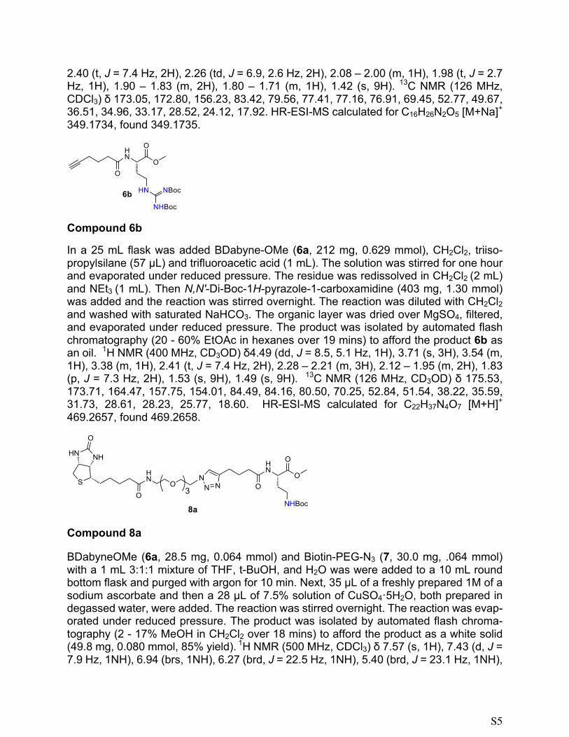
2.40 (t, J = 7.4 Hz, 2H), 2.26 (td, J = 6.9, 2.6 Hz, 2H), 2.08 – 2.00 (m, 1H), 1.98 (t, J = 2.7 Hz, 1H), 1.90 – 1.83 (m, 2H), 1.80 – 1.71 (m, 1H), 1.42 (s, 9H). 13C NMR (126 MHz, CDCl3) δ 173.05, 172.80, 156.23, 83.42, 79.56, 77.41, 77.16, 76.91, 69.45, 52.77, 49.67, 36.51, 34.96, 33.17, 28.52, 24.12, 17.92. HR-ESI-MS calculated for C16H26N2O5 [M+Na]+ 349.1734, found 349.1735.
Compound 6b
In a 25 mL flask was added BDabyne-OMe (6a, 212 mg, 0.629 mmol), CH2Cl2, triiso-propylsilane (57 µL) and trifluoroacetic acid (1 mL). The solution was stirred for one hour and evaporated under reduced pressure. The residue was redissolved in CH2Cl2 (2 mL) and NEt3 (1 mL). Then N,N′-Di-Boc-1H-pyrazole-1-carboxamidine (403 mg, 1.30 mmol) was added and the reaction was stirred overnight. The reaction was diluted with CH2Cl2 and washed with saturated NaHCO3. The organic layer was dried over MgSO4, filtered, and evaporated under reduced pressure. The product was isolated by automated flash chromatography (20 - 60% EtOAc in hexanes over 19 mins) to afford the product 6b as an oil. 1H NMR (400 MHz, CD3OD) δ4.49 (dd, J = 8.5, 5.1 Hz, 1H), 3.71 (s, 3H), 3.54 (m, 1H), 3.38 (m, 1H), 2.41 (t, J = 7.4 Hz, 2H), 2.28 – 2.21 (m, 3H), 2.12 – 1.95 (m, 2H), 1.83 (p, J = 7.3 Hz, 2H), 1.53 (s, 9H), 1.49 (s, 9H). 13C NMR (126 MHz, CD3OD) δ 175.53, 173.71, 164.47, 157.75, 154.01, 84.49, 84.16, 80.50, 70.25, 52.84, 51.54, 38.22, 35.59, 31.73, 28.61, 28.23, 25.77, 18.60. HR-ESI-MS calculated for C22H37N4O7 [M+H]+ 469.2657, found 469.2658.
Compound 8a
BDabyneOMe (6a, 28.5 mg, 0.064 mmol) and Biotin-PEG-N3 (7, 30.0 mg, .064 mmol) with a 1 mL 3:1:1 mixture of THF, t-BuOH, and H2O was were added to a 10 mL round bottom flask and purged with argon for 10 min. Next, 35 µL of a freshly prepared 1M of a sodium ascorbate and then a 28 µL of 7.5% solution of CuSO4·5H2O, both prepared in degassed water, were added. The reaction was stirred overnight. The reaction was evap-orated under reduced pressure. The product was isolated by automated flash chroma-tography (2 - 17% MeOH in CH2Cl2 over 18 mins) to afford the product as a white solid (49.8 mg, 0.080 mmol, 85% yield). 1H NMR (500 MHz, CDCl3) δ 7.57 (s, 1H), 7.43 (d, J = 7.9 Hz, 1NH), 6.94 (brs, 1NH), 6.27 (brd, J = 22.5 Hz, 1NH), 5.40 (brd, J = 23.1 Hz, 1NH),
ON NN
O
NHHN
S
OO
HN
O
NHBoc
OHN
3
8a
S6
5.24 (brs, 1NH), 4.62 (td, J = 8.5, 4.5 Hz, 1H), 4.54 – 4.48 (m, 3H), 4.35 – 4.30 (m, 1H), 3.88 (t, J = 5.1 Hz, 2H), 3.72 (s, 3H), 3.64 – 3.56 (m, 8H), 3.54 (t, J = 5.1 Hz, 2H), 3.48 – 3.31 (m, 3H), 3.14 (td, J = 7.3, 4.8 Hz, 1H), 3.00 (dt, J = 13.5, 5.7 Hz, 1H), 2.90 (dd, J = 12.8, 4.9 Hz, 1H), 2.77 (t, J = 7.4 Hz, 2H), 2.73 (d, J = 12.8 Hz, 1H), 2.33 (t, J = 7.4 Hz, 2H), 2.24 – 2.13 (m, 2H), 2.08 – 1.96 (m, 4H), 1.86 – 1.57 (m, 5H), 1.42 (s, 10H). 13C NMR (126 MHz, CDCl3) δ173.65, 173.52, 173.50, 163.87, 156.21, 147.29, 122.54, 79.49, 70.61, 70.52, 70.49, 70.15, 70.06, 69.64, 61.96, 60.21, 55.73, 52.68, 50.22, 49.86, 40.70, 39.24, 36.78, 35.82, 35.36, 32.54, 28.56, 28.25, 28.17, 25.69, 25.49, 24.86. HR-ESI-MS calculated for C34H58N8O10S [M+Na]+ 793.3889, found 793.3885.
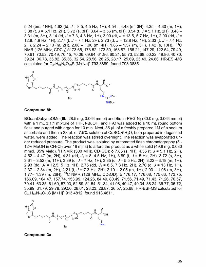
Compound 8b
BGuanDabyneOMe (6b, 28.5 mg, 0.064 mmol) and Biotin-PEG-N3 (30.0 mg, 0.064 mmol) with a 1 mL 3:1:1 mixture of THF, t-BuOH, and H2O was added to a 10 mL round bottom flask and purged with argon for 10 min. Next, 35 µL of a freshly prepared 1M of a sodium ascorbate and then a 28 µL of 7.5% solution of CuSO4·5H2O, both prepared in degassed water, were added. The reaction was stirred overnight. The reaction was evaporated un-der reduced pressure. The product was isolated by automated flash chromatography (5 - 12% MeOH in CH2Cl2 over 19 mins) to afford the product as a white solid (49.8 mg, 0.080 mmol, 85% yield). 1H NMR (500 MHz, CD3OD): δ 7.85 (s, 1H), 4.55 (t, J = 5.1 Hz, 2H), 4.52 – 4.47 (m, 2H), 4.31 (dd, J1 = 8, 4.5 Hz, 1H), 3.89 (t, J = 5 Hz, 2H), 3.72 (s, 3H), 3.61 – 3.52 (m, 11H), 3.39 (q, J = 7 Hz, 1H), 3.35 (q, J = 5.5 Hz, 2H), 3.22 – 3.18 (m, 1H), 2.93 (dd, J1 = 12.5, 5 Hz, 1H), 2.75 (dd, J1 = 8.5, 7.3 Hz, 2H), 2.70 (d, J = 13 Hz, 1H), 2.37 – 2.34 (m, 2H), 2.21 (t, J = 7.3 Hz, 2H), 2.10 – 2.05 (m, 1H), 2.03 – 1.96 (m, 3H), 1.77– 1.39 (m, 26H); 13C NMR (126 MHz, CD3OD): δ 176.17, 176.08, 175.63, 173.75, 166.09, 164.47, 157.74, 153.99, 124.26, 84.49, 80.49, 71.56, 71.49, 71.43, 71.26, 70.57, 70.41, 63.35, 61.60, 57.03, 52.89, 51.54, 51.34, 41.08, 40.47, 40.34, 38.24, 36.77, 36.72, 35.99, 31.79, 29.78, 29.50, 28.61, 28.23, 26.87, 26.57, 25.68. HR-ESI-MS calculated for C40H68N10O12S [M+H]+ 913.4812, found 913.4811.
Compound 3a
ON NN
O
NHHN
S
OO
HN
O
HN
OHN
3
8bNHBoc
NBoc
OHN NN
O
NHHN
S
OO
HN
O
NH2
OHN
3
3a

S7
BDabOMeBiotin (8a, 27 mg, 0.035 mmol), 1.25 mL of MeOH, and 0.42 mL of 0.1 M LiOH solution in water (1 mg, .042 mmol) were added to a 10 mL round bottom flask and stirred overnight. The compound was desalted on a C-18 sep-pak (waters) to provide the product 3a as a white solid (22 mg, 0.029 mmol, 82% yield). 1H NMR (500 MHz, CD3OD) δ 7.87 (s, 1H), 4.57 – 4.53 (m, 2H), 4.49 (ddd, J = 7.9, 4.9, 0.7 Hz, 1H), 4.33 – 4.27 (m, 2H), 3.91 – 3.87 (m, 2H), 3.63 – 3.56 (m, 10H), 3.53 (t, J = 5.5 Hz, 2H), 3.35 (t, J = 5.5 Hz, 2H), 3.19 (tt, J = 3.6, 3.2 Hz, 2H), 3.06 – 2.98 (m, 1H), 2.92 (dd, J = 12.7, 5.0 Hz, 1H), 2.75 (t, J = 7.6 Hz, 2H), 2.70 (d, J = 12.7 Hz, 1H), 2.32 (t, J = 7.5 Hz, 2H), 2.21 (t, J = 7.4 Hz, 2H), 2.07 – 1.94 (m, 4H), 1.78 – 1.54 (m, 6H), 1.45 – 1.40 (m, 11H).; 13C NMR (126 MHz, CD3OD): δ 176.17, 176.08, 175.63, 173.75, 166.09, 164.47, 157.74, 153.99, 124.26, 84.49, 80.49, 71.56, 71.49, 71.43, 71.26, 70.57, 70.41, 63.35, 61.60, 57.03, 52.89, 51.54, 51.34, 41.08, 40.47, 40.34, 38.24, 36.77, 36.72, 35.99, 31.79, 29.78, 29.50, 28.61, 28.23, 26.87, 26.57, 25.68; HR-ESI-MS calculated for C33H56N8O10S [M+Na]+ 779.3737, found 779.3732.
Compound 3b
BGDabOMeBiotin (8b, 27 mg, 0.035 mmol), 1.25 mL of MeOH, and 0.42 mL of 0.1 M LiOH solution in water (1 mg, .042 mmol) were added to a 10 mL round bottom flask and stirred overnight. The compound was desalted on a C-18 sep-pak (waters) to provide the product 3b as a white solid (22 mg, 0.029 mmol, 82% yield). 1H NMR (500 MHz, CD3OD) δ 7.87 (s, 1H), 4.55 (t, J = 5.0 Hz, 2H), 4.49 (dd, J = 7.8, 4.8 Hz, 1H), 4.33 – 4.27 (m, 2H), 3.89 (t, J = 5.1 Hz, 2H), 3.62 – 3.54 (m, 8H), 3.53 (t, J = 5.5 Hz, 2H), 3.35 (t, J = 5.4 Hz, 2H), 3.30 – 3.24 (m, 1H), 3.22 – 3.17 (m, 1H), 2.92 (dd, J = 12.7, 5.0 Hz, 1H), 2.75 (t, J = 7.6 Hz, 2H), 2.70 (d, J = 12.7 Hz, 1H), 2.35 (t, J = 7.3 Hz, 2H), 2.21 (t, J = 7.4 Hz, 2H), 2.17 – 2.09 (m, 1H), 2.04 – 1.96 (m, 2H), 1.88 – 1.78 (m, 1H), 1.77 – 1.55 (m, 5H), 1.52 (s, 9H), 1.48 – 1.39 (m, 11H).; 13C NMR (126 MHz, CD3OD) δ 178.59, 176.14, 174.91, 166.13, 158.21, 149.44, 148.27, 124.30, 79.84, 71.57, 71.50, 71.45, 71.27, 70.55, 70.42, 63.36, 61.61, 57.03, 53.91, 51.31, 41.07, 40.36, 38.43, 36.73, 36.36, 34.47, 29.78, 29.51, 28.80, 26.87, 26.71, 25.80. HR-ESI-MS calculated for C39H66N10O12S [M+H]+ 899.4655, found 899.4653.
OHN NN
O
NHHN
S
OO
HN
O
HN
OHN
3
3bNHBoc
NBoc
S8
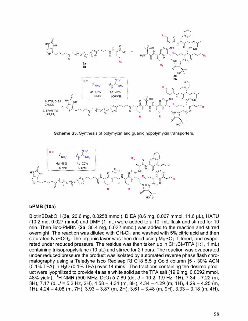
Scheme S3. Synthesis of polymyxin and guanidinopolymyxin transporters.
bPMB (10a)
BiotinBDabOH (3a, 20.6 mg, 0.0258 mmol), DIEA (8.6 mg, 0.067 mmol, 11.6 µL), HATU (10.2 mg, 0.027 mmol) and DMF (1 mL) were added to a 10 mL flask and stirred for 10 min. Then Boc-PMBN (2a, 30.4 mg, 0.022 mmol) was added to the reaction and stirred overnight. The reaction was diluted with CH2Cl2 and washed with 5% citric acid and then saturated NaHCO3. The organic layer was then dried using MgSO4, filtered, and evapo-rated under reduced pressure. The residue was then taken up in CH2Cl2/TFA (1:1, 1 mL) containing triisopropylsilane (10 µL) and stirred for 2 hours. The reaction was evaporated under reduced pressure the product was isolated by automated reverse phase flash chro-matography using a Teledyne Isco Redisep Rf C18 5.5 g Gold column [5 - 30% ACN (0.1% TFA) in H2O (0.1% TFA) over 14 mins]. The fractions containing the desired prod-uct were lyophilized to provide 4a as a white solid as the TFA salt (19.9 mg, 0.0092 mmol, 48% yield). 1H NMR (500 MHz, D2O) δ 7.89 (dd, J = 10.2, 1.9 Hz, 1H), 7.34 – 7.22 (m, 3H), 7.17 (d, J = 5.2 Hz, 2H), 4.58 – 4.34 (m, 8H), 4.34 – 4.29 (m, 1H), 4.29 – 4.25 (m, 1H), 4.24 – 4.08 (m, 7H), 3.93 – 3.87 (m, 2H), 3.61 – 3.48 (m, 9H), 3.33 – 3.18 (m, 4H),
OHN N
N
O
NHHN
S
OO
HN
O
R1
OHN
3
3a3b
NH
HN
NH
OHO
OH
O
R
NH
NH HN
NH
O
OR
HN
O NHHN
OO
R
OHO H
R
N NN
O
NHHN
SO
O
HN
O
R
OHN
3
+H2N
HN
NH
OHO
OH
O
R1
NH
NH HN
NH
O
OR1
HN
O NHHN
OO
R1
OHO H
R1
2a2b
1. HATU, DIEA CH2Cl2
2. TFA/TIPS CH2Cl2
R =
NH2NH
NH2+
NH3+
4b 25%4a 48%bGPMBbPMB
NH
HN
NH
OHO
OH
O
R
NH
NH HN
NH
O
OR
HN
O NHHN
OO
R
OHO H
R
N NN
O
NHHN
SO
O
HN
O
R
OHN
3
R =
NH2NH
NH2+
NH3+
4b 25%4a 48%bGPMBbPMB

S9
3.12 – 2.93 (m, 10H), 2.92 – 2.85 (m, 1H), 2.84 – 2.75 (m, 1H), 2.75 – 2.63 (m, 4H), 2.33 – 2.26 (m, 2H), 2.23 – 1.74 (m, 17H), 1.68 – 1.24 (m, 10H), 1.16 – 1.07 (m, 5H), 0.64 (d, J = 37.2 Hz, 7H). 13C NMR (126 MHz, D2O) δ 176.73, 176.24, 174.90, 173.25, 173.18, 173.09, 172.89, 172.67, 172.62, 172.54, 172.47, 172.27, 172.21, 171.86, 171.61, 171.47, 171.28, 171.24, 165.17, 163.24, 162.82 (TFA, q, J = 35.7 Hz), 146.46, 135.32, 128.83, 127.27, 124.28, 124.15, 116.23 (TFA, q, J = 292.1 Hz), 69.43, 69.29, 69.26, 68.67, 68.43, 68.39, 66.89, 66.66, 66.08, 61.92, 60.08, 59.32, 58.93, 58.79, 55.62, 55.21, 52.66, 52.57, 51.74, 51.64, 51.55, 51.39, 51.25, 51.05, 50.92, 50.36, 50.24, 39.54, 38.94, 38.74, 36.69, 36.27, 36.19, 36.10, 35.94, 35.80, 35.70, 35.27, 34.22, 30.43, 30.31, 29.59, 29.54, 28.67, 28.48, 28.34, 28.09, 27.71, 27.60, 27.54, 25.00, 24.50, 24.44, 23.52, 23.42, 23.30, 22.18, 20.11, 18.95, 18.73, 18.58, 16.13. HR-ESI-MS calculated for C71H120N22O18S [M+Na]+ 1623.8764, found 1623.8766.
bGPMB (4b)
BiotinBGDabOH (3b, 20.2 mg, 0.0223 mmol), DIEA (12.02 mg, 0.093 mmol, 16.2 µL), PyBrop (10.4 mg, 0.0223 mmol) and DMF (1 mL) were added to a 10 mL flask and stirred for 10 min. Then BocGuan-PMBN (2b, 36.0 mg, 0.0186 mmol) was added to the reaction and stirred overnight. The reaction was diluted with CH2Cl2 and washed with 5% citric acid and then saturated NaHCO3. The organic layer was then dried using MgSO4, filtered, and evaporated under reduced pressure. The residue was then taken up in CH2Cl2/TFA (1:1, 1 mL) containing triisopropylsilane (10 µL) and stirred for 2 hours. The reaction was evaporated under reduced pressure the product was isolated by automated reverse phase flash chromatography using a C18 5.5 g Gold column [15 - 35% ACN (0.1% TFA) in H2O (0.1% TFA) over 15 mins]. The fractions were lyophilized to provide 4b as a white solid as the TFA salt (19.9 mg, 0.0092 mmol, 25% yield). 1H NMR (500 MHz, D2O) δ 7.89 – 7.86 (m, 1H), 7.44 – 7.33 (m, 3H), 7.30 – 7.26 (m, 2H), 4.65 – 4.54 (m, 4H), 4.51 – 4.39 (m, 5H), 4.36 – 4.18 (m, 9H), 4.02 – 3.97 (m, 2H), 3.71 – 3.59 (m, 11H), 3.42 – 3.25 (m, 13H), 3.23 – 2.96 (m, 7H), 2.81 – 2.71 (m, 3H), 2.44 – 2.35 (m, 2H), 2.27 (td, J = 7.3, 1.8 Hz, 3H), 2.24 – 2.10 (m, 6H), 2.23 – 1.30 (m, 28H), 1.27 – 1.18 (m, 6H), 0.89 – 0.70 (m, 7H). 13C NMR (126 MHz, D2O) δ 75, 176.59, 176.38, 175.00, 174.28, 173.96, 173.87, 173.83, 173.38, 173.32, 172.93, 172.63, 172.25, 172.25, 172.00, 171.67, 171.48, 171.43, 165.24, 163.35, 163.07, 162.79, 162.50 (TFA, q, J = 35.7 Hz), 156.77, 156.68, 156.64, 156.54, 147.26, 135.40, 128.91, 127.37, 123.55, 117.43, 115.11 (TFA, q, J = 291.5 Hz), 69.57, 69.52, 69.39, 68.78, 68.72, 66.85, 66.54, 66.06, 62.01, 60.19, 59.48, 59.29, 59.04, 55.86, 55.30, 52.59, 51.95, 51.70, 51.50, 51.18, 50.53, 49.84, 39.64, 39.04, 38.85, 37.83, 37.76, 37.39, 36.77, 35.39, 34.47, 34.38, 30.80, 29.76, 29.19, 27.84, 27.66, 25.10, 24.82, 23.95, 23.47, 22.29, 20.21, 19.09, 18.86, 18.72. HR-ESI-MS calculated for C76H130N32O18S [M+3H]3+ 604.6727, found 604.6722.
S10
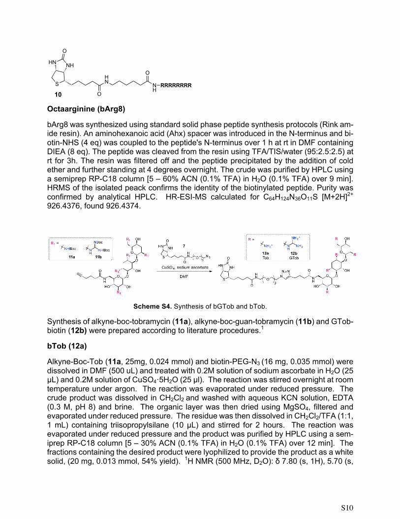
Octaarginine (bArg8)
bArg8 was synthesized using standard solid phase peptide synthesis protocols (Rink am-ide resin). An aminohexanoic acid (Ahx) spacer was introduced in the N-terminus and bi-otin-NHS (4 eq) was coupled to the peptide's N-terminus over 1 h at rt in DMF containing DIEA (8 eq). The peptide was cleaved from the resin using TFA/TIS/water (95:2.5:2.5) at rt for 3h. The resin was filtered off and the peptide precipitated by the addition of cold ether and further standing at 4 degrees overnight. The crude was purified by HPLC using a semiprep RP-C18 column [5 – 60% ACN (0.1% TFA) in H2O (0.1% TFA) over 9 min]. HRMS of the isolated peack confirms the identity of the biotinylated peptide. Purity was confirmed by analytical HPLC. HR-ESI-MS calculated for C64H124N36O11S [M+2H]2+ 926.4376, found 926.4374.
Scheme S4. Synthesis of bGTob and bTob.
Synthesis of alkyne-boc-tobramycin (11a), alkyne-boc-guan-tobramycin (11b) and GTob-biotin (12b) were prepared according to literature procedures.1
bTob (12a)
Alkyne-Boc-Tob (11a, 25mg, 0.024 mmol) and biotin-PEG-N3 (16 mg, 0.035 mmol) were dissolved in DMF (500 uL) and treated with 0.2M solution of sodium ascorbate in H2O (25 µL) and 0.2M solution of CuSO4·5H2O (25 µl). The reaction was stirred overnight at room temperature under argon. The reaction was evaporated under reduced pressure. The crude product was dissolved in CH2Cl2 and washed with aqueous KCN solution, EDTA (0.3 M, pH 8) and brine. The organic layer was then dried using MgSO4, filtered and evaporated under reduced pressure. The residue was then dissolved in CH2Cl2/TFA (1:1, 1 mL) containing triisopropylsilane (10 µL) and stirred for 2 hours. The reaction was evaporated under reduced pressure and the product was purified by HPLC using a sem-iprep RP-C18 column [5 – 30% ACN (0.1% TFA) in H2O (0.1% TFA) over 12 min]. The fractions containing the desired product were lyophilized to provide the product as a white solid, (20 mg, 0.013 mmol, 54% yield). 1H NMR (500 MHz, D2O): δ 7.80 (s, 1H), 5.70 (s,
O
NHHN
S
O
HN N
H
O
RRRRRRRR10

S11
1H), 4.96 (s, 1H), 4.50 (m, 3H), 4.30 (m, 1H), 3.91-3.80 (m, 6H), 3.73 (m, 1H), 3.66 (m, 2H), 3.60-3.40 (m, 16H), 3.38-3.27 (m, 3H), 3.26 (s, 2H), 3.19 (m, 2H), 2.88 (m, 1H), 2.65 (m, 3H), 2.45 (d, 1H, J=12.3 Hz), 2.22 (m, 3H), 2.13 (m, 2H), 1.96-1.83 (m, 4H), 1.62-1.40 (m, 4H), 1.28 (s, 2H). 13C NMR (126 MHz, D2O): δ 176.82, 165.28, 163.30, 163.02, 162.74, 162.45, 146.91, 123.97, 119.77, 117.44, 115.12, 112.80, 100.86, 94.10, 83.56, 77.32, 74.12, 70.92, 70.31, 69.58, 69.53, 69.40, 69.38, 68.78, 68.60, 68.00, 66.48, 64.32, 62.02, 60.20, 55.30, 54.62, 50.15, 49.72, 48.31, 47.70, 39.74, 39.63, 38.94, 38.86, 35.38, 34.86, 29.25, 27.81, 27.71, 27.64, 25.09, 24.93, 23.78. HR-ESI-MS calculated for C42H77N12O14S [M+H]+ 1005.5397, found 1005.5400.
PMB (14)
PMB was isolated from the mixture of isomers by HPLC using a RP-C18 column [5 – 50% ACN (0.1% TFA) in H2O (0.1% TFA) over 40 mins]. Purity was confirmed by analytical HPLC. HR-ESI-MS calculated for C56H98N16O13 [M+Na]+ 1225.7397, found 1225.7395.
GPMB (15)
MeOH (15 mL) and NEt3 (239 mg, 2.36 mmol, 329 µL) were added to 14 (226 mg, 0.157 mmol) followed by N,N′-Di-Boc-1H-pyrazole-1-carboxamidine (195 mg, 0.142 mmol) and stirred overnight. The reaction was evaporated under reduced pressure and CH2Cl2 was added and washed with saturated NaHCO3. The organic layer was dried over MgSO4, filtered, and evaporated under reduced pressure. The residue was then dissolved in CH2Cl2/TFA (1:1, 4 mL) containing triisopropylsilane (44 µL) and stirred for 2 hours. The reaction was diluted with 5 mL of CH2Cl2 and extracted with 10 mL of H2O. The water was evaporated under reduced pressure and the product was isolated by HPLC using a RP-C18 column [5-50% ACN (0.1% TFA) in H2O (0.1% TFA) over 40 mins]. The fractions containing the desired product were lyophilized to provide the TFA salt of GPMB as a white solid, (74.7 mg, 0.0378 mmol, 24% yield). 1H NMR (400 MHz, D2O): δ 7.42 – 7.29 (m, 3H), 7.26 (d, J = 6.9 Hz, 2H), 4.56 – 4.50 (m, 1H), 4.45 (ddd, J = 17.2, 8.7, 5.3 Hz, 3H), 4.35 – 4.21 (m, 6H), 4.20 – 4.14 (m, 2H), 3.43 – 2.98 (m, 14H), 2.33 (t, J = 7.2 Hz,
NH
HN
NH
OHO
OH
O
R
NH
NH HN
NH
O
OR
HN
O NHHN
OO
R
OHO H
RO
HN
O
R
R =
NH2NH
NH2+
NH3+
14PMB
15GPMB

S12
2H), 2.22 – 1.74 (m, 12H), 1.67 – 1.03 (m, 17H), 0.82 (t, J = 6.8 Hz, 6H), 0.75 (s, 3H), 0.68 (s, 3H). HR-ESI-MS calculated for C61H108N26O13 [M+2H]2+ 707.4367, found 707.4351.
Figure S1. Cellular uptake in various cell lines. Cellular uptake of GPMB and PMB conjugated to ST-PE-Cy5. CHO-K1, HEK-293, and HEP-3B cells were incubated with conjugate (5nM) at 37 °C for 1 h. Mean fluorescence intensity was measured by flow cytometry. The background signal from untreated cells was subtracted.
Figure S2. Cell viability. CHO-K1 cells (a) and HEK-293 cells (b) were incubated with various concentrations of GPMB-biotin or PMB-biotin in complete media for 72 hours in a 96-well plate. Cell titer blue was added and incubated an additional 4 hours. Cell viability was calculated by measuring the fluorescence intensity at 560/590.
S13
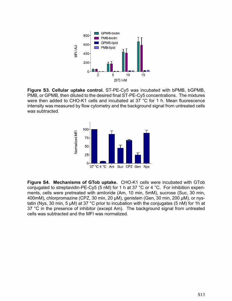
Figure S3. Cellular uptake control. ST-PE-Cy5 was incubated with bPMB, bGPMB, PMB, or GPMB, then diluted to the desired final ST-PE-Cy5 concentrations. The mixtures were then added to CHO-K1 cells and incubated at 37 °C for 1 h. Mean fluorescence intensity was measured by flow cytometry and the background signal from untreated cells was subtracted.
Figure S4. Mechanisms of GTob uptake. CHO-K1 cells were incubated with GTob conjugated to streptavidin-PE-Cy5 (5 nM) for 1 h at 37 °C or 4 °C. For inhibition experi-ments, cells were pretreated with amiloride (Am, 10 min, 5mM), sucrose (Suc, 30 min, 400mM), chlorpromazine (CPZ, 30 min, 20 µM), genistein (Gen, 30 min, 200 µM), or nys-tatin (Nys, 30 min, 5 µM) at 37 °C prior to incubation with the conjugates (5 nM) for 1h at 37 °C in the presence of inhibitor (except Am). The background signal from untreated cells was subtracted and the MFI was normalized.

S14
Figure S5. Cellular uptake of ST-Cy5. CHO-K1 cells were incubated with PMB or GPMB conjugated to streptavidin-PE-Cy5 at various concentrations for 1 h at 37 °C. Mean fluorescence intensity was measured and the background signal from untreated cells was subtracted.
Figure S6. Cellular uptake of saporin. a) Saporin (no streptavidin) was incubated with bPMB or bGPMB for 20 min then dliuted to final saporin concentrations and added to CHO-K1 cells. After four days, the number of viable cells was determined using CellTiter-Blue assay and measuring the flourescence intensity at 560/590. b) pgsA cells were incubated with transporter-streptavidin-saporin conjugates at 37 °C. After four days, the number of viable cells was determined using CellTiter-Blue and measuring fluorescence intensity at 560/590

S15
Z-average (± SD) / nm PDI (± SD) Z-potential (± SD) /
mV
Plain liposomes 143.9 (4.0) 0.122 (.039) 3.78 (0.09)
GPMB liposomes 138.2 (1.9) 0.166 (0.021) 26.9 (0.70)
PMB liposomes 139.7 (1.7) 0.164 (0.056) 22.0 (0.70)
Table S1. Physicochemical characterization of liposomes. Size, polydispersity, and zeta-potential of evaluated liposomes. Plain liposomes were compared to liposomes mixed with 10 mol% GPMB or 10 mol% PMB.

S16
Figure S7. 1H NMR of PMB-biotin (4a, D2O, 500 MHz).
Figure S8. 13C NMR of PMB-biotin (4a, D2O, 126 MHz).
NH
HN
NH
OHO
OH
O
R
NH
NH HN
NH
O
OR
HN
O NHHN
OO
R
OHO H
R
N NN
O
NHHN
SO
O
HN
O
R
OHN
3
R = NH3+
4abiotin-PMB
NH
HN
NH
OHO
OH
O
R
NH
NH HN
NH
O
OR
HN
O NHHN
OO
R
OHO H
R
N NN
O
NHHN
SO
O
HN
O
R
OHN
3
R = NH3+
4abiotin-PMB

S17
Figure S9. 1H NMR of GPMB-biotin (4b, D2O, 500 MHz).
Figure S10. 13C NMR of GPMB-biotin (4b, D2O, 126 MHz)
NH
HN
NH
OHO
OH
O
R
NH
NH HN
NH
O
OR
HN
O NHHN
OO
R
OHO H
R
N NN
O
NHHN
SO
O
HN
O
R
OHN
3
R =NH2N
H
NH2+
4bbiotin-GPMB
NH
HN
NH
OHO
OH
O
R
NH
NH HN
NH
O
OR
HN
O NHHN
OO
R
OHO H
R
N NN
O
NHHN
SO
O
HN
O
R
OHN
3
R =NH2N
H
NH2+
4bbiotin-GPMB

S18
Figure S11. Analytical HPLC trace for bArg8. [RP-C18, 5 – 60% ACN (0.1% TFA) in H2O (0.1% TFA) over 9 min]
Figure S12. Analytical HPLC trace for PMB [RP-C18 column, 5 – 50% ACN (0.1% TFA) in H2O (0.1% TFA) over 15 mins].
Figure S13. Analytical HPLC trace for GPMB [RP-C18 column, 5 – 50% ACN (0.1% TFA) in H2O (0.1% TFA) over 15 mins].
min2 4 6 8 10 12 14 16 18
mAU
-200
0
200
400
600
800
DAD1 A, Sig=220,4 Ref=360,100 (EZE\RUNS\2015\MARCH\3-19-15\R8-KAN-TOB-GNEO ANALYTIC\021-0401.D)
min5 6 7 8 9 10 11 12 13 14
mAU
60
80
100
120
140
160
180
200
DAD1 A, Sig=220,4 Ref=360,100 (FORMER MEMBERS\LISA\GUANPMB\091114_GUANPMB_SEMIPREP\PMBNPREP000010.D)
===================================================================== Area Percent Report ===================================================================== Sorted By : SignalMultiplier: : 1.0000Dilution: : 1.0000Sample Amount: : 20.00000 [ng/ul] (not used in calc.)Use Multiplier & Dilution Factor with ISTDs No peaks found ===================================================================== *** End of Report ***
Data File C:\AGILENT...ORMER MEMBERS\LISA\GUANPMB\091114_GUANPMB_SEMIPREP\PMBNPREP000010.DSample Name: Run2_9 ===================================================================== Acq. Operator : Lisa Seq. Line : 10 Acq. Instrument : Instrument 1 Location : Vial 59 Injection Date : 9/11/2014 3:43:17 PM Inj : 1 Inj Volume : 100.0 µl Different Inj Volume from Sequence ! Actual Inj Volume : 20.0 µl Acq. Method : C:\AGILENT-HPLC (NEW), DATA\LISA\PMB.M Last changed : 5/30/2014 4:52:00 PM by Lisa Analysis Method : C:\AGILENT-HPLC (NEW), DATA\FORMER MEMBERS\LISA\PMB.M Last changed : 12/17/2015 6:13:35 PM by yao (modified after loading) Method Info : 5-50% ACN (0.1% TFA) in H2O (0.1% TFA) over 15 min
=====================================================================
Instrument 1 12/17/2015 6:14:05 PM yao Page 1 of 1
min5 6 7 8 9 10 11 12 13 14
mAU
60
80
100
120
140
160
DAD1 A, Sig=220,4 Ref=360,100 (FORMER MEMBERS\LISA\PMB\PMB_PREPHPLCFRAC_092914\PMBNPREP000019.D)
===================================================================== Area Percent Report ===================================================================== Sorted By : SignalMultiplier: : 1.0000Dilution: : 1.0000Sample Amount: : 20.00000 [ng/ul] (not used in calc.)Use Multiplier & Dilution Factor with ISTDs No peaks found ===================================================================== *** End of Report ***
Data File C:\AGILENT...TA\FORMER MEMBERS\LISA\PMB\PMB_PREPHPLCFRAC_092914\PMBNPREP000019.DSample Name: PMB_Run2_9 ===================================================================== Acq. Operator : Lisa Seq. Line : 19 Acq. Instrument : Instrument 1 Location : Vial 29 Injection Date : 9/30/2014 1:39:31 AM Inj : 1 Inj Volume : 100.0 µl Different Inj Volume from Sequence ! Actual Inj Volume : 20.0 µl Acq. Method : C:\AGILENT-HPLC (NEW), DATA\LISA\PMB.M Last changed : 5/30/2014 4:52:00 PM by Lisa Analysis Method : C:\AGILENT-HPLC (NEW), DATA\FORMER MEMBERS\LISA\PMB.M Last changed : 12/17/2015 6:13:35 PM by yao (modified after loading) Method Info : 5-50% ACN (0.1% TFA) in H2O (0.1% TFA) over 15 min
=====================================================================
Instrument 1 12/17/2015 6:16:48 PM yao Page 1 of 1
S19
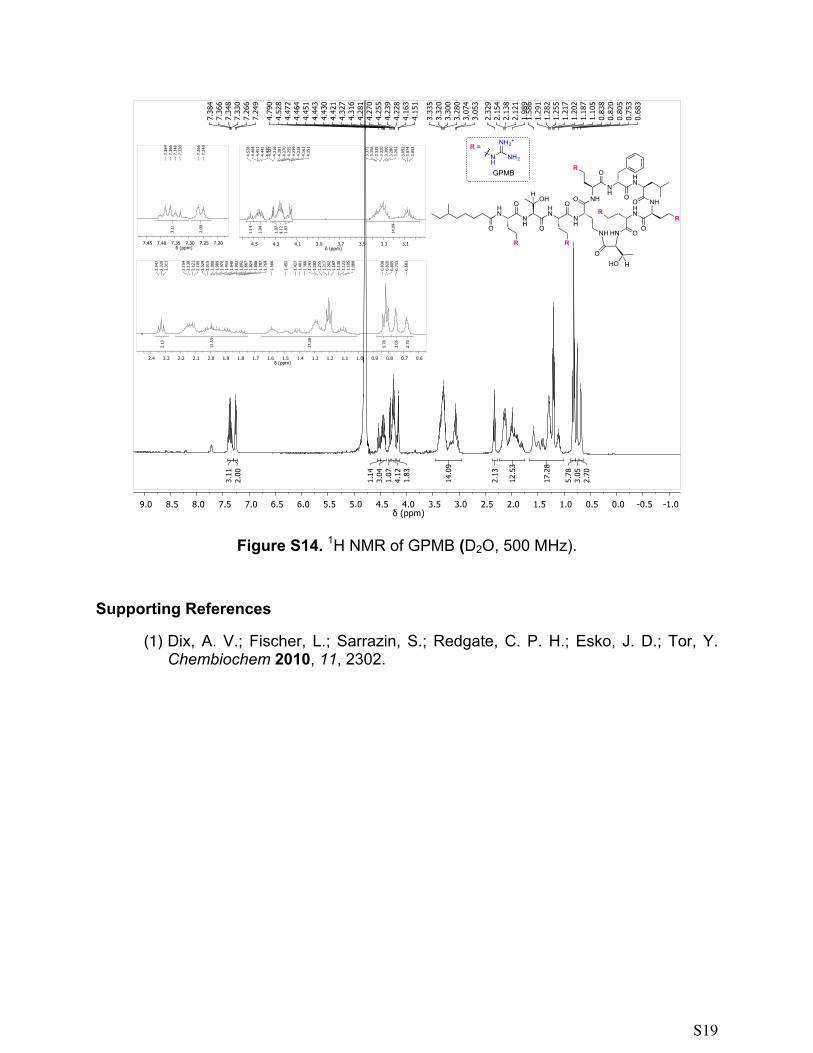
Figure S14. 1H NMR of GPMB (D2O, 500 MHz).
Supporting References
(1) Dix, A. V.; Fischer, L.; Sarrazin, S.; Redgate, C. P. H.; Esko, J. D.; Tor, Y. Chembiochem 2010, 11, 2302.
NH
HN
NH
OHO
OH
O
R
NH
NH HN
NH
O
OR
HN
O NHHN
OO
R
OHO H
RO
HN
O
R
R =NH2N
H
NH2+
GPMB
Related Documents