Polymorphism of amyloid β peptide in different environments: implications for membrane insertion and pore formation † Fernando Terán Arce 1,a , Hyunbum Jang 1,b , Srinivasan Ramachandran a , Preston B. Landon a , Ruth Nussinov *,b,c , and Ratnesh Lal *,a a Department of Bioengineering and Department of Mechanical and Aerospace Engineering, University of California, San Diego, La Jolla, CA 92093, U.S.A. b Center for Cancer Research Nanobiology Program, SAIC-Frederick, Inc., NCI-Frederick, Frederick, Maryland 21702, U.S.A. c Sackler Inst. of Molecular Medicine, Department of Human Genetics and Molecular Medicine, Sackler School of Medicine, Tel Aviv University, Tel Aviv 69978, Israel Abstract Amyloid-β (Aβ) peptides are thought to be involved in neurodegenerative diseases such as Alzheimer's disease and Down's syndrome. They form a large number of polymorphic structures, including heterogeneous ionic pores in membranes as well as different types of fibrillar and globular structures on surfaces and in solution. Understanding the origin of these structures and the factors that influence their occurrence is of great biomedical interest because of the possible relationship between structure and pathogenicity. Here, we use atomic force microscopy (AFM) and molecular dynamics (MD) simulations to demonstrate that at room temperature a truncated Aβ peptide which is generated in vivo and shown to be toxic in vitro forms fibrillar structures on hydrophobic graphite surfaces, but not on hydrophilic mica or lipid bilayers. Our results suggest that the toxic pores and fibrillar polymorphic organizations can be explained in terms of the U- shaped β-strand-turn-β-strand structural motif observed for full length Aβ and other amyloids, as well as the physicochemical properties at the interfaces. The interactions of the hydrophobic, truncated Aβ with its environment illustrate that the universal amyloid motif can provide a link between the pore and fibrillar structures and indicate that surfaces with different physicochemical properties can shift the polymorphic landscape toward other conformational states. 1. Introduction The N-terminal truncated β-amyloid Aβ 17–42 peptide (known as p3, with the sequence LVFFAEDVGSNKGAIIGLMVGGVVIA) is found in brain tissues of patients with Alzheimer's disease (AD) and Down's Syndrome (DS). 1–4 The p3 peptide is produced from its amyloid precursor protein (APP) via enzymatic cleavage by two enzymes, α and γ secretases. 1, 2 Most of the amino acids (81%) comprising p3 are hydrophobic with an average hydropathy index of 1.45, 5, 6 compared to 0.2 for the full length Aβ 1–42 or 0.75 for the smaller Aβ 26–35 fragment. Full length Aβ peptides (Aβ 1–42 and Aβ 1–40 ) as well as the truncated p3 possess hydrophilic extracellular and hydrophobic transmembrane domains, 7–9 allowing them to form polymorhphic structures: globular, annular and fibrillar (filaments, † Electronic Supplementary Information (ESI) available: [details of any supplementary information available should be included here]. © The Royal Society of Chemistry * [email protected] . * [email protected] . 1 These authors contributed equally NIH Public Access Author Manuscript Soft Matter. Author manuscript; available in PMC 2012 May 9. Published in final edited form as: Soft Matter. 2011 May 9; 7(11): 5267–5273. doi:10.1039/C1SM05162H. NIH-PA Author Manuscript NIH-PA Author Manuscript NIH-PA Author Manuscript

Welcome message from author
This document is posted to help you gain knowledge. Please leave a comment to let me know what you think about it! Share it to your friends and learn new things together.
Transcript
Polymorphism of amyloid β peptide in different environments:implications for membrane insertion and pore formation†
Fernando Terán Arce1,a, Hyunbum Jang1,b, Srinivasan Ramachandrana, Preston B.Landona, Ruth Nussinov*,b,c, and Ratnesh Lal*,aaDepartment of Bioengineering and Department of Mechanical and Aerospace Engineering,University of California, San Diego, La Jolla, CA 92093, U.S.A.bCenter for Cancer Research Nanobiology Program, SAIC-Frederick, Inc., NCI-Frederick,Frederick, Maryland 21702, U.S.A.cSackler Inst. of Molecular Medicine, Department of Human Genetics and Molecular Medicine,Sackler School of Medicine, Tel Aviv University, Tel Aviv 69978, Israel
AbstractAmyloid-β (Aβ) peptides are thought to be involved in neurodegenerative diseases such asAlzheimer's disease and Down's syndrome. They form a large number of polymorphic structures,including heterogeneous ionic pores in membranes as well as different types of fibrillar andglobular structures on surfaces and in solution. Understanding the origin of these structures andthe factors that influence their occurrence is of great biomedical interest because of the possiblerelationship between structure and pathogenicity. Here, we use atomic force microscopy (AFM)and molecular dynamics (MD) simulations to demonstrate that at room temperature a truncated Aβpeptide which is generated in vivo and shown to be toxic in vitro forms fibrillar structures onhydrophobic graphite surfaces, but not on hydrophilic mica or lipid bilayers. Our results suggestthat the toxic pores and fibrillar polymorphic organizations can be explained in terms of the U-shaped β-strand-turn-β-strand structural motif observed for full length Aβ and other amyloids, aswell as the physicochemical properties at the interfaces. The interactions of the hydrophobic,truncated Aβ with its environment illustrate that the universal amyloid motif can provide a linkbetween the pore and fibrillar structures and indicate that surfaces with different physicochemicalproperties can shift the polymorphic landscape toward other conformational states.
1. IntroductionThe N-terminal truncated β-amyloid Aβ17–42 peptide (known as p3, with the sequenceLVFFAEDVGSNKGAIIGLMVGGVVIA) is found in brain tissues of patients withAlzheimer's disease (AD) and Down's Syndrome (DS).1–4 The p3 peptide is produced fromits amyloid precursor protein (APP) via enzymatic cleavage by two enzymes, α and γsecretases.1, 2 Most of the amino acids (81%) comprising p3 are hydrophobic with anaverage hydropathy index of 1.45,5, 6 compared to 0.2 for the full length Aβ1–42 or 0.75 forthe smaller Aβ26–35 fragment. Full length Aβ peptides (Aβ1–42 and Aβ1–40) as well as thetruncated p3 possess hydrophilic extracellular and hydrophobic transmembrane domains,7–9
allowing them to form polymorhphic structures: globular, annular and fibrillar (filaments,
†Electronic Supplementary Information (ESI) available: [details of any supplementary information available should be included here].© The Royal Society of Chemistry* [email protected] . * [email protected] .1These authors contributed equally
NIH Public AccessAuthor ManuscriptSoft Matter. Author manuscript; available in PMC 2012 May 9.
Published in final edited form as:Soft Matter. 2011 May 9; 7(11): 5267–5273. doi:10.1039/C1SM05162H.
NIH
-PA Author Manuscript
NIH
-PA Author Manuscript
NIH
-PA Author Manuscript
protofibrils, fibrils and mature fibers).9–18 Polymorphism is a fundamental property ofamyloid peptides, and aggregates can acquire different preferred fibril architecturesdepending on sequence or environmental factors.19, 20 However, the mechanism and extentof structural polymorphism of amyloid peptides in different environments is not yet fullyunderstood.
Recently, X-ray microcrystallography and solid state nuclear magnetic resonance (NMR)provided detailed three-dimensional (3D) structural information of Aβ fibrillarstructures.21–23 The 3D structure of the small Aβ1–42 protofibril is based on two-dimensional NMR in combination with solid state NMR (ssNMR), mutational data andelectron microscopy (EM).22 Because the N-terminal is disordered, only Aβ17–42coordinates were defined. These illustrated that the Aβ peptide adopted a U-shaped, β-strand-turn-β-strand motif as predicted by modeling of Aβ16–35.24 Similarly, Aβ9–40 peptidecoordinates derived from the ssNMR-based Aβ9–40 oligomer also exhibit the U-shapedstructure.23 The U-shaped conformation is a general motif in amyloid organization in Aβand in other amyloids.25, 26 However, amyloid fibrils exhibit different fibril morphologieswhen grown from preformed seeds, validating amyloid fibril polymorphism.27
Using molecular dynamics (MD) simulations, atomic force microscopy (AFM) and othercomplementary experimental techniques, we recently demonstrated that p3 peptides insertinto lipid bilayers and form annular structures, which self-organize into heteromeric, mobileand loosely-connected oligomeric subunits.4, 28–32 Those results showed that the toxic porescan be explained in terms of the U-shaped β-strand-turn-β-strand structural motif observedfor full length Aβ22, 23 and other amyloids.25, 26 Here we address the question whether thismotif is retained at interfaces with different physicochemical properties. Our current resultssuggest that this universal amyloid motif can provide a link between the pore and fibrillarstructures and indicate that surfaces with different physicochemical properties can shift thepolymorphic landscape towards different preferred conformational states and polymorphicorganizations.20, 33–35
2. Materials and Methods2.1 AFM imaging
A Multimode AFM equipped with a Nanoscope IVa controller (Veeco, Santa Barbara, CA)was used. Oxide sharpened cantilevers with spring constants of 0.12 N/m or 0.58 N/m wereemployed. Since experiments were performed in liquid, a fluid cell (Veeco) was utilized. Inmost experiments, the fluid cell was cleaned in detergent (for ~20 min) and rinsedvigorously in a continuous flow of ultrapure water (Millipore, Billerica, MA). To test thepossible influence of surfactant components of the detergent in the fibrillar structuresobserved experimentally, the fluid cell was also sonicated for ~15 min in ultrapure water andethanol in a detergent-free environment. Similar fibrillar structures were observed usingboth cleaning protocols. Time sequential AFM images were typically acquired in tappingmode at oscillation frequencies of ~9 kHz and drive amplitudes between 10 – 20 mV.Although filaments were observed primarily using tapping mode, they could also beobserved in contact mode by minimizing the applied contact force. The filaments width wasmeasured as the full width at half maximum (FWHM) in the height profile. Thicknessmeasurements were performed in contact mode, whereby a portion of the film was removedby scanning at a higher force and subsequently the same region was imaged at a lower force.The force was minimized by adjusting the setpoint to the lowest possible value before the tipcame out of contact with the surface. All experiments were performed at room temperature.
Arce et al. Page 2
Soft Matter. Author manuscript; available in PMC 2012 May 9.
NIH
-PA Author Manuscript
NIH
-PA Author Manuscript
NIH
-PA Author Manuscript
2.2 Sample preparationMuscovite mica disks and Highly Oriented Pyrolitic Graphite (grade SPI-1) were purchasedfrom SPI supplies (West Chester, PA). p3 peptides (purchased from Anaspec, Santa Ana,Ca) were aliquoted in 1% NH4OH (Sigma-Aldrich, St. Louis, MO) aqueous solutions andfurther diluted (1:10 by vol) in PBS without nominal Ca2+ or Mg2+ content (FisherScientific, Pittsburgh, PA). For time sequential imaging at low concentrations (1.5 μM),peptides were diluted in PBS solutions without NH4OH and sonicated for 20 min in an icewater bath at temperatures of ~4 °C. A similar procedure was followed at higherconcentrations (7.5 μM), except that the 1% NH4OH solution was used. For all AFMimaging on graphite and mica surfaces, samples were freshly cleaved before eachexperiment and a region of the sample was imaged for ~1 hr as reference before injecting p3peptides into the AFM fluid cell.
The contact angles of water on these surfaces have been reported to be ~86° for graphite and~23° for mica,36–41 although these values decreased to ~46° for freshly cleaved, highlyoriented pyrolitic graphite,37, 38 and ~10° or not possible to measure due to static charge forcleaned or freshly cleaved mica samples.36, 39 The latter results correlate better with theconditions of our experiments, as the larger contact angles values were measured in graphitesamples with low degrees of crystal orientation,38 or mica samples stored for several days.39
DOPC (Dioleoyl, 1,2-dioleoyl-sn-glycero-3-phosphocholine, purchased from Avanti PolarLipids, Alabaster, Al) supported lipid bilayers were prepared by rupture and fusion ofliposomes. These were deposited on mica at lipid concentrations of 0.1 – 1 mg/mL andallowed to adsorb for periods of time of ~1 min. Samples were rinsed with PBS buffer.Peptide reconstitution in supported lipid bilayers and imaging of pore structures was carriedout as described elsewhere.4, 11, 13 Sonication of the liposome-peptide mixture wasperformed in an ice water bath at ~ 4 C° temperature, as measured before sonication. Weobserved pore structures only when liposomes were sonicated, but not when peptides wereadded to supported lipid bilayers, suggesting that water pore formation under sonoporation,in addition to local heating due to sonication, might provide a favorable mechanism forpeptide insertion into the bilayer and subsequent pore formation.42
2.3 Explicit molecular dynamics (MD) simulationsThe initial p3 (Aβ17–42) monomer coordinates were taken from the three-dimensionalstructure of the Alzheimer's Aβ1–42 fibrils (PDB code: 2BEG; deposited as pentamers;Residues 1–16 are missing due to disorder).22 The p3 monomer conformation was used toconstruct 16-mer p3 filament with a linear shape. The 16-mer p3 filament was minimizedwith a rigid body motion for the peptides in order to enhance the formation of backbonehydrogen bonds (H-bonds) within a β-sheet, and then deposited on the surfaces of graphiteand DOPC bilayers. In the graphite simulations, three layers of graphite in a hexagonalphase were created to mimic highly ordered pyrolytic graphite (HOPG). The lateraldimensions of the HOPG substrate were chosen to be 123.8 × 125.0 Å2. The distancebetween graphite layers was set to 3.354 Å. The graphite sheets were located in the middleof a unit cell within the periodic boundary, and TIP3P water molecules were added at bothsides. To neutralize the system, 16 counterions (Na+) were inserted. In addition to thecounter ions, the system contains NaCl to satisfy a total ions concentration near 100 mM.For the graphite, we adopted the potential parameter for the atom type “CA”. In the bilayersimulations, 400 DOPCs (200 DOPCs each side) constitute the lateral cell dimension 119.3× 119.3 Å2. In the bilayer construction, our method closely follows previous β-sheet channelsimulations.4, 28–30, 32
The CHARMM program43 was used to construct the set of starting points and to relax thesystems to a production-ready stage. In the pre-equilibrium stages, the initial configurations
Arce et al. Page 3
Soft Matter. Author manuscript; available in PMC 2012 May 9.
NIH
-PA Author Manuscript
NIH
-PA Author Manuscript
NIH
-PA Author Manuscript

were gradually relaxed, with the peptides held rigid. A series of dynamic cycles wereperformed with the harmonically restrained peptides in the filaments, and then the harmonicrestraints were gradually diminished with the full Ewald electrostatics calculation andconstant temperature (Nose′-Hoover) thermostat/barostat at 300 K. For production runs to50 ns, the NAMD code44 on a Biowulf cluster at the National Institutes of Health, Bethesda,MD (http://biowulf.nih.gov) was used for the starting point.
Averages were taken after 10 ns discarding initial transient conformations. Since AFMimages of filament growth were acquired on timescales of minutes, MD simulations are notable to provide information on the growth of filament structures.
2.4 Thioflavin stainingAmyloid fibrillogenesis was monitored using Thioflavin-T (ThT) fluorescencemeasurements. Briefly, 1 mg of p3 peptide (Anaspec, Freemont, CA) was dissolved in 100μl of 1% ammonium hydroxide (Sigma-Aldrich) and diluted to 1mg/ml in phosphatebuffered saline (Thermo Scientific) and aliquoted into 50 μl and stored at −20 °C. A vialwas thawed and diluted to 1.5 and 7.5 μM concentrations to which ThT (Anaspec) at a finalconcentration of 20 μM was mixed. 100 μl of this mixture is deposited in 8 wells (replicates)in an opaque black walled 96 well plate (Nunc) and incubated at RT. Fluorescence wasrecorded (Ex/Em: 450/482 nm) on a spectrofluorometer (Spectra Max XS, MolecularDevices) at 0.5, 4, 6, 12 and 24 hrs. Aβ1–42 at 10 μM and 100 μM concentrations were usedas a positive control and ThT mixed with PBS alone was used as a blank. Blankfluorescence was subtracted from all treatments and plotted against time.
3. Results and discussion3.1 p3 fibril morphology on graphite
Linear fibrillar structures with heights of ~1 nm, widths ~5 nm and varying lengths wereobserved on highly ordered pyrolytic graphite (HOPG) surfaces exposed to aqueoussolutions of p3 peptides (Figure 1a). Based on their dimensions, geometrical shape andexisting literature,45, 46 these structures are identified as filaments, the simplest form foundfor amyloid peptides. MD simulations suggest that the irregular cylindrical morphology inFigure 1a correlates well with stable filaments formed from rows of p3 peptides aligned inparallel. For the simulations, the 16-mer p3 filament was initially deposited on the HOPGsurface with the hydrophobic C-terminal β-strands in contact with the surface. The filamentwas constructed from U-shaped p3 peptides containing two parallel β-sheets, which are ~1nm apart. The filament length along its long axis is ~8 nm, and the width ~4.5 nm. Theresulting height and width of MD simulations agree closely with the values measured withAFM in Figure 1a.
Two independent simulations for the preassembled p3 filament with different orientationswere performed (Figure 1b–e). The p3 filaments with its fibril axis were initially aligned 45°(Trajectory 1) and parallel (Trajectory 2) to the reference axis. During the simulations, thep3 filaments appeared very stable on the HOPG surface, with less than 1.5 Å root-mean-square deviation (RMSD) with respect to the starting point, suggesting a favorableinteraction between the filament hydrophobic face and the graphite surface (Figure 1c,e).The p3 filaments tend to orient their long axis parallel to the reference axis. Peaks around θ= 0° indicate high probability of the filament orientational angle with respect to thereference axis on the hexagonal lattice (Figure 1f). An additional simulation for the p3filament with the polar/hydrophilic N-terminal facing the graphite surface led to a disruptedfilament structure which drifted from the graphite surface, suggesting highly unfavorableinteractions (see Figure S1 in ESI).
Arce et al. Page 4
Soft Matter. Author manuscript; available in PMC 2012 May 9.
NIH
-PA Author Manuscript
NIH
-PA Author Manuscript
NIH
-PA Author Manuscript
3.2 Kinetics of p3 filament growth on graphiteFigure 2 shows a time series of AFM images illustrating the morphology and growth patternof p3 filaments on a graphite surface. The same atomically flat terraces separated by stepswith heights of 1 – 2 atomic layers (~0.33 – 0.66 nm) are observed in all images. Filamentformation on the graphite terraces starts after ~100 min of p3 (C ≈ 1.5 μM) contact with thesurface (Figure 2b and Figure 3a). The filaments are ordered, arranged in parallel, and withan order parameter, S=(1/N)Σ[(3cos2θi-1)/2], with the sum extended over all terracefilaments. In Figure 2, S = 1 in each terrace, independent of time.
After a slow initial growth phase at low concentrations, with no observed filaments, thefilaments surface density increased at a rate of ~1.6 filaments/(μm2·min) (solid squares inFigure 3a). Their lengths increased at rates of ~2.5 nm/min (Figure 3b), compatible with amechanism of sudden growth followed by long pauses, similar to Aβ25–35 and Aβ1–42.47 Inaddition to this mechanism, where lengths increase by monomeric or oligomeric peptidesjoining the ends (marked by black, red, and green arrows in Figure 2), filaments in closeproximity can merge together (marked by white arrow in Figure 2) and this mechanism canpredominate at high surface densities.
At higher peptide concentrations (C ≈ 7.5 μM), filaments still grow predominantly inparallel, although occasionally forming criss-cross patterns (Figure 4a) with S ~ 0.5 and~60° angles between filaments. At these concentrations, the lag phase is only 10 min and thesurface density subsequently increases at a rate of ~11 filaments/(μm2·min) (open squares inFigure 3a). In contrast, lengths increase at rates similar to those at lower concentrations.Therefore, merging of pre-existing filaments becomes the predominant filament growthmechanism. After extended periods of time, a film with ~1.1 nm thickness with globularstructures and fairly amorphous appearance is formed (Figure 4b,c). While the parallelalignment of filaments observed with PBS appears to be lost, higher resolution images(Figure 4c,d), show it is still subtly preserved at the nm scale.
The MD results shown in Figure 1f and the ~60° angles between filaments observed ondifferent as well as the same graphite terraces (Figures 2 and 4a) suggest a strong interactionof filaments with the graphite surface, forcing the filaments to follow the graphite'shexagonal lattice symmetry. These results, along with the globular substructure of thefilaments (Figure 1a), suggest an aggregation mechanism driven by hydrophobic oligomer-oligomer (or oligomer-monomer) interactions whose orientation is largely influenced byhydrophobic p3-graphite interactions. This surface-dependent mechanism was alsosuggested by staining experiments in which thioflavin, known to become fluorescent uponbinding to amyloid fibrils (Figure 3c),48 was used to stain possible filaments present insolution. Since no significant fluorescence change for p3 compared to Aβ1–42 was detectedeven after 24 hrs, we concluded that the p3 filaments observed on the graphite surface werenot formed in solution under these experimental conditions. Higher fluorescence for Aβ1–42at 0.5 hrs suggests the presence of pre-existing seeds and significant fiber formation.
3.3 Unfavorable p3 adsorption on micaFollowing the same protocol as on graphite, no adsorption of p3 peptides was observed onmica surfaces. Considering their high hydropathy index,5 p3 peptides do not adsorb, orphysisorb too weakly, to withstand the shear forces exerted by the AFM tip during imaging.Such a behavior contrasts the structures formed by Aβ peptides with lower hydropathy index(e.g. Aβ26–35 or Aβ1–42) on the mica surface, which adsorb in globular form or producefibrillar structures similar to their native structure in solution.9, 13, 49 The averagehydropathy index of the Aβ1–16 segment in full-length Aβ is −1.8, conferring hydrophilic
Arce et al. Page 5
Soft Matter. Author manuscript; available in PMC 2012 May 9.
NIH
-PA Author Manuscript
NIH
-PA Author Manuscript
NIH
-PA Author Manuscript
properties on Aβ1–42/Aβ1–40 peptides. These results correlate well with a recent study thatshowed the importance of hydrophobic interfaces in amyloid fibrilization.50
3.4 Absence of p3 filaments on a lipid bilayerFollowing our results on (hydrophobic) graphite and (hydrophilic) mica surfaces, weinvestigated the possible formation of p3 filamentous structures on lipid bilayers. In the MDsimulations the filament was placed on the DOPC bilayer surface with the hydrophobic C-terminal β-strands in contact with the surface. Our results indicated the filaments' instability,as significant loss of contact between the initial filaments and the bilayer surface is observedafter 50 ns (Figure 5a). Consequently, there is a significant difference between the initial andfinal filament structures on DOPC bilayers (Figure 5b). These results are in stark contrastwith the stability of p3 filaments on graphite, where the initial and final structures almostcoincided (Figures 1c, e). An additional simulation also indicates the instability of the p3filament when its polar/hydrophilic N-terminal face interacts with the bilayer surface,obtaining a disrupted filament structure (see Figure S2 in ESI). In both cases, whether thehydrophobic or the polar/hydrophilic β-strands face the DOPC bilayer surface, theinteractions between the pre-assembled 16-mer p3 filaments and the bilayer surface areunfavorable. The destabilized hydrophobic β-strands lead to the disruption of the p3filaments, either because they interact with the polar bilayer surface (former case) orbecause they are exposed to solution (latter case). Unlike the highly ordered fibrillarstructures on graphite, the bilayer surface does not provide the hydrophobic-surface-inducedp3 aggregation. In contrast, recent studies demonstrated that Aβ1–40 and Aβ26–35 peptidescan form filaments on supported lipid bilayers,7, 51 which correlates with their lowerhydropathy indices.
In good agreement with the MD results described above, p3 filaments were not observed onDOPC bilayers by AFM, although on few occasions the AFM images suggested thepresence of globular oligomers adsorbed on the bilayer surface (see Figure S3 in ESI). Thecombined AFM and MD, along with the presence of filaments on graphite surfaces and theabsence of thioflavin staining in aqueous solutions, strongly suggest a surface-inducedoligomerization mechanism that allows p3 monomers to associate into stable filamentstructures on graphite surfaces. These promote p3 aggregation by providing peptide-peptideand peptide-graphite hydrophobic interactions.
3.5 p3 pore formation in the lipid bilayerThe absence of p3 aggregation into large fibrillar structures on DOPC bilayer surfaces,combined with our observations of pore structures embedded in lipid bilayers (Figure 5c,d),4suggest the pathway that p3 follows in channel formation. Larger p3 aggregates are not ableto insert into the bilayer because of the high energy penalty; however, individual or small p3oligomers can insert with subsequent associations of the small mobile subunits in the fluidicbilayer, leading to the observed pores.4, 28–32
During the proteoliposome sample preparation, water pores created via sonoporation in lipidbilayers52 or local heating during sonication might provide possible gaps in the bilayer forthe peptides to insert, thus presenting a possible favorable mechanism. As p3 monomers/oligomers are weakly adsorbed on the liposome surface and form unstable structures at thelipid bilayer/solution interface, individual peptides find a more energetically favorableenvironment inside the lipid bilayer, where they can associate and dissociate into dynamictoxic pore structures with varying number of subunits.4, 28–32, 53
In agreement with the simulations, pore-like structures with outer (inner) diameters of ~6 –10 nm (~1 – 2 nm) and variable number of subunits are observed in AFM images (Figure
Arce et al. Page 6
Soft Matter. Author manuscript; available in PMC 2012 May 9.
NIH
-PA Author Manuscript
NIH
-PA Author Manuscript
NIH
-PA Author Manuscript
5d). Significantly, the same structural motif adopted by filaments on graphite also appears tobe adopted by p3 peptide pores in lipid bilayers,4 thus suggesting that p3 can assemble intopores in the hydrophobic bilayer environment or organize in parallel filaments at thegraphite/aqueous solution interface. On graphite, the hydrophobic C-terminus, with ahydropathy index of 2.5, interacts with the surface via hydrophobic interactions, while theN-terminus (hydropathy index of 1.4) is exposed to the aqueous environment. Inside thebilayer the C-terminus interacts with the hydrocarbon chains of the lipid molecules, whilethe N-terminus lines to form a solvated pore. In both cases, the presence of a hydrophobicsurface/aqueous solution interface allows the p3 peptides to adopt organized polymorphicpores or fibrillar structures, while these are not observed at the interface of a hydrophilicsurface and an aqueous solution.
Thus, our results are compatible with a common structural motif for fibrillar and poreorganizations of the N-terminal truncated Aβ peptides in hydrophobic environments orsurfaces. Associations of these peptides when adopting the U-shaped motif can lead tofilament and pore morphologies. This however is not the case when placed on thehydrophilic mica or zwitterionic bilayer surfaces, where weakly adsorbed alternatepolymorphic states present higher populations; thus no filament formation. The absence ofthe Aβ hydrophilic/charged N-terminal shifts the polymorphic free energy landscape.Therefore, the physicochemical properties of the Aβ sequence lead to different preferredamyloid organizations in different environments. Polymorphic states at the protofilamentlevel were also observed for mature Aβ1–40 fibrils.27 Further, different β-arches (such as theU-shaped peptides discussed here) can stack into polymorphic β-arcade fibrillarorganizations with parallel in-register β-sheets.54 These observations combine to illustratethe highly polymorphic range of Aβ conformations, and the shift in the prevailingconformational states induced by the sequence properties and the environments and thus theinteractions. Within this framework, our results indicate that the universal U-shaped motif-containing states can populate the landscape for toxic pore and fibrillar organizations, and assuch, substantiate its importance.
4. ConclusionsIn conclusion, we have investigated the structural polymorphism of the p3 peptide. Weobserved that the preferred structural organizations depend on the physicochemicalproperties of the peptide and its environment: N-terminal truncated Aβ (p3) is able to formfibrillar structures on a hydrophobic graphite surface and heterogeneous pores inside a lipidbilayer; however, no filaments or protofibrils form on the bilayer or on mica at roomtemperature. The differential hydrophobicity of the peptide termini, with a hydrophobic C-terminus and hydrophilic N-terminus, determines the final structures based on peptide-peptide, peptide-surface and peptide-solvent interactions. Importantly, these results suggestwhy in vivo deposition of such β-strand-turn-β-strand motifs on the lipid bilayers favors theformation of toxic pores, which is currently believed to be the key to their pathology ratherthan a fibrillar organization.
Supplementary MaterialRefer to Web version on PubMed Central for supplementary material.
AcknowledgmentsThis research was supported by the National Institutes of Health (National Institute on Aging) extramural program(RL). This project has been funded in whole or in part with Federal funds from the National Cancer Institute,National Institutes of Health, under contract number HHSN261200800001E. This research was supported (in part)by the Intramural Research Program of the NIH, National Cancer Institute, Center for Cancer Research. All
Arce et al. Page 7
Soft Matter. Author manuscript; available in PMC 2012 May 9.
NIH
-PA Author Manuscript
NIH
-PA Author Manuscript
NIH
-PA Author Manuscript
simulations had been performed using the high-performance computational facilities of the Biowulf PC/Linuxcluster at the National Institutes of Health, Bethesda, MD (http://biowulf.nih.gov).
References1. Selkoe DJ. Nat Cell Biol. 2004; 6:1054–1061. [PubMed: 15516999]2. Thinakaran G, Koo EH. J Biol Chem. 2008; 283:29615–29619. [PubMed: 18650430]3. Lalowski M, Golabek A, Lemere CA, Selkoe DJ, Wisniewski HM, Beavis RC, Frangione B,
Wisniewski T. J Biol Chem. 1996; 271:33623–33631. [PubMed: 8969231]4. Jang H, Arce FT, Ramachandran S, Capone R, R. A, B.L. K, Nussinov R, Lal R. Proc. Natl. Acad.
Sci. USA. 2010; 107:6538–6543. [PubMed: 20308552]5. Voet, D.; Voet, JG. Biochemistry. 3rd edn.. Wiley; Hoboken, NJ: 2004.6. Kyte J, Doolittle RF. J. Mol. Biol. 1982; 157:105–132. [PubMed: 7108955]7. Zhang L, Zhong J, Huang LX, Wang LJ, Hong YK, Sha YL. J. Phys. Chem. B. 2008; 112:8950–
8954. [PubMed: 18593149]8. Zheng J, Jang H, Ma B, Tsai CJ, Nussinov R. Biophys J. 20079. Kowalewski T, Holtzman DM. Proc. Natl Acad Sci USA. 1999; 96:3688–3693. [PubMed:
10097098]10. Dobson CM. Nature. 2003; 426:884–890. [PubMed: 14685248]11. Lin H, Bhatia R, Lal R. Faseb J. 2001; 15:2433–2444. [PubMed: 11689468]12. Querfurth HW, LaFerla FM. New Engl. J. Med. 2010; 362:329–344. [PubMed: 20107219]13. Quist A, Doudevski I, Lin H, Azimova R, Ng D, Frangione B, Kagan B, Ghiso J, Lal R. Proc Natl
Acad Sci U S A. 2005; 102:10427–10432. [PubMed: 16020533]14. Kayed R, Pensalfini A, Margol L, Sokolov Y, Sarsoza F, Head E, Hall J, Glabe C. J Biol Chem.
2009; 284:4230–4237. [PubMed: 19098006]15. Lashuel HA, Hartley D, Petre BM, Walz T, Lansbury PT Jr. Nature. 2002; 418:291. [PubMed:
12124613]16. Glabe CG. J. Biol. Chem. 2008; 283:29639–29643. [PubMed: 18723507]17. Schmechel A, Zentgraf H, Scheuermann S, Fritz G, Pipkorn R, Reed J, Beyreuther K, Bayer TA,
Multhaup G. J Biol Chem. 2003; 278:35317–35324. [PubMed: 12840025]18. Butterfield SM, Lashuel HA. Angew. Chem. Int. Ed. 2010; 49:5628–5654.19. Marshall KE, Serpell LC. Soft Matter. 2010; 6:2110–2114.20. Miller Y, Ma B, Nussinov R. Chem Rev. 2010; 110:4820–4838. [PubMed: 20402519]21. Nelson R, Sawaya MR, Balbirnie M, Madsen AO, Riekel C, Grothe R, Eisenberg D. Nature. 2005;
435:773–778. [PubMed: 15944695]22. Luhrs T, Ritter C, Adrian M, Riek-Loher D, Bohrmann B, Dobeli H, Schubert D, Riek R. Proc
Natl Acad Sci U S A. 2005; 102:17342–17347. [PubMed: 16293696]23. Petkova AT, Yau WM, Tycko R. Biochemistry. 2006; 45:498–512. [PubMed: 16401079]24. Ma B, Nussinov R. Proc Natl Acad Sci U S A. 2002; 99:14126–14131. [PubMed: 12391326]25. Ferguson N, Becker J, Tidow H, Tremmel S, Sharpe TD, Krause G, Flinders J, Petrovich M,
Berriman J, Oschkinat H, Fersht AR. Proc Natl Acad Sci U S A. 2006; 103:16248–16253.[PubMed: 17060612]
26. Iwata K, Fujiwara T, Matsuki Y, Akutsu H, Takahashi S, Naiki H, Goto Y. Proc Natl Acad Sci U SA. 2006; 103:18119–18124. [PubMed: 17108084]
27. Petkova AT, Leapman RD, Guo Z, Yau W-M, Mattson MP, Tycko R. Science. 2005; 307:262–265. [PubMed: 15653506]
28. Jang H, Arce FT, Ramachandran S, Capone R, Lal R, Nussinov R. J Phys Chem B. 2010;114:9445–9451. [PubMed: 20608696]
29. Jang H, Arce FT, Capone R, Ramachandran S, Lal R, Nussinov R. Biophys J. 2009; 97:3029–3037. [PubMed: 19948133]
30. Jang H, Zheng J, Nussinov R. Biophys J. 2007; 93:1938–1949. [PubMed: 17526580]
Arce et al. Page 8
Soft Matter. Author manuscript; available in PMC 2012 May 9.
NIH
-PA Author Manuscript
NIH
-PA Author Manuscript
NIH
-PA Author Manuscript
31. Jang H, Arce FT, Ramachandran S, Capone R, Lal R, Nussinov R. J Mol Biol. 2010; 404:917–934.[PubMed: 20970427]
32. Jang H, Zheng J, Lal R, Nussinov R. Trends Biochem Sci. 2008; 33:91–100. [PubMed: 18182298]33. Miller Y, Ma BY, Nussinov R. Biophys. J. 2009; 97:1168–1177. [PubMed: 19686665]34. Miller Y, Ma BY, Nussinov R. Proc. Natl. Acad. Sci. USA. 2010; 107:9490–9495. [PubMed:
20448202]35. Miller Y, Ma BY, Tsai CJ, Nussinov R. Proc. Natl. Acad. Sci. USA. 2010; 107:14128–14133.
[PubMed: 20660780]36. Bryant EM, Bowman RS, Buckley JS. J. Petrol. Sci. Eng. 2006; 52:244–252.37. Helmy AK, Ferreiro EA, de Bussetti SG. Appl. Surf. Sci. 2007; 253:4966–4969.38. Schrader ME. J. Phys. Chem. 1980; 84:2774–2779.39. Spagnoli C, Loos K, Ulman A, Cowman MK. J. Am. Chem. Soc. 2003; 125:7124–7128. [PubMed:
12783566]40. Werder T, Walther JH, Jaffe RL, Halicioglu T, Koumoutsakos P. J. Phys. Chem. B. 2003;
107:1345–1352.41. Adamson, AA. Physical Chemistry of Surfaces. Fifth edn.. Wiley; New York: 1990.42. Koshiyama K, Yano T, Kodama T. Phys. Rev. Lett. 2010; 10543. Brooks BR, Bruccoleri RE, Olafson BD, States DJ, Swaminathan S, Karplus M. J. Comp. Chem.
1983; 4:187–217.44. Phillips JC, Braun R, Wang W, Gumbart J, Tajkhorshid E, Villa E, Chipot C, Skeel RD, Kale L,
Schulten K. J. Comp. Chem. 2005; 26:1781–1802. [PubMed: 16222654]45. Harper JD, Lieber CM, Lansbury PT. Chem Biol. 1997; 4:951–959. [PubMed: 9427660]46. Ionescu-Zanetti C, Khurana R, Gillespie JR, Petrick JS, Trabachino LC, Minert LJ, Carter SA,
Fink AL. Proc. Natl. Acad. Sci. USA. 1999; 96:13175–13179. [PubMed: 10557293]47. Kellermayer MSZ, Karsai A, Benke M, Soos K, Penke B. Proc Natl Acad Sci USA. 2008;
105:141–144. [PubMed: 18162558]48. Ban T, Hamada D, Hasegawa K, Naiki H, Goto Y. J. Biol. Chem. 2003; 278:16462–16465.
[PubMed: 12646572]49. Arimon M, Diez-Perez I, Kogan MJ, Durany N, Giralt E, Sanz F, Fernandez-Busquets X. Faseb J.
2005; 19:1344–+. [PubMed: 15919759]50. Pronchik J, He XL, Giurleo JT, Talaga DS. J. Am. Chem. Soc. 2010; 132:9797–9803. [PubMed:
20578692]51. Yip CM, McLaurin J. Biophys. J. 2001; 80:1359–1371. [PubMed: 11222297]52. Koshiyama K, Yano T, Kodama T. Phys. Rev. Lett. 2010; 10553. Jang H, Arce FT, Ramachandran S, Capone R, Lal R, Nussinov R. J Phys Chem B. 2010;
114:9445–9451. [PubMed: 20608696]54. Kajava AV, Baxa U, Steven AC. FASEB J. 2010; 24:1311–1319. [PubMed: 20032312]
Arce et al. Page 9
Soft Matter. Author manuscript; available in PMC 2012 May 9.
NIH
-PA Author Manuscript
NIH
-PA Author Manuscript
NIH
-PA Author Manuscript
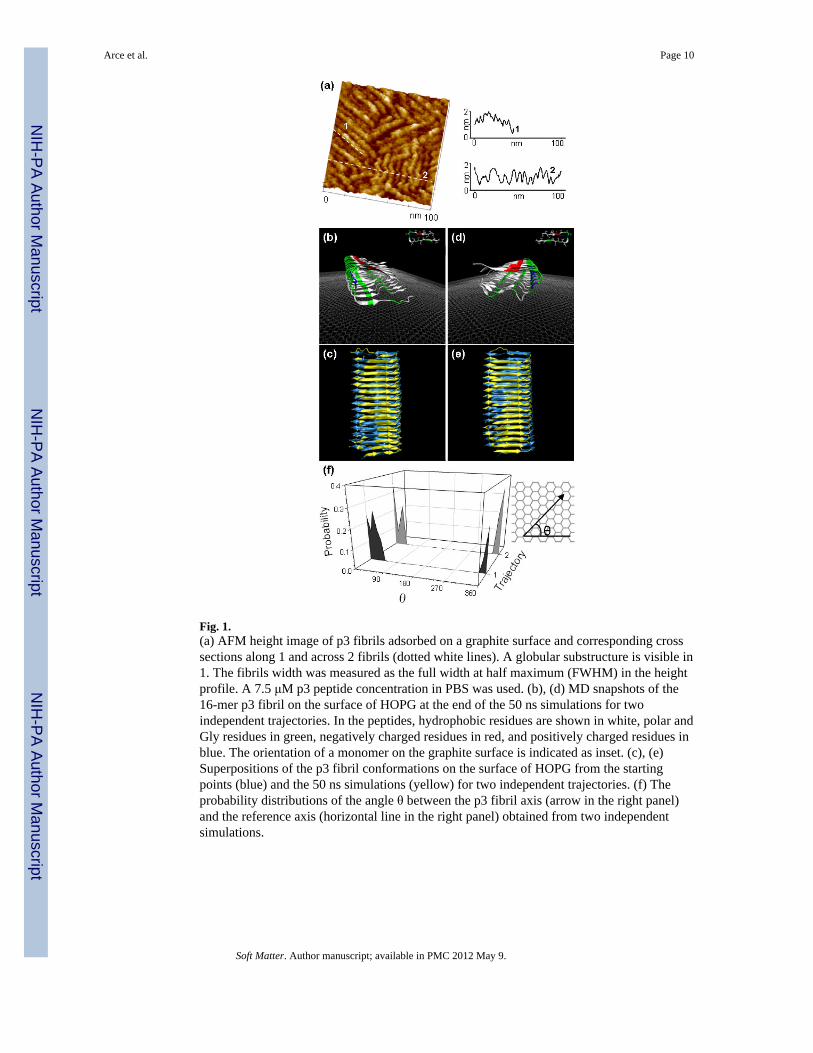
Fig. 1.(a) AFM height image of p3 fibrils adsorbed on a graphite surface and corresponding crosssections along 1 and across 2 fibrils (dotted white lines). A globular substructure is visible in1. The fibrils width was measured as the full width at half maximum (FWHM) in the heightprofile. A 7.5 μM p3 peptide concentration in PBS was used. (b), (d) MD snapshots of the16-mer p3 fibril on the surface of HOPG at the end of the 50 ns simulations for twoindependent trajectories. In the peptides, hydrophobic residues are shown in white, polar andGly residues in green, negatively charged residues in red, and positively charged residues inblue. The orientation of a monomer on the graphite surface is indicated as inset. (c), (e)Superpositions of the p3 fibril conformations on the surface of HOPG from the startingpoints (blue) and the 50 ns simulations (yellow) for two independent trajectories. (f) Theprobability distributions of the angle θ between the p3 fibril axis (arrow in the right panel)and the reference axis (horizontal line in the right panel) obtained from two independentsimulations.
Arce et al. Page 10
Soft Matter. Author manuscript; available in PMC 2012 May 9.
NIH
-PA Author Manuscript
NIH
-PA Author Manuscript
NIH
-PA Author Manuscript
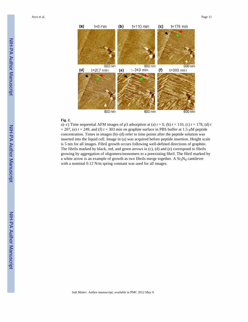
Fig. 2.a)–c) Time sequential AFM images of p3 adsorption at (a) t = 0, (b) t = 110, (c) t = 178, (d) t= 207, (e) t = 249, and (f) t = 303 min on graphite surface in PBS buffer at 1.5 μM peptideconcentration. Times in images (b)–(d) refer to time points after the peptide solution wasinserted into the liquid cell. Image in (a) was acquired before peptide insertion. Height scaleis 5 nm for all images. Fibril growth occurs following well-defined directions of graphite.The fibrils marked by black, red, and green arrows in (c), (d) and (e) correspond to fibrilsgrowing by aggregation of oligomers/monomers to a preexisting fibril. The fibril marked bya white arrow is an example of growth as two fibrils merge together. A Si3N4 cantileverwith a nominal 0.12 N/m spring constant was used for all images.
Arce et al. Page 11
Soft Matter. Author manuscript; available in PMC 2012 May 9.
NIH
-PA Author Manuscript
NIH
-PA Author Manuscript
NIH
-PA Author Manuscript
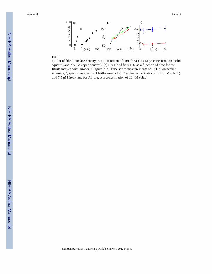
Fig. 3.a) Plot of fibrils surface density, ρ, as a function of time for a 1.5 μM p3 concentration (solidsquares) and 7.5 μM (open squares). (b) Length of fibrils, L, as a function of time for thefibrils marked with arrows in Figure 2. c) Time series measurements of ThT fluorescenceintensity, I, specific to amyloid fibrillogenesis for p3 at the concentrations of 1.5 μM (black)and 7.5 μM (red), and for Aβ1–42, at a concentration of 10 μM (blue).
Arce et al. Page 12
Soft Matter. Author manuscript; available in PMC 2012 May 9.
NIH
-PA Author Manuscript
NIH
-PA Author Manuscript
NIH
-PA Author Manuscript
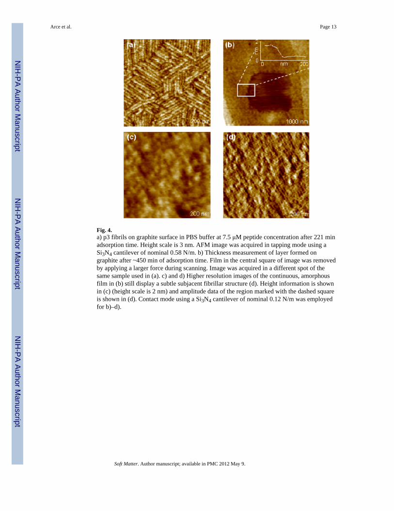
Fig. 4.a) p3 fibrils on graphite surface in PBS buffer at 7.5 μM peptide concentration after 221 minadsorption time. Height scale is 3 nm. AFM image was acquired in tapping mode using aSi3N4 cantilever of nominal 0.58 N/m. b) Thickness measurement of layer formed ongraphite after ~450 min of adsorption time. Film in the central square of image was removedby applying a larger force during scanning. Image was acquired in a different spot of thesame sample used in (a). c) and d) Higher resolution images of the continuous, amorphousfilm in (b) still display a subtle subjacent fibrillar structure (d). Height information is shownin (c) (height scale is 2 nm) and amplitude data of the region marked with the dashed squareis shown in (d). Contact mode using a Si3N4 cantilever of nominal 0.12 N/m was employedfor b)–d).
Arce et al. Page 13
Soft Matter. Author manuscript; available in PMC 2012 May 9.
NIH
-PA Author Manuscript
NIH
-PA Author Manuscript
NIH
-PA Author Manuscript
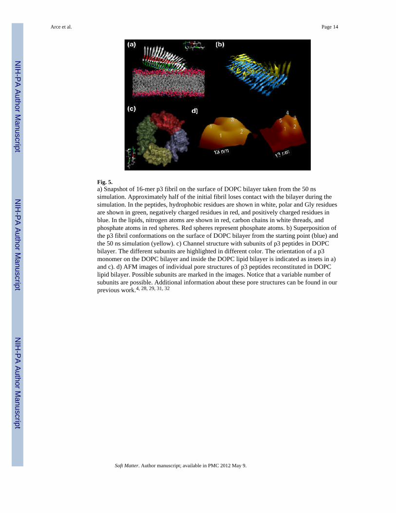
Fig. 5.a) Snapshot of 16-mer p3 fibril on the surface of DOPC bilayer taken from the 50 nssimulation. Approximately half of the initial fibril loses contact with the bilayer during thesimulation. In the peptides, hydrophobic residues are shown in white, polar and Gly residuesare shown in green, negatively charged residues in red, and positively charged residues inblue. In the lipids, nitrogen atoms are shown in red, carbon chains in white threads, andphosphate atoms in red spheres. Red spheres represent phosphate atoms. b) Superposition ofthe p3 fibril conformations on the surface of DOPC bilayer from the starting point (blue) andthe 50 ns simulation (yellow). c) Channel structure with subunits of p3 peptides in DOPCbilayer. The different subunits are highlighted in different color. The orientation of a p3monomer on the DOPC bilayer and inside the DOPC lipid bilayer is indicated as insets in a)and c). d) AFM images of individual pore structures of p3 peptides reconstituted in DOPClipid bilayer. Possible subunits are marked in the images. Notice that a variable number ofsubunits are possible. Additional information about these pore structures can be found in ourprevious work.4, 28, 29, 31, 32
Arce et al. Page 14
Soft Matter. Author manuscript; available in PMC 2012 May 9.
NIH
-PA Author Manuscript
NIH
-PA Author Manuscript
NIH
-PA Author Manuscript
Related Documents