518 Am. J. Trop. Med. Hyg., 61(4), 1999, pp. 518–525 Copyright q 1999 by The American Society of Tropical Medicine and Hygiene POLYMORPHISM AT THE MEROZOITE SURFACE PROTEIN-3a LOCUS OF PLASMODIUM VIVAX: GLOBAL AND LOCAL DIVERSITY MARIAN C. BRUCE, MARY R. GALINSKI, JOHN W. BARNWELL, GEORGES SNOUNOU, AND KAREN P. DAY Wellcome Trust Centre for the Epidemiology of Infectious Disease, University of Oxford, Oxford, United Kingdom; Division of Infectious Diseases, Department of Medicine, Emory University, School of Medicine, Atlanta, Georgia; Biology and Diagnostic Branch, Division of Parasitic Diseases, Centers for Disease Control and Prevention, Atlanta, Georgia; Department of Infection and Tropical Medicine, Wellcome Centre for Clinical Tropical Medicine, Imperial College School of Medicine, University of London, London, United Kingdom Abstract. Allelic diversity at the Plasmodium vivax merozoite surface protein-3a (PvMsp-3a) locus was investi- gated using a combined polymerase chain reaction/restriction fragment length polymorphism (PCR/RFLP) protocol. Symptomatic patient isolates from global geographic origins showed a high level of polymorphism at the nucleotide level. These samples were used to validate the sensitivity, specificity, and reproducibility of the PCR/RFLP method. It was then used to investigate PvMsp3a diversity in field samples from children living in a single village in a malaria- endemic region of Papua New Guinea, with the aim of assessing the usefulness of this locus as an epidemiologic marker of P. vivax infections. Eleven PvMsp-3a alleles were distinguishable in 16 samples with single infections, revealing extensive parasite polymorphism within this restricted area. Multiple infections were easily detected and accounted for 5 (23%) of 22 positive samples. Pairs of samples from individual children provided preliminary evidence for high turnover of P. vivax populations. Epidemiologic analyses of the population structure of Plasmodium parasites within and between endemic areas is essential for understanding the role of parasite diversity in the transmission of malaria as well as for designing and eval- uating malaria vaccines. 1 Several large-scale studies have been conducted for P. falciparum, where the presence and dynamics of either single or multiple polymorphic antigen- encoding genes have been investigated. 2–6 Comparable stud- ies using highly polymorphic markers have yet to be re- ported for P. vivax. Here we present a P. vivax polymerase chain reaction/restriction fragment length polymorphism (PCR/RFLP) protocol that will facilitate such analyses. Us- ing this protocol, we demonstrate that multiple genotypes of P. vivax are present in an endemic area of Papua New Guin- ea and provide preliminary evidence for a rapid turnover of P. vivax genotypes within individuals. The analysis is based on the evaluation of the presence and number of P. vivax merozoite surface protein-3a (PvMsp-3a) alleles. 7 Proteins on the surface of Plasmodium merozoites are tar- gets of naturally acquired 8–10 and vaccine-induced immunity against malaria. 11,12 The PvMsp-3a 7 is a member of an Msp3 gene family that is genetically related to P. falciparum mer- ozoite surface protein-3 (PfMsp-3). 9 In P. falciparum, this molecule is known to induce antibody-dependent cellular- mediated inhibition, a form of immunity naturally acquired through repeated malaria infection. 13,14 Thus, there is an add- ed interest in the P. vivax Msp-3a antigen family as immu- nogens and vaccine candidates. Amplification and sequence analysis of a series of PvMsp- 3a genes from laboratory maintained and patient isolates of P. vivax from diverse geographic regions (Central and South America, India, and southern Asia) indicate a high degree of polymorphism in these genes and encoded proteins. The considerable sequence diversity tends to be largely restricted to the central domain of the molecule. Nevertheless, con- served flanking areas exist that enable PCR-based amplifi- cation of this gene almost in its entirety from most P. vivax samples tested to date (Galinski MR, Barnwell JW, unpub- lished data). The polymorphic nature of these molecules sug- gests that they may be under immune selection pressure. Due to the necessity of having to analyze a large number of samples before drawing conclusions about the genetic make-up of parasite populations, molecular epidemiologic markers must have a high degree of polymorphism that is quickly and easily detectable. Plasmodium vivax markers re- ported to date have either insufficient polymorphism, 15 re- quire complex methods or sequencing for detection of the polymorphism, 16–18 or require large volumes of blood for se- rologic 19 and biochemical methods. 20,21 Thus, we sought a molecular epidemiologic marker that could be used for anal- ysis of P. vivax populations. Here we present a PCR/RFLP protocol based on PvMsp-3a genes and demonstrate its util- ity in analyses of P. vivax parasites from geographically di- verse symptomatic cases and asymptomatic infections from children living in a single village in Papua New Guinea. MATERIALS AND METHODS Parasites. Samples of parasites were of 4 types. 1) Symptomatic patient samples. Venous blood was col- lected in EDTA Vacutainerst (Becton Dickinson UK, Ltd., Oxford, United Kingdom) from symptomatic residents in the United Kingdom attending the Northwick Park Hospital dur- ing 1997, who had contracted malaria while traveling abroad. The geographic origin of the isolates is as follows: 14/97, 16/97, 23/97, and 24/97—India; 26/97—Sudan; 47/ 97—Sri Lanka. Five samples had microscopy counts of less than 200 parasites/ml of blood, while one, 47/97, had a count of 1,500 parasites/ml. Samples were pretested for the pres- ence of other species of Plasmodium using species-specific PCR amplification 22 and were found to contain only P. vivax. 2) Asymptomatic field samples. Fingerprick blood samples were obtained from asymptomatic children between 4 and 14 years of age residing in 11 houses in the village of Gonoa, Madang, Papua New Guinea. Gonoa is situated in an area of intense malaria transmission where all 4 species of human Plasmodium (P. falciparum, P. vivax, P. malariae, and P. ovale) are transmitted. 23 The mean number of P. vivax in- fectious bites in this region is 133 bites/person/year. 24 Chil-

Welcome message from author
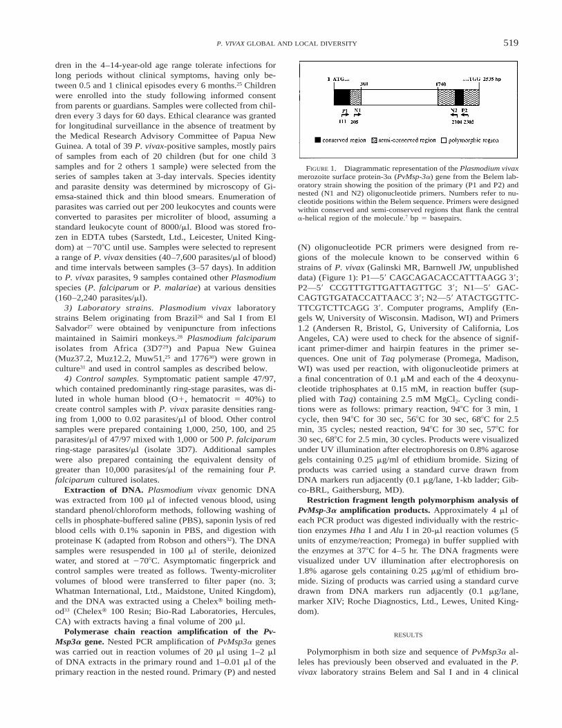
This document is posted to help you gain knowledge. Please leave a comment to let me know what you think about it! Share it to your friends and learn new things together.
Transcript

518
Am. J. Trop. Med. Hyg., 61(4), 1999, pp. 518–525Copyright q 1999 by The American Society of Tropical Medicine and Hygiene
POLYMORPHISM AT THE MEROZOITE SURFACE PROTEIN-3a LOCUS OFPLASMODIUM VIVAX: GLOBAL AND LOCAL DIVERSITY
MARIAN C. BRUCE, MARY R. GALINSKI, JOHN W. BARNWELL, GEORGES SNOUNOU, AND KAREN P. DAYWellcome Trust Centre for the Epidemiology of Infectious Disease, University of Oxford, Oxford, United Kingdom;Division of Infectious Diseases, Department of Medicine, Emory University, School of Medicine, Atlanta, Georgia;
Biology and Diagnostic Branch, Division of Parasitic Diseases, Centers for Disease Control and Prevention, Atlanta, Georgia;Department of Infection and Tropical Medicine, Wellcome Centre for Clinical Tropical Medicine,
Imperial College School of Medicine, University of London, London, United Kingdom
Abstract. Allelic diversity at the Plasmodium vivax merozoite surface protein-3a (PvMsp-3a) locus was investi-gated using a combined polymerase chain reaction/restriction fragment length polymorphism (PCR/RFLP) protocol.Symptomatic patient isolates from global geographic origins showed a high level of polymorphism at the nucleotidelevel. These samples were used to validate the sensitivity, specificity, and reproducibility of the PCR/RFLP method.It was then used to investigate PvMsp3a diversity in field samples from children living in a single village in a malaria-endemic region of Papua New Guinea, with the aim of assessing the usefulness of this locus as an epidemiologicmarker of P. vivax infections. Eleven PvMsp-3a alleles were distinguishable in 16 samples with single infections,revealing extensive parasite polymorphism within this restricted area. Multiple infections were easily detected andaccounted for 5 (23%) of 22 positive samples. Pairs of samples from individual children provided preliminary evidencefor high turnover of P. vivax populations.
Epidemiologic analyses of the population structure ofPlasmodium parasites within and between endemic areas isessential for understanding the role of parasite diversity inthe transmission of malaria as well as for designing and eval-uating malaria vaccines.1 Several large-scale studies havebeen conducted for P. falciparum, where the presence anddynamics of either single or multiple polymorphic antigen-encoding genes have been investigated.2–6 Comparable stud-ies using highly polymorphic markers have yet to be re-ported for P. vivax. Here we present a P. vivax polymerasechain reaction/restriction fragment length polymorphism(PCR/RFLP) protocol that will facilitate such analyses. Us-ing this protocol, we demonstrate that multiple genotypes ofP. vivax are present in an endemic area of Papua New Guin-ea and provide preliminary evidence for a rapid turnover ofP. vivax genotypes within individuals. The analysis is basedon the evaluation of the presence and number of P. vivaxmerozoite surface protein-3a (PvMsp-3a) alleles.7
Proteins on the surface of Plasmodium merozoites are tar-gets of naturally acquired8–10 and vaccine-induced immunityagainst malaria.11,12 The PvMsp-3a7 is a member of an Msp3gene family that is genetically related to P. falciparum mer-ozoite surface protein-3 (PfMsp-3).9 In P. falciparum, thismolecule is known to induce antibody-dependent cellular-mediated inhibition, a form of immunity naturally acquiredthrough repeated malaria infection.13,14 Thus, there is an add-ed interest in the P. vivax Msp-3a antigen family as immu-nogens and vaccine candidates.
Amplification and sequence analysis of a series of PvMsp-3a genes from laboratory maintained and patient isolates ofP. vivax from diverse geographic regions (Central and SouthAmerica, India, and southern Asia) indicate a high degreeof polymorphism in these genes and encoded proteins. Theconsiderable sequence diversity tends to be largely restrictedto the central domain of the molecule. Nevertheless, con-served flanking areas exist that enable PCR-based amplifi-cation of this gene almost in its entirety from most P. vivaxsamples tested to date (Galinski MR, Barnwell JW, unpub-lished data). The polymorphic nature of these molecules sug-gests that they may be under immune selection pressure.
Due to the necessity of having to analyze a large numberof samples before drawing conclusions about the geneticmake-up of parasite populations, molecular epidemiologicmarkers must have a high degree of polymorphism that isquickly and easily detectable. Plasmodium vivax markers re-ported to date have either insufficient polymorphism,15 re-quire complex methods or sequencing for detection of thepolymorphism,16–18 or require large volumes of blood for se-rologic19 and biochemical methods.20,21 Thus, we sought amolecular epidemiologic marker that could be used for anal-ysis of P. vivax populations. Here we present a PCR/RFLPprotocol based on PvMsp-3a genes and demonstrate its util-ity in analyses of P. vivax parasites from geographically di-verse symptomatic cases and asymptomatic infections fromchildren living in a single village in Papua New Guinea.
MATERIALS AND METHODS
Parasites. Samples of parasites were of 4 types.1) Symptomatic patient samples. Venous blood was col-
lected in EDTA Vacutainerst (Becton Dickinson UK, Ltd.,Oxford, United Kingdom) from symptomatic residents in theUnited Kingdom attending the Northwick Park Hospital dur-ing 1997, who had contracted malaria while travelingabroad. The geographic origin of the isolates is as follows:14/97, 16/97, 23/97, and 24/97—India; 26/97—Sudan; 47/97—Sri Lanka. Five samples had microscopy counts of lessthan 200 parasites/ml of blood, while one, 47/97, had a countof 1,500 parasites/ml. Samples were pretested for the pres-ence of other species of Plasmodium using species-specificPCR amplification22 and were found to contain only P. vivax.
2) Asymptomatic field samples. Fingerprick blood sampleswere obtained from asymptomatic children between 4 and14 years of age residing in 11 houses in the village of Gonoa,Madang, Papua New Guinea. Gonoa is situated in an areaof intense malaria transmission where all 4 species of humanPlasmodium (P. falciparum, P. vivax, P. malariae, and P.ovale) are transmitted.23 The mean number of P. vivax in-fectious bites in this region is 133 bites/person/year.24 Chil-
519P. VIVAX GLOBAL AND LOCAL DIVERSITY
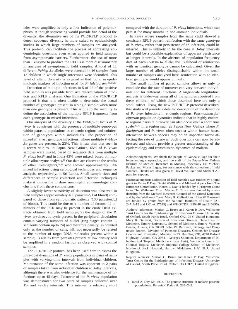
FIGURE 1. Diagrammatic representation of the Plasmodium vivaxmerozoite surface protein-3a (PvMsp-3a) gene from the Belem lab-oratory strain showing the position of the primary (P1 and P2) andnested (N1 and N2) oligonucleotide primers. Numbers refer to nu-cleotide positions within the Belem sequence. Primers were designedwithin conserved and semi-conserved regions that flank the centrala-helical region of the molecule.7 bp 5 basepairs.
dren in the 4–14-year-old age range tolerate infections forlong periods without clinical symptoms, having only be-tween 0.5 and 1 clinical episodes every 6 months.25 Childrenwere enrolled into the study following informed consentfrom parents or guardians. Samples were collected from chil-dren every 3 days for 60 days. Ethical clearance was grantedfor longitudinal surveillance in the absence of treatment bythe Medical Research Advisory Committee of Papua NewGuinea. A total of 39 P. vivax-positive samples, mostly pairsof samples from each of 20 children (but for one child 3samples and for 2 others 1 sample) were selected from theseries of samples taken at 3-day intervals. Species identityand parasite density was determined by microscopy of Gi-emsa-stained thick and thin blood smears. Enumeration ofparasites was carried out per 200 leukocytes and counts wereconverted to parasites per microliter of blood, assuming astandard leukocyte count of 8000/ml. Blood was stored fro-zen in EDTA tubes (Sarstedt, Ltd., Leicester, United King-dom) at 2708C until use. Samples were selected to representa range of P. vivax densities (40–7,600 parasites/ml of blood)and time intervals between samples (3–57 days). In additionto P. vivax parasites, 9 samples contained other Plasmodiumspecies (P. falciparum or P. malariae) at various densities(160–2,240 parasites/ml).
3) Laboratory strains. Plasmodium vivax laboratorystrains Belem originating from Brazil26 and Sal I from ElSalvador27 were obtained by venipuncture from infectionsmaintained in Saimiri monkeys.28 Plasmodium falciparumisolates from Africa (3D729) and Papua New Guinea(Muz37.2, Muz12.2, Muw51,25 and 177630) were grown inculture31 and used in control samples as described below.
4) Control samples. Symptomatic patient sample 47/97,which contained predominantly ring-stage parasites, was di-luted in whole human blood (O1, hematocrit 5 40%) tocreate control samples with P. vivax parasite densities rang-ing from 1,000 to 0.02 parasites/ml of blood. Other controlsamples were prepared containing 1,000, 250, 100, and 25parasites/ml of 47/97 mixed with 1,000 or 500 P. falciparumring-stage parasites/ml (isolate 3D7). Additional sampleswere also prepared containing the equivalent density ofgreater than 10,000 parasites/ml of the remaining four P.falciparum cultured isolates.
Extraction of DNA. Plasmodium vivax genomic DNAwas extracted from 100 ml of infected venous blood, usingstandard phenol/chloroform methods, following washing ofcells in phosphate-buffered saline (PBS), saponin lysis of redblood cells with 0.1% saponin in PBS, and digestion withproteinase K (adapted from Robson and others32). The DNAsamples were resuspended in 100 ml of sterile, deionizedwater, and stored at 2708C. Asymptomatic fingerprick andcontrol samples were treated as follows. Twenty-microlitervolumes of blood were transferred to filter paper (no. 3;Whatman International, Ltd., Maidstone, United Kingdom),and the DNA was extracted using a Chelext boiling meth-od33 (Chelext 100 Resin; Bio-Rad Laboratories, Hercules,CA) with extracts having a final volume of 200 ml.
Polymerase chain reaction amplification of the Pv-Msp3a gene. Nested PCR amplification of PvMsp3a geneswas carried out in reaction volumes of 20 ml using 1–2 mlof DNA extracts in the primary round and 1–0.01 ml of theprimary reaction in the nested round. Primary (P) and nested
(N) oligonucleotide PCR primers were designed from re-gions of the molecule known to be conserved within 6strains of P. vivax (Galinski MR, Barnwell JW, unpublisheddata) (Figure 1): P1—59 CAGCAGACACCATTTAAGG 39;P2—59 CCGTTTGTTGATTAGTTGC 39; N1—59 GAC-CAGTGTGATACCATTAACC 39; N2—59 ATACTGGTTC-TTCGTCTTCAGG 39. Computer programs, Amplify (En-gels W, University of Wisconsin. Madison, WI) and Primers1.2 (Andersen R, Bristol, G, University of California, LosAngeles, CA) were used to check for the absence of signif-icant primer-dimer and hairpin features in the primer se-quences. One unit of Taq polymerase (Promega, Madison,WI) was used per reaction, with oligonucleotide primers ata final concentration of 0.1 mM and each of the 4 deoxynu-cleotide triphosphates at 0.15 mM, in reaction buffer (sup-plied with Taq) containing 2.5 mM MgCl2. Cycling condi-tions were as follows: primary reaction, 948C for 3 min, 1cycle, then 948C for 30 sec, 568C for 30 sec, 688C for 2.5min, 35 cycles; nested reaction, 948C for 30 sec, 578C for30 sec, 688C for 2.5 min, 30 cycles. Products were visualizedunder UV illumination after electrophoresis on 0.8% agarosegels containing 0.25 mg/ml of ethidium bromide. Sizing ofproducts was carried using a standard curve drawn fromDNA markers run adjacently (0.1 mg/lane, 1-kb ladder; Gib-co-BRL, Gaithersburg, MD).
Restriction fragment length polymorphism analysis ofPvMsp-3a amplification products. Approximately 4 ml ofeach PCR product was digested individually with the restric-tion enzymes Hha I and Alu I in 20-ml reaction volumes (5units of enzyme/reaction; Promega) in buffer supplied withthe enzymes at 378C for 4–5 hr. The DNA fragments werevisualized under UV illumination after electrophoresis on1.8% agarose gels containing 0.25 mg/ml of ethidium bro-mide. Sizing of products was carried using a standard curvedrawn from DNA markers run adjacently (0.1 mg/lane,marker XIV; Roche Diagnostics, Ltd., Lewes, United King-dom).
RESULTS
Polymorphism in both size and sequence of PvMsp3a al-leles has previously been observed and evaluated in the P.vivax laboratory strains Belem and Sal I and in 4 clinical
520 BRUCE AND OTHERS
isolates of diverse geographic origin (Galinski MR, BarnwellJW, unpublished data). Here we investigate an additional 6geographically diverse isolates from symptomatic patients.The samples from the symptomatic individuals were used toevaluate the sensitivity, reproducibility, and potential allelicamplification bias in the PCR/RFLP method. Symptomaticinfections were acquired by patients while traveling andwere likely to be the result of infection from a single infectedmosquito bite. These samples were therefore more likely tocontain single P. vivax genotypes than those from superin-fected individuals living under endemic conditions. ThePCR/RFLP method was then used to determine the level ofPvMsp-3a polymorphism within asymptomatic infectionsfrom the very small geographic area of a single malaria en-demic village in Papua New Guinea. By analyzing multiplesamples taken from the same individuals at different timepoints we were able to assess turnover of P. vivax parasitepopulations within semi-immune children.
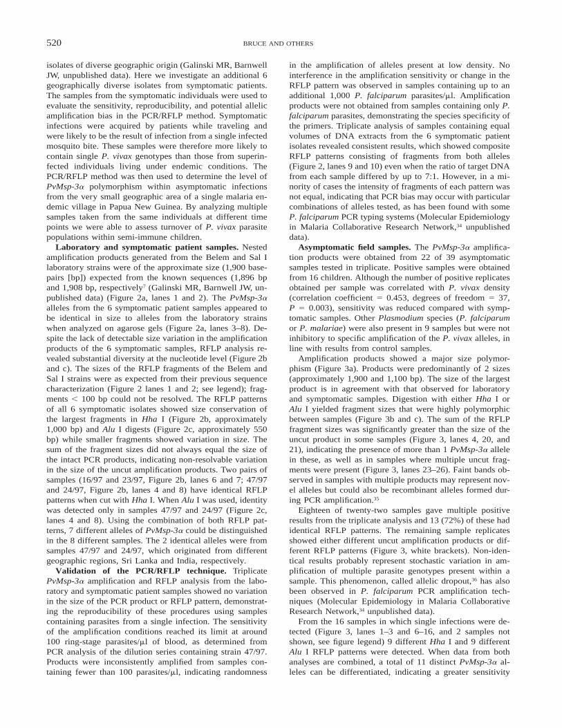
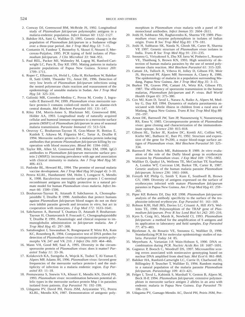
Laboratory and symptomatic patient samples. Nestedamplification products generated from the Belem and Sal Ilaboratory strains were of the approximate size (1,900 base-pairs [bp]) expected from the known sequences (1,896 bpand 1,908 bp, respectively7 (Galinski MR, Barnwell JW, un-published data) (Figure 2a, lanes 1 and 2). The PvMsp-3aalleles from the 6 symptomatic patient samples appeared tobe identical in size to alleles from the laboratory strainswhen analyzed on agarose gels (Figure 2a, lanes 3–8). De-spite the lack of detectable size variation in the amplificationproducts of the 6 symptomatic samples, RFLP analysis re-vealed substantial diversity at the nucleotide level (Figure 2band c). The sizes of the RFLP fragments of the Belem andSal I strains were as expected from their previous sequencecharacterization (Figure 2 lanes 1 and 2; see legend); frag-ments , 100 bp could not be resolved. The RFLP patternsof all 6 symptomatic isolates showed size conservation ofthe largest fragments in Hha I (Figure 2b, approximately1,000 bp) and Alu I digests (Figure 2c, approximately 550bp) while smaller fragments showed variation in size. Thesum of the fragment sizes did not always equal the size ofthe intact PCR products, indicating non-resolvable variationin the size of the uncut amplification products. Two pairs ofsamples (16/97 and 23/97, Figure 2b, lanes 6 and 7; 47/97and 24/97, Figure 2b, lanes 4 and 8) have identical RFLPpatterns when cut with Hha I. When Alu I was used, identitywas detected only in samples 47/97 and 24/97 (Figure 2c,lanes 4 and 8). Using the combination of both RFLP pat-terns, 7 different alleles of PvMsp-3a could be distinguishedin the 8 different samples. The 2 identical alleles were fromsamples 47/97 and 24/97, which originated from differentgeographic regions, Sri Lanka and India, respectively.
Validation of the PCR/RFLP technique. TriplicatePvMsp-3a amplification and RFLP analysis from the labo-ratory and symptomatic patient samples showed no variationin the size of the PCR product or RFLP pattern, demonstrat-ing the reproducibility of these procedures using samplescontaining parasites from a single infection. The sensitivityof the amplification conditions reached its limit at around100 ring-stage parasites/ml of blood, as determined fromPCR analysis of the dilution series containing strain 47/97.Products were inconsistently amplified from samples con-taining fewer than 100 parasites/ml, indicating randomness
in the amplification of alleles present at low density. Nointerference in the amplification sensitivity or change in theRFLP pattern was observed in samples containing up to anadditional 1,000 P. falciparum parasites/ml. Amplificationproducts were not obtained from samples containing only P.falciparum parasites, demonstrating the species specificity ofthe primers. Triplicate analysis of samples containing equalvolumes of DNA extracts from the 6 symptomatic patientisolates revealed consistent results, which showed compositeRFLP patterns consisting of fragments from both alleles(Figure 2, lanes 9 and 10) even when the ratio of target DNAfrom each sample differed by up to 7:1. However, in a mi-nority of cases the intensity of fragments of each pattern wasnot equal, indicating that PCR bias may occur with particularcombinations of alleles tested, as has been found with someP. falciparum PCR typing systems (Molecular Epidemiologyin Malaria Collaborative Research Network,34 unpublisheddata).
Asymptomatic field samples. The PvMsp-3a amplifica-tion products were obtained from 22 of 39 asymptomaticsamples tested in triplicate. Positive samples were obtainedfrom 16 children. Although the number of positive replicatesobtained per sample was correlated with P. vivax density(correlation coefficient 5 0.453, degrees of freedom 5 37,P 5 0.003), sensitivity was reduced compared with symp-tomatic samples. Other Plasmodium species (P. falciparumor P. malariae) were also present in 9 samples but were notinhibitory to specific amplification of the P. vivax alleles, inline with results from control samples.
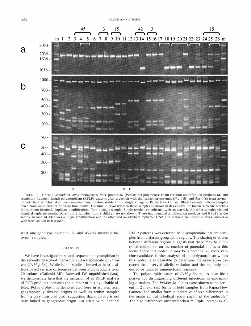
Amplification products showed a major size polymor-phism (Figure 3a). Products were predominantly of 2 sizes(approximately 1,900 and 1,100 bp). The size of the largestproduct is in agreement with that observed for laboratoryand symptomatic samples. Digestion with either Hha I orAlu I yielded fragment sizes that were highly polymorphicbetween samples (Figure 3b and c). The sum of the RFLPfragment sizes was significantly greater than the size of theuncut product in some samples (Figure 3, lanes 4, 20, and21), indicating the presence of more than 1 PvMsp-3a allelein these, as well as in samples where multiple uncut frag-ments were present (Figure 3, lanes 23–26). Faint bands ob-served in samples with multiple products may represent nov-el alleles but could also be recombinant alleles formed dur-ing PCR amplification.35
Eighteen of twenty-two samples gave multiple positiveresults from the triplicate analysis and 13 (72%) of these hadidentical RFLP patterns. The remaining sample replicatesshowed either different uncut amplification products or dif-ferent RFLP patterns (Figure 3, white brackets). Non-iden-tical results probably represent stochastic variation in am-plification of multiple parasite genotypes present within asample. This phenomenon, called allelic dropout,36 has alsobeen observed in P. falciparum PCR amplification tech-niques (Molecular Epidemiology in Malaria CollaborativeResearch Network,34 unpublished data).
From the 16 samples in which single infections were de-tected (Figure 3, lanes 1–3 and 6–16, and 2 samples notshown, see figure legend) 9 different Hha I and 9 differentAlu I RFLP patterns were detected. When data from bothanalyses are combined, a total of 11 distinct PvMsp-3a al-leles can be differentiated, indicating a greater sensitivity
521P. VIVAX GLOBAL AND LOCAL DIVERSITY
FIGURE 2. Uncut Plasmodium vivax merozoite surface protein-3a (PvMsp-3a) polymerase chain reaction amplification products (a) andrestriction fragment length polymorphism (RFLP) patterns after digestion with the restriction enzymes Hha I (b) and Alu I (c) from thelaboratory strains Belem (lane 1) and Sal I (lane 2) and symptomatic patient samples (lanes 3–8: samples 26, 47, 14, 16, 23 and 24/97respectively). Products amplified from mixtures of target DNA from symptomatic patient samples 14 and 16 (lane 9) and 23 and 24 (lane 10)are also shown. DNA size markers are shown in lanes labeled m with sizes shown in basepairs (bp). From nucleotide sequences the predictedRFLP fragment sizes after digestion of nested amplification products from Belem and Sal I are Belem, Hha I (963, 330, 254, 211, 75, and 63bp); Alu I (492, 258, 201, 198, 172, 156, 146, 114, 60, 39, 33, and 27 bp); Sal I, Hha I (975, 465, 405, and 63 bp); Alu I (537, 258, 189,172, 156, 153, 150, 146, 69, 51, and 27 bp)7 (Galinski MR, Barnwell, JW unpublished data).
from the use of 2 restriction enzymes. Three genotypes weredetected in more than 1 sample and 2 genotypes were foundin more than 1 child.
Six pairs of samples from individual children with timeintervals of 3–45 days yielded PCR products. Samples tak-en from 2 children at 3-day intervals showed identity (Fig-
ure 3, lanes 7 and 8, and 15 and 16). Turnover of parasitepopulations was demonstrated in samples taken at time in-tervals of 15 and 42 days (Figure 3, lanes 9 and 10, and13 and 14). In the remaining 2 children, mixed parasitepopulations were detected in 1 or both samples. In eachcase, however, RFLP patterns indicated persistence of at
522 BRUCE AND OTHERS
FIGURE 3. Uncut Plasmodium vivax merozoite surface protein-3a (PvMsp-3a) polymerase chain reaction amplification products (a) andrestriction fragment length polymorphism (RFLP) patterns after digestion with the restriction enzymes Hha I (b) and Alu I (c) from asymp-tomatic field samples taken from semi-immune children resident in a single village in Papua New Guinea. Black brackets indicate samplestaken from same child at different time points. The time interval between these samples is shown in days above the brackets. White bracketsindicate non-identical, duplicate amplifications from a single sample. Single results are indicated with an asterisk. All other samples yieldedidentical replicate results. Data from 2 samples from 2 children are not shown. These had identical amplification products and RFLPs as thesample in lane 14. One was a single amplification and the other had an identical replicate. DNA size markers are shown in lanes labeled mwith sizes shown in basepairs.
least one genotype over the 15- and 45-day intervals be-tween samples.
DISCUSSION
We have investigated size and sequence polymorphism inthe recently described merozoite surface molecule of P. vi-vax (PvMsp-3a). While initial studies showed at least 4 al-leles based on size differences between PCR products from20 isolates (Galinski MR, Barnwell JW, unpublished data),we demonstrate here that the inclusion of an RFLP analysisof PCR products increases the number of distinguishable al-leles. Polymorphism is demonstrated here in isolates fromgeographically diverse origins as well as within samplesfrom a very restricted area, suggesting that diversity is notonly linked to geographic origin. An allele with identical
RFLP patterns was detected in 2 symptomatic patient sam-ples from different geographic regions. The sharing of allelesbetween different regions suggests that there may be func-tional constraints on the number of potential alleles at thislocus. Since this molecule may be a potential P. vivax vac-cine candidate, further analysis of the polymorphism withinthis molecule is desirable to determine the association be-tween the observed allelic variation and the naturally ac-quired or induced immunologic response.
The polymorphic nature of PvMsp-3a makes it an idealmarker for distinguishing different infections in epidemio-logic studies. The PvMsp-3a alleles were shown to be pres-ent in 2 major size forms in field samples from Papua NewGuinea. The smaller form is indicative of size differences inthe major central a-helical repeat region of the molecule.7
The size differences observed when multiple PvMsp-3a al-
523P. VIVAX GLOBAL AND LOCAL DIVERSITY
leles were amplified is only a first indication of polymor-phism. Although sequencing would provide fine detail of thediversity, the alternative use of the PCR/RFLP protocol todetect sequence diversity is more suited to epidemiologicstudies in which large numbers of samples are analyzed.This protocol can facilitate the process of addressing epi-demiologic questions even when applied to field samplesfrom asymptomatic carriers. Furthermore, the use of morethan 1 enzyme to produce the RFLPs is more discriminatoryin analyses of asymptomatic field samples. A total of 11different PvMsp-3a alleles were detected in 16 samples from12 children in which single infections were identified. Thislevel of allelic diversity is as great as that found in epide-miologic markers of infection used for P. falciparum.4,5,37,38
Detection of multiple infections in 5 of 22 of the positivefield samples was possible from size determination of prod-ucts and RFLP analyses. One limitation of the PCR/RFLPprotocol is that it is often unable to determine the actualnumber of genotypes present in a single sample when morethan one genotype is present. This is due to the inability todefine the association of the multiple RFLP fragments fromeach genotype in mixed infections.
Our analysis of the diversity at the PvMsp-3a locus of P.vivax is consistent with the presence of multiple genotypeswithin parasite populations in endemic regions and coinfec-tion of genotypes within individuals. The proportion ofmixed P. vivax genotype infections, where multiple PvMsp-3a genes are present, is 23%. This is less than that seen in2 recent studies. In Papua New Guinea, 65% of P. vivaxsamples were mixed, based on sequence data from multipleP. vivax loci17 and in India 43% were mixed, based on mul-tiple alloenzyme analysis.21 Our data are closest to the resultsof other investigators,18,39 who showed approximately 10%mixed infections using serological techniques and sequenceanalysis, respectively, in Sri Lanka. Small sample sizes anddifferences in sample collection and detection techniquesmake it impossible to draw meaningful epidemiologic con-clusions from these comparisons.
A slightly lower sensitivity of detection was observed infield samples (approximately 400 parasites/ml of blood) com-pared to those from symptomatic patients (100 parasites/mlof blood). This could be due to a number of factors: 1) in-hibitors of the PCR may be present in the crude DNA ex-tracts obtained from field samples; 2) the stages of the P.vivax erythrocytic cycle present in the peripheral circulationcontain varying numbers of nuclei (ring stages contain 1,schizonts contain up to 24) and therefore density, as countedonly as the number of cells, will not necessarily be relatedto the number of target DNA molecules present within asample; 3) alleles from parasites present at low density willbe amplified in a random fashion as observed with controlsamples.
The PCR/RFLP protocol has been used here to assess theintra-host dynamics of P. vivax populations in pairs of sam-ples with varying time intervals from individual children.Maintenance of the same infection was apparent in 2 pairsof samples taken from individual children at 3-day intervals,although there was also evidence for the maintenance of in-fections up to 45 days. Turnover of the P. vivax populationwas demonstrated for two pairs of samples collected over15- and 42-day intervals. This interval is relatively short
compared with the duration of P. vivax infections, which canpersist for many months in non-immune individuals.
In cases where samples from the same child showed aconsistent RFLP pattern, reinfection with the same genotypeof P. vivax, rather than persistence of an infection, could beinferred. This is unlikely to be the case at 3-day intervalsbut could be a possible explanation of apparent persistenceat longer intervals. In the absence of population frequencydata of each PvMsp-3a allele, the likelihood of reinfectionwith an identical genotype cannot be calculated. Given thelarge number of alleles distinguishable within the smallnumber of samples analyzed here, reinfection with an iden-tical genotype would appear unlikely.
The small number of paired samples allows us only toconclude that the rate of turnover can vary between individ-uals and for different infections. A large-scale longitudinalanalysis is underway using all of the samples acquired fromthese children, of which those described here are only asmall subset. Using the new PCR/RFLP protocol described,this study will provide a detailed description of the dynamicsof P. vivax infections in endemic regions. Plasmodium fal-ciparum population dynamics indicate that in highly endem-ic regions parasite turnover can also occur over a short timescale.40,41 In a region such as Papua New Guinea where P.falciparum and P. vivax often coexist within human hosts,interaction between species may be an important factor af-fecting the rate of turnover. Such questions can now be ad-dressed and should provide a greater understanding of theepidemiology and transmission dynamics of malaria.
Acknowledgments: We thank the people of Gonoa village for theirlongstanding cooperation, and the staff of the Papua New GuineaInstitute of Medical Research in Madang, especially Dr. MichaelPacker and Moses Lagog, for assistance with the collection of fieldsamples. Thanks are also given to David Walliker and Michael Al-pers for support.
Financial support: Collection of field samples was funded by a jointgrant to Karen P. Day, David Walliker and Michael Alpers from TheEuropean Commission. Karen P. Day is funded by a Program Grantfrom The Wellcome Trust. Marian C. Bruce was funded by a stu-dentship from the Medical Research Council of the United Kingdomand by the Wellcome Trust. Mary R. Galinski and John W. Barnwellare funded by grants from the National Institutes of Health (AI-24710-12 and U01-AI37545) and WHO/TDR (950440 and 910495).
Authors’ addresses: Marian C. Bruce and Karen P. Day, WellcomeTrust Centre for the Epidemiology of Infectious Disease, Universityof Oxford, South Parks Road, Oxford OX1 3FY, United Kingdom.Mary R. Galinski, Division of Infectious Diseases, Department ofMedicine, Emory University, School of Medicine, Emory VaccineCenter, Atlanta, GA 30329. John W. Barnwell, Biology and Diag-nostic Branch, Division of Parasitic Diseases, Centers for DiseaseControl and Prevention, Mailstop F-13, Building 22B, 4770 BufordHighway, Atlanta, GA 30341. Georges Snounou, Department of In-fection and Tropical Medicine (Lister Unit), Wellcome Centre forClinical Tropical Medicine, Imperial College School of Medicine,Northwick Park Hospital, Harrow, Middlesex, HA1 3UJ, UnitedKingdom.
Reprint requests: Marian C. Bruce and Karen P. Day, WellcomeTrust Centre for the Epidemiology of Infectious Disease, Universityof Oxford, South Parks Road, Oxford OX1 3FY, United Kingdom.
REFERENCES
1. Read A, Day KP, 1992. The genetic structure of malaria parasitepopulations. Parasitol Today 8: 239–242.
524 BRUCE AND OTHERS
2. Conway DJ, Greenwood BM, McBride JS, 1992. Longitudinalstudy of Plasmodium falciparum polymorphic antigens in amalaria-endemic population. Infect Immun 60: 1122–1127.
3. Babiker HA, Satti G, Walliker D, 1995. Genetic changes in thepopulation of Plasmodium falciparum in a Sudanese villageover a three-year period. Am J Trop Med Hyg 53: 7–15.
4. Contamin H, Fandeur T, Bonnefoy S, Skouri F, Ntoumi F, Mer-cereau-Puijalon, 1995. PCR typing of field isolates of Plas-modium falciparum. J Clin Microbiol 33: 944–951.
5. Paul REL, Packer MJ, Walmsley M, Lagog M, Ranford-Cart-wright LC, Paru R, Day KP, 1995. Mating patterns in malariaparasite populations of Papua New Guinea. Science 269:1709–1711.
6. Roper C, Elhassan IA, Hviid L, Giha H, Richardson W, BabikerH, Satti GMH, Theander TG, Amot DE, 1996. Detection ofvery low levels of Plasmodium falciparum infections usingthe nested polymerase chain reaction and reassessment of theepidemiology of unstable malaria in Sudan. Am J Trop MedHyg 54: 325–331.
7. Galinski MR, Corredor-Medina C, Povoa M, Crosby J, Ingra-vallo P, Barnwell JW, 1999. Plasmodium vivax merozoite sur-face protein-3 contains coiled-coil motifs in an alanine-richcentral domain. Mol Biochem Parasitol 101: 131–147.
8. Riley EM, Morris-Jones S, Blackman MJ, Greenwood BM,Holder AA, 1993. Longitudinal study of naturally acquiredcellular and humoral immune responses to a merozoite surfaceprotein (MSP1) of Plasmodium falciparum in area of seasonalmalaria transmission. Parasite Immunol 15: 513–524.
9. Oeuvray C, Bouharoun-Tayoun H, Gras-Masse H, Bottius E,Kaidoh T, Aikawa M, Filgueira M-C, Tartar A, Druilhe P,1994. Merozoite surface protein-3: a malaria protein inducingantibodies that promote Plasmodium falciparum killing by co-operation with blood monocytes. Blood 84: 1594–1602.
10. Taylor RR, Allen SJ, Greenwood BM, Riley EM, 1998. IgG3antibodies to Plasmodium falciparum merozoite surface pro-tein 2 (MSP2): increasing prevalence with age and associationwith clinical immunity to malaria. Am J Trop Med Hyg 58:406–413.
11. Pasloske BL, Howard RL, 1994. The promise of asexual malariavaccine development. Am J Trop Med Hyg 50 (suppl 4): 3–10.
12. Perera KLRL, Handunnetti SM, Holm I, Longacre S, MendisK, 1998. Baculovirus merozoite surface protein 1 C-terminalrecombinant antigens are highly protective in a natural pri-mate model for human Plasmodium vivax malaria. Infect Im-mun 66: 1500–1506.
13. Bouharoun-Tayoun H, Attanath P, Sabchareon A, Chonsupha-jaisiddhi T, Druilhe P, 1990. Antibodies that protect humansagainst Plasmodium falciparum blood stages do not on theirown inhibit parasite growth and invasion in vitro, but act incooperation with monocytes. J Exp Med 172: 1633–1641.
14. Sabchareon A, Burnouf T, Ouattara D, Attanath P, Bouharoun-Tayoun H, Chantavanich P, Foucault C, ChongsuphajaisiddhiT, Druilhe P, 1991. Parasitologic and clinical response to im-munoglobulin administration in falciparum malaria. Am JTrop Med Hyg 45: 297–308.
15. Sattabongkot J, Suwanabun N, Rongnoparut P, Wirtz RA, KainKC, Rosenberg R, 1994. Comparative test of DNA probes fordetection of Plasmodium vivax circumsporozoite protein poly-morphs VK 247 and VK 210. J Infect Dis 169: 464–466.
16. Mann VH, Good MF, Saul A, 1995. Diversity in the circum-sporozoite protein of Plasmodium vivax: does it matter? Par-asitol Today 11: 33–36.
17. Kolakovich KA, Ssengoba A, Wojcik K, Tsuboi T, Al-Yaman F,Alpers MP, Adams JH, 1996. Plasmodium vivax: favored genefrequencies of the merozoite surface protein-1 and the mul-tiplicity of infection in a malaria endemic region. Exp Par-asitol 83: 11–18.
18. Premawansa S, Snewin VA, Khouri E, Mendis KN, David PH,1993. Plasmodium vivax: recombination between potential al-lelic types in the merozoite surface protein Msp 1 in parasitesisolated from patients. Exp Parasitol 76: 192–199.
19. Udagama PV, David PH, Peiris JSM, Ariyarantne YG, PereraKLRL, Mendis KN, 1987. Demonstration of antigenic poly-
morphism in Plasmodium vivax malaria with a panel of 30monoclonal antibodies. Infect Immun 55: 2604–2611.
20. Joshi H, Subbarao SK, Raghavendra K, Sharma VP, 1989. Plas-modium vivax: enzyme polymorphism in isolates of Indianorigin. Trans R Soc Trop Med Hyg 83: 179–181.
21. Joshi H, Subbarao SK, Nanda N, Ghosh SK, Carter R, SharmaVP, 1997. Genetic structure of Plasmodium vivax isolates inIndia. Trans R Soc Trop Med Hyg 91: 231–235.
22. Snounou G, Viriyakosol S, Zhu XP, Jarra W, Pinheiro L, RosarioVE, Thaithong S, Brown KN, 1993. High sensitivity of de-tection of human malaria parasites by the use of nested poly-merase chain reaction. Mol Biochem Parasitol 61: 315–320.
23. Cattani JA, Tulloch JI, Vrbova H, Jolley D, Gibson FD, MoirJS, Heywood PF, Alpers MP, Stevenson A, Clancy R, 1986.The epidemiology of malaria in a population surrounding Ma-dang, Papua New Guinea. Am J Trop Med Hyg 35: 3–15.
24. Burkot TR, Graves PM, Cattani JA, Wirtz RA, Gibson FD,1987. The efficiency of sporozoite transmission in the humanmalarias, Plasmodium falciparum and P. vivax. Bull WorldHealth Organ 65: 375–380.
25. Cox MJ, Kum D, Tavul L, Narara A, Raiko A, Alpers M, Med-ley G, Day KP, 1994. Dynamics of malaria parasitaemia as-sociated with febrile illness in children from a rural area ofMadang, Papua New Guinea. Trans R Soc Trop Med Hyg 88:191–197.
26. Arnot DE, Barnwell JW, Tam JP, Nussenzweig V, NussenzweigRS, Enea V, 1985. Circumsporozoite protein of Plasmodiumvivax: gene cloning and characterization of the immunodom-inant epitope. Science 230: 815–818.
27. Gibson HL, Tucker JE, Kaslow DC, Krettli AU, Collins WE,Kiefer MC, Bathurst IC, Barr PJ, 1992. Structure and expres-sion of the gene for Pv200, a major blood-stage surface an-tigen of Plasmodium vivax. Mol Biochem Parasitol 50: 325–334.
28. Barnwell JW, Nichols ME, Rubinstein P, 1989. In vitro evalu-ation of the role of the Duffy blood group in erythrocyteinvasion by Plasmodium vivax. J Exp Med 169: 1795–1802.
29. Walliker D, Quakyi IA, Wellems TE, McCutchan TF, SzarfmanA, London WT, Corcoran LM, Burkot TR, Carter R, 1987.Genetic analysis of the human malaria parasite Plasmodiumfalciparum. Science 236: 1661–1666.
30. Forsyth KP, Philip G, Smith T, Kum E, Southwell B, BrownGV, 1989. Diversity of antigens expressed on the surface oferythrocytes infected with mature Plasmodium falciparumparasites in Papua New Guinea. Am J Trop Med Hyg 41: 259–265.
31. Piper KP, Roberts DJ, Day KP, 1998. Plasmodium falciparum:analysis of the antibody specificity to the surface of the tro-phozoite-infected erythrocyte. Exp Parasitol 91: 161–169.
32. Robson KJH, Hall JRS, Davies LC, Crisanti A, Hill AVS, Wel-lems TE, 1990. Polymorphism of the TRAP gene of Plas-modium falciparum. Proc R Soc Lond Biol Sci 242: 205–216.
33. Kyes S, Craig AG, Marsh K, Newbold CI, 1993. Plasmodiumfalciparum: a method for the amplification of S antigens andits application to laboratory and field samples. Exp Parasitol77: 473–483.
34. Bjorkman A, do Rosario VE, Snounou G, Walliker D, 1998.Standardizing PCR for molecular epidemiology studies of ma-laria. Parasitol Today 14: 85.
35. Meyerhans A, Vartanian J-P, Wain-Hobson S, 1990. DNA re-combination during PCR. Nucleic Acids Res 18: 1687–1691.
36. Gagneux P, Boesch C, Woodruff DS, 1997. Microsatellite scor-ing errors associated with noninvasive genotyping based onnuclear DNA amplified from shed hair. Mol Ecol 6: 861–868.
37. Babiker HA, Ranford-Cartwright LC, Currie D, Charlwood JD,Billingsley P, Teuscher T, Walliker D, 1994. Random matingin a natural population of the malaria parasite Plasmodiumfalciparum. Parasitology 109: 413–421.
38. Felger I, Tavul L, Kabintik S, Marshall V, Genton B, Alpers M,Beck H-P, 1994. Plasmodium falciparum: extensive polymor-phism in merozoite surface antigen 2 alleles in an area withendemic malaria in Papua New Guinea. Exp Parasitol 79:106–116.
39. Udagama PV, Gamage-Mendis AC, David PH, Peiris JSM, Per-

525P. VIVAX GLOBAL AND LOCAL DIVERSITY
era KLRL, Mendis KN, Carter R, 1990. Genetic complexityof Plasmodium vivax parasites in individual human infectionsanalyzed with monoclonal antibodies against variant epitopeson a single parasite protein. Am J Trop Med Hyg 42: 104–110.
40. Daubersies P, Sallenave Sales S, Magne S, Trape JF, ContaminH, Fandeur T, Rogier C, Mercereau-Puijalon O, Druilhe P,
1996. Rapid turnover of Plasmodium falciparum populationsin asymptomatic individuals living in a high transmissionarea. Am J Trop Med Hyg 54: 18–26.
41. Farnet A, Snounou G, Rooth I, Bjorkman A, 1997. Daily dy-namics of Plasmodium falciparum subpopulations in asymp-tomatic children in a holoendemic area. Am J Trop Med Hyg56: 538–547.
Related Documents