JB-4 has long been a market leader in GMA processing techniques. Our JB-4 Embedding Kit ® is a unique polymer embedding material that gives a higher level of morphological detail than paraffin processed tissues. JB-4 is a water-soluble media that does not require dehydration to absolute alcohol except for dense, bloody and fatty tissue specimens. JB-4 is excellent for small non-decalcified bone specimens, routine stains, special stains and histochemical staining. Clearing agents such as xylene and chloroform are not required. The polymerization of JB-4 is exothermic, which is easily controlled by polymerizing on ice or by using refrigeration at 4° C. Sections of JB-4 embedded material can be cut at 0.5-3.0 microns or thicker. Microtomes designed for plastic sectioning are required as are glass, Ralph or tungsten carbide knives. Immunohistochemical procedures are not recommended for JB-4 as the glycol methacrylate cannot be removed from the section and may block antigen sites for most antibody reactions. As an alternative, we recommend our Osteo-Bed Bone Embedding Kit (Cat. #17734) and Osteo-Bed Plus Embedding Kit (Cat. #24889). The Osteo-Bed and Osteo-Bed Plus methyl methacrylates formulations are well suited for bone or soft tissue embedding techniques that will be subsequently stained with antibodies for immunohistochemistry. polysciences.com PolyFacts BioSciences Vol. 8 | No. 1 . . . . Continued on page 2 News l Views l Insights from IN THIS ISSUE... Featured Article ····················· 1 Resin Talk by Helen Wimer Science Solutions ·················· 4 Tips and Tricks of Working with GMA from Helen Wimer Product Spotlight ··················· 5 • JB-4 Embedding Kits ® • Immuno Bed Kit Resin Talk by Helen Wimer GLYCOL METHACRYLATE PROTOCOL 1. Fixation: Specimens were fixed in Neutral Buffered Formalin, Paraformaldehyde, Bouin’s, Glutaraldehyde/Formaldehyde in Phosphate Buffer and Glutaraldehyde. The last two fixatives are suitable for Electron Microscopy. Formalin is the preferred fixative. Specimens were then stored in 70% Ethanol. 2. Dehydration: Dehydration began in 70% Ethanol and went through a series of ascending graded alcohols ending in 100% Ethanol. Processing times varied depending on size and density of tissue samples. The specimens were processed in a Leica ASP 300 tissue processor overnight with agitation and vacuum. 3. Infiltration: Infiltration solutions were made up according to the directions on the Technical Data Sheet provided by Polysciences, Inc. for their JB-4 Embedding Kit ® . Tissues were then infiltrated in the JB-4 Plus ® Catalyst solution or in the JB-4 Catalyst solution. Immersing the sample with infiltration solution in an individually capped vial and placing the vial in the refrigerator for several hours up to several days depending on the size and density of the sample derived the best end result. The infiltration solution was discarded every two days and replaced with fresh solution, two or three changes. The refrigeration allowed slower infiltration for a longer period of time, which I found to be very effective. When infiltration was complete the tissue looked translucent and sank to the bottom of the vial. Introduction to Glycol Methacrylate Techniques Polysciences, Inc. welcomes Ms. Helen Wimer as a guest technical author for this issue of our Biosciences quarterly newsletter. With over three decades of tenure with the National Museum of Natural History at the Smithsonian Institution, Helen is an expert source for plastic embedding techniques in both GMA and MMA. Helen has earned two distinguished awards from the National Society of Histotechnology in the field of plastic embedding and microtomy techniques: the “Anne Preece Award” (2007) based on hard tissue accomplishments and the “Ostermeier Award” (2009) based on her outstanding achievement for the Smithsonian Institution’s histological slide collection. There are numerous advantages to using glycol methacrylate plastic embedding medium in examining morphological detail over routine paraffin embedding media. High resolution microscopy has been advantageous in both clinical and research specimens for excellent histological detail, morphometrical detail, auto-radiography, cytochemical testing and reproducible reliable results for decades.

Welcome message from author
This document is posted to help you gain knowledge. Please leave a comment to let me know what you think about it! Share it to your friends and learn new things together.
Transcript
JB-4 has long been a market leader in GMA processing techniques. Our JB-4 Embedding Kit® is a unique polymer embedding material that gives a higher level of morphological detail than paraffin processed tissues. JB-4 is a water-soluble media that does not require dehydration to absolute alcohol except for dense, bloody and fatty tissue specimens. JB-4 is excellent for small non-decalcified bone specimens, routine stains, special stains and histochemical staining. Clearing agents such as xylene and chloroform are not required. The polymerization of JB-4 is exothermic, which is easily controlled by polymerizing on ice or by using refrigeration at 4° C. Sections of JB-4 embedded material can be cut at 0.5-3.0 microns or thicker. Microtomes designed for plastic sectioning are required as are glass, Ralph or tungsten carbide knives. Immunohistochemical procedures are not recommended for JB-4 as the glycol methacrylate cannot be removed from the section and may block antigen sites for most antibody reactions. As an alternative, we recommend our Osteo-Bed Bone Embedding Kit (Cat. #17734) and Osteo-Bed Plus Embedding Kit (Cat. #24889). The Osteo-Bed and Osteo-Bed Plus methyl methacrylates formulations are well suited for bone or soft tissue embedding techniques that will be subsequently stained with antibodies for immunohistochemistry.
polysciences.com
PolyFactsBioSciences Vol. 8 | No. 1
. . . . Continued on page 2
News l V iews l Insights from
IN THIS ISSUE...
Featured Article ····················· 1Resin Talk by Helen Wimer
Science Solutions ·················· 4Tips and Tricks of Working with GMA from Helen Wimer
Product Spotlight ··················· 5• JB-4 Embedding Kits®
• Immuno Bed Kit
Resin Talk by Helen Wimer
GLYCOL METHACRYLATE PROTOCOL1. Fixation: Specimens were fixed in Neutral Buffered Formalin, Paraformaldehyde, Bouin’s, Glutaraldehyde/Formaldehyde in Phosphate Buffer and Glutaraldehyde. The last two fixatives are suitable for Electron Microscopy. Formalin is the preferred fixative. Specimens were then stored in 70% Ethanol.
2. Dehydration: Dehydration began in 70% Ethanol and went through a series of ascending graded alcohols ending in 100% Ethanol. Processing times varied depending on size and density of tissue samples. The specimens were processed in a Leica ASP 300 tissue processor overnight with agitation and vacuum.
3. Infi ltration: Infiltration solutions were made up according to the directions on the Technical Data Sheet provided by Polysciences, Inc. for their JB-4 Embedding Kit®. Tissues were then infiltrated in the JB-4 Plus® Catalyst solution or in the JB-4 Catalyst solution. Immersing the sample with infiltration solution in an individually capped vial and placing the vial in the refrigerator for several hours up to several days depending on the size and density of the sample derived the best end result. The infiltration solution was discarded every two days and replaced with fresh solution, two or three changes. The refrigeration allowed slower infiltration for a longer period of time, which I found to be very effective. When infiltration was complete the tissue looked translucent and sank to the bottom of the vial.
Introduction to Glycol Methacrylate TechniquesPolysciences, Inc. welcomes Ms. Helen Wimer as a guest technical author for this issue of our Biosciences quarterly newsletter. With over three decades of tenure with the National Museum of Natural History at the Smithsonian Institution, Helen is an expert source for plastic embedding techniques in both GMA and MMA. Helen has earned two distinguished awards from the National Society of Histotechnology in the field of plastic embedding and microtomy techniques: the “Anne Preece Award” (2007) based on hard tissue accomplishments and the “Ostermeier Award” (2009) based on her outstanding achievement for the Smithsonian Institution’s histological slide collection.
There are numerous advantages to using glycol methacrylate plastic embedding medium in examining morphological detail over routine paraffin embedding media. High resolution microscopy has been advantageous in both clinical and research specimens for excellent histological detail, morphometrical detail, auto-radiography, cytochemical testing and reproducible reliable results for decades.

2
4. Embedding: If the specimens were small enough, they were placed with forceps in a GMA embedding mold according to the desired orientation. The embedding molds were then filled with embedding resin and topped with a round embedding mold holder. If the specimens were too large for the GMA mold, they were embedded in a “Peel-A- Way” mold and filled with resin. The resin was made up according to the manufacturer’s specifications. During embedding, the embedding resin was kept in an ice bath or ice tray to retard premature polymerization.
5. Polymerization: The embedding molds were placed in a Tupperware® container with a small amount of desiccant with the lid on tightly. They were kept in the refrigerator for one or two days. Then, they were removed from the refrigerator and placed under a fume hood at room temperature overnight. The blocks were removed from the molds. If they were still tacky, I inverted them and allowed them to remain overnight under the fume hood.
6. Re-embedding: If the desired orientation of the specimen is not obtained during polymerization, the specimen may be re-embedded. This was accomplished by removing the hardened block from the embedding mold and trimming the block with a band saw as a square as close as possible to the tissue sample. Make sure the end that is to be sectioned is flat so that it will stand up on its own in an embedding mold without being held in place, then re-embed with fresh GMA embedding resin and begin the polymerization process.
7. Sectioning: Blocks were sectioned on automated Leica microtome using Tungsten Carbide Knives (Cat. #24234). Blocks were generally cut dry because too much moisture will soften the block sufficiently enough to make sectioning impossible. However, I found at times if the tissue seemed extremely dry, that if I just barely moistened the block with damp gauze or my thumb, it actually aided sectioning. The first few sections would not be usable but after a few “throw aways” the following sections were usable. Sections are lifted from the knife-edge using fine tip forceps or a brush (Red Sable Brush, Cat. #08411). A flat edge brush is used to help flatten the sections. They are then floated on a 35-40˚ C water bath, which is illuminated using a small overhead lamp. I use a 125 x 65 Pyrex dish as a water bath. The tissue will then spin and stretch. If the tissue samples do not spin, wipe the water bath with a Kimwipe® to clean and increase the surface tension of the water. This stretching on the water bath minimizes compression of the tissue. Sections are lifted from the water bath with the aid of a very fine bristled brush and placed on an acid cleaned slide. A brush is used when removing sections from the water bath to facilitate proper orientation of the sections on the slide. The use of an acid cleaned slide will eliminate background staining that can be seen if a positively charged or adhesive coated slide is used (Tissue Tack™ Microscope Slides, Cat. # 24216).
8. Staining: Prior to staining, the slides were dried in a 70˚ C hot air oven or on a hot plate overnight. This ensured adherence of the tissue to the slide. Slides were stained without Xylene or Alcohol pretreatment, as Glycol Methacrylate is water-soluble. Finished slides were stained with Hematoxlyin and Eosin, PASMY, PASMY with Hematoxylin, and Thionin. Slides were cleared in Xylene and cover slipped using DPX mounting medium.Source: Helen Wimer, HT (ASCP), Department of Vertebrate Zoology, Smithsonian Institution
Processing Schedule – Glycol Methacrylate: Fish Gonads and Mouse Embryos
70% Ethanol/ 1 hour80% Ethanol/ 1 hour95% Ethanol/ 1 hour95% Ethanol/ 2 hours
The total processing time was 8 hours. The specimens were infiltrated in solutions according to the manufacturer’s specifications.Source: Helen F. Wimer, HT (ASCP), Department of Vertebrate Zoology, Smithsonian Institution
Hematoxylin & Eosin Stain for GMA Sections(FMRI Histotechnology- August 1995) Modified by Helen Wimer
Staining Procedure
1. Weigert’s Hematoxlyin, 6 minutes. Let excess stain drain back onto staining solution.
2. Differentiate in:2a. 70% acid ethanol (pH=2.5), 2 changes, 3 seconds each 1400 ml ethanol, 600 ml water, 2ml Hydrochloric Acid
2b. Acid distilled water (ph=2.5), 2 changes, 4 seconds each. 2000 ml distilled water, 1 ml Hydrochloric Acid
2c. Running tap water, 15 minutes.
3. Stain in Eosin - Phloxine counter stain for two minutes.
4. Differentiate and dehydrate in 95% ethanol, two changes, 30 secs each.
5. Completely dehydrate in two changes of absolute ethanol 3 quick dips.
6. Clear in Xylene 2 minutes each.
7. Mount using DPX mounting medium.
Note: Weigert’s Hematoxylin and Eosin with Phyloxine were commercially purchased. (Weigert’s Hematoxylin Kit Solution A & B Cat. # 25373)
RESULTS:Nuclei: deep blue-violetCytoplasm: varying shades of pink and orangeCollagen: pale pinkMuscle: deep pink
16045 Bouin’s Fixative (Bouin’s Fluid) 1L, 1 gal
08379 Formalin, 10% neutral buffered (phosphate buffer) 20 L, 3.75 L
04018 Formaldehyde, 10%, methanol free, Ultra Pure 1L, 4x1 L
18428 Glutaraldehyde, EM Grade, 50% 100 ml, 10 x 10 ml 5 x 100 ml22872 Karnovsky’s Fixative 5 kits
18646D Peel-A-Way® Disposable Embedding Molds 1 package Sampler Pack
CAT # Product Size
100% Ethanol/ 1 hour100% Ethanol/ 1 hour100% Ethanol/ 1 hour
24200 Periodic Acid Schiff’s (PAS) Stain 1 kit
25373 Weigert’s Hematoxylin Kit (Solution A & B) 250 ml
CAT # Product Size
3
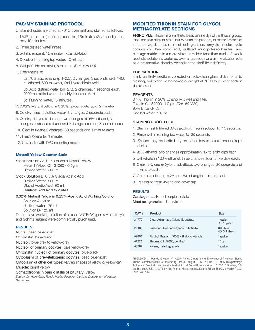
PAS/MY STAINING PROTOCOLUnstained slides are dried at 70º C overnight and stained as follows:
1. 1% Periodic acid (aqueous) oxidation, 15 minutes. (Scalloped gonads only 10 minutes).
2. Three distilled water rinses.
3. Schiff’s reagent, 15 minutes. (Cat. #24200)
4. Develop in running tap water, 10 minutes.
5. Weigert’s Hematoxlyin, 6 minutes. (Cat. #25373)
6. Differentiate in:
6a. 70% acid ethanol (pH=2.5), 2 changes, 3 seconds each 1400 ml ethanol, 600 ml water, 2ml Hydrochloric Acid
6b. Acid distilled water (ph=2.5), 2 changes, 4 seconds each. 2000ml distilled water, 1 ml Hydrochloric Acid
6c. Running water, 15 minutes.
7. 0.02% Metanil yellow in 0.25% glacial acetic acid, 2 minutes.
8. Quickly rinse in distilled water, 3 changes, 2 seconds each.
9. Quickly dehydrate through two changes of 95% ethanol, 3 changes of absolute ethanol and 2 changes acetone, 2 seconds each.
10. Clear in Xylene 2 changes, 30 seconds and 1 minute each.
11. Fresh Xylene for 1 minute.
12. Cover slip with DPX mounting media.
Metanil Yellow Counter Stain
Stock solution A: 0.1% aqueous Metanil YellowMetanil Yellow, CI 134065 - 0.5gmDistilled Water - 500 ml
Stock Solution B: 0.5% Glacial Acetic AcidDistilled Water - 950 mlGlacial Acetic Acid- 50 mlCaution: Add Acid to Water!
0.02% Metanil Yellow in 0.25% Acetic Acid Working SolutionSolution A- 50 mlDistilled water - 75 mlSolution B- 125 ml
Do not save working solution after use. NOTE: Weigert’s Hematoxylin and Schiff’s reagent were commercially purchased.
RESULTS:Nuclei: deep blue-violetChromatin: blue-blackNucleoli: blue-grey to yellow-greyNucleoli of primary oocytes: pale yellow-greyChromatin nucleoli of primary oocytes: blue-blackCytoplasm of pre-vitellogenic oocytes: deep blue-violetCytoplasm of other cell types: varying shades of yellow or yellow-tanMuscle: bright yellowSomatotrophs in pars distalis of pituitary: yellowSource: Dr. Harry Grier, Florida Marine Research Institute, Department of Natural Resources
MODIFIED THIONIN STAIN FOR GLYCOL METHACRYLATE SECTIONSPRINCIPLE: Thionin is a synthetic basic aniline dye of the thiazin group. It is used as a nuclear stain, but exhibits the property of metachromasia; in other words, mucin, mast cell granules, amyloid, nucleic acid compounds, hyaluronic acid, sulfated mucopolysaccharides, and cartilage matrix stain a more violet or redder tone than nuclei. A weak alcoholic solution is preferred over an aqueous one as the alcohol acts as a preservative, thereby extending the shelf life indefinitely.
PREPARATION4 micron GMA sections collected on acid-clean glass slides; prior to staining, slides should be baked overnight at 70˚ C to prevent section detachment.
REAGENTS0.4% Thionin in 20% Ethanol Mix well and filter.Thionin C.I. 52000- 1.0 gm (Cat. #01220)95% Ethanol- 53 mlDistilled water- 197 ml
STAINING PROCEDURE
1. Stain in freshly filtered 0.4% alcoholic Thionin solution for 15 seconds.
2. Rinse well in running tap water for 30 seconds.
3. Section may be blotted dry on paper towels before proceeding if desired.
4. 95% ethanol, two changes approximately six to eight dips each.
5. Dehydrate in 100% ethanol, three changes, four to five dips each.
6. Clear in Xylene or Xylene substitute, two changes, 30 seconds and 1 minute each.
7. Complete clearing in Xylene, two changes 1 minute each
8. Transfer to fresh Xylene and cover slip.
RESULTS: Cartilage matrix: red purple to violetMast cell granules: deep violet
REFERENCES: 1. Pamela E Nagle, HT (ASCP) Florida Department of Environmental Protection, Florida Marine Research Institute, St. Petersburg, Florida - August 1995. 2. Lillie, R.D. 1965, Histopathologic Technic and Practical Histochemistry, third edition. McGraw-Hill, New York. p. 116, 508 3. Sheehan, D.C. and Hrapchak, B.B. 1980. Theory and Practice Histotechnology, Second Edition. The C.V.> Mosby Co., St. Louis, Mo., p 149.
24770 Clear-Advantage Xylene Substitute 1 gallon 4 x 1 gallon
22463 ParaClear Odorless Xylene Substitute 3.8 liters 4 X 3.8 liters
09860 Alcohol Reagent, 100% - Histology Grade 1 gallon
01220 Thionin, C.I. 52000, certified 10 g
08389 Xylene, histology grade 1 gallon
CAT # Product Size

Science SolutionsScience
4
• Embedded blocks are clear for excellent tissue visibility• Excellent for IHC, special stains and routine stains• Tissue Infiltration• Embedding & Sectioning
Immuno-Bed, designed for light microscopy immunohistochemical (IHC) techniques, is best suited for IHC techniques requiring small antibodies and low molecular weight chromagens, such as aminoethylcarbazole (AEC). The Immuno-Bed kit is a glycol methacrylate based kit resembling JB-4 medium in tissue infiltration, embedding and cutting procedures.
Are you working with IHC? Try Immuno-Bed!
Cat. # Description Size
17324 Immuno-Bed Kit 1 kit
GMA Embedded Micrographs from Helen Wimer
(Left to right ) 1. Horseshoe crab shell with ovary - PASMY stain 2. Implant material - Von Kossa with Giemsa 3. Paddlefi sh ovary - PASMY stain4. Paddlefi sh ovary - PASMY stain 5. Paddlefi sh ovary - PASMY stain with Weigert’s Hematoxylin 6. Paddlefi sh ovary PASMY stain with Weigert’s Hematoxylin7. Cells in a polyethylene tube embedded in JB-4
Helen Wimer’s Tips and Tricks of Working with GMA!1. Make sure to use accurate measurements in the preparation of the
infiltrating/embedding solutions.
2. Keep specimens in a refrigerator during infiltration to allow longer, slower infiltration.
3. During polymerization, place blocks in a refrigerator for about 2 days. Then place them in the fume hood for 24 hours at room temperature. Pop the blocks out of the molds. If the blocks feel a little tacky, leave them under the fume hood face up, out of the mold for an additional 24 hours.
4. If the tissue squeaks or seems dry while sectioning, just barely touch the tissue with damp gauze or your thumb. You may get a usable section after you trim away the first two or three sections. The first two or three will not be usable.
5. After sectioning, place the slides on a 70˚ C hot plate or oven overnight to ensure adherence of sections to the slide during staining.
6. Clean water bath with a Kimwipe® to increase the water’s surface tension and facilitate spinning out of the section.
7. Make sure the water in the water bath is about 35-40˚ C. This gentle heat will increase the ability of the section to flatten out. If you cannot see the sections floating on the water, place a small amount of water under the staining dish.
8. Use acid cleaned slides.
9. Use Xylene as a clearing agent during staining.
10. DPX 1 mounting medium is recommended to use to coverslip to avoid artifacts.
• Complete dehydration is not necessary• Water soluble• Can produce very thin sections
• No soaking of block• Less shrinkage of tissue• Greater cellular detail
The Joys of Working with GMA

5
JB-4 Product Line
JB-4 Embedding Kit®
Intended for use in the preparation of embedded samples for high resolution light microscopy. Widely used for research and clinical diagnosis. Clear casts are obtained in 90 minutes or less at room temperature. (Cat. #00226)
JB-4 Plus® Embedding KitAll the benefits of JB-4, plus the advantage of a cooler acting accelerator and the production of clear blocks. A clear polymer embedding matrix is essential to numerous histological & histochemical procedures, most notably in ophthalmic applications. Excellent for routine stains, special stains and histochemical staining. (Cat. #18570)
JB-4 Mini Embedding KitMini, pre-measured and ready-to-use version of the JB-4 Embedding Kit®, designed for use with a small number of samples or for one larger specimen. Convenient size for introducing new users to the advantages of the JB-4 Embedding Kit®. (Cat. #22507)
Product Spotlight
JB-4 Embedding Kit® JB-4 Plus® Sample 20X
Karnovsky’s FixativeFormaldehyde/Glutaraldehyde fixative commonly used in EM for structural preservation. Pre-measured in ampoules for one-step preparation.
Kit Contains: 1x10ml 50% Glutaraldehyde, 2x10ml 16% Formaldehyde, 50ml 0.2M Phosphate Buffer. (Cat. #22872)
Formaldehyde, 10%, Methanol Free, Ultra PureSuitable for both electron and light microscopy. Easily penetrates large blocks of tissue. When used in combination with glutaraldehyde, it fixes delicate tissues such as brain in vascular perfusion. Avoids the problem of having to depolymerize paraformaldehyde. Used in Karnovsky’s fixative in conjunction with your own buffer system. Source of the formaldehyde is paraformaldehyde. (Cat. #04018)
Bouin’s Fixative (Bouin’s Fluid)Picric acid, formalin and acetic acid fixative. Bouin’s fixative is excellent for use in preserving soft and delicate tissue structures. The shrinking induced by the picric acid is offset by the swelling of the glacial acetic acid. (Cat. #16045)
Additional Products for Fixation
Glutaraldehyde, EM Grade, 50%Polysciences was the first to develop a superior grade of glutaraldehyde suitable for immunochemical techniques. Each batch of vacuum distilled glutaraldehyde manufactured by Polysciences is analyzed to maintain consistent high quality and freedom from contaminants which destroy antigenicity. Each ampoule is fitted with an ampoule cracker for added safety.
EM grade is recommended for histochemical or immunological techniques. (Cat. #18428)
Additional Stains for GMA
Cat. # Description Size
25086
24614
08379
25087
25091
25001
08824
24901
25008
16280
24199
Alcian Blue/PAS Kit
Amyloid Stain Kit (Congo Red)
Formalin, 10% neutral buffered (phosphate buffer)
Grocott Methenamine Silver Stain (GMS)
Jones PAS-M Stain Kit
Leishman Stain
Multiple Stain Solution (Paragon Multiple Stain)
Picrosirius Red Stain Kit
Sudan Black B, C.I. 26150, Certified
Villanueva Osteochrome Bone Stain
Prussian Blue Iron Stain Kit
1 kit
1 kit
20 L, 3.75 L
1 kit
1 kit
25g
50 ml, 100 ml
250 ml, 500 ml
25 g
450 ml
1 kit
Complete line of Block Holders, Chucks, and Molding Cup Trays for Plastic Resin Embedding. Please visit www.polysciences.com/plastic
JB-4 is a unique, GMA based polymer embedding material that yields a higher level of morphological detail than paraffin processed tissues. JB-4 is water-soluble and does not require dehydration to absolute alcohol except for dense, bloody or fatty tissue specimens. JB-4 is excellent for small non-decalcified bone specimens, routine stains, special stains and histochemical staining. Polymerization of JB-4 is exothermic, which is easily controlled by polymerizing on ice or by using refrigeration at 4°C. Sections of JB-4 embedded material can be cut at 0.5-3.0 microns or thicker. Microtomes designed for plastic sectioning are required as are glass, Ralph or tungsten carbide knives.

400 Valley Road, Warrington, PA 18976
Follow Us!
PolyFacts Vol. 8 | No. 1
Resin Talk
by Helen Wimer . . . . . . . . . . . . . . . . . 1
Tips and Tricks of Working
with GMA from Helen Wimer . . . . . . . . . . 4
Product Spotlight . . . . . . . . . . . . . . . . . 5
U.S. Corporate Headquarters | 400 Valley Rd, Warrington, PA 18976 | 1(800) 523-2575 (215) 343-6484 | Fax 1(800) 343-3291 | [email protected] Europe GmbH | Handelsstrasse 3 D-69214 Eppelheim, Germany | +(49) 6221-765767 | Fax +(49) 6221-764620 | [email protected] Asia Pacifi c, Inc. | 2F-1, 207 DunHua N. Rd. Taipei, Taiwan 10595 | (886) 2 8712 0600 | Fax (886) 2 8712 2677 | [email protected]
POLYSCIENCES 2013 CATALOG
Over 3,000 Products & 300 New Additions!• Microspheres & Particles• Electronic Encapsulants & Adhesives
• Histology & Microscopy• Monomers & Polymers
Request a FREE Copy Today!Visit polysciences.com/newcat
Histological Applications & Techniques for Bone, Biomaterials and Medical Device Implants
Saturday, May 4th, 2013 | 8:00 a.m. - 5:00 p.m. EDTHyatt Regency Cambridge | Cambridge, MA
Join us in Cambridge, MA for this one day event! It will be an interactive day focused on applications and new technologies for hard tissue. This workshop is an informative, inexpensive way to provide continuing education and gain contact hours.
Workshop includes 4 expert speakers, all course materials, 6 contact hours through the National Society of Histotechnology, meals and program book of the event.
$149.00 NSH Members $199.00 Non-Members
Register online at www.polysciences.com/histoforum
Histological Applications & Techniques for
REGISTER NOW!
Related Documents