Polycaprolactone scaffold and reduced rhBMP-7 dose for the regeneration of critical-sized defects in sheep tibiae Amaia Cipitria a, 1 , Johannes C. Reichert b, c, 1 , Devakar R. Epari b , Siamak Saifzadeh b , Arne Berner b, d , Hanna Schell a , Manav Mehta a , Michael A. Schuetz b , Georg N. Duda a , Dietmar W. Hutmacher b, * a Julius Wolff Institute & Center for Musculoskeletal Surgery, Berlin-Brandenburg Center for Regenerative Therapies, Charité e Universitätsmedizin Berlin, Augustenburger Platz 1, 13353 Berlin, Germany b Institute of Health and Biomedical Innovation, Queensland University of Technology, 60 Musk Ave, Kelvin Grove, Queensland 4049, Australia c Department of Orthopaedic Surgery, König-Ludwig-Haus, Center for Musculoskeletal Research, Julius-Maximilians-University, Brettreichstr.11, 97074 Würzburg, Germany d Department of Trauma Surgery, University of Regensburg, Franz-Josef-Strauß-Allee 11, 93053 Regensburg, Germany article info Article history: Received 25 June 2013 Accepted 4 September 2013 Available online xxx Keywords: Polycaprolactone BMP (bone morphogenetic protein) Ovine animal model Bone tissue engineering abstract The transplantation of autologous bone graft as a treatment for large bone defects has the limitation of harvesting co-morbidity and limited availability. This drives the orthopaedic research community to develop bone graft substitutes. Routinely, supra-physiological doses of bone morphogenetic proteins (BMPs) are applied perpetuating concerns over undesired side effects and cost of BMPs. We therefore aimed to design a composite scaffold that allows maintenance of protein bioactivity and enhances growth factor retention at the implantation site. Critical-sized defects in sheep tibiae were treated with the autograft and with two dosages of rhBMP-7, 3.5 mg and 1.75 mg, embedded in a slowly degradable medical grade poly(ε-caprolactone) (PCL) scaffold with b-tricalcium phosphate microparticles (mPCL eTCP). Specimens were characterised by biomechanical testing, microcomputed tomography and his- tology. Bridging was observed within 3 months for the autograft and both rhBMP-7 treatments. No significant difference was observed between the low and high rhBMP-7 dosages or between any of the rhBMP-7 groups and autograft implantation. Scaffolds alone did not induce comparable levels of bone formation compared to the autograft and rhBMP-7 groups. In summary, the mPCLeTCP scaffold with the lower rhBMP-7 dose led to equivalent results to autograft transplantation or the high BMP dosage. Our data suggest a promising clinical future for BMP application in scaffold-based bone tissue engineering, lowering and optimising the amount of required BMP. Crown Copyright Ó 2013 Published by Elsevier Ltd. All rights reserved. 1. Introduction The reconstruction of large segmental bone defects in humans usually requires the transplantation of autologous bone grafts (ABG). The limiting factors associated with this approach, however, include availability of ABG material, co-morbidity, and insufficient integration into the damaged bone. The delivery of recombinant growth factor proteins has emerged as a promising alternative to bone grafting to promote endogenous repair mechanisms and tis- sue regeneration. This approach has been translated into routine clinical applications for the treatment of acute fractures and non- unions [1,2]. Currently, bone morphogenetic proteins (BMPs) are applied by use of collagen carriers equipped with water-solubilised protein. The recommended dose of rhBMP-7 applied in humans suffering from recalcitrant long bone non-unions remains 7 mg [3] regardless of defect size and shape. However, undesired side effects associated with rapid protein degradation and diffusion from these carriers, excessive or ectopic bone formation, immunological re- action or correlation between extremely high doses of BMP and cancer incidence [4e7], as well as concerns over the incremental effectiveness and cost of BMP on fracture healing, necessitate the optimisation of drug delivery regarding both drug quantity and mode of release by carrier systems. Most importantly, these con- siderations must account for site-specific requirements regarding therapeutic doses, which depend on the local degree of vascular- isation, defect size, and the number of resident responding cells. Over the last 10 years our group has demonstrated the potential of biodegradable composite scaffolds consisting of medical grade * Corresponding author. Tel.: þ61 7 3138 6077; fax: þ61 7 3138 6030. E-mail address: [email protected] (D.W. Hutmacher). 1 These authors contributed equally to the presented work. Contents lists available at ScienceDirect Biomaterials journal homepage: www.elsevier.com/locate/biomaterials 0142-9612/$ e see front matter Crown Copyright Ó 2013 Published by Elsevier Ltd. All rights reserved. http://dx.doi.org/10.1016/j.biomaterials.2013.09.011 Biomaterials xxx (2013) 1e9 Please cite this article in press as: Cipitria A, et al., Polycaprolactone scaffold and reduced rhBMP-7 dose for the regeneration of critical-sized defects in sheep tibiae, Biomaterials (2013), http://dx.doi.org/10.1016/j.biomaterials.2013.09.011

Welcome message from author
This document is posted to help you gain knowledge. Please leave a comment to let me know what you think about it! Share it to your friends and learn new things together.
Transcript

lable at ScienceDirect
Biomaterials xxx (2013) 1e9
Contents lists avai
Biomaterials
journal homepage: www.elsevier .com/locate/biomater ia ls
Polycaprolactone scaffold and reduced rhBMP-7 dose forthe regeneration of critical-sized defects in sheep tibiae
Amaia Cipitria a, 1, Johannes C. Reichert b, c, 1, Devakar R. Epari b, Siamak Saifzadeh b,Arne Berner b, d, Hanna Schell a, Manav Mehta a, Michael A. Schuetz b, Georg N. Duda a,Dietmar W. Hutmacher b, *
a Julius Wolff Institute & Center for Musculoskeletal Surgery, Berlin-Brandenburg Center for Regenerative Therapies, Charité e Universitätsmedizin Berlin,Augustenburger Platz 1, 13353 Berlin, Germanyb Institute of Health and Biomedical Innovation, Queensland University of Technology, 60 Musk Ave, Kelvin Grove, Queensland 4049, Australiac Department of Orthopaedic Surgery, König-Ludwig-Haus, Center for Musculoskeletal Research, Julius-Maximilians-University, Brettreichstr. 11, 97074Würzburg, Germanyd Department of Trauma Surgery, University of Regensburg, Franz-Josef-Strauß-Allee 11, 93053 Regensburg, Germany
a r t i c l e i n f o
Article history:Received 25 June 2013Accepted 4 September 2013Available online xxx
Keywords:PolycaprolactoneBMP (bone morphogenetic protein)Ovine animal modelBone tissue engineering
* Corresponding author. Tel.: þ61 7 3138 6077; faxE-mail address: [email protected] (D
1 These authors contributed equally to the present
0142-9612/$ e see front matter Crown Copyright � 2http://dx.doi.org/10.1016/j.biomaterials.2013.09.011
Please cite this article in press as: Cipitria Adefects in sheep tibiae, Biomaterials (2013),
a b s t r a c t
The transplantation of autologous bone graft as a treatment for large bone defects has the limitation ofharvesting co-morbidity and limited availability. This drives the orthopaedic research community todevelop bone graft substitutes. Routinely, supra-physiological doses of bone morphogenetic proteins(BMPs) are applied perpetuating concerns over undesired side effects and cost of BMPs. We thereforeaimed to design a composite scaffold that allows maintenance of protein bioactivity and enhancesgrowth factor retention at the implantation site. Critical-sized defects in sheep tibiae were treated withthe autograft and with two dosages of rhBMP-7, 3.5 mg and 1.75 mg, embedded in a slowly degradablemedical grade poly(ε-caprolactone) (PCL) scaffold with b-tricalcium phosphate microparticles (mPCLeTCP). Specimens were characterised by biomechanical testing, microcomputed tomography and his-tology. Bridging was observed within 3 months for the autograft and both rhBMP-7 treatments. Nosignificant difference was observed between the low and high rhBMP-7 dosages or between any of therhBMP-7 groups and autograft implantation. Scaffolds alone did not induce comparable levels of boneformation compared to the autograft and rhBMP-7 groups. In summary, the mPCLeTCP scaffold with thelower rhBMP-7 dose led to equivalent results to autograft transplantation or the high BMP dosage. Ourdata suggest a promising clinical future for BMP application in scaffold-based bone tissue engineering,lowering and optimising the amount of required BMP.
Crown Copyright � 2013 Published by Elsevier Ltd. All rights reserved.
1. Introduction
The reconstruction of large segmental bone defects in humansusually requires the transplantation of autologous bone grafts(ABG). The limiting factors associated with this approach, however,include availability of ABG material, co-morbidity, and insufficientintegration into the damaged bone. The delivery of recombinantgrowth factor proteins has emerged as a promising alternative tobone grafting to promote endogenous repair mechanisms and tis-sue regeneration. This approach has been translated into routineclinical applications for the treatment of acute fractures and non-unions [1,2]. Currently, bone morphogenetic proteins (BMPs) are
: þ61 7 3138 6030..W. Hutmacher).ed work.
013 Published by Elsevier Ltd. All
, et al., Polycaprolactone scafhttp://dx.doi.org/10.1016/j.bi
applied by use of collagen carriers equipped with water-solubilisedprotein. The recommended dose of rhBMP-7 applied in humanssuffering from recalcitrant long bone non-unions remains 7 mg [3]regardless of defect size and shape. However, undesired side effectsassociated with rapid protein degradation and diffusion from thesecarriers, excessive or ectopic bone formation, immunological re-action or correlation between extremely high doses of BMP andcancer incidence [4e7], as well as concerns over the incrementaleffectiveness and cost of BMP on fracture healing, necessitate theoptimisation of drug delivery regarding both drug quantity andmode of release by carrier systems. Most importantly, these con-siderations must account for site-specific requirements regardingtherapeutic doses, which depend on the local degree of vascular-isation, defect size, and the number of resident responding cells.
Over the last 10 years our group has demonstrated the potentialof biodegradable composite scaffolds consisting of medical grade
rights reserved.
fold and reduced rhBMP-7 dose for the regeneration of critical-sizedomaterials.2013.09.011
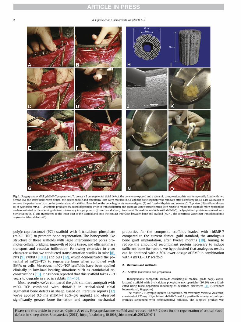
Fig. 1. Surgery and scaffold/rhBMP-7 preparation. To create a 3 cm segmental tibial defect, the bone was exposed and a dynamic compression plate was temporarily fixed with twoscrews (A), the screw holes were drilled, the defect middle and osteotomy lines were marked (B, C), and the bone segment was removed after osteotomy (D, E). Care was taken toremove the periosteum 1 cm on the proximal and distal tibial. Bone before the bone fragments were realigned (F) and fixed with plate and screws (G). Top view (H) and lateral view(I) of cylindrical mPCLeTCP scaffold produced via fused deposition. Prior to transplantation, the scaffolds were surface treated with NaOH to render the scaffolds more hydrophilicas demonstrated in the scanning electron microscopy images prior to (J, insert) and after (J) treatment. To load the scaffolds with rhBMP-7, the lyophilised protein was mixed withsterile saline (K, L) and transferred to the inner duct of the scaffold and onto the contact interfaces between bone and scaffold (M, N). The constructs were then transplanted intosegmental tibial defects (O).
A. Cipitria et al. / Biomaterials xxx (2013) 1e92
poly(ε-caprolactone) (PCL) scaffold with b-tricalcium phosphate(mPCLeTCP) to promote bone regeneration. The honeycomb likestructure of these scaffolds with large interconnected pores pro-motes cellular bridging, ingrowth of bone tissue, and efficient masstransport and vascular infiltration. Following extensive in vitrocharacterisation, we conducted transplantation studies in mice [8],rats [9], rabbits [10,11] and pigs [12], which demonstrated the po-tential of mPCLeTCP to regenerate bone when combined withBMPs or cells. Moreover, mPCLeTCP scaffolds have been appliedclinically in low-load bearing situations such as craniofacial re-constructions [13]. It has been reported that this scaffold takes 2e3years to degrade in vivo in rabbits [14e16].
Most recently, we’ve compared the gold standard autograft withmPCLeTCP combined with rhBMP-7 in critical-sized tibialsegmental bone defects in sheep. Based on literature reports [17]we’ve applied 3.5 mg rhBMP-7 (0.5e0.6 mg/mL) and observedsignificantly greater bone formation and superior mechanical
Please cite this article in press as: Cipitria A, et al., Polycaprolactone scafdefects in sheep tibiae, Biomaterials (2013), http://dx.doi.org/10.1016/j.bi
properties for the composite scaffolds loaded with rhBMP-7compared to the current clinical gold standard, the autologousbone graft implantation, after twelve months [18]. Aiming toreduce the amount of recombinant protein necessary to inducesufficient bone formation, we hypothesised that analogous resultscan be obtained with a 50% lower dosage of BMP in combinationwith a mPCLeTCP scaffold.
2. Materials and methods
2.1. Scaffold fabrication and preparation
Biodegradable composite scaffolds consisting of medical grade poly(ε-capro-lactone) scaffold with b-tricalcium phosphate microparticles (80:20) were fabri-cated using fused deposition modelling as described elsewhere [18] (OsteoporeInternational, Singapore).
The rhBMP-7 (Olympus Biotech Corporation, Mt Waverley, Victoria, Australia)consisted of 1.75 mg of lyophilised rhBMP-7 on 0.5 g purified bovine type I collagengranules suspended with carboxymethyl cellulose. The supplied product was
fold and reduced rhBMP-7 dose for the regeneration of critical-sizedomaterials.2013.09.011

A. Cipitria et al. / Biomaterials xxx (2013) 1e9 3
reconstituted with 1.5 mL of saline to form an implantable paste, which was thentransferred to the inner duct of the scaffold and the contact interfaces between boneand scaffold (Fig. 1). Compared to a previous study [18], where 3.5 mg of lyophilisedrhBMP-7 on 1 g purified bovine type I collagen granules were reconstituted with3 mL of saline, the amount of paste manually administered to the inner duct of thescaffold was halved.
2.2. Segmental bone defect model
Mid-diaphyseal segmental tibial defects in Merino sheep (age 7e8 years,average weight 43.3 kg) were created as described previously [18]. The study wasapproved by the animal ethics committee of the Queensland University of Tech-nology (Brisbane, Australia; approval number: 700000915, 900000906,800000825). Defects were reconstructed with the scaffold only (n ¼ 8), and scaffoldand 1.75 mg rhBMP-7 (n ¼ 8) or scaffold and 3.5 mg rhBMP-7 (n ¼ 8). For defectfixation a 10-hole, stainless steel, dynamic compression plate (DCP) (Synthes,Switzerland) [19] was used (Fig. 1). Previously, we have demonstrated the critical-sized nature of the defect model and achieved defect bridging in all cases withABG treatment after 3 months [18]. Animals were allowed unrestricted weightbearing and sacrificed after 3 months.
2.3. Radiographic analysis
Conventional X-ray analysis (3.2 mAs; 65 kV) was performed in two standardplanes (anterioreposterior andmedialelateral) directly after surgery and after 6 and12 weeks.
2.4. Biomechanical testing
After sacrifice, both tibial ends were embedded in 80 mL Paladur (Heraeus-Kulzer, Wehrheim, Germany) and mounted in a biaxial testing machine (Instron8874, Melbourne, Australia). A torsion test was conducted under angular displace-ment control at an angular velocity of 0.5 deg/s and a constant compressive pre-loadof 0.05 kNuntil the first signs of fracturewere visible in the form of a discontinuity inthe torque-angular displacement curves. The process was stopped instantly toensure structural integrity of the bones. For each animal the contralateral tibia wasused as a paired reference. The torsional moment and torsional stiffness values werecalculated from the slope of the torque-angular displacement curves and normalisedagainst the values of the contralateral tibiae. Next, the specimens were fixed in 10%neutral buffered formalin solution prior to further processing.
Fig. 2. Representative X-ray images showing a defect treated with the scaffold only (A), andrhBMP-7 (C) after 3 months. The results of defects reconstructed with the mPCLeTCP scaffoldand are included for comparison purposes.
Please cite this article in press as: Cipitria A, et al., Polycaprolactone scafdefects in sheep tibiae, Biomaterials (2013), http://dx.doi.org/10.1016/j.bi
2.5. Microcomputed tomography (mCT)
The newly formedmineralised tissuewas analysed as described previously using aViva40 mCT (Scanco Medical AG, Bruettisellen, Switzerland) [18]. Briefly, specimenswere scanned at a voltage of 70 kVp, a current of 114 mA and a voxel size of 19 mm.Acquired images were segmented with a Gaussian filter of a width of 0.8 and supportof 1.0. A threshold of 210 (519.2mg hydroxyapatite per cm3)was chosen for the defectsand a threshold of 230 (591.6 mg hydroxyapatite per cm3) for the contralateral tibiae.These were determined by visually evaluating 20 randomly chosen tomograms perspecimen of four samples per group. A global threshold that best showed themorphology of mineralised tissue and excluded the scaffold and soft tissue wasselected [20]. The bone volume within the defect, its axial and radial distribution, thepolar moment of inertia (Jz), and tissue mineral density were determined.
2.6. Histology
For histological processing, 3 mm thick slices were cut and dehydrated in agraded series of ethanol prior to embedding in poly-methyl-methacrylate (PMMA)(Technovit 9100 NEU, Heraeus Kulzer). Sections of 6 mm thickness were prepared(Leica SM 2500S, Munich, Germany) and stained with Safranin-Orange/von Kossa(black: mineralised tissue) and Movat Pentachrome (yellow: collagen type I fibres;light blue: reticular fibres; red: elastin fibres and muscle tissue). Samples werevisualised using a light microscope (Leica DMRB, Munich, Germany and Zeiss Axi-oCam MRc, Zeiss, Oberkochen, Germany).
2.7. Statistical analysis
Statistical comparison between groups was performed with the non-parametricKruskaleWallis multiple group test (IBM SPSS 18.0). A two-tailed ManneWhitney-U-test was then used for pair-wise comparison. A confidence level of 95% was used(p < 0.05) and the p-values were adjusted according to BonferronieHolm. The boxplots represent the median, and 1st and 3rd quartile; the error bars indicatemaximum and minimum values.
3. Results
The present study reports on the effect of different dosages ofrhBMP-7 on bone healing in a critical sized defect model in sheep.The results corresponding to ABG, mPCLeTCP scaffold only andmPCLeTCP loaded with 3.5 mg rhBMP-7 have previously been
defects augmented with scaffold and 1.75 mg rhBMP-7 (B) or with scaffold and 3.5 mgonly and mPCLeTCP loaded with 3.5 mg rhBMP-7 have been published elsewhere [18]
fold and reduced rhBMP-7 dose for the regeneration of critical-sizedomaterials.2013.09.011

A. Cipitria et al. / Biomaterials xxx (2013) 1e94
published elsewhere [18] and are included in Figs. 2e5 forcomparative purposes.
3.1. Radiographic analysis
X-ray analysis showed a union rate of 37.5% for the scaffold only,100% for scaffold and 1.75 mg rhBMP-7 and 100% for scaffold and3.5 mg rhBMP-7 after 3 months (Fig. 2).
3.2. Biomechanical testing
Biomechanical testing revealed significantly higher torsionalmoment and torsional stiffness values for both rhBMP-7 groups
Fig. 3. Biomechanical testing and mCT results after 3 months. Torsional moment (A) and torsand 1.75 mg rhBMP-7, and scaffold and 3.5 mg rhBMP-7 (n ¼ 8 each). Data are median valueabsolute values (C). Median polar moment of inertia (D) and tissue mineral density (E) normand 3rd quartile; the error bars indicate maximum and minimum values. The asterisks (*) inmPCLeTCP scaffold only and mPCLeTCP loaded with 3.5 mg rhBMP-7 have been published
Please cite this article in press as: Cipitria A, et al., Polycaprolactone scafdefects in sheep tibiae, Biomaterials (2013), http://dx.doi.org/10.1016/j.bi
compared to the scaffold only group. No significant difference wasfound between both rhBMP-7 dosages or when these groups werecompared to ABG. Interestingly, a smaller variance of mechanicalproperties was observed with the reduced rhBMP-7 dosage(Fig. 3A, B).
3.3. Microcomputed tomography (mCT)
Bone volumes and polar moment of inertia values determinedby quantitative mCT analysis were significantly higher for bothrhBMP-7 groups when compared to the scaffold only, correlatingwell with the biomechanical testing results. Again no significantdifference was seen between the different BMP dosages or when
ional stiffness (B) represent biomechanics of empty defects, ABG, scaffold only, scaffolds normalised against corresponding contralateral tibiae. Bone volumes are presented asalised against corresponding contralateral tibiae. The box plots represent median, 1st
dicate statistical significance (p < 0.05). The results of defect reconstruction with ABG,elsewhere [18] and are included for comparison purposes.
fold and reduced rhBMP-7 dose for the regeneration of critical-sizedomaterials.2013.09.011

Fig. 4. Microcomputed tomography analysis of axial and radial bone distribution after 3 months (n ¼ 8). The longitudinal and transversal mCT tomograms depict the different VOI(A). To describe the axial bone volume distribution the total defect length was divided into three equal parts: proximal, middle and distal third. The radial bone volume distributionwas characterised by analysing the bone volume within the scaffold’s inner duct, its wall and the periphery. Volumes of bone formed in the distal, middle and proximal thirds (B).Bone volumes in the scaffold’s inner duct, wall and periphery (C). Radial bone volume distribution normalised against the total volume of interest (D). The box plots representmedian, 1st and 3rd quartile; the error bars indicate maximum and minimum values. The asterisks (*) indicate statistical significance (p < 0.05). The results of defects reconstructedwith the mPCLeTCP scaffold only and mPCLeTCP loaded with 3.5 mg rhBMP-7 have been published elsewhere [18] and are included for comparison purposes.
A. Cipitria et al. / Biomaterials xxx (2013) 1e9 5
these groups were compared to ABG. With 1.75 mg of rhBMP-7administered, the variance in bone formation appeared smallerwhen compared to 3.5 mg rhBMP-7 (Fig. 3C, D). No differences intissue mineral density were determined amongst any of the groups(Fig. 3E).
Axial bone volume distribution was assessed by dividing thetotal length of the defect into three parts of equal length. In allgroups, a non-significant tendency towards greater bone formationin the regions adjacent to the interfaces with native bone wasobserved, when compared to the defect middle, indicating thatbone ingrowth initiated at the interface with cortical bone. A non-significant trend to the proximal interface was determined, whichis better vascularised than the distal end (Fig. 4B). The amount ofnewly formed bone in the periphery of the scaffold was comparableto the bone in the scaffold wall and inner duct (Fig. 4C). Radial bonedistribution per unit volume of scaffold wall and inner duct washomogeneous in the scaffold only and 3.5 mg rhBMP-7 groups. Inthe groupwith the reduced BMP dosage, however, the bone volume
Please cite this article in press as: Cipitria A, et al., Polycaprolactone scafdefects in sheep tibiae, Biomaterials (2013), http://dx.doi.org/10.1016/j.bi
within the inner duct appeared significantly higher (Fig. 4D). Anadditional movie file shows a three-dimensional mCT reconstruc-tion of a representative defect augmented with mPCLeTCP scaffoldand 1.75 mg rhBMP-7 three months after surgery (see Supple-mentary file 1).
Supplementary video related to this article can be found athttp://dx.doi.org/10.1016/j.biomaterials.2013.09.011.
3.4. Histology
The calculated bone volumes, bone volume distribution, andmechanical properties correlated well withmacroscopic findings in3D mCT reconstructions and histological sections (Fig. 5), depictingthe minor bone formation in the scaffold only group, as opposed tothe rhBMP-7 groups, where full defect bridging was observed after3 months. The histological sections of all samples under investi-gation shown in Fig. 5 illustrate the variability in bone formationwithin each group. Although the 3.5 mg rhBMP-7 group consisted
fold and reduced rhBMP-7 dose for the regeneration of critical-sizedomaterials.2013.09.011
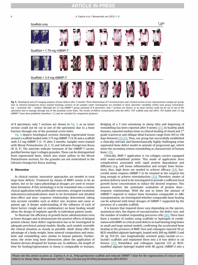
Fig. 5. Histological and mCT imaging analyses of bone defects after 3 months. Three-dimensional mCT reconstructions and a frontal section of one representative sample per group,next to Safranin-Orange/von Kossa stained histology sections of all samples under investigation are included to show specimen variability within each group (orientation:top ¼ proximal; left ¼ medial). Although the 3.5 mg rhBMP-7 group consisted of 8 specimens, only 7 sections are shown, as an intact section could not be cut in one of thespecimens due to damage through one of the proximal screw holes. The results of defects reconstructed with the mPCLeTCP scaffold only and mPCLeTCP loaded with 3.5 mgrhBMP-7 have been published elsewhere [18] and are included for comparison purposes.
A. Cipitria et al. / Biomaterials xxx (2013) 1e96
of 8 specimens, only 7 sections are shown in Fig. 5, as an intactsection could not be cut in one of the specimens due to a bonefracture through one of the proximal screw holes.
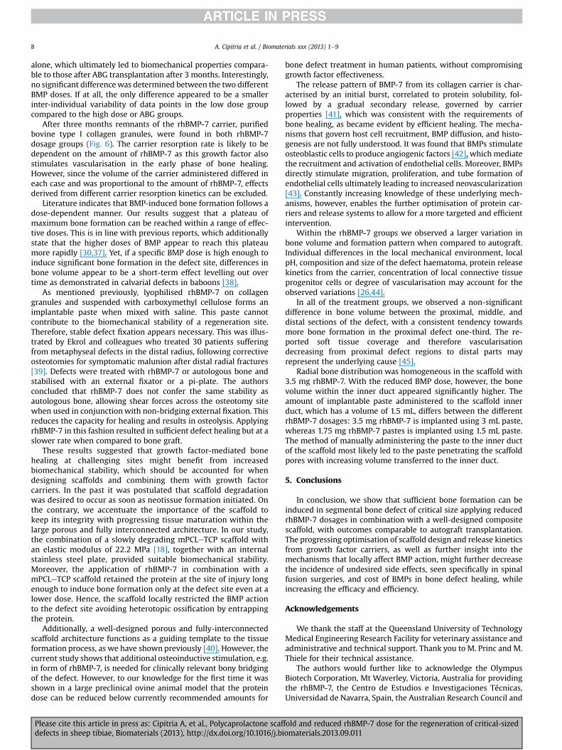
Fig. 6 depicts histological sections showing regenerated tissuearound a scaffold loaded with 1.75mg rhBMP-7 (A, B) and a scaffoldwith 3.5 mg rhBMP-7 (CeF) after 3 months. Samples were stainedwith Movat Pentachrome (A, C, E) and Safranin-Orange/von Kossa(B, D, F). The asterisks indicate remnants of the rhBMP-7 carrier,purified bovine type I collagen granules. These can be distinguishedfrom regenerated bone, which also stains yellow in the MovatPentachrome sections, for the granules are not mineralised in theSafranin-Orange/von Kossa staining.
4. Discussion
In clinical routine, innovative approaches are needed to treatlarge bone defects. Treatment by means of BMPs seems to be anoption, but so far supra-physiological dosages are used to ensurebone formation. If this technology is to be translated into a routineclinical application with predictable outcomes, stringent treatmentindications/contraindications need to be formulated and detaileddose-effect and doseeresponse relationships determined, takinginto account variables such as defect size, location and cause orpatient age. A deeper understanding of the influence of each ofthese factors (single and in combination) on bone healing wouldallow the design of implants specific to individual patient needs.
To illustrate the efficiency of growth factor administration evenat lower dosages and to demonstrate the positive effects of delayedprotein release, bone defect regeneration has to be investigated inadequate preclinical animal models. Ideally, such models resemblethe clinical situation as closely as possible. Adult sheep offer theadvantage of a body weight, bone mineral composition and meta-bolic and remodelling rates similar to human patients. Further-more, the ovine long bone dimensions allow the application offixation devices designed for human use. In addition, the length oftime for healing/regeneration in sheep is comparable to humans.
Please cite this article in press as: Cipitria A, et al., Polycaprolactone scafdefects in sheep tibiae, Biomaterials (2013), http://dx.doi.org/10.1016/j.bi
Bridging of a 3 mm osteotomy in sheep tibia and beginning ofremodelling has been reported after 9 weeks [21]. In healthy adulthumans, reported median times to clinical healing of closed and 1st
grade transverse and oblique tibial fractures range from 103 to 136days between [22,23]. Thus, our group has successfully establisheda clinically relevant and biomechanically highly challenging ovinesegmental bone defect model in animals of progressed age, whichshow the secondary osteon remodelling as characteristic of humanbone [19].
Clinically, BMP-7 application is via collagen carriers equippedwith water-solubilised protein. This mode of application facescomplications associated with rapid protein degradation anddiffusion (e.g. soft tissue inflammation and ectopic bone forma-tion), thus, high doses are needed to achieve efficacy [24]. Suc-cessful union requires rhBMP-7 to be retained at the surgical sitelong enough to achieve osteoinduction [25]. Therefore, modes ofprotein delivery need to be investigated to provide a sufficient localgrowth factor concentration to induce the desired response. Thisprocess involves the systematic evaluation of protein doseeresponse relationships. With the aim to lower the amount ofrhBMP-7 required to induce bone formation comparable to ABGtransplantation, we investigated whether comparable bone healingcan be achieved with lower dosages of rhBMP-7 supported by thepresence of a suitable scaffold.
It is known that required doses vary depending on the species,anatomical sites, the degree of vascularisation, the defect size, andthe number of resident responding precursor cells [26]. There havebeen a number of studies using scaffolds or hydrogels in combi-nationwith BMPs in critical sized defects in load bearing long bonesin small and large animal models, confirming the accelerated bonehealing in the presence of BMP. Oest and colleagues injected 50 mLRGD-modified alginate hydrogels, loadedwith 200 ng rhBMP-2 and20 ng TGF-b3, into longitudinally oriented poly(L-lactide-co-D,L-lactide) scaffolds and implanted them into a 8 mm defect in ratfemora [27]. Kolambkar and colleagues injected 125 mL RGD-modified alginate hydrogel loaded with 40 mg/mL rhBMP-2 into a
fold and reduced rhBMP-7 dose for the regeneration of critical-sizedomaterials.2013.09.011

Fig. 6. Histological sections showing regenerated tissue around a scaffold loaded with 1.75 mg rhBMP-7 (A, B) and scaffold with 3.5 mg rhBMP-7 (CeF), after 3 months, stained withMovat Pentachrome (A, C, E) and Safranin-Orange/von Kossa (B, D, F). The asterisks indicate remnants of the rhBMP-7 carrier, purified bovine type I collagen granules. Orientation:top ¼ proximal; left ¼ medial.
A. Cipitria et al. / Biomaterials xxx (2013) 1e9 7
electrospun poly(ε-caprolactone) nanofiber mesh tube andimplanted it into a 8 mm defect in rat femora [28]. Cook and col-leagues implanted 125 mg of demineralised bovine bone matrixreconstituted with 3.13, 6.25, 12.5, 25, 50, 100, 200, 300, or 400 mgrhBMP-7 and implanted it in a 15 mm ulnar segmental defect inadult rabbits [29]. In a second study, Cook and colleagues implanted500 mg of demineralised bovine bone matrix reconstituted with1.2 mg rhBMP-7 and implanted it in a 25 mm ulnar segmentaldefect in male dogs [30]. In a third study, Cook and colleaguescreated 20 mm mid-diaphyseal tibial defects in African greenmonkeys (Cercopithecus aethiops) [31]. The ulnar defects were filledwith 400 mg of bovine bone collagen carrier reconstituted with1 mg rhBMP-7. The tibial defects were filled with 400 mg of bovinebone collagen carrier reconstituted with 0.25, 0.5, 1 or 2mg rhBMP-7.
Kirker-Head and colleagues implanted 1.5 mg rhBMP-2 mixedwith inactivated demineralised ovine bone matrix in a 25 mmfemoral segmental defect in sheep [32]. Following this, Kirker-Headand colleagues implanted 2e4 mg rhBMP-2 mixed with autologousblood, into 25 mm segmental defects in sheep femora, using
Please cite this article in press as: Cipitria A, et al., Polycaprolactone scafdefects in sheep tibiae, Biomaterials (2013), http://dx.doi.org/10.1016/j.bi
poly(D,L-(lactideeCOeglycolide)) bioerodible polymer microparti-cles [33]. Den Boer and colleagues loaded 2.5 mg rhBMP-7 intogranular porous hydroxyapatite and implanted it into a 30 mmsegmental defect in sheep tibiae [34]. Kokubo and colleaguesimplanted poly(L-lactide-co-glycolide) copolymer-coated gelatinsponges impregnated with 0.4 mg/mL of rhBMP-2 into 25 mmsegmental defects in dog tibiae [35]. Regauer and colleaguesimplanted 5 mg rhBMP-7 together with inactivated demineralisedbone matrix and 5 mL autogenous bone marrow in a 50 mmsegmental defect in sheep tibiae [36]. Based on a prospective,randomised, double-blind study involving 24 patients, who un-derwent tibial osteotomy and were treated with rhBMP-7 on acollagen type I carrier [17], and the current recommendations forthe treatment of non-unions, we have chosen dosages of 3.5 mg(0.5e0.6 mg/mL) and 1.75 mg rhBMP-7 (0.25e0.3 mg/mL) to bereconstituted in a mPCLeTCP scaffold and implanted in a 30 mmtibial segmental defect.
Transplanting the biodegradable composite scaffolds to thesedefects we found that the local BMP delivery resulted in a signifi-cant increase in bone formation when compared to the scaffold
fold and reduced rhBMP-7 dose for the regeneration of critical-sizedomaterials.2013.09.011
A. Cipitria et al. / Biomaterials xxx (2013) 1e98
alone, which ultimately led to biomechanical properties compara-ble to those after ABG transplantation after 3 months. Interestingly,no significant differencewas determined between the two differentBMP doses. If at all, the only difference appeared to be a smallerinter-individual variability of data points in the low dose groupcompared to the high dose or ABG groups.
After three months remnants of the rhBMP-7 carrier, purifiedbovine type I collagen granules, were found in both rhBMP-7dosage groups (Fig. 6). The carrier resorption rate is likely to bedependent on the amount of rhBMP-7 as this growth factor alsostimulates vascularisation in the early phase of bone healing.However, since the volume of the carrier administered differed ineach case and was proportional to the amount of rhBMP-7, effectsderived from different carrier resorption kinetics can be excluded.
Literature indicates that BMP-induced bone formation follows adose-dependent manner. Our results suggest that a plateau ofmaximum bone formation can be reached within a range of effec-tive doses. This is in line with previous reports, which additionallystate that the higher doses of BMP appear to reach this plateaumore rapidly [30,37]. Yet, if a specific BMP dose is high enough toinduce significant bone formation in the defect site, differences inbone volume appear to be a short-term effect levelling out overtime as demonstrated in calvarial defects in baboons [38].
As mentioned previously, lyophilised rhBMP-7 on collagengranules and suspended with carboxymethyl cellulose forms animplantable paste when mixed with saline. This paste cannotcontribute to the biomechanical stability of a regeneration site.Therefore, stable defect fixation appears necessary. This was illus-trated by Ekrol and colleagues who treated 30 patients sufferingfrom metaphyseal defects in the distal radius, following correctiveosteotomies for symptomatic malunion after distal radial fractures[39]. Defects were treated with rhBMP-7 or autologous bone andstabilised with an external fixator or a pi-plate. The authorsconcluded that rhBMP-7 does not confer the same stability asautologous bone, allowing shear forces across the osteotomy sitewhen used in conjunctionwith non-bridging external fixation. Thisreduces the capacity for healing and results in osteolysis. ApplyingrhBMP-7 in this fashion resulted in sufficient defect healing but at aslower rate when compared to bone graft.
These results suggested that growth factor-mediated bonehealing at challenging sites might benefit from increasedbiomechanical stability, which should be accounted for whendesigning scaffolds and combining them with growth factorcarriers. In the past it was postulated that scaffold degradationwas desired to occur as soon as neotissue formation initiated. Onthe contrary, we accentuate the importance of the scaffold tokeep its integrity with progressing tissue maturation within thelarge porous and fully interconnected architecture. In our study,the combination of a slowly degrading mPCLeTCP scaffold withan elastic modulus of 22.2 MPa [18], together with an internalstainless steel plate, provided suitable biomechanical stability.Moreover, the application of rhBMP-7 in combination with amPCLeTCP scaffold retained the protein at the site of injury longenough to induce bone formation only at the defect site even at alower dose. Hence, the scaffold locally restricted the BMP actionto the defect site avoiding heterotopic ossification by entrappingthe protein.
Additionally, a well-designed porous and fully-interconnectedscaffold architecture functions as a guiding template to the tissueformation process, as we have shown previously [40]. However, thecurrent study shows that additional osteoinductive stimulation, e.g.in form of rhBMP-7, is needed for clinically relevant bony bridgingof the defect. However, to our knowledge for the first time it wasshown in a large preclinical ovine animal model that the proteindose can be reduced below currently recommended amounts for
Please cite this article in press as: Cipitria A, et al., Polycaprolactone scafdefects in sheep tibiae, Biomaterials (2013), http://dx.doi.org/10.1016/j.bi
bone defect treatment in human patients, without compromisinggrowth factor effectiveness.
The release pattern of BMP-7 from its collagen carrier is char-acterised by an initial burst, correlated to protein solubility, fol-lowed by a gradual secondary release, governed by carrierproperties [41], which was consistent with the requirements ofbone healing, as became evident by efficient healing. The mecha-nisms that govern host cell recruitment, BMP diffusion, and histo-genesis are not fully understood. It was found that BMPs stimulateosteoblastic cells to produce angiogenic factors [42], whichmediatethe recruitment and activation of endothelial cells. Moreover, BMPsdirectly stimulate migration, proliferation, and tube formation ofendothelial cells ultimately leading to increased neovascularization[43]. Constantly increasing knowledge of these underlying mech-anisms, however, enables the further optimisation of protein car-riers and release systems to allow for a more targeted and efficientintervention.
Within the rhBMP-7 groups we observed a larger variation inbone volume and formation pattern when compared to autograft.Individual differences in the local mechanical environment, localpH, composition and size of the defect haematoma, protein releasekinetics from the carrier, concentration of local connective tissueprogenitor cells or degree of vascularisation may account for theobserved variations [26,44].
In all of the treatment groups, we observed a non-significantdifference in bone volume between the proximal, middle, anddistal sections of the defect, with a consistent tendency towardsmore bone formation in the proximal defect one-third. The re-ported soft tissue coverage and therefore vascularisationdecreasing from proximal defect regions to distal parts mayrepresent the underlying cause [45].
Radial bone distribution was homogeneous in the scaffold with3.5 mg rhBMP-7. With the reduced BMP dose, however, the bonevolume within the inner duct appeared significantly higher. Theamount of implantable paste administered to the scaffold innerduct, which has a volume of 1.5 mL, differs between the differentrhBMP-7 dosages: 3.5 mg rhBMP-7 is implanted using 3 mL paste,whereas 1.75 mg rhBMP-7 pastes is implanted using 1.5 mL paste.The method of manually administering the paste to the inner ductof the scaffold most likely led to the paste penetrating the scaffoldpores with increasing volume transferred to the inner duct.
5. Conclusions
In conclusion, we show that sufficient bone formation can beinduced in segmental bone defect of critical size applying reducedrhBMP-7 dosages in combination with a well-designed compositescaffold, with outcomes comparable to autograft transplantation.The progressing optimisation of scaffold design and release kineticsfrom growth factor carriers, as well as further insight into themechanisms that locally affect BMP action, might further decreasethe incidence of undesired side effects, seen specifically in spinalfusion surgeries, and cost of BMPs in bone defect healing, whileincreasing the efficacy and efficiency.
Acknowledgements
We thank the staff at the Queensland University of TechnologyMedical Engineering Research Facility for veterinary assistance andadministrative and technical support. Thank you to M. Princ and M.Thiele for their technical assistance.
The authors would further like to acknowledge the OlympusBiotech Corporation, Mt Waverley, Victoria, Australia for providingthe rhBMP-7, the Centro de Estudios e Investigaciones Técnicas,Universidad de Navarra, Spain, the Australian Research Council and
fold and reduced rhBMP-7 dose for the regeneration of critical-sizedomaterials.2013.09.011

A. Cipitria et al. / Biomaterials xxx (2013) 1e9 9
the Wesley Research Foundation, Brisbane, and the Berlin-Bran-denburg Center for Regenerative Medicine (BCRT) for funding.
References
[1] Govender S, Csimma C, Genant HK, Valentin-Opran A. Recombinant humanbone morphogenetic protein-2 for treatment of open tibial fractures e aprospective, controlled, randomized study of four hundred and fifty patients.J Bone Jt Surg Am 2002;84A(12):2123e34.
[2] Einhorn TA. Clinical applications of recombinant human BMPs: early experi-ence and future development. J Bone Jt Surg Am 2003;85A:82e8.
[3] Haidar ZS, Hamdy RC, Tabrizian M. Delivery of recombinant bone morpho-genetic proteins for bone regeneration and repair. Part A: current challengesin BMP delivery. Biotechnol Lett 2009;31(12):1817e24.
[4] Harwood PJ, Giannoudis PV. Application of bone morphogenetic proteins inorthopaedic practice: their efficacy and side effects. Expert Opin Drug Saf2005;4(1):75e89.
[5] Mines D, Gu Y, Kou TD, Cooper GS. Recombinant human bone morphogeneticprotein-2 and pancreatic cancer: a retrospective cohort study. Pharmacoepi-demiol Drug Saf 2011;20(2):111e8.
[6] Lad SP, Bagley JH, Ugiliweneza B, Babu R, Karikari I, Patil CG, et al. BMP andcancer risk: results of a large, propensity matched study. Neurosurgery2012;71(2):E554.
[7] DeVine JG, Dettori JR, France JC, Brodt E, McGuire RA. The use of rhBMP in spinesurgery: is there a cancer risk? Evid Based Spine Care J 2012;3(2):35e41.
[8] Chim H, Hutmacher DW, Chou AM, Oliveira AL, Reis RL, Lim TC, et al.A comparative analysis of scaffold material modifications for load-bearingapplications in bone tissue engineering. Int J Oral Maxillofac Surg2006;35(10):928e34.
[9] Sawyer AA, Song SJ, Susanto E, Chuan P, Lam CXF, Woodruff MA, et al. Thestimulation of healing within a rat calvarial defect by mPCLeTCP/collagenscaffolds loaded with rhBMP-2. Biomaterials 2009;30:2479e88.
[10] Swieszkowski W, Tuan BHS, Kurzydlowski KJ, Hutmacher DW. Repair andregeneration of osteochondral defects in the articular joints. Biomol Eng2007;24:489e95.
[11] Shao XX, Hutmacher DW, Ho ST, Goh JCH, Lee EH. Evaluation of a hybridscaffold/cell construct in repair of high-load-bearing osteochondral defects inrabbits. Biomaterials 2006;27:1071e80.
[12] Abbah SA, Lam CX, Hutmacher DW, Goh JC, Wong HK. Biological performanceof a polycaprolactone-based scaffold used as fusion cage device in a largeanimal model of spinal reconstructive surgery. Biomaterials 2009;30(28):5086e93.
[13] Woodruff MA, Lange C, Reichert J, Berner A, Chen F, Fratzl P, et al. Bone tissueengineering: from bench to bedside. Mater Today 2012;15(10):430e5.
[14] Pitt CG, Chasalow FI, Hibionada YM, Klimas DM, Schindler A. Aliphatic poly-esters. 1. The degradation of poly(epsilon-caprolactone) in-vivo. J Appl PolymSci 1981;26(11):3779e87.
[15] Pitt CG, Gratzl MM, Kimmel GL, Surles J, Schindler A. Aliphatic polyesters. 2.The degradation of poly(DL-lactide), poly(epsilon-caprolactone), and theircopolymers in-vivo. Biomaterials 1981;2(4):215e20.
[16] Lam CXF, Hutmacher DW, Schantz J-T, Woodruff MA, Teoh SH. Evaluation ofpolycaprolactone scaffold degradation for 6 months in vitro and in vivo.J Biomed Mater Res A 2009;90(3):906e19.
[17] Geesink RGT, Hoefnagels NHM, Bulstra SK. Osteogenic activity of OP-1 bonemorphogenetic protein (BMP-7) in a human fibular defect. J Bone Jt Surg Br1999;81B(4):710e8.
[18] Reichert J, Cipitria A, Epari D, Saifzadeh S, Krishnakanth P, Berner A, et al.A tissue engineering solution for segmental defect regeneration in long bones.Sci Transl Med 2012;4(141):141ra93.
[19] Reichert JC, Epari DR, Wullschleger ME, Saifzadeh S, Steck R, Lienau J, et al.Establishment of a preclinical ovine model for tibial segmental bone defectrepair by applying bone tissue engineering strategies. Tissue Eng Part B Rev2010;16(1):93e104.
[20] Guldberg RE, Duvall CL, Peister A, Oest ME, Lin ASP, Palmer AW, et al. 3Dimaging of tissue integration with porous biomaterials. Biomaterials2008;29(28):3757e61.
[21] Epari DR, Schell H, Bail HJ, Duda GN. Instability prolongs the chondral phaseduring bone healing in sheep. Bone 2006;38(6):864e70.
[22] Heckman JD, Ryaby JP, McCabe J, Frey JJ, Kilcoyne RF. Acceleration of tibialfracture-healing by noninvasive, low-intensity pulsed ultrasound. J Bone JtSurg Am 1994;76A(1):26e34.
Please cite this article in press as: Cipitria A, et al., Polycaprolactone scafdefects in sheep tibiae, Biomaterials (2013), http://dx.doi.org/10.1016/j.bi
[23] Schmitz MA, Finnegan M, Natarajan R, Champine J. Effect of smoking on tibialshaft fracture healing. Clin Orthop Relat Res 1999;365:184e200.
[24] Boerckel JD, Kolambkar YM, Dupont KM, Uhrig BA, Phelps EA, Stevens HY,et al. Effects of protein dose and delivery system on BMP-mediated boneregeneration. Biomaterials 2011;32(22):5241e51.
[25] Wei G, Jin Q, Giannobile WV, Ma PX. The enhancement of osteogenesis bynano-fibrous scaffolds incorporating rhBMP-7 nanospheres. Biomaterials2007;28(12):2087e96.
[26] Obert L, Deschaseaux F, Garbuio P. Critical analysis and efficacy of BMPs inlong bones non-union. Injury 2005;36:38e42.
[27] Oest ME, Dupont KM, Kong H-J, Mooney DJ, Guldberg RE. Quantitativeassessment of scaffold and growth factor-mediated repair of critically sizedbone defects. J Orthop Res 2007;25(7):941e50.
[28] Kolambkar YM, Dupont KM, Boerckel JD, Huebsch N, Mooney DJ,Hutmacher DW, et al. An alginate-based hybrid system for growth factordelivery in the functional repair of large bone defects. Biomaterials2011;32(1):65e74.
[29] Cook SD, Baffes GC, Wolfe MW, Sampath TK, Rueger DC, Whitecloud TS. Theeffect of recombinant human osteogenic protein-1 on healing of largesegmental bone defects. J Bone Jt Surg Am 1994;76A(6):827e38.
[30] Cook SD, Baffes GC, Wolfe MW, Sampath TK, Rueger DC. Recombinant humanbone morphogenetic protein-7 induces healing in a canine long-bonesegmental defect model. Clin Orthop Relat Res 1994;301:302e12.
[31] Cook SD, Wolfe MW, Salkeld SL, Rueger DC. Effect of recombinant humanosteogenic protein-1 on healing of segmental defects in nonhuman-primates.J Bone Jt Surg Am 1995;77A(5):734e50.
[32] Kirker-Head CA, Gerhart TN, Schelling SH, Hennig GE, Wang E, Holtrop ME.Long-term healing of bone using recombinant human bone morphogeneticprotein-2. Clin Orthop Relat Res 1995;318:222e30.
[33] Kirker-Head CA, Gerhart TN, Armstrong R, Schelling SH, Carmel LA. Healingbone using recombinant human bone morphogenetic protein 2 and copol-ymer. Clin Orthop Relat Res 1998;349:205e17.
[34] Den Boer FC, Wippermann BW, Blokhuis TJ, Patka P, Bakker FC, Haarman H.Healing of segmental bone defects with granular porous hydroxyapatiteaugmented with recombinant human osteogenic protein-1 or autologousbone marrow. J Orthop Res 2003;21(3):521e8.
[35] Kokubo S, Mochizuki Manabu, Fukushima S, Ito T, Nozaki K, Iwai T, et al. Long-term stability of bone tissues induced by an osteoinductive biomaterial, re-combinant human bone morphogenetic protein-2 and a biodegradable car-rier. Biomaterials 2004;25:1795e803.
[36] Regauer M, Jurgens I, Kotsianos D, Stutzle H, Mutschler W, Schieker M. New-bone formation by osteogenic protein-1 and autogenic bone marrow in acritical tibial defect model in sheep. Zentralbl Chir 2005;130(4):338e45.
[37] Lee SC, Shea M, Battle MA, Kozitza K, Ron E, Turek T, et al. Healing of largesegmental defects in rat femurs is aided by rhBMP-2 in PLGA matrix. J BiomedMater Res 1994;28(10):1149e56.
[38] Ripamonti U, VandenHeever B, Sampath TK, Tucker MM, Rueger DC, Reddi AH.Complete regeneration of bone in the baboon by recombinant human oste-ogenic protein-1 (hOP-1, bone morphogenetic protein-7). Growth Factors1996;13(3e4):273e89.
[39] Ekrol I, Hajducka C, Court-Brown C, McQueen MM. A comparison of rhBMP-7(OP-1) and autogenous graft for metaphyseal defects after osteotomy of thedistal radius. Injury 2008;39:S73e82.
[40] Cipitria A, Lange C, Schell H, Wagermaier W, Reichert J, Hutmacher D, et al.Porous scaffold architecture guides tissue formation. J Bone Miner Res2012;27(6):1275e88.
[41] Uludag H, D’Augusta D, Golden J, Li J, Timony G, Riedel R, et al. Implantation ofrecombinant human bone morphogenetic proteins with biomaterial carriers:a correlation between protein pharmacokinetics and osteoinduction in the ratectopic model. J Biomed Mater Res 2000;50(2):227e38.
[42] Kanczler JM, Oreffo ROC. Osteogenesis and angiogenesis: the potential forengineering bone. Eur Cell Mater 2008;15:100e14.
[43] Ten Dijke P, Fu JY, Schaap P, Roelen BAJ. Signal transduction of bonemorphogenetic proteins in osteoblast differentiation. J Bone Jt Surg Am2003;85A:34e8.
[44] Takigami H, Kumagai K, Latson L, Togawa D, Bauer T, Powell K, et al. Boneformation following OP-1 implantation is improved by addition of autogenousbone marrow cells in a canine femur defect model. J Orthop Res 2007;25(10):1333e42.
[45] Triffitt PD, Gregg PJ. Depression of bone blood-flow after blunt trauma e afracture study in the adult rabbit. Acta Orthop Scand 1994;65(2):195e8.
fold and reduced rhBMP-7 dose for the regeneration of critical-sizedomaterials.2013.09.011
Related Documents