Friedrich-Schiller-Universität Jena Chemisch-Geowissenschaftliche Fakultät Poly(2-oxazoline)s – Synthesis, self-assembly and biomedical applications Dissertation (kumulativ) zur Erlangung des akademischen Grades doctor rerum naturalium (Dr. rer. nat.) vorgelegt dem Rat der Chemisch-Geowissenschaftlichen Fakultät der Friedrich-Schiller-Universität Jena von Meike Nicole Leiske geboren am 09.10.1989 in Delmenhorst

Welcome message from author
This document is posted to help you gain knowledge. Please leave a comment to let me know what you think about it! Share it to your friends and learn new things together.
Transcript
Friedrich-Schiller-Universität Jena
Chemisch-Geowissenschaftliche Fakultät
Poly(2-oxazoline)s – Synthesis, self-assembly and
biomedical applications
Dissertation
(kumulativ)
zur Erlangung des akademischen Grades
doctor rerum naturalium (Dr. rer. nat.)
vorgelegt dem Rat der Chemisch-Geowissenschaftlichen Fakultät
der Friedrich-Schiller-Universität Jena
von Meike Nicole Leiske
geboren am 09.10.1989 in Delmenhorst

1. Prof. Dr. Ulrich S. Schubert, Friedrich-Schiller-Universität Jena
2. Prof. Dr. Felix H. Schacher, Friedrich-Schiller-Universität Jena
3. Prof. Dr. Stefan Spange, Technische Universität Chemnitz
Tag der öffentlichen Verteidigung: 11. Juli 2018

Table of contents
1
Table of contents
Table of contents ........................................................................................................................ 1
Documentation of authorship ..................................................................................................... 2
1. Introduction ......................................................................................................................... 8
2. Poly(2-oxazoline)s in biomedical applications ................................................................. 11
2.1. In vitro elucidation of the potential of P(Ox)s for biomedical applications .............. 12
2.2. In vivo biocompatibility and therapeutic efficiency of P(Ox)s ................................. 13
3. Synthesis and polymerization of functional 2-oxazolines ................................................ 16
3.1. Monomer synthesis and polymerization mechanism ................................................ 16
3.2. Polymerization kinetics ............................................................................................. 18
3.3. Synthesis of polymers containing P(Ox) and poly(urea) .......................................... 22
4. P(Ox) containing nanostructures ...................................................................................... 30
4.1. P(Ox) mediated nanoparticle stabilization ................................................................ 30
4.2. Self-assembly of P(Ox) block copolymers ................................................................ 38
5. Gene delivery systems ...................................................................................................... 46
6. Drug delivery systems ...................................................................................................... 53
6.1. POxylation of proteins............................................................................................... 53
6.2. DOX conjugated P(Ox) nanogels .............................................................................. 55
7. Summary ........................................................................................................................... 59
8. Zusammenfassung ............................................................................................................ 62
9. References ......................................................................................................................... 65
List of abbreviations................................................................................................................. 70
Curriculum vitae ...................................................................................................................... 73
Publication list.......................................................................................................................... 74
Acknowledgement / Danksagung ............................................................................................ 77
Declaration of authorship / Selbstständigkeitserklärung ......................................................... 79
Publications P1 to P8 ............................................................................................................... 80
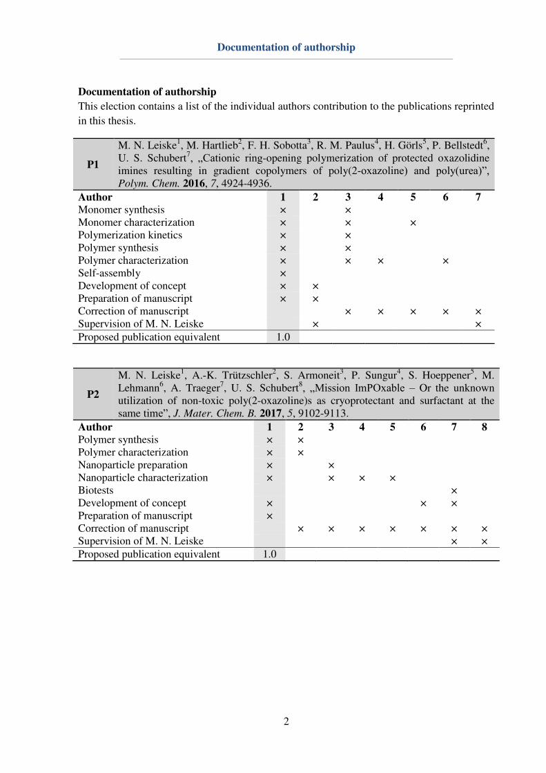
Documentation of authorship
2
Documentation of authorship
This election contains a list of the individual authors contribution to the publications reprinted
in this thesis.
P1
M. N. Leiske1, M. Hartlieb2, F. H. Sobotta3, R. M. Paulus4, H. Görls5, P. Bellstedt6, U. S. Schubert7, „Cationic ring-opening polymerization of protected oxazolidine imines resulting in gradient copolymers of poly(2-oxazoline) and poly(urea)”, Polym. Chem. 2016, 7, 4924-4936.
Author 1 2 3 4 5 6 7
Monomer synthesis × × Monomer characterization × × × Polymerization kinetics × × Polymer synthesis × × Polymer characterization × × × × Self-assembly × Development of concept × × Preparation of manuscript × × Correction of manuscript × × × × × Supervision of M. N. Leiske × × Proposed publication equivalent 1.0
P2
M. N. Leiske1, A.-K. Trützschler2, S. Armoneit3, P. Sungur4, S. Hoeppener5, M. Lehmann6, A. Traeger7, U. S. Schubert8, „Mission ImPOxable – Or the unknown utilization of non-toxic poly(2-oxazoline)s as cryoprotectant and surfactant at the same time”, J. Mater. Chem. B. 2017, 5, 9102-9113.
Author 1 2 3 4 5 6 7 8
Polymer synthesis × × Polymer characterization × × Nanoparticle preparation × × Nanoparticle characterization × × × × Biotests × Development of concept × × × Preparation of manuscript × Correction of manuscript × × × × × × × Supervision of M. N. Leiske × × Proposed publication equivalent 1.0
Documentation of authorship
3
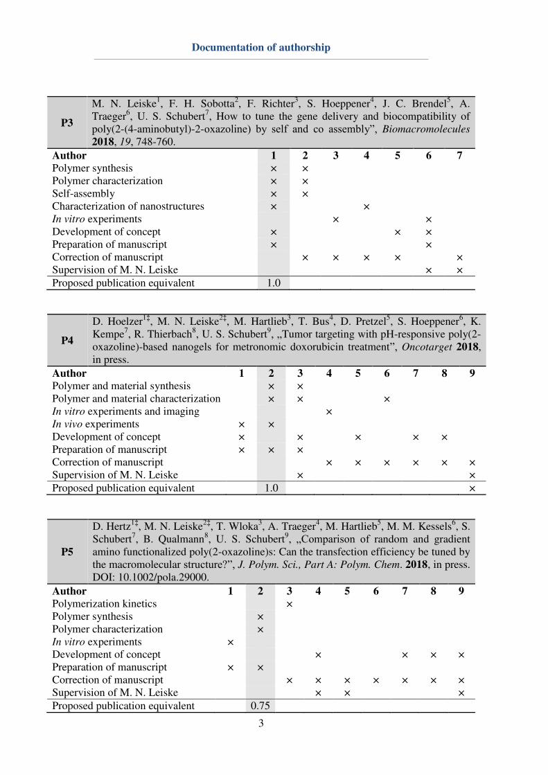
P3
M. N. Leiske1, F. H. Sobotta2, F. Richter3, S. Hoeppener4, J. C. Brendel5, A. Traeger6, U. S. Schubert7, How to tune the gene delivery and biocompatibility of poly(2-(4-aminobutyl)-2-oxazoline) by self and co assembly”, Biomacromolecules 2018, 19, 748-760.
Author 1 2 3 4 5 6 7
Polymer synthesis × × Polymer characterization × × Self-assembly × × Characterization of nanostructures × × In vitro experiments × × Development of concept × × × Preparation of manuscript × × Correction of manuscript × × × × × Supervision of M. N. Leiske × × Proposed publication equivalent 1.0
P4
D. Hoelzer1‡, M. N. Leiske2‡, M. Hartlieb3, T. Bus4, D. Pretzel5, S. Hoeppener6, K. Kempe7, R. Thierbach8, U. S. Schubert9, „Tumor targeting with pH-responsive poly(2-oxazoline)-based nanogels for metronomic doxorubicin treatment”, Oncotarget 2018, in press.
Author 1 2 3 4 5 6 7 8 9
Polymer and material synthesis × × Polymer and material characterization × × × In vitro experiments and imaging × In vivo experiments × × Development of concept × × × × × Preparation of manuscript × × × Correction of manuscript × × × × × × Supervision of M. N. Leiske × × Proposed publication equivalent 1.0 ×
P5
D. Hertz1‡, M. N. Leiske2‡, T. Wloka3, A. Traeger4, M. Hartlieb5, M. M. Kessels6, S. Schubert7, B. Qualmann8, U. S. Schubert9, „Comparison of random and gradient amino functionalized poly(2-oxazoline)s: Can the transfection efficiency be tuned by the macromolecular structure?”, J. Polym. Sci., Part A: Polym. Chem. 2018, in press. DOI: 10.1002/pola.29000.
Author 1 2 3 4 5 6 7 8 9
Polymerization kinetics × Polymer synthesis × Polymer characterization × In vitro experiments × Development of concept × × × × Preparation of manuscript × × Correction of manuscript × × × × × × × Supervision of M. N. Leiske × × × Proposed publication equivalent 0.75
Documentation of authorship
4
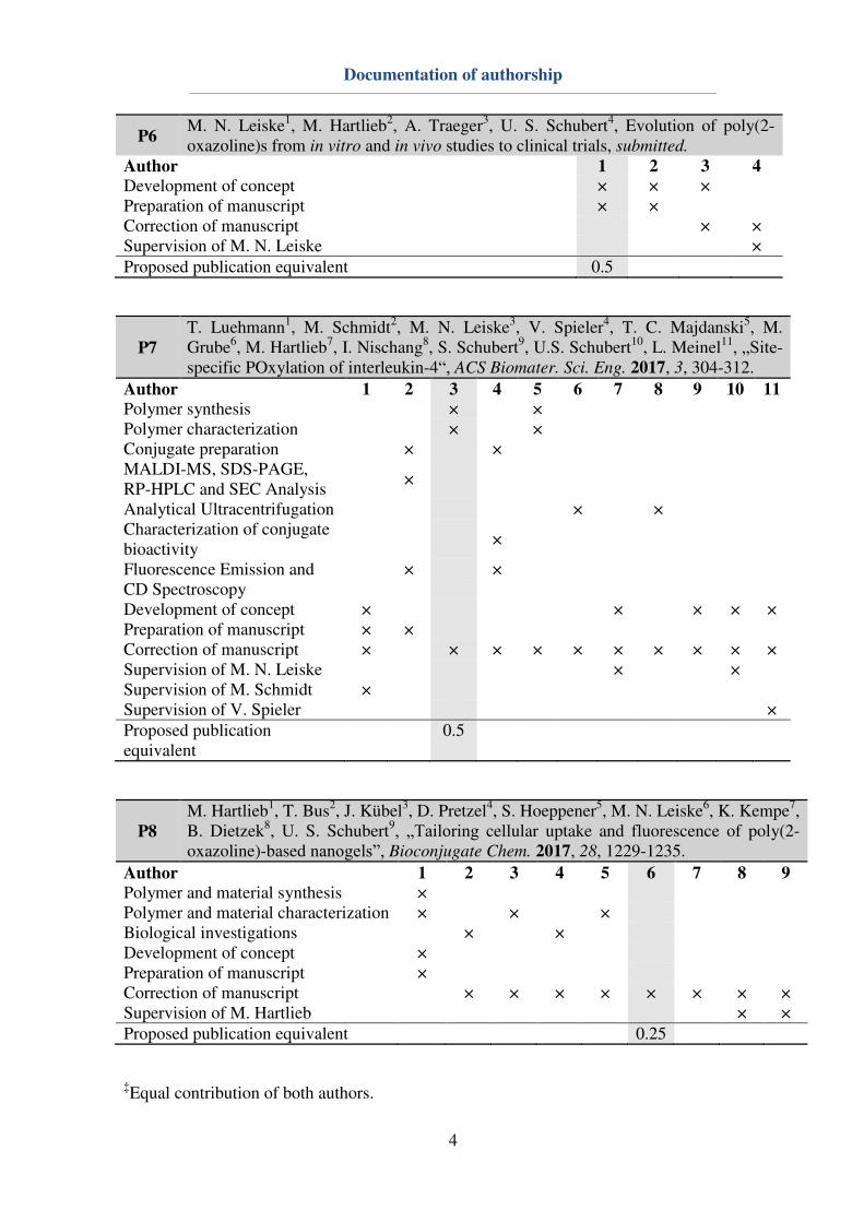
P6 M. N. Leiske1, M. Hartlieb2, A. Traeger3, U. S. Schubert4, Evolution of poly(2-oxazoline)s from in vitro and in vivo studies to clinical trials, submitted.
Author 1 2 3 4
Development of concept × × × Preparation of manuscript × × Correction of manuscript × × Supervision of M. N. Leiske × Proposed publication equivalent 0.5
P7
T. Luehmann1, M. Schmidt2, M. N. Leiske3, V. Spieler4, T. C. Majdanski5, M. Grube6, M. Hartlieb7, I. Nischang8, S. Schubert9, U.S. Schubert10, L. Meinel11, „Site-specific POxylation of interleukin-4“, ACS Biomater. Sci. Eng. 2017, 3, 304-312.
Author 1 2 3 4 5 6 7 8 9 10 11
Polymer synthesis × × Polymer characterization × × Conjugate preparation × × MALDI-MS, SDS-PAGE, RP-HPLC and SEC Analysis
×
Analytical Ultracentrifugation × × Characterization of conjugate bioactivity
×
Fluorescence Emission and × × CD Spectroscopy Development of concept × × × × × Preparation of manuscript × × Correction of manuscript × × × × × × × × × × Supervision of M. N. Leiske × × Supervision of M. Schmidt × Supervision of V. Spieler × Proposed publication equivalent
0.5
P8
M. Hartlieb1, T. Bus2, J. Kübel3, D. Pretzel4, S. Hoeppener5, M. N. Leiske6, K. Kempe7, B. Dietzek8, U. S. Schubert9, „Tailoring cellular uptake and fluorescence of poly(2-oxazoline)-based nanogels”, Bioconjugate Chem. 2017, 28, 1229-1235.
Author 1 2 3 4 5 6 7 8 9
Polymer and material synthesis × Polymer and material characterization × × × Biological investigations × × Development of concept × Preparation of manuscript × Correction of manuscript × × × × × × × × Supervision of M. Hartlieb × × Proposed publication equivalent 0.25
‡Equal contribution of both authors.

5
6
Erklärung zu den Eigenanteilen des Promovenden sowie der weiteren
Doktoranden/Doktorandinnen als Koautoren an Publikationen und
Zweitpublikationsrechten bei einer kumulativen Dissertation
Für alle in dieser kumulativen Dissertation verwendeten Manuskripte liegen die notwendigen
Genehmigungen der Verlage („Reprint permissions“) für die Zweitpublikation vor.
Die Co-Autoren der in dieser kumulativen Dissertation verwendeten Manuskripte sind sowohl
über die Nutzung als auch über die oben angegebenen Eigenanteile informiert und stimmen
dem zu.
Die Anteile der Co-Autoren an den Publikationen sind in den vorausgehenden Tabellen
aufgeführt.
Ich bin mit der Abfassung der Dissertation als publikationsbasiert, d.h. kumulativ,
einverstanden und bestätige die vorstehenden Angaben. Eine entsprechend begründete
Befürwortung mit Angabe des wissenschaftlichen Anteils des Doktoranden an den
verwendeten Publikationen werde ich parallel an den Rat der Fakultät der Chemisch-
Geowissenschaftlichen Fakultät richten.
Prof. Dr. Ulrich S. Schubert Jena, den ____________________
Meike N. Leiske Jena, den ____________________

7
1. Introduction
8
1. Introduction
Modern nanomedicine is divided into viral, lipoid and polymeric gene- and drug-carrier
systems.[1]
Herein, viral systems generally suffer from high production costs and upscaling
difficulties.[2]
Lipoid carriers, such as liposomes or lipoplexes, can be produced more cost-
efficiently, however, their storage is challenging.[2]
Polymer-based carriers require the
utilization of tailored, and biodegradable or biocompatible polymers. During the last years,
the utilization of poly(2-oxazoline)s (P(Ox)s) in terms of biomedical applications has
increased significantly.[3-4]
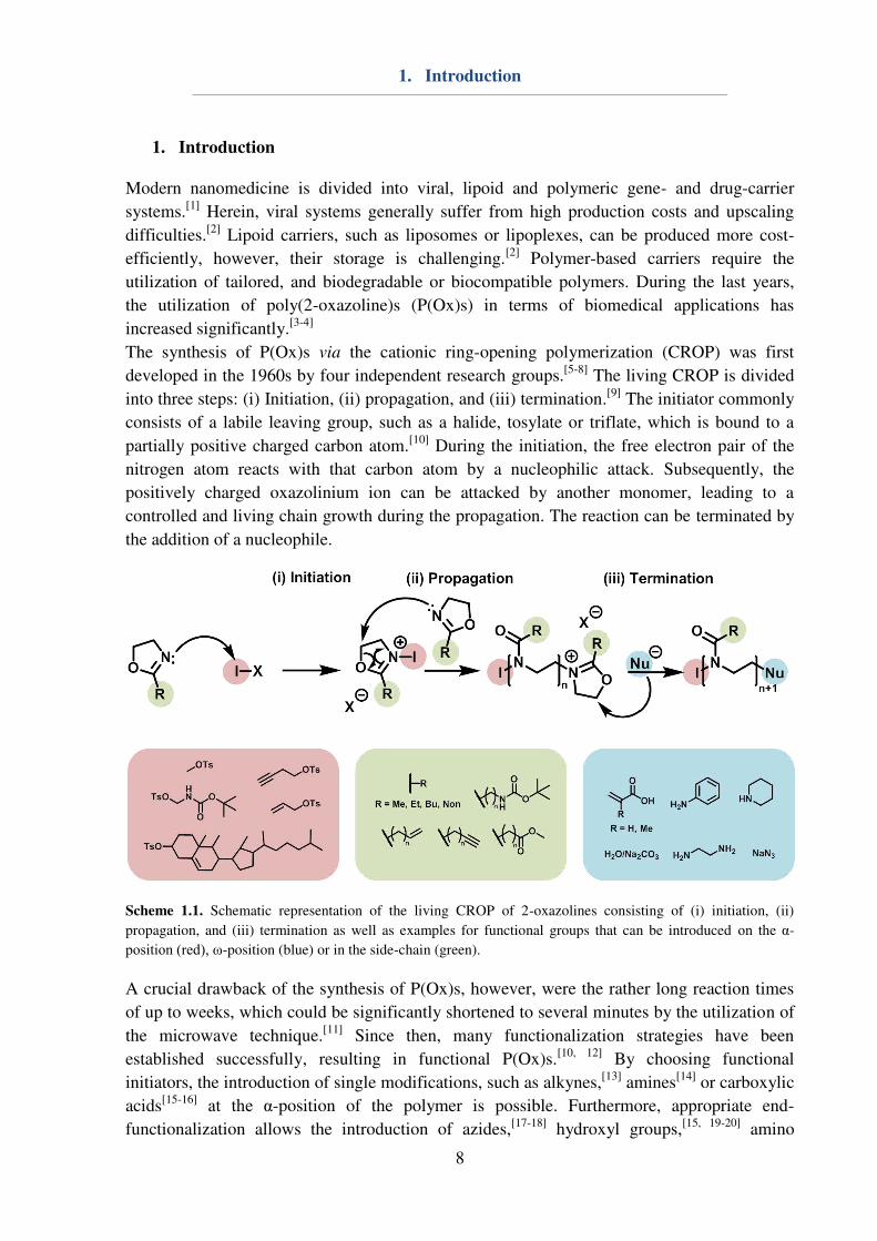
The synthesis of P(Ox)s via the cationic ring-opening polymerization (CROP) was first
developed in the 1960s by four independent research groups.[5-8]
The living CROP is divided
into three steps: (i) Initiation, (ii) propagation, and (iii) termination.[9]
The initiator commonly
consists of a labile leaving group, such as a halide, tosylate or triflate, which is bound to a
partially positive charged carbon atom.[10]
During the initiation, the free electron pair of the
nitrogen atom reacts with that carbon atom by a nucleophilic attack. Subsequently, the
positively charged oxazolinium ion can be attacked by another monomer, leading to a
controlled and living chain growth during the propagation. The reaction can be terminated by
the addition of a nucleophile.
Scheme 1.1. Schematic representation of the living CROP of 2-oxazolines consisting of (i) initiation, (ii)
propagation, and (iii) termination as well as examples for functional groups that can be introduced on the α-
position (red), ω-position (blue) or in the side-chain (green).
A crucial drawback of the synthesis of P(Ox)s, however, were the rather long reaction times
of up to weeks, which could be significantly shortened to several minutes by the utilization of
the microwave technique.[11]
Since then, many functionalization strategies have been
established successfully, resulting in functional P(Ox)s.[10, 12]
By choosing functional
initiators, the introduction of single modifications, such as alkynes,[13]
amines[14]
or carboxylic
acids[15-16]
at the α-position of the polymer is possible. Furthermore, appropriate end-
functionalization allows the introduction of azides,[17-18]
hydroxyl groups,[15, 19-20]
amino
1. Introduction
9
groups,[21-23] or large hydrophobic ω-end-groups.[20, 24-26] In addition, the utilization of 2-
substituted monomers can be used for the introduction of multiple (distinct) functionalities
into the polymer chain.[10, 27] Common examples for functional side-chains are alkynes,[28-30]
azides,[31-33] thiols,[34] carboxylic acids,[35-36] alkenyls,[37-38] aldehydes,[39] and amino
groups.[40-41] These numerous modification opportunities enable the preparation of tailored
polymers with adjustable properties. The resulting polymers are pseudopeptides, which are
biotolerable with respect to their type and amount of functional groups, making P(Ox)s
promising candidates for biomedical applications. For this reason, Chapter 2 will provide an
overview of the state of the art of P(Ox)s with respect to approaches regarding self-assembly,
in vitro and in vivo experiments as well as the first clinical trial.
In order to correctly evaluate the effects of an applied macromolecular structure on the
properties regarding self-assembly as well as drug- or gene-delivery, it is important to
evaluate the reactivity ratios of different monomers utilized for the preparation of copolymers.
Numerous publications already discussed the effect of the counter-ion of the initiator on the
polymerization kinetics.[42-45] Furthermore, the effect of the substituent in 2-position was
investigated.[46-48] Copolymerization of different 2-oxazolines resulted in random,[9, 49]
gradient[27, 50-51] or quasi block copolymers.[52-53] Herein, differences within the copolymer
composition can for instance be caused by length of the alkyl chain in 2-position.[9, 49] In
Chapter 3, two amino functionalized monomers are compared directly: (i) 2-(4-((tert-
Butoxycarbonyl)amino)butyl)-2-oxazoline (BocOx), which is already known from
literature[41] and (ii) tert-butyl 2-iminooxazolidine-3-carboxylate (BocOI), which was newly
synthesized within this thesis.[54] Herein, the synthesis routes of both monomers are shortly
compared. Afterwards, polymerization kinetics of BocOI and 2-ethyl-2-oxazoline (EtOx), BocOx
and EtOx as well as BocOx and 2-methyl-2-oxazoline (MeOx) are conducted to gain an overview
of the reactivity ratios of the different monomers during copolymerization. Since the
polymerization of BocOI and the assumed mechanism have been unknown up to this point, a
series of BocOI homopolymers and copolymers with EtOx are synthesized and deprotected to
yield polymers consisting of poly(urea) and P(Ox). All resulting polymers and tert-
butyloxycarbonyl (Boc) protected precursors are characterized and compared regarding their
properties. Furthermore, BocOx containing statistic and block copolymers are synthesized and
characterized. Hereby the acidic deprotection of BocOx containing copolymers yielding 2-(4-
aminobutyl)-2-oxazoline (AmOx) groups will be shown.
As already mentioned, possible applications of P(Ox)s are positioned in biomedical
applications, such as drug or gene delivery.[3-4] Herein, the utilization of P(Ox)s is versatile,
ranging from nano- and microparticles,[50] as well as nanocapsules[55] to crosslinked networks
such as hydrogels,[41] surface coatings[56] or nanogels.[51, 57] Herein, the assembly of
amphiphilic block copolymers has been described.[53, 58-60] Chapter 4 deals with P(Ox)
containing nanostructures. Firstly, the utilization of water-soluble P(Ox)s for the stabilization
of hydrophobic nanoparticles consisting of poly(lactide-co-glycolide) (PLGA) during
preparation, purification and lyophilization, aiming an all-in-one system to replace common
particle stabilizers, i.e. poly(vinyl alcohol), Pluronic F127, glucose, saccharose and trehalose.
Furthermore, amphiphilic block copolymers consisting of P(Ox)s were self- and co-assembled
in water as well as characterized regarding their pH responsiveness. In addition to that, AmOx
containing block copolymers are self-assembled in chloroform and reversibly core cross-
1. Introduction
10
linked using glutaraldehyde (GA) to form imines. Additionally the covalent attachment of 6-
amino-fluorescein (6AF) and doxorubicin (DOX) to the resulting nanogels will be shown.
In addition to the synthesis and assembly of P(Ox)s, their application in biomedical sciences
has been widely explored during the last decade.[3-4] Herein, the application of cationic P(Ox)s
in terms of gene delivery represents one interesting field of research.[38, 61-62] Chapter 5
describes the utilization of AmOx containing copolymers for gene transfection. Hereby,
copolymers containing either EtOx (random), MeOx (gradient) or 2-nonyl-2-oxazoline
(NonOx, block) representing the comonomer are compared regarding their cyto- and
hemocompatibility, cellular uptake as well as transfection efficiency. A further possible
application for functional P(Ox)s is the delivery of conjugated drugs, e.g. proteins or small
molecules. Hereby, the most important issue concerns the activity of the active
pharmaceutical ingredient (API). For this reason, Chapter 6 describes the preparation of two
different P(Ox) drug conjugates, i.e. the P(Ox)ylation of interleukin-4 (IL-4) to water soluble
P(Ox)s using biorthogonal copper catalyzed click chemistry (CuAAC) and the conjugation of
doxorubicin (DOX) to AmOx containing nanogels via Schiff-base chemistry.
The aim of this thesis is to push forward the potential of 2-oxazolines in terms of synthesis,
self-assembly and biomedical utilization. Hereby, the synthesis of two different monomers,
namely BocOx and BocOI will be presented. Homo- and copolymerization kinetics with
EtOx, respectively MeOx will provide an overview of the monomer distribution within the
polymers, which is important for further implementations of the polymers in terms of self-
assembly as well as gene- and drug-delivery. Furthermore, the contribution of different P(Ox)
regarding the preparation of colloidal nanostructures will be shown. Herein, P(Ox)
homopolymers will be utilized as surfactants and cryoprotectants for the stabilization of
nanoparticles. It will be demonstrated that different P(Ox) block copolymers can be utilized to
form distinct nanostructures, suitable for gene-and drug delivery applications. In addition to
that, the usage of cationic, pH responsive nanostructures with respect to gene-delivery will be
conducted. Herein, nanostructures will be compared to water soluble cationic random and
gradient copolymers. Furthermore, the contribution of P(Ox) to successful and targeted drug
delivery will be shown. This thesis contributes to the knowledge of P(Ox) in a wide range,
covering not only the synthesis, however, also the preparation and characterization self-
assembled structures and their biomedical potential.
2. Poly(2-oxazoline)s in biomedical applications
11
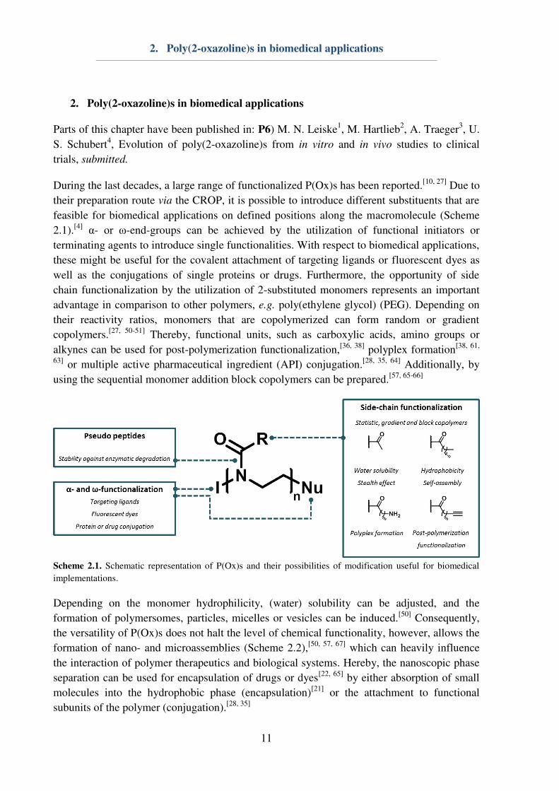
2. Poly(2-oxazoline)s in biomedical applications
Parts of this chapter have been published in: P6) M. N. Leiske1, M. Hartlieb
2, A. Traeger
3, U.
S. Schubert4, Evolution of poly(2-oxazoline)s from in vitro and in vivo studies to clinical
trials, submitted.
During the last decades, a large range of functionalized P(Ox)s has been reported.[10, 27]
Due to
their preparation route via the CROP, it is possible to introduce different substituents that are
feasible for biomedical applications on defined positions along the macromolecule (Scheme
2.1).[4]
α- or ω-end-groups can be achieved by the utilization of functional initiators or
terminating agents to introduce single functionalities. With respect to biomedical applications,
these might be useful for the covalent attachment of targeting ligands or fluorescent dyes as
well as the conjugations of single proteins or drugs. Furthermore, the opportunity of side
chain functionalization by the utilization of 2-substituted monomers represents an important
advantage in comparison to other polymers, e.g. poly(ethylene glycol) (PEG). Depending on
their reactivity ratios, monomers that are copolymerized can form random or gradient
copolymers.[27, 50-51]
Thereby, functional units, such as carboxylic acids, amino groups or
alkynes can be used for post-polymerization functionalization,[36, 38]
polyplex formation[38, 61,
63] or multiple active pharmaceutical ingredient (API) conjugation.
[28, 35, 64] Additionally, by
using the sequential monomer addition block copolymers can be prepared.[57, 65-66]
Scheme 2.1. Schematic representation of P(Ox)s and their possibilities of modification useful for biomedical
implementations.
Depending on the monomer hydrophilicity, (water) solubility can be adjusted, and the
formation of polymersomes, particles, micelles or vesicles can be induced.[50]
Consequently,
the versatility of P(Ox)s does not halt the level of chemical functionality, however, allows the
formation of nano- and microassemblies (Scheme 2.2),[50, 57, 67]
which can heavily influence
the interaction of polymer therapeutics and biological systems. Hereby, the nanoscopic phase
separation can be used for encapsulation of drugs or dyes[22, 65]
by either absorption of small
molecules into the hydrophobic phase (encapsulation)[21]
or the attachment to functional
subunits of the polymer (conjugation).[28, 35]

2. Poly(2-oxazoline)s in biomedical applications
12
Scheme 2.2. Schematic representation of different assembled P(Ox) structures: (i) Nanocapsules,[55]
(ii)
micelles,[21]
(iii) nanogels,[51]
(iv) hydrogels,[51]
and (v) surface coatings.[68]
Herein, the hydrohphilic poly(2-ethyl-2-oxazoline) (P(EtOx)) or poly(2-methyl-2-oxazoline)
(P(MeOx)) shell induces a shielding towards unspecific interactions with biological matter
and prevents undesired protein interactions comparable to PEG.[24-25, 69-70]
Furthermore, the
preparation of cross-linked materials, such as nanogels,[51, 57]
capsules,[55]
and hydrogels[41, 51]
is possible by either covalent or physical interactions.[51]
In general, these materials are
utilized, e.g. for drug delivery,[35]
gene purification[56]
or as nano reaction compartment.[71]
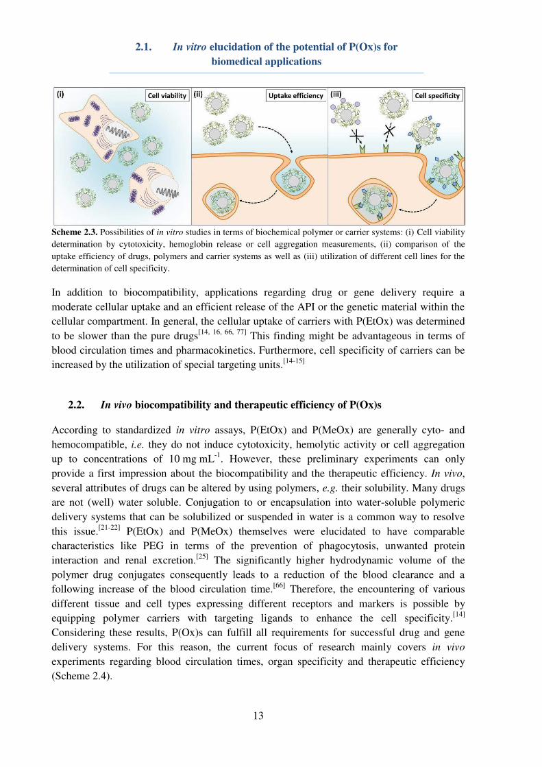
2.1. In vitro elucidation of the potential of P(Ox)s for biomedical applications
In order to evaluate the potential of P(Ox)s for biomedical applications, in vitro investigations
(i.e. cell tests) are indespensable. Hereby, the initial experiments mainly cover the elucidation
of the cytocompatibility, respectively cell viability as well as the hemocompatibility, i.e.
erythrocyte aggregation and hemolysis experiments (Scheme 2.3). Herein, P(EtOx) and
P(MeOx) of varios molar masses did not cause any cytotoxycity, erythrocyte aggregation or
hemoglobin release in short-term experiments.[72-73]
Long-term experiments revealed a slight
decrease of the cell viability dependent on the molar masss of the polymers and the incubation
time.[72-73]
In addition to that, experiments showed an increase of the cytotoxicity of P(EtOx)
in dependence on the degree of hydrolysis.[74]
However, investigations on the hydrolysis of
P(EtOx) under physiological conditions have shown that a degree of hydrolysis above 10% is
unlikely.[75]
Furthermore, P(Ox) micelles with a poly(lactic acid) (PLA) or poly(2-butyl-2-
oxazoline) core were determined to be non-toxic.[67, 76]
2.1. In vitro elucidation of the potential of P(Ox)s for
biomedical applications
13
Scheme 2.3. Possibilities of in vitro studies in terms of biochemical polymer or carrier systems: (i) Cell viability
determination by cytotoxicity, hemoglobin release or cell aggregation measurements, (ii) comparison of the
uptake efficiency of drugs, polymers and carrier systems as well as (iii) utilization of different cell lines for the
determination of cell specificity.
In addition to biocompatibility, applications regarding drug or gene delivery require a
moderate cellular uptake and an efficient release of the API or the genetic material within the
cellular compartment. In general, the cellular uptake of carriers with P(EtOx) was determined
to be slower than the pure drugs[14, 16, 66, 77]
This finding might be advantageous in terms of
blood circulation times and pharmacokinetics. Furthermore, cell specificity of carriers can be
increased by the utilization of special targeting units.[14-15]
2.2. In vivo biocompatibility and therapeutic efficiency of P(Ox)s
According to standardized in vitro assays, P(EtOx) and P(MeOx) are generally cyto- and
hemocompatible, i.e. they do not induce cytotoxicity, hemolytic activity or cell aggregation
up to concentrations of 10 mg mL-1
. However, these preliminary experiments can only
provide a first impression about the biocompatibility and the therapeutic efficiency. In vivo,
several attributes of drugs can be altered by using polymers, e.g. their solubility. Many drugs
are not (well) water soluble. Conjugation to or encapsulation into water-soluble polymeric
delivery systems that can be solubilized or suspended in water is a common way to resolve
this issue.[21-22]
P(EtOx) and P(MeOx) themselves were elucidated to have comparable
characteristics like PEG in terms of the prevention of phagocytosis, unwanted protein
interaction and renal excretion.[25]
The significantly higher hydrodynamic volume of the
polymer drug conjugates consequently leads to a reduction of the blood clearance and a
following increase of the blood circulation time.[66]
Therefore, the encountering of various
different tissue and cell types expressing different receptors and markers is possible by
equipping polymer carriers with targeting ligands to enhance the cell specificity.[14]
Considering these results, P(Ox)s can fulfill all requirements for successful drug and gene
delivery systems. For this reason, the current focus of research mainly covers in vivo
experiments regarding blood circulation times, organ specificity and therapeutic efficiency
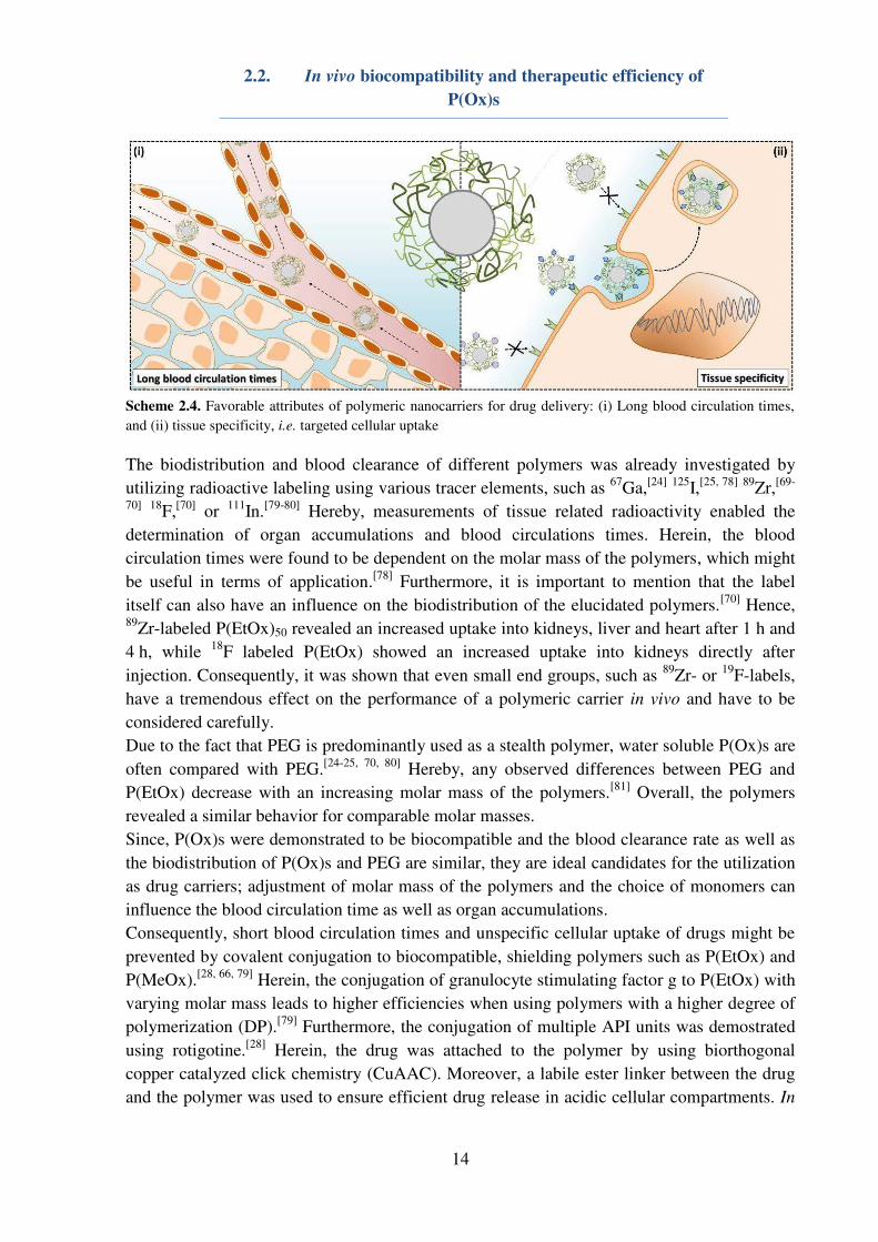
(Scheme 2.4).
2.2. In vivo biocompatibility and therapeutic efficiency of
P(Ox)s
14
Scheme 2.4. Favorable attributes of polymeric nanocarriers for drug delivery: (i) Long blood circulation times,
and (ii) tissue specificity, i.e. targeted cellular uptake
The biodistribution and blood clearance of different polymers was already investigated by
utilizing radioactive labeling using various tracer elements, such as 67
Ga,[24]
125
I,[25, 78]
89
Zr,[69-
70]
18F,
[70] or
111In.
[79-80] Hereby, measurements of tissue related radioactivity enabled the
determination of organ accumulations and blood circulations times. Herein, the blood
circulation times were found to be dependent on the molar mass of the polymers, which might
be useful in terms of application.[78]
Furthermore, it is important to mention that the label
itself can also have an influence on the biodistribution of the elucidated polymers.[70]
Hence, 89
Zr-labeled P(EtOx)50 revealed an increased uptake into kidneys, liver and heart after 1 h and
4 h, while 18
F labeled P(EtOx) showed an increased uptake into kidneys directly after
injection. Consequently, it was shown that even small end groups, such as 89
Zr- or 19
F-labels,
have a tremendous effect on the performance of a polymeric carrier in vivo and have to be
considered carefully.
Due to the fact that PEG is predominantly used as a stealth polymer, water soluble P(Ox)s are
often compared with PEG.[24-25, 70, 80]
Hereby, any observed differences between PEG and
P(EtOx) decrease with an increasing molar mass of the polymers.[81]
Overall, the polymers
revealed a similar behavior for comparable molar masses.
Since, P(Ox)s were demonstrated to be biocompatible and the blood clearance rate as well as
the biodistribution of P(Ox)s and PEG are similar, they are ideal candidates for the utilization
as drug carriers; adjustment of molar mass of the polymers and the choice of monomers can
influence the blood circulation time as well as organ accumulations.
Consequently, short blood circulation times and unspecific cellular uptake of drugs might be
prevented by covalent conjugation to biocompatible, shielding polymers such as P(EtOx) and
P(MeOx).[28, 66, 79]
Herein, the conjugation of granulocyte stimulating factor g to P(EtOx) with
varying molar mass leads to higher efficiencies when using polymers with a higher degree of
polymerization (DP).[79]
Furthermore, the conjugation of multiple API units was demostrated
using rotigotine.[28]
Herein, the drug was attached to the polymer by using biorthogonal
copper catalyzed click chemistry (CuAAC). Moreover, a labile ester linker between the drug
and the polymer was used to ensure efficient drug release in acidic cellular compartments. In
2.2. In vivo biocompatibility and therapeutic efficiency of
P(Ox)s
15
vivo investigations revealed steady plasma drug concentrations over several days and,
consequently, reduced unwanted side-effects such as dyskinesia.[64]
In particular in terms of cancer therapy, the enhanced blood circulation times might be
advantageous to ensure an efficient delivery of the cytostatic agent into the tumor cells, e.g.
by exploiting the EPR effect. Hereby, conjugation via azide cleavable hydrazone linker,[16, 35]
the encapsulation into P(Ox) based micelles,[14-16, 19, 76-77, 82] or liposomes[26, 83] leads to an
enhanced solubility[67] of the cytostatic agents as well as longer blood circulation times[22, 83]
and higher therapeutic efficiencies[14-15, 21, 67, 76] while expressing a high biocompatibility.[22, 77]
3. Synthesis and polymerization of functional 2-oxazolines
16
3. Synthesis and polymerization of functional 2-oxazolines
Parts of this chapter have been published in: P1) M. N. Leiske, M. Hartlieb, F. H. Sobotta, R.
M. Paulus, H. Görls, P. Bellstedt, U. S. Schubert, Polym. Chem. 2016, 7, 4924-4936. P4) D.
Hoelzer‡, M. N. Leiske‡, M. Hartlieb, T. Bus, D. Pretzel, S. Hoeppener, K. Kempe, R.
Thierbach, U. S. Schubert, Oncotarget 2018, in press. P5) D. Hertz‡, M. N. Leiske‡, T.
Wloka, A. Traeger, M. Hartlieb, M. M. Kessels, S. Schubert, B. Qualmann, U. S. Schubert, J.
Polym. Sci., Part A: Polym. Chem. 2018, in press. DOI: 10.1002/pola.29000. P8) M.
Hartlieb‡, T. Bus‡, J. Kübel, D. Pretzel, S. Hoeppener, M. N. Leiske, K. Kempe, B. Dietzek,
U. S. Schubert, Bioconjugate Chem. 2017, 28, 1229-1235. ‡Equal contribution of both
authors.
The synthesis of P(Ox)s via CROP facilitates the synthesis of functional homo- and
copolymers with a tailored structure. Within this chapter, the synthesis and polymerization
route of the Boc protected 2-oxazoline 2-(4-((tert-butoxycarbonyl(amino)butyl)-2-oxazoline
(BocOx), which is known from literature, will be compared with the newly synthesized tert-
butyl 2-iminooxazolidine-3-carboxylate (BocOI). Since the polymerization kinetic of a monomer is
for instance dependent on the substituent in 2-position, kinetic investigations on the polymerization
rate constant (kp) are indispensable. Furthermore, the copolymerization of different monomers can
result in random, gradient or quasi block copolymers, depending on the reactivity ratios of the used
monomers. For this reason, detailed kinetic investigations on the homopolymerization of the newly
synthesized BocOI as well as on the copolymerization of BocOI and EtOx will be performed. In
order to obtain information about the monomer distribution within water soluble cationic P(Ox)
copolymers, the copolymerization of BocOx and EtOx as well as BocOx and MeOx will also be
investigated. On the basis of the resulting kp values, a series of functional homo- and copolymers
will be synthesized and characterized.
3.1. Monomer synthesis and polymerization mechanism
During the last decade, amino functionalized P(Ox)s have been widely explored. While post-
polymerization functionalization represents an effective method to introduce primary,[35, 38, 62,
84-85] secondary,[38] or tertiary amino moieties,[38, 85] suitable protection groups like the acid
labile tert-butyloxycarbonyl (Boc) protection group enable the possibility of a polymerization
of amino functionalized 2-oxazoline monomers.[40-41, 61] M. Hartlieb et al. synthesized the Boc
protected 2-oxazoline BocOx in a three step synthesis approach (Figure 3.1A).[41] In the first
reaction step (a), the amino group of 5-aminovaleric acid is protected using di-tert-butyl
dicarbonate (DiBoc). Subsequently, the carboxylic acid can be activated by the addition of
ethyl chloroformate to facilitate the reaction with 2-chloroethylamine (b). Finally, the ring
closure can be conducted under basic conditions (c) to obtain BocOx (3), which is suitable for
the CROP.
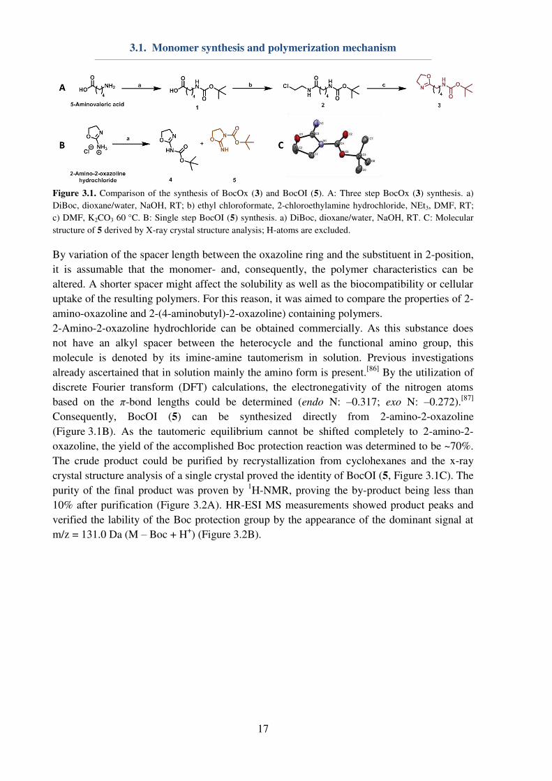
3.1. Monomer synthesis and polymerization mechanism
17
Figure 3.1. Comparison of the synthesis of BocOx (3) and BocOI (5). A: Three step BocOx (3) synthesis. a)
DiBoc, dioxane/water, NaOH, RT; b) ethyl chloroformate, 2-chloroethylamine hydrochloride, NEt3, DMF, RT;
c) DMF, K2CO3 60 °C. B: Single step BocOI (5) synthesis. a) DiBoc, dioxane/water, NaOH, RT. C: Molecular
structure of 5 derived by X-ray crystal structure analysis; H-atoms are excluded.
By variation of the spacer length between the oxazoline ring and the substituent in 2-position,
it is assumable that the monomer- and, consequently, the polymer characteristics can be
altered. A shorter spacer might affect the solubility as well as the biocompatibility or cellular
uptake of the resulting polymers. For this reason, it was aimed to compare the properties of 2-
amino-oxazoline and 2-(4-aminobutyl)-2-oxazoline) containing polymers.
2-Amino-2-oxazoline hydrochloride can be obtained commercially. As this substance does
not have an alkyl spacer between the heterocycle and the functional amino group, this
molecule is denoted by its imine-amine tautomerism in solution. Previous investigations
already ascertained that in solution mainly the amino form is present.[86]
By the utilization of
discrete Fourier transform (DFT) calculations, the electronegativity of the nitrogen atoms
based on the π-bond lengths could be determined (endo N: –0.317; exo N: –0.272).[87]
Consequently, BocOI (5) can be synthesized directly from 2-amino-2-oxazoline
(Figure 3.1B). As the tautomeric equilibrium cannot be shifted completely to 2-amino-2-
oxazoline, the yield of the accomplished Boc protection reaction was determined to be ~70%.
The crude product could be purified by recrystallization from cyclohexanes and the x-ray
crystal structure analysis of a single crystal proved the identity of BocOI (5, Figure 3.1C). The
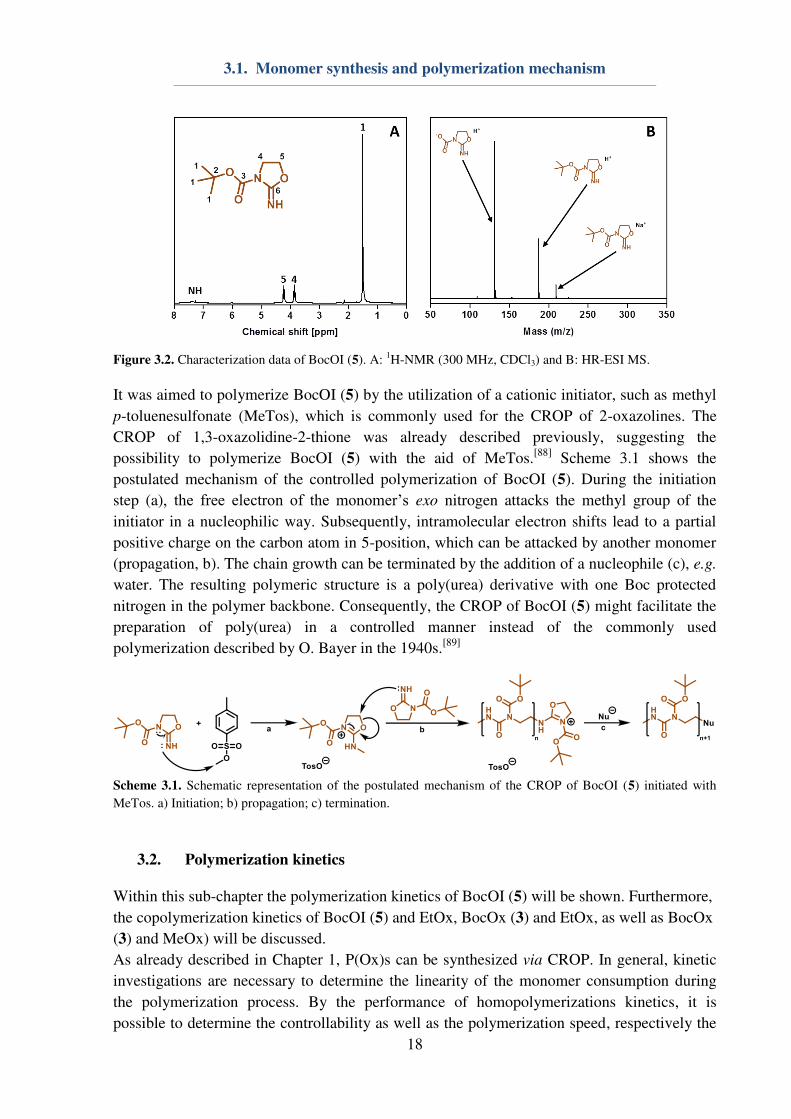
purity of the final product was proven by 1H-NMR, proving the by-product being less than
10% after purification (Figure 3.2A). HR-ESI MS measurements showed product peaks and
verified the lability of the Boc protection group by the appearance of the dominant signal at
m/z = 131.0 Da (M – Boc + H+) (Figure 3.2B).
3.1. Monomer synthesis and polymerization mechanism
18
Figure 3.2. Characterization data of BocOI (5). A:
1H-NMR (300 MHz, CDCl3) and B: HR-ESI MS.
It was aimed to polymerize BocOI (5) by the utilization of a cationic initiator, such as methyl
p-toluenesulfonate (MeTos), which is commonly used for the CROP of 2-oxazolines. The
CROP of 1,3-oxazolidine-2-thione was already described previously, suggesting the
possibility to polymerize BocOI (5) with the aid of MeTos.[88]
Scheme 3.1 shows the
postulated mechanism of the controlled polymerization of BocOI (5). During the initiation
step (a), the free electron of the monomer’s exo nitrogen attacks the methyl group of the
initiator in a nucleophilic way. Subsequently, intramolecular electron shifts lead to a partial
positive charge on the carbon atom in 5-position, which can be attacked by another monomer
(propagation, b). The chain growth can be terminated by the addition of a nucleophile (c), e.g.
water. The resulting polymeric structure is a poly(urea) derivative with one Boc protected
nitrogen in the polymer backbone. Consequently, the CROP of BocOI (5) might facilitate the
preparation of poly(urea) in a controlled manner instead of the commonly used
polymerization described by O. Bayer in the 1940s.[89]
Scheme 3.1. Schematic representation of the postulated mechanism of the CROP of BocOI (5) initiated with
MeTos. a) Initiation; b) propagation; c) termination.
3.2. Polymerization kinetics
Within this sub-chapter the polymerization kinetics of BocOI (5) will be shown. Furthermore,
the copolymerization kinetics of BocOI (5) and EtOx, BocOx (3) and EtOx, as well as BocOx
(3) and MeOx) will be discussed.
As already described in Chapter 1, P(Ox)s can be synthesized via CROP. In general, kinetic
investigations are necessary to determine the linearity of the monomer consumption during
the polymerization process. By the performance of homopolymerizations kinetics, it is
possible to determine the controllability as well as the polymerization speed, respectively the
3.2. Polymerization kinetics
19
reaction rate constant (kp). The kp of the monomer under the investigated conditions could be
calculated by the assumption that ln[�]ln[�]� = � complies with � , eventuating equations
(3.1) and (3.2). ln � − ln �� = � ∙ � (3.1) � = � [�] (3.2)
Within copolymerization reactions, monomers with different kp values can form different
copolymers, i.e. random,[9, 49] gradient[27, 50-51] or quasi block copolymers,[52-53] as depicted in
Chapter 1. In order to obtain a further insight on the monomer distribution within the polymer
chain, detailed kinetic investigations on the copolymerization of different monomers are
indispensable. Herein, reactivity ratios for copolymerization of the monomer pairs are
calculated at four different monomer ratios at 30% conversion of the monomer with the
higher reaction constant as determined using least-linear least square fitting (equation
(3.3)).[90-91]
� = � − +� +� − + −� +� (3.3)
F1: instantaneous mole fraction, f1: monomer fraction of monomer 1, f2: monomer fraction of
monomer 2, r1: reactivity ratio of monomer 1, r2: reactivity ratio of monomer 2
Homopolymerization kinetics of EtOx,[11] MeOx[48] and BocOx[41] are already known from
literature. Consequently, within this sub-chapter, only the homopolymerizations kinetics of
BocOI (5) will be presented. For the purpose of accomplishing a copolymerization of 2-
oxazolines and oxazolidine imines, time-dependent kinetics on the copolymerization of
BocOI (5) and EtOx were conducted. As BocOx, respectively AmOx, containing cationic
polymers were aimed to be used for gene-delivery applications (Chapter 5), the
copolymerization with a non-ionic water soluble monomer is indispensable to enhance its
biocompatibility. For this reason, copolymerization kinetics of BocOx (3) and EtOx as well as
the more reactive MeOx and BocOx (3) were performed.
Initial kinetics were conducted on the homopolymerization of BocOI (5). Due to the fact that
a copolymerization with 2-oxazolines, i.e. EtOx, was planned, preliminary experiments were
conducted at 140 °C, being a well applicable temperature for the polymerization of EtOx
considering the reaction speed and the dispersity of the resulting polymers.[11] Unfortunately,
keff in dependence on the polymerization time was not linear under the investigated
conditions. It was suggested that the monomer suffers from thermal deprotection at high
reaction temperatures as already known from literature for other examples.[92] Furthermore, it
is quite likely that a non-protected 2-imino-1,3-oxazolidine monomer can still be polymerized
by using a CROP, when referring to A. Nagai et al.. Herein, 1,3-oxazolidine-2-thione was
polymerized utilizing methyl trifluoromethanesulfonate without any protection groups for the
endo N.[88] It might be presumed that the 2-imino-1,3-oxazolidine is significantly more
reactive than BocOI (5), because of a lack in steric hindrance, leading to the lack in linearity
3.2. Polymerization kinetics
20
of the conversion in dependence on the time caused by slow initiation speeds compared to the
propagation. A. Nagai et al. therefore polymerized 1,3-oxazolidine-2-thione at low
temperatures of 30 to 40 °C. Unfortunately, by using such low reaction temperatures, the
polymerization rate constant of 2-oxazolines is very low, leading to reaction times of several
days or weeks.[11]
For this reason, the polymerization temperature was lowered to 100 °C, also
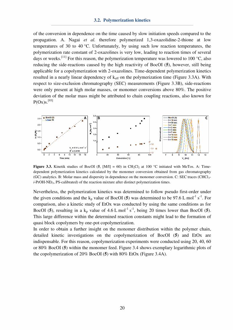
reducing the side-reactions caused by the high reactivity of BocOI (5), however, still being
applicable for a copolymerization with 2-oxazolines. Time-dependent polymerization kinetics
resulted in a nearly linear dependency of keff on the polymerization time (Figure 3.3A). With
respect to size-exclusion chromatography (SEC) measurements (Figure 3.3B), side-reactions
were only present at high molar masses, or monomer conversions above 80%. The positive
deviation of the molar mass might be attributed to chain coupling reactions, also known for
P(Ox)s.[93]
Figure 3.3. Kinetik studies of BocOI (5, [M/I] = 60) in CH2Cl2 at 100 °C initiated with MeTos. A: Time-
dependent polymerization kinetics calculated by the monomer conversion obtained from gas chromatography
(GC) analytics. B: Molar mass and dispersity in dependence on the monomer conversion. C: SEC traces (CHCl3-
i-PrOH-NEt3, PS-calibrated) of the reaction mixture after distinct polymerization times.
Nevertheless, the polymerization kinetics was determined to follow pseudo first-order under
the given conditions and the kp value of BocOI (5) was determined to be 97.6 L mol-1
s-1
. For
comparison, also a kinetic study of EtOx was conducted by using the same conditions as for
BocOI (5), resulting in a kp value of 4.6 L mol-1
s-1
, being 20 times lower than BocOI (5).
This large difference within the determined reaction constants might lead to the formation of
quasi block copolymers by one-pot copolymerization.
In order to obtain a further insight on the monomer distribution within the polymer chain,
detailed kinetic investigations on the copolymerization of BocOI (5) and EtOx are
indispensable. For this reason, copolymerization experiments were conducted using 20, 40, 60
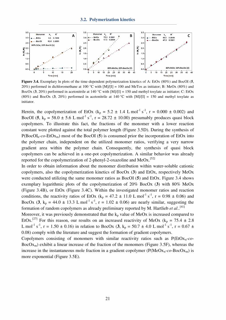
or 80% BocOI (5) within the monomer feed. Figure 3.4 shows exemplary logarithmic plots of
the copolymerization of 20% BocOI (5) with 80% EtOx (Figure 3.4A).
3.2. Polymerization kinetics
21
Figure 3.4. Exemplary ln plots of the time-dependent polymerization kinetics of A: EtOx (80%) and BocOI (5,
20%) performed in dichloromethane at 100 °C with [M]/[I] = 100 and MeTos as initiator; B: MeOx (80%) and
BocOx (3, 20%) performed in acetonitrile at 140 °C with [M]/[I] = 150 and methyl tosylate as initiator; C: EtOx
(80%) and BocOx (3, 20%) performed in acetonitrile at 140 °C with [M]/[I] = 150 and methyl tosylate as
initiator.
Herein, the copolymerization of EtOx (kp = 5.2 ± 1.4 L mol-1
s-1
, r = 0.000 ± 0.002) and
BocOI (5, kp = 58.0 ± 5.6 L mol-1
s-1
, r = 28.72 ± 10.00) presumably produces quasi block
copolymers. To illustrate this fact, the fractions of the monomer with a lower reaction
constant were plotted against the total polymer length (Figure 3.5D). During the synthesis of
P(BocOIn-co-EtOxm) most of the BocOI (5) is consumed prior the incorporation of EtOx into
the polymer chain, independent on the utilized monomer ratios, verifying a very narrow
gradient area within the polymer chain. Consequently, the synthesis of quasi block
copolymers can be achieved in a one-pot copolymerization. A similar behavior was already
reported for the copolymerization of 2-phenyl-2-oxazoline and MeOx.[52]
In order to obtain information about the monomer distribution within water-soluble cationic
copolymers, also the copolymerization kinetics of BocOx (3) and EtOx, respectively MeOx
were conducted utilizing the same monomer ratios as BocOI (5) and EtOx. Figure 3.4 shows
exemplary logarithmic plots of the copolymerization of 20% BocOx (3) with 80% MeOx
(Figure 3.4B), or EtOx (Figure 3.4C). Within the investigated monomer ratios and reaction
conditions, the reactivity ratios of EtOx (kp = 47.2 ± 11.0 L mol-1
s-1
, r = 0.98 ± 0.06) and
BocOx (3, kp = 44.0 ± 13.3 L mol-1
s-1
, r = 1.02 ± 0.06) are nearly similar, suggesting the
formation of random copolymers as already preliminary reported by M. Hartlieb et al..[41]
Moreover, it was previously demonstrated that the kp value of MeOx is increased compared to
EtOx.[27]
For this reason, our results on an increased reactivity of MeOx (kp = 75.4 ± 2.8
L mol-1
s-1
, r = 1.50 ± 0.16) in relation to BocOx (3, kp = 50.7 ± 4.0 L mol-1
s-1
, r = 0.67 ±
0.08) comply with the literature and suggest the formation of gradient copolymers.
Copolymers consisting of monomers with similar reactivity ratios such as P(EtOxn-co-
BocOxm) exhibit a linear increase of the fraction of the monomers (Figure 3.5F), whereas the
increase in the instantaneous mole fraction in a gradient copolymer (P(MeOxn-co-BocOxm) is
more exponential (Figure 3.5E).
3.2. Polymerization kinetics
22
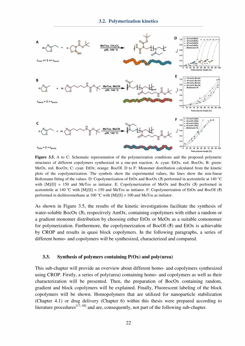
Figure 3.5. A to C: Schematic representation of the polymerization conditions and the proposed polymeric
structures of different copolymers synthesized in a one-pot reaction. A: cyan: EtOx, red: BocOx; B: green:
MeOx, red. BocOx; C: cyan: EtOx; orange: BocOI. D to F: Monomer distribution calculated from the kinetic
plots of the copolymerization. The symbols show the experimental values, the lines show the non-linear
Boltzmann fitting of the values. D: Copolymerization of EtOx and BocOx (3) performed in acetonitrile at 140 °C
with [M]/[I] = 150 and MeTos as initiator. E: Copolymerization of MeOx and BocOx (3) performed in
acetonitrile at 140 °C with [M]/[I] = 150 and MeTos as initiator. F: Copolymerization of EtOx and BocOI (5)
performed in dichloromethane at 100 °C with [M]/[I] = 100 and MeTos as initiator.
As shown in Figure 3.5, the results of the kinetic investigations facilitate the synthesis of
water-soluble BocOx (3), respectively AmOx, containing copolymers with either a random or
a gradient monomer distribution by choosing either EtOx or MeOx as a suitable comonomer
for polymerization. Furthermore, the copolymerization of BocOI (5) and EtOx is achievable
by CROP and results in quasi block copolymers. In the following paragraphs, a series of
different homo- and copolymers will be synthesized, characterized and compared.
3.3. Synthesis of polymers containing P(Ox) and poly(urea)
This sub-chapter will provide an overview about different homo- and copolymers synthesized
using CROP. Firstly, a series of poly(urea) containing homo- and copolymers as well as their
characterization will be presented. Then, the preparation of BocOx containing random,
gradient and block copolymers will be explained. Finally, Fluorescent labeling of the block
copolymers will be shown. Homopolymers that are utilized for nanoparticle stabilization
(Chapter 4.1) or drug delivery (Chapter 6) within this thesis were prepared according to
literature procedures[17, 48]
and are, consequently, not part of the following sub-chapter.
3.3. Synthesis of polymers containing P(Ox) and poly(urea)
23
After obtaining the polymerization kinetics of BocOI, four Boc protected homopolymers with
varying DP from 25 to 100 (P01 to P04) were synthesized and characterized accordingly
using 1H-NMR, SEC, differential scanning calorimetry (DSC) and thermogravimetric analysis
(TGA) measurements (Table 3.1). Thereby, 1H-,
13C-,
1H correlation spectroscopy (COSY)
and 1H-
13C heteronuclear single quantum coherence (HSQC) NMR measurements were
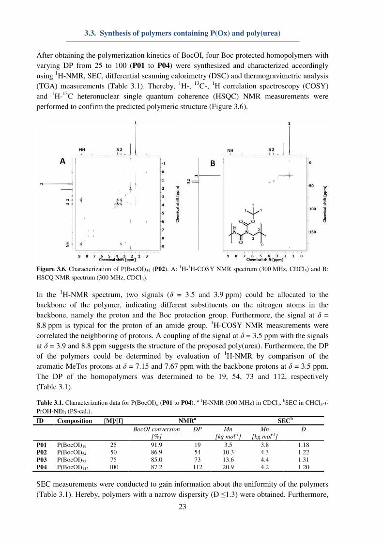
performed to confirm the predicted polymeric structure (Figure 3.6).
Figure 3.6. Characterization of P(BocOI)54 (P02). A:
1H-
1H-COSY NMR spectrum (300 MHz, CDCl3) and B:
HSCQ NMR spectrum (300 MHz, CDCl3).
In the 1H-NMR spectrum, two signals (δ = 3.5 and 3.9 ppm) could be allocated to the
backbone of the polymer, indicating different substituents on the nitrogen atoms in the
backbone, namely the proton and the Boc protection group. Furthermore, the signal at δ =
8.8 ppm is typical for the proton of an amide group. 1H-COSY NMR measurements were
correlated the neighboring of protons. A coupling of the signal at δ = 3.5 ppm with the signals
at δ = 3.9 and 8.8 ppm suggests the structure of the proposed poly(urea). Furthermore, the DP
of the polymers could be determined by evaluation of 1H-NMR by comparison of the
aromatic MeTos protons at δ = 7.15 and 7.67 ppm with the backbone protons at δ = 3.5 ppm.
The DP of the homopolymers was determined to be 19, 54, 73 and 112, respectively
(Table 3.1).
Table 3.1. Characterization data for P(BocOI)n (P01 to P04). a 1H-NMR (300 MHz) in CDCl3.
bSEC in CHCl3-i-
PrOH-NEt3 (PS-cal.).
ID Composition [M]/[I] NMRa
SECb
BocOI conversion
[%]
DP Mn
[kg mol-1
]
Mn
[kg mol-1
]
Ð
P01 P(BocOI)19 25 91.9 19 3.5 3.8 1.18
P02 P(BocOI)54 50 86.9 54 10.3 4.3 1.22
P03 P(BocOI)73 75 85.0 73 13.6 4.4 1.31
P04 P(BocOI)112 100 87.2 112 20.9 4.2 1.20
SEC measurements were conducted to gain information about the uniformity of the polymers
(Table 3.1). Hereby, polymers with a narrow dispersity (Ð ≤1.3) were obtained. Furthermore,
3.3. Synthesis of polymers containing P(Ox) and poly(urea)
24
even though different DPs were calculated using 1H-NMR, die molar masses regarding SEC
measurements did not vary significantly. This might be caused by column interactions of the
polymers, which were not obtained during kinetic studies. A possible explanation for the
different results might be attributed to the differences in the molar mass of the polymers P01
to P04 (min. 3.5 kDa) compared to the kinetic studies (max. 2.7 kDa).
Based on the data of the kinetic investigations, copolymers consisting of BocOI and EtOx are
found to be block-like. This might be advantageous, e.g. in terms of self-assembly properties
(Chapter 4.2), and, consequently, also a series of copolymers consisting of BocOI and EtOx
was synthesized (Table 3.2). Hereby, the DP of the copolymers was kept constant, while the
monomer ratios were varied from 20 to 80% BocOI (Table 3.2). The polymers were
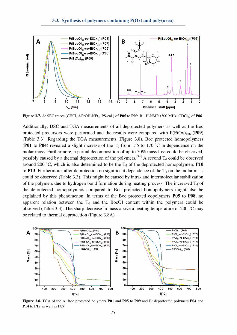
characterized using 1H-NMR and SEC measurements (Figure 3.7).
1H-NMR measurements
showed that all copolymers revealed a similar DP of around 100 and varying BocOI contents.
The dispersity of the polymers was determined to be Ð < 1.4.
Table 3.2. Characterization data for P(BocOIn-co-EtOxm) (P05 to P08) and P(EtOx)116 (P09). a
1H-NMR
(300 MHz) in CDCl3. bSEC in CHCl3-i-PrOH-NEt3 (PS-cal.).
ID Composition NMRa
SECb
DP BocOI
[mol%]
Mn
[kg mol-1
]
Mn
[kg mol-1
]
Ð
P05 P(BocOI16-co-EtOx84) 95 16 11.3 6.1 1.27
P06 P(BocOI36-co-EtOx64) 96 36 13.0 5.3 1.36
P07 P(BocOI52-co-EtOx48) 100 52 14.4 5.9 1.34
P08 P(BocOI84-co-EtOx16) 100 84 17.2 7.4 1.26
P09 P(EtOx)116 116 0 11.5 6.8 1.16
Boc protected urea containing copolymers could be deprotected using trifluoroacetic acid
(TFA, Scheme 3.2), resulting in either poly(urea) homopolymers (Scheme 3.2A) or block like
copolymers from P(EtOx) and P(OI) (Scheme 3.2B). Due to the fact that the deprotected
copolymers were not soluble in any suitable solvent, further analytics, such as mass
spectrometry (MS) or SEC were not possible. However, NMR measurements in deuterated
TFA could be conducted to verify the successful deprotection of the polymers.
Scheme 3.2. Schematic representation of the performed deprotection reactions of A: P(BocOI)n to P(OI)n as well
as B: P(BocOIn-co-EtOxm) to P(OIn-co-EtOxm) utilizing TFA as deprotection agent.
3.3. Synthesis of polymers containing P(Ox) and poly(urea)
25
Figure 3.7. A: SEC traces (CHCl3-i-PrOH-NEt3, PS-cal.) of P05 to P09. B:
1H-NMR (300 MHz, CDCl3) of P06.
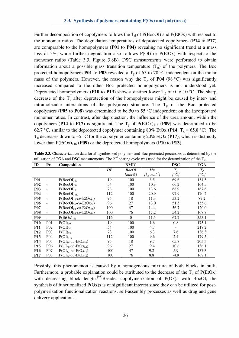
Additionally, DSC and TGA measurements of all deprotected polymers as well as the Boc
protected precursors were performed and the results were compared with P(EtOx)166 (P09)
(Table 3.3). Regarding the TGA measurements (Figure 3.8), Boc protected homopolymers
(P01 to P04) revealed a slight increase of the Td from 155 to 170 °C in dependence on the
molar mass. Furthermore, a partial decomposition of up to 50% mass loss could be observed,
possibly caused by a thermal deprotection of the polymers.[94]
A second Td could be observed
around 200 °C, which is also determined to be the Td of the deprotected homopolymers P10
to P13. Furthermore, after deprotection no significant dependence of the Td on the molar mass
could be observed (Table 3.3). This might be caused by intra- and intermolecular stabilization
of the polymers due to hydrogen bond formation during heating process. The increased Td of
the deprotected homopolymers compared to Boc protected homopolymers might also be
explained by this phenomenon. In terms of the Boc protected copolymers P05 to P08, no
apparent relation between the Td and the BocOI content within the polymers could be
observed (Table 3.3). The sharp decrease in mass above a heating temperature of 200 °C may
be related to thermal deprotection (Figure 3.8A).
Figure 3.8. TGA of the A: Boc protected polymers P01 and P05 to P09 and B: deprotected polymers P04 and
P14 to P17 as well as P09.
3.3. Synthesis of polymers containing P(Ox) and poly(urea)
26
Further decomposition of copolymers follows the Td of P(BocOI) and P(EtOx) with respect to
the monomer ratios. The degradation temperatures of deprotected copolymers (P14 to P17)
are comparable to the homopolymers (P01 to P04) revealing no significant trend at a mass
loss of 5%, while further degradation also follows P(OI) or P(EtOx) with respect to the
monomer ratios (Table 3.3, Figure 3.8B). DSC measurements were performed to obtain
information about a possible glass transition temperature (Tg) of the polymers. The Boc
protected homopolymers P01 to P03 revealed a Tg of 65 to 70 °C independent on the molar
mass of the polymers. However, the reason why the Tg of P04 (98 °C) was significantly
increased compared to the other Boc protected homopolymers is not understood yet.
Deprotected homopolymers (P10 to P13) show a distinct lower Tg of 0 to 10 °C. The sharp
decrease of the Tg after deprotection of the homopolymers might be caused by inter- and
intramolecular interactions of the poly(urea) structure. The Tg of the Boc protected
copolymers (P05 to P08) was determined to be 50 to 55 °C independent on the incorporated
monomer ratios. In contrast, after deprotection, the influence of the urea amount within the
copolymers (P14 to P17) is significant. The Tg of P(EtOx)116 (P09) was determined to be
62.7 °C, similar to the deprotected copolymer containing 80% EtOx (P14, Tg = 65.8 °C). The
Tg decreases down to –5 °C for the copolymer containing 20% EtOx (P17), which is distinctly
lower than P(EtOx)116 (P09) or the deprotected homopolymers (P10 to P13).
Table 3.3. Characterization data for all synthesized polymers and Boc protected precursors as determined by the
utilization of TGA and DSC measurements. The 2nd heating cycle was used for the determination of the Tg.
ID Pre Composition NMRa
DSC TGA
DP BocOI
[mol%]
Mn
[kg mol-1
]
Tg
[°C]
Td
[°C]
P01 - P(BocOI)19 19 100 3.5 69.6 154.3 P02 - P(BocOI)54 54 100 10.3 66.2 164.5 P03 - P(BocOI)73 73 100 13.6 68.9 167.6 P04 - P(BocOI)112 112 100 20.9 97.9 170.2 P05 - P(BocOI16-co-EtOx84) 95 18 11.3 53.2 89.2 P06 - P(BocOI36-co-EtOx64) 96 27 13.0 51.5 155.6 P07 - P(BocOI52-co-EtOx48) 100 47 14.4 56.7 120.0 P08 - P(BocOI84-co-EtOx16) 100 76 17.2 54.2 168.7
P09 - P(EtOx)116 116 0 11.5 62.7 353.1
P10 P01 P(OI)19 19 100 1.6 0.8 175.1 P11 P02 P(OI)54 54 100 4.7 - 218.2 P12 P03 P(OI)73 73 100 6.3 7.6 136.3 P13 P04 P(OI)112 112 100 9.6 2.4 179.5 P14 P05 P(OI16-co-EtOx84) 95 18 9.7 65.8 203.3 P15 P06 P(OI36-co-EtOx64) 96 27 9.4 10.6 136.1 P16 P07 P(OI52-co-EtOx48) 100 47 9.2 5.9 137.3 P17 P08 P(OI84-co-EtOx16) 100 76 8.8 -4.9 168.1
Possibly, this phenomenon is caused by a homogeneous mixture of both blocks in bulk.
Furthermore, a probable explanation could be attributed to the decrease of the Tg of P(EtOx)
with decreasing block length.[95]Besides copolymerization of P(Ox)s with BocOI, the
synthesis of functionalized P(Ox)s is of significant interest since they can be utilized for post-
polymerization functionalization reactions, self-assembly processes as well as drug and gene
delivery applications.
3.3. Synthesis of polymers containing P(Ox) and poly(urea)
27
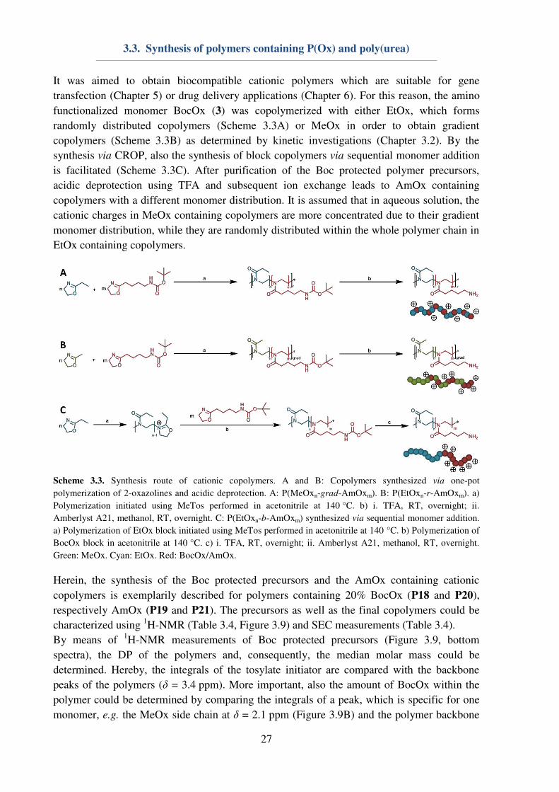
It was aimed to obtain biocompatible cationic polymers which are suitable for gene
transfection (Chapter 5) or drug delivery applications (Chapter 6). For this reason, the amino
functionalized monomer BocOx (3) was copolymerized with either EtOx, which forms
randomly distributed copolymers (Scheme 3.3A) or MeOx in order to obtain gradient
copolymers (Scheme 3.3B) as determined by kinetic investigations (Chapter 3.2). By the
synthesis via CROP, also the synthesis of block copolymers via sequential monomer addition
is facilitated (Scheme 3.3C). After purification of the Boc protected polymer precursors,
acidic deprotection using TFA and subsequent ion exchange leads to AmOx containing
copolymers with a different monomer distribution. It is assumed that in aqueous solution, the
cationic charges in MeOx containing copolymers are more concentrated due to their gradient
monomer distribution, while they are randomly distributed within the whole polymer chain in
EtOx containing copolymers.
Scheme 3.3. Synthesis route of cationic copolymers. A and B: Copolymers synthesized via one-pot
polymerization of 2-oxazolines and acidic deprotection. A: P(MeOxn-grad-AmOxm). B: P(EtOxn-r-AmOxm). a)
Polymerization initiated using MeTos performed in acetonitrile at 140 °C. b) i. TFA, RT, overnight; ii.
Amberlyst A21, methanol, RT, overnight. C: P(EtOxn-b-AmOxm) synthesized via sequential monomer addition.
a) Polymerization of EtOx block initiated using MeTos performed in acetonitrile at 140 °C. b) Polymerization of
BocOx block in acetonitrile at 140 °C. c) i. TFA, RT, overnight; ii. Amberlyst A21, methanol, RT, overnight.
Green: MeOx. Cyan: EtOx. Red: BocOx/AmOx.
Herein, the synthesis of the Boc protected precursors and the AmOx containing cationic
copolymers is exemplarily described for polymers containing 20% BocOx (P18 and P20),
respectively AmOx (P19 and P21). The precursors as well as the final copolymers could be
characterized using 1H-NMR (Table 3.4, Figure 3.9) and SEC measurements (Table 3.4).
By means of 1H-NMR measurements of Boc protected precursors (Figure 3.9, bottom
spectra), the DP of the polymers and, consequently, the median molar mass could be
determined. Hereby, the integrals of the tosylate initiator are compared with the backbone
peaks of the polymers (δ = 3.4 ppm). More important, also the amount of BocOx within the
polymer could be determined by comparing the integrals of a peak, which is specific for one
monomer, e.g. the MeOx side chain at δ = 2.1 ppm (Figure 3.9B) and the polymer backbone
3.3. Synthesis of polymers containing P(Ox) and poly(urea)
28
peak at δ = 3.4 ppm. Furthermore, the success of the deprotection reaction could be verified
by the disappearance of the referring peak at δ = 1.4 ppm (Figure 3.9, top spectra).
Table 3.4. Key properties of the synthesized P(Ox)s determined by either 1H-NMR spectroscopy (300 MHz) in
aCDCl3,
bCD3OD or
cD2O;
dcalculated from Boc protected precursor;
ecalculated from deprotected copolymer.
fSEC in DMAc + 0.21% LiCl, PS-cal.;
gSEC in 0.3% TFA + 0.1 M NaCl, P2VP-cal.;
hSEC (CHCl3-i-PrOH-NEt3
(94:2:4), PS-cal.)
ID Pre Composition NMR SEC
DP mol%
MeOx/
EtOx
mol%
BocOx/
AmOx
Mn
[kg mol-1
]
Mn
[kg mol-1
]
Ð
P18 - P(EtOx150-r-BocOx33) 183a 82
e 18
e 22.8 19.1
f 1.16
f
P19 P18 P(EtOx150-r-AmOx33) 183b,d
82 18 19.6 8.0g 1.41
g
P20 - P(MeOx130-grad-BocOx31) 161a
81e 19
e 18.6 15.2
f 1.33
f
P21 P20 P(MeOx130-grad-AmOx31) 161b,d
81 19 15.5 7.0g 1.48
g
P22 - P(EtOx)98 98
100 - 10.0 5.7h 1.05
h
P23 P222 P(EtOx98-b-BocOx32) 130a 75
a 25
a 17.5
a 8.2
h 1.07
h
P24 P23 P(EtOx98-b-AmOx32) 130c 75
c 25
c 14.2
c 13.8
f 1.11
f
P25 P24 P(EtOx98-b-[AmOx31-stat-
FOx1])
130c 75
c 25
c 15.3
c 14.1
f 1.12
f
SEC measurements were conducted to gain information about the polymer dispersity after
preparation (Table 3.4). The Boc protected polymers exhibited a dispersity of Ð ≤ 1.33, being well-suited for further experiments. Furthermore, measurements on an aqueous SEC provided
information about the stability of the polymers during acidic deprotection. No degradation of
the polymer backbone could be observed.
In addition to random and gradient block copolymers, also the preparation of tailored block
copolymers, suitable for self-assembly and core cross-linking (Chapter 4.2) by CROP is
enabled using the sequential monomer addition technique (Table 3.4). In order to prepare
micelles, a water soluble block copolymer consisting of EtOx and BocOx was synthesized
and deprotected (P24).
Figure 3.9. 1H NMR (300 MHz) spectra of A: P(EtOx150-stat-BocOx33) (P18, CDCl3) and P(EtOx150-stat-
AmOx33) (P19, D2O), as well as B: P(MeOx130-grad-BocOx31) (P20, CDCl3) and P(MeOx130-grad-AmOx31)
(P21), D2O.
3.3. Synthesis of polymers containing P(Ox) and poly(urea)
29
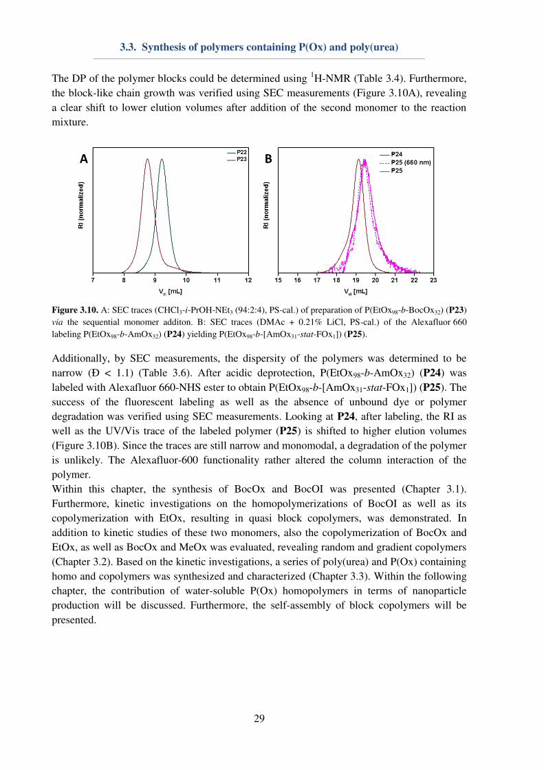
The DP of the polymer blocks could be determined using 1H-NMR (Table 3.4). Furthermore,
the block-like chain growth was verified using SEC measurements (Figure 3.10A), revealing
a clear shift to lower elution volumes after addition of the second monomer to the reaction
mixture.
Figure 3.10. A: SEC traces (CHCl3-i-PrOH-NEt3 (94:2:4), PS-cal.) of preparation of P(EtOx98-b-BocOx32) (P23)
via the sequential monomer additon. B: SEC traces (DMAc + 0.21% LiCl, PS-cal.) of the Alexafluor 660
labeling P(EtOx98-b-AmOx32) (P24) yielding P(EtOx98-b-[AmOx31-stat-FOx1]) (P25).
Additionally, by SEC measurements, the dispersity of the polymers was determined to be
narrow (Ð < 1.1) (Table 3.6). After acidic deprotection, P(EtOx98-b-AmOx32) (P24) was
labeled with Alexafluor 660-NHS ester to obtain P(EtOx98-b-[AmOx31-stat-FOx1]) (P25). The
success of the fluorescent labeling as well as the absence of unbound dye or polymer
degradation was verified using SEC measurements. Looking at P24, after labeling, the RI as
well as the UV/Vis trace of the labeled polymer (P25) is shifted to higher elution volumes
(Figure 3.10B). Since the traces are still narrow and monomodal, a degradation of the polymer
is unlikely. The Alexafluor-600 functionality rather altered the column interaction of the
polymer.
Within this chapter, the synthesis of BocOx and BocOI was presented (Chapter 3.1).
Furthermore, kinetic investigations on the homopolymerizations of BocOI as well as its
copolymerization with EtOx, resulting in quasi block copolymers, was demonstrated. In
addition to kinetic studies of these two monomers, also the copolymerization of BocOx and
EtOx, as well as BocOx and MeOx was evaluated, revealing random and gradient copolymers
(Chapter 3.2). Based on the kinetic investigations, a series of poly(urea) and P(Ox) containing
homo and copolymers was synthesized and characterized (Chapter 3.3). Within the following
chapter, the contribution of water-soluble P(Ox) homopolymers in terms of nanoparticle
production will be discussed. Furthermore, the self-assembly of block copolymers will be
presented.
4. P(Ox) containing nanostructures
30
4. P(Ox) containing nanostructures
Parts of this chapter have been published in: P1) M. N. Leiske, M. Hartlieb, F. H. Sobotta, R.
M. Paulus, H. Görls, P. Bellstedt, U. S. Schubert, Polym. Chem. 2016, 7, 4924-4936. P2) M.
N. Leiske, A.-K. Trützschler, S. Armoneit, P. Sungur, S. Hoeppener, M. Lehmann, A.
Traeger, U. S. Schubert, J. Mater. Chem. B. 2017, 5, 9102-9113. P3) M. N. Leiske, F. H.
Sobotta, F. Richter. S. Hoeppener, J. C. Brendel, A. Traeger, U. S. Schubert,
Biomacromolecules 2018, 19, 748-760. P4) D. Hoelzer‡, M. N. Leiske‡, M. Hartlieb, T. Bus,
D. Pretzel, S. Hoeppener, K. Kempe, R. Thierbach, U. S. Schubert, Oncotarget 2018, in press.
P8) M. Hartlieb‡, T. Bus‡, J. Kübel, D. Pretzel, S. Hoeppener, M. N. Leiske, K. Kempe, B.
Dietzek, U. S. Schubert, Bioconjugate Chem. 2017, 28, 1229-1235. ‡Equal contribution of
both authors.
This chapter is dedicated to the utilization of P(Ox) based homopolymers and copolymers in
the production of colloidal nanostructures. While the first part of the work focuses on the use
of P(Ox) as cryoprotectant and its usage in the stabilization of polymeric nanoparticles, the
second sub chapter describes the self-assembly of P(Ox) block copolymers. Here, amphiphilic
block copolymers were self- and co-assembled in water in order to form mixed micelles.
Furthermore, hydrophilic P(Ox) block copolymers were assembled in chloroform to obtain
nanostructures with a cationic core. These nanostructures were cross-linked to obtain
nanogels.
4.1. P(Ox) mediated nanoparticle stabilization
Nanomedicine represents one promising approach for the curing of various diseases, which
require targeted drug uptake to lower unwanted side-effects.[1] The design and preparation of
potent and safe drug carriers play a pivotal role in pharmaceutical, biomedical and chemical
research, since nanocarriers offer possibilities of cell and organ specificity, e.g. by the
introduction of targeting ligands. Furthermore, a minimization of side-effects can be achieved
by the encapsulation and protection of the active pharmaceutical ingredient (API). Herein,
water-insoluble polyesters, i.e. the Food and Drug Administration (FDA) approved
poly(lactic-co-glycolic acid) (PLGA), are already used in numerous preclinical trials.[96]
However, major obstacles regarding the utilization of nanoparticles are caused by their
preparation,[97-99] purification or storage, respectively lyophilization.[100] In particular, the
encapsulation of hydrophilic drugs is problematic, since it requires the emulsification method
for nanoparticle preparation, and, consequently, the utilization of emulsifiers or
surfactants.[101-102] Poly(vinyl alcohol) (Mowiol 8-88, PVA) and Pluronic F127 represent two
important macromolecules, which are commonly used for nanoparticle stabilization during
preparation,[101-102] while lyophilization is usually conducted using glucose, saccharose or
trehalose as a cryo-protectant.[100-101, 103] In order to reduce the amount of substances being
used for nanoparticle, an all-in-one system being suitable for the stabilization of hydrophobic
nanoparticles during preparation, purification and lyophilization is envisioned.
4.1. P(Ox) mediated nanoparticle stabilization
31
The utilization of P(EtOx) or P(MeOx) could be beneficial, in particular in terms of elongated
blood circulation times of nanoparticles, assuming that the surfactant is (partially)
incorporated into the polymer shell during emulsification. The possibility to dissolve P(EtOx)
and P(MeOx) in water as well as in various organic solvents[27] makes them ideal candidates
for investigations on their properties regarding nanoparticle stabilization. For this reason, a
small library of water-soluble homopolymers (Table 4.1) consisting of either P(EtOx)n or
P(MeOx)n was synthesized accordingly to literature procedures.[48] The polymers were
characterized utilizing 1H-NMR to obtain information about the DP, which was determined to
be 25, 60, 100 or 200 in case of both utilized monomers. Furthermore, SEC measurements
were conducted to gain information about the polymer dispersity. With exception of P35 (Ɖ = 1.38), all polymers revealed a narrow dispersity (Ɖ ≤ 1.2).
Table 4.1. Key properties of the synthesized P(Ox)s determined by a 1H-NMR (300 MHz, CDCl3) and bSEC
(DMAc, 0.21% LiCl, PS-cal.). cSEC (CHCl3-i-PrOH-NEt3 (94:2:4), PS-cal.).
ID Pre Composition NMRa
SECb
DP Mn
[kg mol-1
]
Mn
[kg mol-1
]
Ð
P26 - P(EtOx)25 25 2.5 6.3 1.09 P27 - P(EtOx)61 61 6.0 12.2
7.0c 1.09 1.15c
P28 - P(EtOx)107 107 10.7 19.1 1.16 P29 - P(EtOx)184 184 18.4 25.3 1.21 P30 P27 P(EtOx)61-NH2 61 6.0 3.5c 1.14c
P31 P30 P(EtOx)61-Rhodamine B 61 6.0 4.2c 1.13c
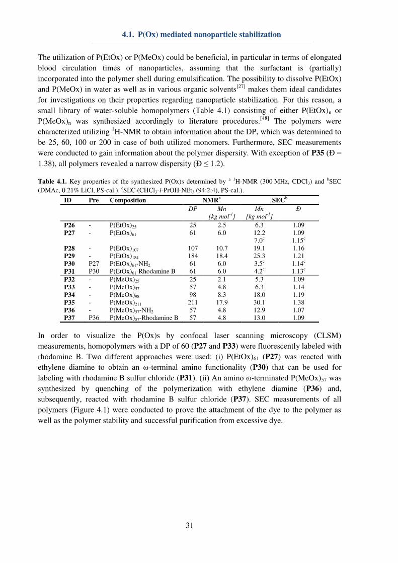
P32 - P(MeOx)25 25 2.1 5.3 1.09 P33 - P(MeOx)57 57 4.8 6.3 1.14 P34 - P(MeOx)98 98 8.3 18.0 1.19 P35 - P(MeOx)211 211 17.9 30.1 1.38 P36 - P(MeOx)57-NH2 57 4.8 12.9 1.07 P37 P36 P(MeOx)57-Rhodamine B 57 4.8 13.0 1.09
In order to visualize the P(Ox)s by confocal laser scanning microscopy (CLSM)
measurements, homopolymers with a DP of 60 (P27 and P33) were fluorescently labeled with
rhodamine B. Two different approaches were used: (i) P(EtOx)61 (P27) was reacted with
ethylene diamine to obtain an ω-terminal amino functionality (P30) that can be used for
labeling with rhodamine B sulfur chloride (P31). (ii) An amino ω-terminated P(MeOx)57 was
synthesized by quenching of the polymerization with ethylene diamine (P36) and,
subsequently, reacted with rhodamine B sulfur chloride (P37). SEC measurements of all
polymers (Figure 4.1) were conducted to prove the attachment of the dye to the polymer as
well as the polymer stability and successful purification from excessive dye.
4.1. P(Ox) mediated nanoparticle stabilization
32
Figure 4.1. A: SEC traces (CHCl3-i-PrOH-NEt3 (94:2:4), PS-cal.) of the rhodamine B labeling of P(EtOx)61
(P27). B: SEC traces (DMAc + 0.21% LiCl, PS-cal.) of the rhodamine B labeling of P(MeOx)57 (P36).
For this purpose, the absorption of the labeled polymers at λ = 540 to 550 nm, which is
corresponding to the rhodamine B label, was measured in addition to the RI. The traces of
P36 and P37 perfectly overlap (Figure 4.1B), indicating successful polymer labeling. Looking
at P(EtOx)61 (P27), after labeling, the RI as well as the UV/Vis trace of the labeled polymer
(P30) is shifted to higher elution volumes (Figure 4.1A). Since the traces are still narrow and
monomodal, a degradation of the polymer is improbable. More likely, the rhodamine B ω-
functionality altered the column interaction of the polymer on the chloroform based SEC. The
labeling efficiencies of the polymers were not determined, as it was not of importance for the
planned application as nanoparticle stabilizers. For this reason, both labeling strategies were
defined to be of equal success.
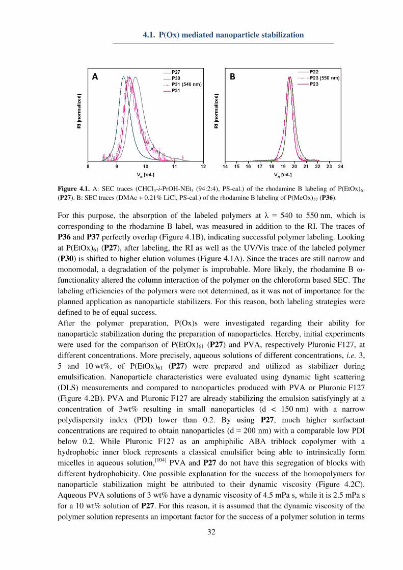
After the polymer preparation, P(Ox)s were investigated regarding their ability for
nanoparticle stabilization during the preparation of nanoparticles. Hereby, initial experiments
were used for the comparison of P(EtOx)61 (P27) and PVA, respectively Pluronic F127, at
different concentrations. More precisely, aqueous solutions of different concentrations, i.e. 3,
5 and 10 wt%, of P(EtOx)61 (P27) were prepared and utilized as stabilizer during
emulsification. Nanoparticle characteristics were evaluated using dynamic light scattering
(DLS) measurements and compared to nanoparticles produced with PVA or Pluronic F127
(Figure 4.2B). PVA and Pluronic F127 are already stabilizing the emulsion satisfyingly at a
concentration of 3wt% resulting in small nanoparticles (d < 150 nm) with a narrow
polydispersity index (PDI) lower than 0.2. By using P27, much higher surfactant
concentrations are required to obtain nanoparticles (d ≈ 200 nm) with a comparable low PDI below 0.2. While Pluronic F127 as an amphiphilic ABA triblock copolymer with a
hydrophobic inner block represents a classical emulsifier being able to intrinsically form
micelles in aqueous solution,[104]
PVA and P27 do not have this segregation of blocks with
different hydrophobicity. One possible explanation for the success of the homopolymers for
nanoparticle stabilization might be attributed to their dynamic viscosity (Figure 4.2C).
Aqueous PVA solutions of 3 wt% have a dynamic viscosity of 4.5 mPa s, while it is 2.5 mPa s
for a 10 wt% solution of P27. For this reason, it is assumed that the dynamic viscosity of the
polymer solution represents an important factor for the success of a polymer solution in terms

4.1. P(Ox) mediated nanoparticle stabilization
33
of nanoparticle stabilization. Another important factor might be the increased solubility of
P27 in water and organic solvents. This phenomenon is also known from PVA, which
represents a very good emulsifier. With respect to PVA, the stabilization of the interphase
between water and the organic droplet is achieved by a different solution behavior of the
hydrophobic polymer backbone and the hydrophilic hydroxyl groups within the polymer side-
chains.[105]
In the case of P(Ox)s similar characteristics are determined, leading to an
enhanced stabilization of the interphase and, consequently, a good stabilization of the
emulsion (Figure 4.2A).
Figure 4.2. A: Schematic representation of the nanoparticle preparation via the nanoemulsion technique. A
hydrophobic drug and the polymer are dissolved in a not water miscible organic solvent and water is added.
Surfactants are added and the solution is emulsified by sonication. After evaporation of the organic solvent,
nanoparticles are obtained. Magnification of the nanoparticle-aqueous phase boundary layer is presented,
showing the potential behavior of polymer surfactants in the nanoemulsion process. B: Properties of PLGA
nanoparticles prepared via the nanoemulsion technique (water and ethyl acetate), using different surfactants as
determined by DLS measurements (n = 3, 5 measurements each). Hashes represent the position of non-
investigated concentrations. C: Dynamic viscosity of aqueous surfactant solutions in dependence in the polymer
concentrations.
After these prelimary investigations, further experiments concentrating on differences in the
side-chain hydrophobicity and the DP of the utilized polymers were conducted. Hereby,
P(EtOx)n (P26 to P29) and P(MeOx)n (P32 to P35) were used in a concentration of 10 wt%
within the aqueous layer during emulsification. The resulting nanoparticles were characterized
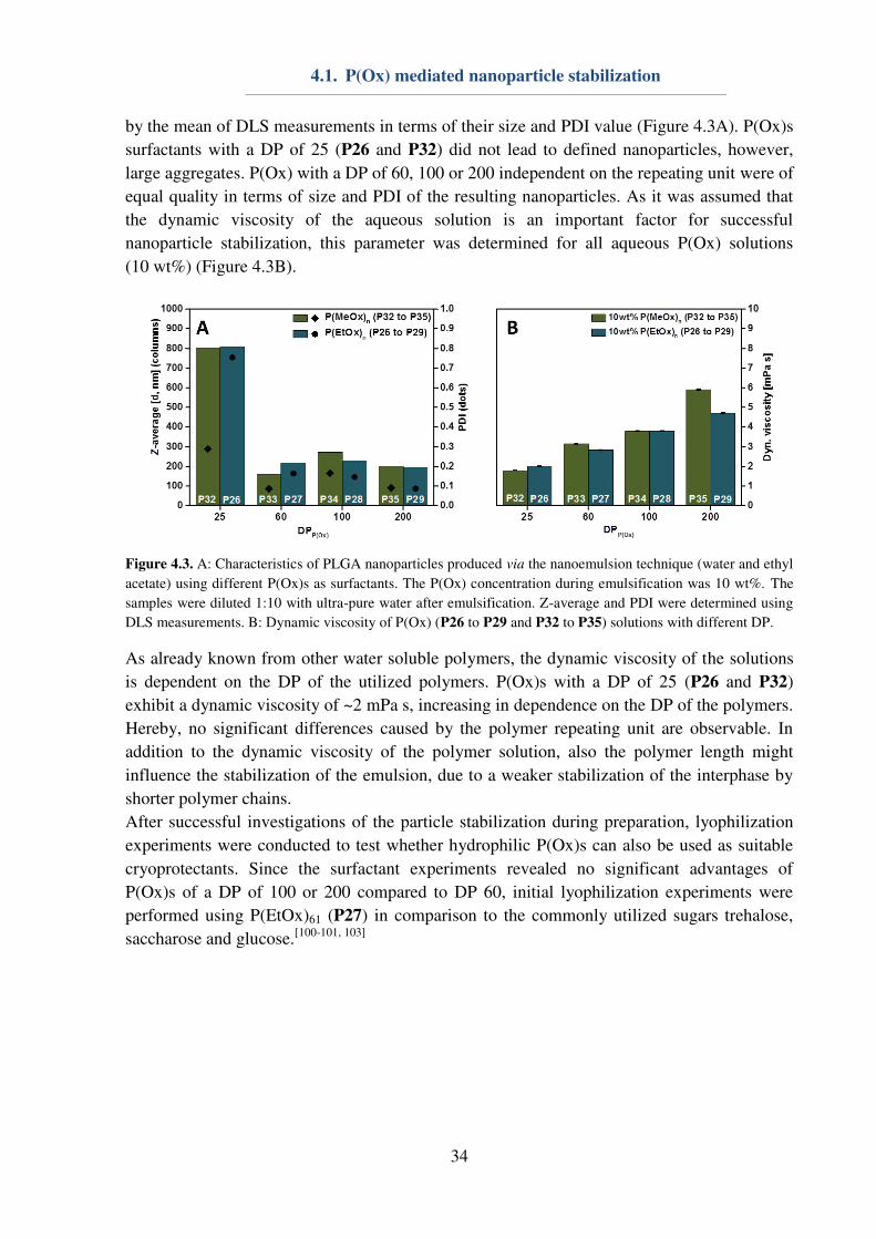
4.1. P(Ox) mediated nanoparticle stabilization
34
by the mean of DLS measurements in terms of their size and PDI value (Figure 4.3A). P(Ox)s
surfactants with a DP of 25 (P26 and P32) did not lead to defined nanoparticles, however,
large aggregates. P(Ox) with a DP of 60, 100 or 200 independent on the repeating unit were of
equal quality in terms of size and PDI of the resulting nanoparticles. As it was assumed that
the dynamic viscosity of the aqueous solution is an important factor for successful
nanoparticle stabilization, this parameter was determined for all aqueous P(Ox) solutions
(10 wt%) (Figure 4.3B).
Figure 4.3. A: Characteristics of PLGA nanoparticles produced via the nanoemulsion technique (water and ethyl
acetate) using different P(Ox)s as surfactants. The P(Ox) concentration during emulsification was 10 wt%. The
samples were diluted 1:10 with ultra-pure water after emulsification. Z-average and PDI were determined using
DLS measurements. B: Dynamic viscosity of P(Ox) (P26 to P29 and P32 to P35) solutions with different DP.
As already known from other water soluble polymers, the dynamic viscosity of the solutions
is dependent on the DP of the utilized polymers. P(Ox)s with a DP of 25 (P26 and P32)
exhibit a dynamic viscosity of ~2 mPa s, increasing in dependence on the DP of the polymers.
Hereby, no significant differences caused by the polymer repeating unit are observable. In
addition to the dynamic viscosity of the polymer solution, also the polymer length might
influence the stabilization of the emulsion, due to a weaker stabilization of the interphase by
shorter polymer chains.
After successful investigations of the particle stabilization during preparation, lyophilization
experiments were conducted to test whether hydrophilic P(Ox)s can also be used as suitable
cryoprotectants. Since the surfactant experiments revealed no significant advantages of
P(Ox)s of a DP of 100 or 200 compared to DP 60, initial lyophilization experiments were
performed using P(EtOx)61 (P27) in comparison to the commonly utilized sugars trehalose,
saccharose and glucose.[100-101, 103]
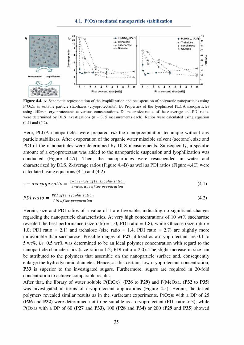
4.1. P(Ox) mediated nanoparticle stabilization
35
Figure 4.4. A: Schematic representation of the lyophilization and resuspension of polymeric nanoparticles using
P(Ox)s as suitable particle stabilizers (cryoprotectants). B: Properties of the lyophilized PLGA nanoparticles
using different cryoprotectants at various concentrations. Diameter size ratios of the z-average and PDI ratios
were determined by DLS investigations (n = 3, 5 measurements each). Ratios were calculated using equation
(4.1) and (4.2).
Here, PLGA nanoparticles were prepared via the nanoprecipitation technique without any
particle stabilizers. After evaporation of the organic water miscible solvent (acetone), size and
PDI of the nanoparticles were determined by DLS measurements. Subsequently, a specific
amount of a cryoprotectant was added to the nanoparticle suspension and lyophilization was
conducted (Figure 4.4A). Then, the nanoparticles were resuspended in water and
characterized by DLS. Z-average ratios (Figure 4.4B) as well as PDI ratios (Figure 4.4C) were
calculated using equations (4.1) and (4.2). � − �� �� ����� = −�� �� � � � � ℎ��� ���−�� �� � � � � ����� (4.1)
��� ����� = ��� � � � � ℎ��� ������ � � � � ����� (4.2)
Herein, size and PDI ratios of a value of 1 are favorable, indicating no significant changes
regarding the nanoparticle characteristics. At very high concentrations of 10 wt% saccharose
revealed the best performance (size ratio = 1.0; PDI ratio = 1.8), while Glucose (size ratio =
1.0; PDI ratio = 2.1) and trehalose (size ratio = 1.4, PDI ratio = 2.7) are slightly more
unfavorable than saccharose. Possible ranges of P27 utilized as a cryoprotectant are 0.1 to
5 wt%, i.e. 0.5 wt% was determined to be an ideal polymer concentration with regard to the
nanoparticle characteristics (size ratio = 1.2; PDI ratio = 2.0). The slight increase in size can
be attributed to the polymers that assemble on the nanoparticle surface and, consequently
enlarge the hydrodynamic diameter. Hence, at this certain, low cryoprotectant concentration,
P33 is superior to the investigated sugars. Furthermore, sugars are required in 20-fold
concentration to achieve comparable results.
After that, the library of water soluble P(EtOx)n (P26 to P29) and P(MeOx)n (P32 to P35)
was investigated in terms of cryoprotectant applications (Figure 4.5). Herein, the tested
polymers revealed similar results as in the surfactant experiments. P(Ox)s with a DP of 25
(P26 and P32) were determined not to be suitable as a cryoprotectant (PDI ratio > 3), while
P(Ox)s with a DP of 60 (P27 and P33), 100 (P28 and P34) or 200 (P29 and P35) showed
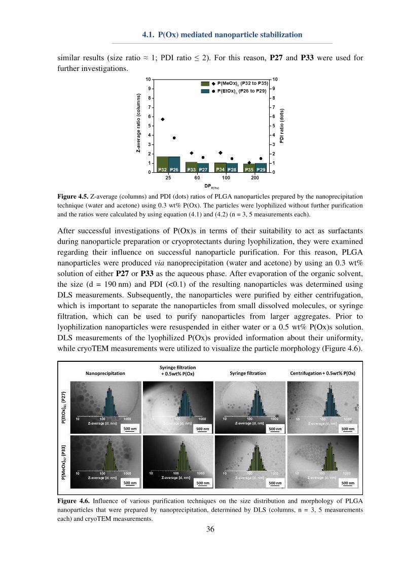
4.1. P(Ox) mediated nanoparticle stabilization
36
similar results (size ratio ≈ 1; PDI ratio ≤ 2). For this reason, P27 and P33 were used for
further investigations.
Figure 4.5. Z-average (columns) and PDI (dots) ratios of PLGA nanoparticles prepared by the nanoprecipitation
technique (water and acetone) using 0.3 wt% P(Ox). The particles were lyophilized without further purification
and the ratios were calculated by using equation (4.1) and (4.2) (n = 3, 5 measurements each).
After successful investigations of P(Ox)s in terms of their suitability to act as surfactants
during nanoparticle preparation or cryoprotectants during lyophilization, they were examined
regarding their influence on successful nanoparticle purification. For this reason, PLGA
nanoparticles were produced via nanoprecipitation (water and acetone) by using an 0.3 wt%
solution of either P27 or P33 as the aqueous phase. After evaporation of the organic solvent,
the size (d = 190 nm) and PDI (<0.1) of the resulting nanoparticles was determined using
DLS measurements. Subsequently, the nanoparticles were purified by either centrifugation,
which is important to separate the nanoparticles from small dissolved molecules, or syringe
filtration, which can be used to purify nanoparticles from larger aggregates. Prior to
lyophilization nanoparticles were resuspended in either water or a 0.5 wt% P(Ox)s solution.
DLS measurements of the lyophilized P(Ox)s provided information about their uniformity,
while cryoTEM measurements were utilized to visualize the particle morphology (Figure 4.6).
Figure 4.6. Influence of various purification techniques on the size distribution and morphology of PLGA
nanoparticles that were prepared by nanoprecipitation, determined by DLS (columns, n = 3, 5 measurements
each) and cryoTEM measurements.
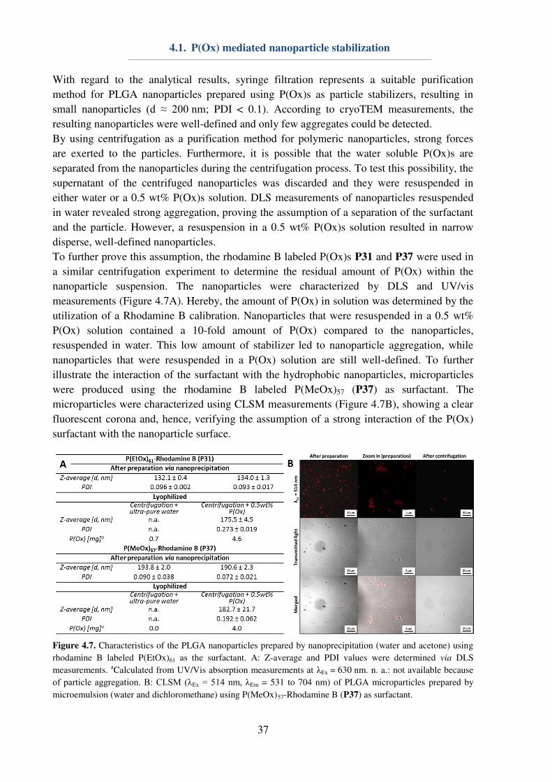
4.1. P(Ox) mediated nanoparticle stabilization
37
With regard to the analytical results, syringe filtration represents a suitable purification
method for PLGA nanoparticles prepared using P(Ox)s as particle stabilizers, resulting in
small nanoparticles (d ≈ 200 nm; PDI < 0.1). According to cryoTEM measurements, the
resulting nanoparticles were well-defined and only few aggregates could be detected.
By using centrifugation as a purification method for polymeric nanoparticles, strong forces
are exerted to the particles. Furthermore, it is possible that the water soluble P(Ox)s are
separated from the nanoparticles during the centrifugation process. To test this possibility, the
supernatant of the centrifuged nanoparticles was discarded and they were resuspended in
either water or a 0.5 wt% P(Ox)s solution. DLS measurements of nanoparticles resuspended
in water revealed strong aggregation, proving the assumption of a separation of the surfactant
and the particle. However, a resuspension in a 0.5 wt% P(Ox)s solution resulted in narrow
disperse, well-defined nanoparticles.
To further prove this assumption, the rhodamine B labeled P(Ox)s P31 and P37 were used in
a similar centrifugation experiment to determine the residual amount of P(Ox) within the
nanoparticle suspension. The nanoparticles were characterized by DLS and UV/vis
measurements (Figure 4.7A). Hereby, the amount of P(Ox) in solution was determined by the
utilization of a Rhodamine B calibration. Nanoparticles that were resuspended in a 0.5 wt%
P(Ox) solution contained a 10-fold amount of P(Ox) compared to the nanoparticles,
resuspended in water. This low amount of stabilizer led to nanoparticle aggregation, while
nanoparticles that were resuspended in a P(Ox) solution are still well-defined. To further
illustrate the interaction of the surfactant with the hydrophobic nanoparticles, microparticles
were produced using the rhodamine B labeled P(MeOx)57 (P37) as surfactant. The
microparticles were characterized using CLSM measurements (Figure 4.7B), showing a clear
fluorescent corona and, hence, verifying the assumption of a strong interaction of the P(Ox)
surfactant with the nanoparticle surface.
Figure 4.7. Characteristics of the PLGA nanoparticles prepared by nanoprecipitation (water and acetone) using
rhodamine B labeled P(EtOx)61 as the surfactant. A: Z-average and PDI values were determined via DLS
measurements. aCalculated from UV/Vis absorption measurements at λEx = 630 nm. n. a.: not available because
of particle aggregation. B: CLSM (λEx = 514 nm, λEm = 531 to 704 nm) of PLGA microparticles prepared by
microemulsion (water and dichloromethane) using P(MeOx)57-Rhodamine B (P37) as surfactant.

4.1. P(Ox) mediated nanoparticle stabilization
38
Within this study, it could be shown that water soluble P(Ox)s, i.e. P(EtOx) and P(MeOx) are
utilizable for the stabilization of hydrophobic PLGA nanoparticles. Furthermore, the
stabilization of poly(methacrylate) based nanoparticles could be demonstrated (data not
shown). Hereby, a minimum DP of 60 was necessary to obtain reproducible and satisfying
results during preparation, purification and lyophilization. Hence, an all in one system usable
for further applications could be invented.
4.2. Self-assembly of P(Ox) block copolymers
In addition to P(Ox) mediated nanoparticle stabilization, the self-assembly of different block
copolymers was investigated. Hence, this subchapter is dedicated to the preparation of
polymer colloids consisting of different block copolymers. The resulting nanostructures can
be utilized to facilitate the transport of genetic material (Chapter 5) or APIs (Chapter 6).
Firstly, poly(urea) containing quasi block copolymers were assembled in water. Herein,
polymers with 28 wt% poly(urea) were found to form very uniform nanostructures
accordingly to DLS measurements (d = 297 ±8 nm; PDI = 0.020 ± 0.015; Figure 4.8A). Cryo
transmission electron-microscopy (cryoTEM) measurements were conducted to confirm the
results obtained by DLS (Figure 4.8A), revealing brush-like spherical structures
(Figure 4.8B). It is assumable that the investigated polymers form strong hydrogen bonds in
water and, for that reason, self-assemble. However, due to lack of accessible functionalities,
the prepared nanostructures could not be used for the complexation of genetic material or the
conjugation of an API and were, consequently, not further elucidated within this thesis.
Figure 4.8. Self-assembly of quasi block copolymers consisting of poly(urea) and (PEtOx) (P(OI46-co-EtOx117)).
A: cryoTEM image. B: Schematic representation of hydrogen bond stabilized nanostructures.
Furthermore, cationic P(Ox) nanostructures were prepared by self- and co-assembly of AmOx
and EtOx containing amphiphilic block copolymers. The utilization of these nanostructures as
polymeric vectors for the transfection of cells will be further discussed in Chapter 5. For the
successful complexation of genetic material and polymers, cationic charges are indispensable.
However, they are also known to force membrane disruption.[106]
In fact, a shielding of the
cationic charges, e.g. by EtOx, can reduce membrane disruption by shielding of the cationic
charges.[107]
For this reason, pH responsive system, in which the cationic charges are shielded
at a physiological pH value of 7.4, while they stretch after protonation at lower
endolysosomal pH values of 5 to trigger the endosomal release, was aimed. In order to access

4.2. Self-assembly of P(Ox) block copolymers
39
functional P(Ox) based nanostructures, two different block copolymers with NonOx
representing the hydrophobic block were synthesized. Herein, the hydrophilic block of the
macromolecules consisted of either AmOx (P39), which allows polyplex formation of the
resulting nanostructure, or EtOx (P38) to mediate stealth effect and enhanced
cytocompatibility. Block copolymers were prepared and deprotected accordingly to
copolymer synthesis described in Chapter 3.3. The polymers were characterized using 1H-
NMR for determination of the block ratios as well as using SEC to gain insight about the
dispersity (Table 4.2). In both polymers, the DP of NonOx was kept similar, leading to
different weight ratios of the hydrophobic and the hydrophilic block. P(EtOx155-b-NonOx76
(P38) consisted 55 wt% NonOx, leading to a favorable assembly into worm-like structures,[60]
while the cationic block copolymer P(NonOx52-b-AmOx184 (P39) contained 28 wt% NonOx,
favorably leading to spherical micelles during the assembly process.
Table 4.2. Key properties of the synthesized polymers. aSEC (eluent: DMAc, 0.21% LiCl; PS-standard); b 1H-
NMR (300 MHz). cCalculated from Boc-protected precursor polymer). eSEC (eluent: 0.1 M NaCl(aq) + 0.3%
TFA; P2VP-standard).
ID Composition NMR SEC
DP Wt%
(EtOx/AmOx)
Wt%
(NonOx)
Mn
[kg mol-1
]
Mn
[kg mol-1
]
Ð
P38 P(EtOx155-b-NonOx76) 231 45 55 30.4 38.3 1.14 P39 P(NonOx52-b-AmOx184) 236c 72c 28c 36.4 24.3 1.26
In order to obtain polymeric micelles which express high transfection efficiencies in
combination with an enhanced cytocompatibility, mixed micelles were prepared. For the
preparation of the nanostructures, the polymers were mixed in different ratios prior to
assembly to obtain nanostructures with 0, 20, 40, 60, 80 or 100% of the cationic polymer
(P39) within the shell. Final nanostructures were prepared in 0.9 wt% NaCl representing
physiological salt concentrations.
Preliminary characterization of the nanostructures was conducted by means of DLS
measurements to obtain information about the size and uniformity of the micelles
(Figure 4.9A). Interestingly, all nanostructures containing at least 20 wt% P38 are
significantly larger (d = 80 to 100 nm) than the nanostructures consisting of 100 wt% P39 (d
= 60 nm). Furthermore, the PDI of the micelles was also shown to be dependent on the ratio
of polymers used for preparation, slightly increasing from 0.2 to 0.3 with increasing amount
of cationic polymers.
Additionally, cryoTEM measurements were performed to obtain information about the shape
of the produced nanostructures (Figure 4.9B). Micelles that did not contain cationic
copolymer were determined to be rod- or sheet-like, whereas darker rods are presumably
sheets with a parallel orientation with respect to the electron beam. Previously, rod-like
structures from ABA triblock copolymers with NonOx representing the hydrophobic inner
and MeOx as the hydrophilic outer blocks have been reported.[60]
Nanostructures consisting of the cationic block copolymer (P39) were found to be spherical.
More interestingly, all mixed micelles showed a rod-like shape regarding the cryoTEM
measurements. For this reason, it is quite likely that the prepared nanostructures are mixed
micelles instead of two different species present. Furthermore, the shape was determined to by
dependent on the polymer ratios within the nanostructures as shown in Figure 4.9C.
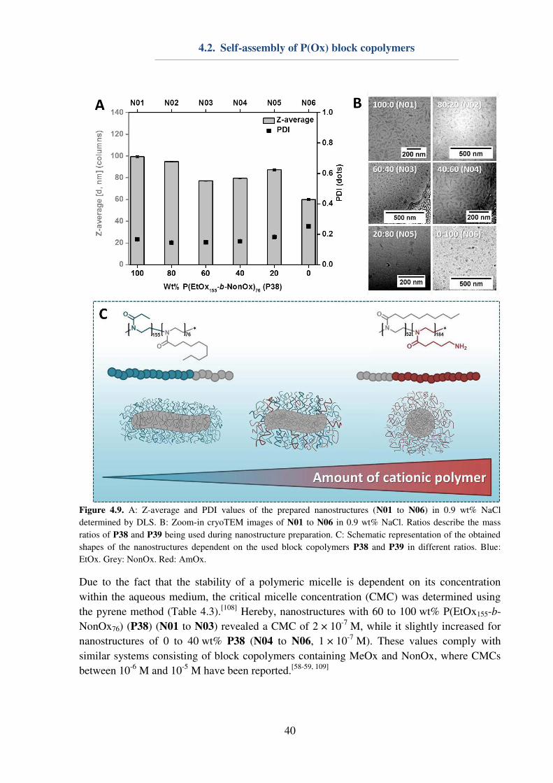
4.2. Self-assembly of P(Ox) block copolymers
40
Figure 4.9. A: Z-average and PDI values of the prepared nanostructures (N01 to N06) in 0.9 wt% NaCl
determined by DLS. B: Zoom-in cryoTEM images of N01 to N06 in 0.9 wt% NaCl. Ratios describe the mass
ratios of P38 and P39 being used during nanostructure preparation. C: Schematic representation of the obtained
shapes of the nanostructures dependent on the used block copolymers P38 and P39 in different ratios. Blue:
EtOx. Grey: NonOx. Red: AmOx.
Due to the fact that the stability of a polymeric micelle is dependent on its concentration
within the aqueous medium, the critical micelle concentration (CMC) was determined using
the pyrene method (Table 4.3).[108]
Hereby, nanostructures with 60 to 100 wt% P(EtOx155-b-
NonOx76) (P38) (N01 to N03) revealed a CMC of 2 × 10⁻⁷
M, while it slightly increased for
nanostructures of 0 to 40 wt% P38 (N04 to N06, 1 × 10⁻⁷
M). These values comply with
similar systems consisting of block copolymers containing MeOx and NonOx, where CMCs
between 10-6
M and 10-5
M have been reported.[58-59, 109]
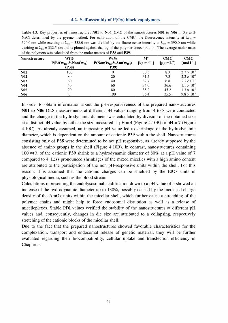
4.2. Self-assembly of P(Ox) block copolymers
41
Table 4.3. Key properties of nanostructures N01 to N06. CMC of the nanostructures N01 to N06 in 0.9 wt%
NaCl determined by the pyrene method. For calibration of the CMC, the fluorescence intensity at λEm =
390.0 nm while exciting at λEx = 338.0 nm was divided by the fluorescence intensity at λEm = 390.0 nm while
exciting at λEx = 332.5 nm and is plotted against the log of the polymer concentration. aThe average molar mass
of the polymers was calculated from the molar masses of P38 and P39.
Nanostructure Wt%
P(EtOx155-b-NonOx76)
(P38)
Wt%
P(NonOx52-b-AmOx184)
(P39)
Ma
[kg mol-1
]
CMC
[µg mL-1
]
CMC
[mol L-1
]
N01 100 0 30.3 8.3 2.7 × 10⁻⁷ N02 80 20 31.5 7.3 2.3 × 10⁻⁷ N03 60 40 32.7 6.8 2.2× 10⁻⁷ N04 40 60 34.0 36.6 1.1 × 10⁻⁶ N05 20 80 35.2 45.2 1.3 × 10⁻⁶ N06 0 100 36.4 35.5 9.8 × 10⁻⁷
In order to obtain information about the pH-responsiveness of the prepared nanostructures
N01 to N06 DLS measurements at different pH values ranging from 4 to 8 were conducted
and the change in the hydrodynamic diameter was calculated by division of the obtained size
at a distinct pH value by either the size measured at pH = 4 (Figure 4.10B) or pH = 7 (Figure
4.10C). As already assumed, an increasing pH value led to shrinkage of the hydrodynamic
diameter, which is dependent on the amount of cationic P39 within the shell. Nanostructures
consisting only of P38 were determined to be not pH responsive, as already supposed by the
absence of amino groups in the shell (Figure 4.10B). In contrast, nanostructures containing
100 wt% of the cationic P39 shrink to a hydrodynamic diameter of 80% at a pH value of 7
compared to 4. Less pronounced shrinkages of the mixed micelles with a high amino content
are attributed to the participation of the non pH-responsive units within the shell. For this
reason, it is assumed that the cationic charges can be shielded by the EtOx units in
physiological media, such as the blood stream.
Calculations representing the endolysosomal acidification down to a pH value of 5 showed an
increase of the hydrodynamic diameter up to 130%, possibly caused by the increased charge
density of the AmOx units within the micellar shell, which further cause a stretching of the
polymer chains and might help to force endosomal disruption as well as a release of
micelleplexes. Stable PDI values verified the stability of the nanostructures at different pH
values and, consequently, changes in die size are attributed to a collapsing, respectively
stretching of the cationic blocks of the micellar shell.
Due to the fact that the prepared nanostructures showed favorable characteristics for the
complexation, transport and endosomal release of genetic material, they will be further
evaluated regarding their biocompatibility, cellular uptake and transfection efficiency in
Chapter 5.
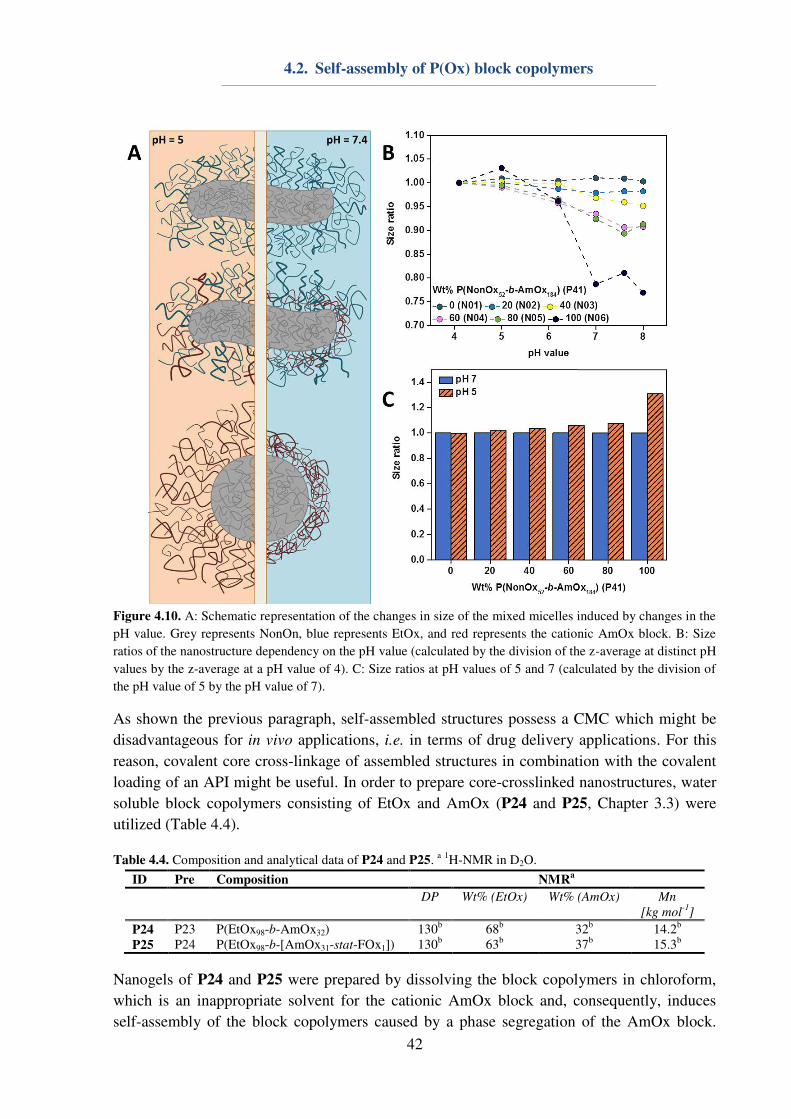
4.2. Self-assembly of P(Ox) block copolymers
42
Figure 4.10. A: Schematic representation of the changes in size of the mixed micelles induced by changes in the
pH value. Grey represents NonOn, blue represents EtOx, and red represents the cationic AmOx block. B: Size
ratios of the nanostructure dependency on the pH value (calculated by the division of the z-average at distinct pH
values by the z-average at a pH value of 4). C: Size ratios at pH values of 5 and 7 (calculated by the division of
the pH value of 5 by the pH value of 7).
As shown the previous paragraph, self-assembled structures possess a CMC which might be
disadvantageous for in vivo applications, i.e. in terms of drug delivery applications. For this
reason, covalent core cross-linkage of assembled structures in combination with the covalent
loading of an API might be useful. In order to prepare core-crosslinked nanostructures, water
soluble block copolymers consisting of EtOx and AmOx (P24 and P25, Chapter 3.3) were
utilized (Table 4.4).
Table 4.4. Composition and analytical data of P24 and P25. a 1H-NMR in D2O.
ID Pre Composition NMRa
DP Wt% (EtOx) Wt% (AmOx) Mn
[kg mol-1
]
P24 P23 P(EtOx98-b-AmOx32) 130b
68b
32b
14.2b
P25 P24 P(EtOx98-b-[AmOx31-stat-FOx1]) 130b
63b
37b
15.3b
Nanogels of P24 and P25 were prepared by dissolving the block copolymers in chloroform,
which is an inappropriate solvent for the cationic AmOx block and, consequently, induces
self-assembly of the block copolymers caused by a phase segregation of the AmOx block.
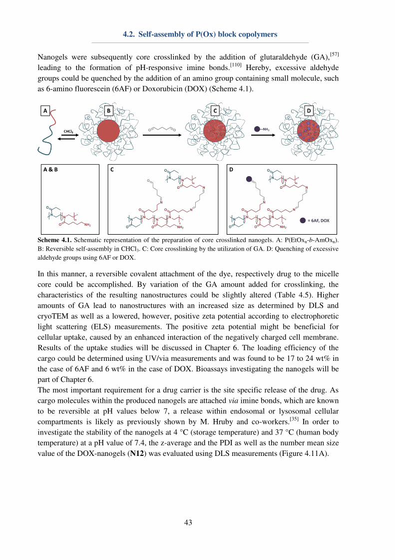
4.2. Self-assembly of P(Ox) block copolymers
43
Nanogels were subsequently core crosslinked by the addition of glutaraldehyde (GA),[57]
leading to the formation of pH-responsive imine bonds.[110]
Hereby, excessive aldehyde
groups could be quenched by the addition of an amino group containing small molecule, such
as 6-amino fluorescein (6AF) or Doxorubicin (DOX) (Scheme 4.1).
Scheme 4.1. Schematic representation of the preparation of core crosslinked nanogels. A: P(EtOxn-b-AmOxn).
B: Reversible self-assembly in CHCl3. C: Core crosslinking by the utilization of GA. D: Quenching of excessive
aldehyde groups using 6AF or DOX.
In this manner, a reversible covalent attachment of the dye, respectively drug to the micelle
core could be accomplished. By variation of the GA amount added for crosslinking, the
characteristics of the resulting nanostructures could be slightly altered (Table 4.5). Higher
amounts of GA lead to nanostructures with an increased size as determined by DLS and
cryoTEM as well as a lowered, however, positive zeta potential according to electrophoretic
light scattering (ELS) measurements. The positive zeta potential might be beneficial for
cellular uptake, caused by an enhanced interaction of the negatively charged cell membrane.
Results of the uptake studies will be discussed in Chapter 6. The loading efficiency of the
cargo could be determined using UV/via measurements and was found to be 17 to 24 wt% in
the case of 6AF and 6 wt% in the case of DOX. Bioassays investigating the nanogels will be
part of Chapter 6.
The most important requirement for a drug carrier is the site specific release of the drug. As
cargo molecules within the produced nanogels are attached via imine bonds, which are known
to be reversible at pH values below 7, a release within endosomal or lysosomal cellular
compartments is likely as previously shown by M. Hruby and co-workers.[35]
In order to
investigate the stability of the nanogels at 4 °C (storage temperature) and 37 °C (human body
temperature) at a pH value of 7.4, the z-average and the PDI as well as the number mean size
value of the DOX-nanogels (N12) was evaluated using DLS measurements (Figure 4.11A).
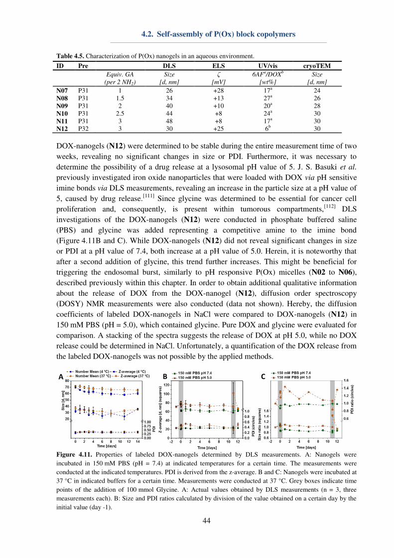
4.2. Self-assembly of P(Ox) block copolymers
44
Table 4.5. Characterization of P(Ox) nanogels in an aqueous environment.
ID Pre DLS ELS UV/vis cryoTEM
Equiv. GA
(per 2 NH2)
Size
[d, nm]
ζ [mV]
6AFa/DOX
b
[wt%]
Size
[d, nm]
N07 P31 1 26 +28 17a
24
N08 P31 1.5 34 +13 27a
26
N09 P31 2 40 +10 20a
28
N10 P31 2.5 44 +8 24a
30
N11 P31 3 48 +8 17a
30
N12 P32 3 30 +25 6b 30
DOX-nanogels (N12) were determined to be stable during the entire measurement time of two
weeks, revealing no significant changes in size or PDI. Furthermore, it was necessary to
determine the possibility of a drug release at a lysosomal pH value of 5. J. S. Basuki et al.
previously investigated iron oxide nanoparticles that were loaded with DOX via pH sensitive
imine bonds via DLS measurements, revealing an increase in the particle size at a pH value of
5, caused by drug release.[111]
Since glycine was determined to be essential for cancer cell
proliferation and, consequently, is present within tumorous compartments,[112]
DLS
investigations of the DOX-nanogels (N12) were conducted in phosphate buffered saline
(PBS) and glycine was added representing a competitive amine to the imine bond
(Figure 4.11B and C). While DOX-nanogels (N12) did not reveal significant changes in size
or PDI at a pH value of 7.4, both increase at a pH value of 5.0. Herein, it is noteworthy that
after a second addition of glycine, this trend further increases. This might be beneficial for
triggering the endosomal burst, similarly to pH responsive P(Ox) micelles (N02 to N06),
described previously within this chapter. In order to obtain additional qualitative information
about the release of DOX from the DOX-nanogel (N12), diffusion order spectroscopy
(DOSY) NMR measurements were also conducted (data not shown). Hereby, the diffusion
coefficients of labeled DOX-nanogels in NaCl were compared to DOX-nanogels (N12) in
150 mM PBS (pH = 5.0), which contained glycine. Pure DOX and glycine were evaluated for
comparison. A stacking of the spectra suggests the release of DOX at pH 5.0, while no DOX
release could be determined in NaCl. Unfortunately, a quantification of the DOX release from
the labeled DOX-nanogels was not possible by the applied methods.
Figure 4.11. Properties of labeled DOX-nanogels determined by DLS measurements. A: Nanogels were
incubated in 150 mM PBS (pH = 7.4) at indicated temperatures for a certain time. The measurements were
conducted at the indicated temperatures. PDI is derived from the z-average. B and C: Nanogels were incubated at
37 °C in indicated buffers for a certain time. Measurements were conducted at 37 °C. Grey boxes indicate time
points of the addition of 100 mmol Glycine. A: Actual values obtained by DLS measurements (n = 3, three
measurements each). B: Size and PDI ratios calculated by division of the value obtained on a certain day by the
initial value (day -1).
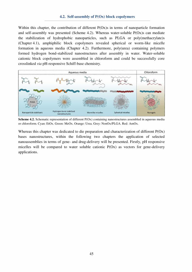
4.2. Self-assembly of P(Ox) block copolymers
45
Within this chapter, the contribution of different P(Ox)s in terms of nanoparticle formation
and self-assembly was presented (Scheme 4.2). Whereas water-soluble P(Ox)s can mediate
the stabilization of hydrophobic nanoparticles, such as PLGA or poly(methacrylate)s
(Chapter 4.1), amphiphilic block copolymers revealed spherical or worm-like micelle
formation in aqueous media (Chapter 4.2). Furthermore, poly(urea) containing polymers
formed hydrogen bond-stabilized nanostructures after assembly in water. Water-soluble
cationic block copolymers were assembled in chloroform and could be successfully core
crosslinked via pH-responsive Schiff-base chemistry.
Scheme 4.2. Schematic representation of different P(Ox) containing nanostructures assembled in aqueous media
or chloroform. Cyan: EtOx. Green: MeOx. Orange: Urea. Grey: NonOx/PLGA. Red: AmOx.
Whereas this chapter was dedicated to die preparation and characterization of different P(Ox)
bases nanostructures, within the following two chapters the application of selected
nanoassemblies in terms of gene- and drug-delivery will be presented. Firstly, pH responsive
micelles will be compared to water soluble cationic P(Ox) as vectors for gene-delivery
applications.
5. Gene delivery systems
46
5. Gene delivery systems
Parts of this chapter have been published in: P3) M. N. Leiske, F. H. Sobotta, F. Richter. S.
Hoeppener, J. C. Brendel, A. Traeger, U. S. Schubert, Biomacromolecules 2018, 19, 748-760.
P5) D. Hertz‡, M. N. Leiske‡, T. Wloka, A. Traeger, M. Hartlieb, M. M. Kessels, S. Schubert,
B. Qualmann, U. S. Schubert, J. Polym. Sci., Part A: Polym. Chem. 2018, in press. DOI:
10.1002/pola.29000. ‡Equal contribution of both authors.
Besides lipoplexes, also cationic polymers are used for gene delivery, since they are capable
of forming polyplexes with the negatively charged phosphate backbone of nucleic acids.
Poly(ethylene imine) (PEI) is one of the most commonly used materials for gene delivery
applications and for a long time it was claimed to be the gold standard for the transfection of
genetic material.[113] Its high charge density leads to the formation of physiological stable
PEI-DNA polyplexes. Disadvantages of PEI-based systems are their high in vitro and in vivo
toxicity and their resistance against biodegradation, leading to the accumulation of the
polymer in the cells or tissue, which can elicit further toxicity effects.[114] Furthermore, the
cytotoxicity has been shown to be dependent on the molar mass of the polymers,[115] but can
be improved by the introduction of stealth units, i.e. EtOx, into the polymer chain.[74, 116]
These drawbacks lead to a necessity to search for alternative polymer systems for gene-
delivery applications, which reveal high transfection efficiencies while expressing a low
cytotoxicity.
For this reason, cationic copolymers containing non-ionic comonomers with increasing
hydrophobicity (MeOx < EtOx < NonOx) were prepared and compared regarding their
transfection efficiency. Due to the fact that cytocompatibility as well as transfection efficiency
of cationic polymers are dependent on the amount of cationic charges within the
macromolecule, water soluble polymers containing 20% (P25 and P19), 40% (P40 and P43),
60% (P41 and P44) or 80% (P44 and P45) AmOx were synthesized accordingly to Chapter
3.3 (Table 5.1). The copolymers consisted of AmOx and either MeOx in order to form
gradient copolymers (P25, P40 to P42) or EtOx forming random copolymers (P19, P43 to
P45) Furthermore, P(EtOx3-b-AmOx157) (P46) was synthesized and is from now on denoted
as P(AmOx). Cationic polymers were characterized by means of 1H-NMR and SEC (Table
5.1). 1H-NMR measurements provided information about the DP of the polymers, which was
determined to be between 130 and 190. SEC measurements confirmed the stability of the
macromolecules during acidic deprotection. In addition to the water-soluble cationic
copolymers, nanostructures (N01 to N06) consisting of amphiphilic block copolymers were
investigated. The preparation and characterization of the pH-responsive nanostructures with
varying AmOx content in the shell was already discussed in Chapter 4.2.
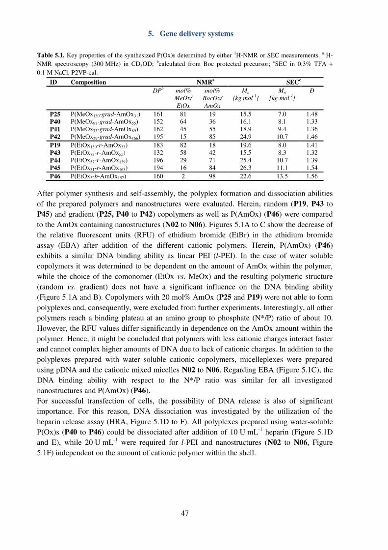
5. Gene delivery systems
47
Table 5.1. Key properties of the synthesized P(Ox)s determined by either 1H-NMR or SEC measurements. a1H-
NMR spectroscopy (300 MHz) in CD3OD; bcalculated from Boc protected precursor; cSEC in 0.3% TFA +
0.1 M NaCl, P2VP-cal.
ID Composition NMRa
SECc
DPb
mol%
MeOx/
EtOx
mol%
BocOx/
AmOx
Mn
[kg mol-1
] Mn
[kg mol-1
] Ð
P25 P(MeOx130-grad-AmOx31) 161 81 19 15.5 7.0 1.48 P40 P(MeOx97-grad-AmOx55) 152 64 36 16.1 8.1 1.33 P41 P(MeOx73-grad-AmOx89) 162 45 55 18.9 9.4 1.36 P42 P(MeOx29-grad-AmOx166) 195 15 85 24.9 10.7 1.46
P19 P(EtOx150-r-AmOx33) 183 82 18 19.6 8.0 1.41 P43 P(EtOx77-r-AmOx55) 132 58 42 15.5 8.3 1.32 P44 P(EtOx57-r-AmOx139) 196 29 71 25.4 10.7 1.39 P45 P(EtOx31-r-AmOx163) 194 16 84 26.3 11.1 1.54
P46 P(EtOx3-b-AmOx157) 160 2 98 22.6 13.5 1.56
After polymer synthesis and self-assembly, the polyplex formation and dissociation abilities
of the prepared polymers and nanostructures were evaluated. Herein, random (P19, P43 to
P45) and gradient (P25, P40 to P42) copolymers as well as P(AmOx) (P46) were compared
to the AmOx containing nanostructures (N02 to N06). Figures 5.1A to C show the decrease of
the relative fluorescent units (RFU) of ethidium bromide (EtBr) in the ethidium bromide
assay (EBA) after addition of the different cationic polymers. Herein, P(AmOx) (P46)
exhibits a similar DNA binding ability as linear PEI (l-PEI). In the case of water soluble
copolymers it was determined to be dependent on the amount of AmOx within the polymer,
while the choice of the comonomer (EtOx vs. MeOx) and the resulting polymeric structure
(random vs. gradient) does not have a significant influence on the DNA binding ability
(Figure 5.1A and B). Copolymers with 20 mol% AmOx (P25 and P19) were not able to form
polyplexes and, consequently, were excluded from further experiments. Interestingly, all other
polymers reach a binding plateau at an amino group to phosphate (N*/P) ratio of about 10.
However, the RFU values differ significantly in dependence on the AmOx amount within the
polymer. Hence, it might be concluded that polymers with less cationic charges interact faster
and cannot complex higher amounts of DNA due to lack of cationic charges. In addition to the
polyplexes prepared with water soluble cationic copolymers, micelleplexes were prepared
using pDNA and the cationic mixed micelles N02 to N06. Regarding EBA (Figure 5.1C), the
DNA binding ability with respect to the N*/P ratio was similar for all investigated
nanostructures and P(AmOx) (P46).
For successful transfection of cells, the possibility of DNA release is also of significant
importance. For this reason, DNA dissociation was investigated by the utilization of the
heparin release assay (HRA, Figure 5.1D to F). All polyplexes prepared using water-soluble
P(Ox)s (P40 to P46) could be dissociated after addition of 10 U mL-1 heparin (Figure 5.1D
and E), while 20 U mL-1 were required for l-PEI and nanostructures (N02 to N06, Figure
5.1F) independent on the amount of cationic polymer within the shell.
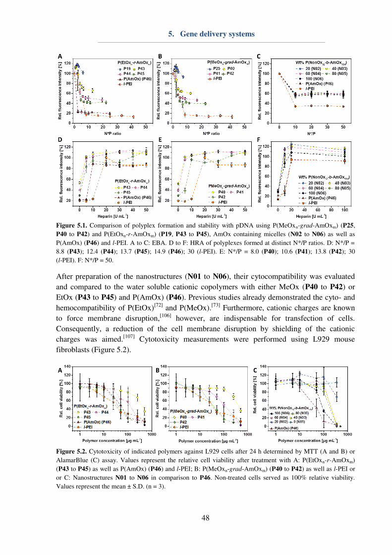
5. Gene delivery systems
48
Figure 5.1. Comparison of polyplex formation and stability with pDNA using P(MeOxn-grad-AmOxm) (P25,
P40 to P42) and P(EtOxn-r-AmOxm) (P19, P43 to P45), AmOx containing micelles (N02 to N06) as well as
P(AmOx) (P46) and l-PEI. A to C: EBA. D to F: HRA of polyplexes formed at distinct N*/P ratios. D: N*/P =
8.8 (P43); 12.4 (P44); 13.7 (P45); 14.9 (P46); 30 (l-PEI). E: N*/P = 8.0 (P40); 10.6 (P41); 13.8 (P42); 30
(l-PEI). F: N*/P = 50.
After preparation of the nanostructures (N01 to N06), their cytocompatibility was evaluated
and compared to the water soluble cationic copolymers with either MeOx (P40 to P42) or
EtOx (P43 to P45) and P(AmOx) (P46). Previous studies already demonstrated the cyto- and
hemocompatibility of P(EtOx)[72]
and P(MeOx).[73]
Furthermore, cationic charges are known
to force membrane disruption,[106]
however, are indispensable for transfection of cells.
Consequently, a reduction of the cell membrane disruption by shielding of the cationic
charges was aimed.[107]
Cytotoxicity measurements were performed using L929 mouse
fibroblasts (Figure 5.2).
Figure 5.2. Cytotoxicity of indicated polymers against L929 cells after 24 h determined by MTT (A and B) or
AlamarBlue (C) assay. Values represent the relative cell viability after treatment with A: P(EtOxn-r-AmOxm)
(P43 to P45) as well as P(AmOx) (P46) and l-PEI; B: P(MeOxn-grad-AmOxm) (P40 to P42) as well as l-PEI or
or C: Nanostructures N01 to N06 in comparison to P46. Non-treated cells served as 100% relative viability.
Values represent the mean ± S.D. (n = 3).
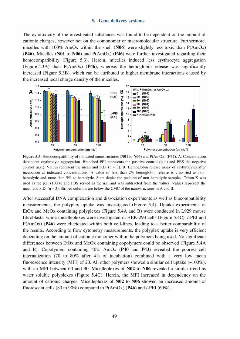
5. Gene delivery systems
49
The cytotoxicity of the investigated substances was found to be dependent on the amount of
cationic charges, however not on the comonomer or macromolecular structure. Furthermore,
micelles with 100% AmOx within the shell (N06) were slightly less toxic than P(AmOx)
(P46). Micelles (N01 to N06) and P(AmOx) (P46) were further investigated regarding their
hemocompatibility (Figure 5.3). Herein, micelles induced less erythrocyte aggregation
(Figure 5.3A) than P(AmOx) (P46), whereas the hemoglobin release was significantly
increased (Figure 5.3B), which can be attributed to higher membrane interactions caused by
the increased local charge density of the micelles.
Figure 5.3. Hemocompatibility of indicated nanostructures (N01 to N06) and P(AmOx) (P47). A: Concentration
dependent erythrocyte aggregation. Branched PEI represents the positive control (p.c.) and PBS the negative
control (n.c.). Values represent the mean and S.D. (n = 3). B: Hemoglobin release assay of erythrocytes after
incubation at indicated concentrations. A value of less than 2% hemoglobin release is classified as non-
hemolytic and more than 5% as hemolytic. Stars depict the position of non-hemolytic samples. Triton-X was
used as the p.c. (100%) and PBS served as the n.c. and was subtracted from the values. Values represent the
mean and S.D. (n = 3). Striped columns are below the CMC of the nanostructures in A and B.
After successful DNA complexation and dissociation experiments as well as biocompatibility
measurements, the polyplex uptake was investigated (Figure 5.4). Uptake experiments of
EtOx and MeOx containing polyplexes (Figure 5.4A and B) were conducted in L929 mouse
fibroblasts, while micelleplexes were investigated in HEK-293 cells (Figure 5.4C). l-PEI and
P(AmOx) (P46) were elucidated within both cell-lines, leading to a better comparability of
the results. According to flow cytometry measurements, the polyplex uptake is very efficient
depending on the amount of cationic monomer within the polymers being used. No significant
differences between EtOx and MeOx containing copolymers could be observed (Figure 5.4A
and B). Copolymers containing 40% AmOx (P40 and P43) revealed the poorest cell
internalization (70 to 80% after 4 h of incubation) combined with a very low mean
fluorescence intensity (MFI) of 20. All other polymers showed a similar cell uptake (~100%),
with an MFI between 60 and 90. Micelleplexes of N02 to N06 revealed a similar trend as
water soluble polyplexes (Figure 5.4C). Herein, the MFI increased in dependency on the
amount of cationic charges. Micelleplexes of N02 to N06 showed an increased amount of
fluorescent cells (80 to 90%) compared to P(AmOx) (P46) and l-PEI (60%).
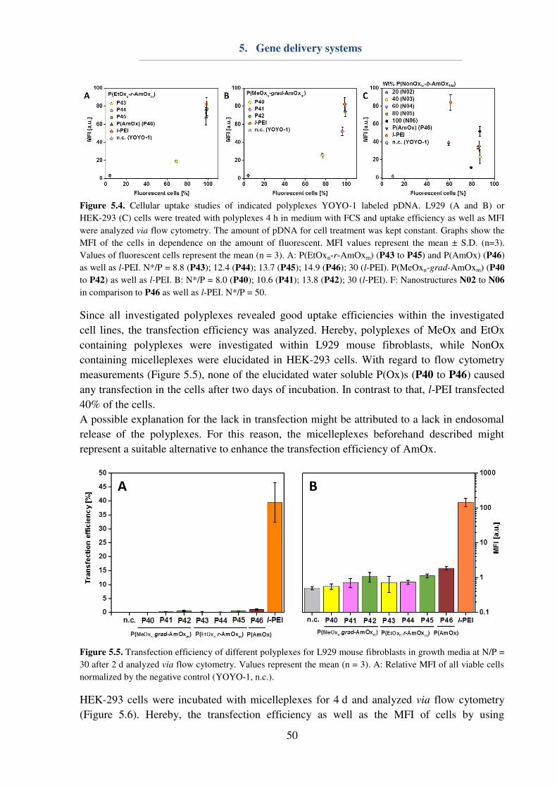
5. Gene delivery systems
50
Figure 5.4. Cellular uptake studies of indicated polyplexes YOYO-1 labeled pDNA. L929 (A and B) or
HEK-293 (C) cells were treated with polyplexes 4 h in medium with FCS and uptake efficiency as well as MFI
were analyzed via flow cytometry. The amount of pDNA for cell treatment was kept constant. Graphs show the
MFI of the cells in dependence on the amount of fluorescent. MFI values represent the mean ± S.D. (n=3).
Values of fluorescent cells represent the mean (n = 3). A: P(EtOxn-r-AmOxm) (P43 to P45) and P(AmOx) (P46)
as well as l-PEI. N*/P = 8.8 (P43); 12.4 (P44); 13.7 (P45); 14.9 (P46); 30 (l-PEI). P(MeOxn-grad-AmOxm) (P40
to P42) as well as l-PEI. B: N*/P = 8.0 (P40); 10.6 (P41); 13.8 (P42); 30 (l-PEI). F: Nanostructures N02 to N06
in comparison to P46 as well as l-PEI. N*/P = 50.
Since all investigated polyplexes revealed good uptake efficiencies within the investigated
cell lines, the transfection efficiency was analyzed. Hereby, polyplexes of MeOx and EtOx
containing polyplexes were investigated within L929 mouse fibroblasts, while NonOx
containing micelleplexes were elucidated in HEK-293 cells. With regard to flow cytometry
measurements (Figure 5.5), none of the elucidated water soluble P(Ox)s (P40 to P46) caused
any transfection in the cells after two days of incubation. In contrast to that, l-PEI transfected
40% of the cells.
A possible explanation for the lack in transfection might be attributed to a lack in endosomal
release of the polyplexes. For this reason, the micelleplexes beforehand described might
represent a suitable alternative to enhance the transfection efficiency of AmOx.
Figure 5.5. Transfection efficiency of different polyplexes for L929 mouse fibroblasts in growth media at N/P =
30 after 2 d analyzed via flow cytometry. Values represent the mean (n = 3). A: Relative MFI of all viable cells
normalized by the negative control (YOYO-1, n.c.).
HEK-293 cells were incubated with micelleplexes for 4 d and analyzed via flow cytometry
(Figure 5.6). Hereby, the transfection efficiency as well as the MFI of cells by using

5. Gene delivery systems
51
micelleplexes consisting of at least 40% AmOx within the micelle shell (N03 to N06) was
enhanced compared to P(AmOx) (P46) (Figure 5.6A and B). Furthermore, the cell viability of
all micelleplexes (80 to 95%) was superior to l-PEI and P(AmOx) (P46) (60%). For this
reason, the transfection efficiency of all cells was also determined (Figure 5.6C), revealing a
superiority of N05 and N06 compared to l-PEI.
Figure 5.6. Transfection efficiency of different polyplexes for adherent HEK-293 cells in growth media at N*/P
= 50 after 4 d analyzed via flow cytometry. Values represent the mean (n = 3). A: Relative MFI of all viable cells
normalized by the negative control (n.c.). B: Transfection efficiency of all viable cells. C: Transfection
efficiency of all cells.
Within this chapter, water soluble cationic copolymers consisting of EtOx and AmOx
(random) or MeOx and AmOx (gradient) as well as cationic nanostructures with EtOx and
AmOx within the shell were compared to P(AmOx) and l-PEI regarding their potential as
biocompatible non-viral vectors. Herein, the transfection efficiency of AmOx containing
polyplexes could be enhanced by using copolymers with hydrophobic NonOx blocks that
were self-assembled into micelles prior to complexation of the genetic material. It might be
assumed that the endosomal release can be triggered by stretching of the cationic AmOx
blocks during acidification of the endolysosomes (Scheme 5.1). Consequently, amphiphilic
copolymers were determined to be more efficient than fully water soluble polymeric carrier
systems.
Scheme 5.1. Schematic presentation of the cellular uptake and endosomal release of different polyplexes. Left:
water soluble polyplexes of random or gradient hydrophilic copolymers might not be able to be released from the
endosomes and, consequently, lack in transfection efficiency. Right: Micelleplexes that were prepared by using
amphiphilic polymeric micelles can trigger the endosomal burst by stretching of the cationic polymer blocks
during acidification of the endolysosomes and, hence, successfully transfect cells. Cyan: EtOx. Green: MeOx.
Red: AmOx. Grey: NonOx. Purple: pDNA.

5. Gene delivery systems
52
Within the following chapter (Chapter 6), water soluble functional P(Ox)s will be applied in
terms of drug-delivery systems by covalent conjugation of drugs to the hydrophilic polymers
and their activity, respectively therapeutic efficiency will be elucidated and compared to the
pure drugs.
6. Drug delivery systems
53
6. Drug delivery systems
Parts of this chapter have been published in: P4) D. Hoelzer‡, M. N. Leiske‡, M. Hartlieb, T.
Bus, D. Pretzel, S. Hoeppener, K. Kempe, R. Thierbach, U. S. Schubert, Oncotarget 2018, in
press. P7) T. Luehmann, M. Schmidt, M. N. Leiske, V. Spieler, T. C. Majdanski, M. Grube,
M. Hartlieb, I. Nischang, S. Schubert, U.S. Schubert, L. Meinel, ACS Biomater. Sci. Eng.
2017, 3, 304-312. P8) M. Hartlieb‡, T. Bus‡, J. Kübel, D. Pretzel, S. Hoeppener, M. N.
Leiske, K. Kempe, B. Dietzek, U. S. Schubert, Bioconjugate Chem. 2017, 28, 1229-1235. ‡Equal contribution of both authors.
In addition to complexation of genetic material, functional groups of polymers can also be
utilized for drug conjugation. As already mentioned in Chapter 2, many drugs (i.e. small
molecules) are poorly water soluble. Conjugation to water soluble polymers can enhance the
solubility significantly.[21-22] Furthermore, the blood circulation times can be increased[79] by
preventing unspecific cellular uptake, immune response or renal excretion.
Within this work, two different possibilities for the attachment of drugs to polymers were
conducted. Firstly, single P(Ox) chains with a defined azide functionalized ω-end group were
used for the POxylation of interleukin-4 (IL-4). Hereby, irreversible conjugation of polymers
to proteins is mostly known from PEGylation, aiming longer blood circulation times and
consequently altering the pharmacokinetics.[117-118] By further evaluating the PEGylation of
proteins, some drawbacks are obvious by the nature of the unspecific reaction carried out: (i)
Different protein molecules can be attacked at different positions, (ii) the active center of the
protein can be blocked, (iii) mono-, di-, or multi-PEGylation occurs, and (iv) 25% of the
human population already have PEG antibodies. These unwanted effects can lead to a
significant change within the pharmacokinetics of the protein based pharmaceutical, since it
cannot be determined precisely how much of the protein is still active or how significantly the
blood circulation times are altered. For this reason, CuAAC might be used for the preparation
of defined protein P(Ox) conjugates.
6.1. POxylation of proteins
In order to obtain mono-POxylated proteins, an alkyne modified amber codon was
synthesized previously.[119] This amino acid can be inserted into the protein structure during
protein biosynthesis and, hence, reveals a single, defined position within the protein.
Consequently, the synthesis of mono azide functionalized P(Ox)s via ω-end
functionalization[17-18] was conducted. In order to obtain conjugates of varying size, three
different homopolymers of different molar mass were synthesized accordingly to Chapter 3.3
and characterized using NMR as well as SEC (Table 6.1) to gain information about the size
and dispersity of the polymers. Polymers P47 to P49 revealed a narrow dispersity (Ð ≤ 1.2) while having a molar mass of 2,600, 4,300 or 9,200 g mol-1. In addition to the P(MeOx)
homopolymers, gradient copolymers of MeOx and AmOx (P50 to P52) were synthesized to
enhance the cellular uptake mediated by the cationic charges. However, the amount of AmOx
units was kept low to minimize cytotoxic effects caused by the cationic charges as reported
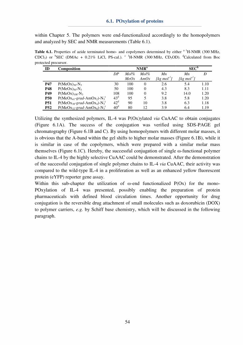
6.1. POxylation of proteins
54
within Chapter 5. The polymers were end-functionalized accordingly to the homopolymers
and analyzed by SEC and NMR measurements (Table 6.1).
Table 6.1. Properties of azide terminated homo- and copolymers determined by either a 1H-NMR (300 MHz,
CDCl3) or bSEC (DMAc + 0.21% LiCl, PS-cal.). c 1H-NMR (300 MHz, CD3OD). dCalculated from Boc
protected precursor.
ID Composition NMRa
SECb
DP Mol%
MeOx
Mol%
AmOx
Mn
[kg mol-1
]
Mn
[kg mol-1
]
Ð
P47 P(MeOx)30-N3 30 100 0 2.6 5.4 1.10 P48 P(MeOx)50-N3 50 100 0 4.3 8.3 1.11 P49 P(MeOx)108-N3 108 100 0 9.2 14.0 1.20 P50 P(MeOx41-grad-AmOx2)-N3
c 43d 95 5 3.8 5.8 1.20 P51 P(MeOx38-grad-AmOx4)-N3
c 42d 90 10 3.8 6.3 1.18 P52 P(MeOx32-grad-AmOx8)-N3
c 40d 80 12 3.9 6.4 1.19
Utilizing the synthesized polymers, IL-4 was P(Ox)ylated via CuAAC to obtain conjugates
(Figure 6.1A). The success of the conjugation was verified using SDS-PAGE gel
chromatography (Figure 6.1B and C). By using homopolymers with different molar masses, it
is obvious that the A-band within the gel shifts to higher molar masses (Figure 6.1B), while it
is similar in case of the copolymers, which were prepared with a similar molar mass
themselves (Figure 6.1C). Hereby, the successful conjugation of single ω-functional polymer
chains to IL-4 by the highly selective CuAAC could be demonstrated. After the demonstration
of the successful conjugation of single polymer chains to IL-4 via CuAAC, their activity was
compared to the wild-type IL-4 in a proliferation as well as an enhanced yellow fluorescent
protein (eYFP) reporter gene assay.
Within this sub-chapter the utilization of ω-end functionalized P(Ox) for the mono-
POxylation of IL-4 was presented, possibly enabling the preparation of protein
pharmaceuticals with defined blood circulation times. Another opportunity for drug
conjugation is the reversible drug attachment of small molecules such as doxorubicin (DOX)
to polymer carriers, e.g. by Schiff base chemistry, which will be discussed in the following
paragraph.
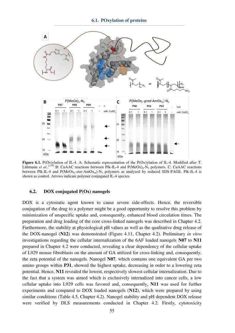
6.1. POxylation of proteins
55
Figure 6.1. P(Ox)ylation of IL-4. A: Schematic representation of the P(Ox)ylation of IL-4. Modified after T.
Lühmann et al..[119]
B: CuAAC reactions between Plk-IL-4 and P(MeOx)n-N3 polymers. C: CuAAC reactions
between Plk-IL-4 and P(MeOxn-stat-AmOxm)-N3 polymers as analyzed by reduced SDS-PAGE. Plk-IL-4 is
shown as control. Arrows indicate polymer conjugated IL-4 species.
6.2. DOX conjugated P(Ox) nanogels
DOX is a cytostatic agent known to cause severe side-effects. Hence, the reversible
conjugation of the drug to a polymer might be a good opportunity to resolve this problem by
minimization of unspecific uptake and, consequently, enhanced blood circulation times. The
preparation and drug loading of the core cross-linked nanogels was described in Chapter 4.2.
Furthermore, the stability at physiological pH values as well as the qualitative drug release of
the DOX-nanogel (N12) was demonstrated (Figure 4.11, Chapter 4.2). Preliminary in vitro
investigations regarding the cellular internalization of the 6AF loaded nanogels N07 to N11
prepared in Chapter 4.2 were conducted, revealing a clear dependency of the cellular uptake
of L929 mouse fibroblasts on the amount of GA utilized for cross-linking and, consequently,
the zeta potential of the nanogels. Nanogel N07, which contains one equivalent GA per two
amino groups within P31, showed the highest uptake, decreasing in order to a lowering zeta
potential. Hence, N11 revealed the lowest, respectively slowest cellular internalization. Due to
the fact that a system was aimed which is exclusively internalized into cancer cells, a low
cellular uptake into L929 cells was favored and, consequently, N11 was used for further
experiments and compared to DOX loaded nanogels (N12), which were prepared by using
similar conditions (Table 4.5, Chapter 4.2). Nanogel stability and pH dependent DOX release
were verified by DLS measurements conducted in Chapter 4.2. Firstly, cytotoxicity
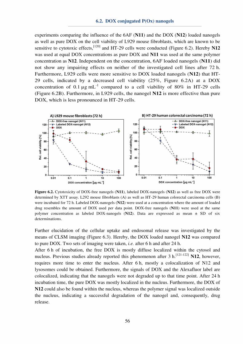
6.2. DOX conjugated P(Ox) nanogels
56
experiments comparing the influence of the 6AF (N11) and the DOX (N12) loaded nanogels
as well as pure DOX on the cell viability of L929 mouse fibroblasts, which are known to be
sensitive to cytotoxic effects,[120]
and HT-29 cells were conducted (Figure 6.2). Hereby N12
was used at equal DOX concentrations as pure DOX and N11 was used at the same polymer
concentration as N12. Independent on the concentration, 6AF loaded nanogels (N11) did
not show any impairing effects on neither of the investigated cell lines after 72 h.
Furthermore, L929 cells were more sensitive to DOX loaded nanogels (N12) that HT-
29 cells, indicated by a decreased cell viability (25%, Figure 6.2A) at a DOX
concentration of 0.1 µg mL-1
compared to a cell viability of 80% in HT-29 cells
(Figure 6.2B). Furthermore, in L929 cells, the nanogel N12 is more effective than pure
DOX, which is less pronounced in HT-29 cells.
Figure 6.2. Cytotoxicity of DOX-free nanogels (N11), labeled DOX-nanogels (N12) as well as free DOX were
determined by XTT assay. L292 mouse fibroblasts (A) as well as HT-29 human colorectal carcinoma cells (B)
were incubated for 72 h. Labeled DOX-nanogels (N12) were used at a concentration where the amount of loaded
drug resembles the amount of DOX used per data point. DOX-free nanogels (N11) were used at the same
polymer concentration as labeled DOX-nanogels (N12). Data are expressed as mean ± SD of six
determinations.
Further elucidation of the cellular uptake and endosomal release was investigated by the
means of CLSM imaging (Figure 6.3). Hereby, the DOX loaded nanogel N12 was compared
to pure DOX. Two sets of imaging were taken, i.e. after 6 h and after 24 h.
After 6 h of incubation, the free DOX is mostly diffuse localized within the cytosol and
nucleus. Previous studies already reported this phenomenon after 3 h.[121-122]
N12, however,
requires more time to enter the nucleus. After 6 h, mostly a colocalization of N12 and
lysosomes could be obtained. Furthermore, the signals of DOX and the Alexafluor label are
colocalized, indicating that the nanogels were not degraded up to that time point. After 24 h
incubation time, the pure DOX was mostly localized in the nucleus. Furthermore, the DOX of
N12 could also be found within the nucleus, whereas the polymer signal was localized outside
the nucleus, indicating a successful degradation of the nanogel and, consequently, drug
release.

6.2. DOX conjugated P(Ox) nanogels
57
Figure 6.3. CLSM images of free DOX as and labeled DOX-nanogels (N12) incubated with HT-29 colorectal
carcinoma for 6 h or 24 h. Lysosomal cellular compartments were stained green using LysoTracker Green DND-
26 and the nucleus was labeled with Hoechst 33342 (blue). The fluorescence of DOX is depicted in red and the
Alexafluor label of the polymer is shown in white.
With regard to the in vitro results, an in vivo study using the xenograft mouse model was
conducted using the DOX-loaded nanogel N12 in comparison to pure DOX. 6AF loaded
nanogels (N11) as well as physiological sodium chloride solution represented the control
groups. All substances were dissolved in sterile sodium chloride solution and mice were
treated via tail vein injection. Preliminary experiments on the biocompatibility revealed no
toxic effects of the nanogels at a DOX concentration of 1 mg kg-1
. For this reason, this
amount of DOX was used for the determination of the therapeutic efficiency in vivo. HT-29
tumor cells were injected to male nude mice. After the tumor growth reached a certain level,
therapy was conducted by the injection of six doses. Mice treated with DOX or N12 revealed
a reduced tumor growth whereas N11 and NaCl did not have an influence. Interestingly, the
DOX loaded nanogel N12 was slightly more efficient than the pure DOX (Figure 6.4A) and,
furthermore, increased the median survival time significantly (N12: 73 d; DOX: 39 d;
Figure 6.4B).
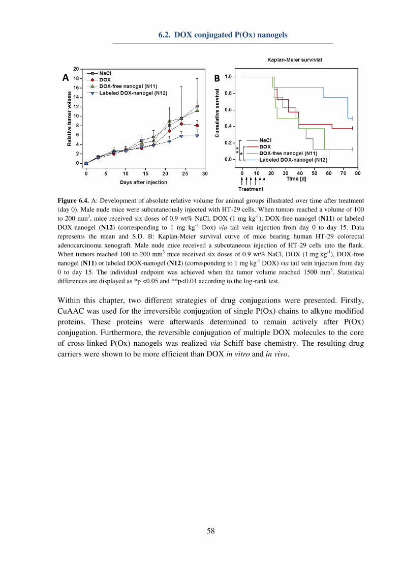
6.2. DOX conjugated P(Ox) nanogels
58
Figure 6.4. A: Development of absolute relative volume for animal groups illustrated over time after treatment
(day 0). Male nude mice were subcutaneously injected with HT-29 cells. When tumors reached a volume of 100
to 200 mm3, mice received six doses of 0.9 wt% NaCl, DOX (1 mg kg
-1), DOX-free nanogel (N11) or labeled
DOX-nanogel (N12) (corresponding to 1 mg kg-1
Dox) via tail vein injection from day 0 to day 15. Data
represents the mean and S.D. B: Kaplan-Meier survival curve of mice bearing human HT-29 colorectal
adenocarcinoma xenograft. Male nude mice received a subcutaneous injection of HT-29 cells into the flank.
When tumors reached 100 to 200 mm3 mice received six doses of 0.9 wt% NaCl, DOX (1 mg kg
-1), DOX-free
nanogel (N11) or labeled DOX-nanogel (N12) (corresponding to 1 mg kg-1
DOX) via tail vein injection from day
0 to day 15. The individual endpoint was achieved when the tumor volume reached 1500 mm3. Statistical
differences are displayed as *p <0.05 and **p<0.01 according to the log-rank test.
Within this chapter, two different strategies of drug conjugations were presented. Firstly,
CuAAC was used for the irreversible conjugation of single P(Ox) chains to alkyne modified
proteins. These proteins were afterwards determined to remain actively after P(Ox)
conjugation. Furthermore, the reversible conjugation of multiple DOX molecules to the core
of cross-linked P(Ox) nanogels was realized via Schiff base chemistry. The resulting drug
carriers were shown to be more efficient than DOX in vitro and in vivo.
7. Summary
59
7. Summary
Since their invention in the 1960s, research on the pseudopeptides poly(2-oxazoline)s
(P(Ox)s) has risen significantly. Whereas initial investigations mainly concentrated on the
synthesis of defined P(Ox)s with different substituents in 2-position and their chemical
characterization, the field of biomedical applications has been focused increasingly during the
last decade. Due to this broad scope in academic research on P(Ox)s, the aim of this thesis
was to contribute to the understanding of the synthesis and polymerization, self-assembly
behavior as well as biomedical applications of P(Ox)s (Figure 7.1).
Figure 7.1. With the aid of detailed kinetic investigations, the CROP facilitates the synthesis of tailored
polymers consisting of 2-oxazolines and/or urea (orange boxes). P(Ox)s can be utilized for stabilization of
hydrophobic polymer particles as well as for direct self- and co-assembly (blue boxes). Functional P(Ox)s are
used in a wide range of applications in drug as well as gene delivery (green boxes).
By using distinct synthesis routes, different Boc protected amino-functionalized 2-oxazolines
can be obtained. Firstly, the synthesis of 2-(4-((tert-butoxycarbonyl)amino)butyl)-2-oxazoline
(BocOx), which is already known from literature, is proceeded by using a three step synthesis
utilizing 5-aminovaleric acid as starting material. The synthesis resulted in a Boc protected 2-
oxazoline, which can be polymerized using the cationic ring-opening polymerization (CROP).
On the other hand, tert-butyl 2-iminooxazolidine-3-carboxylate (BocOI) can be synthesized from
2-amino-2oxazoline in a single step reaction. Hereby, the endo-nitrogen of the molecule was found
to be more reactive than the exo-nitrogen and, hence, leads to the formation of a Boc protected 2-
iminooxazolidine instead of a 2-oxazoline. BocOI was found to be polymerizable by CROP,
resulting in Boc protected poly(urea) derivatives, which could be deprotected under acidic
conditions to yield poly(urea). Thus, the synthesis of poly(urea) was proceeded in a controlled
chain growth polymerization, yielding polymers with a narrow dispersity (Ð ≤ 1.3) instead of the less controllable step growth polymerization, invented by Otto von Bayer. Furthermore, by using
the CROP for the preparation of poly(urea) it was possible to prepare copolymers of P(Ox)s and
poly(urea) in a one-pot polymerization. Hereby, the polymerization constant of BocOI was found
7. Summary
60
to be more than ten times higher than 2-ethyl-2-oxazoline (EtOx), suggesting the formation of
quasi block copolymers in a one-pot reaction.
Furthermore, the copolymerization kinetics of the Boc protected 2-oxazoline BocOx and EtOx,
respectively 2-methyl-2-oxazoline (MeOx) were evaluated. Here, BocOx was found to be as
reactive as EtOx, leading to the formation of random copolymers, while MeOx was more reactive
and, consequently, forms gradient copolymers with BocOx. Consequently, BocOI is much more
reactive than BocOx, which might be caused by the monomer structure and the differences in the
polymerization mechanism. Then, different functional copolymers were synthesized and
characterized. The polymers were used for the preparation of colloidal structures and used for gene
and drug delivery applications within this thesis.
The second main objective of this thesis concentrated on the application of P(Ox)s for the
preparation of nanostructures. Herein, water soluble homopolymers of P(EtOx) or P(MeOx) with
different degrees of polymerization (DP; 25, 60, 100 and 200) were synthesized and investigated
regarding their properties within the stabilization of hydrophobic nanoparticles during preparation,
purification and lyophilization. In general, no significant differences between P(EtOx) and
P(MeOx) could be observed. P(Ox)s with a DP of 25 were not suitable for neither of the aimed
applications, possible caused by their low dynamic viscosity in aqueous solutions or their short
chain length, which cannot stabilize the interphase between the hydrophobic nanoparticle and the
aqueous solution in a satisfying manner. Though, P(Ox)s with a DP of 60, 100 or 200 were found
to be suitable as stabilizers for nanoparticle preparation, purification and lyophilization at distinct
concentrations. Consequently, it could be shown that non-ionic P(Ox)s can be utilized as an all-in-
one system to stabilize hydrophobic polymer nanoparticles.
In terms of nano-assembly, the utilization of amphiphilic P(Ox)s for the preparation of pH-
responsive nanostructures was investigated. Herein, P(Ox) block copolymers were synthesized
consisting of either 2-nonyl-2-oxazoline (NonOx) and EtOx or NonOx and 2-(4-aminobutyl)-2-
oxazoline) (AmOx) and self- as well as co-assembled in a simple mixed micelle approach. In both
cases, NonOx served as the hydrophobic building block of the block copolymers, forming the
micellar core during the assembly process. AmOx was chosen as the cationic core block to
facilitate the complexation of genetic material for gene delivery applications. In contrast to that,
EtOx served as the non-ionic building block suitable to enhance the biocompatibility of the
nanostructures by shielding the cationic charges of AmOx. Nanostructures were prepared with
different amino contents by simply mixing the two polymers in different weight ratios prior
assembly. By this means, nanostructures with 0, 20, 40, 60, 80, or 100 wt% of the AmOx block
copolymer could be prepared and elucidated regarding their size, morphology, critical micelle
concentration (CMC), and pH responsiveness. All nanostructures containing the EtOx copolymer
were found to be worm- or sheet-like with an average diameter of 100 nm, while the micelles
consisting of 100 wt% of the AmOx copolymer where found to be slightly smaller (d ≈ 70 nm) and
spherical. The CMC was very low (10-7 to 10-6 M) in all cases and the pH responsiveness increased
with an increasing AmOx amount within the shell of the micelles.
In addition to assembled micelles, also core cross-linked nanogels were prepared, utilizing
AmOx and EtOx containing block copolymers. Hereby, block copolymers consisting of EtOx
and AmOx were assembled in chloroform to form nanostructures with a cationic AmOx core.
These nanostructures could be successfully prepared and reversible drug (6AF or DOX)
conjugation was conducted using Schiff-base chemistry. The nanogels were found to be stable
7. Summary
61
at 4 °C and 37 °C at physiological pH values, while the DOX was shown to be successfully
released at a pH value of 5.0 in the presence of the proteinogenic amino acid glycine.
The transport of genetic material by cationic polymers into cells is one important topic of this
thesis. For this reason, all AmOx containing nanostructures as well as a series of water soluble
copolymers consisting of EtOx and AmOx (random copolymers), respectively MeOx and AmOx
(gradient copolymers) was investigated. The cationic polymers were able to bind and release the
genetic material. Complexes of plasmid DNA (pDNA) and AmOx containing micelles
(micelleplexes) were evaluated regarding their transfection efficiency in vitro. Herein, micelles
consisting of 80 or 100 wt% AmOx copolymer could cope with l-PEI when comparing the
absolute transfection efficiency of the cell population. Hereby, also P(AmOx) was used for
comparison, revealing no detectable transfection efficiency. As the transfection efficiency was
proven to be enhanced for the amphiphilic micelleplexes in comparison to the AmOx
homopolymers, also water-soluble copolymers of AmOx and EtOx (random) or AmOx and
MeOx (gradient) were elucidated. Unfortunately, none of the investigated polyplexes were
able to successfully transfect cells. For this reason, it was concluded that the hydrophobic
NonOx block of the polymers enhances the transfection efficiency of AmOx. Furthermore,
the assembled morphology could have an influence on the endosomal burst and the
subsequent release of the micelleplexes from the endolysosomes.
In addition to gene delivery systems, also drug delivery represents an important aspect for the
application of P(Ox)s. In general, the encapsulation into or the conjugation to polymeric
carrier systems aims an enhanced solubility of hydrophobic drugs, longer blood circulation
times and reduced side-effects by targeted uptake. Since encapsulation of drugs might lead to
unspecific diffusion within an organism, covalent conjugation might be more promising to
fulfill these aims. Drugs can be conjugated irreversible or reversible to a polymer based
carrier system. Within this thesis, irreversible conjugation was conducted using the copper
catalyzed azide alkyne click reaction of the alkyne modified protein IL-4 and azide ω-end
group functionalized water soluble P(Ox)s. Hereby, the conjugation of different P(Ox)s could
be demonstrated in a very selective manner. SDS-PAGE analysis showed the specific
attachment of single polymer chains to the proteins. Furthermore, the remaining activity of the
protein was proven by the expression of eYFP. Furthermore, DOX containing nanogels were
elucidated in terms of therapeutic efficiency in vitro and in vivo, revealing enhanced tumor
suppression in combination with longer median survival times.
In summary, within this thesis new P(Ox) and poly(urea) containing polymers could be
synthesized from amino-functionalized 2-oxazolines and compared to amino-functionalized
P(Ox) copolymers regarding their polymerization kinetics and reactivity ratios. The urea
containing polymers could be characterized. Furthermore, water soluble P(Ox)s could be used
as a suitable all-in-one system for the stabilization of hydrophobic polymer nanoparticles,
while amphiphilic P(Ox)s could be self- and co-assembled and successfully applied as gene
carriers. They were advantageous to water soluble gene vectors in terms of cell transfection.
However, water soluble P(Ox)s could be applied as drug delivery systems by irreversible or
reversible drug conjugation.
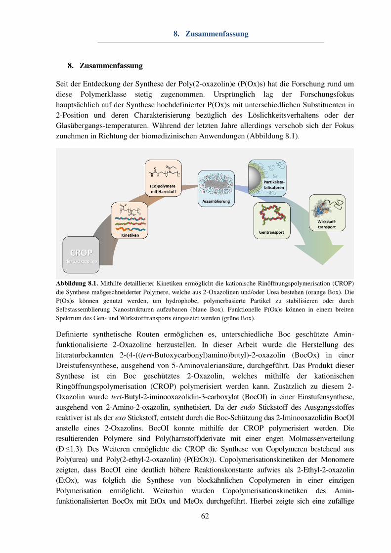
8. Zusammenfassung
62
8. Zusammenfassung
Seit der Entdeckung der Synthese der Poly(2-oxazolin)e (P(Ox)s) hat die Forschung rund um
diese Polymerklasse stetig zugenommen. Ursprünglich lag der Forschungsfokus
hauptsächlich auf der Synthese hochdefinierter P(Ox)s mit unterschiedlichen Substituenten in
2-Position und deren Charakterisierung bezüglich des Löslichkeitsverhaltens oder der
Glasübergangs-temperaturen. Während der letzten Jahre allerdings verschob sich der Fokus
zunehmen in Richtung der biomedizinischen Anwendungen (Abbildung 8.1).
Abbildung 8.1. Mithilfe detaillierter Kinetiken ermöglicht die kationische Rinöffnungspolymerisation (CROP)
die Synthese maßgeschneiderter Polymere, welche aus 2-Oxazolinen und/oder Urea bestehen (orange Box). Die
P(Ox)s können genutzt werden, um hydrophobe, polymerbasierte Partikel zu stabilisieren oder durch
Selbstassemblierung Nanostrukturen aufzubauen (blaue Box). Funktionelle P(Ox)s können in einem breiten
Spektrum des Gen- und Wirkstofftransports eingesetzt werden (grüne Box).
Definierte synthetische Routen ermöglichen es, unterschiedliche Boc geschützte Amin-
funktionalisierte 2-Oxazoline herzustellen. In dieser Arbeit wurde die Herstellung des
literaturbekannten 2-(4-((tert-Butoxycarbonyl)amino)butyl)-2-oxazolin (BocOx) in einer
Dreistufensynthese, ausgehend von 5-Aminovaleriansäure, durchgeführt. Das Produkt dieser
Synthese ist ein Boc geschütztes 2-Oxazolin, welches mithilfe der kationischen
Ringöffnungspolymerisation (CROP) polymerisiert werden kann. Zusätzlich zu diesem 2-
Oxazolin wurde tert-Butyl-2-iminooxazolidin-3-carboxylat (BocOI) in einer Einstufensynthese,
ausgehend von 2-Amino-2-oxazolin, synthetisiert. Da der endo Stickstoff des Ausgangsstoffes
reaktiver ist als der exo Stickstoff, entsteht durch die Boc-Schützung das 2-Iminooxazolidin BocOI
anstelle eines 2-Oxazolins. BocOI konnte mithilfe der CROP polymerisiert werden. Die
resultierenden Polymere sind Poly(harnstoff)derivate mit einer engen Molmassenverteilung
(Ð ≤1.3). Des Weiteren ermöglichte die CROP die Synthese von Copolymeren bestehend aus
Poly(urea) und Poly(2-ethyl-2-oxazolin) (P(EtOx)). Copolymerisationskinetiken der Monomere
zeigten, dass BocOI eine deutlich höhere Reaktionskonstante aufwies als 2-Ethyl-2-oxazolin
(EtOx), was folglich die Synthese von blockähnlichen Copolymeren in einer einzigen
Polymerisation ermöglicht. Weiterhin wurden Copolymerisationskinetiken des Amin-
funktionalisierten BocOx mit EtOx und MeOx durchgeführt. Hierbei zeigte sich eine zufällige

8. Zusammenfassung
63
Monomerverteilung bei der Polymerisation von BocOx und EtOx, während Copolymere bestehend
aus BocOx und 2-Methyl-2-oxazolin (MeOx) eine graduelle Monomerverteilung aufwiesen.
Ausgehend von den durchgeführten Kinetiken konnten verschiedene Copolymere synthetisiert und
charakterisiert werden.
Ein zweiter Schwerpunkt dieser Arbeit bestand darin, P(Ox) enthaltende Nanostrukturen
herzustellen und zu charakterisieren. Hierbei wurden zunächst wasserlösliche Homopolymere
bestehend aus EtOx oder MeOx in Bezug auf ihre Fähigkeiten als Stabilisatoren für
hydrophobe Nanopartikel untersucht. Homopolymere mit unterschiedlichem
Polymerisationsgrad (DP; 25, 60, 100, 200) wurden synthetisiert und bezüglich ihrer Eignung
als Emulgatoren für die Nanoemulsionsmethode mit den kommerziellen Emulgatoren
Poly(vinylalkohol) und Pluronic F127 verglichen. Zusätzlich wurden diese Polymere als
Kryoprotektoren eingesetzt und mit den standardmäßig genutzten Zuckern Glukose,
Trehalose und Saccharose verglichen. Polymere mit einem DP von 60, 100 oder 200 erwiesen
sich hierbei als sehr gute Stabilisatoren als „all-in-one“ System während der Präparation,
Aufreinigung und Gefriertrocknung der Nanopartikel.
Ein weiterer Schwerpunkt dieses Kapitels beschäftigte sich mit der Selbst- und Co-
Assemblierung von P(Ox)-basierten Copolymeren. Es konnte gezeigt werden, dass
blockähnliche Copolymere bestehend aus P(EtOx) und Poly(harnstoff) in wässrigem Medium
assembliert werden können und die resultierenden Nanostrukturen eine hohe Uniformität
aufweisen. Diese Nanostrukturen werden vermutlich durch Wasserstoffbrückenbindungen
stabilisiert.
Weiterhin konnten amphiphile Block-Copolymere bestehend aus einem hydrophoben 2-
Nonyl-2-oxazolin (NonOx) Block sowie einem hydrophilen EtOx oder 2-(4-Aminobutyl)-2-
oxazolin (AmOx) Block selbst- und co-assembliert werden. Nanostrukturen, welche aus
ausschließlich mit dem AmOx enthaltenden Copolymere präpariert wurden, waren hierbei
sphärisch, während alle Co-Assemblierungen sowie die Selbst-Assemblierungen des EtOx
enthaltenden Copolymers stäbchenförmig waren. Es konnte eine kritische
Mizellenkonzentration von 10-7 bis 10-6 M bestimmt werden. Des Weiteren konnte mithilfe
von dynamischer Lichtstreuung eine pH-responsive Größenänderung der Nanostrukturen
abhängig von Anteil an AmOx bestimmt werden. Diese Nanostrukturen wurden in einem
späteren Abschnitt dieser Arbeit für den Transport genetischen Materials genutzt.
Nanostrukturen, genauer gesagt Nanogele, welche für den Transport von Wirkstoffen
bestimmt waren, wurden aus wasserlöslichen Block-Copolymeren, bestehend aus AmOx und
EtOx, hergestellt. Diese Copolymere wurden in Chloroform assembliert und anschließend
über die Aminogruppen mittels einer Imin-Bindung quervernetzt, um definierte Nanogele zu
erhalten. Des Weiteren wurde der Quervernetzungsprozess dazu genutzt einen Wirkstoff
kovalent in den Kern des Nanogels zu binden. Mittels DLS Messungen konnten die
Reversibilität dieser Nanogele nachgewiesen werden. Die Freigabe des Wirkstoffes ist
deshalb wahrscheinlich, jedoch nicht quantifizierbar.
Der Transport genetischen Materials in Zellen stellt ein wichtiges Themengebiet dieser Arbeit
dar. Aus diesem Grund wurden die zuvor hergestellten pH responsiven Polymernano-
strukturen diesbezüglich untersucht. Zusätzlich zu den Nanostrukturen wurden hydrophile
Copolymere bestehend aus AmOx und EtOx oder AmOx und MeOx untersucht. Es zeigte
sich, dass die kationischen Copolymere das genetische Material reversibel binden konnten.
8. Zusammenfassung
64
Zudem wurde eine erwartete Abhängigkeit der Zytotoxizität vom Amin-Gehalt der
verwendeten Polymere bestätigt. Obwohl die wasserlöslichen Polymere und die
Nanostrukturen ein ähnliches Verhalten bezüglich der zellulären Aufnahme zeigten, konnten
nur Mizellen-basierte Systeme erfolgreich transfizieren. Diese Überlegenheit gegenüber den
wasserlöslichen System kann sowohl auf dem hydrophoben Anteil der Polymere als auch der
Anordnung zur Nanostruktur beruhen.
Im letzten Kapitel dieser Arbeit, welches sich mit dem Transport von kovalent gebundenen
Wirkstoffen befasste, wurden ebenfalls hydrophile Polymere genutzt. Hierbei konnte zum
einen die gezielte Bindung Azid-endfunktionalisierter Polymere mittels Kupfer katalysierter
Click-Chemie demonstriert werden. Zum anderen konnten die wasserlöslichen DOX
enthaltenden Nanostrukturen in vitro und in vivo bezüglich ihrer zellulären Aufnahme und der
therapeutischen Effizient untersucht werden. Es konnte festgestellt werden, dass unter in vitro
Bedingungen eine verlangsamte zelluläre Aufnahme der Nanogele im Vergleich zum reinen
Wirkstoff stattfindet. Des Weiteren konnte die Zytotoxizität in HT-29 Zell-Linien gesteigert
werden. In vivo Experimente zeigten eine erhöhte Lebensdauer der mit DOX Nanogelen
behandelten Tiere im Vergleich zu Tieren, welche mit DOX behandelt wurden.
Zusammengefasst konnte in dieser Arbeit die Synthese neuer aus P(Ox) und Poly(urea)
bestehenden Polymere gezeigt werden. Diese Polymere wurden ausgehend von Amin-
funktionalisierten 2-Oxazolinen hergestellt und bezüglich ihrer Syntheseroute als auch ihrer
Polymerisationskinetiken mit Amin-funktionalisierten P(Ox) verglichen. Weiterhin konnten
wasserlösliche P(Ox)-basierte Homopolymere als Nanopartikelstabilisatoren während der
Herstellung, Aufreinigung und Lyophilisierung verwendet werden. Des Weiteren wurden
Blockcopolymere assembliert und die Nanostrukturen charakterisiert. Diese Systeme konnten
schließlich erfolgreich zum Transport von Wirkstoffen und genetischem Material verwendet
werden.
9. References
65
9. References
[1] R. Duncan, R. Gaspar, Mol. Pharm. 2011, 8, 2101-2141. [2] C. Tros de Ilarduya, Y. Sun, N. Düzgüneş, Eur. J. Pharm. Sci. 2010, 40, 159-170. [3] N. Adams, U. S. Schubert, Adv. Drug Deliv. Rev. 2007, 59, 1504-1520. [4] R. Luxenhofer, Y. Han, A. Schulz, J. Tong, Z. He, A. V. Kabanov, R. Jordan,
Macromol. Rapid Commun. 2012, 33, 1613-1631. [5] T. Kagiya, S. Narisawa, T. Maeda, K. Fukui, J. Polym. Sci. C: Polym. Lett. 1966, 4,
441-445. [6] W. Seeliger, E. Aufderhaar, W. Diepers, R. Feinauer, R. Nehring, W. Thier, H.
Hellmann, Angew. Chem. Int. Ed. 1966, 5, 875-888. [7] D. Tomalia, D. Sheetz, J. Polym. Sci. Part A: Polym. Chem. 1966, 4, 2253-2265. [8] T. Bassiri, A. Levy, M. Litt, J. Polym. Sci. C: Polym. Lett. 1967, 5, 871-879. [9] R. Hoogenboom, Macromol. Chem. Phys. 2007, 208, 18-25. [10] B. Guillerm, S. Monge, V. Lapinte, J. J. Robin, Macromol. Rapid Commun. 2012, 33,
1600-1612. [11] F. Wiesbrock, R. Hoogenboom, C. H. Abeln, U. S. Schubert, Macromol. Rapid
Commun. 2004, 25, 1895-1899. [12] H. Schlaad, C. Diehl, A. Gress, M. Meyer, A. L. Demirel, Y. Nur, A. Bertin,
Macromol. Rapid Commun. 2010, 31, 511-525. [13] M. W. Fijten, C. Haensch, B. M. van Lankvelt, R. Hoogenboom, U. S. Schubert,
Macromol. Chem. Phys. 2008, 209, 1887-1895. [14] L.-Y. Qiu, L. Yan, L. Zhang, Y.-M. Jin, Q.-H. Zhao, Int. J. Pharm. 2013, 456, 315-
324. [15] Y. Gao, Y. Li, Y. Li, L. Yuan, Y. Zhou, J. Li, L. Zhao, C. Zhang, X. Li, Y. Liu,
Nanoscale 2015, 7, 597-612. [16] J. Li, Y. Zhou, C. Li, D. Wang, Y. Gao, C. Zhang, L. Zhao, Y. Li, Y. Liu, X. Li,
Bioconjugate Chem. 2014, 26, 110-119. [17] K. Kempe, R. Hoogenboom, M. Jaeger, U. S. Schubert, Macromolecules 2011, 44,
6424-6432. [18] G. Volet, T.-X. Lav, J. Babinot, C. Amiel, Macromol. Chem. Phys. 2011, 212, 118-
124. [19] Y. Zhao, Y. Zhou, D. Wang, Y. Gao, J. Li, S. Ma, L. Zhao, C. Zhang, Y. Liu, X. Li,
Acta Biomater. 2015, 17, 182-192. [20] P. H. Kierstead, H. Okochi, V. J. Venditto, T. C. Chuong, S. Kivimae, J. M. Fréchet,
F. C. Szoka, J. Controlled Release 2015, 213, 1-9. [21] Z. He, A. Schulz, X. Wan, J. Seitz, H. Bludau, D. Y. Alakhova, D. B. Darr, C. M.
Perou, R. Jordan, I. Ojima, J. Controlled Release 2015, 208, 67-75. [22] Z. He, X. Wan, A. Schulz, H. Bludau, M. A. Dobrovolskaia, S. T. Stern, S. A.
Montgomery, H. Yuan, Z. Li, D. Alakhova, Biomaterials 2016, 101, 296-309. [23] L. Tauhardt, M. Frant, D. Pretzel, M. Hartlieb, C. Bücher, G. Hildebrand, B. Schröter,
C. Weber, K. Kempe, M. Gottschaldt, J. Mater. Chem. B 2014, 2, 4883-4893. [24] M. C. Woodle, C. M. Engbers, S. Zalipsky, Bioconjugate Chem. 1994, 5, 493-496. [25] S. Zalipsky, C. B. Hansen, J. M. Oaks, T. M. Allen, J. Pharm. Sci. 1996, 85, 133-137. [26] H. Xu, W. Zhang, Y. Li, F. Y. Fei, P. P. Yin, X. Yu, M. N. Hu, Y. S. Fu, C. Wang, D.
J. Shang, Pharm. Res. 2014, 31, 3038-3050. [27] M. Glassner, M. Vergaelen, R. Hoogenboom, Polym. Int. 2018, 67, 32-45.
9. References
66
[28] K. L. Eskow Jaunarajs, D. G. Standaert, T. X. Viegas, M. D. Bentley, Z. Fang, B. Dizman, K. Yoon, R. Weimer, P. Ravenscroft, T. H. Johnston, Mov. Disord. 2013, 28, 1675-1682.
[29] R. Luxenhofer, R. Jordan, Macromolecules 2006, 39, 3509-3516. [30] K. Kempe, C. Weber, K. Babiuch, M. Gottschaldt, R. Hoogenboom, U. S. Schubert,
Biomacromolecules 2011, 12, 2591-2600. [31] G. Le Fer, C. Le Cœur, J.-M. Guigner, C. Amiel, G. Volet, Langmuir 2017, 33, 2849-
2860. [32] G. Le Fer, C. Amiel, G. Volet, Eur. Polym. J. 2015, 71, 523-533. [33] T.-X. Lav, P. Lemechko, E. Renard, C. Amiel, V. Langlois, G. Volet, React. Funct.
Polym. 2013, 73, 1001-1008. [34] S. Cesana, A. Kurek, M. A. Baur, J. Auernheimer, O. Nuyken, Macromol. Rapid
Commun. 2007, 28, 608-615. [35] O. Sedlacek, B. D. Monnery, J. Mattova, J. Kucka, J. Panek, O. Janouskova, A.
Hocherl, B. Verbraeken, M. Vergaelen, M. Zadinova, R. Hoogenboom, M. Hruby, Biomaterials 2017, 146, 1-12.
[36] G. Vancoillie, W. L. A. Brooks, M. A. Mees, B. S. Sumerlin, R. Hoogenboom, Polym.
Chem. 2016, 7, 6725-6734. [37] A. Gress, A. Völkel, H. Schlaad, Macromolecules 2007, 40, 7928-7933. [38] A. C. Rinkenauer, L. Tauhardt, F. Wendler, K. Kempe, M. Gottschaldt, A. Traeger, U.
S. Schubert, Macromol. Biosci. 2015, 15, 414-425. [39] C. Taubmann, R. Luxenhofer, S. Cesana, R. Jordan, Macromol. Biosci. 2005, 5, 603-
612. [40] S. Cesana, J. Auernheimer, R. Jordan, H. Kessler, O. Nuyken, Macromol. Chem. Phys.
2006, 207, 183-192. [41] M. Hartlieb, D. Pretzel, K. Kempe, C. Fritzsche, R. M. Paulus, M. Gottschaldt, U. S.
Schubert, Soft Matter 2013, 9, 4693-4704. [42] R. Hoogenboom, M. W. M. Fijten, U. S. Schubert, J. Polym. Sci. Part A: Polym.
Chem. 2004, 42, 1830-1840. [43] M. Glassner, D. R. D’hooge, J. Young Park, P. H. M. Van Steenberge, B. D.
Monnery, M.-F. Reyniers, R. Hoogenboom, Eur. Polym. J. 2015, 65, 298-304. [44] T. Saegusa, S. Kobayashi, A. Yamada, Macro. Chem. Phys. 1976, 177, 2271-2283. [45] M. W. M. Fijten, R. Hoogenboom, U. S. Schubert, J. Polym. Sci. Part A: Polym.
Chem. 2008, 46, 4804-4816. [46] R. Hoogenboom, M. W. M. Fijten, H. M. L. Thijs, B. M. van Lankvelt, U. S.
Schubert, Des. Monomers Polym. 2005, 8, 659-671. [47] H. Goossens, S. Catak, M. Glassner, V. R. de la Rosa, B. D. Monnery, F. De Proft, V.
Van Speybroeck, R. Hoogenboom, ACS Macro Lett. 2013, 2, 651-654. [48] F. Wiesbrock, R. Hoogenboom, M. A. M. Leenen, M. A. R. Meier, U. S. Schubert,
Macromolecules 2005, 38, 5025-5034. [49] R. Hoogenboom, M. W. Fijten, S. Wijnans, A. M. van den Berg, H. M. Thijs, U. S.
Schubert, J. Comb. Chem. 2006, 8, 145-148. [50] P. Wilson, P. C. Ke, T. P. Davis, K. Kempe, Eur. Polym. J. 2017, 88, 486-515. [51] M. Hartlieb, K. Kempe, U. S. Schubert, J. Mater. Chem. B 2015, 3, 526-538. [52] R. Hoogenboom, H. M. L. Thijs, M. W. M. Fijten, B. M. van Lankvelt, U. S.
Schubert, J. Polym. Sci. Part A: Polym. Chem. 2007, 45, 416-422. [53] C.-A. Fustin, N. Lefèvre, R. Hoogenboom, U. S. Schubert, J.-F. Gohy, Macromol.
Chem. Phys. 2007, 208, 2026-2031. [54] M. N. Leiske, M. Hartlieb, F. H. Sobotta, R. M. Paulus, H. Gorls, P. Bellstedt, U. S.
Schubert, Polym. Chem. 2016, 7, 4924-4936.
9. References
67
[55] K. Kempe, S. L. Ng, K. F. Noi, M. Müllner, S. T. Gunawan, F. Caruso, ACS Macro
Lett. 2013, 2, 1069-1072. [56] M. N. Leiske, M. Hartlieb, C. Paulenz, D. Pretzel, M. Hentschel, C. Englert, M.
Gottschaldt, U. S. Schubert, Adv. Funct. Mater. 2015, 25, 2458-2466. [57] M. Hartlieb, D. Pretzel, M. Wagner, S. Hoeppener, P. Bellstedt, M. Görlach, C.
Englert, K. Kempe, U. S. Schubert, J. Mater. Chem. B 2015, 3, 1748-1759. [58] T. B. Bonné, K. Lüdtke, R. Jordan, P. Štěpánek, C. M. Papadakis, Colloid Polym. Sci.
2004, 282, 833-843. [59] R. Ivanova, T. Komenda, T. B. Bonné, K. Lüdtke, K. Mortensen, P. K. Pranzas, R.
Jordan, C. M. Papadakis, Macromol. Chem. Phys. 2008, 209, 2248-2258. [60] S. Jaksch, A. Schulz, Z. Di, R. Luxenhofer, R. Jordan, C. M. Papadakis, Macromol.
Chem. Phys. 2016, 217, 1448-1456. [61] Z. He, L. Miao, R. Jordan, D. S-Manickam, R. Luxenhofer, A. V. Kabanov,
Macromol. Biosci. 2015, 15, 1004-1020. [62] T. Bus, C. Englert, M. Reifarth, P. Borchers, M. Hartlieb, A. Vollrath, S. Hoeppener,
A. Traeger, U. S. Schubert, J. Mater. Chem. B 2017, 5, 1258-1274. [63] M. N. Leiske, F. H. Sobotta, S. Hoeppener, J. C. Brendel, A. Traeger, U. S. Schubert,
Biomacromolecules 2018, 19, 748-760. [64] R. W. Moreadith, T. X. Viegas, M. D. Bentley, J. M. Harris, Z. Fang, K. Yoon, B.
Dizman, R. Weimer, B. P. Rae, X. Li, Eur. Polym. J. 2017, 88, 524-552. [65] Y. Seo, A. Schulz, Y. Han, Z. He, H. Bludau, X. Wan, J. Tong, T. K. Bronich, M.
Sokolsky, R. Luxenhofer, Polym. Adv. Technol. 2015, 26, 837-850. [66] J. Tong, X. Yi, R. Luxenhofer, W. A. Banks, R. Jordan, M. C. Zimmerman, A. V.
Kabanov, Mol. Pharm. 2012, 10, 360-377. [67] R. Luxenhofer, A. Schulz, C. Roques, S. Li, T. K. Bronich, E. V. Batrakova, R.
Jordan, A. V. Kabanov, Biomaterials 2010, 31, 4972-4979. [68] L. Tauhardt, K. Kempe, M. Gottschaldt, U. S. Schubert, Chem. Soc. Rev. 2013, 42,
7998-8011. [69] T. Verbrugghen, B. D. Monnery, M. Glassner, S. Stroobants, R. Hoogenboom, S.
Staelens, J. Controlled Release 2016, 235, 63-71. [70] M. Glassner, L. Palmieri, B. D. Monnery, T. Verbrugghen, S. Deleye, S. Stroobants,
S. Staelens, L. Wyffels, R. Hoogenboom, Biomacromolecules 2016, 18, 96-102. [71] Y. Liu, Y. Wang, Y. Wang, J. Lu, V. Pin III, M. Weck, J. Am. Chem. Soc. 2011,
133, 14260-14263. [72] M. Bauer, C. Lautenschlaeger, K. Kempe, L. Tauhardt, U. S. Schubert, D. Fischer,
Macromolecular Bioscience 2012, 12, 986-998. [73] M. Bauer, S. Schroeder, L. Tauhardt, K. Kempe, U. S. Schubert, D. Fischer, J. Polym.
Sci. Part A: Polym. Chem. 2013, 51, 1816-1821. [74] R. Shah, Z. Kroneková, A. Zahoranová, L. Roller, N. Saha, P. Sáha, J. Kronek, J.
Mater. Sci. Mater. Med. 2015, 26, 157. [75] H. P. C. Van Kuringen, J. Lenoir, E. Adriaens, J. Bender, B. G. De Geest, R.
Hoogenboom, Macromol. Biosci. 2012, 12, 1114-1123. [76] Y. Gao, Y. Zhou, L. Zhao, C. Zhang, Y. Li, J. Li, X. Li, Y. Liu, Acta Biomater. 2015,
23, 127-135. [77] M.-J. Shieh, C.-L. Peng, W.-L. Chiang, C.-H. Wang, C.-Y. Hsu, S.-J. J. Wang, P.-S.
Lai, Mol. Pharm. 2010, 7, 1244-1253. [78] P. Goddard, L. E. Hutchinson, J. Brown, L. J. Brookman, J. Controlled Release 1989,
10, 5-16. [79] A. Mero, Z. Fang, G. Pasut, F. M. Veronese, T. X. Viegas, J. Controlled Release
2012, 159, 353-361.
9. References
68
[80] F. C. Gaertner, R. Luxenhofer, B. Blechert, R. Jordan, M. Essler, J. Controlled
Release 2007, 119, 291-300. [81] L. wyffels, T. Verbrugghen, B. D. Monnery, M. Glassner, S. Stroobants, R.
Hoogenboom, S. Staelens, J. Controlled Release 2016, 235, 63-71. [82] R. Gaspar, R. Duncan, Adv. Drug Deliv. Rev. 2009, 61, 1220-1231. [83] H. Xu, M. Hu, X. Yu, Y. Li, Y. Fu, X. Zhou, D. Zhang, J. Li, Eur. J. Pharm.
Biopharm. 2015, 91, 66-74. [84] C. Englert, A.-K. Trützschler, M. Raasch, T. Bus, P. Borchers, A. S. Mosig, A.
Traeger, U. S. Schubert, J. Controlled Release 2016, 241, 1-14. [85] M. A. Mees, R. Hoogenboom, Macromolecules 2015, 48, 3531-3538. [86] I. Forfar, C. Jarry, J.-P. Fayet, A. Carpy, Arch. Pharm. 1992, 325, 541-543. [87] J.-J. Bosc, I. Forfar, C. Jarry, J. Ouhabi, J.-M. Leger, A. Carpy, Arch. Pharm. 1990,
323, 561-566. [88] A. Nagai, T. Miyagawa, H. Kudo, T. Endo, Macromolecules 2003, 36, 9335-9339. [89] O. Bayer, Angew. Chem. 1947, 59, 257-272. [90] V. E. Meyer, G. G. Lowry, J. Polym. Sci. A: Gen. Pap. 1965, 3, 2843-2851. [91] S. M. Shawki, A. E. Hamielec, J. Appl. Polym. Sci. 1979, 23, 3155-3166. [92] T. Yamamoto, M. Yoshizawa, A. Mahmut, M. Abe, S.-i. Kuroda, T. Imase, S. Sasaki,
J. Polym. Sci. A: Polym. Chem. 2005, 43, 6223-6232. [93] F. Wiesbrock, R. Hoogenboom, M. Leenen, S. F. G. M. van Nispen, M. van der Loop,
C. H. Abeln, A. M. J. van den Berg, U. S. Schubert, Macromolecules 2005, 38, 7957-7966.
[94] T.-H. Liu, W.-T. Cheng, K.-T. Hunang, J. Photopolym. Sci. Technol. 2010, 23, 529-533.
[95] K. Kempe, E. F.-J. Rettler, R. M. Paulus, A. Kuse, R. Hoogenboom, U. S. Schubert, Polymer 2013, 54, 2036-2042.
[96] F. Danhier, E. Ansorena, J. M. Silva, R. Coco, A. Le Breton, V. Préat, J. Controlled
Release 2012, 161, 505-522. [97] J. M. Barichello, M. Morishita, K. Takayama, T. Nagai, Drug Dev. Ind. Pharm. 1999,
25, 471-476. [98] H. Fessi, F. Puisieux, J. P. Devissaguet, N. Ammoury, S. Benita, Int. J. Pharm. 1989,
55, R1-R4. [99] C. Song, V. Labhasetwar, H. Murphy, X. Qu, W. Humphrey, R. Shebuski, R. Levy, J.
Controlled Release 1997, 43, 197-212. [100] P. Fonte, S. Soares, F. v. Sousa, A. Costa, V. Seabra, S. Reis, B. Sarmento,
Biomacromolecules 2014, 15, 3753-3765. [101] P. Fonte, S. Reis, B. Sarmento, J. Controlled Release 2016, 225, 75-86. [102] H. K. Makadia, S. J. Siegel, Polymers 2011, 3, 1377-1397. [103] S. Bozdag, K. Dillen, J. Vandervoort, A. Ludwig, J. Pharm. Pharmacol. 2005, 57,
699-707. [104] R. Basak, R. Bandyopadhyay, Langmuir 2013, 29, 4350-4356. [105] S. Galindo-Rodriguez, E. Allémann, H. Fessi, E. Doelker, Pharm. Res. 2004, 21,
1428-1439. [106] K. Jain, P. Kesharwani, U. Gupta, N. K. Jain, Int. J. Pharm. 2010, 394, 122-142. [107] E. Betthausen, M. Drechsler, M. Fortsch, F. H. Schacher, A. H. E. Muller, Soft Matter
2011, 7, 8880-8891. [108] M. Wilhelm, C. L. Zhao, Y. Wang, R. Xu, M. A. Winnik, J. L. Mura, G. Riess, M. D.
Croucher, Macromolecules 1991, 24, 1033-1040. [109] T. B. Bonné, C. M. Papadakis, K. Lüdtke, R. Jordan, Colloid Polym. Sci. 2007, 285,
491-497.
9. References
69
[110] A. W. Jackson, D. A. Fulton, Macromolecules 2012, 45, 2699-2708. [111] J. S. Basuki, H. T. Duong, A. Macmillan, R. B. Erlich, L. Esser, M. C. Akerfeldt, R.
M. Whan, M. Kavallaris, C. Boyer, T. P. Davis, ACS Nano 2013, 7, 10175-10189. [112] I. Amelio, F. Cutruzzolá, A. Antonov, M. Agostini, G. Melino, Trends Biochem. Sci.
2014, 39, 191-198. [113] W. Godbey, M. A. Barry, P. Saggau, K. K. Wu, A. G. Mikos, J. Biomed. Mater. Res.
2000, 51, 321-328. [114] C.-H. Ahn, S. Y. Chae, Y. H. Bae, S. W. Kim, J. Controlled Release 2002, 80, 273-
282. [115] B. D. Monnery, M. Wright, R. Cavill, R. Hoogenboom, S. Shaunak, J. H. Steinke, M.
Thanou, Int. J. Pharm. 2017, 521, 249-258. [116] G.-H. Hsiue, H.-Z. Chiang, C.-H. Wang, T.-M. Juang, Bioconjugate Chem. 2006, 17,
781-786. [117] J. M. Harris, N. E. Martin, M. Modi, Clin. Pharmacokinet. 2001, 40, 539-551. [118] M. Hamidi, A. Azadi, P. Rafiei, Drug Delivery 2006, 13, 399-409. [119] T. Lühmann, V. Spieler, V. Werner, M.-G. Ludwig, J. Fiebig, T. D. Mueller, L.
Meinel, ChemBioChem 2016, 17, 2123-2128. [120] B. Thonemann, G. Schmalz, K.-A. Hiller, H. Schweikl, Dent. Mater. 2002, 18, 318-
323. [121] T. Yildirim, A. Traeger, P. Sungur, S. Hoeppener, C. Kellner, I. Yildirim, D. Pretzel,
S. Schubert, U. S. Schubert, Biomacromolecules 2017, 18, 3280-3290. [122] K. K. Upadhyay, A. N. Bhatt, A. K. Mishra, B. S. Dwarakanath, S. Jain, C. Schatz, J.-
F. Le Meins, A. Farooque, G. Chandraiah, A. K. Jain, Biomaterials 2010, 31, 2882-2892.
List of abbreviations
70
List of abbreviations
1H-NMR - Proton nuclear magnetic resonance spectroscopy
6AF - 6-Amino fluorescein
AmOx - 2-(4-Aminobutyl)-2-oxazoline)
API - Active pharmaceutical ingredient
Boc - tert-Butyloxycarbonyl
BocOI - tert-Butyl 2-iminooxazolidine-3-carboxylatePoly
BocOx - 2-(4-((tert-Butoxycarbonyl)amino)butyl)-2-oxazoline
CLSM - Confocal laser scanning microscopy
CMC - Critical micelle concentration
COSY - - Correlation spectroscopy
CROP - Cationic ring-opening polymerization
cryoTEM - Cryo transmission electron microscopy
CuAAC - Copper catalyzed alkyne azide click chemistry
Ð - Dispersity
d - Diameter
Da - Dalton
DFT - Discrete Fourier transform
DiBoc - Di-tert-butyl dicarbonate
DLS - Dynamic light scattering
DOSY - Diffusion ordered spectroscopy
DOX - Doxorubicin
DMAc - N,N-Dimethylacetamide
DMF - Dimethyl formamide
DNA - Deoxyribonucleic acid
DSC - Differential scanning calorimetry
DP - Degree of polymerization
EBA - Ethidium bromide assay
ELS - Electrophoretic light scattering
Em - Emission
EtBr - Ethidium bromide
EtOx - 2-Ethyl-2-oxazoline
Ex - Excitation
eYFP - Enhanced yellow fluorescent protein
GA - Glutaraldehyde
GC - Gas chromatography
HEK-293 - Human embryonic kidney cells 293
HR-ESI MS - High resolution electrospray ionization mass spectrometry
HRA - Heparin release assay
HSQC - Heteronuclear single quantum coherence
HT-29 - Human colorectal adenocarcinoma cell line
i-PrOH - iso-Propanol

List of abbreviations
71
I - Initiator
IL-4 - Interleukin-4
kp - Reaction rate constant
L929 - L929 mouse fibroblasts
lPEI - linear poly(ethylene imine)
µW - Microwave
M - Monomer
m/z - Mass to charge ratio
MeOx - 2-Methyl-2-oxazoline
MeTos - Methyl p-toluenesulfonate
MFI - Mean fluorescence intensity
Mn - Number average molar mass
N - Nanostructure
N/P - Nitrogen to phosphate ratio
N*/P - Amino group to phosphate ratio
n.a. - Not available
NEt3 - Triethylamine
NHS - N-Hydroxysuccinimide
NMR- - Nuclear magnetic resonance spectroscopy
NonOx - 2-Nonyl-2-oxazoline
p - para
P - Polymer
P2VP - Poly(2-vinylpyridine)
P(AmOx) - Poly(2-(4-aminobutyl)-2-oxazoline)
P(BocOI) - Poly(tert-butyl 2-iminooxazolidine-3-carboxylate)
P(EtOx) - Poly(2-ethyl-2-oxazoline)
P(MeOx) - Poly(2-methyl-2-oxazoline)
P(OI) - Poly(2-iminooxazolidine-3-carboxylate)
P(Ox) - Poly(2-oxazoline)
PBS - Phosphate buffered saline
PDI - Polydispersity index
pDNA - Plasmid deoxyribonucleic acid
PEG - Poly(ethylene glycol)
PEI - Poly(ethylene imine)
PLA - Poly(lactic acid)
PLGA - Poly(lactic-co-glycolic acid)
ppm - parts per million
Pre - Precursor
PS - Poly(styrene)
PVA - Poly(vinyl alcohol)
r - Reactivity ratio
RFU - Relative fluorescent unit
RI - Refractive index
RNA - Ribonucleic acid
List of abbreviations
72
RT - Room temperature
SD - Standard deviation
SEC - Size-exclusion chromatography
Td - Decomposition temperature
TFA - Trifluoroacetic acid
Tg - Glass transition temperature
TGA - Thermogravimetrical analysis
Wt - Weight

Curriculum vitae
73
Curriculum vitae
09/10/1989 Born in Delmenhorst, Germany
1996 – 2000 Grundschule am Grünen Kamp, Delmenhorst
2000 – 2002 Hermann-Allmers Schule, Delmenhorst
2002 – 2009 Gymnasium an der Willmsstraβe, Delmenhorst General qualification for university entrance
2009 – 2012 Academic studies in Biology and Chemistry (B.Sc.)
Car von Ossietzky University, Oldenburg
2011/2012 Student assistant in General and Inorganic Chemistry
2012 Bachelor thesis in the group of Prof. Dr. Ralf. A. Rabus
Carl von Ossietzky University, Oldenburg
Title: Enzymatic studies on the degradation of branched chain
amino acids in Phaeobacter gallaeciensis
2012 – 2014 Academic studies in Chemical Biology (M.Sc.)
Friedrich Schiller University Jena, Jena
2013 - 2014 Student assistant in the group of Prof. Dr. Ulrich S. Schubert
2014 Master thesis in the group of Prof. Ulrich S. Schubert
Friedrich Schiller University Jena, Jena
Title: Poly(2-oxazoline)s for DNA applications
2013 Internship in the group of Prof. Dr. Michael Haley
University of Oregon, Eugene (USA)
Since 2015 PhD student at the Laboratory of Organic and Macromolecular
Chemistry (IOMC) at the Friedrich Schiller University Jena in the
group of Prof. Dr. Ulrich S. Schubert
Thesis: Poly(2-oxazoline)s: Synthesis, self-assembly and
biomedical applications
Meike Nicole Leiske 22.03.2018 Jena _____________________
Publication list
74
Publication list
Peer reviewed publications
D. Hoelzer‡, M. N. Leiske‡, M. Hartlieb, T. Bus, D. Pretzel, S. Hoeppener, K. Kempe, R.
Thierbach, U. S. Schubert, “Tumor targeting with pH-responsive poly(2-oxazoline)-based
nanogels for metronomic doxorubicin treatment”, Oncotarget 2018, in press.
D. Hertz‡, M. N. Leiske‡, T. Wloka, A. Traeger, M. Hartlieb, M. M. Kessels, S. Schubert, B.
Qualmann, U. S. Schubert, “Comparison of statistical and gradient amino functionalized poly(2-oxazoline)s: Can the transfection efficiency be tuned by the macromolecular
structure?”, J. Polym. Sci. Part A: Polym. Chem. 2018, in press.
DOI: 10.1002/pola.29000.
M. Grube, M. N. Leiske, U. S. Schubert, I. Nischang, “POx as an alternative to PEG? A
hydrodynamic and light scattering study”, Macromolecules 2018, 51, 1905-1916.
DOI: 10.1021/acs.macromol.7b02665.
M. N. Leiske, F. H. Sobotta, F. Richter, S. Hoeppener, J. C. Brendel, A. Traeger, U. S.
Schubert, “How to tune the gene delivery and biocompatibility of poly(2-(4-aminobutyl)-2-
oxazoline) by self- and co-assembly”, Biomacromolecules 2018, 19, 748-760.
DOI: 10.1021/acs.biomac.7b01535.
M. N. Leiske, A.-K. Trützschler, S. Armoneit, P. Sungur, S. Hoeppener, M. Lehmann, A.
Traeger, U. S. Schubert, “Mission imPOxable - Or the unknown utilization of non-toxic
poly(2-oxazoline)s as cryoprotectant and surfactant at the same time”, J. Mater. Chem. B
2017, 5, 9102-9113.
DOI: 10.1039/C7TB02443F.
M. Hartlieb‡, T. Bus‡, J. Kübel, D. Pretzel, S. Hoeppener, M. N. Leiske, K. Kempe, B.
Dietzek, U.S. Schubert, “Tailoring cellular uptake and fluorescence of poly(2-oxazoline)-
based nanogels”, Bioconjugate Chem. 2017, 28, 1229-1235.
DOI: 10.1021/acs.bioconjchem.7b00067.
T. Lühmann, M. Schmidt, M. N. Leiske, V. Spieler, T. C. Majdanski, M. Grube, M. Hartlieb,
I. Nischang, S. Schubert, U. S. Schubert, L. Meinel, „Site-specific POxylation of interleukin-
4“, ACS Biomater. Sci. Eng. 2017, 3, 304-312.
DOI: 10.1021/acsbiomaterials.6b00578.
M. N. Leiske, M. Hartlieb, F. H. Sobotta, R. M. Paulus, H. Gorls, P. Bellstedt, U. S. Schubert,
“Cationic ring-opening polymerization of protected oxazolidine imines resulting in gradient
copolymers of poly(2-oxazoline) and poly(urea)”, Polym. Chem. 2016, 7, 4924-4936.
DOI: 10.1039/C6PY00785F.

Publication list
75
M. N. Leiske, M. Hartlieb, C. Paulenz, D. Pretzel, M. Hentschel, C. Englert, M. Gottschaldt,
U. S. Schubert, „Lab in a tube: Purification, amplification and detection of DNA using
poly(2-oxazoline) multilayers.“ Adv. Func. Mat. 2015, 25, 2458-2466.
DOI: 10.1002/adfm.201404510.
Submitted manuscripts
M. N. Leiske, M. Hartlieb, A. Traeger, U. S. Schubert, “Evolution of poly(2-oxazoline)s from
in vitro and in vivo studies to clinical trials.” Submitted.
‡Equal contribution of both authors.
Declared patents
Friedrich-Schiller-Universität Jena. Organische Polymerpartikel enthaltend Poly(oxazolin)-
Stabilisatoren und Verwendung von Poly(oxazolinen) zur Stabilisierung von organischen
Polymerpartikeln. Erfinder: Meike N. Leiske, Anja Traeger und Ulrich S. Schubert.
14.03.2017. DE 10 2017 002 454.5.
Friedrich-Schiller-Universität Jena. Stabilisierung von Zellkulturen durch Polyoxazoline.
Erfinder: Meike N. Leiske, Anja Traeger und Ulrich S. Schubert. 26.05.2017. DE 10 2017
005 048.1.
Non-peer reviewed conference proceedings
252nd ACS national meeting (Philadelphia, PA, USA):
M. N. Leiske§, M. Hartlieb, F. H. Sobotta, R. M. Paulus, H. Gorls, P. Bellstedt, U. S. Schubert
(2016): Co-polymers of poly(2-oxazoline) and substituted poly(urea) as an easy access to
hydrogen-bond stabilized nanostructures (final paper number: POLY 60).
252nd ACS National meeting (Philadelphia, PA, USA):
M. N. Leiske§, M. Hartlieb, C. Paulenz, D. Pretzel, M. Hentschel, C. Englert, M. Gottschaldt,
U. S. Schubert (2016): Lab in a tube: Purification, amplification, and detection of DNA using
poly(2-oxazoline) multilayers (final paper number: POLY 276).
Printing Future Days 2015 (Chemnitz, Germany):
M. Herzig, M. N. Leiske, E. Preußger, T. Luehmann, M. Hartlieb, S. Hoeppener, S. Hoelzer,
L. Meinel, U. S. Schubert (2015): Reactive inkjet printing of functional poly(oxazoline)s for
biomedical applications.

Publication list
76
Oral presentations
FSU Jena – U. Tokyo workshop 2017 (Jena, Germany):
M. N. Leiske§, A.-K. Trützschler, S. Armoneit, P. Sungur, S. Hoeppener, M. Lehmann, A.
Traeger, U. S. Schubert (2017): Mission imPOxable – Or the unknown utilization of non-
toxic poly(2-oxazoline)s as cryoprotectant and surfactant at the same time.
4th Euro BioMAT 2017 (Weimar, Germany):
M. N. Leiske§, M. Hartlieb, C. Paulenz, D. Pretzel, M. Hentschel, C. Englert, M. Gottschaldt,
U. S. Schubert (2017): Lab in a tube: Or the combination of DNA purification and
amplification by using a poly(2-oxazoline) based all-in one system.
252nd ACS national meeting (Philadelphia, PA, USA):
M. N. Leiske§, M. Hartlieb, F. H. Sobotta, R. M. Paulus, H. Gorls, P. Bellstedt, U. S. Schubert
(2016): Co-polymers of poly(2-oxazoline) and substituted poly(urea) as an easy access to
hydrogen-bond stabilized nanostructures (final paper number: POLY 60).
252nd ACS National meeting (Philadelphia, PA, USA):
M. N. Leiske§, M. Hartlieb, C. Paulenz, D. Pretzel, M. Hentschel, C. Englert, M. Gottschaldt,
U. S. Schubert (2016): Lab in a tube: Purification, amplification, and detection of DNA using
poly(2-oxazoline) multilayers (final paper number: POLY 276).
IRTG workshop, Polymers: Random coils and beyond (Wittenberg, Germany, November
2015):
M. N. Leiske§ (2015): Interaction between DNA and polyamines in solution.
§Presenter
Poster presentations
Macromolecular Colloquium Freiburg (Freiburg, Germany):
M. N. Leiske§, F. H. Sobotta, S. Hoeppener, J. C. Brendel, A. Traeger, U. S. Schubert (2018):
How to tune the cellular uptake of amino functionalized poly(2-oxazoline)s by appropriate
self-assembly.
The 12th International Conference on Advanced Polymers via Macromolecular Engineering
(Ghent, Belgium):
M. N. Leiske§, M. Hartlieb, D. Poburski, T. Bus, D. Pretzel, S. Hoeppener, K. Kempe, R.
Thierbach, U. S. Schubert (2017): Core-crosslinked poly(2-oxazoline) nanogels as
doxorubicin carriers for cancer therapy.
§Presenter

Acknowledgement / Danksagung
77
Acknowledgement / Danksagung
Last but not least I want to thank all the people who supported me over all the years and
worked hard to make all this possible. At first, I want to express my deep grant to my
scientific supervisor Prof. Dr. Ulrich S. Schubert. Thank you, Uli, for all the support and the
trust in my work over the years. I really enjoyed working in such an interdisciplinary,
international and incredibly well equipped group. It is very impressive what you developed in
Jena and I am keen to see what the future will bring.
Furthermore, I want to thank Dr. Matthias Hartlieb for his continuous support and supervision
over the years. Matthias, thank you for bringing me to the subject of polymer synthesis, for
your patience and your never-ending help and discussions. During the last years, your tutoring
helped me improving my skills and made my thesis joyful, even on a rocky road. I am happy
to see how your carrier develops in the future and who else you are going to rise during the
next years.
I also want to thank Dr. Anja Träger for daily supervision. Anja, you always trusted in my
knowledge and helped me to realize my ideas and to fill them with life. I am also very
thankful that I could find a sympathetic ear in you whenever it was needed. Thank you for
challenging all the small problems at work with me. I wish you all the best for your future
research group.
I would also like to thank the Bundesministerium für Bildung und Forschung for funding
(project: smart-dye-livery, #13N13416).
In the following I want to acknowledge all the coworkers within or outside the Schubert
group who supported me and cooperated with me over the years. Firstly, I want to thank
Fabian Sobotta who was so kind to act as my first guinea pig as a master student. Fabi, thank
you for all the help and support in the lab. You did a great job and I wish you all the best for
your future carrier. I would also like to thank Dr. Johannes C. Brendel, Dr. Christine Weber
and Dr. Marc Lehmann for all the discussions and the support.
Additionally, I want to thank the people who helped me working out my projects with their
great contributions. Thanks to Anne-Kristin Trützschler, David Hertz, Fabian Sobotta,
Friederike Richter, Mandy Grube, Marcel Schmidt, Pelin Sungur, Renzo Paulus, Sabine
Armoneit, Tanja Bus, Thomas Wloka, Valerie Spieler, Dr. Anja Träger, Dr. David Pretzel,
Dr. Dörte Hölzer, Dr. Helmar Görls, Dr. Ivo Nischang, Dr. Joachim Kübel, Dr. Johannes C.
Brendel, Dr. Kristian Kempe, Dr. Dr. Marc Lehmann, Matthias Hartlieb, Dr. Michael Kessels,
Dr. Peter Bellstedt, Dr. Rene Thierbach, Dr. Stephanie Schubert, Dr. Stephanie Höppener, Dr.
Tessa Lühmann, Prof. Dr. Benjamin Dietzek, Prof. Dr. Britta Qualmann and Prof. Dr. Dr.
Lorenz Meinel.
Further thanks go to all the students who did practical courses and contributed to my work.
Thank you, Nora Engel, Linda Lattermann, Andre Schumann and Stefanie Raps. And thank
you, Thomas Wloka. I am sorry I have to mention you with the interns, but this was too funny
for me. I know you can deal with it, you workaholic kinetics beast!
Furthermore, I want to thank the people, who keep the whole system running. I want to thank
Sandra Köhn, Kristin Schreyer, Nicole Fritz, Carolin Kellner, Annett Urbanek, Dr. Grit

Acknowledgement / Danksagung
78
Festag, Dr. Jürgen Vitz, Renzo Paulus, Alexander Meyer, Maximilian Kleinsteuber, Jens
Ulbrich, Friederike Pielenz, Steffi Stumpf and Gabi Sentis for instrument introductions,
maintenance and service measurements. Furthermore, I would like to thank Dr. Uwe Köhn
and Silvia Pfeifer for ordering management.
I further would like to thank Franca Frister, Sylvia Braunsdorf, Simone Burchardt and Doreen
Küchler for managing the bureaucracy. Without you, we all would have been stranded.
I would also like to thank the people who helped me to survive the nerve-racking times by
supporting my work with jokes, coffee breaks and senseless discussions. Thank you, Franca
Frister, Anne-Kristin Trützschler, Renzo Paulus, Dr. Christian Friebi, Tina Mede, Thomas
“Das Tier” Wloka, Michi Dirauf, Susi Seupel and Michael “The Lat” Pröhl.
In this regard, I would also like to thank the people in my old and new home for cheering me
up, whenever necessary. Thank you Hannah Eilers, Tini Witte, Stella Kaiser and Thesi Wilke.
You are the best friends I could have found. Thank you so much for accepting (or at least
ignoring) all my quirks for several years.
I also want to thank my parents Heidrun and Jürgen Leiske, my grandparents Margarete and
Heinz Leiske as well as my brother Heiko Leiske and my sister-in-law Stephanie Leiske for
their continuous support in all situations in life.
Finally, I want to thank my boyfriend and soulmate Thomas, who is already waiting in
Melbourne to experience the great adventure of living at the end of the world with me.
Contigo al fin del mundo.
Declaration of authorship / Selbstständigkeitserklärung
79
Declaration of authorship / Selbstständigkeitserklärung
Hiermit erkläre ich, dass ich die vorliegende Arbeit selbständig angefertigt, nicht anderweitig zu Prüfungszwecken vorgelegt und keine anderen als die angegebenen Hilfsmittel verwendet habe. Sämtliche wissentlich verwendete Textausschnitte, Zitate oder Inhalte anderer Verfasser wurden ausdrücklich als solche gekennzeichnet. I hereby certify that the work disclosed here is, to the best of my knowledge, original and the result of my own investigations, except as acknowledged, and has not been submitted, either in part or whole, for a degree at this or any other university. Jena, den 22.03.2018 ____________________
Meike Nicole Leiske

Publications P1 to P8
80
Publications P1 to P8
P1: Reprinted by permission of Royal Society of Chemistry. Copyright 2016.
P2: Reprinted by permission of Royal Society of Chemistry. Copyright 2017.
P3: Reprinted by permission of the American Chemical Society. Copyright 2018.
P4: Reprinted by permission of Impact Journals. Copyright 2018.
P5: Reprinted by permission of Wiley VCH. Copyright 2018.
P6: Reprinted by permission of M. N. Leiske, M. Hartlieb, A. Traeger and U. S. Schubert.
P7: Reprinted by permission of the American Chemical Society. Copyright 2017.
P8: Reprinted by permission of the American Chemical Society. Copyright 2017.

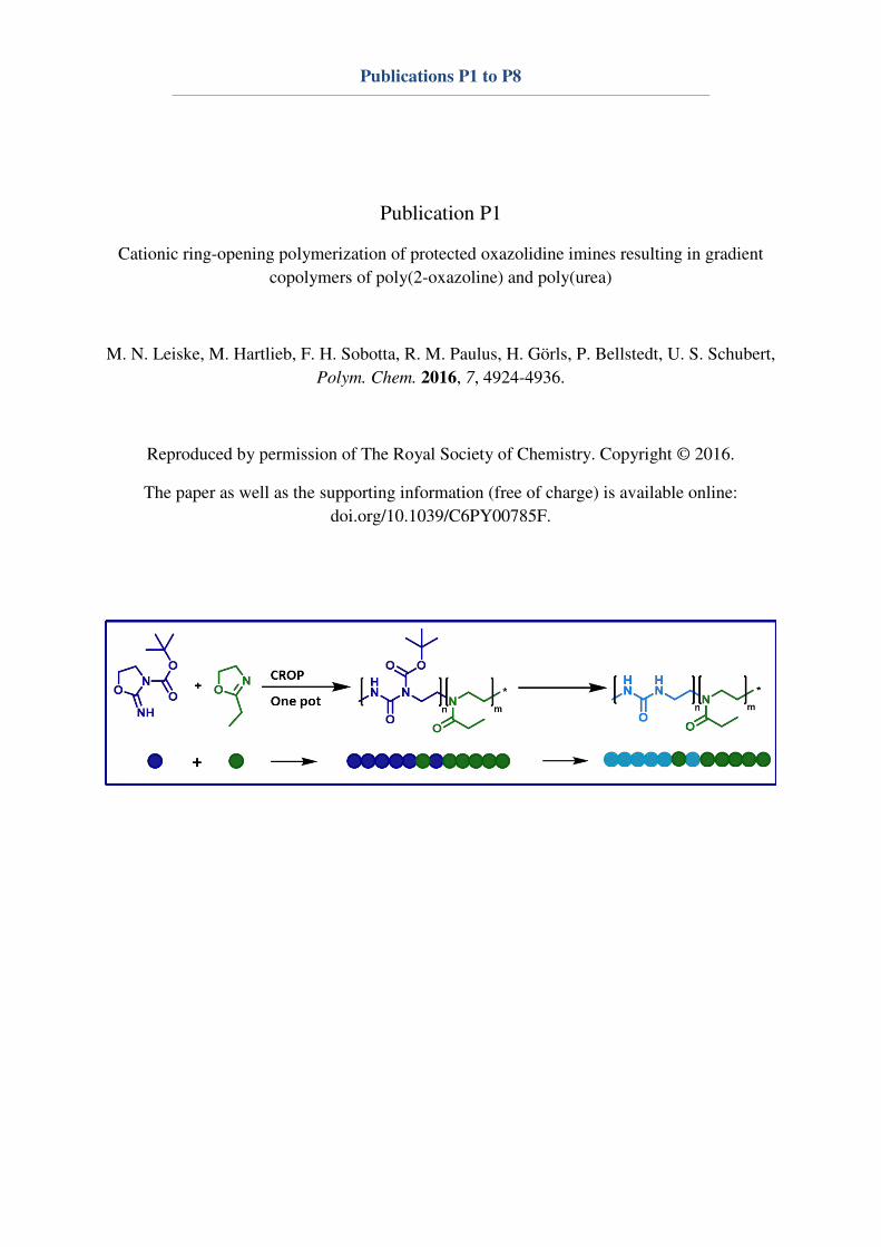
Publications P1 to P8
Publication P1
Cationic ring-opening polymerization of protected oxazolidine imines resulting in gradient
copolymers of poly(2-oxazoline) and poly(urea)
M. N. Leiske, M. Hartlieb, F. H. Sobotta, R. M. Paulus, H. Görls, P. Bellstedt, U. S. Schubert,
Polym. Chem. 2016, 7, 4924-4936.
Reproduced by permission of The Royal Society of Chemistry. Copyright © 2016.
The paper as well as the supporting information (free of charge) is available online:
doi.org/10.1039/C6PY00785F.
PolymerChemistry
PAPER
Cite this: Polym. Chem., 2016, 7,
4924
Received 4th May 2016,
Accepted 24th June 2016
DOI: 10.1039/c6py00785f
www.rsc.org/polymers
Cationic ring-opening polymerization of protectedoxazolidine imines resulting in gradientcopolymers of poly(2-oxazoline) and poly(urea)†
Meike N. Leiske,a,b Matthias Hartlieb,‡a,b Fabian H. Sobotta,a,b Renzo M. Paulus,a,b
Helmar Görls,c Peter Bellstedta and Ulrich S. Schubert*a,b
Poly(urea)s are a polymer class widely used in industry. Their utilization in biomedical applications is
already described, however, the use of controlled polymerization methods instead of polycondensation
approaches would allow a better control over the degree of polymerization and the dispersity of the
resulting polymers, improving their suitability for this particular field of application. Cationic ring-opening
polymerization (CROP) as a chain growth polymerization enables those requirements and, additionally,
allows the copolymerization with 2-oxazolines, which are generally known for their biocompatibility. In
this report, a Boc protected oxazolidine imine monomer is synthesized and polymerized in a homopoly-
merization, as well as in a copolymerization with 2-ethyl-2-oxazoline (EtOx) via CROP. The synthesized
polymers were analyzed regarding their chemical and physical properties, using NMR, GC, MALDI-MS,
SEC, TGA and DSC. Copolymerization kinetics revealed the formation of quasi-block copolymers, able to
self-assemble in aqueous solution as indicated by DLS.
Introduction
Poly(urea)s are commonly synthesized by polyaddition of di-isocyanates and diamines, a method, which was first describedby Otto Bayer in 1947.1 Their major applications are in situ
formed foams,1 anti-fouling coatings,2 grease3 and anticorro-sives.4 However, also biomedical applications such as siRNAdelivery are described.5–9
Modifications of the polymerization technique allow theformation of nanocapsules by interfacial polyaddition ininverse mini emulsion reactions9 or ring-opening polyaddi-
tion-condensations.10 One major advantage of these methodsis their insensitivity to moisture,1 however, the described reac-tion times extend to several hours depending on theirfunctionalization,6,11,12 which can be significantly reduced to afew minutes by using the microwave technique.13
Nevertheless, the step-growth polyaddition is not suited forthe synthesis of well-defined poly(urea)s with controlledmacromolecular architectures, molar mass as well as a head-to-tail structure.
Living and controlled polymerization techniques like theliving anionic and cationic as well as controlled radicalpolymerizations facilitate an improved control over the molarmasses and the use of functionalized monomers.14 The groupof Hedrick already described a method to synthesize well-defined poly(carbonate)s via controlled ring-opening polymeriz-ation instead of using polycondensation methods,15–17 leadingto materials which can be applied for biomedical applications.18
Moreover, the controlled cationic ring-opening polymerization(CROP) of 1,3-oxazolidine-2-thione with methyl triflate wasalready performed,19 yielding a sulfur analogue to poly(urea)s.The focus of this report is the CROP of an oxazolidine-2-imineand a possible copolymerization with 2-oxazolines to producepoly(urea)s or copolymeric systems with poly(oxazoline)s (POx).
The polymerization of 2-oxazolines via CROP20 was firstdescribed in the 1960s by four independent researchgroups.21–24 The use of microwave technology allows a signifi-
†Electronic supplementary information (ESI) available: NMR, SEC, TGA, DSC,HR-ESI-MS, ESI-MS, DLS and X-ray data, a detailed overview about all kineticstudies. Crystallographic data (excluding structure factors) has been deposited
with the Cambridge Crystallographic Data Centre as supplementary publication.CCDC 1477579 for 1b. For ESI and crystallographic data in CIF or other elec-
tronic format see DOI: 10.1039/c6py00785f‡Current address: Department of Chemistry, University of Warwick, Gibbet HillRoad, Coventry, CV4 7AL, UK.
aLaboratory of Organic and Macromolecular Chemistry (IOMC), Friedrich Schiller
University Jena, Humboldtstraße 10, 07743 Jena, Germany.
E-mail: [email protected] Center for Soft Matter (JCSM), Friedrich Schiller University Jena,
Philosophenweg 7, 07743 Jena, GermanycInstitute of Inorganic and Analytical Chemistry, Friedrich Schiller University Jena,
Humboldtstraße 8, 07743 Jena, Germany
4924 | Polym. Chem., 2016, 7, 4924–4936 This journal is © The Royal Society of Chemistry 2016
Publi
shed
on 2
4 J
une
2016. D
ow
nlo
aded
by T
huer
inger
Univ
ersi
tats
Lan
des
bib
lioth
ek J
ena
on 1
7/0
2/2
017 1
1:2
7:0
5.
View Article OnlineView Journal | View Issue

cant reduction of the polymerization time by increasing thereaction temperatures.25
By varying the initiator, moieties like alkyl chains26–29 orunsaturated double and triple bonds30–32 can be introduced totune the solubility or to enable post-modification strategies ofthe polymers. Furthermore, termination of the living chainend using nucleophiles results in end-functionalized poly-mers.33,34 The substituent in 2-position of the monomer canbe varied in order to alter the polymeric side group20 anddifferent monomers can be combined to yield copolymericsystems of various architectures.35
A further advantage of POx is their suitability as biomater-ial, as derivatives with small substituents like 2-ethyl-2-oxazo-line (EtOx) and 2-methyl-2-oxazoline (MeOx) are known to bebiocompatible and possess a stealth effect similar to polyethyl-ene glycol (PEG).36–39 In particular, EtOx is polymerizable witha high degree of control40 and shows PEG-like characteristicsconcerning biodistribution and excretion.41–43 During the lastyears, polymers based on EtOx have been widely characterizedregarding toxicity and cellular uptake.37,39,44 Currently, the in
vivo behavior of POx is receiving an increasing attention45,46
due to their numerous previously described benefits in
vitro.47,48 The usage of POx in drug delivery systems by conju-gation of low molar mass therapeutics49 and proteins50 arepromising for clinical studies. Additionally, the formation ofmicelles as drug carriers,51 nanogels44 and surface coatings forbio-analytics52 supports the increasing importance of POx.
The aim of this study is to combine the advantages of POxand poly(urea)s by an easy CROP based copolymerizationprocess. The mechanism and kinetic behavior of the homo-polymerization of a substituted oxazolidine imine, as well as thecopolymerization with EtOx are described. Homopolymers andcopolymers are characterized in detail regarding structure andphysical properties. Finally, the copolymer systems were investi-gated in terms of reactivity ratios and their potential to formnanostructures by hydrogen bond mediated self-assembly.
Results and discussionMonomer synthesis
In order to synthesize a monomer, which can be polymerizedby CROP to yield a poly(urea) structure, the commercially avail-
able 2-amino-2-oxazoline hydrochloride was reacted with di-tert-butyl dicarbonate (DiBoc) since the Boc protection groupis stable during CROP and can be removed under acidic con-ditions.53 As the starting material is in a tautomeric equili-brium, the molecule can react on two positions, at the endo-and the exo-nitrogen (Fig. 1A).
It was already described in literature that the endo-nitrogenof 2-amino-2-oxazoline, which is mainly present in solution,54
is more reactive as compared to the exo-nitrogen.55 The reacti-vity was predicted by density functional theory (DFT) from themolecule’s geometry derived by X-ray crystal analysis. Based onthe π-bond lengths, the electronegativity of the atoms could becalculated (endo-nitrogen: −0.317, exo-nitrogen: −0.272),explaining the differences in reactivity.
As a result, the monomer tert-butyl 2-iminooxazolidine-3-carboxylate (1b, BocOI) can be synthesized from 2-amino-2-oxazoline hydrochloride by reaction with DiBoc in the pres-ence of triethyl amine (TEA). Since the tautomeric equilibriumcannot be shifted completely to the 2-amino-2-oxazoline, thecrude product is a mixture of the two possible products tert-butyl (4,5-dihydrooxazol-2-yl)carbamate (∼30%, 1a) and BocOI(∼70%, 1b) (Fig. 1A) according to proton NMR experiments(data not shown). The monomer was purified by recrystalliza-tion and the structure was analyzed by 1H-NMR (Fig. S1A†),13C-NMR (Fig. S1B†), Fourier transform infrared spectroscopy(FTIR, Fig. S3†) and high-resolution electrospray ionizationmass spectrometry (HR-ESI-MS, Fig. S2†). HR-ESI-MS showsthe lability of the Boc protection group, as indicated by theappearance of the dominant signal at m/z = 131.0 [M − Boc +H+]. Information about the exact structure of the synthesizedmonomer was obtained by X-ray structure analysis of a singlecrystal (Fig. 1B, crystallographic data in Table S1†), proving theidentity of BocOI (1b).
Homopolymerization of BocOI (1b)
A kinetic study of the homopolymerization of BocOI (1b) wasperformed by preparing a stock solution of BocOI (1b) andmethyl tosylate (MeTos) ([M]/[I] = 60) in dry dichloromethaneunder a stream of argon and aliquoting the solution overseveral microwave vials, which were heated in a microwavesynthesizer for various times. The conversion was determinedvia gas chromatography (GC) by comparing the monomer con-centration with the initial concentration in the stock solution.
Fig. 1 A: Schematic representation of the Boc protection of 2-amino-2-oxazoline hydrochloride using DiBoc resulting in two possible tautomeric
products (tert-butyl (4,5-dihydrooxazol-2-yl)carbamate (1a) and tert-butyl 2-iminooxazolidine-3-carboxylate (1b)), and B: Molecular structure of 1b
derived by X-ray crystal structure analysis; H-atoms are excluded.
Polymer Chemistry Paper
This journal is © The Royal Society of Chemistry 2016 Polym. Chem., 2016, 7, 4924–4936 | 4925
Publi
shed
on 2
4 J
une
2016. D
ow
nlo
aded
by T
huer
inger
Univ
ersi
tats
Lan
des
bib
lioth
ek J
ena
on 1
7/0
2/2
017 1
1:2
7:0
5.
View Article Online

As the concentrations of BocOI (1b) and MeTos were equal inall reaction vessels, dichloromethane could be used as aninternal standard. By plotting the natural logarithm of thequotient of these amounts, the reaction constant (kp) could bedetermined from the slope of the linear fit and the initialinitiator concentration (Fig. 2A).
Since the monomer should be copolymerized with EtOx,140 °C was chosen as the reaction temperature being theoptimum for the synthesis of P(EtOx)s using the microwaveapproach.25 However, at 140 °C the time dependence of kp isnot linear (Fig. S4A†). Possible explanations for this phenom-enon might include the thermal deprotection of BocOI (1b), aswell as an increased reactivity of BocOI (1b), as compared toEtOx. Hence, a reaction temperature of 100 °C was applied.
The time-dependent polymerization kinetics calculated bythe monomer conversion obtained from GC-analytics of BocOI(1b) at 100 °C shows a nearly linear dependence of conversionwith time, however, side-reactions at higher molar masses arestill present as can be seen in the conversion dependent Mn
values and dispersities (Fig. 2B). The positive deviation of themolar mass from linearity might be attributed to side reac-tions, such as chain coupling at high conversions. Neverthe-less, a pseudo-1st-order kinetics can be postulated for theCROP of BocOI (1b, Fig. 2A), having a kp of 97.6 L mol−1 s−1,which is about 20 times higher than the kp of EtOx at thistemperature (kpEtOx = 4.6 L mol−1 s−1, Fig. S6B†).
SEC measurements were performed to obtain informationabout the increase in molar mass as well as the dispersity ofthe resulting polymers (Fig. 2B and C), showing a linearincrease in molar mass and narrow dispersities below 1.3 andmono-modal elution curves, indicating a living polymerizationat the given conditions. The increase in dispersity over timecould be explained on the same basis as the non-linearity ofthe polymerization at 140 °C. The decrease in temperature,however, lowers those unintended side-reactions.
NMR spectra of the purified polymer were collected toobtain information about the polymeric structure (Fig. 3). Inthe 1H-NMR spectrum, two signals for the backbone protonsare present at δ = 3.5 and 3.9 ppm, indicating different substi-tuted nitrogens in the polymer backbone as expected forP(BocOI) (Scheme 1). Additionally, the peak at δ = 8.8 ppm is
typical for an amide group. Further information was obtainedfrom the 1H–
1H-COSY-NMR spectrum, correlating protons onneighboring carbon-atoms in the polymer. It is clearly visiblethat the protons at δ = 3.5 and 3.9 ppm are coupling with eachother. Furthermore, the proton of the downfield shifted back-bone signal couples with the amide group at δ = 8.8 ppm indi-cating a poly(urea) like polymeric structure.
Since BocOI (1b) shows comparable structural character-istics as 1,3-oxazolidine-2-thione,19 a plausible mechanism forthe CROP of this monomer can be postulated (Scheme 1). Theelectrophilic methyl group of the initiator can be attacked bythe electron pair of the monomer’s imine functionality. Thisresults in a positively charged oxazolidinium ion as an inter-mediate and a finally partial positively charged carbon atom in5-position, that can be attacked by another imine group. Thisleads to a ring-opening and the final poly(urea) like Boc pro-tected backbone structure.
Fig. 2 Kinetic studies of BocOI ([M]/[I] = 60) in CH2Cl2 at 100 °C. A: Time-dependent polymerization kinetics calculated by the monomer conver-
sion obtained from GC-analytics. B: Molar mass and dispersity in dependence on the monomer conversion. C: SEC-traces (CHCl3-i-PrOH-TEA, PS-
cal.; 1st system peak after 12 min) of the reaction mixture after several polymerization times.
Fig. 3 1H–1H-COSY NMR spectrum (300 MHz, CDCl3) of P(BocOI)n.
Paper Polymer Chemistry
4926 | Polym. Chem., 2016, 7, 4924–4936 This journal is © The Royal Society of Chemistry 2016
Publi
shed
on 2
4 J
une
2016. D
ow
nlo
aded
by T
huer
inger
Univ
ersi
tats
Lan
des
bib
lioth
ek J
ena
on 1
7/0
2/2
017 1
1:2
7:0
5.
View Article Online
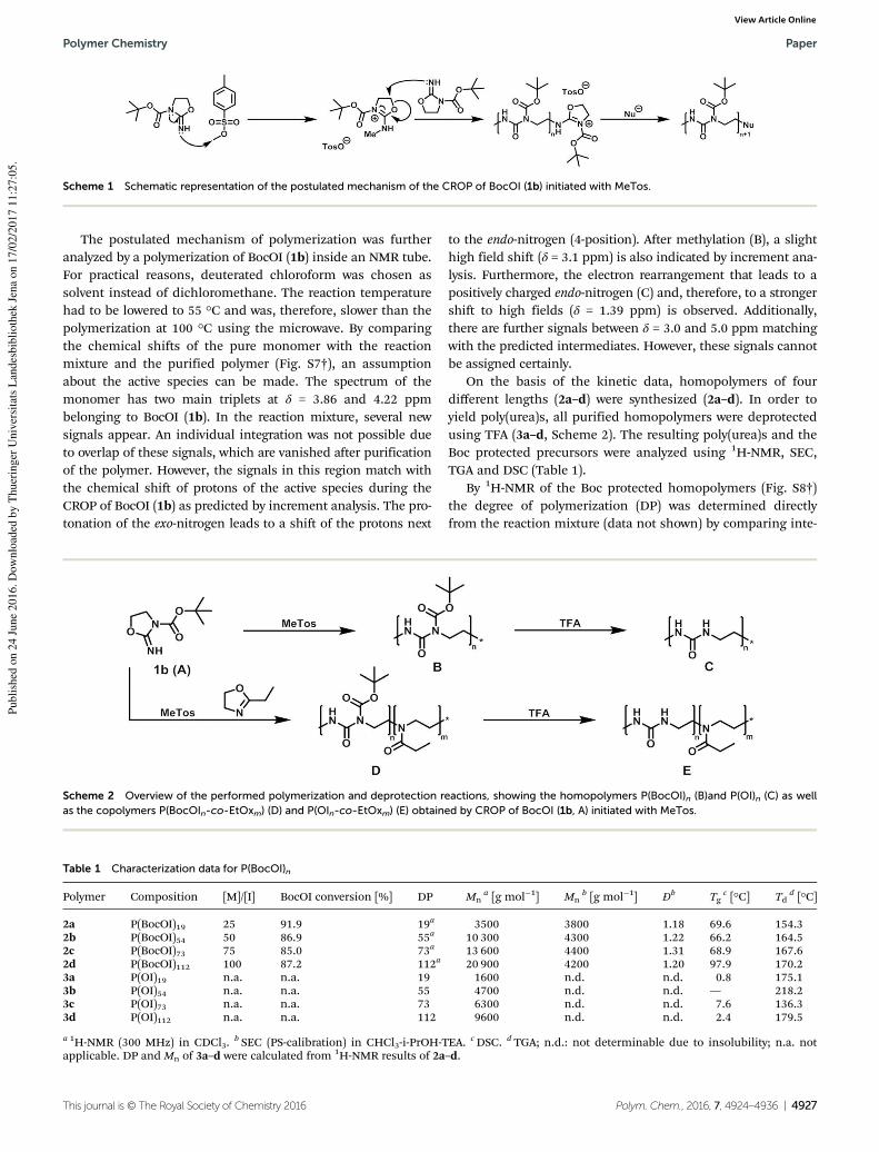
The postulated mechanism of polymerization was furtheranalyzed by a polymerization of BocOI (1b) inside an NMR tube.For practical reasons, deuterated chloroform was chosen assolvent instead of dichloromethane. The reaction temperaturehad to be lowered to 55 °C and was, therefore, slower than thepolymerization at 100 °C using the microwave. By comparingthe chemical shifts of the pure monomer with the reactionmixture and the purified polymer (Fig. S7†), an assumptionabout the active species can be made. The spectrum of themonomer has two main triplets at δ = 3.86 and 4.22 ppmbelonging to BocOI (1b). In the reaction mixture, several newsignals appear. An individual integration was not possible dueto overlap of these signals, which are vanished after purificationof the polymer. However, the signals in this region match withthe chemical shift of protons of the active species during theCROP of BocOI (1b) as predicted by increment analysis. The pro-tonation of the exo-nitrogen leads to a shift of the protons next
to the endo-nitrogen (4-position). After methylation (B), a slighthigh field shift (δ = 3.1 ppm) is also indicated by increment ana-lysis. Furthermore, the electron rearrangement that leads to apositively charged endo-nitrogen (C) and, therefore, to a strongershift to high fields (δ = 1.39 ppm) is observed. Additionally,there are further signals between δ = 3.0 and 5.0 ppm matchingwith the predicted intermediates. However, these signals cannotbe assigned certainly.
On the basis of the kinetic data, homopolymers of fourdifferent lengths (2a–d) were synthesized (2a–d). In order toyield poly(urea)s, all purified homopolymers were deprotectedusing TFA (3a–d, Scheme 2). The resulting poly(urea)s and theBoc protected precursors were analyzed using 1H-NMR, SEC,TGA and DSC (Table 1).
By 1H-NMR of the Boc protected homopolymers (Fig. S8†)the degree of polymerization (DP) was determined directlyfrom the reaction mixture (data not shown) by comparing inte-
Scheme 1 Schematic representation of the postulated mechanism of the CROP of BocOI (1b) initiated with MeTos.
Scheme 2 Overview of the performed polymerization and deprotection reactions, showing the homopolymers P(BocOI)n (B)and P(OI)n (C) as well
as the copolymers P(BocOIn-co-EtOxm) (D) and P(OIn-co-EtOxm) (E) obtained by CROP of BocOI (1b, A) initiated with MeTos.
Table 1 Characterization data for P(BocOI)n
Polymer Composition [M]/[I] BocOI conversion [%] DP Mna [g mol−1] Mn
b [g mol−1] Đb Tg
c [°C] Tdd [°C]
2a P(BocOI)19 25 91.9 19a 3500 3800 1.18 69.6 154.32b P(BocOI)54 50 86.9 55a 10 300 4300 1.22 66.2 164.52c P(BocOI)73 75 85.0 73a 13 600 4400 1.31 68.9 167.62d P(BocOI)112 100 87.2 112a 20 900 4200 1.20 97.9 170.23a P(OI)19 n.a. n.a. 19 1600 n.d. n.d. 0.8 175.13b P(OI)54 n.a. n.a. 55 4700 n.d. n.d. — 218.23c P(OI)73 n.a. n.a. 73 6300 n.d. n.d. 7.6 136.33d P(OI)112 n.a. n.a. 112 9600 n.d. n.d. 2.4 179.5
a 1H-NMR (300 MHz) in CDCl3.b SEC (PS-calibration) in CHCl3-i-PrOH-TEA. cDSC. d TGA; n.d.: not determinable due to insolubility; n.a. not
applicable. DP and Mn of 3a–d were calculated from 1H-NMR results of 2a–d.
Polymer Chemistry Paper
This journal is © The Royal Society of Chemistry 2016 Polym. Chem., 2016, 7, 4924–4936 | 4927
Publi
shed
on 2
4 J
une
2016. D
ow
nlo
aded
by T
huer
inger
Univ
ersi
tats
Lan
des
bib
lioth
ek J
ena
on 1
7/0
2/2
017 1
1:2
7:0
5.
View Article Online

grals of the aromatic protons of the MeTos initiator (δ = 7.15and 7.67 ppm) with one of the polymer backbone peaks (δ =3.5 ppm). As a consequence, DP values of 19, 54, 73 and 112could be obtained, respectively. A polymerization at [M]/[I] =300 was attempted, however resulted in increased dipseristies,which is a result of the longer reaction times required leadingto more side reactions (ESI Fig. S31†) According to the 1H-NMRinvestigations, the monomer consumption was between 85and 90% (Table 1). Therefore, the positive deviation of theobtained DP from the aimed [M]/[I] can most probably beexplained by inaccuracies while weighting the solid monomer.Additionally, 13C-NMR spectra of all Boc protected homopoly-mers were recorded (Fig. S10†). These spectra provide qualita-tive information about the polymeric structure. At δ =28.0 ppm, the peak of the Boc protection group is visible.Moreover, the methyl group of the initiator results in a peak atδ = 30.9 ppm. The two different methylene carbons of the poly-meric backbone split into two peaks at δ = 39.9 and 43.3 ppm.At δ = 126.0 and 128.6 ppm, the carbonyl carbons can beidentified. Furthermore, SEC measurements of the Boc pro-tected polymers (Fig. S13†) were performed to gain infor-mation about their dispersity, revealing narrow values(Table 1). The molar mass does not vary markedly with respectto the DP, which might be due to column interactions.Although it is not obvious from the kinetic studies (Fig. 2), themaximum molar mass for kinetic samples was Mn = 2700 gmol−1 and, therefore, is significantly lower than the length ofthe herein synthesized homopolymers. Additionally, anESI-MS spectrum (Fig. S14†) of P(BocOI)19 (2a) was recorded toverify the mass of the repeating unit. The spectrum showsspecific peaks for P(BocOI) as well as partially and fully de-protected poly(urea).
FTIR measurements provided further information aboutthe Boc protected (Fig. S11†) and deprotected (Fig. S12†)homopolymers. The spectra of the protected polymers showspecific bands at λ = 1533 and 3336 cm−1, representing the NHoscillations of the polymers. At λ = 2937 to 3009 cm−1, the CH2
vibrations of the polymeric backbone are visible as threedifferent peaks. Furthermore, the Boc protection group showsoscillations at λ = 754, 852 and 1371 cm−1, respectively. Carbo-nyl vibrations in the FTIR spectrum are visible at λ = 1157,1231, 1681 and 1719 cm−1, respectively.
The spectra of the deprotected polymers lack specific bandsat λ = 1371 and 852 cm−1 verifying the deprotection. Further-more, the carbonyl band at λ = 1719 cm−1 shift to a lower valuearound λ = 1681 cm−1, overlapping with the previous amideband, after deprotection.
1H-NMR measurements of the deprotected homopolymerswere performed in deuterated hydrochloric acid and verifiedthe success of the deprotection by disappearance of the Bocpeak in the spectrum (Fig. S9†) while keeping the backbonesignals at δ = 3.4 to 3.8 ppm. The peaks at high ppm values(δ = 7.15 and 7.67 ppm) could be attributed to a degradation ofthe polymer in the presence of high DCl concentrations. SECand MS measurements of deprotected polymers could not beperformed due to the insolubility in the available solvents.
To elucidate the thermal properties of the polymers, TGAand DSC measurements were performed. The TGA resultsrevealed a Td of 150 to 170 °C for the protected homopolymers(2a–d) that increases slightly with the polymer length(Table 1). Since this temperature does not change significantlywith the polymer length and the graph (Fig. S26A†) only showsa partial decomposition (∼50%), a thermal deprotection of thepolymers can be assumed. Issues regarding this phenomenonhave already been reported56 and might also be the trigger ofthe lack in controllability while polymerizing BocOI at 140 °C.After this first mass loss, all polymers show a comparabledecomposition behavior with a second Td around 200 °C. Like-wise, this is roughly the Td of the deprotected polymers (3a–d)(Fig. S27A†), verifying the hypothesis of a thermal de-protection. The lack of a clear trend of the Td of 3a–d might becaused by intra- and intermolecular stabilization of the poly-mers due to hydrogen-bond formation during the heatingprocess, which is also explaining the higher degradation temp-eratures of the polymers.
According to the DSC measurements (Fig. S28A†), the Bocprotected homopolymers (2a–d) show a Tg between 65 and70 °C, except polymer 2d which shows a significantly increasedTg of 98 °C (Table 1). The reason for this sudden increase in Tgis not understood jet. The deprotected homopolymers (3a–d)have a glass-transition temperature between 0 and 10 °C(Fig. S29A†), again showing no dependency of the chain lengthof the polymers. This sharp decrease in the Tg is caused byintra- and intermolecular interactions of the urea structure.
Copolymerization of BocOI and EtOx
After the successful homopolymerization of BocOI (1b) via
CROP, the copolymerization with EtOx was investigated. Theformation of gradient or quasi-block structured polymers wasassumed to be likely due to the high difference in the reactionconstants between the two monomers based on the homopoly-merization kinetics. To confirm this assumption, the reactivityratios were assessed by determination of velocity constants ofboth monomers while copolymerizing at different monomerratios. The general reaction conditions were kept equal to thehomopolymerization. Four stock solutions bearing differentmonomer ratios were prepared under a stream of argon, dividedover several reaction vessels and heated to 100 °C in a microwavesynthesizer for predetermined times. After the reaction times,the kp values for both monomers were calculated from the linearfit of the monomer conversion determined by GC measurements(Fig. 4). BocOI shows slight deviation from ideal linear behavior,which could be assigned to interactions with EtOx, as linearityincreases at higher BocOI contents. The resulting averaged con-stant for BocOI (kp = 58.0 ± 5.6 L mol−1 s−1) is about ten timeshigher than for EtOx (kp = 5.2 ± 1.4 L mol−1 s−1).
The formation of gradient copolymers from 2-oxazolinemonomers having different kp values is described in literaturefor several monomer pairs like 2-nonyl-2-oxazoline and2-phenyl-2-oxazoline (PhOx),51 as well as MeOx and 2-(3-butenyl)-2-oxazoline.57,58 However, it should be pointed outthat the difference between EtOx and BocOI is significantly
Paper Polymer Chemistry
4928 | Polym. Chem., 2016, 7, 4924–4936 This journal is © The Royal Society of Chemistry 2016
Publi
shed
on 2
4 J
une
2016. D
ow
nlo
aded
by T
huer
inger
Univ
ersi
tats
Lan
des
bib
lioth
ek J
ena
on 1
7/0
2/2
017 1
1:2
7:0
5.
View Article Online

larger, leading to the assumption of a quasi-block polymericstructure. Using the data from the kinetic studies, the reactivityratios (r) were calculated by non-linear least square fitting(Fig. 5A).59 For these calculations, the kinetic plots were usedto determine the ratio of consumed monomers at 30% of con-version of the faster monomer (BocOI, 1b) to ensure thatmonomer feed ratios are unaffected by the polymerization,excluding a falsification of the calculation by changingmonomer ratios at high conversions. Subsequently, the conver-sion of EtOx as the less reactive monomer is calculated at thistime. By plotting the incorporated EtOx fraction (F1) againstthe theoretical EtOx fraction in the monomer feed ( f1), a non-linear dependence is obtained. The reactivity ratios were calcu-lated using eqn (3) as adapted from literature,60 excludingnegative values. A similar behavior was already reported byHoogenboom et al.61 and Kagiya et al.62 for MeOx and PhOx,respectively.
The calculations resulted in rBocOI = 28.72 ± 10.07 ≫ 1 ≫
rEtOx = 0.000 ± 0.002. The very large difference between the two
monomers as well as the fact that the reactivity ratio of EtOx isclose to zero, while BocOI monomers are present in the reac-tion mixture, leads to the conclusion of a very narrow gradientarea in the polymer chain. To illustrate this fact, the monomerdistribution over the polymer chain (Fig. 5B) was calculatedbased on the kinetic plots (Fig. 4). Symbols represent theactual values of the EtOx fraction in the polymer chain. Thesevalues were fitted by a non-linear Boltzmann fitting (lines) toobtain information about the monomer distribution in thepolymer from initiation to termination, showing that mostBocOI (1b) is polymerized prior to the EtOx incorporation.
Based on the information obtained by the kinetic data, aseries of copolymers with different monomer ratios was syn-thesized and deprotected with TFA. Polymerizations werecarried out under microwave irradiation at 100 °C under argonatmosphere. Aiming for a DP of 100, polymers with 20%, 40%,60% and 80% BocOI (1b) content were synthesized. Theanalytical details for all copolymers can be found in Table 2.The DP was determined by 1H-NMR directly from the reaction
Fig. 4 Time-dependent polymerization kinetics calculated by the monomer conversion obtained from GC-analytics for BocOI (1b) and EtOx at four
monomer compositions, namely 20, 40, 60 and 80 mol% BocOI (1b), performed in dichloromethane at 100 °C with [M]/[I] = 100 and methyl tosylate
as initiator.
Polymer Chemistry Paper
This journal is © The Royal Society of Chemistry 2016 Polym. Chem., 2016, 7, 4924–4936 | 4929
Publi
shed
on 2
4 J
une
2016. D
ow
nlo
aded
by T
huer
inger
Univ
ersi
tats
Lan
des
bib
lioth
ek J
ena
on 1
7/0
2/2
017 1
1:2
7:0
5.
View Article Online
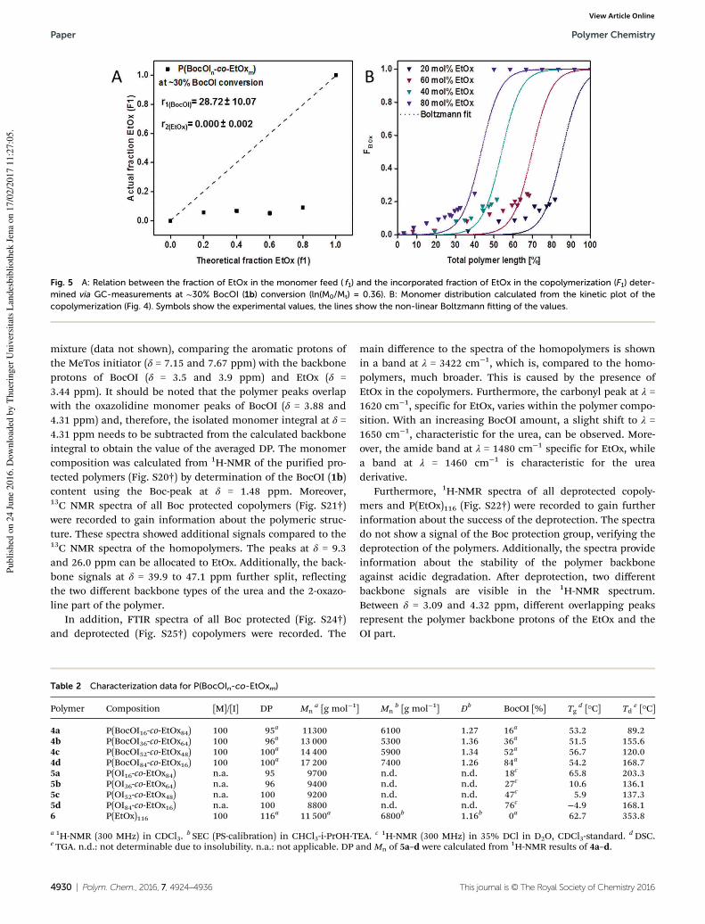
mixture (data not shown), comparing the aromatic protons ofthe MeTos initiator (δ = 7.15 and 7.67 ppm) with the backboneprotons of BocOI (δ = 3.5 and 3.9 ppm) and EtOx (δ =3.44 ppm). It should be noted that the polymer peaks overlapwith the oxazolidine monomer peaks of BocOI (δ = 3.88 and4.31 ppm) and, therefore, the isolated monomer integral at δ =4.31 ppm needs to be subtracted from the calculated backboneintegral to obtain the value of the averaged DP. The monomercomposition was calculated from 1H-NMR of the purified pro-tected polymers (Fig. S20†) by determination of the BocOI (1b)content using the Boc-peak at δ = 1.48 ppm. Moreover,13C NMR spectra of all Boc protected copolymers (Fig. S21†)were recorded to gain information about the polymeric struc-ture. These spectra showed additional signals compared to the13C NMR spectra of the homopolymers. The peaks at δ = 9.3and 26.0 ppm can be allocated to EtOx. Additionally, the back-bone signals at δ = 39.9 to 47.1 ppm further split, reflectingthe two different backbone types of the urea and the 2-oxazo-line part of the polymer.
In addition, FTIR spectra of all Boc protected (Fig. S24†)and deprotected (Fig. S25†) copolymers were recorded. The
main difference to the spectra of the homopolymers is shownin a band at λ = 3422 cm−1, which is, compared to the homo-polymers, much broader. This is caused by the presence ofEtOx in the copolymers. Furthermore, the carbonyl peak at λ =1620 cm−1, specific for EtOx, varies within the polymer compo-sition. With an increasing BocOI amount, a slight shift to λ =1650 cm−1, characteristic for the urea, can be observed. More-over, the amide band at λ = 1480 cm−1 specific for EtOx, whilea band at λ = 1460 cm−1 is characteristic for the ureaderivative.
Furthermore, 1H-NMR spectra of all deprotected copoly-mers and P(EtOx)116 (Fig. S22†) were recorded to gain furtherinformation about the success of the deprotection. The spectrado not show a signal of the Boc protection group, verifying thedeprotection of the polymers. Additionally, the spectra provideinformation about the stability of the polymer backboneagainst acidic degradation. After deprotection, two differentbackbone signals are visible in the 1H-NMR spectrum.Between δ = 3.09 and 4.32 ppm, different overlapping peaksrepresent the polymer backbone protons of the EtOx and theOI part.
Fig. 5 A: Relation between the fraction of EtOx in the monomer feed ( f1) and the incorporated fraction of EtOx in the copolymerization (F1) deter-
mined via GC-measurements at ∼30% BocOI (1b) conversion (ln(M0/Mt) = 0.36). B: Monomer distribution calculated from the kinetic plot of the
copolymerization (Fig. 4). Symbols show the experimental values, the lines show the non-linear Boltzmann fitting of the values.
Table 2 Characterization data for P(BocOIn-co-EtOxm)
Polymer Composition [M]/[I] DP Mna [g mol−1] Mn
b [g mol−1] Đb BocOI [%] Tg
d [°C] Tde [°C]
4a P(BocOI16-co-EtOx84) 100 95a 11300 6100 1.27 16a 53.2 89.24b P(BocOI36-co-EtOx64) 100 96a 13 000 5300 1.36 36a 51.5 155.64c P(BocOI52-co-EtOx48) 100 100a 14 400 5900 1.34 52a 56.7 120.04d P(BocOI84-co-EtOx16) 100 100a 17 200 7400 1.26 84a 54.2 168.75a P(OI16-co-EtOx84) n.a. 95 9700 n.d. n.d. 18c 65.8 203.35b P(OI36-co-EtOx64) n.a. 96 9400 n.d. n.d. 27c 10.6 136.15c P(OI52-co-EtOx48) n.a. 100 9200 n.d. n.d. 47c 5.9 137.35d P(OI84-co-EtOx16) n.a. 100 8800 n.d. n.d. 76c −4.9 168.16 P(EtOx)116 100 116a 11 500a 6800b 1.16b 0a 62.7 353.8
a 1H-NMR (300 MHz) in CDCl3.b SEC (PS-calibration) in CHCl3-i-PrOH-TEA. c 1H-NMR (300 MHz) in 35% DCl in D2O, CDCl3-standard.
dDSC.e TGA. n.d.: not determinable due to insolubility. n.a.: not applicable. DP and Mn of 5a–d were calculated from 1H-NMR results of 4a–d.
Paper Polymer Chemistry
4930 | Polym. Chem., 2016, 7, 4924–4936 This journal is © The Royal Society of Chemistry 2016
Publi
shed
on 2
4 J
une
2016. D
ow
nlo
aded
by T
huer
inger
Univ
ersi
tats
Lan
des
bib
lioth
ek J
ena
on 1
7/0
2/2
017 1
1:2
7:0
5.
View Article Online
The molar masses, as determined by SEC (Fig. S23†) arealmost constant for all protected polymers, whereas the differ-ences might originate from changes in monomer compo-sitions and the associated differences in molar mass as well asinteractions of the column material with P(BocOI). SEC investi-gations of the deprotected P(OI) copolymers were not possiblesince the polymers assembled in the SEC eluent.
TGA and DSC measurements of all copolymers andP(EtOx)116 (6) were performed to obtain information about thethermal properties. TGA analysis of the protected copolymers(4a–d) shows varying Td values, however, no apparent relationbetween the BocOI (1b) amount and the temperature can beobserved. Nevertheless, all Boc protected copolymers reveal thesame sharp decrease in mass as the homopolymers below200 °C, caused by thermal deprotection. Further decompo-sition follows the behavior of P(BocOI) and P(EtOx)(Fig. S26B†). The deprotected copolymers (5a–d) show asimilar degradation behavior as the homopolymers(Fig. S27B†). The low Td observed for protected polymers isattributed to thermal deprotection, which is not possible fordeprotected polymers. It should be noted that Td is defined asthe temperature at which 5% of mass loss is observed anddoes not necessarily mean a full decomposition. According tothe OI amount in the polymers, no obvious trend is observed.However, the Td of copolymers (100 to 200 °C) is in the samerange as observed for the homopolymers.
The Boc protected copolymers (4a–d) reveal a Tg around 50to 55 °C (Fig. S28B†), independent on the monomer ratio, asdetermined by DSC measurements.
After deprotection, a significant influence of the polymercomposition on the Tg values is visible, caused most probablyby intermolecular interactions of the macromolecules.P(BocOI) precursors (2a–d) as well as P(EtOx)116 (6) possess Tgvalues around 60 °C, resulting in an overall Tg between 50 and55 °C. Until now, we do not have an explanation for this
decrease in Tg of the copolymers compared to the homopoly-mers. The deprotected macromolecules (5a–d), however, showa distinct decrease in the Tg with increasing OI amount(Fig. S29B†). As the Tg of the P(OI) homopolymers is signifi-cantly lower than the Tg of P(EtOx)116, this could as well be theresult of a homogenious mixture of both blocks in bulk. Pre-vious investigations on POx also described a decrease in Tgassociated with the decreasing length of the EtOx block.63
Self-assembly
The results of the copolymerization studies from BocOI andEtOx show the formation of quasi-block copolymers withnarrow dispersity. As the P(OI) homopolymers are not watersoluble, block-like copolymers of P(OI) and P(EtOx) areexpected to self-assemble or aggregate in aqueous solution. Toevaluate the formation of such hydrogen bond stabilized64,65
nano-assemblies, the polymers were dissolved in TFA, which isknown to break hydrogen bonds, and MilliQ water was addedslowly under stirring to induce phase segregation. The resul-ting solution was dialyzed to remove the acid and to obtainnanostructures in solution. Larger aggregates were removed bysyringe filtration (0.45 μm) and the size distribution was deter-mined by DLS measurements (Table 3, Fig. S30†).
P(EtOx)116 (6) and copolymers with a low OI content (∼20 to30%, 5a,b) have a z-average around 4 nm in diameter, indica-ting unimers or small aggregates. In contrast, copolymers witha high OI amount (5c,d) form nanostructures in aqueous solu-tion, having diameters of 55.3 ± 13.4 and 107.7 ± 53.4 withincreasing co-monomer content. Furthermore, the zeta poten-tial of the aggregates was investigated. As shown in Table 3,P(EtOx)116 (6) shows a slightly negative zeta potential. Thecopolymers, on the other hand, show a positive zeta potential,which could be attributed to a partial degradation of PEtOxresulting in a minor amount of poly(ethylene imine) units inthe PEtOx segment, caused by the acidic conditions of de-
Table 3 Characterization of the formed nanostructures after precipitation of the copolymers in MilliQ water by DLS
Polymer Composition Concentrationa [mg mL−1] Sizeb [d, nm] ζ [mV]
5a P(OI16-co-EtOx84) 1 4.7 ± 0.1 18.6 ± 15.75b P(OI36-co-EtOx64) 1 5.1 ± 0.6 8.2 ± 0.55c P(OI52-co-EtOx48) 1 55.3 ± 13.4 9.1 ± 3.35d P(OI84-co-EtOx16) 0.1 107.7 ± 53.4 1.3 ± 1.86 P(EtOx)116 10 4.3 ± 0.7 −5.0 ± 0.4
aDetermined after freeze-drying of the dialyzed solutions. bNumber PSD.
Scheme 3 Schematic representation of the possible macrostructures derived from P(OIn-co-EtOxm) bearing different co-monomer ratios in water.
Polymer Chemistry Paper
This journal is © The Royal Society of Chemistry 2016 Polym. Chem., 2016, 7, 4924–4936 | 4931
Publi
shed
on 2
4 J
une
2016. D
ow
nlo
aded
by T
huer
inger
Univ
ersi
tats
Lan
des
bib
lioth
ek J
ena
on 1
7/0
2/2
017 1
1:2
7:0
5.
View Article Online
protection and a self assembly process (the P(EtOx) homopoly-mer was not treated with TFA prior to DLS measurements).Due to the low solubility of the polymers and the resulting lowresolution of the 1H NMR spectra it was not possible to provethis hypothesis.
It should be noted that the results on the self-assembly be-havior are preliminary and the optimization of copolymer com-position regarding aggregation and possibly micelleformation, as well as the investigation on the self-assemblingprocess and the resulting nanostructures will be the subject ofsubsequent studies (Scheme 3).
Conclusion
A Boc protected oxazolidine imine was synthesized, polymer-ized via CROP and deprotected to form poly(urea)s with a lowdispersity. Homopolymers of different lengths were syn-thesized and characterized via NMR, SEC, MALDI-MS, TGAand DSC. A copolymerization kinetic with EtOx was performedand the resulting reactivity ratios of the two monomerssuggest the formation of quasi-block copolymers. Copolymerswith different monomer ratios were synthesized and character-ized regarding their composition and thermal properties. Self-assembly of those polymers in aqueous solution resulted innano-assemblies as detected by DLS measurements. Thesestructures are believed to be a result of hydrogen bond for-mation between the P(OI) segments. The presented copolymersystem offers an easy access to quasi-block systems with thepotential to self-assemble in solution.
Further research will focus on the self-assembly processand the nature of the resulting nano-structures, as well as theassessment of their suitability for biomedical applications.
Experimental partMaterial and instrumentation
All chemicals were purchased from Sigma-Aldrich, Merck, TCIand Synthon-Chemicals. Triethylamine (TEA), 2-ethyl-2-oxazo-line (EtOx) and methyl tosylate (MeTos) were distilled todryness under argon atmosphere prior to usage. Dichloro-methane was obtained from a solvent purification system(MB-SPS-800 by MBraun) and stored under argon.
Polymerization reactions were performed under microwaveirradiation, using an Initiator Sixty single-mode microwavesynthesizer from Biotage, equipped with a noninvasive IRsensor (accuracy: 2%). Microwave vials were heated overnightat 100 °C under vacuum and allowed to cool to RT underargon before usage. Polymerizations were performed usingtemperature control.
Size-exclusion chromatography (SEC) of the protectedhomo- and copolymers was performed on a Shimadzu systemequipped with a SCL-10A system controller, a LC-10AD pump,a RID-10A refractive index detector and a PSS SDV columnwith chloroform-TEA-2-propanol (94 : 4 : 2) as eluent. The
column oven was set to 50 °C and a polystyrene (PS) standardwas used for calibration.
Proton NMR spectroscopy (1H-NMR) was performed at RTusing a Bruker Avance I 300 MHz spectrometer, utilizing eitherCDCl3 or 35% DCl in D2O as solvent. The chemical shifts aregiven in ppm relative to the signal from the residual non-deuterated chloroform. Measurements in DCl were performedwith an additional CDCl3 standard.
1H–1H-COSY NMR and 1H-NMR experiments of the
polymerization inside the NMR tube were performed at RT or55 °C on a Bruker Avance II 400 MHz, using CDCl3 as solvent.
IR spectra were recorded using an Affinity-1 FT-IR from Shi-madzu, utilizing the reflection technique.
High resolution electrospray ionization (HR-ESI) mass spec-trometry (MS) was performed on a micrOTOF Q-II (Bruker Dal-tonics) mass spectrometer equipped with an automatic syringepump from KD Scientific for sample injection at 4.5 kV at adesolvation temperature of 180 C. The mass spectrometer wasoperating in the positive ion mode.
Batch dynamic light scattering (DLS) was performed on aZetasizer Nano ZS (Malvern Instruments, Herrenberg,Germany). All measurements were performed in folded capil-lary cells (DTS1071, Malvern Instruments, Herrenberg,Germany). After an equilibration time of 180 s, 3 × 30 s runswere carried out at 25 °C (λ = 633 nm). Scattered light wasdetected at an angle of 173°. Each measurement was per-formed in triplicates. Apparent hydrodynamic radii, Rh, werecalculated according to the Stokes–Einstein equation.
Differential scanning calorimetry (DSC) experiments wereperformed on a Netzsch DSC 204 F1 Phoenix under a nitrogenatmosphere with a heating rate of 20 K min−1 from −20 to140 °C, if not indicated differently. Three cycles were recordedfor each sample. The glass transition temperature (Tg) valuesare reported for the second heating run. Thermo-gravimetricanalysis (TGA) was performed under a nitrogen atmosphere ona Netzsch TG 209 F1 Iris in the range from room temperatureto 800 °C with a heating rate of 10 K min−1.
For crystal structure determination, the intensity data werecollected on a Nonius KappaCCD diffractometer, using graph-ite-monochromated Mo-Kα radiation. Data were corrected forLorentz and polarization effects; absorption was taken intoaccount on a semi-empirical basis using multiple-scans.66–68
The structure was solved by direct methods (SHELXS69) andrefined by full-matrix least squares techniques against Fo
2
(SHELXL-97 69). All hydrogen atoms were located by differenceFourier synthesis and refined isotropically. All non-hydrogenatoms were refined anisotropically. MERCURY70 was used forstructure representations.
tert-Butyl 2-iminooxazolidine-3-carboxylate (BocOI) (1b)
2-Amino-2-oxazoline hydrochloride (20 g, 163 mmol) was dis-solved in a mixture of 1,4-dioxane (300 mL) and aqueoussodium hydroxide (3 wt%, 300 mL). Dry TEA (16.5 g,163 mmol) was added to this mixture to remove hydrochloricacid. DiBoc, (35 g, 163 mmol) was dissolved in 1,4-dioxane(150 mL) and added dropwise to the solution of 2-amino-2-oxa-
Paper Polymer Chemistry
4932 | Polym. Chem., 2016, 7, 4924–4936 This journal is © The Royal Society of Chemistry 2016
Publi
shed
on 2
4 J
une
2016. D
ow
nlo
aded
by T
huer
inger
Univ
ersi
tats
Lan
des
bib
lioth
ek J
ena
on 1
7/0
2/2
017 1
1:2
7:0
5.
View Article Online
zoline The reaction mixture was stirred at RT for 24 h and, sub-sequently, extracted with dichloromethane (3 × 200 mL). Theorganic phase was dried over sodium sulfate before evapor-ation of the solvent under reduced pressure. The crudeproduct was further purified via recrystallization from cyclo-hexanes to give the final product as a white crystalline solid(24.0 g, 87.2%).
1H-NMR (CDCl3, 300 MHz): δ = 7.38 (1H, s, NH), 4.23 (2H, t,CH2-oxazolidine), 3.86 (2H, t, CH2-oxazolidine), 1.51 (9H, s,CH3) ppm.
13C-NMR (CDCl3, 300 MHz): δ = 154.89 (O–CNH–N), 151.37(N–CO–O), 83.57 (O–C–(CH3)3), 62.21 (CH2-oxazolidine), 44.39(CH2-oxazolidine), 28.06 (CH3) ppm.
HR-ESI: m/z calc. for C8H14N2O3H [M + H]: 187.1007, found:187.1081 (error: 1.9 ppm).
FTIR: λ [cm−1] = 3422 (w, carbonyl), 3336 (m, NH), 2939 (s,CH2 CH3), 1620, 1570 (s, amide), 1165 (m, CH3).
Crystal data for 1b. C8H14N2O3, Mr = 186.21 g mol−1, colour-less prism, size 0.098 × 0.088 × 0.068 mm3, monoclinic, spacegroup P21/n, a = 9.7713(7), b = 9.5154(7), c = 11.5372(9) Å, β =113.738(4)°, V = 981.95(13) Å3, T = −140 °C, Z = 4, ρcalcd =1.260 g cm−3, µ (Mo-Kα) = 0.97 cm−1, multi-scan, transmin:0.6395, transmax: 0.7456, F(000) = 400, 6408 reflections inh(−12/12), k(−11/11), l(−14/14), measured in the range 2.32° ≤Θ ≤ 26.37°, completeness Θmax = 99.9%, 2004 independentreflections, Rint = 0.0448, 1736 reflections with Fo > 4σ(Fo), 174parameters, 0 restraints, R1obs = 0.0682, wR2
obs = 0.1284, R1all =0.0802, wR2all = 0.1320, GOOF = 1.307, largest difference peakand hole: 0.237/−0.213 e Å−3.
Kinetic studies
For kinetic investigations a stock solution of BocOI, (and EtOxfor copolymerizations), MeTos and CH2Cl2 ([M] = 1 mol−1,[M]/[I] = 60) was prepared, aliquoted into microwave vials(1 mL per vial) and heated in a microwave synthesizer (100 °C,varying reaction times). After polymerization, the conversionsof the monomers were determined using GC using the solventas an internal standard. The reaction rate constants kp of themonomers were determined using eqn (1) and (2) assumingthat the slope of the linear fit of ln([M]0/[M]t) = f (t ) complieswith keff.
ln M0 � ln Mt ¼ keff t ð1Þ
keff ¼ kp½I� ð2Þ
Reactivity ratios of both monomers were calculated for fourdifferent monomer ratios at 30% BocOI conversion (deter-mined by GC) using non-linear least square fitting60 (eqn (3)):
F1 ¼ðr1 � 1Þf12 þ f1
ðr1 þ r2 � 2Þf12 þ 2ð1� r2Þf1þr2; r1 � 0 ð3Þ
F1 = instantaneous mole fraction; f1 = mole fraction ofmonomer EtOx; f2 = mole fraction of monomer BocOI; r1 =reactivity ratio of EtOx; r2 = reactivity ratio of BocOI.
Homopolymerization inside an NMR tube
In an NMR tube BocOI (112 mg, 0.6 mmol), MeTos (0.8 µL,0.024 mmol) and CDCl3 (550 µL) were mixed under inert con-ditions and the reaction mixture was heated to 55 °C for a pre-determined time. 1H-NMR spectra at RT were recorded atspecific points of time to determine the reactive speciesduring polymerization.
Homopolymerization of BocOI (2a–d)
The experimental procedure for homopolymerizations is exem-plarily described on polymers with [M]/[I] = 25 (2a).
In a microwave vial BocOI (2793 mg, 15.0 mmol), MeTos(19.8 µL, 0.6 mmol) and dichloromethane (14.9 mL) weremixed under inert conditions and the reaction mixture washeated to 100 °C for a predetermined time. The resulting solu-tion was purified via BioBeads SX-1 column using CH2Cl2 aseluent. After combining the polymer fractions, the solvent wasevaporated under reduced pressure to obtain the product as awhite crystalline solid.
1H-NMR (CDCl3, 300 MHz): δ = 8.70 (0.8H, s, NH-back-bone), 3.87 (2H, m, backbone), 3.41 (2H, m, backbone), 2.80(0.06H, d, CH3), 1.46 (9H, m, CH3) ppm.
SEC (eluent: CHCl3-i-propanol-TEA, PS-standard): Mn =3700 g mol−1, Mw = 4700 g mol−1, Đ = 1.18.
FTIR: λ [cm−1] = 3336 (m, NH (backbone)), 3009, 2978, 2937(s, CH2 (backbone), CH3 (Boc)). 1719, 1681 (s, carbonyl), 1533(s, NH (backbone)), 1371 (s, CH3 (Boc)), 1231 (s, carbonyl),1157 (s, carbonyl), 852 (m, CH3 (Boc)), 754 (m, CH2
(backbone)).
Deprotection of P(BocOI) (3a–d)
The experimental procedure for deprotection is exemplarilydescribed for the polymers with a [M]/[I] = 25 (3a).
10 mL of TFA were added to the polymer and the solutionwas stirred at RT overnight. Subsequently, the reaction mixturewas diluted with methanol and precipitated from ice-colddiethyl ether (300 mL). The polymer was filtered off and driedin a high vacuum to obtain the product as a white crystallinesolid.
FTIR: λ [cm−1] = 3333 (s, NH (backbone)), 2939 (s, CH2
(backbone)), 1620, 1570 (s, amide (backbone)), 1446 (CH2
(backbone)), 1141 (s, carbonyl).
Copolymerization of BocOI and EtOx (4a–d)
The experimental procedure for copolymerizations is exempla-rily described for the polymers with a [M]/[I] = 100 aiming aBocOI amount of 20% (4a).
In a microwave vial BocOI (745 mg, 4.0 mmol), EtOx(1586 µL, 16.0 mmol), MeTos (30.3 µL, 0.2 mmol) and di-chloromethane (8.35 mL) were mixed under inert conditionsand the reaction mixture was heated to 100 °C for 15 h. Theresulting solution was purified by precipitation into ice colddiethyl ether. The white solid was filtered off and re-dissolvedin CH2Cl2 and the solvent was evaporated under reducedpressure to obtain the product as a white crystalline solid.
Polymer Chemistry Paper
This journal is © The Royal Society of Chemistry 2016 Polym. Chem., 2016, 7, 4924–4936 | 4933
Publi
shed
on 2
4 J
une
2016. D
ow
nlo
aded
by T
huer
inger
Univ
ersi
tats
Lan
des
bib
lioth
ek J
ena
on 1
7/0
2/2
017 1
1:2
7:0
5.
View Article Online
1H-NMR (CDCl3, 300 MHz): δ = 8.73 (0.1H, s, NH-back-bone), 3.90 (0.3H, m, backbone), 3.45 (3.6H, s, backbone), 2.40(1.7H, s, CH2 (EtOx)), 1.51 (1.3H, d, CH3 (BocOI)), 1.12 (2.5H,s, CH3 (EtOx)) ppm.
SEC (eluent: CHCl3-i-propanol-TEA, PS-standard): Mn =6100 g mol−1, Mw = 7700 g mol−1, Đ = 1.27.
FTIR: λ [cm−1] = 3422 (w, carbonyl), 3336 (m, NH (back-bone)), 2939 (s, CH2 (backbone), CH3 (Boc)), 1720 (s, carbonyl),1632 (m, amide), 1431 (m, CH3 (Boc), CH2 (backbone)), 1373(m, CH3 (Boc)), 1141 (s, carbonyl), 754 (m, CH2 (backbone)).
Deprotection of P(BocOI-co-EtOx) (5a–d)
The experimental procedure for deprotection is exemplarilydescribed for the polymers with a [M]/[I] = 100 aiming a BocOIamount of 20% (5a).
10 mL of TFA were added to P(BocOI-co-EtOx) and stirred atRT overnight. Subsequently, the solution was diluted withmethanol and purified by precipitation into ice cold diethylether (300 mL). The white solid was filtered off and dried in ahigh vacuum to obtain the product as a white crystalline solid.
1H-NMR (35% DCl in D2O, CDCl3-standard, 300 MHz):3.09–4.32 (4H, m, backbone), 2.63 (1.7H, s, CH2 (EtOx)), 1.11(2.4H, s, CH3 (EtOx)) ppm.
FTIR: λ [cm−1] = 3422 (w, carbonyl), 3336 (m, NH (back-bone)), 2939 (s, CH2 (backbone), CH3 (Boc)), 1616 (s, amide(BocOI)), 1580, 1562 (s, amide (EtOx)), 1138 (m, CH2, CH3
(EtOx)). 813 (m, CH2, CH3 (EtOx), CH3 (EtOx)).
Homopolymerization of EtOx (6)
In a microwave vial, EtOx (8076 µL, 80.0 mmol), MeTos(121.1 µL, 0.8 mmol) and acetonitrile (11.8 mL) were mixedunder inert condition and the reaction mixture was heated to140 °C for a predetermined time and subsequently quenchedby the addition of 500 µL of deionized H2O. The resulting solu-tion was purified via precipitation into ice cold diethyl ether.The polymer was filtered off and re-dissolved in CH2Cl2 andthe solvent was evaporated under reduced pressure to obtainthe product as a white crystalline solid.
1H-NMR (CDCl3, 300 MHz): δ = 4.34 (0.1H, s, backbone-OH), 3.44 (4.0H, s, backbone), 3.02 (0.3H, s, CH3-backbone),2.4 (1.7H, m, CH2 (EtOx)), 1.11 (2.5H, s, CH3 (EtOx)) ppm.
1H-NMR (35% DCl in D2O, CDCl3-standard, 400 MHz): 4.08(4H, s, backbone), 2.55 (2.4H, d, CH2 (EtOx)),1.10 (2.8H, s, CH3
(EtOx)) ppm.SEC (eluent: CHCl3-i-propanol-TEA, PS-standard): Mn =
6800 g mol−1, Mw = 7900 g mol−1, Đ = 1.16.FTIR: λ [cm−1] = 3422 (w, carbonyl), 2939 (s, CH2, CH3),
1620, 1570 (s, amide (backbone)), 1427 (s, CH2, CH3), 1160(m, CH2, CH3).
Self-assembly of P(OI-co-EtOx)
30 mg of deprotected copolymer (5a–d) were dissolved in TFA(600 µL). 3.0 mL of MilliQ water were added via syringe pump(10 mL h−1) while stirring (1400 rpm). Directly after theaddition, the solution was dialyzed against deionized H2O(MWCO 3500 g mol−1) for 72 h. The final concentration of the
solution was provided by lyophilization and determined as1 mg mL−1 for 5a–c, 0.1 mg mL for 5d and 10 mg mL−1.
Acknowledgements
The authors gratefully acknowledge the Bundesministeriumfür Bildung und Forschung (Germany) (project: smart-dye-livery, 081220/127) for funding. The authors thank Tina Schlot-thauer for ESI-MS measurements. M. H. gratefully acknowledgesthe German Research Foundation (DFG, GZ: HA 7725/1-1) forfunding.
References
1 O. Bayer, Angew. Chem., 1947, 59, 257–272.2 S. Holberg and C. Bischoff, Prog. Org. Coat., 2014, 77, 1591–
1595.3 S. L. Kwolek, J. Polym. Sci., Part A: Polym. Chem., 1964, 2,
5149–5160.4 L. Feng and J. O. Iroh, Prog. Org. Coat., 2014, 77, 590–599.5 P. Cass, W. Knower, T. Hinton, S. Shi, F. Grusche, M. Tizard
and P. Gunatillake, Acta Biomater., 2013, 9, 8299–8307.6 R. Kumar, R. Narayan, T. Aminabhavi and K. V. S. N. Raju,
J. Polym. Res., 2014, 21, 1–16.7 G. Morral-Ruíz, P. Melgar-Lesmes, M. L. García, C. Solans
and M. J. García-Celma, Polymer, 2012, 53, 6072–6080.8 G. Morral-Ruíz, C. Solans, M. L. García and M. J. García-
Celma, Langmuir, 2012, 28, 6256–6264.9 E.-M. Rosenbauer, K. Landfester and A. Musyanovych, Lang-
muir, 2009, 25, 12084–12091.10 Y. Saegusa and S. Nakamura, Macromol. Rapid Commun.,
1998, 19, 177–180.11 S. Li, J. Zhao, Z. Zhang, J. Zhang and W. Yang, RSC Adv.,
2015, 5, 6843–6852.12 H. Mighani, Carbon, 2012, 10, 235oC.13 S. Mallakpour and Z. Rafiee, J. Appl. Polym. Sci., 2004, 91,
2103–2113.14 K. Stridsberg, M. Ryner and A.-C. Albertsson, in Degradable
Aliphatic Polyesters, Springer, Berlin, Heidelberg, 2002, vol.157, pp. 41–65.
15 K. R. Carter, R. Richter, H. R. Kricheldorf and J. L. Hedrick,Macromolecules, 1997, 30, 6074–6076.
16 X. Ding, C. Yang, T. P. Lim, L. Y. Hsu, A. C. Engler,J. L. Hedrick and Y.-Y. Yang, Biomaterials, 2012, 33, 6593–6603.
17 D. P. Sanders, K. Fukushima, D. J. Coady, A. Nelson,M. Fujiwara, M. Yasumoto and J. L. Hedrick, J. Am. Chem.
Soc., 2010, 132, 14724–14726.18 F. Nederberg, Y. Zhang, J. P. K. Tan, K. Xu, H. Wang,
C. Yang, S. Gao, X. D. Guo, K. Fukushima, L. Li,J. L. Hedrick and Y.-Y. Yang, Nat. Chem., 2011, 3, 409–414.
19 A. Nagai, T. Miyagawa, H. Kudo and T. Endo, Macro-
molecules, 2003, 36, 9335–9339.
Paper Polymer Chemistry
4934 | Polym. Chem., 2016, 7, 4924–4936 This journal is © The Royal Society of Chemistry 2016
Publi
shed
on 2
4 J
une
2016. D
ow
nlo
aded
by T
huer
inger
Univ
ersi
tats
Lan
des
bib
lioth
ek J
ena
on 1
7/0
2/2
017 1
1:2
7:0
5.
View Article Online
20 K. Kempe, M. Lobert, R. Hoogenboom and U. S. Schubert,J. Polym. Sci., Part A: Polym. Chem., 2009, 47, 3829–3838.
21 T. G. Bassiri, A. Levy and M. Litt, J. Polym. Sci., Part B:
Polym. Lett., 1967, 5, 871–879.22 T. Kagiya, S. Narisawa, T. Maeda and K. Fukui, J. Polym.
Sci., Part B: Polym. Lett., 1966, 4, 441–445.23 W. Seeliger, E. Aufderhaar, W. Diepers, R. Feinauer,
R. Nehring, W. Thier and H. Hellmann, Angew. Chem., Int.
Ed. Engl., 1966, 5, 875–888.24 D. A. Tomalia and D. P. Sheetz, J. Polym. Sci., Part A: Polym.
Chem., 1966, 4, 2253–2265.25 F. Wiesbrock, R. Hoogenboom, C. H. Abeln and
U. S. Schubert, Macromol. Rapid Commun., 2004, 25, 1895–1899.
26 R. Jordan, K. Martin, H. J. Räder and K. K. Unger, Macro-
molecules, 2001, 34, 8858–8865.27 T. Saegusa and H. Ikeda, Macromolecules, 1973, 6, 808–811.28 G. Volet, V. Chanthavong, V. Wintgens and C. Amiel, Macro-
molecules, 2005, 38, 5190–5197.29 C. J. Waschinski, V. Herdes, F. Schueler and J. C. Tiller,
Macromol. Biosci., 2005, 5, 149–156.30 Y. Chujo, E. Ihara, H. Ihara and T. Saegusa, Macromolecules,
1989, 22, 2040–2043.31 M. W. M. Fijten, C. Haensch, B. M. van Lankvelt,
R. Hoogenboom and U. S. Schubert, Macromol. Chem.
Phys., 2008, 209, 1887–1895.32 B. Guillerm, V. Darcos, V. Lapinte, S. Monge, J. Coudane
and J.-J. Robin, Chem. Commun., 2012, 48, 2879–2881.33 K. Kempe, R. Hoogenboom, M. Jaeger and U. S. Schubert,
Macromolecules, 2011, 44, 6424–6432.34 S. Kobayashi, H. Uyama and H. Shirasaka, Die Makromol.
Chem., Rapid Commun., 1990, 11, 11–14.35 M. M. Bloksma, S. Rogers, U. S. Schubert and
R. Hoogenboom, J. Polym. Sci., Part A: Polym. Chem., 2011,49, 2790–2801.
36 Z. K. Juraj Kronek, J. Lustoň, E. Paulovičová, L. Paulovičováand B. Mendrek, J. Mater. Sci. Mater. Med., 2011, 22, 1725–1734.
37 R. Luxenhofer, G. Sahay, A. Schulz, D. Alakhova,T. K. Bronich, R. Jordan and A. V. Kabanov, J. ControlledRelease, 2011, 153, 73–82.
38 M. Barz, R. Luxenhofer, R. Zentel and M. J. Vicent, Polym.
Chem., 2011, 2, 1900–1918.39 K. Knop, R. Hoogenboom, D. Fischer and U. S. Schubert,
Angew. Chem., Int. Ed., 2010, 49, 6288–6308.40 F. Wiesbrock, R. Hoogenboom, M. Leenen, S. F. G. M. van
Nispen, M. van der Loop, C. H. Abeln, A. M. J. van denBerg and U. S. Schubert, Macromolecules, 2005, 38, 7957–7966.
41 F. C. Gaertner, R. Luxenhofer, B. Blechert, R. Jordan andM. Essler, J. Controlled Release, 2007, 119, 291–300.
42 S. Zalipsky, C. B. Hansen, J. M. Oaks and T. M. Allen,J. Pharm. Sci., 1996, 85, 133–137.
43 A. Mero, G. Pasut, L. D. Via, M. W. M. Fijten,U. S. Schubert, R. Hoogenboom and F. M. Veronese,J. Controlled Release, 2008, 125, 87–95.
44 M. Hartlieb, D. Pretzel, M. Wagner, S. Hoeppener,P. Bellstedt, M. Gorlach, C. Englert, K. Kempe andU. S. Schubert, J. Mater. Chem. B, 2015, 3, 1748–1759.
45 Z. He, A. Schulz, X. Wan, J. Seitz, H. Bludau,D. Y. Alakhova, D. B. Darr, C. M. Perou, R. Jordan, I. Ojima,A. V. Kabanov and R. Luxenhofer, J. Controlled Release,2015, 208, 67–75.
46 A. Mero, Z. Fang, G. Pasut, F. M. Veronese and T. X. Viegas,J. Controlled Release, 2012, 159, 353–361.
47 J. Kronek, Z. Kroneková, J. Lustoň, E. Paulovičová,L. Paulovičová and B. Mendrek, J. Mater. Sci. Mater. Med.,2011, 22, 1725–1734.
48 J. Kronek, E. Paulovičová, L. Paulovičová, Z. Kroneková andJ. Lustoň, J. Mater. Sci. Mater. Med., 2012, 23, 1457–1464.
49 K. L. Eskow Jaunarajs, D. G. Standaert, T. X. Viegas,M. D. Bentley, Z. Fang, B. Dizman, K. Yoon, R. Weimer,P. Ravenscroft, T. H. Johnston, M. P. Hill, J. M. Brotchieand R. W. Moreadith, Mov. Disord., 2013, 28, 1675–1682.
50 J. Tong, X. Yi, R. Luxenhofer, W. A. Banks, R. Jordan,M. C. Zimmerman and A. V. Kabanov, Mol. Pharm., 2013,10, 360–377.
51 A. C. Rinkenauer, L. Tauhardt, F. Wendler, K. Kempe,M. Gottschaldt, A. Traeger and U. S. Schubert, Macromol.
Biosci., 2015, 15, 414–425.52 M. N. Leiske, M. Hartlieb, C. Paulenz, D. Pretzel,
M. Hentschel, C. Englert, M. Gottschaldt andU. S. Schubert, Adv. Funct. Mater., 2015, 25, 2458–2466.
53 M. Hartlieb, D. Pretzel, K. Kempe, C. Fritzsche,R. M. Paulus, M. Gottschaldt and U. S. Schubert, Soft
Matter, 2013, 9, 4693–4704.54 I. Forfar, C. Jarry, J.-P. Fayet and A. Carpy, Arch. Pharm.,
1992, 325, 541–543.55 J.-J. Bosc, I. Forfar, C. Jarry, J. Ouhabi, J.-M. Leger and
A. Carpy, Arch. Pharm., 1990, 323, 561–566.56 T.-H. Liu, W.-T. Cheng and K.-T. Hunang, J. Photopolym.
Sci. Technol., 2010, 23, 529–533.57 H. M. L. Lambermont-Thijs, M. J. H. C. Jochems,
R. Hoogenboom and U. S. Schubert, J. Polym. Sci., Part A:
Polym. Chem., 2009, 47, 6433–6440.58 K. Kempe, S. Jacobs, H. M. L. Lambermont-Thijs,
M. M. W. M. Fijten, R. Hoogenboom and U. S. Schubert,Macromolecules, 2010, 43, 4098–4104.
59 S. M. Shawki and A. E. Hamielec, J. Appl. Polym. Sci., 1979,23, 3155–3166.
60 V. E. Meyer and G. G. Lowry, J. Polym. Sci., Part A: Polym.
Chem., 1965, 3, 2843–2851.61 R. Hoogenboom, H. M. L. Thijs, M. W. M. Fijten, B. M. van
Lankvelt and U. S. Schubert, J. Polym. Sci., Part A: Polym.
Chem., 2007, 45, 416–422.62 T. Kagiya, T. Matsuda, M. Nakato and R. Hirata, J. Macro-
mol. Sci., Part A: Pure Appl.Chem., 1972, 6, 1631–1652.63 K. Kempe, E. F. J. Rettler, R. M. Paulus, A. Kuse,
R. Hoogenboom and U. S. Schubert, Polymer, 2013, 54,2036–2042.
64 M. M. Coleman, M. Sobkowiak, G. J. Pehlert, P. C. Painterand T. Iqbal, Macromol. Chem. Phys., 1997, 198, 117–136.
Polymer Chemistry Paper
This journal is © The Royal Society of Chemistry 2016 Polym. Chem., 2016, 7, 4924–4936 | 4935
Publi
shed
on 2
4 J
une
2016. D
ow
nlo
aded
by T
huer
inger
Univ
ersi
tats
Lan
des
bib
lioth
ek J
ena
on 1
7/0
2/2
017 1
1:2
7:0
5.
View Article Online
65 J. Mattia and P. Painter, Macromolecules, 2007, 40, 1546–1554.
66 R. Hooft, Collect, Data Collection Software, Nonius BV, Delft,1998.
67 Z. Otwinowski and W. Minor, in Methods Enzymol, Aca-demic Press, 1997, vol. 276, pp. 307–326.
68 B.-A. Inc., Madison, WI, USA, 2002.69 G. Sheldrick, Acta Crystallogr., Sect. A: Fundam. Crystallogr.,
2008, 64, 112–122.70 C. F. Macrae, P. R. Edgington, P. McCabe, E. Pidcock,
G. P. Shields, R. Taylor, M. Towler and J. van de Streek,J. Appl. Crystallogr., 2006, 39, 453–457.
Paper Polymer Chemistry
4936 | Polym. Chem., 2016, 7, 4924–4936 This journal is © The Royal Society of Chemistry 2016
Publi
shed
on 2
4 J
une
2016. D
ow
nlo
aded
by T
huer
inger
Univ
ersi
tats
Lan
des
bib
lioth
ek J
ena
on 1
7/0
2/2
017 1
1:2
7:0
5.
View Article Online
Publications P1 to P8
Publication P2
Mission ImPOxable - Or the unknown utilization of non-toxic poly(2-oxazoline)s as
cryoprotectant and surfactant at the same time
M. N. Leiske, A.-K. Trützschler, S. Armoneit, P. Sungur, S. Hoeppener, M. Lehmann, A.
Traeger, U. S. Schubert, J. Mater. Chem. B. 2017, 5, 9102 - 9113.
Reproduced by permission of The Royal Society of Chemistry. Copyright © 2017.
The paper as well as the supporting information (free of charge) is available online:
doi.org/10.1039/C7TB02443F.
Journal of
Materials Chemistry BMaterials for biology and medicinersc.li/materials-b
ISSN 2050-750X
PAPER
Anja Traeger, Ulrich S. Schubert et al.Mission ImPOxable – or the unknown utilization of non-toxic poly(2-oxazoline)s as cryoprotectants and surfactants at the same time
Volume 5 Number 46 14 December 2017 Pages 9047–9240
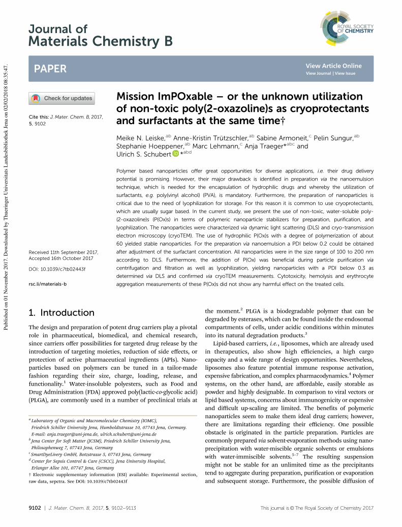
9102 | J. Mater. Chem. B, 2017, 5, 9102--9113 This journal is©The Royal Society of Chemistry 2017
Cite this: J.Mater. Chem. B, 2017,
5, 9102
Mission ImPOxable – or the unknown utilizationof non-toxic poly(2-oxazoline)s as cryoprotectantsand surfactants at the same time†
Meike N. Leiske,ab Anne-Kristin Trutzschler,ab Sabine Armoneit,c Pelin Sungur,ab
Stephanie Hoeppener,ab Marc Lehmann,c Anja Traeger*abc andUlrich S. Schubert *abd
Polymer based nanoparticles offer great opportunities for diverse applications, i.e. their drug delivery
potential is promising. However, their major drawback is identified in preparation via the nanoemulsion
technique, which is needed for the encapsulation of hydrophilic drugs and whereby the utilization of
surfactants, e.g. poly(vinyl alcohol) (PVA), is mandatory. Furthermore, the preparation of nanoparticles is
critical due to the need of lyophilization for storage. For this reason it is common to use cryoprotectants,
which are usually sugar based. In the current study, we present the use of non-toxic, water-soluble poly-
(2-oxazoline)s (P(Ox)s) in terms of polymeric nanoparticle stabilizers for preparation, purification, and
lyophilization. The nanoparticles were characterized via dynamic light scattering (DLS) and cryo-transmission
electron microscopy (cryoTEM). The use of hydrophilic P(Ox)s with a degree of polymerization of about
60 yielded stable nanoparticles. For the preparation via nanoemulsion a PDI below 0.2 could be obtained
after adjustment of the surfactant concentration. All nanoparticles were in the size range of 100 to 200 nm
according to DLS. Furthermore, the addition of P(Ox) was beneficial during particle purification via
centrifugation and filtration as well as lyophilization, yielding nanoparticles with a PDI below 0.3 as
determined via DLS and confirmed via cryoTEM measurements. Cytotoxicity, hemolysis and erythrocyte
aggregation measurements of these P(Ox)s did not show any harmful effect on the treated cells.
1. Introduction
The design and preparation of potent drug carriers play a pivotal
role in pharmaceutical, biomedical, and chemical research,
since carriers offer possibilities for targeted drug release by the
introduction of targeting moieties, reduction of side effects, or
protection of active pharmaceutical ingredients (APIs). Nano-
particles based on polymers can be tuned in a tailor-made
fashion regarding their size, charge, loading, release, and
functionality.1 Water-insoluble polyesters, such as Food and
Drug Administration (FDA) approved poly(lactic-co-glycolic acid)
(PLGA), are commonly used in a number of preclinical trials at
the moment.2 PLGA is a biodegradable polymer that can be
degraded by esterases, which can be found inside the endosomal
compartments of cells, under acidic conditions within minutes
into its natural degradation products.3
Lipid-based carriers, i.e., liposomes, which are already used
in therapeutics, also show high efficiencies, a high cargo
capacity and a wide range of design opportunities. Nevertheless,
liposomes also feature potential immune response activation,
expensive fabrication, and complex pharmacodynamics.4 Polymer
systems, on the other hand, are affordable, easily storable as
powder and highly designable. In comparison to viral vectors or
lipid based systems, concerns about immunogenicity or expensive
and difficult up-scaling are limited. The benefits of polymeric
nanoparticles seem to make them ideal drug carriers; however,
there are limitations regarding their efficiency. One possible
obstacle is originated in the particle preparation. Particles are
commonly prepared via solvent-evaporation methods using nano-
precipitation with water-miscible organic solvents or emulsions
with water-immiscible solvents.5–7 The resulting suspension
might not be stable for an unlimited time as the precipitants
tend to aggregate during preparation, purification or evaporation
and subsequent storage. Furthermore, the possible diffusion of
a Laboratory of Organic and Macromolecular Chemistry (IOMC),
Friedrich Schiller University Jena, Humboldtstrasse 10, 07743 Jena, Germany.
E-mail: [email protected], [email protected] Jena Center for Soft Matter (JCSM), Friedrich Schiller University Jena,
Philosophenweg 7, 07743 Jena, Germanyc SmartDyeLivery GmbH, Botzstrasse 5, 07743 Jena, GermanydCenter for Sepsis Control & Care (CSCC), Jena University Hospital,
Erlanger Allee 101, 07747 Jena, Germany
† Electronic supplementary information (ESI) available: Experimental section,
raw data, sepctra. See DOI: 10.1039/c7tb02443f
Received 11th September 2017,Accepted 16th October 2017
DOI: 10.1039/c7tb02443f
rsc.li/materials-b
Journal ofMaterials Chemistry B
PAPER
Publi
shed
on 0
1 N
ovem
ber
2017. D
ow
nlo
aded
by T
huer
inger
Univ
ersi
tats
Lan
des
bib
lioth
ek J
ena
on 0
2/0
2/2
018 0
8:3
5:4
7. View Article Online
View Journal | View Issue

This journal is©The Royal Society of Chemistry 2017 J. Mater. Chem. B, 2017, 5, 9102--9113 | 9103
hydrophilic drugs in water-based particle suspension might
reduce the drug loading in a time dependent manner. To avoid
these limitations, the particles can be lyophilized using cryopro-
tectants, which are necessary due to the difficult resuspension of
pure particles. Saccharides, such as trehalose, sucrose, and
glucose, are the most commonly used cryoprotectants.8 In the
literature, the common amount of sugar used is 5.0 wt% or even
higher.9,10 Unfortunately sugars are hygroscopic, which can be a
disadvantage for long-term storage. Furthermore, many saccharides
themselves already owe biochemical activity by specific uptake
mechanisms via GLUT transporters.11 For this reason, research on
finding alternatives for these cryoprotectants was already started by
some groups.12,13
The second issue regarding nanoparticles is related to their
preparation for encapsulation of hydrophilic drugs, e.g. siRNA.
These drugs cannot be easily incorporated by nanoprecipitation
methods, but require double emulsion techniques instead.7 As
this preparation method uses a solvent, which is not miscible
with water, the emulsion has to be further stabilized by using a
surfactant. The most common surfactant in this context is the
water-soluble poly(vinyl alcohol) (PVA).8,14 In nanoscience it is
well-known that the utilization of amphiphilic particle stabilizers,
such as PVA, is beneficial and can reduce the surface tension of the
nanoparticles. The resulting nanoparticles are uniform with a
narrow size distribution but, however, PVA influences the physical
properties and the cellular uptake of nanoparticles.15 Furthermore,
it is declared to be possibly carcinogenic and might influence the
cellular uptake of the nanoparticles.15–17 As a consequence, the use
of PVA leads to the necessity of excessive purification of the
nanoparticle formulations before administration.
Usually in terms of purification, crude particles are centri-
fuged, filtered, or dialyzed.3 In particular during centrifugation,
immense forces operate on the particles and potentially influence
the particle characteristics.
Taking all these facts into account, the research for new,
biocompatible surfactants and cryoprotectants is indispensable.
Biocompatible poly(2-oxazoline) (P(Ox)) polymers might be
interesting candidates to address this issue. Since their inven-
tion in the 1960s by four different research groups,18–21 the
interest in this polymer class has risen exponentially. Fundamen-
tal studies mainly included monomer synthesis and polymeriza-
tion parameter optimization as well as the characterization of the
resulting polymers.22–26 As the polymerization process of the
cationic ring-opening polymerization (CROP) of 2-oxazolines is
slow, it requires high temperatures in order to reach useful
reaction times. The usage of the microwave technique (poly-
merization under pressure) since 2004 has decreased the
synthesis time from several days to only a few minutes.27
Since this circumstance makes P(Ox)s affordable, biocompat-
ibility and pharmaceutical studies have come to the fore.28
FDA approved polymers consisting of the water-soluble
monomer 2-ethyl-2-oxazoline (EtOx)29 as well as 2-methyl-2-
oxazoline (MeOx) are of major interest in this context.
Their biocompatibility and biodistribution mechanisms have
already been tested in vitro30 and in vivo,31 and at the moment
the first clinical trial is running.32
P(Ox)s have already been studied in terms of surfactant
abilities, e.g., by investigations on the surface activity of different
block copolymer compositions.33–35 Kobayashi et al. additionally
studied hydrophilic P(Ox) homopolymers providing a hydrophobic
end-group.36 Furthermore, P(Ox)s were investigated as stabilizers
for hydroxyapatite37 and metal-based nanoparticles.38
The aim of this study is to use purely hydrophilic P(Ox)
homopolymers as an all-in-one-system for polymeric nano-
particles to replace both the surfactant necessary for particle
preparation and the cryoprotectant required for particle storage. A
series of poly(2-ethyl-2-oxazoline)s (P(EtOx)) and poly(2-methyl-2-
oxazoline)s (P(MeOx)) with varying degree of polymerization (DP)
were synthesized. In detail, P(EtOx)61 was used as a surfactant for
PLGA nanoparticle preparation in comparison with commercial
PVA and Pluronics F127. Furthermore, P(EtOx)61 was compared
with P(EO)57 and saccharides regarding its cryoprotectant proper-
ties for PLGA nanoparticles. After optimizing the conditions
regarding stabilizer concentration, P(Ox)s of varying DPs were
compared in terms of surfactant and cryoprotectant abilities.
Additionally, the purification techniques (centrifugation and
filtration) were evaluated. Their potential as additives in the
encapsulation of hydrophilic drugs was exemplarily shown for
Nile red and PKC 412, a kinase inhibitor, e.g. for protein kinase
C. The nanoparticles were analyzed using DLS and cryoTEM.
2. Results and discussion2.1. Surfactant abilities
2.1.1. Comparison of P(Ox)s and commercial surfactants.
Since nanoparticles for drug delivery are often prepared via
nanoemulsion, which requires the use of surfactants, whose
absolute removal is nearly impossible, investigations regarding
in vitro cytotoxicity (AlamarBlues) and hemolysis behavior
(hemoglobin release) of potential candidates were conducted
(Fig. 1). Therefore the potential surfactants were tested in a
range of 0.0003 to 5.0 wt%. Due to difficulties with their
solubility, PVA and Pluronics F127 could not be tested at
5.0 wt%. Even at a high polymer concentration of 5.0 wt%
none of the tested surfactants is cytotoxic as revealed by the
AlamarBlues assay (Fig. 1A); however, hemolysis experiments
showed an enhanced hemolytic activity of PVA and Pluronics
F127 at concentrations of 1.0 wt% and higher, while the
investigated P(Ox)s are not hemolytic even at a concentration
of 5.0 wt%, proving their good biocompatibility (Fig. 1B).
Biotests regarding the erythrocyte aggregation (Fig. S1, ESI†)
and an LDH-assay (Fig. S2, ESI†) showed no significant differences
between the tested surfactants PVA, Pluronics F127, P(EtOx)61 and
P(MeOx)57.
Initial particle preparation experiments were performed
using the nanoprecipitation method (Fig. S3A, ESI†) for PVA,
Pluronics F127 and P(EtOx)61 as nanoparticle stabilizers using
concentrations of 0.3, 0.5 and 1.0 wt%. In fact, the precipitation
method results in good PLGA nanoparticles without using
surfactants. However, PLGA nanoparticles, which are used for
therapeutical purposes, often carry drugs and therefore need to
Paper Journal of Materials Chemistry B
Publi
shed
on 0
1 N
ovem
ber
2017. D
ow
nlo
aded
by T
huer
inger
Univ
ersi
tats
Lan
des
bib
lioth
ek J
ena
on 0
2/0
2/2
018 0
8:3
5:4
7.
View Article Online
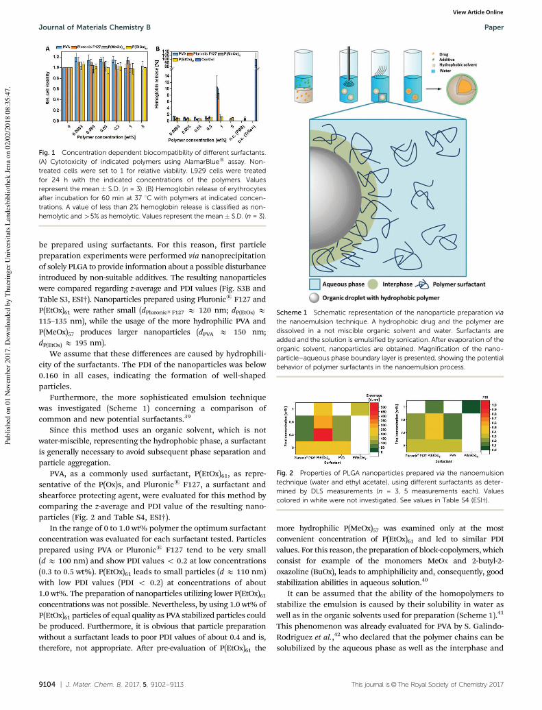
9104 | J. Mater. Chem. B, 2017, 5, 9102--9113 This journal is©The Royal Society of Chemistry 2017
be prepared using surfactants. For this reason, first particle
preparation experiments were performed via nanoprecipitation
of solely PLGA to provide information about a possible disturbance
introduced by non-suitable additives. The resulting nanoparticles
were compared regarding z-average and PDI values (Fig. S3B and
Table S3, ESI†). Nanoparticles prepared using Pluronics F127 and
P(EtOx)61 were rather small (dPluronicsF127 E 120 nm; dP(EtOx) E
115–135 nm), while the usage of the more hydrophilic PVA and
P(MeOx)57 produces larger nanoparticles (dPVA E 150 nm;
dP(EtOx) E 195 nm).
We assume that these differences are caused by hydrophili-
city of the surfactants. The PDI of the nanoparticles was below
0.160 in all cases, indicating the formation of well-shaped
particles.
Furthermore, the more sophisticated emulsion technique
was investigated (Scheme 1) concerning a comparison of
common and new potential surfactants.39
Since this method uses an organic solvent, which is not
water-miscible, representing the hydrophobic phase, a surfactant
is generally necessary to avoid subsequent phase separation and
particle aggregation.
PVA, as a commonly used surfactant, P(EtOx)61, as repre-
sentative of the P(Ox)s, and Pluronics F127, a surfactant and
shearforce protecting agent, were evaluated for this method by
comparing the z-average and PDI value of the resulting nano-
particles (Fig. 2 and Table S4, ESI†).
In the range of 0 to 1.0 wt% polymer the optimum surfactant
concentration was evaluated for each surfactant tested. Particles
prepared using PVA or Pluronics F127 tend to be very small
(d E 100 nm) and show PDI values o 0.2 at low concentrations
(0.3 to 0.5 wt%). P(EtOx)61 leads to small particles (d E 110 nm)
with low PDI values (PDI o 0.2) at concentrations of about
1.0 wt%. The preparation of nanoparticles utilizing lower P(EtOx)61concentrations was not possible. Nevertheless, by using 1.0 wt% of
P(EtOx)61 particles of equal quality as PVA stabilized particles could
be produced. Furthermore, it is obvious that particle preparation
without a surfactant leads to poor PDI values of about 0.4 and is,
therefore, not appropriate. After pre-evaluation of P(EtOx)61 the
more hydrophilic P(MeOx)57 was examined only at the most
convenient concentration of P(EtOx)61 and led to similar PDI
values. For this reason, the preparation of block-copolymers, which
consist for example of the monomers MeOx and 2-butyl-2-
oxazoline (BuOx), leads to amphiphilicity and, consequently, good
stabilization abilities in aqueous solution.40
It can be assumed that the ability of the homopolymers to
stabilize the emulsion is caused by their solubility in water as
well as in the organic solvents used for preparation (Scheme 1).41
This phenomenon was already evaluated for PVA by S. Galindo-
Rodriguez et al.,42 who declared that the polymer chains can be
solubilized by the aqueous phase as well as the interphase and
Fig. 1 Concentration dependent biocompatibility of different surfactants.(A) Cytotoxicity of indicated polymers using AlamarBlues assay. Non-treated cells were set to 1 for relative viability. L929 cells were treatedfor 24 h with the indicated concentrations of the polymers. Valuesrepresent the mean � S.D. (n = 3). (B) Hemoglobin release of erythrocytesafter incubation for 60 min at 37 1C with polymers at indicated concen-trations. A value of less than 2% hemoglobin release is classified as non-hemolytic and 45% as hemolytic. Values represent the mean � S.D. (n = 3).
Scheme 1 Schematic representation of the nanoparticle preparation via
the nanoemulsion technique. A hydrophobic drug and the polymer aredissolved in a not miscible organic solvent and water. Surfactants areadded and the solution is emulsified by sonication. After evaporation of theorganic solvent, nanoparticles are obtained. Magnification of the nano-particle–aqueous phase boundary layer is presented, showing the potentialbehavior of polymer surfactants in the nanoemulsion process.
Fig. 2 Properties of PLGA nanoparticles prepared via the nanoemulsiontechnique (water and ethyl acetate), using different surfactants as deter-mined by DLS measurements (n = 3, 5 measurements each). Valuescolored in white were not investigated. See values in Table S4 (ESI†).
Journal of Materials Chemistry B Paper
Publi
shed
on 0
1 N
ovem
ber
2017. D
ow
nlo
aded
by T
huer
inger
Univ
ersi
tats
Lan
des
bib
lioth
ek J
ena
on 0
2/0
2/2
018 0
8:3
5:4
7.
View Article Online
This journal is©The Royal Society of Chemistry 2017 J. Mater. Chem. B, 2017, 5, 9102--9113 | 9105
the organic phase. Inter- and intrachain interactions of the
surfactant polymer lead to repulsion and steric effects that might
stabilize the droplet. Furthermore, a reduction of the interfacial
tension is beneficial for the stabilization of an emulsion as
known from the literature. This explanation could also fit to
the 2-oxazoline homopolymers used in this study. For this
reason, the surface tension of surfactant containing water dro-
plets was measured for all the surfactants used (Fig. S4, ESI†).
From the comparison of results, all surfactants were found to
reduce the surface tension of water at least slightly. However, the
comparison of the different P(Ox) solutions does not show any
dependence of the surface tension on the concentration or the
molar mass of the polymer (gP(EtOx)n E 60 N m�1, gP(MeOx)n E
70 N m�1), while PVA and Pluronics F127 do (gPVAn E 55 to
45 N m�1, gPluronicsF127 E 45 to 40 N m�1). Therefore, the
concentration dependent surfactant ability might be mainly
caused by the viscosity of the solutions (Fig. 4A). It is already
known from the literature that the viscosity of the aqueous P(Ox)
solution can be influenced by the molar mass as well as the
polymer concentration.43 As shown in Fig. 4A, the dynamic
viscosity of the used PVA solution (3 wt%) is between 4.5 and
5.0 mPA s, whereas the utilized P(EtOx)60 and P(MeOx)60 solu-
tions have much lower viscosities. At a concentration of 10 wt%
the P(Ox) solutions have a dynamic viscosity of B3.0 mPA s,
which seems to be an applicable value for the stabilization of the
emulsion. Even though PVA has great surfactant abilities at the
determined values of surface tension and viscosity, P(Ox)s also
work at significantly lower viscosities and reduced surface ten-
sion. Since Pluronics F127 has very good potential to reduce the
surface tension of water, the lower viscosity of the solution might
not be disadvantageous. These findings support the hypothesis
that the stabilization of the particles is influenced by the surface
tension and the viscosity of the used surfactant.
2.1.2. Influence of the molar mass of the surfactant. To
prove whether the success of surfactant ability of the different
polymers depends on the polymer size, hydrophilic P(Ox)s
with a varying DP (B25, B60, B100, B200) were tested and
characterized regarding the z-average and PDI values of the
resulting particles using DLS (Fig. 3 and Fig. S5, ESI†).
While using the nanoprecipitation technique, the resulting
nanoparticles are of comparable z-average (d E 160 nm) and
have a narrow dispersity (PDI o 0.1) (Fig. S5 and Table S5,
ESI†). By using the more sophisticated nanoemulsion tech-
nique (Fig. 3 and Table S5, ESI†), P(Ox)s with a DP of 60, 100
and 200 produce larger nanoparticles (d E 200 nm) with a
moderate dispersity (PDI o 0.2), while the P(Ox)s with a DP of
25 cannot be used for the production of stable, narrow disperse
nanoparticles (d E 800 nm, PDI Z 0.3). These results indicate
the dependence of the surfactant ability on the DP of the used
polymers for the nanoemulsion technique. A possible explana-
tion for this phenomenon might be given by the viscosity of the
polymer solutions. Fig. 4B shows the strong dependence of the
viscosity on the DP of the used P(Ox)s. T. X. Viegas et al. have
already investigated this using a different molar mass range of
P(EtOx)n.43 If we consider these results, a DP of 60 is the
minimum possible for successful particle stabilization within
the tested range. Low molar mass polymers with a DP below 60
are not capable of stabilizing the PLGA nanoparticles. However,
after crossing a certain point, which was a DP of 60 in the
current study, an increase of the molar mass seems to be
neither beneficial nor disadvantageous for the stabilization of
the nanoparticles.
2.2. Lyophilization experiments
2.2.1. Comparison of P(Ox)s and commercial cryoprotec-
tants. Successful nanoparticle lyophilization and resuspension
are crucial elements for particle preparation and storage
(Fig. 5A). Usually, polymeric nanoparticles show difficulties in
resuspension after lyophilization and lack in quality. Therefore,
first experiments concentrated on the possible capability of
P(Ox)s to act as a stabilizing agent during lyophilization of
nanoparticles after successful preparation.
P(EtOx)61 was used for initial lyophilization experiments to
determine the polymer amount necessary for particle stabili-
zation. PLGA nanoparticles were prepared via the nanoprecipi-
tation technique without using any stabilizers. Their z-average
and PDI values were determined by DLS measurements. Sub-
sequently, different amounts of the cryoprotectants ranging
from 0.05 to 10 wt% were added to the crude nanoparticle
suspension, which was lyophilized afterwards and tested
regarding the z-average and PDI values of the particles and
Fig. 3 Properties of PLGA nanoparticles prepared by nanoemulsion(water and ethyl acetate), using hydrophilic P(Ox)s with varying DP assurfactants (1.0 wt%). z-Average and PDI values were determined via DLSinvestigations (n = 3, 5 measurements each). See values in Table S5 (ESI†).
Fig. 4 Dynamic viscosity of different polymer solutions. (A) Dynamicviscosity dependent on the polymer concentration. (B) Dynamic viscosityof P(Ox) solutions with different DP.
Paper Journal of Materials Chemistry B
Publi
shed
on 0
1 N
ovem
ber
2017. D
ow
nlo
aded
by T
huer
inger
Univ
ersi
tats
Lan
des
bib
lioth
ek J
ena
on 0
2/0
2/2
018 0
8:3
5:4
7.
View Article Online

9106 | J. Mater. Chem. B, 2017, 5, 9102--9113 This journal is©The Royal Society of Chemistry 2017
compared to the previous results (Fig. 5B and Table S3, ESI†).
z-Average and PDI ratios of 1.0 are favorable in this context. In
Fig. 5B the results of the dynamic light scattering of the
particles after lyophilization are summarized compared to the
particle properties before. Thereby the ratios of the z-average
and the PDI were calculated as shown in eqn (1) and (2).
z-average ratio ¼z-average after lyophilization
z-average after preparation(1)
PDI ratio ¼PDI after lyophilization
PDI after preparation(2)
In this experimental setup, saccharose showed the best
performance (z-average ratio = 1.0; PDI ratio = 1.8) at a used
sugar concentration of 10 wt%. Glucose (z-average ratio = 1.0;
PDI ratio = 2.1) and trehalose (z-average ratio = 1.4; PDI ratio = 2.7)
were slightly more unfavorable at concentrations of 10 and 5 wt%,
respectively. In contrast, at a low cryoprotectant concentration of
about 0.5 wt% P(EtOx)61, the z-average (ratio = 1.5) and PDI
(ratio = 2.0) mostly remain constant, while increasing signifi-
cantly by the utilization of other cryoprotectants. Even though
saccharose shows a better performance at a high concentration
of 5.0 wt%, P(Ox)s at low concentrations offer better stabili-
zation. Using P(Ox)s, the increase in the size can be explained
by the cryoprotectant addition. P(EtOx)61 chains themselves
seem to assemble on the PLGA particle surface, whereby the
size of the particle increases. It should be noted that a PDI ratio
of two, which means a doubling in the actual values, results in
PDI values of around 0.2, which still represents a very good value.
For comparison, also poly(ethylene oxide) (P(EO)), i.e. P(EO)57,
was tested regarding its cryoprotectant abilities, since P(Ox) and
P(EO) are often claimed to show comparable characteristics.
Nanoparticles that were stabilized using P(EO)57 showed a
huge increase in z-average (ratio4 2.5) and PDI (ratio4 4.5) in
all tested concentrations ranging from 0.05 to 5 wt%. For this
reason, P(EO)57 does not seem to be suitable for PLGA
nanoparticle stabilization, while P(EtOx)61 is superior to commonly
used cryoprotectants when used at low concentrations. The tested
sugars show their best cryoprotective ability at a concentration of
10.0 wt%, which is twenty times higher than the investigated
P(EtOx)61. Since sugars are known to be hygroscopic, the use of
P(Ox)s, which can be used at much lower amounts, represents a
possible alternative for the common cryoprotectants.
2.2.2. Influence of the molar mass of the cryoprotectant.
After initial experiments using P(EtOx)61, P(Ox)s with varying
molar masses were investigated. Herein, P(EtOx)n and the more
hydrophilic P(MeOx)n were used for the surfactant experiments
(DP E 25, 60, 100, 200). As an all-in-one system was targeted,
nanoparticle samples prepared with those polymers acting as
surfactants were directly lyophilized and re-suspended in dou-
ble deionized water. The resulting size and PDI values were
compared to the values that were determined directly after
preparation and are plotted in Fig. 6.
By using the P(Ox)s with a DP of 60, 100 and 200 the
characteristics of the PLGA nanoparticles can be maintained
during lyophilization (size ratioE 1, PDI ratio: 1 to 2), while the
Fig. 5 (A) Schematic representation of the lyophilization and resuspension of polymeric nanoparticles using P(Ox)s as suitable particle stabilizers(cryoprotectants). (B) Properties of the lyophilized PLGA nanoparticles using different cryoprotectants at various concentrations. Diameter size ratios ofthe z-average and PDI ratios of the nanoparticles were determined by DLS investigations (n = 3, 5 measurements each). Ratios were calculated usingeqn (1) and (2). Values colored in white were not investigated. See values in Table S3 (ESI†).
Fig. 6 z-Average and PDI ratios of PLGA nanoparticles prepared by thenanoprecipitation technique using 0.3 wt% P(Ox). The particles werelyophilized without further purification and the ratios were calculatedusing eqn (1) and (2) (n = 3, 5 measurements each). See values inTable S6 (ESI†).
Journal of Materials Chemistry B Paper
Publi
shed
on 0
1 N
ovem
ber
2017. D
ow
nlo
aded
by T
huer
inger
Univ
ersi
tats
Lan
des
bib
lioth
ek J
ena
on 0
2/0
2/2
018 0
8:3
5:4
7.
View Article Online
This journal is©The Royal Society of Chemistry 2017 J. Mater. Chem. B, 2017, 5, 9102--9113 | 9107
usage of P(Ox)s with a DP of 25 does not seem to be beneficial
(size ratio E 2, PDI ratio: 4 to 6).
According to the determined ratios, a P(Ox) based all-in-one
system is possible. Direct lyophilization of the PLGA nano-
particles after preparation leads to nanoparticles of defined size
and PDI values which seem to be improved by using P(Ox)s with
higher molar masses. However, drug encapsulation usually
leads to the necessity of particle purification to eliminate free drug
from the particle suspension. Furthermore, P(Ox)s with lower
molar masses are known to undergo faster renal excretion44 and
are, therefore, further examined in the current study.
2.3. Particle purification
After successful investigation of P(Ox)s as surfactants, the
influence of particle purification (centrifugation or filtration)
on their size and PDI values was elucidated. For this reason
PLGA nanoparticles were produced by nanoprecipitation using
0.3 wt% P(Ox) as the surfactant. The resulting nanoparticles
were either centrifuged for 45 min at 11 000 rpm and 4 1C or
filtered using a 0.45 mm nylon filter before lyophilization. To
see whether it is necessary to use an additional amount of P(Ox)
solution for good resuspension, the same amounts of P(Ox)
solutions were added before lyophilization to either centrifuged
or filtered nanoparticles. For comparison, nanoparticle suspen-
sions which have not been purified before were lyophilized as
well (Fig. 7A and Table S7, ESI†). The z-average distribution
values of the particles before and after purification were investi-
gated via DLS and cryoTEM measurements and are summarized
in Fig. 7B.
After preparation, the nanoparticles are of comparable size
(d E 190 nm) and dispersity (PDI o 0.1) and, furthermore,
suitable for syringe filtration as well as centrifugation. The
particles moreover feature a perfect spherical shape. Syringe
filtration was conducted using a 0.45 mm nylon syringe filter.
Standard sterile filtration would be usually performed using a
0.2 mm syringe filter but to be sure not to filter out possible
aggregation of particles a larger filter was used.
DLS measurements provided initial information about
the stability of the nanoparticles. Nanoparticles prepared with
P(MeOx)57 resulted in a diameter of about 200 nm and the PDI
was E 0.1, while for nanoparticles prepared by using P(EtOx)61the addition of a P(Ox) solution was necessary (d = 165.1 �
1.0 nm; PDI = 0.172 � 0.044) after purification to avoid particle
aggregation (d = 1075 � 112 nm; PDI = 0.948 � 1). However, by
having a close look at the particle morphology via cryoTEM, it is
also visible that most of the nanoparticles are still well defined
and only a few aggregates are present. Nanoparticle sizes
obtained by DLS measurements could mostly be confirmed by
cryoTEM measurements. Regarding the DLS and cryoTEM results,
this purification has no influence on the particle morphology and
is, therefore, a suitable purification method that can be used, e.g.,
to separate the particles from larger aggregates.
Centrifugation of nanoparticles is a common purification
method for their separation from small and dissolved mole-
cules. This technique potentially has stronger forces which are
exerted onto the nanoparticles; hence, the stability of the
Fig. 7 (A) Schematic representation of nanoparticle purification by either syr-inge filtration or centrifugation using P(Ox) as the particle stabilizer. (B) Influenceof various purification techniques on the size distribution and morphology ofPLGAnanoparticles thatwere prepared by nanoprecipitation, determined byDLS(green columns, n= 3, 5measurements each) and cryoTEMmeasurements. Seevalues in Table S7 (ESI†). See raw cryoTEM images in Fig. S6 (ESI†).
Paper Journal of Materials Chemistry B
Publi
shed
on 0
1 N
ovem
ber
2017. D
ow
nlo
aded
by T
huer
inger
Univ
ersi
tats
Lan
des
bib
lioth
ek J
ena
on 0
2/0
2/2
018 0
8:3
5:4
7.
View Article Online
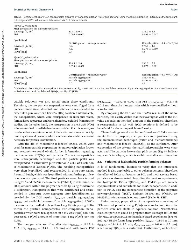
9108 | J. Mater. Chem. B, 2017, 5, 9102--9113 This journal is©The Royal Society of Chemistry 2017
particle solutions was also tested under these conditions.
Therefore, the raw particle suspensions were centrifuged for a
predetermined time, decanted and afterwards resuspended in
either ultra-pure water or a 0.5 wt% P(Ox) solution. Unfortunately,
the nanoparticles, which were resuspended in ultra-pure water,
formed large aggregates and were, therefore, excluded from further
studies. On the other hand, the resuspension in a 0.5 wt% P(Ox)
solution resulted in well-defined nanoparticles. For this reason, we
conclude that a certain amount of the surfactant is washed out by
centrifugation and has to be added afterwards to reach the amount
necessary for particle stabilization.
With the aid of rhodamine B labeled P(Ox)s, which were
used for nanoparticle preparation via nanoprecipitation (water
and acetone), we could obtain further information regarding
the interaction of P(Ox)s and particles. The raw nanoparticles
were subsequently centrifuged and the particle pellet was
resuspended in either ultra-pure water or in a 0.5 wt% solution
of rhodamine B labeled P(Ox)s. The resulting nanoparticles
were then lyophilized and resuspended in ultra-pure water.
A control batch, which was lyophilized without further purifica-
tion, was also prepared. The final particles were characterized
using DLS and UV/Vis measurements (Table 1) to determine the
P(Ox) amount within the polymer particle by using rhodamine
B calibration. Nanoparticles that were centrifuged and resus-
pended in ultra-pure water aggregate after lyophilization as
revealed by the DLS results (dP(MeOx): particle aggregation;
dP(EtOx) not available because of particle aggregation). UV/Vis
measurements resulted in less than 1 mg P(Ox)s per mg PLGA
within the purified nanoparticles. For comparison, nano-
particles which were resuspended in a 0.5 wt% P(Ox) solution
possessed a P(Ox) amount of more than 4 mg P(Ox)s per mg
PLGA.
The nanoparticles are of smaller size (dP(MeOx) = 182.7 �
21.7 nm; dP(EtOx) = 175.4 � 4.5 nm) and with lower PDI
(PDIP(MeOx) = 0.192 � 0.062 nm; PDI P(EtOx)(MeOx) = 0.273 �
0.019 nm) than the nanoparticles which were purified without
a surfactant.
By comparing the DLS and the UV/Vis results of the nano-
particles, it is clearly visible that the z-average as well as the PDI
value depends on the P(Ox) amount of the particles. Therefore,
a resuspension in 0.5 wt% P(Ox) solutions is defined to be
beneficial for the nanoparticle uniformity.
These findings could also be confirmed via CLSM measure-
ments. For this purpose, microparticles were produced using
the microemulsion technique (water and dichloromethane)
and rhodamine B labeled P(MeOx)57 as the surfactant. After
evaporation of the solvent, the PLGA microparticles were char-
acterized. The particles have a clear pink corona (Fig. 8), indicat-
ing a surfactant layer, which is visible even after centrifugation.
2.4. Variation of hydrophobic particle forming polymers
It is of fundamental interest to verify that the introduced
method is also applicable to other polymer systems. Therefore,
the effect of P(Ox) surfactants on PCL and methacrylate based
particles was also evaluated. Regarding the previous experiments,
the hydrophilic P(Ox)s P(EtOx)61 and P(MeOx)57 are suitable
cryoprotectants and surfactants for PLGA nanoparticles. In addi-
tion to PLGA, also the nanoparticle formation of the polymers
poly(caprolactone) (P(CL)), Eudragit RS100, and P(MMA97-co-
MAEMA32) via nanoemulsion was tested (Table S8, ESI†).
Unfortunately, preparation of nanoparticles consisting of
PCL was not possible using P(Ox)s as a surfactant, since the
particles were not stable in aqueous solution. Nevertheless,
excellent particles could be prepared from Eudragit RS100 and
P(MMA97-co-MAEMA32) methacrylate based copolymers (Fig. 9).
Eudragit RS100 forms large particles (dP(EtOx) = 214.0 � 0.7 nm;
dP(MeOx) = 244.3 � 2.5 nm; dultra-purewater = 101.6 � 0.5 nm),
when using P(Ox)s as a surfactant. Furthermore, well-defined
Table 1 Characteristics of PLGA nanoparticles prepared by nanoprecipitation (water and acetone) using rhodamine B labeled P(EtOx)61 as the surfactant.z-Average and PDI values were determined via DLS measurements
P(EtOx)61-rhodamineAfter preparation via nanoprecipitationz-Average [d, nm] 132.1 � 0.4 134.0 � 1.3PDI 0.096 � 0.002 0.093 � 0.017
LyophilizedCentrifugation + ultra-pure water Centrifugation + 0.5 wt% P(Ox)
z-Average [d, nm] n.a. 175.5 � 4.5PDI n.a. 0.273 � 0.019P(Ox)a [mg] 0.7 4.6
P(MeOx)57-rhodamineAfter preparation via nanoprecipitationz-Average [d, nm] 193.8 � 2.0 190.6 � 2.3PDI 0.090 � 0.038 0.072 � 0.021
LyophilizedCentrifugation + ultra-pure water Centrifugation + 0.5 wt% P(Ox)
z-Average [d, nm] Particle aggregation 182.7 � 21.7PDI Particle aggregation 0.192 � 0.062P(Ox)a [mg] 0.0 4.0
a Calculated from UV/Vis absorption measurements at lEx = 630 nm. n.a.: not available because of particle aggregation. For absorbance andemission spectra of the labelled P(Ox)s, see Fig. S7 (ESI).
Journal of Materials Chemistry B Paper
Publi
shed
on 0
1 N
ovem
ber
2017. D
ow
nlo
aded
by T
huer
inger
Univ
ersi
tats
Lan
des
bib
lioth
ek J
ena
on 0
2/0
2/2
018 0
8:3
5:4
7.
View Article Online

This journal is©The Royal Society of Chemistry 2017 J. Mater. Chem. B, 2017, 5, 9102--9113 | 9109
nanoparticles can only be achieved by utilization of P(Ox)
surfactants (PDIP(EtOx) = 0.063 � 0.018; PDIP(MeOx) = 0.081 �
0.012; PDIultra-purewater = 0.246 � 0.020).
When using P(MMA97-co-MAEMA32), the more hydrophilic
surfactant P(MeOx)57 leads to less disperse (PDIP(MeOx) =
0.087 � 0.023; PDIP(EtOx) = 0.147 � 0.030) nanoparticles. How-
ever, at this point, it should also be pointed out that the PDI
values of the nanoparticles prepared using P(EtOx)61 are still
very good. However, the prepared particles are only slightly less
disperse than particles prepared without using a surfactant
(PDIultra-purewater = 0.191 � 0.012). Therefore, at first glance, the
particles do not seem to benefit the usage of P(Ox)s as a surfactant.
Since the non-ionic P(Ox)s have a neutral zeta potential, the zeta
potential of the nanoparticles (zP(MeOx) 4 30 mV; zP(EtOx) 4 30 mV;
zMilliQ 4 40 mV) is more or less unaffected by the surfactant as it
remains being strongly positive.
More interestingly, for P(MMA97-co-MAEMA32) both P(Ox)s
seem to have a comparable ability of being a surfactant
agent; however, the prepared particles are only slightly less
disperse than particles prepared without using a surfactant
(PDIultra-pure water = 0.191 � 0.012). Therefore, at first glance,
the particles do not seem to benefit the usage of P(Ox)s as a
surfactant. In both cases the particle size is dependent on the
surfactant as well as on the type of hydrophibic polymer being
used. As this study mainly concentrates on the investigation
of P(Ox)s as suitable surfactants for all tested polymers,
the particles consisting of different types of polymers are not
compared with each other directly. However, the influence of
P(Ox) based surfactants on the nanoparticle size and PDI value
was examined. Purification via centrifugation and filtration as
well as lyophilization experiments with these nanoparticles
were also investigated (Fig. 10 and Tables S9, S10, ESI†).
The purification of Eudragit RS100 yielded stable nanoparticles
when prepared using P(Ox) based surfactants (size ratio o 1.3;
PDI ratio: o 1.7 (except centrifugation and resuspension in ultra-
pure water)), while nanoparticles without surfactants aggregated
after lyophilization (Fig. 10A). While purifying P(MMA97-co-
MAEMA32) filtration did not work in any case. However, the
addition of a P(Ox) solution after filtration was beneficial even
for P(MMA97-co-MAEMA32) nanoparticles prepared without a sur-
factant. Centrifugation and resuspension in ultra-pure water
before lyophilization also yielded stable nanoparticles with respect
to their size and PDI ratios, which are below a value of two.
Fig. 8 CLSM (lEx = 514 nm, lEm = 531 to 704 nm) of PLGA microparticlesprepared by microemulsion (water and dichloromethane) using P(MeOx)57-rhodamine as a surfactant. For absorbance and emission spectra of thelabelled P(Ox)s, see Fig. S7 (ESI†).
Fig. 9 Properties of the nanoparticles prepared by nanoemulsion (waterand ethyl acetate), using P(Ox)s as surfactants. z-Average and PDI valueswere determined via DLS (n = 3, 5 measurements each). See values inTable S8 (ESI†).
Fig. 10 Dependence of z-average and PDI ratios of (A) Eudragit RS100and (B) P(MMA97-co-MAEMA32) nanoparticles on the purification methodby either direct lyophilization of the particle suspension (None), centri-fugation at 11 000 rpm for 45 min and resuspension in ultra-pure water (A),centrifugation at 11 000 rpm for 45 min and resuspension in a 0.5% P(Ox)solution (B) or syringe filtration using a 0.45 mm nylon filter (C). Nano-particles were prepared via nanoprecipitation (water and acetone). Seevalues in Tables S9 and S10 (ESI†).
Paper Journal of Materials Chemistry B
Publi
shed
on 0
1 N
ovem
ber
2017. D
ow
nlo
aded
by T
huer
inger
Univ
ersi
tats
Lan
des
bib
lioth
ek J
ena
on 0
2/0
2/2
018 0
8:3
5:4
7.
View Article Online
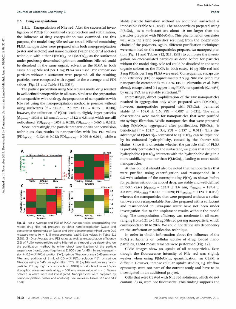
9110 | J. Mater. Chem. B, 2017, 5, 9102--9113 This journal is©The Royal Society of Chemistry 2017
2.5. Drug encapsulation
2.5.1. Encapsulation of Nile red. After the successful inves-
tigation of P(Ox)s for combined cryoprotection and stabilization,
the influence of drug encapsulation was examined. For this
purpose, the model drug Nile red was tested. Nile red containing
PLGA nanoparticles were prepared with both nanoprecipitation
(water and acetone) and nanoemulsion (water and ethyl acetate)
technique with either P(EtOx)61 or P(MeOx)57 as the surfactant
under previously determined optimum conditions. Nile red could
be dissolved in the same organic solvent as the PLGA in both
cases. 10 mg Nile red per 1 mg PLGA was used. For comparison
particles without a surfactant were prepared. All the resulting
particles were compared with regard to the z-average and PDI
values (Fig. 11 and Table S11, ESI†).
The particle preparation using Nile red as a model drug resulted
in well-defined nanoparticles in all cases. Similar to the preparation
of nanoparticles without drug, the preparation of nanoparticles with
Nile red using the nanoprecipitation method is possible without
using surfactants (d = 145.2 � 2.5 nm; PDI = 0.075 � 0.018);
however, the utilization of P(Ox)s leads to slightly larger particles
(dP(EtOx) = 160.8� 1.5 nm; dP(MeOx) = 151.2� 0.8 nm), which are still
well-defined (PDIP(EtOx) = 0.053 � 0.028; PDIP(MeOx) = 0.065 � 0.021).
More interestingly, the particle preparation via nanoemulsion
techniques also results in nanoparticles with low PDI values
(PDIP(EtOx) = 0.124 � 0.013; PDIP(MeOx) = 0.099 � 0.014), while a
stable particle formation without an additional surfactant is
impossible (Table S11, ESI†). The nanoparticles prepared using
P(EtOx)61 as a surfactant are about 10 nm larger than the
particles prepared with P(MeOx)57. This phenomenon correlates
well with the steric properties resulting from the longer side-
chains of the polymers. Again, different purification techniques
were examined on the nanoparticles prepared via nanoprecipita-
tion (Fig. 11 and Tables S12, S13, ESI†) to complete the investi-
gation on encapsulated particles as done before for particles
without the model drug. Nile red could be dissolved in the same
organic solvent as the PLGA in both cases. 10 mg Nile red and
3 mg P(Ox)s per 1 mg PLGA were used. Consequently, encapsula-
tion efficiency (EE) of approximately 2.5 mg Nile red per 1 mg
nanoparticle corresponds to 100% EE. P. Pietzonka et al. have
already encapsulated 0.1 mg per 1mg PLGA nanoparticle (0.1 wt%)
by using PVA as a suitable surfactant.45
Interestingly, direct lyophilization of the raw nanoparticles
resulted in aggregation only when prepared with P(MeOx)57;
however, nanoparticles prepared with P(EtOx)61 remained
stable (d = 168.0 � 1.6; PDI = 0.087 � 0.031). The same
observations were made for nanoparticles that were purified
via syringe filtration. While nanoparticles that were prepared
using P(MeOx)57 aggregated after purification, P(EtOx)61 is
beneficial (d = 161.7 � 3.4; PDI = 0.157 � 0.011). This dis-
advantage of P(MeOx)57 compared to P(EtOx)61 can be explained
by its enhanced hydrophilicity, caused by the shorter side
chains. Since it is uncertain whether the particle shell of PLGA
is probably permeated by the surfactant, we guess that the more
hydrophobic P(EtOx)61 interacts with the hydrophobic drug in a
more stabilizing manner than P(MeOx)57 leading to more stable
nanoparticles.
At this point it should also be noted that nanoparticles that
were purified using centrifugation and resuspended in a
0.5 wt% solution of the corresponding P(Ox), as shown before
for particles without the model drug, are stable and well-defined
in both cases (dP(EtOx) = 184.3 � 1.6 nm; dP(MeOx) = 187.4 �
3.2 nm; PDIP(EtOx) = 0.145 � 0.028; PDIP(MeOx) = 0.123 � 0.052),
whereas the nanoparticles that were prepared without a surfac-
tant were not resuspendable. Particles prepared with a surfactant
and resuspended in ultra-pure water have not been under
investigation due to the unpleasant results without the model
drug. The encapsulation efficiency was moderate in all cases,
ranging from 0.21 to 0.52 mg Nile red per mg nanoparticle, which
corresponds to 10 to 20%. We could not define any dependency
on the surfactant or purification technique.
In order to obtain information about the influence of the
P(Ox) surfactants on cellular uptake of drug loaded nano-
particles, CLSM measurements were performed (Fig. 12).
CLSM images show an uptake of all nanoparticles. Even
though the fluorescence intensity of Nile red was slightly
weaker when using P(MeOx)57, quantification via CLSM is
difficult. However, intense cellular uptake studies, e.g. via flow
cytometry, were not part of the current study and have to be
investigated in an additional project.
Cells that were treated with Nile red solutions, which do not
contain PLGA, were not fluorescent. This finding supports the
Fig. 11 (A) z-Average and PDI of PLGA nanoparticles encapsulating themodel drug Nile red, prepared by either nanoprecipitation (water andacetone) or nanoemulsion (water and ethyl acetate) determined using DLSmeasurements (n = 3, 5 measurements each). See values in Table S11(ESI†). (B–D) z-Average and PDI ratios as well as encapsulation efficiency(EE) of PLGA nanoparticles using Nile red as a model drug depending onthe purification method by either direct lyophilization of the particlesuspension (none), centrifugation at 11 000 rpm for 45 min and resuspen-sion in 0.5 wt% P(Ox) solution (‘‘A’’), syringe filtration using a 0.45 mm nylonfilter and addition of 1 mL of 0.5 wt% P(Ox) solution (‘‘B’’) or syringefiltration using a 0.45 mm nylon filter (‘‘C’’). EE (mg Nile red per mg nano-particle, 2.5 mg mg�1 corresponds to 100%) is calculated from UV/visabsorption measurements at lEx = 630 nm, mean value of n = 3. Valuescolored in white were not investigated. Nanoparticles were prepared bynanoprecipitation (water and acetone). See values in Tables S12 and S13(ESI†).
Journal of Materials Chemistry B Paper
Publi
shed
on 0
1 N
ovem
ber
2017. D
ow
nlo
aded
by T
huer
inger
Univ
ersi
tats
Lan
des
bib
lioth
ek J
ena
on 0
2/0
2/2
018 0
8:3
5:4
7.
View Article Online
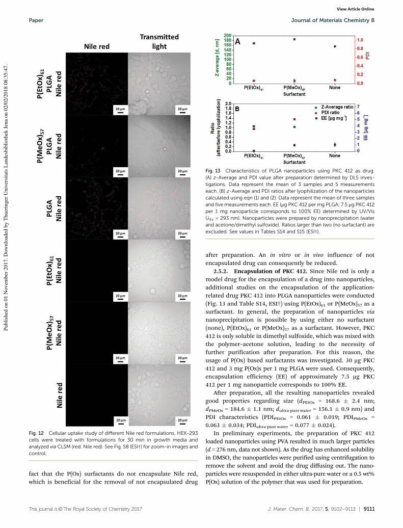
This journal is©The Royal Society of Chemistry 2017 J. Mater. Chem. B, 2017, 5, 9102--9113 | 9111
fact that the P(Ox) surfactants do not encapsulate Nile red,
which is beneficial for the removal of not encapsulated drug
after preparation. An in vitro or in vivo influence of not
encapsulated drug can consequently be reduced.
2.5.2. Encapsulation of PKC 412. Since Nile red is only a
model drug for the encapsulation of a drug into nanoparticles,
additional studies on the encapsulation of the application-
related drug PKC 412 into PLGA nanoparticles were conducted
(Fig. 13 and Table S14, ESI†) using P(EtOx)61 or P(MeOx)57 as a
surfactant. In general, the preparation of nanoparticles via
nanoprecipitation is possible by using either no surfactant
(none), P(EtOx)61 or P(MeOx)57 as a surfactant. However, PKC
412 is only soluble in dimethyl sulfoxide, which was mixed with
the polymer–acetone solution, leading to the necessity of
further purification after preparation. For this reason, the
usage of P(Ox) based surfactants was investigated. 30 mg PKC
412 and 3 mg P(Ox)s per 1 mg PLGA were used. Consequently,
encapsulation efficiency (EE) of approximately 7.5 mg PKC
412 per 1 mg nanoparticle corresponds to 100% EE.
After preparation, all the resulting nanoparticles revealed
good properties regarding size (dPEtOx = 168.6 � 2.4 nm;
dPMeOx = 184.6 � 1.1 nm; dultra-pure water = 156.1 � 0.9 nm) and
PDI characteristics (PDIPEtOx = 0.061 � 0.019; PDIPMeOx =
0.063 � 0.034; PDIultra-pure water = 0.077 � 0.024).
In preliminary experiments, the preparation of PKC 412
loaded nanoparticles using PVA resulted in much larger particles
(d = 276 nm, data not shown). As the drug has enhanced solubility
in DMSO, the nanoparticles were purified using centrifugation to
remove the solvent and avoid the drug diffusing out. The nano-
particles were resuspended in either ultra-pure water or a 0.5 wt%
P(Ox) solution of the polymer that was used for preparation.
Fig. 12 Cellular uptake study of different Nile red formulations. HEK-293cells were treated with formulations for 30 min in growth media andanalyzed via CLSM (red: Nile red). See Fig. S8 (ESI†) for zoom-in images andcontrol.
Fig. 13 Characteristics of PLGA nanoparticles using PKC 412 as drug.(A) z-Average and PDI value after preparation determined by DLS inves-tigations. Data represent the mean of 3 samples and 5 measurementseach. (B) z-Average and PDI ratios after lyophilization of the nanoparticlescalculated using eqn (1) and (2). Data represent the mean of three samplesand five measurements each. EE (mg PKC 412 per mg PLGA; 7.5 mg PKC 412per 1 mg nanoparticle corresponds to 100% EE) determined by UV/Vis(lEx = 293 nm). Nanoparticles were prepared by nanoprecipitation (waterand acetone/dimethyl sulfoxide). Ratios larger than two (no surfactant) areexcluded. See values in Tables S14 and S15 (ESI†).
Paper Journal of Materials Chemistry B
Publi
shed
on 0
1 N
ovem
ber
2017. D
ow
nlo
aded
by T
huer
inger
Univ
ersi
tats
Lan
des
bib
lioth
ek J
ena
on 0
2/0
2/2
018 0
8:3
5:4
7.
View Article Online

9112 | J. Mater. Chem. B, 2017, 5, 9102--9113 This journal is©The Royal Society of Chemistry 2017
As expected, nanoparticles prepared without any additives
aggregate when resuspended after lyophilization. In contrast, the
nanoparticles prepared using P(Ox)s are well resuspendable and
maintain their size (dP(EtOx) = 178.9 � 2.0 nm; dP(MeOx) = 190.4 �
2.4 nm) and PDI characteristics (PDIP(EtOx) = 0.058 � 0.025;
PDIP(MeOx) = 0.086 � 0.022). Surprisingly, nanoparticles prepared
by using P(EtOx)61 do not show any drug encapsulation, while
the encapsulation efficiency for nanoparticles prepared with
P(MeOx)57 is around 0.92 � 0.08 mg mg�1 nanoparticle, corres-
ponding to 12% EE. Regarding these characteristics, the encap-
sulation of PKC 412 is possible by using P(MeOx)57. Preliminary
experiments using PVA as a stabilizer showed much lower
encapsulation efficiencies (B1%, data not shown).
3. Conclusion
In the presented study, we demonstrated the ability of water
soluble and biocompatible P(Ox)s to act as stabilizing agents
for polymer based nanoparticles. First, we showed their ability
to replace the commonly used sugar based cryoprotectants and
result in resuspendable particles with constant properties. After
successful lyophilization experiments, we also used the poly-
mers as biocompatible surfactants. By adjusting the optimum
P(Ox) concentration, PLGA nanoparticles could be prepared via
nanoprecipitation and nanoemulsion techniques omitting the
use of PVA, which allows the preparation of well-defined
nanoparticles at the expense of cytotoxicity and additionally
required purification steps. Furthermore, we demonstrated the
possibility to use P(Ox)s as an all-in-one system for the stabili-
zation of polymeric nanoparticles during preparation, purifica-
tion and lyophilization. After a proof of concept using PLGA
serving as the shell-polymer for the nanoparticles, some other
polymers were investigated including PCL and methacrylate
based copolymers, i.e. Eudragit RS100. Finally, the P(Ox) based
nanoparticle surfactants were used to stabilize nanoparticles
while encapsulating the model drug Nile red or the application-
relevant drug PKC 412. Thereby, the properties, especially the
stability in purification processes, of the nanoparticles could be
improved as well. Since these non-toxic polymers are not
known to mediate cellular responses like P(EO) or specific
uptake mechanisms like sugars, they could be used as promis-
ing stabilizing agents for nanoparticles that are supposed to be
used in biomedical applications. Further experiments will
concentrate on the preparation of micro- and nanoparticles,
using labeled polymers as suitable surfactants for cell-specific
targeting.
We are confident that it will be possible to improve the
performance of P(Ox)s for use in drug encapsulation and with
this their application in biomedical treatment will be of high
interest to the pharmaceutical community.
Conflicts of interest
There are no conflicts to declare. A. Traeger and M. Lehmann
are shareholders of the SmartDyeLivery GmbH, a university
spin-off company engaged in the development of a platform for
nanoformulated drugs to restore critical cellular signalling
functions.
Acknowledgements
The authors gratefully acknowledge the Bundesministerium fur
Bildung und Forschung (BMBF, Germany, #13N13416 smart-
dye-livery, #031A518B Vectura, and #01EO1502 TarOrgSterol
CSCC 2.0) and the Thuringer Ministerium fur Wirtschaft,
Wissenschaft, und Digitale Gesellschaft (TMWWDG, ProExzel-
lenzII, NanoPolar) for funding. A. Traeger acknowledges the
Carl Zeiss Foundation for funding. The authors thankfully
acknowledge Carolin Kellner and Claudia Meier for the con-
duction of AlamarBlue, hemolysis, and aggregation assays,
Steffi Stumpf for surface tension measurements and Dr. Jurgen
Vitz for providing the P(EO)57. Cryo-TEM investigations were
performed at the Electron Microsocpy Facilities of the Jena
Center for Soft Matter (JCSM), which was established with grants
from the Deutsche Forschungsgemeinschaft (DFG) and the
European Fund for Regional Development (EFRE). The LSM880
ELYRA PS.1 was further funded with a grant from the DFG.
Notes and references
1 G. De Crozals, R. Bonnet, C. Farre and C. Chaix, Nano Today,
2016, 11, 435–463.
2 F. Danhier, E. Ansorena, J. M. Silva, R. Coco, A. Le Breton
and V. Preat, J. Controlled Release, 2012, 161, 505–522.
3 A. T. Press, A. Traeger, C. Pietsch, A. Mosig, M. Wagner,
M. G. Clemens, N. Jbeily, N. Koch, M. Gottschaldt, N. Beziere,
V. Ermolayev, V. Ntziachristos, J. Popp, M. M. Kessels,
B. Qualmann, U. S. Schubert and M. Bauer, Nat. Commun.,
2014, 5, 5565.
4 M. C. Pedroso de Lima, S. Simoes, P. Pires, H. Faneca and
N. Duzgunes-, Adv. Drug Delivery Rev., 2001, 47, 277–294.
5 J. M. Barichello, M. Morishita, K. Takayama and T. Nagai,
Drug Dev. Ind. Pharm., 1999, 25, 471–476.
6 H. Fessi, F. Puisieux, J. P. Devissaguet, N. Ammoury and
S. Benita, Int. J. Pharm., 1989, 55, R1–R4.
7 C. X. Song, V. Labhasetwar, H. Murphy, X. Qu, W. R.
Humphrey, R. J. Shebuski and R. J. Levy, J. Controlled
Release, 1997, 43, 197–212.
8 P. Fonte, S. Reis and B. Sarmento, J. Controlled Release,
2016, 225, 75–86.
9 S. Bozdag, K. Dillen, J. Vandervoort and A. Ludwig, J. Pharm.
Pharmacol., 2005, 57, 699–707.
10 P. Fonte, S. Soares, F. Sousa, A. Costa, V. Seabra, S. Reis and
B. Sarmento, Biomacromolecules, 2014, 15, 3753–3765.
11 L.-Q. Chen, L. S. Cheung, L. Feng, W. Tanner and W. B.
Frommer, Annu. Rev. Biochem., 2015, 84, 865–894.
12 Y. Takashima, R. Saito, A. Nakajima, M. Oda, A. Kimura,
T. Kanazawa and H. Okada, Int. J. Pharm., 2007, 343,
262–269.
Journal of Materials Chemistry B Paper
Publi
shed
on 0
1 N
ovem
ber
2017. D
ow
nlo
aded
by T
huer
inger
Univ
ersi
tats
Lan
des
bib
lioth
ek J
ena
on 0
2/0
2/2
018 0
8:3
5:4
7.
View Article Online
This journal is©The Royal Society of Chemistry 2017 J. Mater. Chem. B, 2017, 5, 9102--9113 | 9113
13 P. Fonte, P. R. Lino, V. Seabra, A. J. Almeida, S. Reis and
B. Sarmento, Int. J. Pharm., 2016, 503, 163–173.
14 H. K. Makadia and S. J. Siegel, Polymers, 2011, 3, 1377–1397.
15 S. K. Sahoo, J. Panyam, S. Prabha and V. Labhasetwar,
J. Controlled Release, 2002, 82, 105–114.
16 M. F. Zambaux, F. Bonneaux, R. Gref, P. Maincent,
E. Dellacherie, M. J. Alonso, P. Labrude and C. Vigneron,
J. Controlled Release, 1998, 50, 31–40.
17 G. Wang, Y. Fang, P. Kim, A. Hayek, M. R. Weatherspoon,
J. W. Perry, K. H. Sandhage, S. R. Marder and S. C. Jones,
Adv. Funct. Mater., 2009, 19, 2768.
18 T. Kagiya, S. Narisawa, T. Maeda and K. Fukui, J. Polym. Sci.,
Part B: Polym. Phys., 1966, 4, 441–445.
19 W. Seeliger, E. Aufderhaar, W. Diepers, R. Feinauer,
R. Nehring, W. Thier and H. Hellmann, Angew. Chem., Int.
Ed., 1966, 5, 875–888.
20 T. G. Bassiri, A. Levy and M. Litt, J. Polym. Sci., Part B: Polym.
Phys., 1967, 5, 871–879.
21 D. A. Tomalia and D. P. Sheetz, J. Polym. Sci., Part A: Polym.
Chem., 1966, 4, 2253–2265.
22 K. Kempe, E. F. J. Rettler, R. M. Paulus, A. Kuse, R. Hoogenboom
and U. S. Schubert, Polymer, 2013, 54, 2036–2042.
23 H. M. L. Lambermont-Thijs, M. W. M. Fijten, A. J. van der
Linden, B. M. van Lankvelt, M. M. Bloksma, U. S. Schubert
and R. Hoogenboom, Macromolecules, 2011, 44, 4320–4325.
24 H. M. L. Lambermont-Thijs, M. J. H. C. Jochems,
R. Hoogenboom and U. S. Schubert, J. Polym. Sci., Part A:
Polym. Chem., 2009, 47, 6433–6440.
25 K. Kempe, S. Jacobs, H. M. L. Lambermont-Thijs, M. M. W. M.
Fijten, R. Hoogenboom and U. S. Schubert, Macromolecules,
2010, 43, 4098–4104.
26 K. Kempe, M. Lobert, R. Hoogenboom and U. S. Schubert,
J. Polym. Sci., Part A: Polym. Chem., 2009, 47, 3829–3838.
27 F. Wiesbrock, R. Hoogenboom, C. H. Abeln and U. S.
Schubert, Macromol. Rapid Commun., 2004, 25, 1895–1899.
28 R. Luxenhofer, Y. Han, A. Schulz, J. Tong, Z. He, A. V.
Kabanov and R. Jordan, Macromol. Rapid Commun., 2012,
33, 1613–1631.
29 F. a. D. Administration, in 21, Vol. 21CFR175, 105, ed. D. o.
H. a. H. Services, 2016.
30 Z. K. J. Kronek, J. Luston, E. Paulovicova, L. Paulovicova and
B. Mendrek, J. Mater. Sci.: Mater. Med., 2011, 22, 1725–1734.
31 L. Wyffels, T. Verbrugghen, B. D. Monnery, M. Glassner,
S. Stroobants, R. Hoogenboom and S. Staelens, J. Controlled
Release, 2016, 235, 63–71.
32 K. L. Eskow Jaunarajs, D. G. Standaert, T. X. Viegas,
M. D. Bentley, Z. Fang, B. Dizman, K. Yoon, R. Weimer,
P. Ravenscroft, T. H. Johnston, M. P. Hill, J. M. Brotchie and
R. W. Moreadith, Mov. Disord., 2013, 28, 1675–1682.
33 M. Miyamoto, K. Aoi, H. Yamanaka and T. Saegusa, Polym.
J., 1992, 24, 405–409.
34 S. Kobayashi, Macromolecules, 1991, 24, 5473–5475.
35 C. Giardi, V. Lapinte, C. Charnay and J. J. Robin, React.
Funct. Polym., 2009, 69, 643–649.
36 S. Kobayashi, Macromolecules, 1987, 20, 1729–1734.
37 Z. Amjad and D. Morgan, Phosphorus Res. Bull., 2011, 25,
33–38.
38 O. Poncelet, R. Borlet and D. Getto, Google Patents, 2014.
39 T. Trimaille, C. Pichot, A. Elaıssari, H. Fessi, S. Briançon and
T. Delair, Colloid Polym. Sci., 2003, 281, 1184–1190.
40 R. Luxenhofer, A. Schulz, C. Roques, S. Li, T. K. Bronich,
E. V. Batrakova, R. Jordan and A. V. Kabanov, Biomaterials,
2010, 31, 4972–4979.
41 T. T. Chiu, B. P. Thill and W. J. Fairchok, in Water-Soluble
Polymers, American Chemical Society, 1986, vol. 213,
pp. 425–433.
42 S. Galindo-Rodriguez, E. Allemann, H. Fessi and E. Doelker,
Pharm. Res., 2004, 21, 1428–1439.
43 T. X. Viegas, M. D. Bentley, J. M. Harris, Z. Fang, K. Yoon,
B. Dizman, R. Weimer, A. Mero, G. Pasut and F. M. Veronese,
Bioconjugate Chem., 2011, 22, 976–986.
44 L. E. H. P. Goddard, J. Brown and L. J. Brookman,
J. Controlled Release, 1989, 10, 5–16.
45 P. Pietzonka, B. Rothen-Rutishauser, P. Langguth, H. Wunderli-
Allenspach, E. Walter and H. P. Merkle, Pharm. Res., 2002,
19, 595–601.
Paper Journal of Materials Chemistry B
Publi
shed
on 0
1 N
ovem
ber
2017. D
ow
nlo
aded
by T
huer
inger
Univ
ersi
tats
Lan
des
bib
lioth
ek J
ena
on 0
2/0
2/2
018 0
8:3
5:4
7.
View Article Online
Publications P1 to P8
Publication P3
How to tune the gene delivery and biocompatibility of poly(2-(4-aminobutyl)-2-oxazoline) by
self and co assembly
M. N. Leiske, F. H. Sobotta, F. Richter, S. Hoeppener, J. C. Brendel, A. Traeger, U. S.
Schubert, Biomacromolecules 2018, 19, 748 - 760.
Reproduced by permission of The American Chemical Society. Copyright © 2018.
The paper as well as the supporting information (free of charge) is available online:
doi.org/10.1021/acs.biomac.7b01535.

How To Tune the Gene Delivery and Biocompatibility of Poly(2-(4-aminobutyl)-2-oxazoline) by Self- and Coassembly
Meike N. Leiske,†,‡ Fabian H. Sobotta,†,‡ Friederike Richter,†,‡ Stephanie Hoeppener,†,‡
Johannes C. Brendel,†,‡ Anja Traeger,*,†,‡ and Ulrich S. Schubert*,†,‡
†Laboratory of Organic and Macromolecular Chemistry (IOMC) and ‡Jena Center for Soft Matter (JCSM), Friedrich SchillerUniversity Jena, Humboldtstrasse 10, 07743 Jena, Germany
*S Supporting Information
ABSTRACT: Despite their promising potential in gene transfection, the toxicity andlimited efficiency of cationic polymers as nonviral vectors are major obstacles for theirbroader application. The large amount of cationic charges, for example, in poly(ethyleneimine) (PEI) is known to be advantageous in terms of their transfection efficiency butgoes hand-in-hand with a high toxicity. Consequently, an efficient shielding of thecharges is required to minimize toxic effects. In this study, we use a simple mixed-micelleapproach to optimize the required charge density for efficient DNA complex formationand to minimize toxicity by using a biocompatible polymer. In detail, we coassembledmixed poly(2-oxazoline) nanostructures (d ≈ 100 nm) consisting of a hydrophobic-cationic block copolymer (P(NonOx52-b-AmOx184)) and a hydrophobic−hydrophilicstealth block copolymer (P(EtOx155-b-NonOx76) in ratios of 0, 20, 40, 60, 80, and 100wt % P(NonOx52-b-AmOx184). All micelles with cationic polymers exhibited a very goodDNA binding efficiency and dissociation ability, while the bio- and hemocompatibilityimproved with increasing EtOx content. Analytics via confocal laser scanningmicroscopy and flow cytometry showed an enhanced cellular uptake, transfection ability, and biocompatibility of all preparedmicelleplexes compared to AmOx homopolymers. Micelleplexes with 80 or 100 wt % revealed a similar transfection efficiency asPEI, while the cell viability was significantly higher (80 to 90% compared to 60% for PEI).
■ INTRODUCTION
Modern gene therapy uses two different types of gene carriers,namely viral and nonviral systems. Because of their hightransfection efficiency and approval in clinical trials, virus-basedsystems are more common in recent gene therapy approaches.1
Although viruses are predestinated for gene delivery, caused bytheir evolutionary optimization, there are some disadvantages,which hamper the use of viruses in gene therapy. One majordrawback of viral vectors is the occurring immunogenicity,which may cause an activation of inflammatory cells leading tothe degeneration of treated tissue. Moreover, toxin productionand insertional mutagenesis were observed in some cases.Because of their size, viral vectors are limited by the transgeniccapacity; furthermore, the upscaling of these systems ischallenging.2
Thus, even if the efficiency of nonviral systems is lowercompared to viral systems, there are significant advantages,which constitute nonviral systems as relevant alternatives in thearea of gene delivery. In general, nonviral vectors showrelatively low host immunogenicity; moreover, they allow analmost unlimited transgene size and provide the ability ofrepeated application.3 Additionally, compared to viruses,nonviral systems benefit from low-cost production and theirability for an easy upscaling. Most commonly, cationic polymersare used for gene delivery since they are capable of formingpolyplexes with the negatively charged phosphate backbone of
nucleic acids. Because of the positive charges of the polymer,the polyplex can interact with the negatively charged cellmembrane and is consequently internalized via endocytosis.Subsequently, the complexes need to undergo endosomalescape into the cytoplasm, resulting in the release of thepolyplexes.4 P. Stayton et al. demonstrated that pH-dependentamphiphilic nanocarriers are capable to trigger an enhancedendosomal release by interaction with the endosomalmembrane.5 Thus, changes in the pH value and proteininteractions trigger the dissociation of the polyplex, and thus,the genetic material can enter the nucleus to transfect the cells.Poly(ethylene imine) (PEI) is one of the most commonly usedmaterials for gene delivery applications and for a long time itwas claimed to be the gold standard for the transfection ofgenetic material.6 Its high charge density leads to the formationof physiological stable PEI−DNA polyplexes. In general, thehigh efficiency of PEI is based on the ability of the aminegroups to buffer the pH value over a wide range, causing anefficient endosomal escape (proton sponge effect).7 Disadvan-tages of PEI-based systems are their high in vitro and in vivotoxicity and their resistance against biodegradation, leading tothe accumulation of the polymer in the cells or tissue, which
Received: October 26, 2017Revised: December 11, 2017Published: December 20, 2017
Article
pubs.acs.org/BiomacCite This: Biomacromolecules 2018, 19, 748−760
© 2017 American Chemical Society 748 DOI: 10.1021/acs.biomac.7b01535Biomacromolecules 2018, 19, 748−760
can elicit further toxicity effects.8 Furthermore, the cytotoxicityhas been shown to be dependent on the molar mass of thepolymers9 but can be improved by the introduction of stealthunits, that is, EtOx, into the polymer chain.10,11 Thesedrawbacks lead to a necessity to search for alternative polymersystems for gene delivery applications, which reveal hightransfection efficiencies while expressing a low cytotoxicity. K.Miyata et al. introduced primary and secondary amines into theside chains of poly(aspartamide) to induce pH-sensitivemembrane destabilization at an endosomal pH value of 5,resulting in enhanced cytocompatibility at physiological pHvalues of 7.12 Another possibility to fulfill this aim was shown tobe the introduction of hydrophobic units, such as cholesterol13
or stearic acid,14 to the cationic polymer chains. Thehydrophobic units could be shown to increase the cellularuptake as well as the transfection efficiency, however, not thecytotoxicity.The aim of this study was the development of a micellar
polymeric gene delivery system with low cytotoxicity combinedwith enhanced cellular uptake and transfection efficiency byadjusting the ratio of stealth and cationic units. Poly(2-oxazoline)s (P(Ox)s) are a class of polymers that wereintensively studied during the past years in the context ofseveral potential applications.15−17 In the field of nanomedicine,the combination of enhanced biocompatibility and structurevariability has been shown to be an essential benefit of thispolymer class.17−19 The synthesis of P(Ox)s by the livingcationic ring-opening polymerization (CROP) provides evenaccess to sophisticated polymer architectures such as blockcopolymers20 and star-shaped,21,22 hyperbranched,23 and cross-linked networks.24,25 The combination of monomer unitsbearing side-chains with different hydrophilicities leads toamphiphilic copolymers, which can self-assemble into differentnanostructures.25 Block copolymers of two or more chemicallydifferent polymer chains,26 which potentially phase separate inbulk or selective solvents, provide access to several defined self-assembled structures. Furthermore, cationic P(Ox)s havealready shown their potential in gene delivery applications.27,28
For these reasons, we synthesized two different amphiphilicblock copolymers. The first copolymer consisted of NonOx forthe formation of a hydrophobic core and the amino-functionalized AmOx to facilitate polyplex formation with thegenetic material. The second copolymer consisted of the samehydrophobic unit; however, EtOx served as the hydrophilicblock since it is known for its stealth properties.29 Finally,mixed nanostructures with different weight ratios of these twoblock copolymers were prepared and characterized regardingthe micelle size, PDI value, pH-responsiveness, CMC, toxicity,polyplex formation, and dissociation as well as their cellularuptake and transfection efficiency.
■ MATERIALS AND METHODS
Materials and Instrumentation. Triethylamine (TEA, Sigma-Aldrich), butyronitrile (VWR), 2-ethyl-2-oxazoline (EtOx, Sigma-Aldrich), 2-nonyl-2-oxazoline (NonOx, Henkel), and methyl tosylate(MeTos, Sigma-Aldrich) were distilled to dryness over calciumhydride (VWR) under argon atmosphere prior to usage. Ethyl acetate(EtOAc) and acetone, hydrochloric acid, N,N-dimethylformamide(DMF), and tetrahydrofuran (THF) were purchased from VWRChemicals. Acetonitrile was obtained from a solvent purificationsystem (MB-SPS-800 by MBraun) and stored under argon. All othersolvents used were obtained from standard suppliers. Ethidiumbromide solution (1%, 10 mg mL−1) was purchased from Carl Roth(Karlsruhe, Germany). AlamarBlue YOYO-1 iodide and Hoechst
33342 trihydrochloride as well as all other indicated CLSM dyes wereobtained from Life Technologies (Thermo Fisher Scientific,Germany). If not stated otherwise, cell culture media and solutions(L-glutamine, antibiotics) were obtained from Biochrom (Berlin,Germany). Plasmid eGFP (pEGFP-N1, 4.7 kb, Clontech, USA)enhanced green fluorescent protein (eGFP) was isolated with the GigaPlasmid Kit provided by Qiagen (Hilden, Germany). Plasmid pCMV-GFP was obtained from PlasmidFactory (Bielefeld, Germany).
The synthesis of 2-(4-((tert-butoxycarbonyl)amino)butyl)-2-oxazo-line (BocOx) was described previously in our research group.24
Cryo transmission electron microscopy (cryoTEM) investigationswere conducted with a FEI Tecnai G2 20 at 200 kV accelerationvoltage. Specimens were vitrified by a Vitrobot Mark V system onQuantifoil grids (R2/2). The blotting time was 1 s with an amount ofsolution of 8.5 μL. Samples were plunge frozen in liquid ethane andstored under liquid nitrogen until transfer to the Gatan cryo-holderand brought into the microscope. Images were acquired with anOlympus Mega View camera (Olympus Soft Imaging Solutions; 1376× 1032 pixels) or an Eagle 4 × 4 k CCD camera system.
Proton NMR spectroscopy (1H NMR) was performed at roomtemperature using a Bruker Avance I 300 MHz spectrometer, utilizingeither CDCl3, CD3OD, or D2O as solvent. The chemical shifts weregiven in ppm relative to the signal from the residual nondeuteratedsolvent.
Size exclusion chromatography (SEC) of the copolymers wasperformed on an Agilent 1200 series system, equipped with a PSSdegasser, G1329A pump, a PSS GRAM guard/30/1000 Å with 10 μmparticle size, and a G1362 refractive index (RI) detector. DMAccontaining 0.21% LiCl served as eluent. The column oven was set to40 °C at a flow rate of 1 mL min−1 and polystyrene (PS, 400−1 000 000 g mol−1) served as the calibration.
SEC of the Boc-protected P(EtOx3-b-BocOx157) was performed ona Shimadzu system equipped with a CBM-20A controller, GDU-14Adegasser and a LC-10AD VP pump, a PSS SDV guard/linear S columnwith 5 μm particle size, and a RID-10A RI detector. CHCl3-iso-propanol (i-PrOH)-NEt3 (94:2:4) served as eluent. The column ovenwas set to 40 °C using a flow rate of 1 mL min−1. PS (400−100 000 gmol−1) served as the calibration.
SEC of the P(EtOx3-b-AmOx157) was conducted using a Jascosystem equipped with a DG-980−50 degasser and a PU-980 pump,PSS SUPREMA-MAX guard/300 Å column with 10 μm particle size,and a RI-930 RI detector. 0.3% (v/v) TFA containing 0.1 M NaClserved as aqueous eluent. The column oven was set to 30 °C utlizininga flow rate of 1 mL min−1. Poly(2-vinylpyridine) (P2VP, 1 300−81 000 g mol−1) served as the calibration.
Lyophilization of the nanostructure suspensions was conductedusing an Alpha 1−2 LDplus freeze-dryer from Martin ChristGefriertrocknungsanlagen GmbH (Germany). Absorbance and fluo-rescence measurements of the bioassays were performed at RT using aTECAN Infinite M200 PRO.
Confocal laser scanning microscopy (CLSM) was performed withan LSM880 ELYRA PS.1 system (Zeiss, Oberkochen, Germany)applying a 63 × 1.4 NA plan apochromat oil objective.
Batch dynamic light scattering (DLS) was performed on a ZetasizerNano ZS (Malvern Instruments, Herrenberg, Germany). All measure-ments were performed in standard polypropylene semi micro cuvettes,Malvern Instruments, Herrenberg, Germany). After an equilibrationtime of 180 s, 3 × 300 s runs were carried out at 25 °C (λ = 633 nm).Scattered light was detected at an angle of 173°. Each measurementwas performed in triplicates (three measurements consisting of threeruns each per sample). Apparent hydrodynamic radii, Rh, werecalculated according to the Stokes−Einstein eq 1:
πη=R
kT
D6h
(1)
Synthesis of P(Ox)s. All polymerization solutions were preparedwithin a glovebox under nitrogen atmosphere.
Polymerization reactions of 2-oxazolines were performed undermicrowave irradiation using an Initiator Sixty single-mode microwave
Biomacromolecules Article
DOI: 10.1021/acs.biomac.7b01535Biomacromolecules 2018, 19, 748−760
749

synthesizer from Biotage, equipped with a noninvasive IR sensor(accuracy: 2%). Microwave vials were heated overnight at 100 °Cunder vacuum and allowed to cool to room temperature under argonbefore usage. Polymerizations were performed under temperaturecontrol. According to the polymer characteristics, SEC of the polymerswas performed on different systems and noted in the respective part.The synthesis of P(Ox)s was described previously.30
P(EtOx3-b-BocOx157). In a microwave vial, MeTos (1.9 mg, 0.01mmol), EtOx (3.0 mg, 0.03 mmol), and acetonitrile (392.6 mg) weremixed under inert conditions and heated in the microwave to 140 °Cfor 63.5 min. Subsequently the vial was opened under an inertatmosphere, and BocOx (477.4 mg, 1.97 mmol) was added. Thereaction mixture was heated in the microwave at 140 °C for additional18.0 min. The resulting polymer diluted with chloroform andprecipitated in ice-cold diethyl ether. The precipitate was filtered offand redissolved in chloroform. The solvent was evaporated underreduced pressure to obtain the product as a white solid (391 mg, 82%).Deprotection of P(EtOx3-b-BocOx157) Yielding P(EtOx3-b-
AmOx157). P(EtOx3-b-BocOx157) (390 mg, 10.2 mmol) was dissolvedin 10 mL of MeOH, and 2 mL of concentrated hydrochloric acid wasadded. The reaction mixture was stirred at room temperature for 24 h.Subsequently, the solvent was evaporated under reduced pressure, andthe crude product was redissolved in 10 mL of MeOH andprecipitated in ice cold diethyl ether. Then the precipitate was filteredoff and redissolved in 100 mL of MeOH. Amberlyst A21 was addedand the mixture was stirred slowly (100 rpm) overnight at roomtemperature. Then the Amberlyst A21 was filtered off and the organicsolvent was evaporated under reduced pressure. The polymer wasredissolved in 10 mL of deionized water and lyophilized to obtain theproduct as a white powder (186 mg, 81%).P(EtOx155-b-NonOx76). In a microwave vial, MeTos (24.8 mg, 0.13
mmol), EtOx (1.98 g, 20.0 mmol), and butyronitrile (13.0 g) weremixed under inert conditions and heated in the microwave to 140 °Cfor 130 min. Subsequently, the vial was opened under an inertatmosphere, a sample of 100 μL was taken, and NonOx (1.58 g, 8.0mmol) was added. The reaction mixture was heated in the microwaveat 140 °C for another 120 min. The resulting polymer was precipitatedin ice-cold diethyl ether and centrifuged at 11 000 rpm for 5 min. Thesupernatant was discarded, and the solid was redissolved in CH2Cl2.The solvent was evaporated under reduced pressure to obtain theproduct as a white solid (1.7 g, 86%).
P(NonOx52-b-BocOx184). In a microwave vial, MeTos (7.8 mg,0.04 mmol), NonOx (493 mg, 2.5 mmol), and butyronitrile (6.1 g)were mixed under inert conditions and heated in the microwave to 140°C for 120 min. Subsequently, the vial was opened under an inertatmosphere, a sample of 100 μL was taken, and BocOx (1.75 g, 7.2mmol) was added. The reaction mixture was heated in the microwaveat 140 °C for another 90 min. The resulting polymer was precipitatedin ice-cold diethyl ether, and the solid was resuspended in deionizedwater and centrifuged at 11 000 rpm for 5 min. The supernatant wasdiscarded, and the solid was suspended in deionized water. Thesolvent was lyophilized under reduced pressure to obtain the productas a white powder (740 mg, 33%).
Deprotection of P(NonOx52-b-BocOx184) Yielding P-(NonOx52-b-AmOx184). P(NonOx52-b-BocOx184) (700 mg, 12.8mmol) was dissolved in 5 mL of TFA and stirred at room temperatureovernight. Subsequently, 5 mL of MeOH was added, and the polymerwas precipitated in ice-cold diethyl ether. The precipitate wasredissolved in MeOH, Amberlyst A21 was added, and the solutionwas stirred slowly at room temperature for 72 h. Afterward, AmberlystA21 was filtered off, and the solvent was evaporated under reducedpressure. The polymer was resuspended in deionized water, and thesolvent was lyophilized under reduced pressure to obtain the productas a white powder (457 mg, 98%).
Self-Assembly. Fifty milligrams of polymer was dissolved in 10mL of DMAc by vortexing and ultrasonification. Subsequently, 10 mLof ultrapure water was added slowly using a syringe pump (5 mL h−1)under continuous stirring (1000 rpm). After that, the resultingsolution was transferred to a dialysis tube (cellulose, MWCO 3.5 kDa)and dialyzed against distilled water for 4 days by daily water exchange.Subsequently, it was diluted using a 1.8 wt % aqueous NaCl solution toadjust the salt concentration to 0.9 wt % (pH = 6). The resultingnanostructures were filtered using a 0.2 μm syringe filter andcharacterized by DLS measurements.
The concentration of the polymer in the resulting solution wasdetermined gravimetrically (n = 3) after lyophilization of the samples.For this reason, also a 0.9 wt % aq. NaCl solution was lyophilized (n =3). The mean of the mass of the NaCl samples was subtracted fromeach polymer containing sample to obtain the absolute polymer mass.
The average molar mass of the micelles was calculated by using eq2:
=× ‐ ‐ + × ‐ ‐
MM Wt b M Wt b%(P(NonOx AmOx ) %(P(EtOx NonOx )
100n 52 184 n 155 76
(2)
pH Responsive Behavior. The pH-responsiveness of thenanostructures was determined by mixing 1 mL of a 1.0 mg mL−1
solution with 1 mL of the following buffers (Table S1).The solutions were incubated at room temperature overnight at 200
rpm (BioShake iQ, Qantifoil Intruments GmbH, Jena, Germany).Afterward the pH value was checked and the Z-average and PDI weredetermined by DLS measurements.Determination of Critical Micelle Concentration (CMC). The
determination of the CMC via pyrene method was describedpreviously.31 Fluorescence was recorded with a Jasco FP 6500. Aserial dilution of the nanostructure suspension in 0.9 wt % aq. NaClwas prepared.A saturated pyrene solution in 0.9 wt % aq. NaCl was prepared as
follows. Pyrene was dissolved in acetone (1 mg mL−1) and addeddropwise to a solution of 0.9 wt % in ultrapure water until a slightprecipitation (visible turbidity) occurred. The solution was stirred for48 h at room temperature (1000 rpm) to evaporate the acetone. Thenthe solution was filtered using a pleated filtered to remove anyprecipitate. Subsequently, the same volume of the saturated solution ofpyrene in 0.9 wt % aq. NaCl was added to each dilution of the micellesto obtain a total volume of 2 mL. The mixtures were incubatedovernight at room temperature at 200 rpm (BioShake iQ, QantifoilIntruments GmbH, Jena, Germany). Excitation spectra were collected
at λEm = 390 nm and λEx = 300−380 nm. The pyrene stock solutionserved as the calibration sample and was subtracted from all spectraprior to calculations to remove any fluorescence artifacts. Forcalculation of the CMC, the resulting spectra were used. Thefluorescence intensity at λEm = 390.0 nm while exciting at λEx2 = 338.0nm was divided by the fluorescence intensity at λEm = 390.0 nm whileexciting at λEx2 = 332.5 nm and plotted against the log of the polymerconcentration. A nonlinear Boltzmann fitting and a subsequent linearfitting were conducted using OriginPro 2015G. Hereby, theBoltzmann fitting was used to visualize visible areas, while the linearfit was utilized to obtain the cross-point of two different linear areas,which corresponds to the CMC of the nanostructures.
Cell Culture. HEK-293 cells (CRL-1573) were cultured in DMEMmedium with L-glutamine (Biochrom, Berlin, Germany) supplementedwith 1 g L−1 glucose, 10% fetal calf serum (FCS, v/v), 100 μg mL−1
streptomycin and 100 IU mL−1 penicillin at 37 °C in a humidified 5%CO2 atmosphere.
Cytotoxicity. The cytotoxicity was tested with L929 cells, as thiscell line is recommended by ISO10993−5. In detail, cells were seededat 104 cells per well in a 96-well plate and incubated for 24 h. No cellswere seeded in the outer wells. After exchanging the media with freshone and 30 min incubation, polymers at the indicated endconcentrations were added, and the cells were incubated at 37 °C
Biomacromolecules Article
DOI: 10.1021/acs.biomac.7b01535Biomacromolecules 2018, 19, 748−760
750

for additional 24 h. Subsequently, the medium was replaced by freshmedia and AlamarBlue (Life Technologies, Darmstadt, Germany) asrecommended by the supplier. After incubation for 4 h, thefluorescence was measured at λEx = 570 nm, λEm = 610 nm, withuntreated cells on the same well plate serving as controls. Theexperiments were performed independently three times on threedifferent well-plates.Hemolysis Assay and Erythrocyte Aggregation. All animal
husbandry is performed in compliance with the relevant European andGerman laws, institutional guidelines, and to state the institutionalanimal committee. The sheep blood was taken for general veterinarymanagement of the animal health.To assess the hemolytic activity of the polymer solutions, blood
from sheep, collected in heparinized-tubes (Institut fur Versuch-stierkunde and Tierschutz/Laboratory of Animal Science and AnimalWelfare, Friedrich Schiller University Jena), was centrifuged at 4500 ×g for 5 min, and the pellet was washed three times with cold 1.5 mmolL−1 phosphate buffered saline (PBS, pH = 7.4). After dilution withPBS in a ratio of 1:7, aliquots of erythrocyte suspension were mixed1:1 with the polymer solution and incubated in a water bath at 37 °Cfor 60 min. After centrifugation at 2400 × g for 5 min, the hemoglobinrelease into the supernatant was determined spectrophotometricallyusing a microplate reader at λEx = 544 nm wavelength. Completehemolysis (100%) was achieved using 1% Triton X-100 serving aspositive control. Thereby, PBS served as negative control (0%). Avalue less than 2% hemolysis rate was taken as nonhemolytic.Experiments were run in triplicates and were performed with threedifferent blood donors. The hemolytic activity of the polycations wascalculated by eq 3:
= ×
−A A
A% Hemolysis 100
( )sample negative control
positive control (3)
For the examination of the erythrocyte aggregation, erythrocytes wereisolated as described above. An erythrocytes suspension was mixedwith the same volume of polymeric micelle solution in a clear flatbottomed 96-well plate. The cells were incubated at 37 °C for 2 h, andthe absorbance was measured at λEx = 645 nm in a microplate reader.b-PEI (25 kDa, 50 μg mL−1) was used as positive control and PBS-treated cells served as the negative control. Absorbance values of thetest solutions lower than the negative control were regarded asaggregation. Experiments are the result of triplicates and wereperformed with three different donor blood batches.Polyplex Formation. Polyplexes of pDNA and polymeric micelles
were prepared by mixing stock solutions of 1.5 μL pDNA (1 mgmL−1) and different amounts of polymeric micelle solutions (1 mgmL−1) to obtain various N*/P ratios (amines of polymer to phosphate
of pDNA) 150 mM aq. NaCl solution. 150 mM aq. NaCl was used toequalize the volumina of the different solutions. The solutions werevortexed for 10 s at maximal speed (2700 min−1) and incubated atroom temperature for 20 min to ensure complex formation.
Ethidium Bromide Quenching Assay (EBA). Briefly, a mastermix containing 69 μg mL−1 pDNA and 4.6 μg mL−1 ethidium bromidewas prepared in 150 mM NaCl and incubated in the dark for 10 min atroom temperature. Subsequently, polyplexes were prepared in black96-well plates (Nunc Thermo Fisher) by adding different amounts ofpolymeric micelles (various N*/P ratios) to 20 μL of master mix perwell. The differences in the final volume of polymer were equalized byfilling up with 150 mM NaCl to 230 μL per well (for exact amounts,see also Table S2; for final polymer concentrations, see Table S3).
The samples were incubated in the dark at room temperature for 5min. The fluorescence of the samples was measured at λEx = 525 nmand λEm = 605 nm using a microplate reader. A sample containing thesame amount of pDNA and ethidium bromide diluted using 150 mMNaCl was used as a reference for 100% fluorescence to calculate thepercentage of dye displaced upon polyplex formation (eq 4):
= ×
F
FRFU [%] 100
sample
pDNA (4)
Here, RFU is the relative fluorescence, and Fsample and FpDNA are thefluorescence intensities of a given sample and the ethidium bromideintercalated into pDNA alone.
Heparin Dissociation Assay. To investigate the release of pDNAfrom polyplexes, the heparin dissociation assay was performed.Polyplexes with an N*/P ratio of 50 were prepared as describedabove in a total volume of 115 μL of 150 mM NaCl containingethidium bromide (0.4 μg mL−1) (for exact amounts, see also TableS2). After incubation in the dark at room temperature for 10 min, themaster mix was transferred into 1.5 mL reaction tubes (one perpolymer) and polymers were added. Subsequently, the polyplexes weretransferred into a black 96-well plate, and heparin of indicatedconcentrations was added. The solution was mixed and incubated forfurther 15 min at 37 °C in the dark.
Flow Cytometry. For transfection and uptake studies, HEK-293cells were used. In detail, 5 × 10⁴ cells were seeded in each well of a24-well plate and cultured for 24 h. One hour prior to the addition ofthe polyplexes, the medium was changed to 0.5 mL of fresh culturemedia. For kinetic studies, pDNA was labeled with YOYO-1 iodide(YOYO-1) prior to the polyplex preparation. For labeling of 1 μg ofpDNA, 0.026 μL of 1 M YOYO-1 solution was mixed with pDNA andincubated for 20 min at 4 °C protected from light. The polyplexeswere prepared as described above, and at least 50 μL was added to thecells (dependent on the N*/P ratio of the polymers, for the exact
Scheme 1. Schematic Representation of Block Copolymerization of 2-Oxazolines via CROPa
a(A) P(EtOx155-b-NonOx72) was synthesized using the sequential monomer addition of EtOx and NonOx. (B) P(NonOx52-b-AmOx184) wassynthesized via the sequential monomer addition of NonOx and BocOx. The resulting block copolymers were deprotected using TFA andneutralized with Amberlyst A21. Blue: EtOx. Grey: NonOx. Red: BocOx/AmOx.
Biomacromolecules Article
DOI: 10.1021/acs.biomac.7b01535Biomacromolecules 2018, 19, 748−760
751

amounts see also Table S4). The amount of pDNA added to the cellswas kept constant (0.75 μg pDNA). The plates were incubated for theindicated time point at 37 °C, 5% CO2. Afterward, the cells wereharvested by trypsinization and were resuspended in PBSsupplemented with 1% FCS. To determine the transfection efficiencyor polyplex uptake of the polyplexes, 10 000 cells were measured byflow cytometry using a Cytomics FC 500 (Beckman Coulter). Theamount of viable cells showing YOYO-1 or eGFP signals were gatedand the mean fluorescence intensity (MFI) of all viable cells werecompared. To quench the outer fluorescence of YOYO-1 labeledpolyplexes, 10% trypan blue was added prior to the measurement.Dead cells were identified via shift in the side and forward scatter ofcells.32 The experiments were performed at least three times.Confocal Microscopy. For CLSM studies, 5 × 10⁴ cells were
seeded on glass-bottomed dishes (CellView cell culture dishes withfour compartments, Greiner bio-one) and cultivated for 24 h. Onehour prior to the polymer addition, the medium was changed to 0.5mL of fresh growth media. The polyplexes were formed using YOYO-labeled pDNA as described above, added to the cells, and incubatedfor additional 4 h. Subsequently, medium was replaced by fresh culturemedium supplemented with Hoechst 33342 for nucleus staining,LysoTracker Red DND-99 (all from Thermo Fisher Scientific) forlysosome staining respecting the instructions given by the Supplier.Prior to imaging, 10% trypan blue was added to quench the outerfluorescence of YOYO-1 labeled pDNA.Live-cell imaging was performed on an LSM880, Elyra PS.1 system
(Zeiss, Oberkochen, Germany). Three color channels were recorded:blue (nucleus, Hoechst 33342, λEx = 405 nm,), green (pDNA, YOYO-1 Iodide, λEx = 488 nm), and red (lysosome, LysoTracker Red DND-99, λEx = 561 nm). To avoid possible cellular motions in the timeframe of the experiment, a quick measurement was warranted by thesimultaneous acquisition of all three color channels.
■ RESULTS AND DISCUSSION
Polymer Synthesis. The aim of this study was to obtainmixed polymeric micelles, with a cationic and a stealth polymerwithin the shell for efficient cellular uptake, transfection, andreduced cyto- and hemotoxicity. For this purpose, two differentblock copolymers were synthesized via the sequential monomeraddition method (Scheme 1). By using this preparationmethod, the order of the block sequence is of significanceimportance for the dispersity of the resulting block copolymers.Utilizing the less reactive monomer first might lead to slowerinitiation speeds during the polymerization of the second block,and, consequently a loss of the living/controlled character of
the polymerization. For this reason, the nonionic copolymerwas polymerized with EtOx representing the first and NonOxas the second block (Scheme 1A). For a successful preparationof the cationic block copolymer, on the other hand, it wasnecessary to use NonOx as the first block since thepolymerization of Boc-protected primary amine functionalized2-oxazolines with common initiators leads to side-reactionsduring polymerization.33
NonOx served as the hydrophobic block in both copolymers,while the hydrophilic block either contained AmOx, to obtaingood DNA binding capabilities, or EtOx to enhance thebiocompatibility of the micelles due to its stealth properties.The polymers were analyzed using 1H NMR and SECmeasurements (Table 1, Figures S1−S5). In both polymers,the degree of polymerization (DP) of the NonOx block waskept similar, resulting in different weight ratios of thehydrophobic and the hydrophilic blocks, namely 55% to 45%in case of P(EtOx155-b-NonOx76) and 28% to 72% in case ofP(NonOx52-b-AmOx184), respectively. The DP of the AmOxblock was adjusted higher than the EtOx block to facilitate theendosomal release of the micelle−DNA complexes (micelle-plexes) and, consequently, of the genetic material by stretchingof the cationic blocks within the endolysosome (fusion of andendosome and a lysosome) caused by a decrease of the pHvalue from 7.4 to 5. Prelimary polymerizations in acetonitrile(data not shown) lead to the precipitation of NonOxcontaining polymers. For this reason, butyronitrile was chosenas a suitable solvent for block copolymerization, even thoughthe obtained dispersity of polymers is in general increasedcompared to polymers synthesized in acetonitrile. Nevertheless,both block copolymers also showed a comparable total DP aswell as a narrow dispersity (Đ < 1.2) regarding SECmeasurements in DMAc (Figure S4). In addition to that, anentirely hydrophilic polymer, mainly consisting of AmOx(P(EtOx3-b-AmOx157)), was synthesized using an oligo(EtOx)initiator and characterized for comparison. Since the controll-ability of the polymer dispersity was determined to beenhanced by using acetonitrile as the solvent for polymerizationcompared to butyronitrile, this polymer was prepared inacetonitrile. Because of its high AmOx content, it is entitled asAmOx homopolymer in the following discussion.
Table 1. Key Properties of Synthesized Polymers
ID SECa 1H NMRb
Mn Mw Đ DP DP mol % mol % wt % wt %
[kDa] [kDa] hydrophilic NonOx hydrophilic NonOx hydrophilic NonOx
block block block block block block
P(EtOx)155 22.6 25.0 1.11 155 n.a.f 100 n.a. 100 n.a.
7.6d 9.4d 1.24d
P(EtOx155-b-NonOx76) 38.3 43.6 1.14 155 76 67 33 45 55
17.2d 18.4d 1.07d
P(NonOx)52 12.4 13.0 1.04 n.a. 52 n.a. 100 n.a. 100
9.0d 9.7d 1.08d
P(NonOx52-b-BocOx184) 32.2 38.3 1.19 184 52 78 22 81 19
22.1d 22.9d 1.29d
P(NonOx52-b-AmOx184) 24.3 30.5 1.26 184c 52c 78c 22c 72c 28c
P(EtOx3-b-BocOx157) 15.3d 16.8d 1.10d 160 0 100 0 100 0
P(EtOx3-b-AmOx157) 13.5e 21.0e 1.56e 160c 0c 100c 0c 100c 0c
aSEC (eluent: DMAc, 0.21% LiCl; PS-standard). b1H NMR (300 MHz). cCalculated from Boc-protected precursor polymer. dSEC (eluent: CHCl3-i-PrOH-NEt3 94:2:4; PS-standard).
eSEC (eluent: 0.1 M NaCl(aq) + 0.3% TFA; P2VP-standard). fn.a., not available.
Biomacromolecules Article
DOI: 10.1021/acs.biomac.7b01535Biomacromolecules 2018, 19, 748−760
752
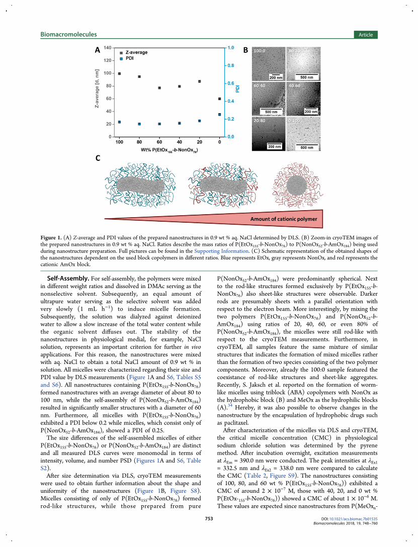
Self-Assembly. For self-assembly, the polymers were mixedin different weight ratios and dissolved in DMAc serving as thenonselective solvent. Subsequently, an equal amount ofultrapure water serving as the selective solvent was addedvery slowly (1 mL h−1) to induce micelle formation.Subsequently, the solution was dialyzed against deionizedwater to allow a slow increase of the total water content whilethe organic solvent diffuses out. The stability of thenanostructures in physiological medial, for example, NaClsolution, represents an important criterion for further in vivoapplications. For this reason, the nanostructures were mixedwith aq. NaCl to obtain a total NaCl amount of 0.9 wt % insolution. All micelles were characterized regarding their size andPDI value by DLS measurements (Figure 1A and S6, Tables S5and S6). All nanostructures containing P(EtOx155-b-NonOx76)formed nanostructures with an average diameter of about 80 to100 nm, while the self-assembly of P(NonOx52-b-AmOx184)resulted in significantly smaller structures with a diameter of 60nm. Furthermore, all micelles with P(EtOx155-b-NonOx76)exhibited a PDI below 0.2 while micelles, which consist only ofP(NonOx52-b-AmOx184), showed a PDI of 0.25.The size differences of the self-assembled micelles of either
P(EtOx155-b-NonOx76) or P(NonOx52-b-AmOx184) are distinctand all measured DLS curves were monomodal in terms ofintensity, volume, and number PSD (Figures 1A and S6, TableS2).After size determination via DLS, cryoTEM measurements
were used to obtain further information about the shape anduniformity of the nanostructures (Figure 1B, Figure S8).Micelles consisting of only of P(EtOx155-b-NonOx76) formedrod-like structures, while those prepared from pure
P(NonOx52-b-AmOx184) were predominantly spherical. Nextto the rod-like structures formed exclusively by P(EtOx155-b-NonOx76) also sheet-like structures were observable. Darkerrods are presumably sheets with a parallel orientation withrespect to the electron beam. More interestingly, by mixing thetwo polymers P(EtOx155-b-NonOx76) and P(NonOx52-b-AmOx184) using ratios of 20, 40, 60, or even 80% ofP(NonOx52-b-AmOx184), the micelles were still rod-like withrespect to the cryoTEM measurements. Furthermore, incryoTEM, all samples feature the same mixture of similarstructures that indicates the formation of mixed micelles ratherthan the formation of two species consisting of the two polymercomponents. Moreover, already the 100:0 sample featured thecoexistance of rod-like structures and sheet-like aggregates.Recently, S. Jaksch et al. reported on the formation of worm-like micelles using triblock (ABA) copolymers with NonOx asthe hydrophobic block (B) and MeOx as the hydrophilic blocks(A).34 Hereby, it was also possible to observe changes in thenanostructure by the encapsulation of hydrophobic drugs suchas paclitaxel.After characterization of the micelles via DLS and cryoTEM,
the critical micelle concentration (CMC) in physiologicalsodium chloride solution was determined by the pyrenemethod. After incubation overnight, excitation measurementsat λEm = 390.0 nm were conducted. The peak intensities at λEx1= 332.5 nm and λEx2 = 338.0 nm were compared to calculatethe CMC (Table 2, Figure S9). The nanostructures consistingof 100, 80, and 60 wt % P(EtOx155-b-NonOx76)) exhibited aCMC of around 2 × 10⁻⁷ M, those with 40, 20, and 0 wt %P(EtOx-155-b-NonOx76)) showed a CMC of about 1 × 10⁻⁶ M.These values are expected since nanostructures from P(MeOxn-
Figure 1. (A) Z-average and PDI values of the prepared nanostructures in 0.9 wt % aq. NaCl determined by DLS. (B) Zoom-in cryoTEM images ofthe prepared nanostructures in 0.9 wt % aq. NaCl. Ratios describe the mass ratios of P(EtOx155-b-NonOx76) to P(NonOx52-b-AmOx184) being usedduring nanostructure preparation. Full pictures can be found in the Supporting Information. (C) Schematic representation of the obtained shapes ofthe nanostructures dependent on the used block copolymers in different ratios. Blue represents EtOx, gray represents NonOx, and red represents thecationic AmOx block.
Biomacromolecules Article
DOI: 10.1021/acs.biomac.7b01535Biomacromolecules 2018, 19, 748−760
753
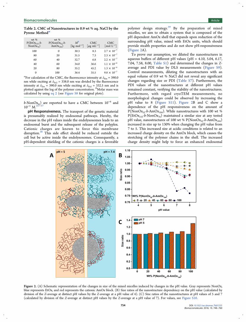
b-NonOxm) are reported to have a CMC between 10−6 and10−5 M.35−37
pH Responsiveness. The transport of the genetic materialis presumably realized by endosomal pathways. Hereby, thedecrease in the pH values inside the endolysosomes leads to anendosomal burst and the subsequent release of the polyplex.Cationic charges are known to force this membranedisruption.38 This side effect should be reduced outside thecell but be active inside the endolysosomes. Consequently, apH-dependent shielding of the cationic charges is a favorable
polymer design strategy.39 By the preparation of mixedmicelles, we aim to obtain a system that is composed of thepH dependent AmOx shell that expands upon reduction of thesurrounding pH value, mixed with EtOx units, which shouldprovide stealth properties and do not show pH-responsiveness(Figure 2A).To prove our assumption, we diluted the nanostructures in
aqueous buffers of different pH values (pH = 4.10, 5.04, 6.17,7.04, 7.58, 8.00, Table S1) and determined the changes in Z-average and PDI value by DLS measurements (Figure S9).Control measurements, diluting the nanostructures with anequal volume of 0.9 wt % NaCl did not reveal any significantchanges regarding size or PDI (Table S7). Furthermore, thePDI values of the nanostructures at different pH valuesremained constant, verifying the stability of the nanostructures.Furthermore, with regard cryoTEM measurements, nomorphological changes could be observed by increasing thepH value to 8 (Figure S11). Figure 2B and C show adependence of the pH responsiveness on the amount ofP(NonOx52-b-AmOx184). While nanostructures with 100 wt %P(EtOx155-b-NonOx76) maintained a similar size at any testedpH value, nanostructures of 100 wt % P(NonOx52-b-AmOx184)increased in size up to 130% when changing the pH value from7 to 5. This increased size at acidic conditions is related to anincreased charge density on the AmOx block, which causes thestretching of the polymer chains in the shell. The increasedcharge density might help to force an enhanced endosomal
Table 2. CMC of Nanostructures in 0.9 wt % aq. NaCl by thePyrene Methoda
wt %P(EtOx155-b-NonOx76)
wt %P(NonOx52-b-AmOx184)
Mb
[kg mol−1]CMC
[μg mL−1]CMC
[mol L−1]
100 0 30.3 8.3 2.7 × 10⁻⁷
80 20 31.5 7.3 2.3 × 10⁻⁷
60 40 32.7 6.8 2.2 × 10⁻⁷
40 60 34.0 36.6 1.1 × 10⁻⁶
20 80 35.2 45.2 1.3 × 10⁻⁶
0 100 36.4 35.5 9.8 × 10⁻⁷
aFor calculation of the CMC, the fluorescence intensity at λEm = 390.0nm while exciting at λEx2 = 338.0 nm was divided by the fluorescenceintensity at λEm = 390.0 nm while exciting at λEx2 = 332.5 nm and isplotted against the log of the polymer concentration. bMolar mass wascalculated by using eq 2 (see Figure S8 for original plots).
Figure 2. (A) Schematic representation of the changes in size of the mixed micelles induced by changes in the pH value. Gray represents NonOx,blue represents EtOx, and red represents the cationic AmOx block. (B) Size ratios of the nanostructure dependency on the pH value (calculated bydivision of the Z-average at distinct pH values by the Z-average at a pH value of 4). (C) Size ratios of the nanostructures at pH values of 5 and 7(calculated by division of the Z-average at distinct pH values by the Z-average at a pH value of 7). For values, see Figure S10.
Biomacromolecules Article
DOI: 10.1021/acs.biomac.7b01535Biomacromolecules 2018, 19, 748−760
754
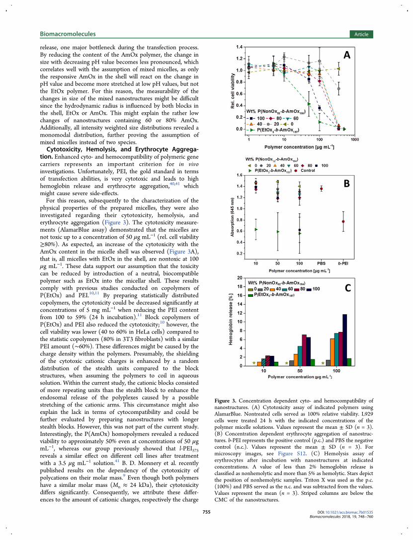
release, one major bottleneck during the transfection process.By reducing the content of the AmOx polymer, the change insize with decreasing pH value becomes less pronounced, whichcorrelates well with the assumption of mixed micelles, as onlythe responsive AmOx in the shell will react on the change inpH value and become more stretched at low pH values, but notthe EtOx polymer. For this reason, the measurability of thechanges in size of the mixed nanostructures might be difficultsince the hydrodynamic radius is influenced by both blocks inthe shell, EtOx or AmOx. This might explain the rather lowchanges of nanostructures containing 60 or 80% AmOx.Additionally, all intensity weighted size distributions revealed amonomodal distribution, further proving the assumption ofmixed micelles instead of two species.Cytotoxicity, Hemolysis, and Erythrocyte Aggrega-
tion. Enhanced cyto- and hemocompatibility of polymeric genecarriers represents an important criterion for in vivoinvestigations. Unfortunately, PEI, the gold standard in termsof transfection abilities, is very cytotoxic and leads to highhemoglobin release and erythrocyte aggregation,40,41 whichmight cause severe side-effects.For this reason, subsequently to the characterization of the
physical properties of the prepared micelles, they were alsoinvestigated regarding their cytotoxicity, hemolysis, anderythrocyte aggregation (Figure 3). The cytotoxicity measure-ments (AlamarBlue assay) demonstrated that the micelles arenot toxic up to a concentration of 50 μg mL−1 (rel. cell viability≥80%). As expected, an increase of the cytotoxicity with theAmOx content in the micelle shell was observed (Figure 3A),that is, all micelles with EtOx in the shell, are nontoxic at 100μg mL−1. These data support our assumption that the toxicitycan be reduced by introduction of a neutral, biocompatiblepolymer such as EtOx into the micellar shell. These resultscomply with previous studies conducted on copolymers ofP(EtOx) and PEI.10,11 By preparing statistically distributedcopolymers, the cytotoxicity could be decreased significantly atconcentrations of 5 mg mL−1 when reducing the PEI contentfrom 100 to 59% (24 h incubation).11 Block copolymers ofP(EtOx) and PEI also reduced the cytotoxicity;10 however, thecell viability was lower (40 to 60% in HeLa cells) compared tothe statistic copolymers (80% in 3T3 fibroblasts) with a similarPEI amount (∼60%). These differences might be caused by thecharge density within the polymers. Presumably, the shieldingof the cytotoxic cationic charges is enhanced by a randomdistribution of the stealth units compared to the blockstructures, when assuming the polymers to coil in aqueoussolution. Within the current study, the cationic blocks consistedof more repeating units than the stealth block to enhance theendosomal release of the polyplexes caused by a possiblestretching of the cationic arms. This circumstance might alsoexplain the lack in terms of cytocompatibility and could befurther evaluated by preparing nanostructures with longerstealth blocks. However, this was not part of the current study.Interestingly, the P(AmOx) homopolymers revealed a reducedviability to approximately 50% even at concentrations of 50 μgmL−1, whereas our group previously showed that l-PEI575reveals a similar effect on different cell lines after treatmentwith a 3.5 μg mL−1 solution.41 B. D. Monnery et al. recentlypublished results on the dependency of the cytotoxicity ofpolycations on their molar mass.9 Even though both polymershave a similar molar mass (Mn ≈ 24 kDa), their cytotoxicitydiffers significantly. Consequently, we attribute these differ-ences to the amount of cationic charges, respectively the charge
Figure 3. Concentration dependent cyto- and hemocompatibility ofnanostructures. (A) Cytotoxicity assay of indicated polymers usingAlamarBlue. Nontreated cells served as 100% relative viability. L929cells were treated 24 h with the indicated concentrations of thepolymer micelle solutions. Values represent the mean ± SD (n = 3).(B) Concentration dependent erythrocyte aggregation of nanostruc-tures. b-PEI represents the positive control (p.c.) and PBS the negativecontrol (n.c.). Values represent the mean ± SD (n = 3). Formicroscopy images, see Figure S12. (C) Hemolysis assay oferythrocytes after incubation with nanostructures at indicatedconcentrations. A value of less than 2% hemoglobin release isclassified as nonhemolytic and more than 5% as hemolytic. Stars depictthe position of nonhemolytic samples. Triton X was used as the p.c.(100%) and PBS served as the n.c. and was subtracted from the values.Values represent the mean (n = 3). Striped columns are below theCMC of the nanostructures.
Biomacromolecules Article
DOI: 10.1021/acs.biomac.7b01535Biomacromolecules 2018, 19, 748−760
755
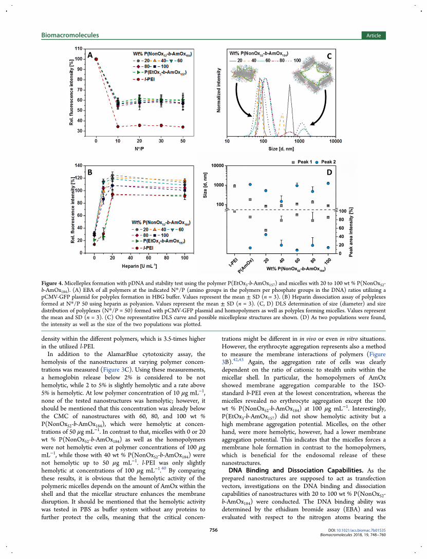
density within the different polymers, which is 3.5-times higherin the utilized l-PEI.In addition to the AlamarBlue cytotoxicity assay, the
hemolysis of the nanostructures at varying polymer concen-trations was measured (Figure 3C). Using these measurements,a hemoglobin release below 2% is considered to be nothemolytic, while 2 to 5% is slightly hemolytic and a rate above5% is hemolytic. At low polymer concentration of 10 μg mL−1,none of the tested nanostructures was hemolytic; however, itshould be mentioned that this concentration was already belowthe CMC of nanostructures with 60, 80, and 100 wt %P(NonOx52-b-AmOx184), which were hemolytic at concen-trations of 50 μg mL−1. In contrast to that, micelles with 0 or 20wt % P(NonOx52-b-AmOx184) as well as the homopolymerswere not hemolytic even at polymer concentrations of 100 μgmL−1, while those with 40 wt % P(NonOx52-b-AmOx184) werenot hemolytic up to 50 μg mL−1. l-PEI was only slightlyhemolytic at concentrations of 100 μg mL−1.40 By comparingthese results, it is obvious that the hemolytic activity of thepolymeric micelles depends on the amount of AmOx within theshell and that the micellar structure enhances the membranedisruption. It should be mentioned that the hemolytic activitywas tested in PBS as buffer system without any proteins tofurther protect the cells, meaning that the critical concen-
trations might be different in in vivo or even in vitro situations.However, the erythrocyte aggregation represents also a methodto measure the membrane interactions of polymers (Figure3B).42,43 Again, the aggregation rate of cells was clearlydependent on the ratio of cationic to stealth units within themicellar shell. In particular, the homopolymers of AmOxshowed membrane aggregation comparable to the ISO-standard b-PEI even at the lowest concentration, whereas themicelles revealed no erythrocyte aggregation except the 100wt % P(NonOx52-b-AmOx184) at 100 μg mL−1. Interestingly,P(EtOx3-b-AmOx157) did not show hemolytic activity but ahigh membrane aggregation potential. Micelles, on the otherhand, were more hemolytic, however, had a lower membraneaggregation potential. This indicates that the micelles forces amembrane hole formation in contrast to the homopolymers,which is beneficial for the endosomal release of thesenanostructures.
DNA Binding and Dissociation Capabilities. As theprepared nanostructures are supposed to act as transfectionvectors, investigations on the DNA binding and dissociationcapabilities of nanostructures with 20 to 100 wt % P(NonOx52-b-AmOx184) were conducted. The DNA binding ability wasdetermined by the ethidium bromide assay (EBA) and wasevaluated with respect to the nitrogen atoms bearing the
Figure 4.Micelleplex formation with pDNA and stability test using the polymer P(EtOx3-b-AmOx157) and micelles with 20 to 100 wt % P(NonOx52-b-AmOx184). (A) EBA of all polymers at the indicated N*/P (amino groups in the polymers per phosphate groups in the DNA) ratios utilizing apCMV-GFP plasmid for polyplex formation in HBG buffer. Values represent the mean ± SD (n = 3). (B) Heparin dissociation assay of polyplexesformed at N*/P 50 using heparin as polyanion. Values represent the mean ± SD (n = 3). (C, D) DLS determination of size (diameter) and sizedistribution of polyplexes (N*/P = 50) formed with pCMV-GFP plasmid and homopolymers as well as polyplex forming micelles. Values representthe mean and SD (n = 3). (C) One representative DLS curve and possible micelleplexe structures are shown. (D) As two populations were found,the intensity as well as the size of the two populations was plotted.
Biomacromolecules Article
DOI: 10.1021/acs.biomac.7b01535Biomacromolecules 2018, 19, 748−760
756
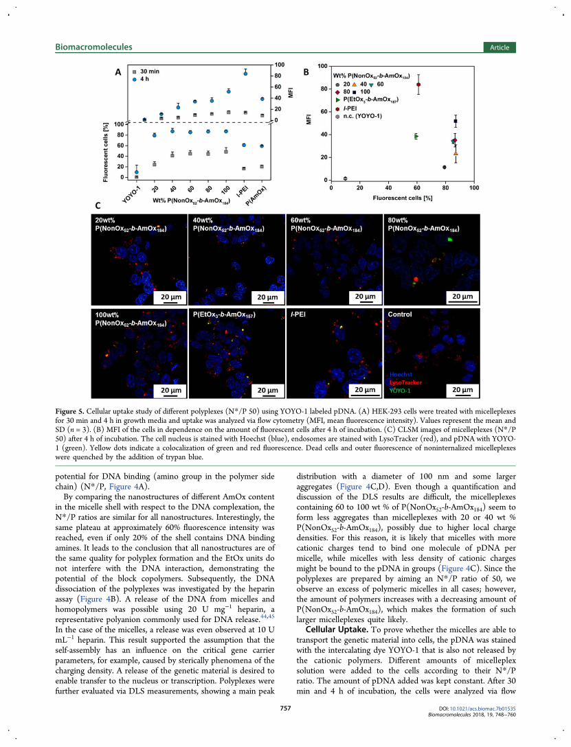
potential for DNA binding (amino group in the polymer sidechain) (N*/P, Figure 4A).By comparing the nanostructures of different AmOx content
in the micelle shell with respect to the DNA complexation, theN*/P ratios are similar for all nanostructures. Interestingly, thesame plateau at approximately 60% fluorescence intensity wasreached, even if only 20% of the shell contains DNA bindingamines. It leads to the conclusion that all nanostructures are ofthe same quality for polyplex formation and the EtOx units donot interfere with the DNA interaction, demonstrating thepotential of the block copolymers. Subsequently, the DNAdissociation of the polyplexes was investigated by the heparinassay (Figure 4B). A release of the DNA from micelles andhomopolymers was possible using 20 U mg−1 heparin, arepresentative polyanion commonly used for DNA release.44,45
In the case of the micelles, a release was even observed at 10 UmL−1 heparin. This result supported the assumption that theself-assembly has an influence on the critical gene carrierparameters, for example, caused by sterically phenomena of thecharging density. A release of the genetic material is desired toenable transfer to the nucleus or transcription. Polyplexes werefurther evaluated via DLS measurements, showing a main peak
distribution with a diameter of 100 nm and some largeraggregates (Figure 4C,D). Even though a quantification anddiscussion of the DLS results are difficult, the micelleplexescontaining 60 to 100 wt % of P(NonOx52-b-AmOx184) seem toform less aggregates than micelleplexes with 20 or 40 wt %P(NonOx52-b-AmOx184), possibly due to higher local chargedensities. For this reason, it is likely that micelles with morecationic charges tend to bind one molecule of pDNA permicelle, while micelles with less density of cationic chargesmight be bound to the pDNA in groups (Figure 4C). Since thepolyplexes are prepared by aiming an N*/P ratio of 50, weobserve an excess of polymeric micelles in all cases; however,the amount of polymers increases with a decreasing amount ofP(NonOx52-b-AmOx184), which makes the formation of suchlarger micelleplexes quite likely.
Cellular Uptake. To prove whether the micelles are able totransport the genetic material into cells, the pDNA was stainedwith the intercalating dye YOYO-1 that is also not released bythe cationic polymers. Different amounts of micelleplexsolution were added to the cells according to their N*/Pratio. The amount of pDNA added was kept constant. After 30min and 4 h of incubation, the cells were analyzed via flow
Figure 5. Cellular uptake study of different polyplexes (N*/P 50) using YOYO-1 labeled pDNA. (A) HEK-293 cells were treated with micelleplexesfor 30 min and 4 h in growth media and uptake was analyzed via flow cytometry (MFI, mean fluorescence intensity). Values represent the mean andSD (n = 3). (B) MFI of the cells in dependence on the amount of fluorescent cells after 4 h of incubation. (C) CLSM images of micelleplexes (N*/P50) after 4 h of incubation. The cell nucleus is stained with Hoechst (blue), endosomes are stained with LysoTracker (red), and pDNA with YOYO-1 (green). Yellow dots indicate a colocalization of green and red fluorescence. Dead cells and outer fluorescence of noninternalized micelleplexeswere quenched by the addition of trypan blue.
Biomacromolecules Article
DOI: 10.1021/acs.biomac.7b01535Biomacromolecules 2018, 19, 748−760
757
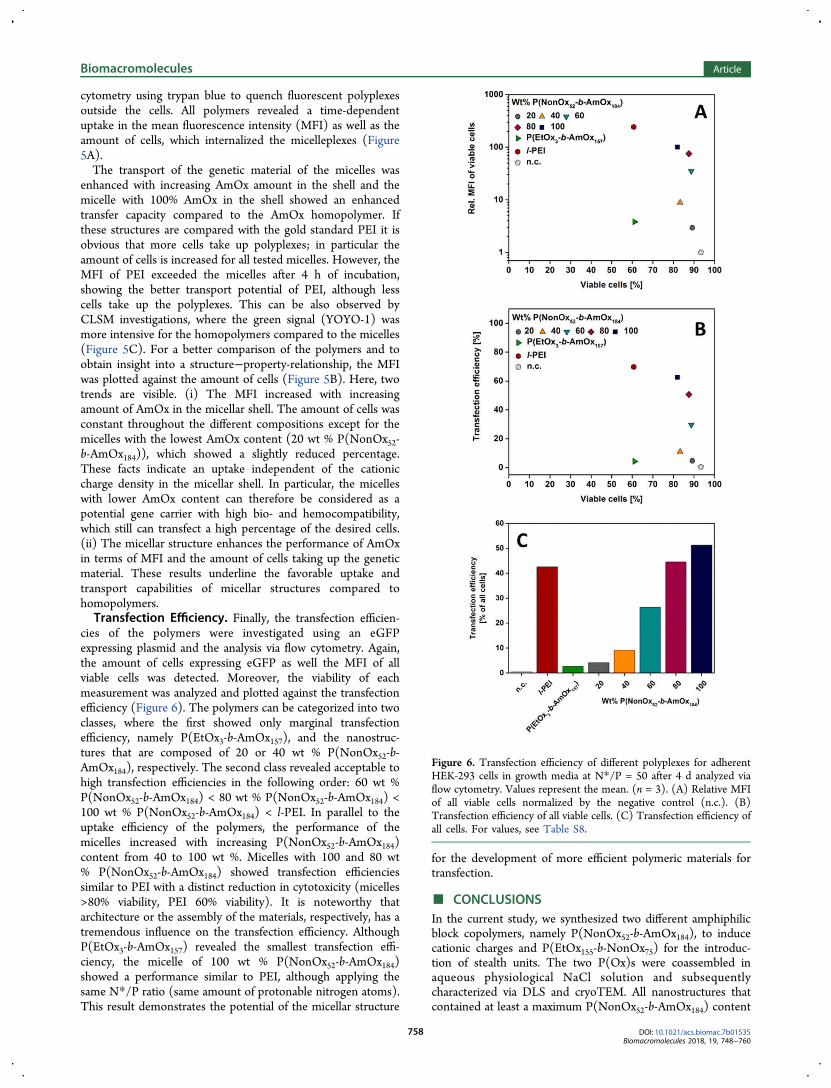
cytometry using trypan blue to quench fluorescent polyplexesoutside the cells. All polymers revealed a time-dependentuptake in the mean fluorescence intensity (MFI) as well as theamount of cells, which internalized the micelleplexes (Figure5A).The transport of the genetic material of the micelles was
enhanced with increasing AmOx amount in the shell and themicelle with 100% AmOx in the shell showed an enhancedtransfer capacity compared to the AmOx homopolymer. Ifthese structures are compared with the gold standard PEI it isobvious that more cells take up polyplexes; in particular theamount of cells is increased for all tested micelles. However, theMFI of PEI exceeded the micelles after 4 h of incubation,showing the better transport potential of PEI, although lesscells take up the polyplexes. This can be also observed byCLSM investigations, where the green signal (YOYO-1) wasmore intensive for the homopolymers compared to the micelles(Figure 5C). For a better comparison of the polymers and toobtain insight into a structure−property-relationship, the MFIwas plotted against the amount of cells (Figure 5B). Here, twotrends are visible. (i) The MFI increased with increasingamount of AmOx in the micellar shell. The amount of cells wasconstant throughout the different compositions except for themicelles with the lowest AmOx content (20 wt % P(NonOx52-b-AmOx184)), which showed a slightly reduced percentage.These facts indicate an uptake independent of the cationiccharge density in the micellar shell. In particular, the micelleswith lower AmOx content can therefore be considered as apotential gene carrier with high bio- and hemocompatibility,which still can transfect a high percentage of the desired cells.(ii) The micellar structure enhances the performance of AmOxin terms of MFI and the amount of cells taking up the geneticmaterial. These results underline the favorable uptake andtransport capabilities of micellar structures compared tohomopolymers.Transfection Efficiency. Finally, the transfection efficien-
cies of the polymers were investigated using an eGFPexpressing plasmid and the analysis via flow cytometry. Again,the amount of cells expressing eGFP as well the MFI of allviable cells was detected. Moreover, the viability of eachmeasurement was analyzed and plotted against the transfectionefficiency (Figure 6). The polymers can be categorized into twoclasses, where the first showed only marginal transfectionefficiency, namely P(EtOx3-b-AmOx157), and the nanostruc-tures that are composed of 20 or 40 wt % P(NonOx52-b-AmOx184), respectively. The second class revealed acceptable tohigh transfection efficiencies in the following order: 60 wt %P(NonOx52-b-AmOx184) < 80 wt % P(NonOx52-b-AmOx184) <100 wt % P(NonOx52-b-AmOx184) < l-PEI. In parallel to theuptake efficiency of the polymers, the performance of themicelles increased with increasing P(NonOx52-b-AmOx184)content from 40 to 100 wt %. Micelles with 100 and 80 wt% P(NonOx52-b-AmOx184) showed transfection efficienciessimilar to PEI with a distinct reduction in cytotoxicity (micelles>80% viability, PEI 60% viability). It is noteworthy thatarchitecture or the assembly of the materials, respectively, has atremendous influence on the transfection efficiency. AlthoughP(EtOx3-b-AmOx157) revealed the smallest transfection effi-ciency, the micelle of 100 wt % P(NonOx52-b-AmOx184)showed a performance similar to PEI, although applying thesame N*/P ratio (same amount of protonable nitrogen atoms).This result demonstrates the potential of the micellar structure
for the development of more efficient polymeric materials fortransfection.
■ CONCLUSIONS
In the current study, we synthesized two different amphiphilicblock copolymers, namely P(NonOx52-b-AmOx184), to inducecationic charges and P(EtOx155-b-NonOx75) for the introduc-tion of stealth units. The two P(Ox)s were coassembled inaqueous physiological NaCl solution and subsequentlycharacterized via DLS and cryoTEM. All nanostructures thatcontained at least a maximum P(NonOx52-b-AmOx184) content
Figure 6. Transfection efficiency of different polyplexes for adherentHEK-293 cells in growth media at N*/P = 50 after 4 d analyzed viaflow cytometry. Values represent the mean. (n = 3). (A) Relative MFIof all viable cells normalized by the negative control (n.c.). (B)Transfection efficiency of all viable cells. (C) Transfection efficiency ofall cells. For values, see Table S8.
Biomacromolecules Article
DOI: 10.1021/acs.biomac.7b01535Biomacromolecules 2018, 19, 748−760
758
of 80 wt % resulted in rod-like micelles with an apparentaverage diameter of 100 nm (assuming spheres by DLSmeasurements), whereas pure P(NonOx52-b-AmOx184) micelleswere spherical. DLS measurements of the nanostructures inbuffers of distinct pH values resulted in a pH-dependentalteration of the size with respect to the pH value and theamount of P(NonOx52-b-AmOx184). A reversible polyplexformation was possible with all amino group containingnanostructures. We observed that the cytotoxicity, erythrocyteaggregation, and hemolytic activity were dependent on thepolymer composition within the nanostructures. The highcharge density of the micelles led to an enhanced hemoglobinrelease compared to the P(AmOx) homopolymer. Examina-tions on the cellular uptake showed that the number offluorescent cells is similar for all nanostructures (∼80%), whilethe MFI increases applying micelles with more cationic charges.In comparison, we observed 60% fluorescent cells using l-PEIor AmOx homopolymers and the MFI of P(AmOx) wasconsiderably lower than for l-PEI. Flow cytometry analysis ofthe transfection efficiency revealed an enhanced viability of thecells when treated with micelleplexes (80−90%) compared topolyplexes of P(AmOx) or l-PEI (60%). The transfectionefficiency was strongly dependent on the amount of cationicpolymer within the micelles and ranged from less than 5% (20wt % P(NonOx52-b-AmOx184)) to more than 60% (100 wt %P(NonOx52-b-AmOx184)). Micelleplexes with 80 or 100 wt %P(NonOx52-b-AmOx184) showed a better allover performancein terms of transfection efficiency than l-PEI, while P(AmOx)was worse than the micelleplexes with 20 wt % P(NonOx52-b-AmOx184). We attribute these advantages to the architecture ofthe micelles and the following accumulation of cationic chargeson their surface due to the cationic blocks. On the basis of thesefindings, we were able to improve the performance of a toxic,poorly transfecting polymer by appropriate self- andcoassembly process to obtain nanostructures with a decreasedcytotoxicity and improved transfection efficiency compared to l-PEI and the AmOx homopolymer.Further studies might concentrate on the utilization of
cationic block copolymers with different lengths to obtainnanostructures, which have an optimum balance betweenshielding by the EtOx blocks and efficient endosomal release bythe stretching of the cationic blocks within the endolysosomes.These experiments might help to find biocompatible andefficient gene carrier systems.
■ ASSOCIATED CONTENT
*S Supporting Information
The Supporting Information is available free of charge on theACS Publications website at DOI: 10.1021/acs.bio-mac.7b01535.
NMR spectra, SEC traces, DLS curves, magnifiedcryoTEM images, CMC determination graphs, eryth-rocyte aggregation microscopy images, raw values of DLSmeasurements (PDF)
■ AUTHOR INFORMATION
Corresponding Authors
*E-mail: [email protected].*E-mail: [email protected].
ORCID
Ulrich S. Schubert: 0000-0003-4978-4670
Author Contributions
The manuscript was written through contributions of allauthors. All authors have given approval to the final version ofthe manuscript.
NotesThe authors declare no competing financial interest.
■ ACKNOWLEDGMENTS
The authors gratefully acknowledge the Bundesministerium furBildung und Forschung (BMBF, Germany, No. 13N13416smart-dye-livery) and the Thuringer Ministerium fur Wirt-schaft, Wissenschaft und Digitale Gesellschaft (ProExzellenzII,NanoPolar). Furthermore, funding of the collaborative researchcenter PolyTarget (SFB 1278) by the Deutsche Forschungsge-meinschaft (DFG) is highly acknowledged. A.T. acknowledgesthe Carl Zeiss Foundation and the BMBF (No. 13XP5034APolyBioMik) for funding. J.C.B. further thanks the DFG forsupport (Emmy-Noether Program, BR 4905/3-1). The authorsthankfully acknowledge Carolin Kellner and Elisabeth Moek forthe conduction of AlamarBlue, hemolysis, and aggregationassays as well as Dr. Grit Festag for SEC measurements on anaqueous system. CryoTEM investigations were performed atthe Electron Microsocpy facilities of the Jena Center for SoftMatter (JCSM), which was established with grants from theDeutsche Forschungsgemeinschaft (DFG) and the EuropeanFund for Regional Development (EFRE). The LSM880ELYRA PS.1 was further funded with a grant from the DFG.
■ REFERENCES
(1) Huang, Y.; Liu, X.; Dong, L.; Liu, Z.; He, X.; Liu, W.Development of viral vectors for gene therapy for chronic pain. PainRes. Treat. 2011, 2011, 968218.(2) Schatzlein, A. G. Non-viral vectors in cancer gene therapy:Principles and progress. Anti-Cancer Drugs 2001, 12 (4), 275−304.(3) Gardlik, R.; Palffy, R.; Hodosy, J.; Lukacs, J.; Turna, J.; Celec, P.Vectors and delivery systems in gene therapy. Med. Sci. Monit. 2005,11 (4), Ra110−21.(4) Zhang, S.; Zhao, B.; Jiang, H.; Wang, B.; Ma, B. Cationic lipidsand polymers mediated vectors for delivery of sirna. J. ControlledRelease 2007, 123 (1), 1−10.(5) Bulmus, V.; Woodward, M.; Lin, L.; Murthy, N.; Stayton, P.;Hoffman, A. A new ph-responsive and glutathione-reactive, endosomalmembrane-disruptive polymeric carrier for intracellular delivery ofbiomolecular drugs. J. Controlled Release 2003, 93 (2), 105−120.(6) Godbey, W. T.; Barry, M. A.; Saggau, P.; Wu, K. K.; Mikos, A. G.Poly(ethylenimine)-mediated transfection: A new paradigm for genedelivery. J. Biomed. Mater. Res. 2000, 51 (3), 321−8.(7) Behr, J.-P. The proton sponge: A trick to enter cells the virusesdid not exploit. Chimia 1997, 51 (1−2), 34−36.(8) Ahn, C. H.; Chae, S. Y.; Bae, Y. H.; Kim, S. W. Biodegradablepoly(ethylenimine) for plasmid DNA delivery. J. Controlled Release2002, 80 (1−3), 273−82.(9) Monnery, B. D.; Wright, M.; Cavill, R.; Hoogenboom, R.;Shaunak, S.; Steinke, J. H. G.; Thanou, M. Cytotoxicity of polycations:Relationship of molecular weight and the hydrolytic theory of themechanism of toxicity. Int. J. Pharm. 2017, 521 (1), 249−258.(10) Hsiue, G. H.; Chiang, H. Z.; Wang, C. H.; Juang, T. M. Nonviralgene carriers based on diblock copolymers of poly(2-ethyl-2-oxazoline) and linear polyethylenimine. Bioconjugate Chem. 2006, 17(3), 781−6.(11) Shah, R.; Kronekova, Z.; Zahoranova, A.; Roller, L.; Saha, N.;Saha, P.; Kronek, J. vitro study of partially hydrolyzed poly(2-ethyl-2-oxazolines) as materials for biomedical applications. J. Mater. Sci.:Mater. Med. 2015, 26 (4), 157.(12) Miyata, K.; Oba, M.; Nakanishi, M.; Fukushima, S.; Yamasaki,Y.; Koyama, H.; Nishiyama, N.; Kataoka, K. Polyplexes from
Biomacromolecules Article
DOI: 10.1021/acs.biomac.7b01535Biomacromolecules 2018, 19, 748−760
759
poly(aspartamide) bearing 1,2-diaminoethane side chains induce ph-selective, endosomal membrane destabilization with amplified trans-fection and negligible cytotoxicity. J. Am. Chem. Soc. 2008, 130 (48),16287−16294.(13) Oba, M.; Miyata, K.; Osada, K.; Christie, R. J.; Sanjoh, M.; Li,W.; Fukushima, S.; Ishii, T.; Kano, M. R.; Nishiyama, N.; Koyama, H.;Kataoka, K. Polyplex micelles prepared from ω-cholesteryl peg-polycation block copolymers for systemic gene delivery. Biomaterials2011, 32 (2), 652−663.(14) Kim, H. J.; Ishii, A.; Miyata, K.; Lee, Y.; Wu, S.; Oba, M.;Nishiyama, N.; Kataoka, K. Introduction of stearoyl moieties into abiocompatible cationic polyaspartamide derivative, pasp(det), withendosomal escaping function for enhanced sirna-mediated geneknockdown. J. Controlled Release 2010, 145 (2), 141−148.(15) Barz, M.; Luxenhofer, R.; Zentel, R.; Vicent, M. J. Overcomingthe peg-addiction: Well-defined alternatives to peg, from structure-property relationships to better defined therapeutics. Polym. Chem.2011, 2 (9), 1900−1918.(16) Hoogenboom, R. Poly(2-oxazoline)s: A polymer class withnumerous potential applications. Angew. Chem., Int. Ed. 2009, 48 (43),7978−94.(17) Luxenhofer, R.; Han, Y.; Schulz, A.; Tong, J.; He, Z.; Kabanov,A. V.; Jordan, R. Poly(2-oxazoline)s as polymer therapeutics.Macromol. Rapid Commun. 2012, 33 (19), 1613−1631.(18) Gaertner, F. C.; Luxenhofer, R.; Blechert, B.; Jordan, R.; Essler,M. Synthesis, biodistribution and excretion of radiolabeled poly(2-alkyl-2-oxazoline)s. J. Controlled Release 2007, 119 (3), 291−300.(19) Salzinger, S.; Huber, S.; Jaksch, S.; Busch, P.; Jordan, R.;Papadakis, C. M. Aggregation behavior of thermo-responsive poly(2-oxazoline)s at the cloud point investigated by fcs and sans. ColloidPolym. Sci. 2012, 290 (5), 385−400.(20) Cai, G.; Litt, M. H. Preparation and characterization of phenyland undecyl oxazoline block copolymers. J. Polym. Sci., Part A: Polym.Chem. 1989, 27 (11), 3603−3618.(21) McAlvin, J. E.; Fraser, C. L. Metal-centered star blockcopolymers: Amphiphilic iron tris(bipyridine)-centered polyoxazolinesand their chemical fragmentation to bipyridine-centered bab triblockcopolymers. Macromolecules 1999, 32 (5), 1341−1347.(22) Jin, R.-H. Water soluble star block poly(oxazoline) withporphyrin label: A unique emulsion and its shape direction. J. Mater.Chem. 2004, 14 (3), 320−327.(23) Lach, C.; Hanselmann, R.; Frey, H.; Mulhaupt, R. Hyper-branched carbosilane oxazoline-macromonomers: Polymerization andcoupling to a trimesic acid core. Macromol. Rapid Commun. 1998, 19(9), 461−465.(24) Hartlieb, M.; Pretzel, D.; Kempe, K.; Fritzsche, C.; Paulus, R.M.; Gottschaldt, M.; Schubert, U. S. Cationic poly(2-oxazoline)hydrogels for reversible DNA binding. Soft Matter 2013, 9 (18),4693−4704.(25) Christova, D.; Velichkova, R.; Goethals, E. J.; Du Prez, F. E.Amphiphilic segmented polymer networks based on poly(2-alkyl-2-oxazoline) and poly(methyl methacrylate). Polymer 2002, 43 (17),4585−4590.(26) Hadjichristidis, N., Pispas, S., Floudas, G. A. Block Copolymers:Synthetic Strategies, Physical Properties, and Applications; Wiley:Weinheim, 2003.(27) He, Z.; Miao, L.; Jordan, R.; S-Manickam, D.; Luxenhofer, R.;Kabanov, A. V. A low protein binding cationic poly(2-oxazoline) asnon-viral vector. Macromol. Biosci. 2015, 15 (7), 1004−1020.(28) Rinkenauer, A. C.; Tauhardt, L.; Wendler, F.; Kempe, K.;Gottschaldt, M.; Traeger, A.; Schubert, U. S. A cationic poly(2-oxazoline) with high in vitro transfection efficiency identified by alibrary approach. Macromol. Biosci. 2015, 15 (3), 414−425.(29) Bauer, M.; Lautenschlaeger, C.; Kempe, K.; Tauhardt, L.;Schubert, U. S.; Fischer, D. Poly(2-ethyl-2-oxazoline) as alternative forthe stealth polymer poly(ethylene glycol): Comparison of in vitrocytotoxicity and hemocompatibility. Macromol. Biosci. 2012, 12 (7),986−998.
(30) Wiesbrock, F.; Hoogenboom, R.; Abeln, C. H.; Schubert, U. S.Single-mode microwave ovens as new reaction devices: Acceleratingthe living polymerization of 2-ethyl-2-oxazoline. Macromol. RapidCommun. 2004, 25 (22), 1895−1899.(31) Wilhelm, M.; Zhao, C. L.; Wang, Y.; Xu, R.; Winnik, M. A.;Mura, J. L.; Riess, G.; Croucher, M. D. Poly(styrene-ethylene oxide)block copolymer micelle formation in water: A fluorescence probestudy. Macromolecules 1991, 24 (5), 1033−1040.(32) Vollrath, A.; Schallon, A.; Pietsch, C.; Schubert, S.; Nomoto, T.;Matsumoto, Y.; Kataoka, K.; Schubert, U. S. A toolbox of differentlysized and labeled pmma nanoparticles for cellular uptake inves-tigations. Soft Matter 2013, 9 (1), 99−108.(33) Cesana, S.; Auernheimer, J.; Jordan, R.; Kessler, H.; Nuyken, O.First poly(2-oxazoline)s with pendant amino groups. Macromol. Chem.Phys. 2006, 207 (2), 183−192.(34) Jaksch, S.; Schulz, A.; Di, Z.; Luxenhofer, R.; Jordan, R.;Papadakis, C. M. Amphiphilic triblock copolymers from poly(2-oxazoline) with different hydrophobic blocks: Changes of the micellarstructures upon addition of a strongly hydrophobic cancer drug.Macromol. Chem. Phys. 2016, 217 (13), 1448−1456.(35) Bonne, T. B.; Papadakis, C. M.; Ludtke, K.; Jordan, R. Role ofthe tracer in characterizing the aggregation behavior of aqueous blockcopolymer solutions using fluorescence correlation spectroscopy.Colloid Polym. Sci. 2007, 285 (5), 491−497.(36) Bonne, T. B.; Ludtke, K.; Jordan, R.; Stepanek, P.; Papadakis, C.M. Aggregation behavior of amphiphilic poly(2-alkyl-2-oxazoline)diblock copolymers in aqueous solution studied by fluorescencecorrelation spectroscopy. Colloid Polym. Sci. 2004, 282 (8), 833−843.(37) Ivanova, R.; Komenda, T.; Bonne, T. B.; Ludtke, K.; Mortensen,K.; Pranzas, P. K.; Jordan, R.; Papadakis, C. M. Micellar structures ofhydrophilic/lipophilic and hydrophilic/fluorophilic poly(2-oxazoline)diblock copolymers in water. Macromol. Chem. Phys. 2008, 209 (21),2248−2258.(38) Jain, K.; Kesharwani, P.; Gupta, U.; Jain, N. K. Dendrimertoxicity: Let’s meet the challenge. Int. J. Pharm. 2010, 394 (1), 122−142.(39) Betthausen, E.; Drechsler, M.; Fortsch, M.; Schacher, F. H.;Muller, A. H. E. Dual stimuli-responsive multicompartment micellesfrom triblock terpolymers with tunable hydrophilicity. Soft Matter2011, 7 (19), 8880−8891.(40) Bus, T.; Englert, C.; Reifarth, M.; Borchers, P.; Hartlieb, M.;Vollrath, A.; Hoeppener, S.; Traeger, A.; Schubert, U. S., 3rdgeneration poly(ethylene imine)s for gene delivery. J. Mater. Chem.B 2017, 5 (6), 1258−1274.(41) Englert, C.; Prohl, M.; Czaplewska, J. A.; Fritzsche, C.;Preußger, E.; Schubert, U. S.; Traeger, A.; Gottschaldt, M. D-fructose-decorated poly(ethylene imine) for human breast cancer cell targeting.Macromol. Biosci. 2017, 17 (8), 1600502.(42) Lee, Y.; Miyata, K.; Oba, M.; Ishii, T.; Fukushima, S.; Han, M.;Koyama, H.; Nishiyama, N.; Kataoka, K. Charge-conversion ternarypolyplex with endosome disruption moiety: A technique for efficientand safe gene delivery. Angew. Chem. 2008, 120 (28), 5241−5244.(43) Rinkenauer, A. C.; Schallon, A.; Gunther, U.; Wagner, M.;Betthausen, E.; Schubert, U. S.; Schacher, F. H. A paradigm change:Efficient transfection of human leukemia cells by stimuli-responsivemulticompartment micelles. ACS Nano 2013, 7 (11), 9621−9631.(44) Mislick, K. A.; Baldeschwieler, J. D. Evidence for the role ofproteoglycans in cation-mediated gene transfer. Proc. Natl. Acad. Sci. U.S. A. 1996, 93 (22), 12349−12354.(45) Xu, Y.; Szoka, F. C. Mechanism of DNA release from cationicliposome/DNA complexes used in cell transfection. Biochemistry 1996,35 (18), 5616−5623.
Biomacromolecules Article
DOI: 10.1021/acs.biomac.7b01535Biomacromolecules 2018, 19, 748−760
760
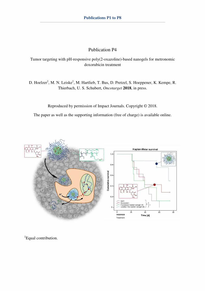
Publications P1 to P8
Publication P4
Tumor targeting with pH-responsive poly(2-oxazoline)-based nanogels for metronomic
doxorubicin treatment
D. Hoelzer‡, M. N. Leiske
‡, M. Hartlieb, T. Bus, D. Pretzel, S. Hoeppener, K. Kempe, R.
Thierbach, U. S. Schubert, Oncotarget 2018, in press.
Reproduced by permission of Impact Journals. Copyright © 2018.
The paper as well as the supporting information (free of charge) is available online.
‡Equal contribution.


at λnanogels the absorbance was detected at λ
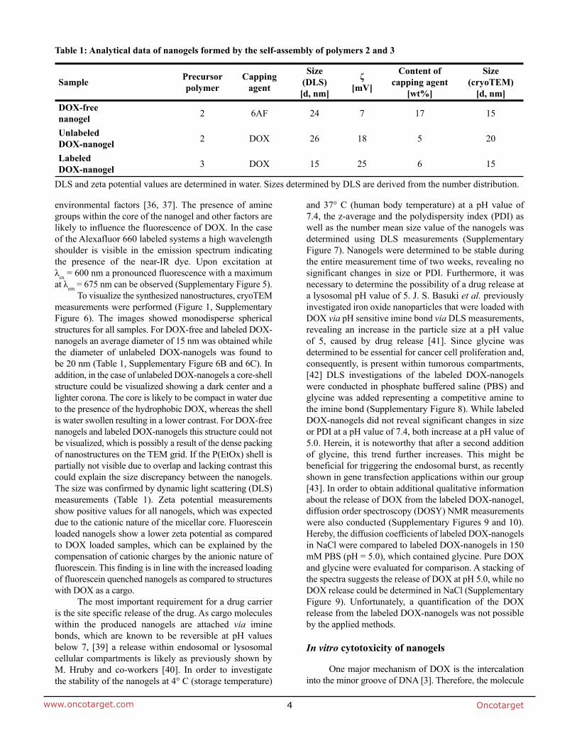
λλ
ζ

L292 mouse fibroblasts (
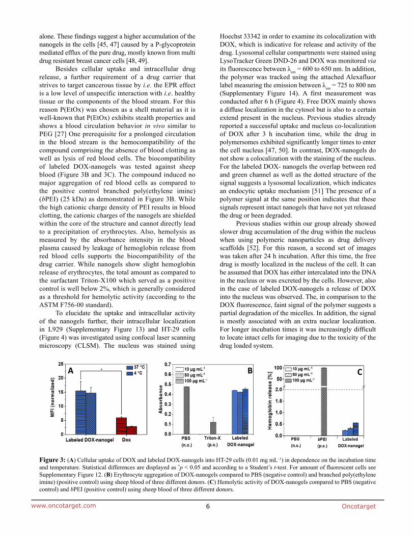
its fluorescence between λ λ
-test. For amount of fluorescent cells see
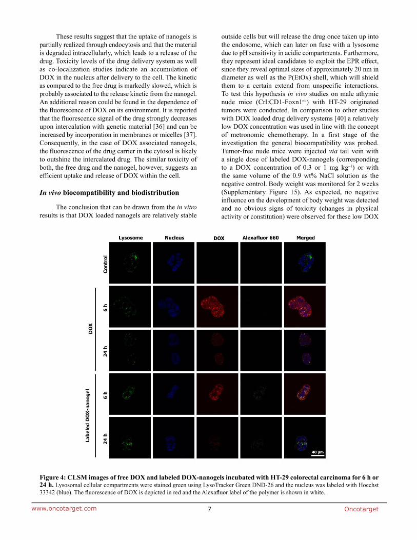
33342 (blue). The fluorescence of DOX is depicted in red and the Alexafluor label of the polymer is shown in white.
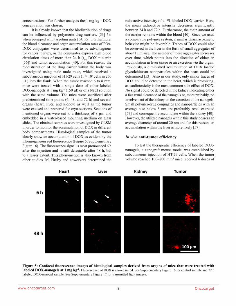
μ
Figure 5: Confocal fluorescence images of histological samples derived from organs of mice that were treated with

subcutaneous injection of HT-29 cells into the flank. When tumors reached 100 to 200 mm
(λ = 633 nm).
in triplicate at 25° C. The zeta potential (ζ) was calculated from the electrophoretic mobility (μ) according to the
20 min). The solution was precipitated in cold (−80° C,
, 300 MHz) (6): δ = 7.66, (d, 8.1 Hz,
mL methanol and precipitated in 400 mL of cold (−80° C) dissolved in de-ionized water and freeze dried (−80° C,
, 300 MHz) (2): δ = 4.9 (s, 2.3 H,
UV/Vis: λ = 660 nm, λ
cold diethyl ether (−80° C). To purify the self-assembled
was detected at an excitation wavelength of λ = 450 nm. emission maximum at λ = 510 nm.
a wavelength of λ = 450 nm and a reference wavelength of λ = 630 nm with untreated cells on the same well
λ =
was measured at λ = 645 nm in a microplate reader
13. Kronek J, Kroneková Z, Lustoň J, Paulovičová E, Paulovičová L, Mendrek B.
24. Kronek J, Paulovičová E, Paulovičová L, Kroneková Z, Lustoň J. Immunomodulatory efficiency of poly(2-
Stroobants S, Hoogenboom R, Staelens S. μPET imaging
R. The Label Matters: μPET Imaging of the Biodistribution
poly(γ-benzyl l-glutamate)-b-hyaluronan polymersomes.
Hyaluronan-block-Poly(γ-benzyl glutamate) Copolymers.
56. Seymour LW, Ulbrich K, Strohalm J, Kopeček J, Duncan
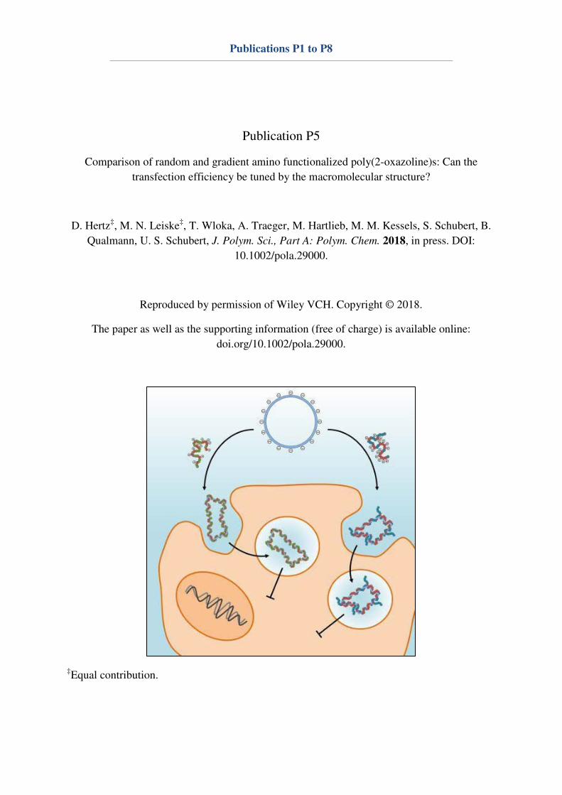
Publications P1 to P8
Publication P5
Comparison of random and gradient amino functionalized poly(2-oxazoline)s: Can the
transfection efficiency be tuned by the macromolecular structure?
D. Hertz‡, M. N. Leiske
‡, T. Wloka, A. Traeger, M. Hartlieb, M. M. Kessels, S. Schubert, B.
Qualmann, U. S. Schubert, J. Polym. Sci., Part A: Polym. Chem. 2018, in press. DOI:
10.1002/pola.29000.
Reproduced by permission of Wiley VCH. Copyright © 2018.
The paper as well as the supporting information (free of charge) is available online:
doi.org/10.1002/pola.29000.
‡Equal contribution.
1
Comparison of random and gradient amino functionalized poly(2-oxazoline)s:
Can the transfection efficiency be tuned by the macromolecular structure?
David Hertz,a,b,#
Meike N. Leiske,b,c,#
Thomas Wloka,b,c
Anja Traeger,b,c
Matthias Hartlieb,b,c,¶
Michael
M. Kessels,a Stephanie Schubert,
b,d Britta Qualmann,
a,b,* Ulrich S. Schubert
b,c,*
aInstitute for Biochemistry I, Jena University Hospital – Friedrich Schiller University Jena, Nonnenplan 2, 07743 Jena, Germany
bJena Center for Soft Matter (JCSM), Friedrich Schiller University Jena, Philosophenweg 7, 07743 Jena, Germany
cLaboratory of Organic and Macromolecular Chemistry (IOMC), Friedrich Schiller University Jena, Humboldtstraße 10, 07743 Jena, Germany
dInstitute of Pharmacy, Pharmaceutical Technology, Friedrich Schiller University Jena, Otto-Schott-Straße 41, 07745 Jena, Germany
¶Current address: Institute of Biomaterial Science, Helmholtz-Zentrum Geesthacht, Kantstr. 55, 14513 Teltow, Germany
#D. Hertz and M. N. Leiske contributed equally to this work.
Correspondence to: B. Qualmann and Ulrich S. Schubert (E-mail: [email protected];
The Supporting Information is available free of charge in the Wiley Online Library.
2
INTRODUCTION
For the introduction of regulative RNAs such as small interfering RNA (siRNA) or short hairpin RNA (shRNA) into cells, delivery vectors represent attractive strategies enabling the treatment of e.g. cancer using gene therapy.1-3 Since free genetic material is rapidly degraded in vivo,4-7 efficient carriers preventing the degradation of nucleic acids are indispensable. These carriers require properties for the association with the plasma membrane of target cells and for internalization into the cytosol.8 Beyond these requirements there are further intracellular hurdles, such as the escape from the endosomal pathway, trafficking through the cytosol, and, in the case of shRNA delivery, entry into the nucleus.2, 9
In general, carriers for nucleic acids complexation and transport are subdivided into viral and non-viral carriers.10, 11 In contrast to viral carriers, which are highly efficient, but induce mutagenic and immunogenic responses,12, 13 non-viral carriers, in particular cationic polymers, are safer, cheaper and simpler in production and storage.14, 15 Furthermore, polymers can be easily modified and exhibit a high loading capacity of genetic material. One of the main disadvantages of polymers is the lower efficiency of transfection in comparison to viral carriers.16 A reason for this problem is the disability of some polyplexes to escape from endosomal vesicles leading to a degradation of nucleic acids by lysosomal enzymes.17 Polymers with efficient release from endosomes possess a pKa value in the
ABSTRACT
Poly(ethylene imine) can be considered as the gold standard for DNA delivery into cells in vitro, but
severe cytotoxic side-effects and inapplicability for targeted approaches in vivo urgently call for the
design of new gene carriers. Since poly(2-oxazoline)s (P(Ox)s) can be easily synthesized and modified,
this polymer class might be ideal for the optimization of polymeric transfection processes. The
utilization of 2-methyl-2-oxazoline (MeOx) and 2-ethyl-2-oxazoline (EtOx) is also known to be
beneficial because these monomers were suggested to overcome solubility issues, mediate stealth
behavior and, consequently, facilitate a reduction of cytotoxicity. A series of amino (AmOx)
functionalized P(Ox) copolymers with either MeOx (gradient copolymers) or EtOx (random
copolymers) was synthesized, deprotected and biochemically characterized regarding cytotoxicity,
polyplex formation ability, cellular uptake and transfection efficiency.
Polymers with percentages of AmOx higher than 35 mol% showed stable polyplex formation but also
an increase in cytotoxicity. All elucidated P(Ox)s revealed a poor transfection efficiency in both L929
and Hepa1-6 cell lines. However, the investigations contribute to the understanding of the influence
of stealth units (MeOx and EtOx) and their distribution within the polymer chain on selected
properties of polyplexes and describe characteristics of amino functionalized P(Ox)s in different cell
lines.
KEYWORDS: Poly(2-oxazoline); DNA interaction; cell uptake; random and gradient copolymers; flow
cytometry
3
physiological pH range showing buffer properties for acidic endosomal vesicles.18 Additionally, a high transfection efficiency of polymeric vectors usually goes hand-in-hand with pronounced cytotoxicity.19, 20 Consequently, the previously mentioned aspects are the major targets of research to improve non-viral carriers. Besides polyamides like poly(L-lysine) or poly(methacrylate)s21, 22, PEI can be considered as the polymeric gold standard for DNA delivery into cells.23 Both types of PEI, namely the linear (l-PEI) and the branched (b-PEI) form, possess high transfection efficiencies in various cell lines, but also severe cytotoxic effects and undesired non-specific interactions in vitro and in vivo.19, 20 To reduce the cytotoxicity and enhance the delivery capacity, new carriers have to be designed. Since the well-defined l-PEI is synthesized by acidic or basic hydrolysis of (P(Ox)s),24 commercially available l-PEI usually contain 5 to 10 mol% non-hydrolyzed P(EtOx) units.25 P(EtOx) and P(MeOx) of various molar masses were already investigated in vitro in terms of biocompatibility, showing no harmful effects on cells in cytotoxicity assays or hemolysis.26, 27 For this reason, it might be interesting to elucidate amino functionalized P(Ox)s with EtOx or MeOx in terms of transfection abilities and biocompatibility. P(Ox)s can be produced via the cationic ring-opening polymerization (CROP) of 2-oxazolines, whi h was first des ri ed i the 9 ’s four independent research groups.28-31 The introduction of the microwave technique in 2004 reduced the reaction times from several days to minutes, by keeping the controlled character of the CROP and resulting in well-defined polymers.32 The application of functional initiators or terminating agents results i α- a d ω-functionalized P(Ox)s.33-35 Furthermore, 2-oxazolines can be easily modified in the 2-position, leading to a high chemical versatility for structural modification through copolymerization. The utilization of the hydrophilic monomers MeOx and EtOx results in water-soluble polymers, which also mediate stealth behavior and facilitate the reduction of
cytotoxicity.33, 36 In 2015, our group showed advantages of amino functionalized P(Ox)s towards l-PEI, by using a library approach.34 They synthesized allyl functionalized P(Ox) precursors, which were amino modified via thiol-ene click-reactions, to obtain different amino functionalized P(Ox)s. Some of the resulting polymers showed very good transfection efficiencies while maintaining a good biocompatibility. However, the use of proper protection groups enables the possibility to directly polymerize different monomers with a high variety of functional groups, such as aldehydes,37, 38 carboxylic acids39 and amino groups.40, 41 The dependence of the polymerization constant (kp) of 2-oxazolines on the substituent in 2-position has been recently summarized.42 Due to the different kp values reported, copolymerization of EtOx with the amino functionalized monomer 2-(4-((tert-butoxycarbonyl)amino)butyl)-2-oxazoline (BocOx) might result in another monomer distribution than a copolymerization of MeOx and BocOx. Comparison of random and gradient copolymers regarding DNA complexation and cellular interaction are still largely unexplored. However, S. Filippov et al. could show that block and gradient copoly(2-oxazoline) micelles show a different behavior with a uniform density profile of the core in block copolymers and a higher density in the outer part of the core for gradient copolymers.43 This could have a significant influence on polyplex formation and cell interaction, in particular with immune cells, regarding a higher shielding capacity in block copolymers in comparison with gradient polymers. In the present study, a series of amino functionalized P(Ox) copolymers with either MeOx or EtOx representing the stealth unit and different amounts of the Boc-protected amino functionalized comonomer BocOx was synthesized, deprotected and fully characterized. Polyplex formation using plasmid DNA (pDNA) in a wide range of nitrogen-to-phosphate group (N/P) ratios as well as the structure, stability, and cytotoxicity of
4
polyplexes were studied. In addition, polyplex uptake and transfection efficiency were determined by flow cytometry and confocal laser scanning microscopy (CLSM).
EXPERIMENTAL
Materials and Instrumentation
EtOx, MeOx and methyl tosylate (MeTos) were purchased from Sigma-Aldrich and were distilled to dryness over calcium hydride (VWR) under argon atmosphere prior to usage. Triethylamine (TEA, Sigma-Aldrich) was distilled under argon atmosphere prior to usage. Acetonitrile was obtained from a solvent purification system (MB-SPS-800, MBraun) and stored under argon. All other solvents used were obtained from standard suppliers. Trifluoroacetic acid (TFA, Sigma-Aldrich), Amberlyst® A21 (Sigma-Aldrich), ethidium bromide (EtBr) solution (10 mg mL-1, Carl Roth), YOYO-1 iodide (Thermo Fisher), LysoTracker Red DND-99 (Thermo Fisher) and Hoechst 33342 trihydrochloride (Thermo Fisher) were used as obtained. Cell culture media and antibiotics were obtained from Biochrom. Plasmid pRNAT-H1.1 (Genscript) was isolated with the Midi Plasmid Kit (Qiagen). Cell Proliferation Kit I (MTT), heparin sodium salt and trypan blue solution (0.4%) were purchased from Sigma-Aldrich. l-PEI and BocOx were synthesized according to literature procedures.25, 33, 44 For the general characterization of l-PEI, please refer to T. Bus et al..33 Polymerization reactions of 2-oxazolines were performed under microwave irradiation, using an Initiator Sixty single-mode microwave synthesizer from Biotage, equipped with a noninvasive IR sensor (accuracy: 2%). Microwave vials were stored overnight at 100 °C under vacuum and allowed to cool to room temperature (RT) under argon before usage. Polymerizations were performed under temperature control. According to the polymer characteristics, size exclusion chromatography (SEC) measurements of the polymers were
performed on different systems and noted in the respective part. SEC measurements of the polymerization kinetics were performed on an Agilent 1200 series system, equipped with a PSS degasser, a G1329A pump, a PSS GRAM guard/30/1000 Å with 10 µm particle size and a G1362 refractive index (RI) detector. Dimethylacetamide (DMAc) containing 0.21% LiCl served as eluent. The column oven was set to 40 °C at a flow rate of 1 mL min-1 and poly(styrene) (PS, 400 to 1,000,000 g mol-1) served as the calibration. SEC of the Boc-protected copolymer precursors was conducted on a Shimadzu system equipped with a SCL-10A VP controller, GDU-14A degasser and a LC-10AD VP pump, a PSS GRAM guard/30/1000 Å with 10 µm particle size and a RID-10A RI detector. DMAc containing 0.21% LiCl served as eluent. The column oven was set to 40 °C using a flow rate of 1 mL min-1 and PS (400 to 1,000,000 g mol-1) served as the calibration. SEC of the Boc-protected P(EtOx3-b-BocOx157) was performed on a Shimadzu system equipped with a CBM-20A controller, GDU-14A degasser and a LC-10AD VP pump, a PSS SDV guard/linear S column with 5 µm particle size and a RID-10A RI detector. CHCl3-iso-propanol (i-PrOH)-NEt3 (94:2:4) served as eluent. The column oven was set to 40 °C using a flow rate of 1 mL min-1. PS (400 to 100,000 g mol-1) served as the calibration. SEC of the deprotected, amino functionalized polymers was conducted on a Jasco system equipped with a DG-980-50 degasser and a PU-980 pump, PSS SUPREMA-MAX guard/300 Å column with 10 µm particle size and a RI-930 RI detector. 0.3% (v/v) TFA containing 0.1 M NaCl served as aqueous eluent. The column oven was set to 30 °C using a flow rate of 1 mL min-1. Poly(2-vinylpyridine) (P2VP, 1,300 to 81,000 g mol-1) served as the calibration. Proton NMR spectroscopy (1H-NMR) was performed at RT using a Bruker Avance I 300 MHz spectrometer, utilizing either CDCl3 or D2O as solvent. The chemical shifts are given in ppm relative to the signal from the residual non-deuterated solvent.

5
Gas chromatography (GC) was performed on a Shimadzu system (GC-2010 plus) equipped with a flame ionization detector, AOC-20s autosampler, AOC-20i injector and a PerkinElmer Elite-5MS column using helium as carrier gas. The stationary phase consists of 5% diphenyl and 95% dimethyl polysiloxane. Lyophilization of the polymers was conducted using an Alpha 1-2 LDplus freeze dryer from Martin Christ Gefriertrocknungsanlagen GmbH. For the characterization of polyplexes and biocompatibility assays, a SpectraMax microplate reader (Molecular Devices) was used. Flow cytometry was conducted on a Cytomics FC 500 (Beckman Coulter). CLSM was performed with a LSM880, Elyra PS.1 system (Zeiss) using the following excitation wavelengths/laser lines 405 nm (for Hoechst 33342), 488 nm (for YOYO-1) and 561 nm (for LysoTracker Red DND-99). Batch dynamic light scattering (DLS) was performed on a Zetasizer Nano ZS (Malvern Instruments). All measurements were performed in folded capillary cells (DTS1070 Malvern Instruments). After an equilibration time of 180 s, 3 × 30 s runs were carried out at 25 °C Ex = 633 nm). Scattered light was detected at an angle of 173°. The mean particle size was approximated as the effective (z-average) diameter and the width of the distribution as the polydispersity index of the particles (PDI) obtained by the cumulants method assuming a spherical shape. Each measurement was performed in triplicates. Electrophoretic light scattering (ELS) was used to easure the zeta pote tial ζ . The measurement was also performed on the Zetasizer Nano ZS by applying laser Doppler velocimetry. For each measurement, 20 runs were carried out using the slow-field reversal and the fast-field reversal mode at 150 V. Each experiment was performed in triplicates at 25 °C. The zeta potential was calculated from the electrophoretic mobilit a ordi g to the Henry equation. Henry coefficient f(ka) was calculated according to Ohshima.45
For the acid/base titration, the copolymers were dissolved in deionized water, reaching a final concentration of 10 mg mL-1 and 10 µL concentrated HCl (12 M) were added. The titration was performed against 0.1 M NaOH using a 765 Dosimat (Metrohm), a digital pH/mV-thermometer GMH 3530 (Greisinger electronic), and the EBS9 M Recorder software. Recorded curves were smoothed using 5-point FFT-fitting in OriginPro 2015G. The resulting curve was derivated two times to determine the pKa value of the copolymers.
Synthesis and Characterization
Kinetic Studies of Copolymerizations
The general procedure for all kinetic investigations is as follows: Under inert conditions, a solution of MeTos and MeOx (respectively EtOx) in acetonitrile (cMeTos = 0.013 mol L-1, [M]/[I] = 3) was prepared in a microwave vial and heated in a microwave synthesizer (140 °C, 102 min, absorption level high). Subsequently, distinct ratios of BocOx and MeOx or EtOx (nMeOx/EtOx:nBocOx; 80:20, 60:40, 40:60, 20:80, [M]/[I] = 147) were added and the solution was aliquoted into microwave vials and heated in a microwave synthesizer (140 °C, varying reaction times, absorption level very high). After polymerization, the conversions of the monomers were determined utilizing GC measurements, using the solvent as an internal standard. The polymerization constants (kp) of the monomers were determined using equations (1) and (2) assuming that the slope of the linear fit of ln([M]0/[M]t) = f(t) complies with keff. lnM − lnMt = k ∙ t (1)
k = kp[I] (2)
The reactivity ratios of both monomers were calculated for four different monomer ratios at 30 mol% BocOx (for the EtOx kinetics) respectively 30 mol% MeOx (for the MeOx

6
kinetics) conversion (determined by GC) using non-linear least square fitting46 (equation (3)): F = r − +r +r − + −r +r (3)
F1 = instantaneous mole fraction; f1 = mole fraction of monomer EtOx/MeOx; f2 = mole fraction of monomer BocOx; r1 = reactivity ratio of MeOx/EtOx; r2 = reactivity ratio of BocOx.
Synthesis of Boc-protected P(Ox)s
The synthesis of P(Ox)s was accomplished as previously described.44 Briefly, it is explained for P(EtOx150-r-BocOx33) (P(E150B33)). In a microwave vial, MeTos (9.3 mg, 0.05 mmol), EtOx (15.1 mg, 0.15 mmol) and acetonitrile (2.87 g) were mixed under inert conditions and heated in the microwave to 140 °C for 93 min. Subsequently, the vial was opened under an inert atmosphere, and EtOx (778.2 mg, 7.85 mmol) and BocOx (484.6 mg, 2.00 mmol) were added. In the case of the homopolymers, only BocOx was added. The reaction mixture was heated in the microwave at 140 °C for another 44 min. The resulting polymer was precipitated in ice-cold diethyl ether and the solid was re-dissolved in CH2Cl2. The solvent was evaporated under reduced pressure to obtain the product as a white solid.
1H-NMR (CDCl3, 300 MHz): δ = 3.48 (s, 4 H, backbone), 3.12 (m, 0.5 H, CH2-CH2-NH (BocOx)), 2.38 (m, 1.91 H, CH2 (EtOx)), 1.43 – 1.65 (m, 2.4 H, CH2-CH2-CO (BocOx) + CH2-CH2-CH2-CH2 (BocOx)), 1.37 (s, 2.4 H, CH3 (EtOx)) ppm.
SEC (eluent: DMAc, 0.21% LiCl, PS-cal.): Mn = 19,100 g mol-1; Mw = 22,200 g mol-1; Ð = 1.16.
Deprotection of Boc-protected P(Ox)s to Yield
Primary Amino Functionalized P(Ox)s
The deprotection of P(E150B33) to yield P(EtOx150-r-AmOx33) (P(E150A33)) is exemplarily described.
P(E150B33) (1.0 g) was dissolved in 5 mL TFA and stirred at RT overnight. Subsequently, it was diluted using 10 mL methanol and precipitated in ice-cold diethyl ether. The resulting solid was filtered off and re-dissolved in methanol. Amberlyst® A21 was added and the polymer solution was stirred at 100 rpm at RT overnight. Then, the Amberlyst® A21 was filtered off and the solvent was evaporated under reduced pressure. The polymer was re-dissolved in deionized water and lyophilized to obtain the product as a white powder.
1H-NMR (D2O, 300 MHz): δ = 3.39 (s, 4 H, backbone), 2.87 (0.4 H, CH2-CH2-NH2 (AmOx)), 2.23 (s, 2 H, CH2 (EtOx) + CH2-CH2-CO (EtOx + AmOx)), 1.50 (s, 0.8 H, CH2-CH2-CH2-CH2 (AmOx)), 0.92 (s, 2.4 H, CH3 (EtOx)) ppm.
SEC (eluent: 0.3% TFA + 0.1 M NaCl, P2VP-cal.): Mn = 8,000 g mol-1; Mw = 11,200 g mol-1; Ð = 1.41.
Polyplex Formation
Polyplexes were prepared by mixing stock solutions of 15 µg mL-1 pDNA and different amounts of polymers to obtain various N/P ratios in HBG buffer (20 mM 4-(2-hydroxyethyl)piperazine-1-ethanesulfonic acid (HEPES) and 5% (w/v) glucose, pH 7.2). The solutions were vortexed for 10 sec at maximum speed (2800 rpm) and incubated at RT for 15 min to ensure complex formation.
EtBr Quenching Assay (EBA)
Formation of polyplexes with pDNA was investigated by quenching of EtBr fluorescence as described previously.47 Briefly, pDNA (15 µg mL-1) in a total volume of 100 µL HBG buffer was incubated with EtBr (10 µg mL-1) for 10 min at RT. Subsequently, polyplexes with increasing concentrations of each polymer (calculated to N/P ratios) were formed in black 96-well plates (Nunc, Thermo Fisher) and incubated at RT for 15 min. The fluorescence of the samples was measured at
Ex = 525 / Em = 605 nm using a microplate
7
reader. A sample containing only pDNA and EtBr was used to calibrate the device to 100% fluorescence against a background of 10 µg mL-1 of EtBr in HBG solution. The percentage of dye displaced upon polyplex formation was calculated using equation (4). RFU [%] = F a e−FF NA−F ∙ (4)
Here, RFU is the relative fluorescence units and Fsample, F0, and FpDNA are the fluorescence intensities of a given sample, EtBr in HBG alone, and EtBr intercalated into pDNA alone. Experiments were conducted in triplicates.
Heparin Dissociation Assay
To investigate the release of pDNA from polyplexes, the heparin dissociation assay was performed. Polyplexes with an N/P ratio of 30 were formed as described above in a total volume of 100 µL HBG buffer containing EtBr (10 µg mL-1). After incubation in the dark at RT for 15 min, the polyplexes were transferred into a black 96-well plate containing heparin in increasing concentrations. The solution was mixed and incubated for further 30 min at 37 °C in the dark. The fluorescence of EtBr was
easured at Ex = 525 / Em = 605 nm utilizing a microplate reader. The percentage of intercalated EtBr was calculated as described before. The experiments were conducted in triplicates.
Cell Culture
Mouse liver cell line Hepa1-6 (CRL-1830TM, ATCC) and mouse fibroblast cell line L929 (CCL-1TM, ATCC) were cultured in growth medium containing Dulbe o’s odified eagle’s medium (DMEM, Lonza) supplemented with 10% fetal calf serum (FCS), 100 U mL-1 penicillin and 100 µg mL-1 streptomycin at 37 °C in a humidified 5% (v/v) CO2 atmosphere.
Cytotoxicity MTT Assay
The cytotoxicity of the investigated polymers was measured by MTT assay using the cell lines Hepa1-6 and L929. Cells were cultured in a 96-well plate for 24 h as described above. The polymers to be tested were added to the cells in a concentration range of 1 to 500 µg mL-1 for 24 h. Subsequently, 10 µL aliquots of the MTT solution were added to each well and the plates were further incubated for 4 h at 37 °C in a humidified 5% (v/v) CO2 atmosphere. The formed formazan crystals were solubilized by addition of 10% sodium dodecyl sulfate (SDS) in 0.01 M HCl. The solubilized formazan product was spectro-photometrically quantified using a
i roplate reader at Ex1 = 550 nm and the refere e at Ex2 = 690 nm. The relative cell viability was calculated using equation (5): Rel. cell viab. [%] = λ x −λ x a eλ x −λ x c ∙ (5)
Data are expressed as mean and standard deviation (S.D.) of three measurements. Furthermore, the 50% cytotoxic concentration (CC50) was determined for comparison of the gradient and random polymers in both cell lines.
Polyplex Uptake
For uptake studies, cells were seeded at a density of 2 × 10⁵ cells mL-1 in 24-well plates (500 µl) and cultured for 24 h. One hour prior to the addition of the polyplexes at concentrations described in Table 1, the medium was changed to DMEM with or without FCS. The polyplexes were formed and 50 µL polyplexes in HBG buffer were added to the cells. The plates were incubated for 2 or 4 h at 37 °C in a humidified 5% (v/v) CO2 atmosphere. For kinetic studies of polyplex uptake, pDNA was labeled with YOYO-1 prior to polyplex preparation. For this reason, 0.02 µL of 1 M YOYO-1 solution was mixed with 1 µg pDNA in HBG buffer and incubated for 20 min at 4 °C protected from light.
8
TABLE 1 Corresponding concentrations of all tested polymers with an N/P ratio of 30.
Polymer system Concentration [µg mL-1]
P(M97A55) 9.79 P(M73A89) 9.47 P(M29A166) 9.10 P(E77A55) 10.41 P(E57A139) 9.56 P(E31A163) 9.27 P(E3A157) 8.99 l-PEI 5.43
Subsequently, polymers were added at an N/P ratio of 30 and the polyplexes were formed as described before (Table 1). After 2 or 4 h of polyplex incubation the cells were washed to remove extracellular polyplexes. To determine the relative uptake of the polyplexes, 10,000 cells were measured by flow cytometry and the amounts of viable cells showing YOYO-1 signal were gated. Uptake of pDNA with YOYO-1 alone was used as control to define the gate. Dead cells were identified via SSC/FSC signal. The experiments were performed at least three times independently. For live cell imaging, Hepa1-6 cells (5 × 10⁵ cells mL-1) were seeded in glass-bottomed, 4-chamber dishes (CELLviewTM, Greiner Bio One) and cultured for 24 h. One hour prior to polymer addition, the cells were rinsed with PBS and the media were changed to DMEM without FCS. Polyplexes were prepared with an N/P ratio of 30 as described above (Table 1) and added to the cells (50 L per well for 4 h. Subsequently, the cells were washed with PBS and incubated for 5 min with a mixture of DMEM and PBS supplemented with LysoTracker Red DND-99 (1:1000) and Hoechst 33342 (1:1000; 10 mg mL-1) for lysosomes as well as nucleus staining, respectively. A 1:10 diluted trypan blue solution was added to the medium immediately before imaging to quench polyplexes not taken up by the cells. The living cells were imaged with a LSM880, Elyra PS.1 system. For the quantification of the amount of YOYO-1 and/or LysoTracker Red DND-99 positive organelles per
cell the software ImageJ and the plugin Coloc2 (version 3.0.0) were used. Since cell borders could not be defined due to overlap of the cells, the average numbers of YOYO-1 and LysoTracker Red DND-99 positive organelles per cell (defined by the number of Hoechst 33342 positive nuclei) per area of 112.5 m × 112.5 m were determined. Cut-off was set to 5 to 1000 pixel units for all organelles.
Transfection Efficiencies in Adherent Cells
Hepa1-6 and L929 cells were cultured as described above. For transfection, the cells were seeded at a density of 105 cells mL−1 in 24-well plates (500 µl) and incubated for 24 h. One hour prior to transfection, the cells were washed with phosphate buffer saline (PBS) and supplemented with DMEM with or without FCS. Polyplexes with an N/P ratio of 30 were prepared as described above and were added to the cells (50 L per well . The orrespo di g concentrations for all tested polymers with an N/P ratio of 30 are shown in Table 1. After an incubation time of 4 h at 37 °C, the supernatant was replaced by fresh medium, and the cells were further incubated for 44 h. For analysis via flow cytometry, the cells were harvested by trypsinization and 10,000 cells were analyzed by flow cytometry. For determination of the viability, dead cells were identified via side scatter/forward scatter (SSC/FSC) signal as described previously.48 Viable cells expressing GFP were gated for analysis of the transfection efficiency. Transfection with pDNA alone was used as control to define the gate. The experiments were performed at least three times independently.
Statistical Analysis
The quantification of YOYO-1 and LysoTracker Red DND-99 positive organelles was performed for normal distribution by Shapiro-Wilk normality test. As the values did not follow normal distributions, data were analyzed by non-parametric Kruskal-Wallis test with Du ’s

∼ ∼ –

10
TABLE 2 Key properties of the synthesized P(Ox)s determined by 1H-NMR spectroscopy (300 MHz) in indicated solvents.
Sample Polymer DP mol% MeOx or
EtOx
mol% BocOx or
AmOx
Mn [g mol-1]
P(M130B31) P(MeOx130-grad-BocOx31)a 161 81d
19d 18,600
P(M97B55) P(MeOx97-grad-BocOx55)a 152 64d
36d 21,600
P(M73B89) P(MeOx73-grad-BocOx89)a 162 45d
55d 27,700
P(M29B166) P(MeOx29-grad-BocOx166)a 195 15d
85d 41,400
P(M130A31) P(MeOx130-grad-AmOx31)b 161c
81 19 15,500
P(M97A55) P(MeOx97-grad-AmOx55)b 152c
64 36 16,100
P(M73A89) P(MeOx73-grad-AmOx89)b 162c
45 55 18,900
P(M29A166) P(MeOx29-grad-AmOx166)b 195c
15 85 24,900
P(E150B33) P(EtOx150-r-BocOx33)a 183 82d
18d 22,800
P(E77B55) P(EtOx77-r-BocOx55)a 132 58d
42d 20,900
P(E57B139) P(EtOx57-r-BocOx139)a 196 29d
71d 39,300
P(E31B163) P(EtOx31-r-BocOx163)a 194 16d
84d 42,500
P(E150A33) P(EtOx150-r-AmOx33)b 183c
82 18 19,600
P(E77A55) P(EtOx77-r-AmOx55)b 132c 58 42 15,500
P(E57A139) P(EtOx57-r-AmOx139)b 196c 29 71 25,400
P(E31A163) P(EtOx31-r-AmOx163)b 194c 16 84 26,300
P(E3B157) P(EtOx3-b-BocOx157)a 160 2d 98d 38,300
P(E3A157) P(EtOx3-b-AmOx157)b 160c 2 98 22,600
aCDCl3; bCD3OD; ccalculated from Boc-protected precursor; dcalculated from deprotected copolymer.
Within the investigated monomer ratios, EtOx and BocOx exhibited similar kp values (kp(EtOx) = 47.2 ± 11.0 L mol-1 s-1; kp(BocOx) = 44.0 ± 13.3 L mol-1 s-1), while MeOx polymerized faster than BocOx (kp(MeOx) = 75.4 ± 2.8 L mol-1 s-1; kp(BocOx) = 50.7 ± 4.0 L mol-1 s-1). This complies with previously published kp values of the homopolymerizations of the investigated monomers.42 To gain an insight into the monomer distribution within the polymer chain, the calculation of the reactivity ratios is indispensable and was performed using equation (3).46, 49, 50 The reactivity ratios of EtOx and BocOx were found to be similar (rBocOx = 1.02 ≈ 1 ≈ rEtOx = 0.98), suggesting the formation of random copolymers.51 On the other hand, copolymerization of MeOx and BocOx resulted in slight gradient copolymers (rMeOx = 1.50 > 1 > rBocOx = 0.67). Regarding the SEC measurements, the polymers showed a linear increase in the molar mass and low dispersities (Ð ≤ 1.2) up to high conversions.
Only polymers with high BocOx amounts revealed small high molar mass shoulders, suggesting side reactions at high BocOx conversions of more than 90 mol%, which are probably caused by the amino side chain. However, these side reactions can be minimized by aiming for a slightly lower conversion during the preparation of copolymers. The investigations of cytotoxicity, polyplex stability and cellular uptake efficiency within this study enable a comparison of gradient (MeOx) and random (EtOx) copolymers by variation of the non-charged comonomers. Differences regarding the polyplex performance might be found in terms of cytotoxicity provoked by an enhanced shielding by gradient copolymers compared to random polymers. Furthermore, differences in the cellular uptake could also be caused by the shielding of the non-ionic comonomer. These two crucial factors might be important for a successful transfection efficiency of the investigated polyplexes.
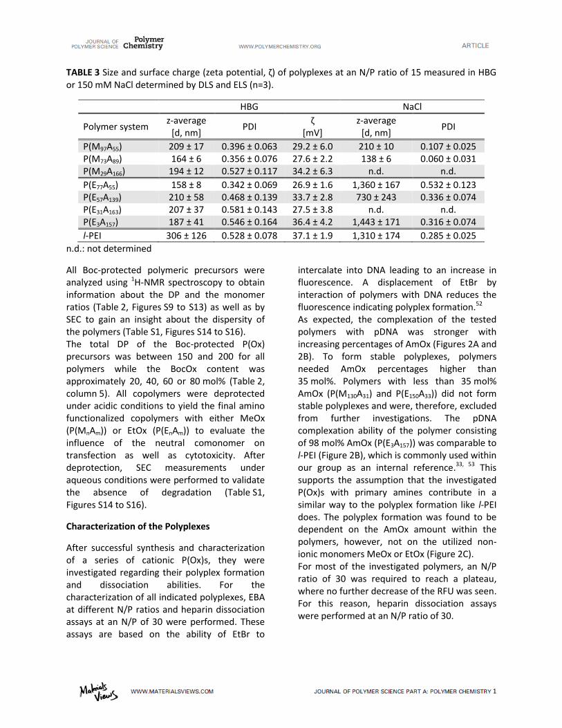
11
TABLE 3 Size a d surfa e harge zeta pote tial, ζ of pol ple es at a N/P ratio of easured i HBG or 150 mM NaCl determined by DLS and ELS (n=3).
HBG NaCl
Polymer system z-average
[d, nm] PDI
ζ [mV]
z-average [d, nm]
PDI
P(M97A55) 209 ± 17 0.396 ± 0.063 29.2 ± 6.0 210 ± 10 0.107 ± 0.025 P(M73A89) 164 ± 6 0.356 ± 0.076 27.6 ± 2.2 138 ± 6 0.060 ± 0.031 P(M29A166) 194 ± 12 0.527 ± 0.117 34.2 ± 6.3 n.d. n.d.
P(E77A55) 158 ± 8 0.342 ± 0.069 26.9 ± 1.6 1,360 ± 167 0.532 ± 0.123 P(E57A139) 210 ± 58 0.468 ± 0.139 33.7 ± 2.8 730 ± 243 0.336 ± 0.074 P(E31A163) 207 ± 37 0.581 ± 0.143 27.5 ± 3.8 n.d. n.d. P(E3A157) 187 ± 41 0.546 ± 0.164 36.4 ± 4.2 1,443 ± 171 0.316 ± 0.074
l-PEI 306 ± 126 0.528 ± 0.078 37.1 ± 1.9 1,310 ± 174 0.285 ± 0.025 n.d.: not determined
All Boc-protected polymeric precursors were analyzed using 1H-NMR spectroscopy to obtain information about the DP and the monomer ratios (Table 2, Figures S9 to S13) as well as by SEC to gain an insight about the dispersity of the polymers (Table S1, Figures S14 to S16). The total DP of the Boc-protected P(Ox) precursors was between 150 and 200 for all polymers while the BocOx content was approximately 20, 40, 60 or 80 mol% (Table 2, column 5). All copolymers were deprotected under acidic conditions to yield the final amino functionalized copolymers with either MeOx (P(MnAm)) or EtOx (P(EnAm)) to evaluate the influence of the neutral comonomer on transfection as well as cytotoxicity. After deprotection, SEC measurements under aqueous conditions were performed to validate the absence of degradation (Table S1, Figures S14 to S16).
Characterization of the Polyplexes
After successful synthesis and characterization of a series of cationic P(Ox)s, they were investigated regarding their polyplex formation and dissociation abilities. For the characterization of all indicated polyplexes, EBA at different N/P ratios and heparin dissociation assays at an N/P of 30 were performed. These assays are based on the ability of EtBr to
intercalate into DNA leading to an increase in fluorescence. A displacement of EtBr by interaction of polymers with DNA reduces the fluorescence indicating polyplex formation.52 As expected, the complexation of the tested polymers with pDNA was stronger with increasing percentages of AmOx (Figures 2A and 2B). To form stable polyplexes, polymers needed AmOx percentages higher than 35 mol%. Polymers with less than 35 mol% AmOx (P(M130A31) and P(E150A33)) did not form stable polyplexes and were, therefore, excluded from further investigations. The pDNA complexation ability of the polymer consisting of 98 mol% AmOx (P(E3A157)) was comparable to l-PEI (Figure 2B), which is commonly used within our group as an internal reference.33, 53 This supports the assumption that the investigated P(Ox)s with primary amines contribute in a similar way to the polyplex formation like l-PEI does. The polyplex formation was found to be dependent on the AmOx amount within the polymers, however, not on the utilized non-ionic monomers MeOx or EtOx (Figure 2C). For most of the investigated polymers, an N/P ratio of 30 was required to reach a plateau, where no further decrease of the RFU was seen. For this reason, heparin dissociation assays were performed at an N/P ratio of 30.
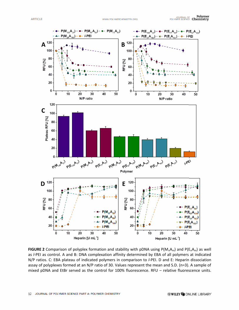
–

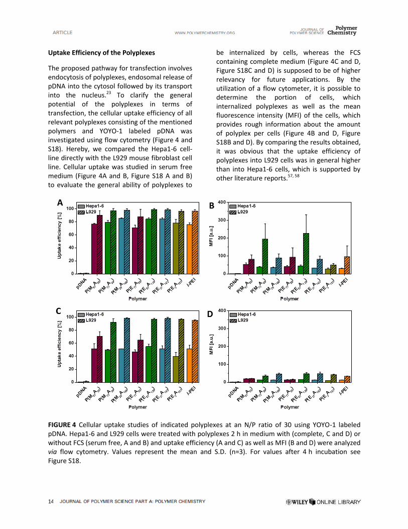

15
Possible explanations for the observed cell type dependency of the uptake and transfection efficiency are different membrane characteristics, intracellular transport mechanisms and DNA-degrading enzymes due to discriminative expression patterns.59 Polyplexes with AmOx amounts of 35 mol% were internalized less well in both cell lines due to the presence of fewer cationic charges.60 Having a closer look at the cells, which were cultivated in serum free medium, L929 cells revealed an uptake efficiency of nearly 100% even after 2 h of incubation, while efficiency was slightly less (80 to 90%) for Hepa1-6 cells (Figure 4A). After 4 h incubation exclusively the MFI per cell in both cell lines further increased suggesting an increased polyplex uptake by several cells, which could have a positive effect on transfection efficiency (Figure S18B). No differences between the P(Ox)s and l-PEI were observed. Interestingly, the uptake efficiency significantly decreased when using 10% FCS containing (complete) medium in combination with Hepa1-6 cells. While the differences between the two media were negligible within the L929 cell line, in Hepa1-6 cells, we observed an uptake efficiency in complete medium of only 50% after 2 h (Figure 4C) and 50 to 70% after 4 h (Figure S18C). This phenomenon might be explained by the interaction of the polyplexes with serum proteins.61 It is noteworthy that similar results were obtained for l-PEI, possibly due to high charge densities within all polyplexes. As there were no difference between the investigated polymers and l-PEI the influence of P(MeOx) and P(EtOx) on polyplex uptake is only of minor importance.
Transfection Efficiency
The previous results indicated that the tested polymers fulfill the prerequisites to be investigated as non-viral gene-delivery agents. As a consequence, they were analyzed regarding transfection efficiency using Hepa1-6 and L929 cells and pDNA encoding green
fluorescent protein (GFP) as visual reporter. The transfection efficiency was analyzed by flow cytometry counting all viable cells (SSC/FSC), which successfully expressed GFP (Figures 5 and S18). For all tested polymers, an N/P ratio of 30 was used to form stable polyplexes. In a first step, we determined the optimal time point for the detection of the GFP expression using l-PEI. After 48 h of transfection in both cell lines (Hepa1-6 and L929) the number of GFP expressing cells doubled, and an increase of the MFI was detected compared to measurements after 24 h (Figure S19). In comparison to L929 cells, Hepa1-6 cells revealed a lower GFP expression. It is well-known that various transfection reagents show different transfection efficiencies in various cell lines and especially L929 cells display high uptake and transfection efficiencies.57, 58, 62 Similar to differences in the uptake efficiency also the transfection efficiency is dependent on the discriminative expression pattern leading to variations in membrane topology, intracellular transport and enzymes for DNA degradation.59 Furthermore, as serum proteins influenced the uptake efficiency, we also investigated the transfection efficiency of l-PEI based polyplexes in growth media supplemented with 10% FCS in comparison to media without serum. In L929 cells, serum containing medium decreased the transfection efficiency by 50% after 24 h and 48 h. Nevertheless, GFP expression was detectable after 48 h in 40% of all cells. Hepa1-6 cells showed a stronger impact upon presence of serum with no transfection efficiencies higher than 5%, neither after 24 h nor 48 h (Figure S19). Based on these results and with the knowledge that stealth moieties are able to reduce the interaction with serum proteins, we investigated transfection after 48 h in medium with (complete) or without serum (serum free). Figure 5 shows the results of the transfection efficiencies of all investigated P(Ox)s comparing Hepa1-6 and L929 cells in medium with (complete, Figure 5A and B) or without FCS (serum free, Figure 5C and D). None of the elucidated P(Ox)s was able to transfect Hepa1-6

alled proto spo ge effe t .
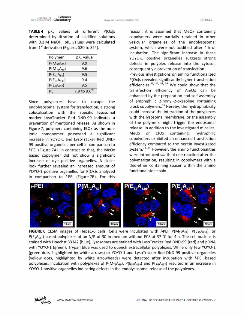
17
TABLE 4 pKa values of different P(Ox)s determined by titration of acidified solutions with 0.1 M NaOH. pKa values were calculated from 1st derivation (Figures S20 to S24).
Polymer pKa value
P(M97A55) 9.9 P(M73A89) 9.6
P(E77A55) 9.5 P(E57A139) 9.4 P(E3A157) 9.5
PEI 7.9 to 9.664
Since polyplexes have to escape the endolysosomal system for transfection, a strong colocalization with the specific lysosomal marker LysoTracker Red DND-99 indicates a prevention of mentioned release. As shown in Figure 7, polymers containing EtOx as the non-ionic comonomer possessed a significant increase in YOYO-1 and LysoTracker Red DND-99 positive organelles per cell in comparison to l-PEI (Figure 7A). In contrast to that, the MeOx based copolymer did not show a significant increase of dye positive organelles. A closer look further revealed an increased amount of YOYO-1 positive organelles for P(Ox)s analyzed in comparison to l-PEI (Figure 7B). For this
reason, it is assumed that MeOx containing copolymers were partially retained in other vesicular organelles of the endolysosomal system, which were not acidified after 4 h of incubation. The significant increase in these YOYO-1 positive organelles suggests strong defects in polyplex release into the cytosol, consequently a prevention of transfection. Previous investigations on amino functionalized P(Ox)s revealed significantly higher transfection efficiencies.33, 34, 41, 53 We could show that the transfection efficiency of AmOx can be enhanced by the preparation and self-assembly of amphiphilic 2-nonyl-2-oxazoline containing block copolymers.53 Hereby, the hydrophobicity could increase the interaction of the polyplexes with the lysosomal membrane, or the assembly of the polymers might trigger the endosomal release. In addition to the investigated micelles, MeOx or EtOx containing, hydrophilic copolymers exhibited an enhanced transfection efficiency compared to the herein investigated system.33, 34 However, the amino functionalities were introduced via thiol-ene reaction after the polymerization, resulting in copolymers with a thio-ether containing spacer within the amino functional side chain.
FIGURE 6 CLSM images of Hepa1-6 cells. Cells were incubated with l-PEI, P(M73A89), P(E57A139), or P(E3A157) based polyplexes at an N/P of 30 in medium without FCS at 37 °C for 4 h. The cell nucleus is stained with Hoechst 33342 (blue), lysosomes are stained with LysoTracker Red DND-99 (red) and pDNA with YOYO-1 (green). Trypan blue was used to quench extracellular polyplexes. While only few YOYO-1 (green dots, highlighted by white arrows) or YOYO-1 and LysoTracker Red DND-99 positive organelles (yellow dots, highlighted by white arrowheads) were detected after incubation with l-PEI based polyplexes, incubation with polyplexes of P(M73A89), P(E57A139) and P(E3A157) resulted in an increase in YOYO-1 positive organelles indicating defects in the endolysosomal release of the polyplexes.

Wallis test with Du ’s post
of the proto spo ge effe t as well as
19
gene delivery by using the investigated conditions. Future studies might concentrate on alternative copolymers with differences in the hydrophobicity and buffer capacity to identify ideal carriers for genetic material, which can ensure biocompatibility while expressing higher transfection efficiencies. Based on the different uptake efficiencies in the investigated cell lines targeting moieties can also increase the uptake and possibly transfection efficiency. Since gradient polymers show higher polyplex stability in physiological sodium chloride solution we would recommend to prefer this polymer structure over random polymers.
ACKNOWLEDGEMENTS
This project was funded by the Thüringer Ministerium für Wirtschaft, Wissenschaft und Digitale Gesellschaft (ProExzellenz II, NanoPolar) to BQ and USS. The funding of the collaborative research center PolyTarget (SFB 1278-C03) by the Deutsche Forschungsgemeinschaft (DFG) is highly acknowledged. MNL acknowledges the German Federal Ministry of Education and Research (BMBF, #13N13416 smart-dye-livery) for funding. AT acknowledges the Carl Zeiss Foundation as well as the BMBF (#13XP5034A, PolyBioMik) for funding. The authors gratefully acknowledge the members of the IOMC Fabian H. Sobotta for the synthesis and characterization of P(E3A157) and Anne-Kristin Trützschler for the supply of l-PEI.
REFERENCES AND NOTES
1 C. Zhang; R. Jin; P. Zhao; C. Lin, Acta Biomater.
2015, 22, 120. 2 D. W. Pack; A. S. Hoffman; S. Pun; P. S. Stayton, Nat. Rev. Drug Discov. 2005, 4, (7), 581. 3 H. J. Kim; A. Kim; K. Miyata; K. Kataoka, Adv.
Drug Deliv. Rev. 2016, 104, 61. 4 K. Kawabata; Y. Takakura; M. Hashida, Pharm.
Res. 1995, 12, (6), 825. 5 J. Hisazumi; N. Kobayashi; M. Nishikawa; Y. Takakura, Pharm. Res. 2004, 21, (7), 1223.
6 R. M. Schiffelers; A. Ansari; J. Xu; Q. Zhou; Q. Tang; G. Storm; G. Molema; P. Y. Lu; P. V. Scaria; M. C. Woodle, Nucleic Acids Res. 2004, 32, (19), e149. 7 Y. Fujiwara; A. Furuta; H. Kikuchi; S. Aizawa; Y. Hatanaka; C. Konya; K. Uchida; A. Yoshimura; Y. Tamai; K. Wada; T. Kabuta, Autophagy 2013, 9, (3), 403. 8 M. Mees; E. Haladjova; D. Momekova; G. Momekov; P. S. Shestakova; C. B. Tsvetanov; R. Hoogenboom; S. Rangelov, Biomacromolecules
2016, 17, (11), 3580. 9 D. Luo; W. M. Saltzman, Nat. Biotech. 2000, 18, (1), 33. 10 N. Nayerossadat; T. Maedeh; P. A. Ali, Adv.
Biomed. Res. 2012, 1, 27. 11 A. El-Aneed, J. Control. Release 2004, 94, (1), 1. 12 T. Friedmann; R. Roblin, Science 1972, 175, (4025), 949. 13 Y. Huang; X. Liu; L. Dong; Z. Liu; X. He; W. Liu, Pain Res. Treat. 2011, 2011, 8. 14 S. Zhang; B. Zhao; H. Jiang; B. Wang; B. Ma, J. Control. Release 2007, 123, (1), 1. 15 A. G. Schatzlein, Anti-Cancer Drugs 2001, 12, (4), 275. 16 M. A. Mintzer; E. E. Simanek, Chem. Rev.
2008, 109, (2), 259. 17 R. Wattiaux; N. Laurent; S. Wattiaux-De Coninck; M. Jadot, Adv. Drug Deliv. Rev. 2000, 41, (2), 201. 18 A. Akinc; R. Langer, Biotechnol. Bioeng. 2002, 78, (5), 503. 19 D. Fischer; T. Bieber; Y. Li; H.-P. Elsässer; T. Kissel, Pharm. Res. 1999, 16, (8), 1273. 20 D. Fischer; Y. Li; B. Ahlemeyer; J. Krieglstein; T. Kissel, Biomaterials 2003, 24, (7), 1121. 21 S. C. De Smedt; J. Demeester; W. E. Hennink, Pharm. Res. 2000, 17, (2), 113. 22 M. E. Favretto; A. Krieg; S. Schubert; U. S. Schubert; R. Brock, J. Control. Release 2015, 209, 1. 23 O. Boussif; F. Lezoualc'h; M. A. Zanta; M. D. Mergny; D. Scherman; B. Demeneix; J. P. Behr, Proc. Natl. Acad. Sci. U S A 1995, 92, (16), 7297. 24 M. Jager; S. Schubert; S. Ochrimenko; D. Fischer; U. S. Schubert, Chem. Soc. Rev. 2012, 41, (13), 4755.
20
25 L. Tauhardt; K. Ke pe; K. K op; E. Altu taş; M. Jäger; S. Schubert; D. Fischer; U. S. Schubert, Macromol. Chem. Phys. 2011, 212, (17), 1918. 26 M. Bauer; C. Lautenschlaeger; K. Kempe; L. Tauhardt; U. S. Schubert; D. Fischer, Macromol.
Biosci. 2012, 12, (7), 986. 27 M. Bauer; S. Schroeder; L. Tauhardt; K. Kempe; U. S. Schubert; D. Fischer, J. Polym. Sci.
A: Polym. Chem. 2013, 51, (8), 1816. 28 T. G. Bassiri; A. Levy; M. Litt, J. Polym. Sci. B:
Polym. Lett. 1967, 5, (9), 871. 29 T. Kagiya; S. Narisawa; T. Maeda; K. Fukui, J. Polym. Sci. B: Polym. Lett. 1966, 4, (7), 441. 30 W. Seeliger; E. Aufderhaar; W. Diepers; R. Feinauer; R. Nehring; W. Thier; H. Hellmann, Angew. Chem. Int. Ed. Eng. 1966, 5, (10), 875. 31 D. A. Tomalia; D. P. Sheetz, J. Polym. Sci. A:
Polym. Chem. 1966, 4, (9), 2253. 32 F. Wiesbrock; R. Hoogenboom; C. H. Abeln; U. S. Schubert, Macromol. Rapid Commun.
2004, 25, (22), 1895. 33 T. Bus; C. Englert; M. Reifarth; P. Borchers; M. Hartlieb; A. Vollrath; S. Hoeppener; A. Traeger; U. S. Schubert, J. Mater. Chem. B 2017, 5, (6), 1258. 34 A. C. Rinkenauer; L. Tauhardt; F. Wendler; K. Kempe; M. Gottschaldt; A. Traeger; U. S. Schubert, Macromol. Biosci. 2015, 15, (3), 414. 35 B. Guillerm; S. Monge; V. Lapinte; J. J. Robin, Macromol. Rapid Commun. 2012, 33, (19), 1600. 36 M. Bauer; C. Lautenschlaeger; K. Kempe; L. Tauhardt; U. S. Schubert; D. Fischer, Macromol.
Biosci. 2012, 12, (7), 986. 37 C. Taubmann; R. Luxenhofer; S. Cesana; R. Jordan, Macromol. Biosci. 2005, 5, (7), 603. 38 C. Legros; M.-C. De Pauw-Gillet; K. C. Tam; S. Lecommandoux; D. Taton, Eur. Polym. J. 2015, 62, (Supplement C), 322. 39 P. J. M. Bouten; D. Hertsen; M. Vergaelen; B. D. Monnery; M. A. Boerman; H. Goossens; S. Catak; J. C. M. van Hest; V. Van Speybroeck; R. Hoogenboom, Polym. Chem. 2015, 6, (4), 514. 40 S. Cesana; J. Auernheimer; R. Jordan; H. Kessler; O. Nuyken, Macromol. Chem. Phys.
2006, 207, (2), 183.
41 Z. He; L. Miao; R. Jordan; D. S-Manickam; R. Luxenhofer; A. V. Kabanov, Macromolecular
Bioscience 2015, 15, (7), 1004. 42 M. Glassner; M. Vergaelen; R. Hoogenboom, Polym. Int. 2017, 67, (1), 32. 43 S. K. Filippov; B. Verbraeken; P. V. Konarev; D. I. Svergun; B. Angelov; N. S. Vishnevetskaya; C. M. Papadakis; S. Rogers; A. Radulescu; T. Courtin, J. Phys. Chem. Lett. 2017, 8, (16), 3800. 44 M. Hartlieb; D. Pretzel; K. Kempe; C. Fritzsche; R. M. Paulus; M. Gottschaldt; U. S. Schubert, Soft Matter 2013, 9, (18), 4693. 45 H. Ohshima, J. Colloid Interface Sci. 1994, 168, (1), 269. 46 V. E. Meyer; G. G. Lowry, J. Polym. Sci. Part
A: Gen. Pap. 1965, 3, (8), 2843. 47 C. Englert; A.-K. Trützschler; M. Raasch; T. Bus; P. Borchers; A. S. Mosig; A. Traeger; U. S. Schubert, J. Control. Release 2016, 241, 1. 48 A. Vollrath; A. Schallon; C. Pietsch; S. Schubert; T. Nomoto; Y. Matsumoto; K. Kataoka; U. S. Schubert, Soft Matter 2013, 9, (1), 99. 49 M. N. Leiske; M. Hartlieb; F. H. Sobotta; R. M. Paulus; H. Gorls; P. Bellstedt; U. S. Schubert, Polymer Chemistry 2016, 7, (30), 4924. 50 S. M. Shawki; A. E. Hamielec, J. Appl. Polym.
Sci. 1979, 23, (11), 3155. 51 K. Kempe; S. Jacobs; H. M. L. Lambermont-Thijs; M. M. W. M. Fijten; R. Hoogenboom; U. S. Schubert, Macromolecules 2010, 43, (9), 4098. 52 R. Mehta; R. Kumari; P. Das; A. K. Bhowmick, J. Mater. Chem. B 2014, 2, (37), 6236. 53 M. N. Leiske; F. H. Sobotta; S. Hoeppener; J. C. Brendel; A. Traeger; U. S. Schubert, Biomacromolecules 2017. 54 R. Luxenhofer; G. Sahay; A. Schulz; D. Alakhova; T. K. Bronich; R. Jordan; A. V. Kabanov, J Control Release 2011, 153, (1), 73. 55 D. Pezzoli; E. Giupponi; D. Mantovani; G. Candiani, Sci. Rep. 2017, 7, 44134. 56 K. A. Curtis; D. Miller; P. Millard; S. Basu; F. Horkay; P. L. Chandran, PLOS ONE 2016, 11, (9), e0158147. 57 A. Rémy-Kristensen; J.-P. Clamme; C. Vuilleumier; J.-G. Kuhry; Y. Mély, BBA -
Biomembranes 2001, 1514, (1), 21.
21
58 N. Zhao; S. Roesler; T. Kissel, Int. J. Pharm.
2011, 411, (1), 197. 59 E. V. B. van Gaal; R. van Eijk; R. S. Oosting; R. J. Kok; W. E. Hennink; D. J. A. Crommelin; E. Mastrobattista, J. Control. Release 2011, 154, (3), 218. 60 A. C. Rinkenauer; L. Tauhardt; F. Wendler; K. Kempe; M. Gottschaldt; A. Traeger; U. S. Schubert, Macromol. Biosci. 2015, 15, (3), 414. 61 S. Tenzer; D. Docter; J. Kuharev; A. Musyanovych; V. Fetz; R. Hecht; F. Schlenk; D. Fischer; K. Kiouptsi; C. Reinhardt; K. Landfester; H. Schild; M. Maskos; S. K. Knauer; R. H. Stauber, Nat. Nanotechnol. 2013, 8, 772. 62 S. Yamano; J. Dai; A. M. Moursi, Molecular
Biotechnology 2010, 46, (3), 287. 63 J.-P. Behr, Chimia 1997, 51, (1-2), 34. 64 J. J. Virgen-Ortiz; J. C. S. dos Santos; A. Berenguer-Murcia; O. Barbosa; R. C. Rodrigues; R. Fernandez-Lafuente, J. Mater. Chem. B 2017, 5, (36), 7461. 65 M. N. Leiske; M. Hartlieb; C. Paulenz; D. Pretzel; M. Hentschel; C. Englert; M. Gottschaldt; U. S. Schubert, Adv. Func. Mater.
2015, 25, (16), 2458. 66 M. Hartlieb; D. Pretzel; C. Englert; M. Hentschel; K. Kempe; M. Gottschaldt; U. S. Schubert, Biomacromolecules 2014, 15, (6), 1970. 67 T. Lühmann; M. Schmidt; M. N. Leiske; V. Spieler; T. C. Majdanski; M. Grube; M. Hartlieb; I. Nischang; S. Schubert; U. S. Schubert; L. Meinel, ACS Biomater. Sci. Eng. 2016. 68 M. Hartlieb; T. Bus; J. Kübel; D. Pretzel; S. Hoeppener; M. N. Leiske; K. Kempe; B. Dietzek; U. S. Schubert, Bioconjugate Chem. 2017, 28, (4), 1229.
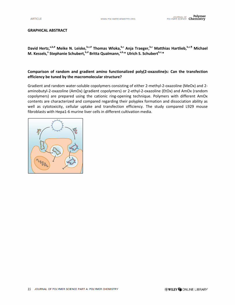
22
GRAPHICAL ABSTRACT
David Hertz,a,b,#
Meike N. Leiske,b,c,#
Thomas Wloka,b,c
Anja Traeger,b,c
Matthias Hartlieb,b,c,¶
Michael
M. Kessels,a Stephanie Schubert,
b,d Britta Qualmann,
a,b,* Ulrich S. Schubert
b,c,*
Comparison of random and gradient amino functionalized poly(2-oxazoline)s: Can the transfection
efficiency be tuned by the macromolecular structure?
Gradient and random water-soluble copolymers consisting of either 2-methyl-2-oxazoline (MeOx) and 2-aminobutyl-2-oxazoline (AmOx) (gradient copolymers) or 2-ethyl-2-oxazoline (EtOx) and AmOx (random copolymers) are prepared using the cationic ring-opening technique. Polymers with different AmOx contents are characterized and compared regarding their polyplex formation and dissociation ability as well as cytotoxicity, cellular uptake and transfection efficiency. The study compared L929 mouse fibroblasts with Hepa1-6 murine liver cells in different cultivation media.
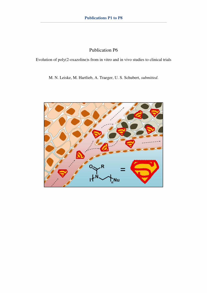
Publications P1 to P8
Publication P6
Evolution of poly(2-oxazoline)s from in vitro and in vivo studies to clinical trials
M. N. Leiske, M. Hartlieb, A. Traeger, U. S. Schubert, submitted.
Evolution of poly(2-oxazoline)s from in
vitro and in vivo studies to clinical trials
Meike N. Leiske,a,b
Matthias Hartlieb,a,b,
† Anja Traeger,a,b
Ulrich S. Schubert a,b,
*
a. Jena Center for Soft Matter (JCSM), Friedrich Schiller University Jena,
Philosophenweg 7, 07743 Jena, Germany. b. Laboratory of Organic and Macromolecular Chemistry (IOMC), Friedrich Schiller
University Jena, Humboldtstraße 10, 07743 Jena, Germany. c. † Current address: Institute of Biomaterial Science, Helmholtz-Zentrum
Geesthacht, Kantstr. 55, 14513 Teltow, Germany.
Research on poly(2-oxazoline)s (P(Ox)s) has significantly evolved over the
last decades. Whereas mainly synthesis and characterization were studied
first, focus is increasingly shifting towrads biomedical applications of the
polymer class, also catalyzed by the drawbacks of commonly used polymers,
such as poly(ethylene glycol) (PEG). The cationic ring-opening
polymerization enables the copolymerization of various functional
monomers, as well as modifications at the α- and ω-terminus. This variety of
functional groups is supposed to be beneficial for self-assembly processes,
drug conjugation or polyplex formation. Copolymers with 2-ethyl-2-
oxazoline (EtOx) or 2-methyl-2-oxazoline (MeOx) repeating units were found
to show stealth ability and, consequently, provide an enhanced
biocompatibility and elongated blood circulation times. For these reasons,
P(Ox)s are progressively used for in vivo studies and clinical trials to find safe
pharmaceuticals. Hence, the synthetic approaches leading to biomedical
relevant P(Ox)s, their biocompatiblity, as well as findings from in vivo studies
using P(Ox)s is summarized and evaluated within this review.
Key learning points
• P(Ox) synthesis and important polymer characteristics for in vivo studies
• Important P(Ox) architectures for in vivo studies
• Influence of P(Ox)s on the blood circulation time and the biodistribution
of drugs
• Controlled drug release by P(Ox) conjugates
• Developments in cancer therapy
Introduction
The first synthetic polymer-based medicines in clinical practice
were developed only three decades ago and the first product
approval occurred in 1990.1 In the 1990s, R. Duncan coined the
term “polymer therapeutics”, which means that the polymer
can either act as the bioactive itself or as a part of the covalent
conjugate, e.g. polymer-drug conjugates and polymer-protein
conjugates.2 Furthermore, self-assembled systems, such as
polymeric micelles and multi-component polyplexes, which are
known as non-viral vectors, are of great importance.1 In
general, biodegradable structures are preferred since the body
needs to be able to deal with the polymers after they fulfilled
their purpose; however, none of them should lead to
undesired toxicity or an immunogenic response before and
after biodegradation.3, 4
The currently most prominent example in this context is
poly(ethylene glycol) (PEG), which is already in use for several
applications against various diseases, e.g. multiple sclerosis
(Copaxone®), hepatitis C (PEGIntron
®) or anaemia (Macugen
®,
Puricase®).
5 However, many advantages and disadvantages of
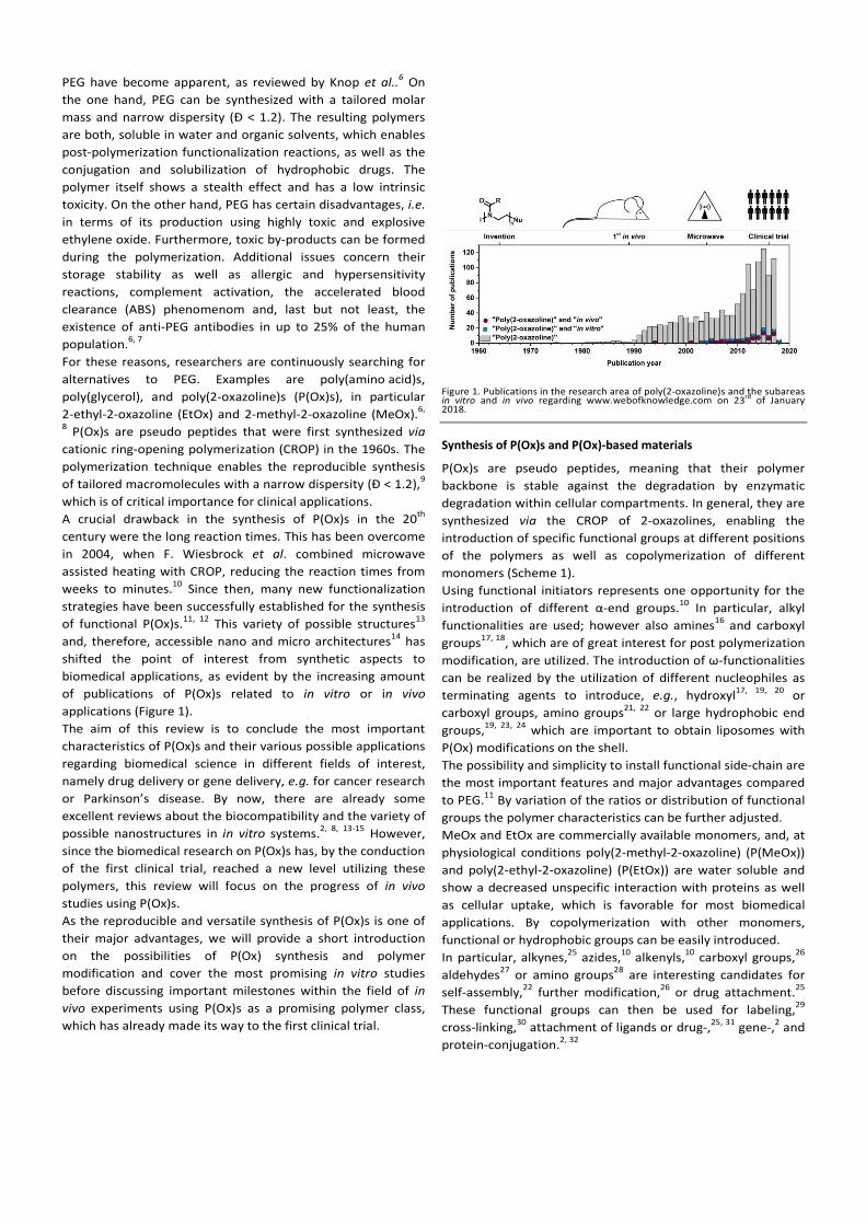
on of different α modification, are utilized. The introduction of ω

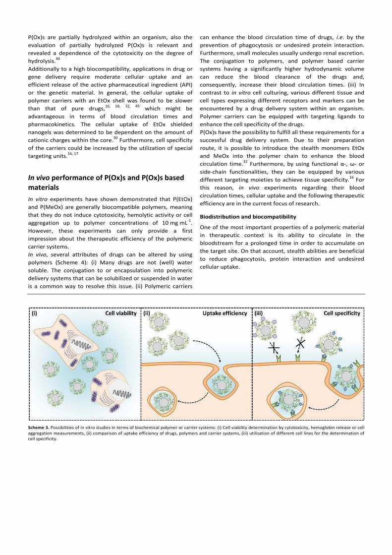
Furthermore, by using functional α, ω

defined P(Ox)s (Ð ≤ 1.1) in a
μ
APIs to the α or ω
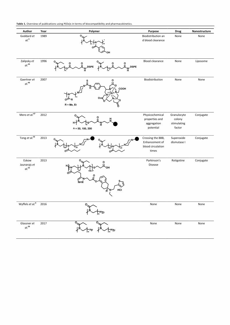
Table 1. Overview of publications using P(Ox)s in terms of biocompatibility and pharmacokinetics.
Author Year Polymer Purpose Drug Nanostructure
Goddard et
al.3
1989
Biodistribution an
d blood clearance
None None
Zalipsky et
al.23
1996
Blood clearance None Liposome
Gaertner et
al.46
2007
Biodistribution None None
Mero et al.47
2012
Physicochemical
properties and
aggregation
potential
Granulocyte
colony
stimulating
factor
Conjugate
Tong et al.32
2013
Crossing the BBB,
Enhancement of
blood circulation
times
Superoxide
dismutase I
Conjugate
Eskow
Jaunarajs et
al.25
2013
Parkinson’s
Disease
Rotigotine Conjugate
Wyffels et al.4 2016
None None None
Glassner et
al.36
2017
None None None
N
O
N
O
OH
n m
∗
N
O
O
O
NH
O
DSPE
n
N
O
O
O
NH
O
DSPE
n
N
RO
N
N NH
S
N
N
N
N
COOH
O
O
O
O
O
O
111In3+
n
R = Me, Et
HN
O
S NH
OHN
n
n = 50, 100, 200
N
O
N
O
N
50 20
N
N
O
N
O
N
50 20
N
HN
N
O
O
S
N N
NO
O
N
S
H HCl
O
OH
190 r10
N89Zr
O
n
N18F
O
n
N89Zr
O
n
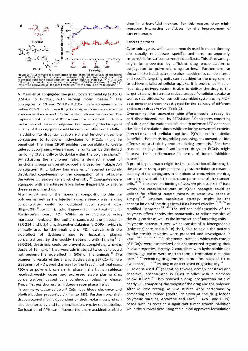

introduction of functional α or ω
1,1′3,3,3′,3′
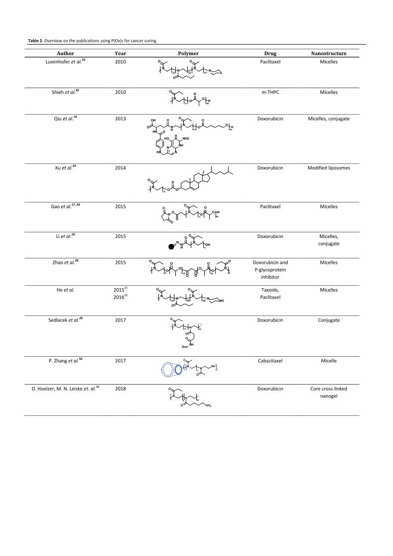
N
O
N
O
NN
O
37 23 37
N
O
O
O
OH
n m
N
O
O
O
OHN
H
O
HN
O
OH
O
HN N
NNH
HNHO NH2
n m
N
O
O O
O
n
N
O
O
O
N
O
O
On
O
O H
m
NNH
O
N
O
OHn
N
O
O
O
O
O
O
O
O
O
N
O
n nmm
N
O
N
O
NN
O
NH33 26 45
N∗
O
N
∗
O
O
NHDox
n m
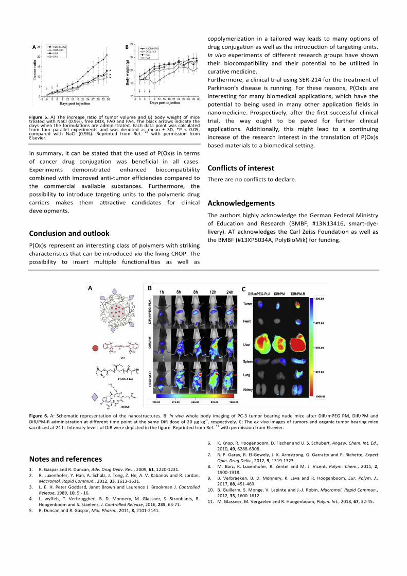

12. H. Schlaad, C. Diehl, A. Gress, M. Meyer, A. L. Demirel, Y. Nur and A. Bertin,
Macromol. Rapid Commun., 2010, 31, 511-525.
13. M. Hartlieb, K. Kempe and U. S. Schubert, J. Mat. Chem. B, 2015, 3, 526-538.
14. P. Wilson, P. C. Ke, T. P. Davis and K. Kempe, Eur. Polym. J., 2016, 88, 486-515.
15. K. Lava, B. Verbraeken and R. Hoogenboom, Eur. Polym. J., 2015, 65, 98-111.
16. L.-Y. Qiu, L. Yan, L. Zhang, Y.-M. Jin and Q.-H. Zhao, Int. J. Pharm., 2013, 456,
315-324.
17. Y. Gao, Y. Li, Y. Li, L. Yuan, Y. Zhou, J. Li, L. Zhao, C. Zhang, X. Li and Y. Liu,
Nanoscale, 2015, 7, 597-612.
18. J. Li, Y. Zhou, C. Li, D. Wang, Y. Gao, C. Zhang, L. Zhao, Y. Li, Y. Liu and X. Li,
Bioconjugate Chem., 2015, 26, 110-119.
19. P. H. Kierstead, H. Okochi, V. J. Venditto, T. C. Chuong, S. Kivimae, J. M. J.
Fréchet and F. C. Szoka, J. Controlled Release, 2015, 213, 1-9.
20. Y. Zhao, Y. Zhou, D. Wang, Y. Gao, J. Li, S. Ma, L. Zhao, C. Zhang, Y. Liu and X. Li,
Acta Biomater., 2015, 17, 182-192.
21. Z. He, A. Schulz, X. Wan, J. Seitz, H. Bludau, D. Y. Alakhova, D. B. Darr, C. M.
Perou, R. Jordan, I. Ojima, A. V. Kabanov and R. Luxenhofer, J. Controlled
Release, 2015, 208, 67-75.
22. Z. He, X. Wan, A. Schulz, H. Bludau, M. A. Dobrovolskaia, S. T. Stern, S. A.
Montgomery, H. Yuan, Z. Li, D. Alakhova, M. Sokolsky, D. B. Darr, C. M. Perou,
R. Jordan, R. Luxenhofer and A. V. Kabanov, Biomaterials, 2016, 101, 296-309.
23. S. Zalipsky, C. B. Hansen, J. M. Oaks and T. M. Allen, J. Pharm. Sci., 1996, 85,
133-137.
24. H. Xu, W. Zhang, Y. Li, F. F. Ye, P. P. Yin, X. Yu, M. N. Hu, Y. S. Fu, C. Wang and
D. J. Shang, Pharm. Res., 2014, 31, 3038-3050.
25. K. L. Eskow Jaunarajs, D. G. Standaert, T. X. Viegas, M. D. Bentley, Z. Fang, B.
Dizman, K. Yoon, R. Weimer, P. Ravenscroft, T. H. Johnston, M. P. Hill, J. M.
Brotchie and R. W. Moreadith, Mov. Disord., 2013, 28, 1675-1682.
26. O. Sedlacek, B. D. Monnery, J. Mattova, J. Kucka, J. Panek, O. Janouskova, A.
Hocherl, B. Verbraeken, M. Vergaelen, M. Zadinova, R. Hoogenboom and M.
Hruby, Biomaterials, 2017, 146, 1-12.
27. C. Legros, M.-C. De Pauw-Gillet, K. C. Tam, S. Lecommandoux and D. Taton,
Eur. Polym. J., 2015, 62, 322-330.
28. Z. He, L. Miao, R. Jordan, D. S-Manickam, R. Luxenhofer and A. V. Kabanov,
Macromol. Biosci., 2015, 15, 1004-1020.
29. M. N. Leiske, M. Hartlieb, C. Paulenz, D. Pretzel, M. Hentschel, C. Englert, M.
Gottschaldt and U. S. Schubert, Adv. Funct. Mater., 2015, 25, 2458-2466.
30. M. Hartlieb, T. Bus, J. Kübel, D. Pretzel, S. Hoeppener, M. N. Leiske, K. Kempe,
B. Dietzek and U. S. Schubert, Bioconjugate Chem., 2017, 28, 1229-1235.
31. R. W. Moreadith, T. X. Viegas, M. D. Bentley, J. M. Harris, Z. Fang, K. Yoon, B.
izman, R. Weimer, B. P. Rae, X. Li, C. Rader, D. Standaert and W. Olanow, Eur.
Polym. J., 2016.
32. J. Tong, X. Yi, R. Luxenhofer, W. A. Banks, R. Jordan, M. C. Zimmerman and A.
V. Kabanov, Mol. Pharm., 2013, 10, 360-377.
33. R. Luxenhofer, A. Schulz, C. Roques, S. Li, T. K. Bronich, E. V. Batrakova, R.
Jordan and A. V. Kabanov, Biomaterials, 2010, 31, 4972-4979.
34. Y. Seo, A. Schulz, Y. Han, Z. He, H. Bludau, X. Wan, J. Tong, T. K. Bronich, M.
Sokolsky, R. Luxenhofer, R. Jordan and A. V. Kabanov, Polym. Adv. Technol.,
2015, 26, 837-850.
35. M. C. Woodle, C. M. Engbers and S. Zalipsky, Bioconjugate Chem., 1994, 5,
493-496.
36. M. Glassner, L. Palmieri, B. D. Monnery, T. Verbrugghen, S. Deleye, S.
Stroobants, S. Staelens, L. wyffels and R. Hoogenboom, Biomacromolecules,
2017, 18, 96-102.
37. K. Kempe, S. L. Ng, K. F. Noi, M. Müllner, S. T. Gunawan and F. Caruso, ACS
Macro Lett., 2013, 2, 1069-1072.
38. T. R. Dargaville, B. G. Hollier, A. Shokoohmand and R. Hoogenboom, Cell Adh.
Migr., 2014, 8, 88-93.
39. Y. Liu, Y. Wang, Y. Wang, J. Lu, V. Piñón and M. Weck, J. Am. Chem. Soc., 2011,
133, 14260-14263.
40. L. Tauhardt, K. Kempe, M. Gottschaldt and U. S. Schubert, Chem. Soc. Rev.,
2013, 42, 7998-8011.
41. M. Bauer, C. Lautenschlaeger, K. Kempe, L. Tauhardt, U. S. Schubert and D.
Fischer, Macromol. Biosci., 2012, 12, 986-998.
42. M. Bauer, S. Schroeder, L. Tauhardt, K. Kempe, U. S. Schubert and D. Fischer, J.
Polym. Sci. A: Polym. Chem., 2013, 51, 1816-1821.
43. Y. Gao, Y. Zhou, L. Zhao, C. Zhang, Y. Li, J. Li, X. Li and Y. Liu, Acta Biomater.,
2015, 23, 127-135.
44. R. Shah, Z. Kronekova, A. Zahoranová, L. Roller, N. Saha, P. Saha and J. Kronek,
J. Mater. Sci. Mater. Med., 2015, 26, 157.
45. M.-J. Shieh, C.-L. Peng, W.-L. Chiang, C.-H. Wang, C.-Y. Hsu, S.-J. J. Wang and
P.-S. Lai, Mol. Pharm., 2010, 7, 1244-1253.
46. F. C. Gaertner, R. Luxenhofer, B. Blechert, R. Jordan and M. Essler, J. Controlled
Release, 2007, 119, 291-300.
47. A. Mero, Z. Fang, G. Pasut, F. M. Veronese and T. X. Viegas, J. Controlled
Release, 2012, 159, 353-361.
48. D. Hoelzer, M. N. Leiske, M. Hartlieb, T. Bus, D. Pretzel, S. Hoeppener, K.
Kempe, R. Thierbach and U. S. Schubert, Oncotarget, 2018, Submitted.
49. H. Xu, M. Hu, X. Yu, Y. Li, Y. Fu, X. Zhou, D. Zhang and J. Li, Eur. J. Pharm.
Biopharm., 2015, 91, 66-74.
50. P. Zhang, X. Qian, Z. Zhang, C. Li, C. Xie, W. Wu and X. Jiang, ACS Appl. Mater.
Interfaces, 2017, 9, 5768-5777.
Publications P1 to P8
Publication P7
Site-specific POxylation of interleukin-4
T. Luehmann, M. Schmidt, M. N. Leiske, V. Spieler, T. C. Majdanski, M. Grube, M. Hartlieb,
I. Nischang, S. Schubert, U.S. Schubert, L. Meinel,
ACS Biomater. Sci. Eng. 2017, 3, 304 - 312.
Reproduced by permission of The American Chemical Society. Copyright © 2017.
The paper as well as the supporting information (free of charge) is available online:
doi.org/10.1021/acsbiomaterials.6b00578.
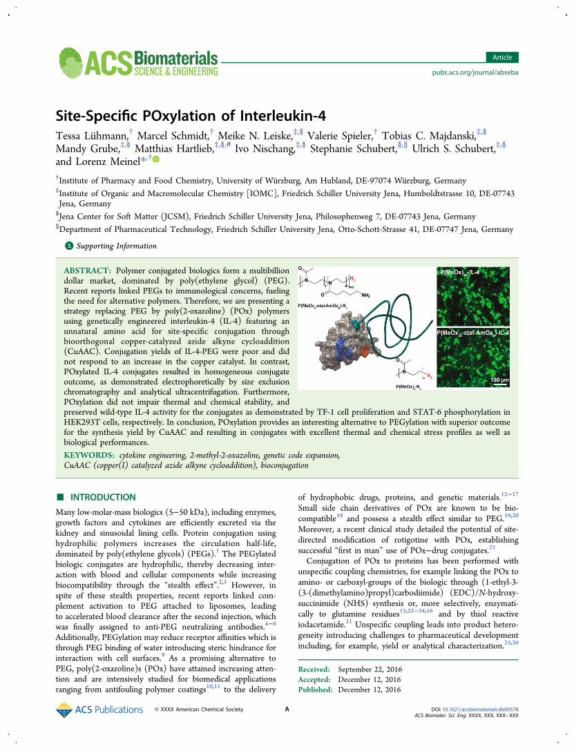
Site-Specific POxylation of Interleukin‑4
Tessa Luhmann,† Marcel Schmidt,† Meike N. Leiske,‡,§ Valerie Spieler,† Tobias C. Majdanski,‡,§
Mandy Grube,‡,§ Matthias Hartlieb,‡,§,# Ivo Nischang,‡,§ Stephanie Schubert,§,∥ Ulrich S. Schubert,‡,§
and Lorenz Meinel*,†
†Institute of Pharmacy and Food Chemistry, University of Wurzburg, Am Hubland, DE-97074 Wurzburg, Germany‡Institute of Organic and Macromolecular Chemistry [IOMC], Friedrich Schiller University Jena, Humboldtstrasse 10, DE-07743Jena, Germany§Jena Center for Soft Matter (JCSM), Friedrich Schiller University Jena, Philosophenweg 7, DE-07743 Jena, Germany∥Department of Pharmaceutical Technology, Friedrich Schiller University Jena, Otto-Schott-Strasse 41, DE-07747 Jena, Germany
*S Supporting Information
ABSTRACT: Polymer conjugated biologics form a multibilliondollar market, dominated by poly(ethylene glycol) (PEG).Recent reports linked PEGs to immunological concerns, fuelingthe need for alternative polymers. Therefore, we are presenting astrategy replacing PEG by poly(2-oxazoline) (POx) polymersusing genetically engineered interleukin-4 (IL-4) featuring anunnatural amino acid for site-specific conjugation throughbioorthogonal copper-catalyzed azide alkyne cycloaddition(CuAAC). Conjugation yields of IL-4-PEG were poor and didnot respond to an increase in the copper catalyst. In contrast,POxylated IL-4 conjugates resulted in homogeneous conjugateoutcome, as demonstrated electrophoretically by size exclusionchromatography and analytical ultracentrifugation. Furthermore,POxylation did not impair thermal and chemical stability, andpreserved wild-type IL-4 activity for the conjugates as demonstrated by TF-1 cell proliferation and STAT-6 phosphorylation inHEK293T cells, respectively. In conclusion, POxylation provides an interesting alternative to PEGylation with superior outcomefor the synthesis yield by CuAAC and resulting in conjugates with excellent thermal and chemical stress profiles as well asbiological performances.
KEYWORDS: cytokine engineering, 2-methyl-2-oxazoline, genetic code expansion,CuAAC (copper(I) catalyzed azide alkyne cycloaddition), bioconjugation
■ INTRODUCTION
Many low-molar-mass biologics (5−50 kDa), including enzymes,growth factors and cytokines are efficiently excreted via thekidney and sinusoidal lining cells. Protein conjugation usinghydrophilic polymers increases the circulation half-life,dominated by poly(ethylene glycols) (PEGs).1 The PEGylatedbiologic conjugates are hydrophilic, thereby decreasing inter-action with blood and cellular components while increasingbiocompatibility through the “stealth effect”.2,3 However, inspite of these stealth properties, recent reports linked com-plement activation to PEG attached to liposomes, leadingto accelerated blood clearance after the second injection, whichwas finally assigned to anti-PEG neutralizing antibodies.4−8
Additionally, PEGylation may reduce receptor affinities which isthrough PEG binding of water introducing steric hindrance forinteraction with cell surfaces.9 As a promising alternative toPEG, poly(2-oxazoline)s (POx) have attained increasing atten-tion and are intensively studied for biomedical applicationsranging from antifouling polymer coatings10,11 to the delivery
of hydrophobic drugs, proteins, and genetic materials.12−17
Small side chain derivatives of POx are known to be bio-compatible18 and possess a stealth effect similar to PEG.19,20
Moreover, a recent clinical study detailed the potential of site-directed modification of rotigotine with POx, establishingsuccessful “first in man” use of POx−drug conjugates.21
Conjugation of POx to proteins has been performed withunspecific coupling chemistries, for example linking the POx toamino- or carboxyl-groups of the biologic through (1-ethyl-3-(3-(dimethylamino)propyl)carbodiimide) (EDC)/N-hydroxy-succinimide (NHS) synthesis or, more selectively, enzymati-cally to glutamine residues15,22−24,16 and by thiol reactiveiodacetamide.21 Unspecific coupling leads into product hetero-geneity introducing challenges to pharmaceutical developmentincluding, for example, yield or analytical characterization.25,26
Received: September 22, 2016Accepted: December 12, 2016Published: December 12, 2016
Article
pubs.acs.org/journal/abseba
© XXXX American Chemical Society A DOI: 10.1021/acsbiomaterials.6b00578ACS Biomater. Sci. Eng. XXXX, XXX, XXX−XXX
Furthermore, unspecific chemistries, leading to heterogeneousproduct outcome, drive another challenge−immunogenicity.27
Through heterogeneity, more species (thereby more conforma-tional variants) are presented to the patient’s immune systemthereby exposing the immune system to a number of alteredepitopes as compared to the wild type, arguably more effec-tively supporting antibody formation against the biologic.These considerations fuel the need for alternatives, leading tohomogeneous product outcome. We are approaching this bygenetically introducing unnatural groups into the biologicsbackbone at one predefined site, providing a distinctivefunctional group. Only at that introduced group will decorationoccur, removing the heterogeneity of the majority of couplingstrategies pursued today.26,28−30 To this end, we recentlyreported on genetic code expansion integrating pyrrolysinederivatives, e.g., N-propargyl-L-lysine (Plk), through recombi-nant protein expression for site-specific modification of growthfactors,31 fluorescent proteins,32 as well as for cytokines33 anduse for the surface decoration of glyco-engineered cells34 orbioresponsive drug delivery35 through bio-orthogonal coppercatalyzed azide alkyne cycloaddition (CuAAC). In thisstudy, we pursue the production of site-specifically decoratedpolymer-interleukin-4 (IL-4) conjugates with the ultimate goalfor unprecedented homogeneity in IL-4 conjugate out-come and maintained potency as compared to the wild type(wt) IL-4. IL-4 is a small and (at physiological conditions)positively charged 15 kDa-cytokine, triggering macrophage(Mφ) polarization along the M2 lineage with possible appli-cation in Mφ associated diseases.36 In light of the favor-able protein-repellent property and good biocompatibility,hydrophilic POx-based polymers of different molar masses(2.5, 4, 10 kDa) and architecture, including an azide group forcycloaddition, were synthesized to approach site-specificconjugation of Plk-IL-4 with the polymer. Conjugation withPEG resulted in poor yield, whereas POx polymers wereeffectively conjugated as a function of the polymers’ weight-average molar mass MW. The IL-4 conjugate products weredetailed with respect to bioactivity, secondary structure, as wellas thermal and chemical stability.
2. MATERIALS AND METHODS
DMEM, RPMI-1640 medium, L-glutamine, L-alanyl-L-glutamine, sodiumpyruvate, bovine serum albumin solution 7.5%, lipid medium supple-ment, copper(II) sulfate, sodium L-ascorbate, and tris(3-hydroxypropyl-triazolylmethyl) amine (THPTA) were purchased from Sigma-Aldrich(Schnelldorf, Germany). Penicillin G and streptomycin solution (Pen/Strep) were purchased from Biochrom AG (Berlin, Germany). Fetalbovine serum (FBS) was from GIBCO life technologies (Carlsbad,USA). HiTrap SP XL and HiTrap SP HP AKTA columns were fromGE Healthcare (Buckinghamshire, GB). Vivaspin centrifugal concen-trators were from Sartorius AG (Gottingen, Germany) and HyperSepC18 desalting columns were from Thermo Scientific (Waltham, USA).WST-1 was purchased from Roche (Basel, Switzerland). All otherchemicals used were at least of pharmaceutical grade and were purchasedfrom Sigma-Aldrich and Merck (unless noted otherwise). Solvents wereobtained by Sigma-Aldrich, VWR, Linde, and Arcos Organics.2.1. Polymer Synthesis and Characterization. A detailed
overview about synthetic procedures and characterization is given inthe Supporting Information.2.2. Chemical Synthesis of Propargyl-L-lysine (Plk). Propargyl-
L-lysine (Plk) was prepared as HCl-salt following procedures describedby Milles et al.37 1H NMR spectra were acquired on a Bruker Advance400 MHz spectrometer for confirmation of the product.31
2.3. Expression of Plk-IL-4 and wt-IL-4. Plk-IL-4 and wt-IL-4were expressed as described previously.33 Briefly, E.coli BL21 DE3,
encoding for TAG(42)-IL-4 and for the PylRS/tRNACUA pair, werecultured at 37 °C and the Plk substrate was added at a finalconcentration of 2 mM at OD600 = 0.3 in standard TB (Terrific Broth)medium. For wt-IL-4 expression, BL21 (DE3) cells, encoding forwt-IL-4, were used. Protein expression was subsequently induced with1 mM IPTG at OD600 = 0.6 at 37 °C. After 6 h, the bacterial cells wereharvested and the pellet was solubilized after sonification andcentrifugation with lysis buffer, containing 5 M guanidine-HCl and2 mM reduced and 0.2 mM oxidized glutathione. This solution wassubsequently refolded using a glutathione redox buffer system asdescribed.38 After centrifugation, the supernatant containing Plk-IL-4was purified by ion exchange affinity chromatography using a FPLCsystem (GE Healthcare Akta Purifier, Life sciences, Freiburg,Germany). After purification, fractions containing IL-4 proteins wereextensively dialyzed against PBS and stored at −80 °C. Wt-IL-4 andPlk-IL-4 concentrations were determined by UV-absorbance at280 nm, using a molar extinction coefficient of 8860 M−1 cm−1.39
2.4. Copper-Catalyzed Azide−Alkyne Huisgen Cycloaddi-tion (CuAAC). Twenty-five μM Plk-IL-4 and a 50-fold molar excess ofeach polymer was used. The click reaction was performed in thepresence of 2.5 mM L-ascorbic acid, 500 μM THPTA, and 100 μMCuSO4 or in the presence of 2.5 mM L-ascorbic acid, 1 mM THPTA,and 1 mM CuSO4 in PBS (137 mM NaCl, 2.7 mM KCl, 4.3 mMNa2HPO4,1.47 mM KH2PO4, pH 7.4). CuSO4 and THPTA waspremixed and incubated with L-ascorbic acid under air exclusion for10 min to quench occurring reactive oxygene species (ROS) asdetailed before.34 The click reaction was performed at room temper-ature overnight and the reaction was stopped by the addition of 5 mMEDTA. The proteins and conjugates were subsequently analyzed bySDS-PAGE and MALDI-MS analysis or used for further purification.
2.5. MALDI-MS. The samples were desalted using ZipTipC18-tipsfollowing the manufacturer’s instructions. Matrix-assisted laser desorp-tion ionization (MALDI-MS) spectra were acquired in the linearpositive mode by using an Autoflex II LRF instrument (Billerica, USA).Mass spectra were calibrated externally with a protein standard I fromBruker Daltonics Inc. (Billerica, USA), containing insulin, ubiquitin,myoglobin, and cytochrome C.
2.6. SDS-PAGE. Expressed proteins and proteins used in clickreactions were analyzed by standard Tris-glycine SDS-PAGE as out-lined before.40 Gels were stained with Coomassie Brilliant Blue G250and photographed using a FluorChem FC2 imaging system (ProteinSimple, Santa Clara, CA).
2.7. RP-HPLC and SEC Analysis. Protein purity was assessed on aRP-HPLC system using a VWR Hitachi LaChrom HPLC system(Darmstadt, Germany). Protein samples and polymers were applied toa ZORBAX Eclipse XDB-C18 column (150 mm * 4.6 mm, particlesize = 5 μm (Agilent, Santa Clara, CA)), equilibrated by watercontaining 0.1% TFA and acetonitrile (ACN) containing 0.1% TFA(90:10 v/v). Polymers, wt-IL-4 and IL-4 conjugates were eluted by alinear gradient of 10−60% ACN containing 0.1% TFA with a gradientof 1% ACN/min and a flow rate of 1 mL/min. Column temperaturewas kept at 24 °C and UV-absorbance was monitored at λ = 214 nmand λ = 280 nm, respectively.
After desalting using HyperSep C18 columns, IL-4-conjugates wereeluated with 45% MeCN, containing 0.1% TFA (v/v). For SEC analysis,approximately 10 μg protein sample was applied to an equilibratedBioSep-SEC s2000 column (300 * 4.6 mm, particle size = 5 μm, poresize = 145 Å (Phenomenex, Torrance, USA)). Polymers, wt-IL-4 andIL-4 conjugates were analyzed in 45% MeCN, containing 0.1% TFA(v/v) at a flow rate of 1 mL/min according to the manufacturer’sinstructions for small peptides and proteins. UV-absorbance wasmonitored at λ = 220 nm. Data analysis, including nonlinear curvefitting and parameter determination (EC50-value range within the 95%confidence interval), was performed with the software GraphPadPrism 7 (San Diego, California, USA).
2.8. Analytical Ultracentrifugation (AUC). Sedimentationvelocity experiments were performed using a ProteomeLab XL-Ianalytical ultracentrifuge (Beckman Coulter Instruments, Brea, CA)with an An-60Ti four-hole rotor using double-sector aluminum center-pieces with a 12 mm optical path length. Interference optics detection
ACS Biomaterials Science & Engineering Article
DOI: 10.1021/acsbiomaterials.6b00578
ACS Biomater. Sci. Eng. XXXX, XXX, XXX−XXXB
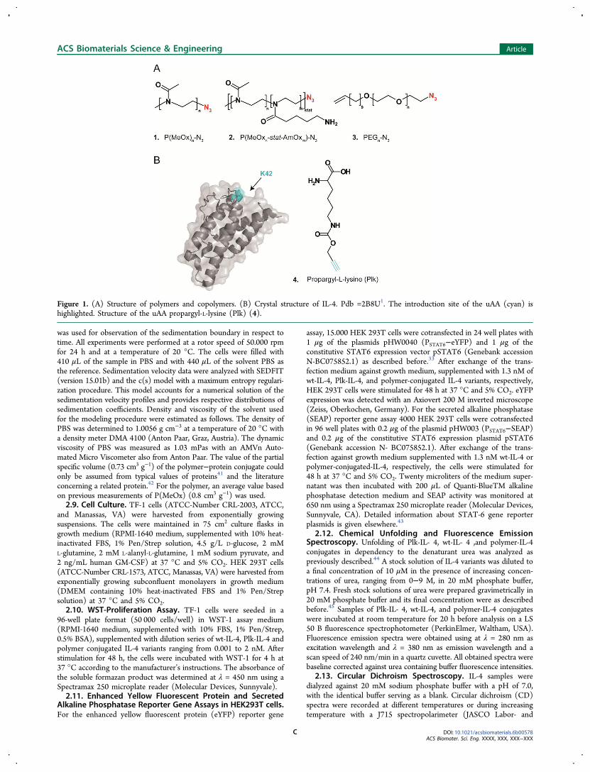
was used for observation of the sedimentation boundary in respect totime. All experiments were performed at a rotor speed of 50.000 rpmfor 24 h and at a temperature of 20 °C. The cells were filled with410 μL of the sample in PBS and with 440 μL of the solvent PBS asthe reference. Sedimentation velocity data were analyzed with SEDFIT(version 15.01b) and the c(s) model with a maximum entropy regulari-zation procedure. This model accounts for a numerical solution of thesedimentation velocity profiles and provides respective distributions ofsedimentation coefficients. Density and viscosity of the solvent usedfor the modeling procedure were estimated as follows. The density ofPBS was determined to 1.0056 g cm−3 at a temperature of 20 °C witha density meter DMA 4100 (Anton Paar, Graz, Austria). The dynamicviscosity of PBS was measured as 1.03 mPas with an AMVn Auto-mated Micro Viscometer also from Anton Paar. The value of the partialspecific volume (0.73 cm3 g−1) of the polymer−protein conjugate couldonly be assumed from typical values of proteins41 and the literatureconcerning a related protein.42 For the polymer, an average value basedon previous measurements of P(MeOx) (0.8 cm3 g−1) was used.2.9. Cell Culture. TF-1 cells (ATCC-Number CRL-2003, ATCC,
and Manassas, VA) were harvested from exponentially growingsuspensions. The cells were maintained in 75 cm2 culture flasks ingrowth medium (RPMI-1640 medium, supplemented with 10% heat-inactivated FBS, 1% Pen/Strep solution, 4.5 g/L D-glucose, 2 mML-glutamine, 2 mM L-alanyl-L-glutamine, 1 mM sodium pyruvate, and2 ng/mL human GM-CSF) at 37 °C and 5% CO2. HEK 293T cells(ATCC-Number CRL-1573, ATCC, Manassas, VA) were harvested fromexponentially growing subconfluent monolayers in growth medium(DMEM containing 10% heat-inactivated FBS and 1% Pen/Strepsolution) at 37 °C and 5% CO2.2.10. WST-Proliferation Assay. TF-1 cells were seeded in a
96-well plate format (50 000 cells/well) in WST-1 assay medium(RPMI-1640 medium, supplemented with 10% FBS, 1% Pen/Strep,0.5% BSA), supplemented with dilution series of wt-IL-4, Plk-IL-4 andpolymer conjugated IL-4 variants ranging from 0.001 to 2 nM. Afterstimulation for 48 h, the cells were incubated with WST-1 for 4 h at37 °C according to the manufacturer’s instructions. The absorbance ofthe soluble formazan product was determined at λ = 450 nm using aSpectramax 250 microplate reader (Molecular Devices, Sunnyvale).2.11. Enhanced Yellow Fluorescent Protein and Secreted
Alkaline Phosphatase Reporter Gene Assays in HEK293T cells.For the enhanced yellow fluorescent protein (eYFP) reporter gene
assay, 15.000 HEK 293T cells were cotransfected in 24 well plates with1 μg of the plasmids pHW0040 (PSTAT6−eYFP) and 1 μg of theconstitutive STAT6 expression vector pSTAT6 (Genebank accessionN-BC075852.1) as described before.33 After exchange of the trans-fection medium against growth medium, supplemented with 1.3 nM ofwt-IL-4, Plk-IL-4, and polymer-conjugated IL-4 variants, respectively,HEK 293T cells were stimulated for 48 h at 37 °C and 5% CO2. eYFPexpression was detected with an Axiovert 200 M inverted microscope(Zeiss, Oberkochen, Germany). For the secreted alkaline phosphatase(SEAP) reporter gene assay 4000 HEK 293T cells were cotransfectedin 96 well plates with 0.2 μg of the plasmid pHW003 (PSTAT6−SEAP)and 0.2 μg of the constitutive STAT6 expression plasmid pSTAT6(Genebank accession N- BC075852.1). After exchange of the trans-fection against growth medium supplemented with 1.3 nM wt-IL-4 orpolymer-conjugated-IL-4, respectively, the cells were stimulated for48 h at 37 °C and 5% CO2. Twenty microliters of the medium super-natant was then incubated with 200 μL of Quanti-BlueTM alkalinephosphatase detection medium and SEAP activity was monitored at650 nm using a Spectramax 250 microplate reader (Molecular Devices,Sunnyvale, CA). Detailed information about STAT-6 gene reporterplasmids is given elsewhere.43
2.12. Chemical Unfolding and Fluorescence EmissionSpectroscopy. Unfolding of Plk-IL- 4, wt-IL- 4 ,and polymer-IL-4conjugates in dependency to the denaturant urea was analyzed aspreviously described.44 A stock solution of IL-4 variants was diluted toa final concentration of 10 μM in the presence of increasing concen-trations of urea, ranging from 0−9 M, in 20 mM phosphate buffer,pH 7.4. Fresh stock solutions of urea were prepared gravimetrically in20 mM phosphate buffer and its final concentration were as describedbefore.45 Samples of Plk-IL- 4, wt-IL-4, and polymer-IL-4 conjugateswere incubated at room temperature for 20 h before analysis on a LS50 B fluorescence spectrophotometer (PerkinElmer, Waltham, USA).Fluorescence emission spectra were obtained using at λ = 280 nm asexcitation wavelength and λ = 380 nm as emission wavelength and ascan speed of 240 nm/min in a quartz cuvette. All obtained spectra werebaseline corrected against urea containing buffer fluorescence intensities.
2.13. Circular Dichroism Spectroscopy. IL-4 samples weredialyzed against 20 mM sodium phosphate buffer with a pH of 7.0,with the identical buffer serving as a blank. Circular dichroism (CD)spectra were recorded at different temperatures or during increasingtemperature with a J715 spectropolarimeter (JASCO Labor- and
Figure 1. (A) Structure of polymers and copolymers. (B) Crystal structure of IL-4. Pdb =2B8U1. The introduction site of the uAA (cyan) ishighlighted. Structure of the uAA propargyl-L-lysine (Plk) (4).
ACS Biomaterials Science & Engineering Article
DOI: 10.1021/acsbiomaterials.6b00578
ACS Biomater. Sci. Eng. XXXX, XXX, XXX−XXXC
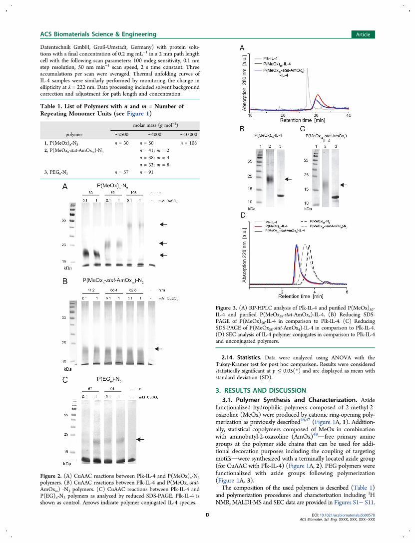
Datentechnik GmbH, Groß-Umstadt, Germany) with protein solu-tions with a final concentration of 0.2 mg mL−1 in a 2 mm path lengthcell with the following scan parameters: 100 mdeg sensitivity, 0.1 nmstep resolution, 50 nm min−1 scan speed, 2 s time constant. Threeaccumulations per scan were averaged. Thermal unfolding curves ofIL-4 samples were similarly performed by monitoring the change inellipticity at λ = 222 nm. Data processing included solvent backgroundcorrection and adjustment for path length and concentration.
2.14. Statistics. Data were analyzed using ANOVA with theTukey-Kramer test for post hoc comparison. Results were consideredstatistically significant at p ≤ 0.05(*) and are displayed as mean withstandard deviation (SD).
3. RESULTS AND DISCUSSION
3.1. Polymer Synthesis and Characterization. Azidefunctionalized hydrophilic polymers composed of 2-methyl-2-oxazoline (MeOx) were produced by cationic ring-opening poly-merization as previously described46,47 (Figure 1A, 1). Addition-ally, statistical copolymers composed of MeOx in combinationwith aminobutyl-2-oxazoline (AmOx)48free primary aminegroups at the polymer side chains that can be used for addi-tional decoration purposes including the coupling of targetingmotifswere synthesized with a terminally located azide group(for CuAAC with Plk-IL-4) (Figure 1A, 2). PEG polymers werefunctionalized with azide groups following polymerization(Figure 1A, 3).The composition of the used polymers is described (Table 1)
and polymerization procedures and characterization including 1HNMR, MALDI-MS and SEC data are provided in Figures S1− S11.
Table 1. List of Polymers with n and m = Number ofRepeating Monomer Units (see Figure 1)
molar mass (g mol−1)
polymer ∼2500 ∼4000 ∼10 000
1, P(MeOx)n-N3 n = 30 n = 50 n = 108
2, P(MeOxn-stat-AmOxm)-N3 n = 41; m = 2
n = 38; m = 4
n = 32; m = 8
3, PEGn-N3 n = 57 n = 91
Figure 2. (A) CuAAC reactions between Plk-IL-4 and P(MeOx)n-N3
polymers. (B) CuAAC reactions between Plk-IL-4 and P(MeOxn-stat-AmOxm) -N3 polymers. (C) CuAAC reactions between Plk-IL-4 andP(EG)n-N3 polymers as analyzed by reduced SDS-PAGE. Plk-IL-4 isshown as control. Arrows indicate polymer conjugated IL-4 species.
Figure 3. (A) RP-HPLC analysis of Plk-IL-4 and purified P(MeOx)50-IL-4 and purified P(MeOx38-stat-AmOx4)-IL-4. (B) Reducing SDS-PAGE of P(MeOx)50-IL-4 in comparison to Plk-IL-4. (C) ReducingSDS-PAGE of P(MeOx38-stat-AmOx4)-IL-4 in comparison to Plk-IL-4.(D) SEC analysis of IL-4 polymer conjugates in comparison to Plk-IL-4and unconjugated polymers.
ACS Biomaterials Science & Engineering Article
DOI: 10.1021/acsbiomaterials.6b00578
ACS Biomater. Sci. Eng. XXXX, XXX, XXX−XXXD
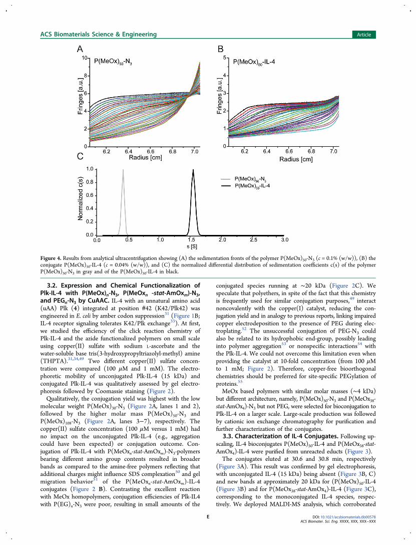
3.2. Expression and Chemical Functionalization ofPlk-IL-4 with P(MeOx)n-N3, P(MeOxn -stat-AmOxm)-N3,and PEGn-N3 by CuAAC. IL-4 with an unnatural amino acid(uAA) Plk (4) integrated at position #42 (K42/Plk42) wasengineered in E. coli by amber codon suppression31 (Figure 1B;IL-4 receptor signaling tolerates K42/Plk exchange33). At first,we studied the efficiency of the click reaction chemistry ofPlk-IL-4 and the azide functionalized polymers on small scaleusing copper(II) sulfate with sodium L-ascorbate and thewater-soluble base tris(3-hydroxypropyltriazolyl-methyl) amine(THPTA).31,34,49 Two different copper(II) sulfate concen-tration were compared (100 μM and 1 mM). The electro-phoretic mobility of unconjugated Plk-IL-4 (15 kDa) andconjugated Plk-IL-4 was qualitatively assessed by gel electro-phoresis followed by Coomassie staining (Figure 2).Qualitatively, the conjugation yield was highest with the low
molecular weight P(MeOx)30-N3 (Figure 2A, lanes 1 and 2),followed by the higher molar mass P(MeOx)50-N3 andP(MeOx)108-N3 (Figure 2A, lanes 3−7), respectively. Thecopper(II) sulfate concentration (100 μM versus 1 mM) hadno impact on the unconjugated Plk-IL-4 (e.g., aggregationcould have been expected) or conjugation outcome. Con-jugation of Plk-IL-4 with P(MeOxn-stat-AmOxm)-N3-polymersbearing different amino group contents resulted in broaderbands as compared to the amine-free polymers reflecting thatadditional charges might influence SDS complexation50 and gelmigration behavior51 of the P(MeOxn-stat-AmOxm)-IL-4conjugates (Figure 2 B). Contrasting the excellent reactionwith MeOx homopolymers, conjugation efficiencies of Plk-IL4with P(EG)n-N3 were poor, resulting in small amounts of the
conjugated species running at ∼20 kDa (Figure 2C). Wespeculate that polyethers, in spite of the fact that this chemistryis frequently used for similar conjugation purposes,49 interactnoncovalently with the copper(I) catalyst, reducing the con-jugation yield and in analogy to previous reports, linking impairedcopper electrodeposition to the presence of PEG during elec-troplating.52 The unsuccessful conjugation of PEG-N3 couldalso be related to its hydrophobic end-group, possibly leadinginto polymer aggregation53 or nonspecific interactions54 withthe Plk-IL-4. We could not overcome this limitation even whenproviding the catalyst at 10-fold concentration (from 100 μMto 1 mM; Figure 2). Therefore, copper-free bioorthogonalchemistries should be preferred for site-specific PEGylation ofproteins.55
MeOx based polymers with similar molar masses (∼4 kDa)but different architecture, namely, P(MeOx)50-N3 and P(MeOx38-stat-AmOx4)-N3 but not PEG, were selected for bioconjugation toPlk-IL-4 on a larger scale. Large-scale production was followedby cationic ion exchange chromatography for purification andfurther characterization of the conjugates.
3.3. Characterization of IL-4 Conjugates. Following up-scaling, IL-4 bioconjugates P(MeOx)50-IL-4 and P(MeOx38-stat-AmOx4)-IL-4 were purified from unreacted educts (Figure 3).The conjugates eluted at 30.6 and 30.8 min, respectively
(Figure 3A). This result was confirmed by gel electrophoresis,with unconjugated IL-4 (15 kDa) being absent (Figure 3B, C)and new bands at approximately 20 kDa for (P(MeOx)50-IL-4(Figure 3B) and for P(MeOx38-stat-AmOx4)-IL-4 (Figure 3C),corresponding to the monoconjugated IL-4 species, respec-tively. We deployed MALDI-MS analysis, which corroborated
Figure 4. Results from analytical ultracentrifugation showing (A) the sedimentation fronts of the polymer P(MeOx)50-N3 (c = 0.1% (w/w)), (B) theconjugate P(MeOx)50-IL-4 (c = 0.04% (w/w)), and (C) the normalized differential distribution of sedimentation coefficients c(s) of the polymerP(MeOx)50-N3 in gray and of the P(MeOx)50-IL-4 in black.
ACS Biomaterials Science & Engineering Article
DOI: 10.1021/acsbiomaterials.6b00578
ACS Biomater. Sci. Eng. XXXX, XXX, XXX−XXXE

these findings. (P(MeOx)50-IL-4 revealed an average centeredmass of 20 000 Da (Figure S12).Conjugation of the statistical copolymer P(MeOx38-stat-
AmOx4)−N3 resulted in an average centered mass of 19.1 kDaand a broad peak reflecting the statistical mass distribution ofthe P(MeOx38-stat-AmOx4) conjugated to IL-4 (Figure S13).The homogeneity of the IL-4-conjugates was further corro-
borated by SEC and AUC experiments. Both bioconjugates(P(MeOx)50-IL-4 and the statistical copolymer P(MeOx38-stat-AmOx4)-IL-4) were indistinguishable by SEC (r.t.: P(MeOx)50-IL-4 = 2.90 min; r.t.: P(MeOx38-stat-AmOx4)-IL-4 = 2.88 min)
and similar to unmodified Plk-IL-4 (r.t. = 2.96 min; Figure 3D),whereas the (unreacted) polymers eluted at later retentiontimes (r.t.: P(MeOx)50-N3 = 3.67 min; r.t.: P(MeOx38-stat-AmOx4)-N3 = 3.42 min) reflecting their lower molar mass ascompared to the conjugates. The presence of one single peakfor both IL-4 bioconjugates suggested homogeneous productoutcome through CuAAC coupling without detectable oligomers.The SEC findings were further detailed by AUC and the
monodisperse P(MeOx)50-N3. P(MeOx)50-N3 had an approx-imate molar mass of 3700 g mol−1 with a diffuse sedimentationprofile and significant back-diffusion (Figure 4A). The resultingdistribution of the sedimentation coefficient (Figure 4C, grayline) was narrow and indicated one single population of species,i.e. homogeneity of the polymer’s molar mass. P(MeOx)50-IL-4conjugates had less diffuse sedimentation profiles as comparedto the unconjugated P(MeOx)50-N3 polymer, reflecting the con-jugate’s higher molar mass (Figure 4B). However, P(MeOx)50-IL-4 conjugates sedimented with a single population of species,indicating the absence of the unconjugated P(MeOx)50 on theone hand or higher oligomeric species on the other hand(Figure 4C, black line). Therefore, these conjugation protocolslead to homogeneous conjugation outcome.
3.4. In Vitro Activity of IL-4 Conjugates. IL-4 signalsthrough two different receptor complexes both of which com-prising IL-4Rα and γc (type I receptor; preferentially expressedon cells of hematopoietic origin) or IL13Rα1 (type II receptor;preferentially on nonhematopoietic cells). Interaction of IL-4with the IL-4Rα subunit has picomolar affinity (KD = 100 pM)56
(step 1) followed by ligand-mediated receptor heterodimeriza-tion (step 2) to recruit the low affinity receptors γc (type Ireceptor) or IL13Rα1 (type II receptor). Proliferation of TF-1suspension cells (hematopoietic cells expressing both the type Iand the type II IL-4 receptor) was detailed in response to IL-4concentration (Figure 5A).Bioactivity of IL-4 conjugates was alike the wt-IL-4,
indicating fully retained IL-4 bioactivity following POxylation.A weak reduction in bioactivity was observed for P(MeOx)50-IL-4 in contrast to Plk-IL-4 (but not wt-IL-4). This data set wascorroborated by analyzing STAT-6 phosphorylation, therebyreporting on IL-4 receptor activation (Figure 5B, C). Kidneyderived HEK293T cells (nonhematopoietic and expressing thetype II receptor) were cotransfected with a STAT-6 expressionvector reporting either for eYFP or for SEAP expression aspreviously described.57 Treatment with both IL-4 conjugatesresulted in similarly strong STAT-6 phosphorylation as com-pared to wt-IL-4 and significant weaker responses in unstimulatedbut transfected control cells (Figure 5C). These results sug-gested that both chemically modified IL-4 conjugates conservedwt-IL-4-activity in cells of hematopoietic and nonhematopoieticorigin.
3.5. Stability of IL-4 Conjugates. The stability of IL-4 wasstudied through fluorescence emission taking advantage of asingle tryptophan (W91) buried within the correctly foldedIL-4 (not fluorescent) and being surface exposed after unfolding(fluorescent; Figure 6A).Chemically induced IL-4 unfolding was studied with
increasing urea concentrations. Identical curves were recordedfor P(MeOx) 50-IL-4 and of P(MeOx38-stat-AmOx4)-IL-4 ascompared to unconjugated wt-IL-4 and Plk-IL-4, respectively.Both IL-4 conjugates maintained their structural integrity afterstressing with up to 6 M of the denaturant urea, highlightingtheir excellent chemical stability. This data was corroborated byexposing the groups to thermal stress tying to previous reports
Figure 5. (A) TF-1 proliferation assay of wt-IL-4, Plk-IL-4, and polymerconjugated IL-4 derivatives (mean ± standard deviation, n = 3). R2
values of the nonlinear fitting as well as the 95% confidence intervals ofthe estimated EC50 values are given. (B) eYFP reporter gene assay ofHEK 293 T cells transfected with pSTAT6−eYFP and pSTAT6 afterstimulation with 1.3 nM wt-IL-4, Plk-IL-4, and polymer conjugated IL-4species. The control shows transfected but unstimulated cells. (C) SEAPreporter gene assay of HEK293 T cells transfected with pSTAT6-SEAPand pSTAT6 after stimulation with 1.3 nM wt-IL-4, Plk-IL-4 andpolymer conjugated IL-4 species. Asterisks indicate statisticallysignificant differences among groups (p ≤ 0.05 (*), n = 5).
ACS Biomaterials Science & Engineering Article
DOI: 10.1021/acsbiomaterials.6b00578
ACS Biomater. Sci. Eng. XXXX, XXX, XXX−XXXF
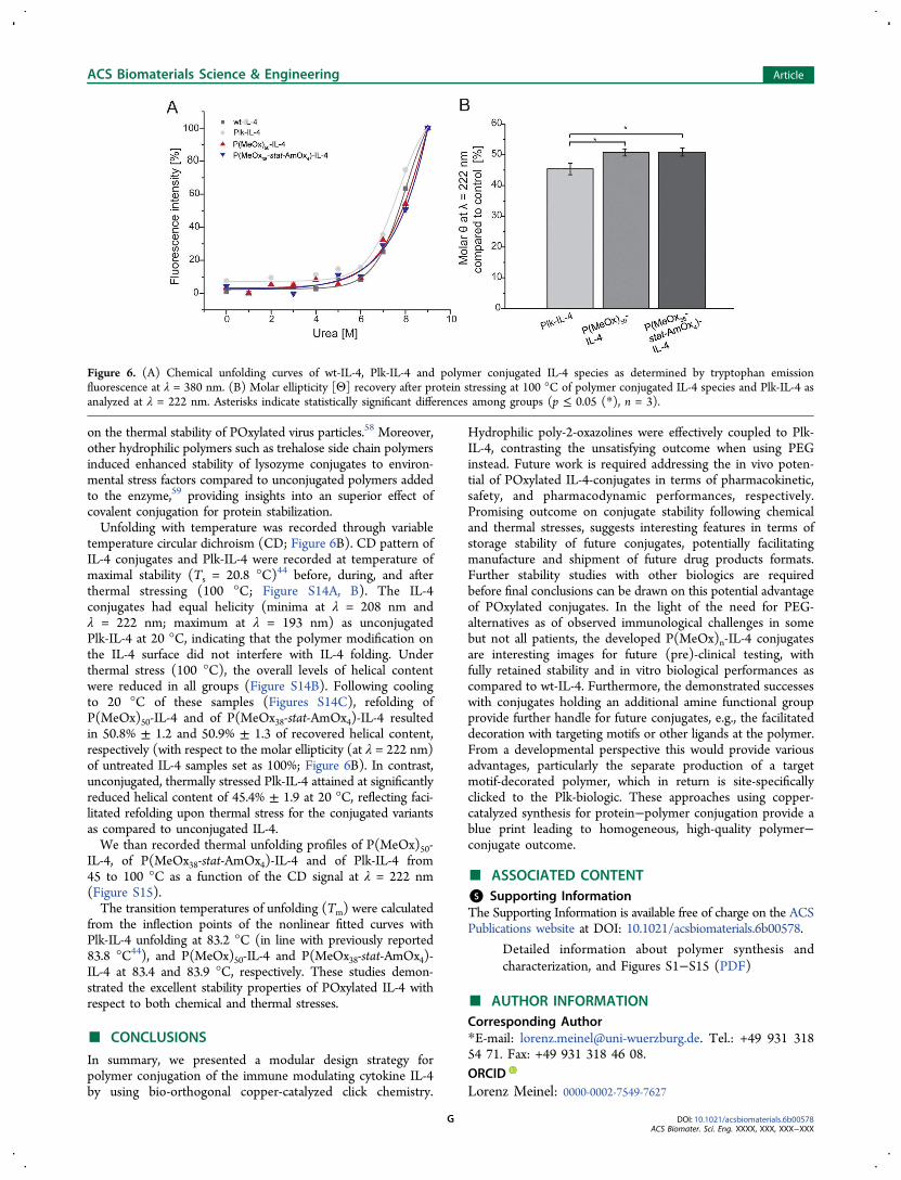
on the thermal stability of POxylated virus particles.58 Moreover,other hydrophilic polymers such as trehalose side chain polymersinduced enhanced stability of lysozyme conjugates to environ-mental stress factors compared to unconjugated polymers addedto the enzyme,59 providing insights into an superior effect ofcovalent conjugation for protein stabilization.Unfolding with temperature was recorded through variable
temperature circular dichroism (CD; Figure 6B). CD pattern ofIL-4 conjugates and Plk-IL-4 were recorded at temperature ofmaximal stability (Ts = 20.8 °C)44 before, during, and afterthermal stressing (100 °C; Figure S14A, B). The IL-4conjugates had equal helicity (minima at λ = 208 nm andλ = 222 nm; maximum at λ = 193 nm) as unconjugatedPlk-IL-4 at 20 °C, indicating that the polymer modification onthe IL-4 surface did not interfere with IL-4 folding. Underthermal stress (100 °C), the overall levels of helical contentwere reduced in all groups (Figure S14B). Following coolingto 20 °C of these samples (Figures S14C), refolding ofP(MeOx)50-IL-4 and of P(MeOx38-stat-AmOx4)-IL-4 resultedin 50.8% ± 1.2 and 50.9% ± 1.3 of recovered helical content,respectively (with respect to the molar ellipticity (at λ = 222 nm)of untreated IL-4 samples set as 100%; Figure 6B). In contrast,unconjugated, thermally stressed Plk-IL-4 attained at significantlyreduced helical content of 45.4% ± 1.9 at 20 °C, reflecting faci-litated refolding upon thermal stress for the conjugated variantsas compared to unconjugated IL-4.We than recorded thermal unfolding profiles of P(MeOx)50-
IL-4, of P(MeOx38-stat-AmOx4)-IL-4 and of Plk-IL-4 from45 to 100 °C as a function of the CD signal at λ = 222 nm(Figure S15).The transition temperatures of unfolding (Tm) were calculated
from the inflection points of the nonlinear fitted curves withPlk-IL-4 unfolding at 83.2 °C (in line with previously reported83.8 °C44), and P(MeOx)50-IL-4 and P(MeOx38-stat-AmOx4)-IL-4 at 83.4 and 83.9 °C, respectively. These studies demon-strated the excellent stability properties of POxylated IL-4 withrespect to both chemical and thermal stresses.
■ CONCLUSIONS
In summary, we presented a modular design strategy forpolymer conjugation of the immune modulating cytokine IL-4by using bio-orthogonal copper-catalyzed click chemistry.
Hydrophilic poly-2-oxazolines were effectively coupled to Plk-IL-4, contrasting the unsatisfying outcome when using PEGinstead. Future work is required addressing the in vivo poten-tial of POxylated IL-4-conjugates in terms of pharmacokinetic,safety, and pharmacodynamic performances, respectively.Promising outcome on conjugate stability following chemicaland thermal stresses, suggests interesting features in terms ofstorage stability of future conjugates, potentially facilitatingmanufacture and shipment of future drug products formats.Further stability studies with other biologics are requiredbefore final conclusions can be drawn on this potential advantageof POxylated conjugates. In the light of the need for PEG-alternatives as of observed immunological challenges in somebut not all patients, the developed P(MeOx)n-IL-4 conjugatesare interesting images for future (pre)-clinical testing, withfully retained stability and in vitro biological performances ascompared to wt-IL-4. Furthermore, the demonstrated successeswith conjugates holding an additional amine functional groupprovide further handle for future conjugates, e.g., the facilitateddecoration with targeting motifs or other ligands at the polymer.From a developmental perspective this would provide variousadvantages, particularly the separate production of a targetmotif-decorated polymer, which in return is site-specificallyclicked to the Plk-biologic. These approaches using copper-catalyzed synthesis for protein−polymer conjugation provide ablue print leading to homogeneous, high-quality polymer−conjugate outcome.
■ ASSOCIATED CONTENT
*S Supporting Information
The Supporting Information is available free of charge on the ACSPublications website at DOI: 10.1021/acsbiomaterials.6b00578.
Detailed information about polymer synthesis andcharacterization, and Figures S1−S15 (PDF)
■ AUTHOR INFORMATION
Corresponding Author
*E-mail: [email protected]. Tel.: +49 931 31854 71. Fax: +49 931 318 46 08.
ORCID
Lorenz Meinel: 0000-0002-7549-7627
Figure 6. (A) Chemical unfolding curves of wt-IL-4, Plk-IL-4 and polymer conjugated IL-4 species as determined by tryptophan emissionfluorescence at λ = 380 nm. (B) Molar ellipticity [Θ] recovery after protein stressing at 100 °C of polymer conjugated IL-4 species and Plk-IL-4 asanalyzed at λ = 222 nm. Asterisks indicate statistically significant differences among groups (p ≤ 0.05 (*), n = 3).
ACS Biomaterials Science & Engineering Article
DOI: 10.1021/acsbiomaterials.6b00578
ACS Biomater. Sci. Eng. XXXX, XXX, XXX−XXXG
Present Address#M.H. is currently at Department of Chemistry, University ofWarwick, Gibbet Hill Road, Coventry CV4 7AL, UK.
Notes
The authors declare no competing financial interest.
■ ACKNOWLEDGMENTS
Support by DFG (grant ME 3920/3-1 ‘Macrophage plasticitydeployed for efficient bone (re-) generation’), the Sino-Germancenter, and the Bundesministerium fur Bildung und Forschung(Germany) (13N13454) are gratefully acknowledged. We alsoacknowledge funding from the Carl-Zeiss Foundation (JCSMStrukturantrag) and the Thuringer Ministerium fur Wirtschaft,Wissenschaft, und Digitale Gesellschaft (TMWWDG, Pro-Exzellenz II, NanoPolar). M.H. gratefully acknowledges theGerman Research Foundation (DFG, GZ: HA 7725/1-1) forfunding. M.N.L. gratefully acknowledges the Bundesministe-rium fur Bildung und Forschung (Germany) (project: smart-dye-livery, 081220/127) for funding. The authors thank AnnettUrbanek for MALDI-MS measurements. We thankfully acknowl-edge the kind support by Caroline Kisker (University ofWurzburg) and her group with the CD measurements.
■ REFERENCES
(1) Alconcel, S. N. S.; Baas, A. S.; Maynard, H. D. FDA-approvedpoly(ethylene glycol)-protein conjugate drugs. Polym Chem 2011, 2(7), 1442−1448.(2) Knop, K.; Hoogenboom, R.; Fischer, D.; Schubert, U. S.Poly(ethylene glycol) in Drug Delivery: Pros and Cons as Well asPotential Alternatives. Angew. Chem., Int. Ed. 2010, 49 (36), 6288−6308.(3) Schottler, S.; Becker, G.; Winzen, S.; Steinbach, T.; Mohr, K.;Landfester, K.; Mailander, V.; Wurm, F. R. Protein adsorption isrequired for stealth effect of poly(ethylene glycol)- and poly-(phosphoester)-coated nanocarriers. Nat. Nanotechnol. 2016, 11 (4),372−377.(4) Chanan-Khan, A.; Szebeni, J.; Savay, S.; Liebes, L.; Rafique, N.M.; Alving, C. R.; Muggia, F. M. Complement activation following firstexposure to pegylated liposomal doxorubicin (Doxil): possible role inhypersensitivity reactions. Ann. Oncol 2003, 14 (9), 1430−1437.(5) Sroda, K.; Rydlewski, J.; Langner, M.; Kozubek, A.; Grzybek, M.;Sikorski, A. F. Repeated injections of PEG-PE liposomes generate anti-PEG antibodies. Cell Mol. Biol. Lett. 2005, 10 (1), 37−47.(6) Dams, E. T. M.; Laverman, P.; Oyen, W. J. G.; Storm, G.;Scherphof, G. L.; Van der Meer, J. W. M.; Corstens, F. H. M.;Boerman, O. C. Accelerated blood clearance and altered biodis-tribution of repeated injections of sterically stabilized liposomes. J.Pharmacol. Exp. Ther. 2000, 292 (3), 1071−1079.(7) Tagami, T.; Nakamura, K.; Shimizu, T.; Ishida, T.; Kiwada, H.The relationship between PEGylated siRNA-lipoplex and anti-PEGIgM on the induction of accelerated blood clearance (ABC)phenomenon. Yakugaku Zasshi 2008, 128, 109−110.(8) Ishida, T.; Kiwada, H. Accelerated blood clearance (ABC)phenomenon upon repeated injection of PEGylated liposomes. Int. J.Pharm. 2008, 354 (1−2), 56−62.(9) Veronese, F. M. Peptide and protein PEGylation: a review ofproblems and solutions. Biomaterials 2001, 22 (5), 405−17.(10) Pidhatika, B.; M?ller, J.; Benetti, E. M.; Konradi, R.;Rakhmatullina, E.; Muhlebach, A.; Zimmermann, R.; Werner, C.;Vogel, V.; Textor, M. The role of the interplay between polymerarchitecture and bacterial surface properties on the microbial adhesionto polyoxazoline-based ultrathin films. Biomaterials 2010, 31 (36),9462−9472.(11) Chen, Y.; Pidhatika, B.; von Erlach, T.; Konradi, R.; Textor, M.;Hall, H.; Luhmann, T. Comparative assessment of the stability ofnonfouling poly(2-methyl-2-oxazoline) and poly(ethylene glycol)
surface films: an in vitro cell culture study. Biointerphases 2014, 9(3), 031003.(12) Sedlacek, O.; Hruby, M.; Studenovsky, M.; Vetvicka, D.;Svoboda, J.; Kankova, D.; Kovar, J.; Ulbrich, K. Polymer conjugates ofacridine-type anticancer drugs with pH-controlled activation. Bioorg.Med. Chem. 2012, 20 (13), 4056−63.(13) von Erlach, T.; Zwicker, S.; Pidhatika, B.; Konradi, R.; Textor,M.; Hall, H.; Luhmann, T. Formation and characterization of DNA-polymer-condensates based on poly(2-methyl-2-oxazoline) graftedpoly(L-lysine) for non-viral delivery of therapeutic DNA. Biomaterials2011, 32 (22), 5291−303.(14) Rinkenauer, A. C.; Tauhardt, L.; Wendler, F.; Kempe, K.;Gottschaldt, M.; Traeger, A.; Schubert, U. S. A cationic poly(2-oxazoline) with high in vitro transfection efficiency identified by alibrary approach. Macromol. Biosci. 2015, 15 (3), 414−25.(15) Tong, J.; Luxenhofer, R.; Yi, X.; Jordan, R.; Kabanov, A. V.Protein modification with amphiphilic block copoly(2-oxazoline)s as anew platform for enhanced cellular delivery. Mol. Pharmaceutics 2010,7 (4), 984−92.(16) Mero, A.; Fang, Z. H.; Pasut, G.; Veronese, F. M.; Viegas, T. X.Selective conjugation of poly(2-ethyl 2-oxazoline) to granulocytecolony stimulating factor. J. Controlled Release 2012, 159 (3), 353−361.(17) He, Z. J.; Wan, X. M.; Schulz, A.; Bludau, H.; Dobrovolskaia, M.A.; Stern, S. T.; Montgomery, S. A.; Yuan, H.; Li, Z. B.; Alakhova, D.;Sokolsky, M.; Darr, D. B.; Perou, C. M.; Jordan, R.; Luxenhofer, R.;Kabanov, A. V. A high capacity polymeric micelle of paclitaxel:Implication of high dose drug therapy to safety and in vivo anti-canceractivity. Biomaterials 2016, 101, 296−309.(18) Luxenhofer, R.; Sahay, G.; Schulz, A.; Alakhova, D.; Bronich, T.K.; Jordan, R.; Kabanov, A. V. Structure-property relationship incytotoxicity and cell uptake of poly(2-oxazoline) amphiphiles. J.Controlled Release 2011, 153 (1), 73−82.(19) Zalipsky, S.; Hansen, C. B.; Oaks, J. M.; Allen, T. M. Evaluationof blood clearance rates and biodistribution of poly(2-oxazoline)-grafted liposomes. J. Pharm. Sci. 1996, 85 (2), 133−137.(20) Platen, M.; Mathieu, E.; Luck, S.; Schubel, R.; Jordan, R.; Pautot,S. Poly(2-oxazoline)-Based Microgel Particles for Neuronal CellCulture. Biomacromolecules 2015, 16 (5), 1516−24.(21) Moreadith, R. W.; Viegas, T. X.; Bentley, M. D.; Harris, J. M.;Fang, Z.; Yoon, K.; Dizman, B.; Weimer, R.; Rae, B. P.; Li, X.; Rader,C.; Standaert, D.; Olanow, W., Clinical development of a poly(2-oxazoline) (POZ) polymer therapeutic for the treatment ofParkinson’s disease − Proof of concept of POZ as a versatile polymerplatform for drug development in multiple therapeutic indications. Eur.Polym. J..201610.1016/j.eurpolymj.2016.09.052(22) Viegas, T. X.; Bentley, M. D.; Harris, J. M.; Fang, Z. F.; Yoon,K.; Dizman, B.; Weimer, R.; Mero, A.; Pasut, G.; Veronese, F. M.Polyoxazoline: Chemistry, Properties, and Applications in DrugDelivery. Bioconjugate Chem. 2011, 22 (5), 976−986.(23) Mero, A.; Pasut, G.; Via, L. D.; Fijten, M. W. M.; Schubert, U.S.; Hoogenboom, R.; Veronese, F. M. Synthesis and characterizationof poly(2-ethyl 2-oxazoline)-conjugates with proteins and drugs:Suitable alternatives to PEG-conjugates? J. Controlled Release 2008,125 (2), 87−95.(24) Glassner, M.; Maji, S.; de la Rosa, V. R.; Vanparijs, N.;Ryskulova, K.; De Geest, B. G.; Hoogenboom, R. Solvent-freemechanochemical synthesis of a bicyclononyne tosylate: a fast routetowards bioorthogonal clickable poly(2-oxazoline)s. Polym. Chem.2015, 6 (48), 8354−8359.(25) Totaro, K. A.; Liao, X.; Bhattacharya, K.; Finneman, J. I.; Sperry,J. B.; Massa, M. A.; Thorn, J.; Ho, S. V.; Pentelute, B. L. SystematicInvestigation of EDC/sNHS-Mediated Bioconjugation Reactions forCarboxylated Peptide Substrates. Bioconjugate Chem. 2016, 27, 994.(26) Agarwal, P.; Bertozzi, C. R. Site-specific antibody-drugconjugates: the nexus of bioorthogonal chemistry, protein engineering,and drug development. Bioconjugate Chem. 2015, 26 (2), 176−92.
ACS Biomaterials Science & Engineering Article
DOI: 10.1021/acsbiomaterials.6b00578
ACS Biomater. Sci. Eng. XXXX, XXX, XXX−XXXH
(27) Caliceti, P.; Veronese, F. M. Pharmacokinetic and biodis-tribution properties of poly(ethylene glycol)-protein conjugates. Adv.Drug Delivery Rev. 2003, 55 (10), 1261−1277.(28) Aravind, S.; Paul, W.; Vasudev, S. C.; Sharma, C. P.Polyethylene glycol (PEG) modified bovine pericardium as abiomaterial: a comparative study on immunogenicity. J. BiomaterAppl. 1998, 13 (2), 158−165.(29) Luhmann, T.; Meinel, L. Nanotransporters for drug delivery.Curr. Opin. Biotechnol. 2016, 39, 35−40.(30) Zhao, H.; Heusler, E.; Jones, G.; Li, L.; Werner, V.;Germershaus, O.; Ritzer, J.; Luehmann, T.; Meinel, L. Decoration ofsilk fibroin by click chemistry for biomedical application. J. Struct. Biol.2014, 186 (3), 420−30.(31) Luhmann, T.; Jones, G.; Gutmann, M.; Rybak, J. C.; Nickel, J.;Rubini, M.; Meinel, L. Bio-orthogonal Immobilization of FibroblastGrowth Factor 2 for Spatial Controlled Cell Proliferation. ACSBiomater. Sci. Eng. 2015, 1 (9), 740−746.(32) Wandrey, G.; Wurzel, J.; Hoffmann, K.; Ladner, T.; Buchs, J.;Meinel, L.; Luhmann, T. Probing unnatural amino acid integrationinto enhanced green fluorescent protein by genetic code expansionwith a high-throughput screening platform. J. Biol. Eng. 2016, 10 (1),11.(33) Luhmann, T.; Spieler, V.; Werner, V.; Ludwig, M. G.; Fiebig, J.;Muller, T.; Meinel, L. Interleukin-4 clicked surfaces drive M2macrophage polarization. ChemBioChem 2016, 17, 2123.(34) Gutmann, M.; Memmel, E.; Braun, A. C.; Seibel, J.; Meinel, L.;Luhmann, T. Biocompatible Azide-Alkyne ″Click″ Reactions forSurface Decoration of Glyco-Engineered Cells. ChemBioChem 2016,17 (9), 866−75.(35) Braun, A. C.; Gutmann, M.; Ebert, R.; Jakob, F.; Gieseler, H.;Luehmann, T.; Meinel, L. Matrix metalloproteinase responsive deliveryof myostatin inhibitors. Pharm. Res. 2016, DOI: 10.1007/s11095-016-2038-6.(36) Chazaud, B. Macrophages: supportive cells for tissue repair andregeneration. Immunobiology 2014, 219 (3), 172−8.(37) Milles, S.; Tyagi, S.; Banterle, N.; Koehler, C.; VanDelinder, V.;Plass, T.; Neal, A. P.; Lemke, E. A. Click strategies for single-moleculeprotein fluorescence. J. Am. Chem. Soc. 2012, 134 (11), 5187−95.(38) van Kimmenade, A.; Bond, M. W.; Schumacher, J. H.; Laquoi,C.; Kastelein, R. A. Expression, renaturation and purification ofrecombinant human interleukin 4 from Escherichia coli. Eur. J.Biochem. 1988, 173 (1), 109−14.(39) Wang, Y.; Shen, B. J.; Sebald, W. A mixed-charge pair in humaninterleukin 4 dominates high-affinity interaction with the receptoralpha chain. Proc. Natl. Acad. Sci. U. S. A. 1997, 94 (5), 1657−62.(40) Germershaus, O.; Schultz, I.; Luhmann, T.; Beck-Broichsitter,M.; Hogger, P.; Meinel, L. Insulin-like growth factor-I aerosolformulations for pulmonary delivery. Eur. J. Pharm. Biopharm. 2013,85 (1), 61−8.(41) Johnson, M. L.; Brand, L. Numerical Computer Methods, Part C;Methods in Enzymology; Academic Press: New York, 2000; Vol. 321.(42) Wingfield, P.; Payton, M.; Tavernier, J.; Barnes, M.; Shaw, A.;Rose, K.; Simona, M. G.; Demczuk, S.; Williamson, K.; Dayer, J. M.Purification and characterization of human interleukin-1 beta ex-pressed in recombinant Escherichia coli. Eur. J. Biochem. 1986, 160(3), 491−7.(43) Christen, E. H.; Karlsson, M.; Kampf, M. M.; Schoenmakers, R.;Gubeli, R. J.; Wischhusen, H. M.; Friedrich, C.; Fussenegger, M.;Weber, W. Conditional DNA-Protein Interactions Confer Stimulus-Sensing Properties to Biohybrid Materials. Adv. Funct. Mater. 2011, 21(15), 2861−2867.(44) Vaz, D. C.; Rodrigues, J. R.; Sebald, W.; Dobson, C. M.; Brito,R. M. M. Enthalpic and entropic contributions mediate the role ofdisulfide bonds on the conformational stability of interleukin-4. ProteinSci. 2006, 15 (1), 33−44.(45) Pace, C. N.; Shirley, B. A.; Thomson, J. A.; Measuring theconformational stability of a protein. In Protein Structure: A PracticalApproach; Oxford University Press: New York, 1997; 299−321.
(46) Volet, G.; Lav, T. X.; Babinot, J.; Amiel, C. Click-Chemistry: AnAlternative Way to Functionalize Poly(2-methyl-2-oxazoline). Macro-mol. Chem. Phys. 2011, 212 (2), 118−124.(47) Kempe, K.; Hoogenboom, R.; Jaeger, M.; Schubert, U. S. Three-Fold Metal-Free Efficient (″Click″) Reactions onto a MultifunctionalPoly(2-oxazoline) Designer Scaffold. Macromolecules 2011, 44 (16),6424−6432.(48) Hartlieb, M.; Pretzel, D.; Englert, C.; Hentschel, M.; Kempe, K.;Gottschaldt, M.; Schubert, U. S. Matrix supported poly(2-oxazoline)-based hydrogels for DNA catch and release. Biomacromolecules 2014,15 (6), 1970−8.(49) Presolski, S. I.; Hong, V. P.; Finn, M. G. Copper-CatalyzedAzide-Alkyne Click Chemistry for Bioconjugation. Curr. Protoc. Chem.Biol. 2011, 3 (4), 153−162.(50) Zheng, C.; Ma, G.; Su, Z. Native PAGE eliminates the problemof PEG-SDS interaction in SDS-PAGE and provides an alternative toHPLC in characterization of protein PEGylation. Electrophoresis 2007,28 (16), 2801−7.(51) Park, E. J.; Kim, M. S.; Lee, H. S.; Lee, K. C.; Na, D. H.Differences in electrophoretic behavior between linear and branchedPEG-conjugated proteins. Electrophoresis 2015, 36 (6), 918−923.(52) Hebert, K. R.; Adhikari, S.; Houser, J. E. Chemical mechanismof suppression of copper electrodeposition by poly (ethylene glycol). J.Electrochem. Soc. 2005, 152 (5), C324−C329.(53) Israelachvili, J. The different faces of poly(ethylene glycol). Proc.Natl. Acad. Sci. U. S. A. 1997, 94 (16), 8378−9.(54) Sheth, S. R.; Leckband, D. Measurements of attractive forcesbetween proteins and end-grafted poly(ethylene glycol) chains. Proc.Natl. Acad. Sci. U. S. A. 1997, 94 (16), 8399−8404.(55) Cho, H.; Daniel, T.; Buechler, Y. J.; Litzinger, D. C.; Maio, Z.;Putnam, A. M.; Kraynov, V. S.; Sim, B. C.; Bussell, S.; Javahishvili, T.;Kaphle, S.; Viramontes, G.; Ong, M.; Chu, S.; GC, B.; Lieu, R.;Knudsen, N.; Castiglioni, P.; Norman, T. C.; Axelrod, D. W.; Hoffman,A. R.; Schultz, P. G.; DiMarchi, R. D.; Kimmel, B. E. Optimized clinicalperformance of growth hormone with an expanded genetic code. Proc.Natl. Acad. Sci. U. S. A. 2011, 108 (22), 9060−9065.(56) Duppatla, V.; Gjorgjevikj, M.; Schmitz, W.; Hermanns, H. M.;Schafer, C. M.; Kottmair, M.; Muller, T.; Sebald, W. IL-4 analogueswith site-specific chemical modification at position 121 inhibit IL-4and IL-13 biological activities. Bioconjugate Chem. 2014, 25 (1), 52−62.(57) Lienemann, P. S.; Karlsson, M.; Sala, A.; Wischhusen, H. M.;Weber, F. E.; Zimmermann, R.; Weber, W.; Lutolf, M. P.; Ehrbar, M.A versatile approach to engineering biomolecule-presenting cellularmicroenvironments. Adv. Healthcare Mater. 2013, 2 (2), 292−6.(58) Manzenrieder, F.; Luxenhofer, R.; Retzlaff, M.; Jordan, R.; Finn,M. G. Stabilization of Virus-like Particles with Poly(2-oxazoline)s.Angew. Chem., Int. Ed. 2011, 50 (11), 2601−2605.(59) Mancini, R. J.; Lee, J.; Maynard, H. D. Trehalose Glycopolymersfor Stabilization of Protein Conjugates to Environmental Stressors. J.Am. Chem. Soc. 2012, 134 (20), 8474−8479.
ACS Biomaterials Science & Engineering Article
DOI: 10.1021/acsbiomaterials.6b00578
ACS Biomater. Sci. Eng. XXXX, XXX, XXX−XXXI
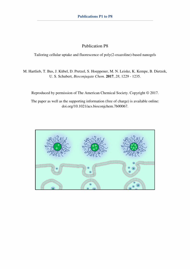
Publications P1 to P8
Publication P8
Tailoring cellular uptake and fluorescence of poly(2-oxazoline)-based nanogels
M. Hartlieb, T. Bus, J. Kübel, D. Pretzel, S. Hoeppener, M. N. Leiske, K. Kempe, B. Dietzek,
U. S. Schubert, Bioconjugate Chem. 2017, 28, 1229 - 1235.
Reproduced by permission of The American Chemical Society. Copyright © 2017.
The paper as well as the supporting information (free of charge) is available online:
doi.org/10.1021/acs.bioconjchem.7b00067.
Tailoring Cellular Uptake and Fluorescence of Poly(2-oxazoline)-Based Nanogels
Matthias Hartlieb,†,‡,#,¶ Tanja Bus,†,‡,# Joachim Kubel,§,∥,⊥ David Pretzel,†,‡ Stephanie Hoeppener,†,‡
Meike N. Leiske,†,‡ Kristian Kempe,†,‡,¶ Benjamin Dietzek,§,∥ and Ulrich S. Schubert*,†,‡
†Laboratory of Organic and Macromolecular Chemistry (IOMC), Friedrich Schiller University Jena, Humboldtstrasse 10, 07743,Jena, Germany‡Jena Center for Soft Matter (JCSM), Friedrich Schiller University Jena, Philosophenweg 7, 07743, Jena, Germany§Institute of Physical Chemistry (IPC) and Abbe Center of Photonics, Friedrich Schiller University Jena, Helmholtzweg 4, 07743Jena, Germany∥Leibniz Institute of Photonic Technology (IPHT), Albert-Einstein-Str. 9, 07745 Jena, Germany
*S Supporting Information
ABSTRACT: Controlling the size and charge of nanometer-sized objects is of upmost importance for their interactionswith cells. We herein present the synthesis of poly(2-oxazoline) based nanogels comprising a hydrophilic shell andan amine containing core compartment. Amine groups werecross-linked using glutaraldehyde resulting in imine basednanogels. As a drug model, amino fluorescein was covalentlyimmobilized within the core, quenching excessive aldehydefunctions. By varying the amount of cross-linker, the zetapotential and, hence, the cellular uptake could be adjusted.The fluorescence of the nanogels was found to be dependenton the cross-linking density. Finally, the hemocompatibility ofthe described systems was studied by hemolysis and erythrocyte aggregation assays. While cellular uptake was shown to bedependent on the zeta potential of the nanogel, no harmful effects to red blood cells was observed, rendering the present systemas an interesting toolbox for the production of nanomaterials with a defined biological interaction profile.
■ INTRODUCTION
Nanomedicine, the use of nanoscopic objects for biomedicalapplications such as diagnostics or treatment of diseases, hasattracted increasing interest in recent years.1,2 By using(polymeric) carriers, it is possible to solubilize, protect, anddeliver drug molecules to the desired site of action in the body.Nanogels, such as (reversibly) cross-linked polymer micelles,3
are particularly valuable in this context as, if the chemistry ischosen appropriately, premature drug release or disassemblycan be reduced.4 In the nanomedicine based treatment ofcancer, the enhanced permeability and retention (EPR) effect isused to generate a tumor specific accumulation of the drug.5
The concept exploits the leaky nature of tumor tissue and thepassive accumulation of nanosized objects within those cavities.However, in order to take advantage of the EPR effect, a drugcarrier has to exhibit long blood circulation times and a lowlevel of unspecific cellular interactions. Many parameters suchas size, shape, hydrophilicity or charge influence the cellularuptake,6,7 and with regard to new nanomedicines, the ability totailor the cellular interaction in an easy way is highly beneficial.It was shown that a positively charged surface significantlyincreases the uptake of nanoparticles.6,8−12 This effect is alsoused in gene therapy approaches in terms of a complexation of
negatively charged genetic material by positively chargedpolymers in order to penetrate cellular membranes.13 However,a positively charged surface usually also increases thecytotoxicity induced by the system.14,15 In addition, in thecontext of the EPR effect, a hydrophilic, low fouling surface isindispensable to maintain low protein adsorption levels. Poly(2-oxazoline)s (POx) display a promising material in a biomedicalcontext, as certain derivatives bearing small side chains, likepoly(2-methyl-2-oxazoline) (PMeOx) or poly(2-ethyl-2-oxazo-line) (PEtOx), show excellent biocompatibility.16−18 Indeed,their performance in biological applications is often comparedto poly(ethylene glycol) (PEG), since they also show a stealtheffect.19,20 Recent studies show that in terms of circulation timein the bloodstream and unspecific accumulation in the body,PEtOx is even more advantageous than PEG.21 Their versatilefunctionalization chemistry displays another advantage.22 Thereare sparse examples of POx based nanogels using PEtOx orPMeOx as a polymer shell23,24 and only a few were utilized forbiomedical applications.25,26
Received: February 7, 2017Revised: February 9, 2017Published: February 16, 2017
Article
pubs.acs.org/bc
© 2017 American Chemical Society 1229 DOI: 10.1021/acs.bioconjchem.7b00067Bioconjugate Chem. 2017, 28, 1229−1235
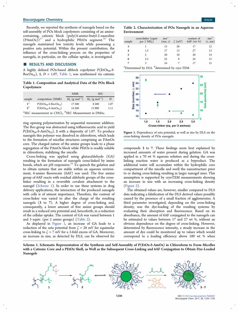
Recently, we reported the synthesis of nanogels based on theself-assembly of POx block copolymers consisting of an amine-containing, cationic block (poly(4-amino-butyl-2-oxazoline(PAmOx))27 and a hydrophilic PEtOx segment.26 Thenanogels maintained low toxicity levels while possessing apositive zeta potential. Within the present contribution, theinfluence of the cross-linking process on the properties ofnanogels, in particular, on the cellular uptake, is investigated.
■ RESULTS AND DISCUSSION
A highly defined POx-based diblock copolymer P(EtOx98-b-BocOx32), 1, Đ = 1.07, Table 1, was synthesized via cationic
ring opening polymerization by sequential monomer addition.The Boc-group was abstracted using trifluoroacetic acid to yieldP(EtOx98-b-AmOx32), 2 with a dispersity of 1.07. To producenanogels this polymer was dissolved in chloroform, which leadsto the formation of micellar structures comprising an PAmOxcore. The charged nature of the amine groups leads to a phasesegregation of the PAmOx block while PEtOx is readily solublein chloroform, stabilizing the micelle.Cross-linking was applied using glutaraldehyde (GA)
resulting in the formation of nanogels cross-linked by iminebonds, which are pH responsive.28 To quench the gelation andto obtain systems that are stable within an aqueous environ-ment, 6-amino fluorescein (6AF) was used. The free aminogroup of 6AF reacts with residual aldehyde groups of the cross-linker resulting in a reversible covalent attachment to thenanogel (Scheme 1). In order to use these systems in drugdelivery applications, the interaction of the produced nanogelswith cells is of utmost importance. Therefore, the content ofcross-linker was varied to alter the charge of the resultingnanogels (3 to 7). A higher degree of cross-linking and,consequently, a lower amount of free amine groups shouldresult in a reduced zeta potential and, henceforth, in a reductionof the cellular uptake. The content of GA was varied between 1and 3 equiv. (per 2 amine groups) (Table 2).As displayed in Figure 1, an increase of GA leads to a
reduction of the zeta potential from ζ = 28 mV for equimolarcross-linking to ζ = 7 mV for a 3-fold excess of GA. Moreover,an increase in size, as detected by DLS, can be observed for
compounds 3 to 7. These findings seem best explained byincreased amounts of water present during gelation. GA wasapplied in a 70 wt % aqueous solution and during the cross-linking reaction water is produced as a byproduct. Theadditional water will accumulate within the hydrophilic corecompartment of the micelle and swell the nanostructure priorto or during cross-linking resulting in larger nanogel sizes. Thisassumption is supported by cryoTEM measurements showingan increase in size with an increasing cross-linking density(Figure 2).The obtained values are, however, smaller compared to DLS
data indicating a falsification of the DLS derived values possiblycaused by the presence of a small fraction of agglomerates. Athird parameter investigated, depending on the cross-linkingdensity, was the dye-loading of the resulting systems byevaluating their absorption and fluorescence. Based on itsabsorbance, the amount of 6AF conjugated to the nanogels canbe estimated to values between 17 and 27 wt %, without anobvious dependence on the degree of cross-linking. However,determined by fluorescence intensity, a steady increase in theamount of dye could be monitored up to values which wouldcorrespond to a loading efficiency above 100 wt % when
Table 1. Composition and Analytical Data of the POx BlockCopolymers
NMR SEC
sample composition (NMR) Mn (g mol−1) Mn (g mol−1) Đ
1a P(EtOx98-b-BocOx32) 17 500 8 200 1.07
2b P(EtOx98-b-AmOx32) 14 200 13 900 1.11aSEC measurement in CHCl3.
bSEC Measurement in DMAc.
Scheme 1. Schematic Representation of the Synthesis and Self-Assembly of P(EtOx-b-AmOx) in Chloroform to Form Micelleswith a Cationic Core and a PEtOx Shell, as Well as the Subsequent Cross-Linking and 6AF Conjugation to Obtain Dye-LoadedNanogels
Table 2. Characterization of POx Nanogels in an AqueousEnvironment
samplecross-linker (equiv
per 2 NH2)sizea
(nm, r) ζ (mV)content of6AF (wt %)
sizeb
(r, nm)
3 1 13 28 17 12
4 1.5 17 13 27 13
5 2 20 10 20 14
6 2.5 22 8 24 15
7 3 24 7 17 15aDetermined by DLS. bdetermined by cryo-TEM.
Figure 1. Dependency of zeta potential, as well as size by DLS on thecross-linking density of POx nanogels.
Bioconjugate Chemistry Article
DOI: 10.1021/acs.bioconjchem.7b00067Bioconjugate Chem. 2017, 28, 1229−1235
1230
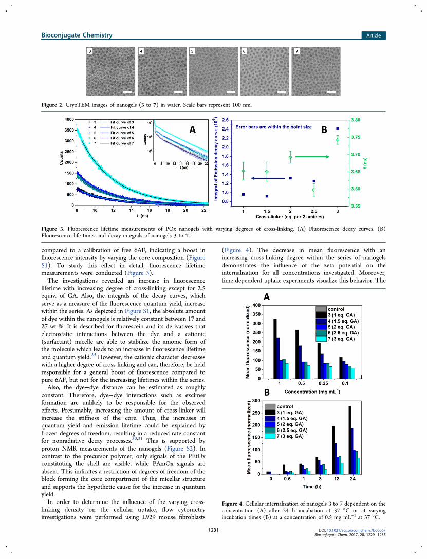
compared to a calibration of free 6AF, indicating a boost influorescence intensity by varying the core composition (FigureS1). To study this effect in detail, fluorescence lifetimemeasurements were conducted (Figure 3).The investigations revealed an increase in fluorescence
lifetime with increasing degree of cross-linking except for 2.5equiv. of GA. Also, the integrals of the decay curves, whichserve as a measure of the fluorescence quantum yield, increasewithin the series. As depicted in Figure S1, the absolute amountof dye within the nanogels is relatively constant between 17 and27 wt %. It is described for fluorescein and its derivatives thatelectrostatic interactions between the dye and a cationic(surfactant) micelle are able to stabilize the anionic form ofthe molecule which leads to an increase in fluorescence lifetimeand quantum yield.29 However, the cationic character decreaseswith a higher degree of cross-linking and can, therefore, be heldresponsible for a general boost of fluorescence compared topure 6AF, but not for the increasing lifetimes within the series.Also, the dye−dye distance can be estimated as roughly
constant. Therefore, dye−dye interactions such as excimerformation are unlikely to be responsible for the observedeffects. Presumably, increasing the amount of cross-linker willincrease the stiffness of the core. Thus, the increases inquantum yield and emission lifetime could be explained byfrozen degrees of freedom, resulting in a reduced rate constantfor nonradiative decay processes.30,31 This is supported byproton NMR measurements of the nanogels (Figure S2). Incontrast to the precursor polymer, only signals of the PEtOxconstituting the shell are visible, while PAmOx signals areabsent. This indicates a restriction of degrees of freedom of theblock forming the core compartment of the micellar structureand supports the hypothetic cause for the increase in quantumyield.In order to determine the influence of the varying cross-
linking density on the cellular uptake, flow cytometryinvestigations were performed using L929 mouse fibroblasts
(Figure 4). The decrease in mean fluorescence with anincreasing cross-linking degree within the series of nanogelsdemonstrates the influence of the zeta potential on theinternalization for all concentrations investigated. Moreover,time dependent uptake experiments visualize this behavior. The
Figure 2. CryoTEM images of nanogels (3 to 7) in water. Scale bars represent 100 nm.
Figure 3. Fluorescence lifetime measurements of POx nanogels with varying degrees of cross-linking. (A) Fluorescence decay curves. (B)Fluorescence life times and decay integrals of nanogels 3 to 7.
Figure 4. Cellular internalization of nanogels 3 to 7 dependent on theconcentration (A) after 24 h incubation at 37 °C or at varyingincubation times (B) at a concentration of 0.5 mg mL−1 at 37 °C.
Bioconjugate Chemistry Article
DOI: 10.1021/acs.bioconjchem.7b00067Bioconjugate Chem. 2017, 28, 1229−1235
1231

difference in the fluorescence intensity between the nanogelswas considered by referencing to the absolute fluorescenceintensity of the measurement. This finding is in agreement withliterature reports where objects having a positive net charge aredescribed to be taken up more efficiently as compared toneutral or anionic structures.32 The reported investigationshows that the cellular uptake, which displays a crucial factor forthe utilization as a drug delivery agent, can be fine-tuned for thepresented nanogel systems.In order to investigate the nature of the cellular internal-
ization, uptake studies at 4 °C were performed (Figure S3).The diminished uptake at low temperatures suggests an energydependent internalization via endocytosis as expected forobjects in such a size range.7,33
Besides cellular uptake, the biocompatibility of the drugcarriers represents an essential parameter. It was reported thatan increase in zeta potential of nanoparticles negatively affectsthe cell viability.14,15 Nanogel 3, investigated in a previousstudy, possesses the highest zeta potential (ζ = +28 mV) withinthe series and is, therefore, expected to induce the highesttoxicity, although the system did not interfere with themetabolism of L929 mouse fibroblasts in a negative way upto a concentration of 5 mg mL−1.26 While this is a promisingindication regarding the biocompatibility of the material, themost important environment a drug delivery system is facing isthe bloodstream. Long circulation times, leading to a passivetargeting, require a low level of interaction with thecomponents of the blood. An interaction with erythrocytesresulting in clotting or disruption is highly undesired. Thehemolytic activity of 6AF loaded nanogels was studieddepending on the applied concentration (Figure 5A). Allnanogels in a concentration range between 10 and 100 μg mL−1
resulted in hemolytic activity values well below 2%, which isdefined as the threshold for a hemolysis (according to theASTM F756−00 standard).Furthermore, the erythrocyte aggregation was investigated
and found to be negligible in the given concentration rangewith absorbance values comparable with the negative control(Figure 5B, Figure S4).These findings are remarkable, since positively charged
nanomaterials are expected to feature a decreased bloodcompatibility. In contrast to nanoparticle systems with analtered surface chemistry, the charge of the nanogels presentedherein results from amine groups within the core of the micellarstructure, whereas the periphery is covered with nonchargedPEtOx chains. While this setup enables tailoring of the cellularinteraction, as shown by the cellular uptake studies, thebiocompatibility of the nanogels is maintained in all cases.
■ CONCLUSION
Within this contribution, we present a straightforward syntheticroute to poly(2-oxazoline)-based polymeric nanogels with atailored cellular uptake. The gels are produced by phasesegregation of a diblock copolymer, containing a cationic and aneutral block forming micellar structures in chloroform. Cross-linking is conducted using glutaraldehyde and the fluorescentdye 6-amino fluorescein is loaded covalently. By changing thecross-linking density, it is possible to alter the properties of thenanogels in terms of fluorescence intensity and zeta potential.Hence, it is possible to adjust their cellular uptake as shown byflow cytometry measurements. Due to the unique nature of thenanogels, which carry the charged units within the core of themicellar structure, the biocompatibility is not affected by thevariation in charge as demonstrated by hemocompatibilityexperiments. Therefore, the herein presented material displaysa versatile toolbox for the production of drug delivery vehicles.Further studies will focus on the extension of the concept to invivo investigations as well as on the loading of anticancer drugssuch as doxorubicin, and the utilization of drug loaded nanogelsin vitro and in vivo.
■ EXPERIMENTAL SECTION
Material and Instrumentation. Chemicals and solventswere purchased from Sigma-Aldrich, Merck, Fluka, and Acros.2-Ethyl-2-oxazoline (EtOx) and methyl tosylate (MeOTos)were distilled to dryness prior to use. EtOx was dried usingbarium oxide before distillation. 2-(4-((tert-Butoxycarbonyl)-amino)butyl)-2-oxazoline (BocOx) was synthesized as de-scribed in a previous publication.27 If not stated otherwise,cell culture media and supplements (L-Glutamin, antibiotics)were obtained from Biochrom (Merck Millipore, Germany).The Initiator Sixty single-mode microwave synthesizer from
Biotage, equipped with a noninvasive IR sensor (accuracy: 2%),was used for polymerizations under microwave irradiation.Microwave vials were heated overnight to 110 °C and allowedto cool to room temperature under an argon atmosphere beforeuse. All polymerizations were carried out under temperaturecontrol. Size-exclusion chromatography (SEC) measurementsof the protected polymers were performed on a Shimadzusystem equipped with a SCL-10A system controller, a LC-10AD pump, a RID-10A refractive index detector, and a PSSSDV column with chloroform/triethylamine (NEt3)/iso-prop-anol (94:4:2) as eluent. The column oven was set to 50 °C.SEC of the deprotected statistical copolymers was performedon a Shimadzu system with a LC-10AD pump, a RID-10Arefractive index detector, a system controller SCL-10A, adegasser DGU-14A, and a CTO-10A column oven using N,N-
Figure 5. Induction of hemolysis (A) as well as erythrocyte aggregation (B) by 6AF loaded nanogels (3 to 7) in a concentration range between 10and 100 μg mL−1 using sheep blood of three different donor batches.
Bioconjugate Chemistry Article
DOI: 10.1021/acs.bioconjchem.7b00067Bioconjugate Chem. 2017, 28, 1229−1235
1232
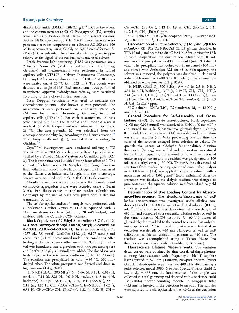
dimethylacetamide (DMAc) with 2.1 g L−1 LiCl as the eluentand the column oven set to 50 °C. Poly(styrene) (PS) sampleswere used as calibration standards for both solvent systems.Proton NMR spectroscopy (1H NMR) measurements wereperformed at room temperature on a Bruker AC 300 and 400MHz spectrometer, using CDCl3 or N,N-dimethylformamide(DMF)-D7 as solvents. The chemical shifts are given in ppmrelative to the signal of the residual nondeuterated solvent.Batch dynamic light scattering (DLS) was performed on a
Zetasizer Nano ZS (Malvern Instruments, Herrenberg,Germany). All measurements were performed in foldedcapillary cells (DTS1071, Malvern Instruments, Herrenberg,Germany). After an equilibration time of 180 s, 3 × 30 s runswere carried out at 25 °C (λ = 633 nm). The counts weredetected at an angle of 173°. Each measurement was performedin triplicate. Apparent hydrodynamic radii, Rh, were calculatedaccording to the Stokes−Einstein equation.Laser Doppler velocimetry was used to measure the
electrokinetic potential, also known as zeta potential. Themeasurements were performed on a Zetasizer Nano ZS(Malvern Instruments, Herrenberg, Germany) in foldedcapillary cells (DTS1071). For each measurement, 15 runswere carried out using the fast-field and slow-field reversalmode at 150 V. Each experiment was performed in triplicate at25 °C. The zeta potential (ζ) was calculated from theelectrophoretic mobility (μ) according to the Henry equation.34
The Henry coefficient, f(ka), was calculated according toOhshima.35
CryoTEM investigations were conducted utilizing a FEITecnai G2 20 at 200 kV acceleration voltage. Specisms werevitrified by a Vitrobot Mark V system on Quantifoil grids (R2/2). The blotting time was 1 s with blotting force offset of 0. Theamount of solution was 7 μL. Samples were plunge frozen inliquid ethane and stored under liquid nitrogen until transferredto the Gatan cryo-holder and brought into the microscope.Images were acquired with a 4k × 4k CCD Eagle camera.Absorbance and fluorescence spectra as well as hemolysis and
erythrocyte aggregation assays were recorded using a TecanM200 Pro fluorescence microplate reader (Crailsheim,Germany) by the use of black well plates with a flat andtransparent bottom.The cellular uptake studies of nanogels were performed with
a Beckmann Coulter Cytomics FC-500 equipped with aUniphase Argon ion laser (488 nm, 20 mW output) andanalyzed with the Cytomics CXP software.Block Copolymer of 2-Ethyl-2-oxazoline (EtOx) and 2-
(4-((tert-Butoxycarbonyl)amino)butyl)-2-oxazoline(BocOx) (P(EtOx-b-BocOx)), (1). In a microwave vial, EtOx(757 μL, 7.5 mmol), MeOTos (16.2 μL, 0.107 mmol) andacetonitrile (3.4 mL) were mixed under inert conditions. Afterheating in the microwave synthesizer at 140 °C for 25 min thevial was introduced into a glovebox with nitrogen atmosphereand BocOx (803 μL, 3.2 mmol) was added. The closed vial washeated again in the microwave synthesizer (140 °C, 20 min).The solution was precipitated in cold (−80 °C, 300 mL)diethyl ether. The white precipitate was filtered and dried inhigh vacuum (1.4 g, 92%).
1H NMR (CDCl3, 300 MHz): δ = 7.66, (d, 8.1 Hz, 0.019 H,tosylate), 7.14 (d, 8.21 Hz, 0.019 H, tosylate), 3.45 (s, 4 H,backbone), 3.10 (s, 0.58 H, CH2−CH2−NH (BocOx)), 2.50−2.15 (m, 1.96 H, CH2 (EtOx)/CH2−CH2−NHBoc), 1.62 (s,0.52 H, CH2−CH2−CH2 (BocOx)), 1.52 (s, 0.52 H, CH2−
CH2−CH2 (BocOx)), 1.42 (s, 2.3 H, CH3 (BocOx)), 1.21(s, 2.1 H, CH3 (EtOx)) ppm.SEC (eluent: CHCl3/iso-propanol/NEt3, PS-standard):
Mn = 8200 g mol−1, Đ = 1.07.Deprotection of P(EtOx-b-BocOx) (1) to yield (P(EtOx-
b-AmOx), (2). P(EtOx-b-BocOx) (1, 1.3 g) was dissolved inTFA (5 mL) and heated to 60 °C for 1 h. After stirring for 12 hat room temperature, the mixture was diluted with 10 mLmethanol and precipitated in 400 mL of cold (−80 °C) diethylether. The precipitate was redissolved in methanol (100 mL)and stirred with Amberlyst A21 for 48 h. Subsequently, thesolvent was removed, the polymer was dissolved in deionizedwater and freeze-dried (−80 °C, 0.003 mbar). The polymer wasobtained as white powder (1.2 g, 92%).
1H NMR (DMF-D7, 300 MHz): δ = 4.9 (s, 2.3 H, NH2),3.51 (s, 4 H, backbone), 3.07 (s, 0.49 H, CH2−CH2−NH2),2.44 (m, 2.1 H, CH2 (EtOx)/CH2−CH2−CO (AmOx)), 1.9−1.54 (m, 0.96 H, CH2−CH2−CH2−CH2 (AmOx)), 1.2 (s, 2,3H, CH3 (EtOx)) ppm.SEC (eluent: DMAc/LiCl, PS-standard): Mn = 13 900 g
mol−1, Đ = 1.11.General Procedure for Self-Assembly and Cross-
Linking (3−7). To create nanostructures, block copolymer(2, 90 mg, 0.006 mmol) was dissolved in CHCl3, (5 mg mL
−1)and stirred for 3 h. Subsequently, glutaraldehyde (30 mg,0.3 mmol, 1.5 equiv per amine (4)) was added and the solutionwas stirred another 3 h. With proceeding reaction time thecolor of the solution changed from colorless to yellow. Toquench the excess of aldehyde functionalities, 6-aminofluorescein (50 mg) was added and the mixture was stirredfor 12 h. Subsequently, the amount of solvent was reducedunder an argon stream and the residual was precipitated in 100mL cold diethyl ether (−80 °C). To purify the self-assembledstructures from residual capping agent and cross-linker, dialysisin MeOH/water (1:4) was applied using a membrane with amolar mass cut off of 3500 g mol−1 (Roth Zellutrans). After theextraction was finished, the dialysis medium was changed topure water and the aqueous solution was freeze-dried to yieldan orange powder.
Determination of Dye Loading Content by Absorb-ance/Fluorescence. The absorbance/fluorescence of 6AFloaded nanostructures was investigated under alkaline con-ditions (1 mol L−1 NaOH in water) in diluted solution (0.1 mgmL−1). The absorbance was determined at a wavelength of490 nm and compared to a sequential dilution series of 6AF inthe same aqueous NaOH solution. A 100-fold excess ofglutaraldehyde was added to the control to ensure that only theimine species of 6AF is present. Emission was detected at anexcitation wavelength of 450 nm. Nanogels as well as 6AFcalibration exhibit an emission maximum at 510 nm. Thereadout was accomplished using a Tecan M200 Profluorescence microplate reader (Crailsheim, Germany).
Fluorescence Lifetime Measurements. The emissiondecay curves were obtained by time-correlated-single-photon-counting. After excitation with a frequency-doubled Ti-sapphirelaser adjusted to 870 nm (Tsunami, Newport Spectra-PhysicsGmbH, pulse-to-pulse repetition rate 400 kHz after passing apulse selector, model 3980, Newport Spectra-Physics GmbH),i.e., at λex = 435 nm, the luminescence of the sample wascollected in a 90°-geometry and detected with a Becker & HicklPMC-100-4 photon-counting module. A long-pass filter(455 nm) is inserted in the detection beam path. The sampleswere adjusted to yield optical densities <0.03 at the excitation
Bioconjugate Chemistry Article
DOI: 10.1021/acs.bioconjchem.7b00067Bioconjugate Chem. 2017, 28, 1229−1235
1233
wavelength in aqueous NaOH (0.1 mol L−1). The measure-ments were accumulated at count rates <3% of the rep.-rateuntil 15 000 counts in the maximum were reached.Blood Compatibility Measurements. To assess the
hemolytic activity of the polymer solutions, blood from sheepcollected in heparinized tubes (Institute of Laboratory AnimalScience and Animal Welfare, Friedrich Schiller University Jena)was centrifuged at 4500 × g for 5 min, and the pellet waswashed three times with cold 1.5 mmol L−1 phosphate bufferedsaline (PBS, pH 7.4). After dilution with PBS in a ratio of 1:7,aliquots of erythrocyte suspension were mixed 1:1 with thepolymer solution and incubated in a water bath at 37 °C for 60min. After centrifugation at 2400 × g for 5 min the hemoglobinrelease was determined by measuring the absorbance of thesupernatant with a microplate reader at 544 nm wavelength.Complete hemolysis (100%) was achieved using 1% Triton X-100 serving as positive control. Thereby, PBS served as negativecontrol (0%). A value less than 2% hemolysis rate was taken asnonhemolytic. Experiments were run in triplicate and wereperformed with three different batches of donor blood.The hemolytic activity of the polycations was calculated as
follows (eq 1):
= ×
−A A
A%Hemolysis 100
( )Sample Negative control
Positive control (1)
For the examination of the erythrocyte aggregation, theerythrocyte suspension was mixed with the same volume ofpolymer solution in a clear flat-bottomed 96-well plate. Thecells were incubated at 37 °C for 2 h, and the absorbance wasmeasured at 645 nm in a microplate reader. 25 kDa bPEI (50μg mL−1) was used as positive control, and as negative control,cells were treated with PBS. Absorbance values of the testsolutions lower than negative control were regarded asaggregation. Experiments are the result of triplicates and wereperformed with three different donor blood batches.Investigation of the Cellular Uptake. The evaluation of
the nanogel uptake was performed with the cell line L929(CCL-1, ATCC). In general, the cells were cultured inDulbecco’s modified Eagle’s medium (DMEM) supplementedwith 10% fetal calf serum (FCS), 100 U mL−1 penicillin and100 μg mL−1 streptomycin at 37 °C in a humidified 5% CO2
atmosphere. For the uptake studies, cells were seeded at105 cells per mL in a 24-well plate and incubated for 24 h.For the time-dependent uptake studies, cells were incubated
with nanogels at a concentration of 0.5 mg mL−1 for 30 min to24 h, whereas the concentration-dependent uptake wasinvestigated over an incubation time of 24 h using nanogelconcentrations in the range between 0.1 and 1 mg mL−1. Cellsincubated with culture medium only served as control. Foruptake studies at low temperature, the cells were incubated withnanogels (0.5 mg mL−1) for 4 h at 4 and 37 °C, respectively,and the internalization was monitored using FC analysis asdescribed above.
■ ASSOCIATED CONTENT
*S Supporting Information
The Supporting Information is available free of charge on theACS Publications website at DOI: 10.1021/acs.bioconj-chem.7b00067.
Details on nanogel characterization regarding fluores-cence, cellular uptake and biocompatibility (PDF)
■ AUTHOR INFORMATION
Corresponding Author
*E-mail: [email protected].
ORCID
Ulrich S. Schubert: 0000-0003-4978-4670
Present Addresses¶Department of Chemistry, University of Warwick, Gibbet HillRoad, Coventry, CV4 7AL, United Kingdom.⊥Department of Chemistry and Molecular Biology Universityof Gothenburg 40530 Gothenburg, Sweden.
Author Contributions#Matthias Hartlieb and Tanja Bus contributed equally.
Notes
The authors declare no competing financial interest.
■ ACKNOWLEDGMENTS
T.B. acknowledges the German Federal Ministry of Education& Research (BMBF, #031A518B Vectura). CryoTEM inves-tigations were performed at the cryoTEM facilities of the JenaCenter for Soft Matter (JCSM). TEM facilities were funded bya grant of the DFG (German Research Foundation) and theEFRE (European Fund for Regional Development). MHgratefully acknowledges the German Research Foundation(DFG, GZ: HA 7725/1-1) for funding. U.S.S. and M.N.L.acknowledge German Federal Ministry of Education &Research (BMBF, #13NI3417, smart-dye-livery).
■ REFERENCES
(1) Duncan, R., and Vicent, M. J. (2013) Polymer therapeutics-prospects for 21st century: The end of the beginning. Adv. DrugDelivery Rev. 65, 60−70.(2) Duncan, R., and Gaspar, R. (2011) Nanomedicine(s) under theMicroscope. Mol. Pharmaceutics 8, 2101−2141.(3) van Nostrum, C. F. (2011) Covalently cross-linked amphiphilicblock copolymer micelles. Soft Matter 7, 3246−3259.(4) Talelli, M., Rijcken, C. J. F., Hennink, W. E., and Lammers, T.(2012) Polymeric micelles for cancer therapy: 3 C’s to enhanceefficacy. Curr. Opin. Solid State Mater. Sci. 16, 302−309.(5) Maeda, H., Greish, K., and Fang, J. (2006) The EPR effect andpolymeric drugs: A paradigm shift for cancer chemotherapy in the 21stcentury, in Polymer Therapeutics II (Satchi-Fainaro, R., and Duncan, R.,Eds.) pp 103−121, Springer, Berlin Heidelberg.(6) Zhu, M., Nie, G., Meng, H., Xia, T., Nel, A., and Zhao, Y. (2013)Physicochemical Properties Determine Nanomaterial Cellular Uptake,Transport, and Fate. Acc. Chem. Res. 46, 622−631.(7) Albanese, A., Tang, P. S., and Chan, W. C. W. (2012) The Effectof Nanoparticle Size, Shape, and Surface Chemistry on BiologicalSystems. Annu. Rev. Biomed. Eng. 14, 1−16.(8) Adjei, I. M., Sharma, B., and Labhasetwar, V. (2014)Nanoparticles: Cellular Uptake and Cytotoxicity, in Nanomaterial:Impacts on Cell Biology and Medicine (Capco, G. D., and Chen, Y.,Eds.) pp 73−91, Springer, Netherlands, Dordrecht.(9) Cho, E. C., Xie, J., Wurm, P. A., and Xia, Y. (2009)Understanding the Role of Surface Charges in Cellular Adsorptionversus Internalization by Selectively Removing Gold Nanoparticles onthe Cell Surface with a I2/KI Etchant. Nano Lett. 9, 1080−1084.(10) Hauck, T. S., Ghazani, A. A., and Chan, W. C. W. (2008)Assessing the Effect of Surface Chemistry on Gold Nanorod Uptake,Toxicity, and Gene Expression in Mammalian Cells. Small 4, 153−159.(11) Yue, Z.-G., Wei, W., Lv, P.-P., Yue, H., Wang, L.-Y., Su, Z.-G.,and Ma, G.-H. (2011) Surface Charge Affects Cellular Uptake andIntracellular Trafficking of Chitosan-Based Nanoparticles. Biomacro-molecules 12, 2440−2446.
Bioconjugate Chemistry Article
DOI: 10.1021/acs.bioconjchem.7b00067Bioconjugate Chem. 2017, 28, 1229−1235
1234

(12) He, C., Hu, Y., Yin, L., Tang, C., and Yin, C. (2010) Effects ofparticle size and surface charge on cellular uptake and biodistributionof polymeric nanoparticles. Biomaterials 31, 3657−3666.(13) Rinkenauer, A. C., Schubert, S., Traeger, A., and Schubert, U. S.(2015) The influence of polymer architecture on in vitro pDNAtransfection. J. Mater. Chem. B 3, 7477−7493.(14) Frohlich, E. (2012) The role of surface charge in cellular uptakeand cytotoxicity of medical nanoparticles. Int. J. Nanomed. 7, 5577−5591.(15) Xia, T., Kovochich, M., Liong, M., Meng, H., Kabehie, S.,George, S., Zink, J. I., and Nel, A. E. (2009) PolyethyleneimineCoating Enhances the Cellular Uptake of Mesoporous SilicaNanoparticles and Allows Safe Delivery of siRNA and DNAConstructs. ACS Nano 3, 3273−3286.(16) Kronek, J., Kronekova, Z., Luston, J., Paulovicova, E.,Paulovicova, L., and Mendrek, B. (2011) In vitro bio-immunologicaland cytotoxicity studies of poly(2-oxazolines). J. Mater. Sci.: Mater.Med. 22, 1725−1734.(17) Luxenhofer, R., Sahay, G., Schulz, A., Alakhova, D., Bronich, T.K., Jordan, R., and Kabanov, A. V. (2011) Structure-propertyrelationship in cytotoxicity and cell uptake of poly(2-oxazoline)amphiphiles. J. Controlled Release 153, 73−82.(18) Kronek, J., Paulovicova, E., Paulovicova, L., Kronekova, Z., andLuston, J. (2012) Immunomodulatory efficiency of poly(2-oxazolines).J. Mater. Sci.: Mater. Med. 23, 1457−1464.(19) Woodle, M. C., Engbers, C. M., and Zalipsky, S. (1994) NewAmphipatic Polymer-Lipid Conjugates Forming Long-CirculatingReticuloendothelial System-Evading Liposomes. Bioconjugate Chem.5, 493−496.(20) Zalipsky, S., Hansen, C. B., Oaks, J. M., and Allen, T. M. (1996)Evaluation of blood clearance rates and biodistribution of poly(2-oxazoline)-grafted liposomes. J. Pharm. Sci. 85, 133−137.(21) Wyffels, L., Verbrugghen, T., Monnery, B. D., Glassner, M.,Stroobants, S., Hoogenboom, R., and Staelens, S. (2016) μPETimaging of the pharmacokinetic behavior of medium and high molarmass 89Zr-labeled poly(2-ethyl-2-oxazoline) in comparison to poly-(ethylene glycol). J. Controlled Release 235, 63−71.(22) Guillerm, B., Monge, S., Lapinte, V., and Robin, J.-J. (2012)How to modulate the chemical structure of polyoxazolines byappropriate functionalization. Macromol. Rapid Commun. 33, 1600−1612.(23) Wilson, P., Ke, P. C., Davis, T. P., and Kempe, K. (2016)Poly(2-oxazoline)-based micro- and nanoparticles: A review. Eur.Polym. J., 1 DOI: 10.1016/j.eurpolymj.2016.09.011.(24) Hartlieb, M., Kempe, K., and Schubert, U. S. (2015) Covalentlycross-linked poly(2-oxazoline) materials for biomedical applications -from hydrogels to self-assembled and templated structures. J. Mater.Chem. B 3, 526−538.(25) Legros, C., Wirotius, A.-L., De Pauw-Gillet, M.-C., Tam, K. C.,Taton, D., and Lecommandoux, S. (2015) Poly(2-oxazoline)-BasedNanogels as Biocompatible Pseudopolypeptide Nanoparticles. Bio-macromolecules 16, 183−191.(26) Hartlieb, M., Pretzel, D., Wagner, M., Hoeppener, S., Bellstedt,P., Gorlach, M., Englert, C., Kempe, K., and Schubert, U. S. (2015)Core cross-linked nanogels based on the self-assembly of doublehydrophilic poly(2-oxazoline) block copolymers. J. Mater. Chem. B 3,1748−1759.(27) Hartlieb, M., Pretzel, D., Kempe, K., Fritzsche, C., Paulus, R. M.,Gottschaldt, M., and Schubert, U. S. (2013) Cationic poly(2-oxazoline) hydrogels for reversible DNA binding. Soft Matter 9,4693−4704.(28) Jackson, A. W., and Fulton, D. A. (2012) Triggering PolymericNanoparticle Disassembly through the Simultaneous Application ofTwo Different Stimuli. Macromolecules 45, 2699−2708.(29) Song, A., Zhang, J., Zhang, M., Shen, T., and Tang, J. a. (2000)Spectral properties and structure of fluorescein and its alkyl derivativesin micelles. Colloids Surf., A 167, 253−262.
(30) Humphry-Baker, R., Graetzel, M., and Steiger, R. (1980) Drasticfluorescence enhancement and photochemical stabilization of cyaninedyes through micellar systems. J. Am. Chem. Soc. 102, 847−848.(31) Wu, W.-C., Chen, C.-Y., Tian, Y., Jang, S.-H., Hong, Y., Liu, Y.,Hu, R., Tang, B. Z., Lee, Y.-T., Chen, C.-T., et al. (2010) Enhancementof Aggregation-Induced Emission in Dye-Encapsulating PolymericMicelles for Bioimaging. Adv. Funct. Mater. 20, 1413−1423.(32) Verma, A., and Stellacci, F. (2010) Effect of Surface Propertieson Nanoparticle−Cell Interactions. Small 6, 12−21.(33) Rejman, J., Oberle, V., Zuhorn, I. S., and Hoekstra, D. (2004)Size-dependent internalization of particles via the pathways of clathrin-and caveolae-mediated endocytosis. Biochem. J. 377, 159−169.(34) Delgado, A. V., Gonzalez-Caballero, F., Hunter, R. J., Koopal, L.K., and Lyklema, J. (2007) Measurement and interpretation ofelectrokinetic phenomena. J. Colloid Interface Sci. 309, 194−224.(35) Ohshima, H. (1994) A Simple Expression for Henry’s Functionfor the Retardation Effect in Electrophoresis of Spherical ColloidalParticles. J. Colloid Interface Sci. 168, 269−271.
Bioconjugate Chemistry Article
DOI: 10.1021/acs.bioconjchem.7b00067Bioconjugate Chem. 2017, 28, 1229−1235
1235
Related Documents


