The treatments for which proteins and peptides are prescribed as therapeutic agents require stable levels of active components over prolonged periods. One way to meet these requirements are sustained release systems, generally based on biodegradable po- lymers, of which polylactide (PLA) and its copolymers (PLGA) with glycolide (GA) are commonly used. An advantage of the lactide/glycolide copolymers is the well docu- mented versatility in polymer properties (via manipulation of the comonomer ratio, mo- lar mass, polymer crystallinity) and the corresponding performance characteristics (e.g., predictable in vivo degradation rates) (1, 2). In addition to the polymer chemistry, drug 215 Acta Pharm. 54 (2004) 215–229 Original research paper Poly(lactide-co-glycolide) microparticles as systems for controlled release of proteins – Preparation and characterization ALEKSANDRA PORJAZOSKA 1 KATERINA GORACINOVA 2 KRISTINA MLADENOVSKA 2 MARIJA GLAVA[ 2 MAJA SIMONOVSKA 2 EMILIJA IVANOVSKA JANJEVI] 3 MAJA CVETKOVSKA 1 * 1 Institute of Organic Technology Faculty of Technology and Metallurgy University »Ss Cyril and Methodius« Skopje, R. Macedonia 2 Institute of Pharmaceutical Technology Faculty of Pharmacy, University »Ss Cyril and Methodius« Skopje, R. Macedonia 3 Institute of Nuclear Medicine and Pathophysiology, Faculty of Medicine University »Ss Cyril and Methodius« Skopje, R. Macedonia Received February 9, 2004 Accepted July 16, 2004 Poly(DL-lactide-co-glycolide) (PDLLGA) and poly(L-lac- tide-co-glycolide) (PLLGA) copolymers were prepared by bulk ring opening polymerization of lactide and glyco- lide and characterized by GPC, FTIR, 1 H NMR and DSC. Copolymers with different molar masses at a constant lactide/glycolide ratio were used for preparation of bo- vine serum albumin (BSA)-loaded microparticles by the double emulsion w/o/w method. The influence of the co- polymer molar mass and composition on the microparti- cle morphology, size, yield, degradation rate, BSA-load- ing efficiency and BSA release profile were studied. For microparticles prepared from PDLLGA copolymers, a bi- phasic profile for BSA release was found and for those made from PLLGA copolymers the release profile was typically triphasic; both of them were characterized by high initial burst release. Possible reasons for such beha- vior are discussed. Keywords: poly(lactide-co-glycolide), microparticles, pro- tein, in vitro release * Correspondence, e-mail: majac@ereb1.mf.ukim.edu.mk; majac@ian.tmf.ukim.edu.mk

Welcome message from author
This document is posted to help you gain knowledge. Please leave a comment to let me know what you think about it! Share it to your friends and learn new things together.
Transcript

The treatments for which proteins and peptides are prescribed as therapeutic agentsrequire stable levels of active components over prolonged periods. One way to meetthese requirements are sustained release systems, generally based on biodegradable po-lymers, of which polylactide (PLA) and its copolymers (PLGA) with glycolide (GA) arecommonly used. An advantage of the lactide/glycolide copolymers is the well docu-mented versatility in polymer properties (via manipulation of the comonomer ratio, mo-lar mass, polymer crystallinity) and the corresponding performance characteristics (e.g.,predictable in vivo degradation rates) (1, 2). In addition to the polymer chemistry, drug
215
Acta Pharm. 54 (2004) 215–229 Original research paper
Poly(lactide-co-glycolide) microparticles as systemsfor controlled release of proteins – Preparation and
characterization
ALEKSANDRA PORJAZOSKA1
KATERINA GORACINOVA2
KRISTINA MLADENOVSKA2
MARIJA GLAVA[2
MAJA SIMONOVSKA2
EMILIJA IVANOVSKA JANJEVI]3
MAJA CVETKOVSKA1*
1 Institute of Organic TechnologyFaculty of Technology and MetallurgyUniversity »Ss Cyril and Methodius«Skopje, R. Macedonia
2 Institute of Pharmaceutical TechnologyFaculty of Pharmacy, University»Ss Cyril and Methodius«Skopje, R. Macedonia
3 Institute of Nuclear Medicine andPathophysiology, Faculty of MedicineUniversity »Ss Cyril and Methodius«Skopje, R. Macedonia
Received February 9, 2004
Accepted July 16, 2004
Poly(DL-lactide-co-glycolide) (PDLLGA) and poly(L-lac-tide-co-glycolide) (PLLGA) copolymers were prepared bybulk ring opening polymerization of lactide and glyco-lide and characterized by GPC, FTIR, 1H NMR and DSC.Copolymers with different molar masses at a constantlactide/glycolide ratio were used for preparation of bo-vine serum albumin (BSA)-loaded microparticles by thedouble emulsion w/o/w method. The influence of the co-polymer molar mass and composition on the microparti-cle morphology, size, yield, degradation rate, BSA-load-ing efficiency and BSA release profile were studied. Formicroparticles prepared from PDLLGA copolymers, a bi-phasic profile for BSA release was found and for thosemade from PLLGA copolymers the release profile wastypically triphasic; both of them were characterized byhigh initial burst release. Possible reasons for such beha-vior are discussed.
Keywords: poly(lactide-co-glycolide), microparticles, pro-tein, in vitro release
* Correspondence, e-mail: [email protected]; [email protected]

release rates can be controlled through variation of the microparticle formulation pa-rameters, and thus the physical characteristics of the resulting particles (3–9). For pro-teins with low or negligible solubility in polymers such as PLGA, the diffusional trans-port through the polymer phase will be prevented. In this case, polymer degradationwill play a crucial role in the mechanism of protein release (2, 10–12). Based on this find-ing tailoring of PLGA devices with suitable degradation properties that will allow a con-trolled release of high molar mass, water-soluble protein for long term therapeutic appli-cation has become an important area of research. Biodegradable microparticles have beenthe most studied devices due to their relatively simple fabrication and facile administra-tion to a variety of locations in vivo. Several methodologies for microencapsulation ofproteins and peptides have been developed. Most of them are essentially based on thephase separation technique, such as the solid-in-oil-in-water (s/o/w) method (6, 12) oremulsion-evaporation technique, such as the water-in-oil-in-water (w/o/w) method (7–9),that enable production of particles with a wide range of mean diameters.
The aim of this study was to evaluate the influence of copolymers’ (PDLLGA andPLLGA) molar mass and composition on the properties of bovine serum albumin (BSA)-loaded microparticles prepared by the w/o/w emulsion technique. The copolymers wereprepared by bulk ring-opening polymerization (13) and characterized by GPC, FTIR, 1HNMR and DSC analyses. The influence of the copolymer molar mass and compositionon the drug release rate and the rate of degradation of polymeric microparticles werealso investigated.
EXPERIMENTAL
Materials
Poly(DL-lactide-co-glycolide) (PDLLGA) and poly(L-lactide-co-glycolide) (PLLGA)were synthesized by bulk ring-opening polymerization (13) and characterized.
Albumin, bovine, fraction V, min. 96% was purchased from Sigma Chemical Co. (UK).Radiolabelling of BSA was performed by 131I obtained from Biointernational (France).Poly(vinyl alcohol) (PVA) (Mw 72 000 g mol–1, 98%, hydrolyzed) was supplied by Merck(Germany). All solvents were of HPLC grade and were supplied by Merck.
Synthesis of poly(lactide-co-glycolide) copolymers
PLGA copolymers were prepared by ring opening polymerization of lactide (LA)and glycolide (GA) in the presence of stannous octoate as catalyst (13). A solution ofstannous octoate in dry chloroform and a mixture of monomers were added to a reac-tion tube (molar ratio monomer to catalyst was 1000). The molar ratio of the feed DL-LA/GA was 85/15, and L-LA/GA was 75/25. The solvent was removed in vacuo, andthe tube was sealed and immersed in a silicon oil bath at 115 °C. At the end of polymer-ization (24 h), the product was dissolved in a small amount of chloroform and precipitedin an excess of methanol. Actual copolymer composition, that is the molar ratio of mo-nomers (DL-LA/GA or L-LA/GA) in copolymers, was found from 1H NMR analysis.For PDLLGA, it was 80/20, and for PLLGA, 68/32.
216
A. Porjazoska et al.: Poly(lactide-co-glycolide) microparticles as systems for controlled release of proteins – Preparation and characte-rization, Acta Pharm. 54 (2004) 215–229.

Copolymer characterization
The synthesized copolymers were characterized by FTIR, 1H NMR, GPC and DSC.The infrared spectra of copolymers were recorded on a Perkin Elmer 983 IR spectro-
meter (Perker Elmer, USA) at room temperature. The following IR peaks assigned to PLGAwere recorded (�, cm–1): 2997–2965 (CH2, CH3), 1759 (C=O), 1360–1450 (CH3), 750 (CH).
1H NMR spectra of the copolymers were obtained on a Bruker AC 200L spectrome-ter (Bruker, USA) at 200 MHz, in deuterated chloroform at 20 °C. 1H NMR data were asfollows: (CDCl3, �, ppm): 1.56 (3H,CH3), 4.77 (2H,CH2), 5.18 (H, OCHCH3). The compo-sition of copolymers was calculated from the ratio of absorbances at 4.77 and 5.18 ppm.
The molar masses of copolymers (mass average, Mr and number average, Mn) weredetermined by gel permeation chromatography (Waters, USA) using the Waters styragelcolumn HT6F and Waters 410 differential refractometer detector. THF was used as theeluting solvent at a flow rate of 1 mL min–1 and polystyrene standards were used for ca-libration purposes.
Thermal characterization of the copolymers was performed using a DuPont DSC910 Model (DuPont, USA) device. The samples were scanned from –140 °C to +140 °C, ata heating rate of 10 °C min–1. DSC samples were first heated under nitrogen to +140 °C,then quenched to –140 °C using liquid nitrogen. This heating/cooling cycle was repea-ted twice. The data were analyzed from the second heating run.
The main characteristics of the prepared copolymers are presented in Table I.
Preparation of PLGA microparticles containing bovine serum albumin
Microparticles were prepared using the w/o/w emulsion technique (14, 15) accord-ing to the following procedure. PLGA (0.05 g) was dissolved at room temperature in 1 mLof dichloromethane (DCM) and cooled at 4 °C. Aqueous solution of 131I-BSA (2%, m/V,0.5 mL) was sonicated into 1 mL solution of PLGA in DCM (5%, m/V) for 15 min to ob-tain the first (w/o) emulsion, which was then vortexed into 10 mL of cooled (4 °C) aque-ous PVA solution (2% m/V) to prepare the w/o/w emulsion. Stirring for 3 h at 600 rpm
217
A. Porjazoska et al.: Poly(lactide-co-glycolide) microparticles as systems for controlled release of proteins – Preparation and characte-rization, Acta Pharm. 54 (2004) 215–229.
Table I. Main characteristics of copolymers
CopolymerFeed ratio of
LA/GA (mol/mol)Composition on the basis
of 1H NMR (mol/mol)Mw PDI Tg (�C)
PDLLGA
85/15
40–41I 81/19 21 850 1.65II 80/20 44 700 1.68III 80/20 83 800 2.94
PLLGA75/25
471 68/32 44 050 2.302 68/32 88 000 1.95
Mw – weight average molar mass; PDI – polydispersity or heterogeneity index (PDI = Mw/Mn, where Mn is thenumber average molar mass); Tg – glass transition temperature

was applied to remove the solvent. The isolated microparticles were successively wa-shed with deionized water, collected by filtration (Sartorius Type 16692, Sartorius, Ger-many) and freeze-dried (200 Pa, –40 °C, Christ � 2–4, Bioblock Scientific, France). Mi-croparticles without BSA were referred to as »blank« ones.
Microparticle characterization
Size distribution analysis. – The mean geometric diameter and size distribution of thepopulation of BSA-loaded PLGA microparticles (before and after freeze-drying) dis-persed in doubly-distilled water were determined using laser diffractometry (Particlesize analysette DLAB/22, Fritsch, Germany). Three independent analyses consisting of100 repetitive measurements were performed.
Morphology studies. – Microparticles were placed on metal grids using double-sidedtape and coated with gold (thickness 2 nm) under vacuum (BAL-TEC MED 020 Coatingsystem, Balzers, Liechtenstein). The shape and surface characteristics of blank and BSA-loaded PLGA microparticles were determined by scanning electron microscopy (HitachiS-450, Hitachi, Japan).
Yield and BSA-loading determination. – The microparticle yield was determined as ra-tio between the mass of isolated, freeze-dried microparticles to the total initial mass ofthe polymer and BSA. The content of radiolabelled 131I-BSA entrapped in PLGA micro-particles was calculated as the percent of total radioactivity in the supernatant and in thefiltrate after microparticles isolation (»well« counter Scaler Type N529 D, EKCO Electro-nics, UK) (16, 17).
In vitro degradation of PLGA microparticles. – Dispersion of blank microparticles (1.5mg mL–1) was used to assess the in vitro degradation rate during incubation in phos-phate-buffered saline (PBS, pH 7.4, 37 °C, 75 strike min–1, horizontal shaker; Haake SWB20, Fisons, Germany). At regular time intervals, the dispersion was centrifuged at 8000rpm for 30 min (Jouan MR 22i, Jouan S. A., Centrifuge, France), the total mass ofmicroparticles isolated and freeze-dried to a constant mass was determined and the per-centage of remaining microparticles was calculated.
In vitro release of BSA from PLGA microparticles. – In vitro release studies were per-formed by suspending the microparticles in isotonic PBS (mg microparticles per mLbuffer: 1.5–2.0 mg mL–1) and by simulating the in vivo conditions as in biodegradationstudies. At each time interval, the 131I-BSA release was determined as the percent of totalradioactivity present in the microparticles and the supernatant. In order to determinethe initial and the last time release, the in vitro release profiles were described using theexponential function: M� – Mt/M = Ae–�t + Be–�t +…, where M� is the total amount ofBSA released, M and Mt is the amount of BSA at zero time and the amount of BSA re-leased at time t, A and B are system-characteristic constants, � and � are rate constantsfor the initial and later time release, obtained by semilogarithmic plots (16–18).
218
A. Porjazoska et al.: Poly(lactide-co-glycolide) microparticles as systems for controlled release of proteins – Preparation and characte-rization, Acta Pharm. 54 (2004) 215–229.
RESULTS AND DISCUSSION
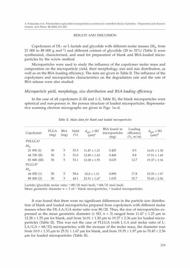
Copolymers of DL- or L-lactide and glycolide with different molar masses (Mw from21 000 to 88 000 g mol–1) and different content of glycolide (20 to 32%) (Table I) weresynthesized, characterized, and used for preparation of blank and BSA-loaded micro-particles by the w/o/w method.
Microparticles were used to study the influence of the copolymer molar mass andcomposition on the microparticle yield, their morphology, size and size distribution, aswell as on the BSA-loading efficiency. The data are given in Table II. The influence of thecopolymers and microparticles characteristics on the degradation rate and the rate ofBSA release were also studied.
Microparticle yield, morphology, size distribution and BSA-loading efficiency
In the case of all copolymers (I–III and 1–2, Table II), the blank microparticles werespherical and non-porous vs. the porous structure of loaded microparticles. Representa-tive scanning electron micrographs are given in Figs. 1a–d.
It was found that there were no significant differences in the particle size distribu-tion of blank and loaded microparticles prepared from copolymers with different molarmasses when the DL-LA/GA molar ratio was 80/20. Thus, the size of microparticles ex-pressed as the mean geometric diameter (� SD, n = 3) ranged from 11.47 � 1.25 �m to12.28 � 1.55 �m for blank, and from 16.01 � 1.30 �m to 19.37 � 2.36 �m for loaded micro-particles (Table II). This was not the case of PLLGA (with L-LA and molar ratio of L-LA/GA = 68/32) microparticles; with the increase of the molar mass, the diameter rosefrom 10.0 � 1.33 �m to 25.51 � 1.67 �m for blank, and from 19.35 � 1.97 �m to 70.45 � 2.56�m for loaded microparticles (Table II).
219
A. Porjazoska et al.: Poly(lactide-co-glycolide) microparticles as systems for controlled release of proteins – Preparation and characte-rization, Acta Pharm. 54 (2004) 215–229.
Table II. Main data for blank and loaded microparticles
CopolymerPLGA(mg)
BSA(mg)
Yield(%)
dgeo � SD(�m)c
BSA found inmicroparticles
(mg)
Loadingefficiency(%, m/m)
dgeo � SD(�m)d
PDLLGAa
Mw
21 850 (I) 50 5 53.3 11.47 � �� 0.425 8.5 16.01 � 1.3044 700 (II) 50 5 51.0 12.89 � 1.43 0.440 8.8 17.91 � �
83 800 (III) 50 5 53.1 12.28 � 1.55 0.635 12.7 19.37 � ��
PLLGAb
Mw
44 050 (1) 50 5 58.6 10.0 � �� 0.890 17.8 19.35 � ��
88 000 (2) 50 5 44.1 25.51 � 1.67 1.635 32.7 70.45 � 2.56
Lactide/glycolide molar ratio: a (80/20 mol/mol), b (68/32 mol/mol).Mean geometric diameter n = 3 of: c blank microparticles, d loaded microparticles.

Influence of the copolymer composition on particle size distribution was observedin the copolymers with the highest Mw. Thus, for copolymers with comparable Mw (�80 000 g mol–1) (copolymers III and 2, Table II), the mean geometric diameter for micro-particles of 68/32 L-LA/GA copolymer was 70.45 �m vs. 19.37 �m for microparticles of80/20 DL-LA/GA copolymer.
The microparticles yield was similar for all series of PLGA microparticles and nearly50% of the total initial mass. However, regarding the drug entrapment, a higher loadingefficiency was observed in the series with higher Mw and a higher content of GA (Table II).
A possible explanation for the increase of the microparticle size as well as the load-ing efficiency with the increase of the copolymers molar mass and the content of GA
220
A. Porjazoska et al.: Poly(lactide-co-glycolide) microparticles as systems for controlled release of proteins – Preparation and characte-rization, Acta Pharm. 54 (2004) 215–229.
Fig. 1. SEM of blank PLGA microparticles: a) copolymer II and b) copolymer 2 and BSA-loadedPLGA microparticles: c) copolymer II and d) copolymer 2. a) and c): bar stands for 10 �m;
b) and d): bar stands for 50 �m). (For microparticles see also Table II).
could be as follows. First, the copolymers with a higher molar mass (III and 2, Table II)provide a more viscous solution. Emulsification of a solution of high viscosity is moredifficult and leads to a larger size of microparticles prepared by the w/o/w solvent evapo-ration procedure. Higher viscosity may result in a faster microsphere hardening as well,and thus in a more difficult diffusion of BSA out of the microsphere into the outer waterphase. Second, PLLGA has a higher content of the hydrophilic segment in the backbone,which may interact with BSA and prevent BSA from diffusing out, resulting in higherencapsulation efficiency. A similar explanation was proposed by Yang et al. (4).
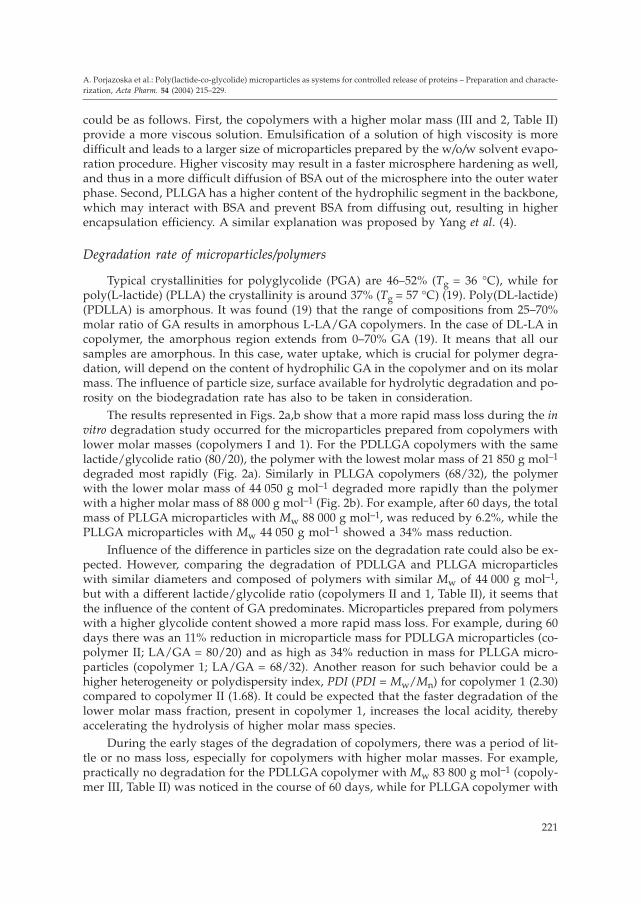
Degradation rate of microparticles/polymers
Typical crystallinities for polyglycolide (PGA) are 46–52% (Tg = 36 °C), while forpoly(L-lactide) (PLLA) the crystallinity is around 37% (Tg = 57 °C) (19). Poly(DL-lactide)(PDLLA) is amorphous. It was found (19) that the range of compositions from 25–70%molar ratio of GA results in amorphous L-LA/GA copolymers. In the case of DL-LA incopolymer, the amorphous region extends from 0–70% GA (19). It means that all oursamples are amorphous. In this case, water uptake, which is crucial for polymer degra-dation, will depend on the content of hydrophilic GA in the copolymer and on its molarmass. The influence of particle size, surface available for hydrolytic degradation and po-rosity on the biodegradation rate has also to be taken in consideration.
The results represented in Figs. 2a,b show that a more rapid mass loss during the invitro degradation study occurred for the microparticles prepared from copolymers withlower molar masses (copolymers I and 1). For the PDLLGA copolymers with the samelactide/glycolide ratio (80/20), the polymer with the lowest molar mass of 21 850 g mol–1
degraded most rapidly (Fig. 2a). Similarly in PLLGA copolymers (68/32), the polymerwith the lower molar mass of 44 050 g mol–1 degraded more rapidly than the polymerwith a higher molar mass of 88 000 g mol–1 (Fig. 2b). For example, after 60 days, the totalmass of PLLGA microparticles with Mw 88 000 g mol–1, was reduced by 6.2%, while thePLLGA microparticles with Mw 44 050 g mol–1 showed a 34% mass reduction.
Influence of the difference in particles size on the degradation rate could also be ex-pected. However, comparing the degradation of PDLLGA and PLLGA microparticleswith similar diameters and composed of polymers with similar Mw of 44 000 g mol–1,but with a different lactide/glycolide ratio (copolymers II and 1, Table II), it seems thatthe influence of the content of GA predominates. Microparticles prepared from polymerswith a higher glycolide content showed a more rapid mass loss. For example, during 60days there was an 11% reduction in microparticle mass for PDLLGA microparticles (co-polymer II; LA/GA = 80/20) and as high as 34% reduction in mass for PLLGA micro-particles (copolymer 1; LA/GA = 68/32). Another reason for such behavior could be ahigher heterogeneity or polydispersity index, PDI (PDI = Mw/Mn) for copolymer 1 (2.30)compared to copolymer II (1.68). It could be expected that the faster degradation of thelower molar mass fraction, present in copolymer 1, increases the local acidity, therebyaccelerating the hydrolysis of higher molar mass species.
During the early stages of the degradation of copolymers, there was a period of lit-tle or no mass loss, especially for copolymers with higher molar masses. For example,practically no degradation for the PDLLGA copolymer with Mw 83 800 g mol–1 (copoly-mer III, Table II) was noticed in the course of 60 days, while for PLLGA copolymer with
221
A. Porjazoska et al.: Poly(lactide-co-glycolide) microparticles as systems for controlled release of proteins – Preparation and characte-rization, Acta Pharm. 54 (2004) 215–229.
Mw 88 000 g mol–1 (copolymer 2, Table II) there was only a 6% reduction in microparticlemass. It means that only a decrease of the molar mass can be expected in this periodwhich was confirmed by GPC measurements (Figs. 2a and b).
Deterioration in microparticle surface morphology
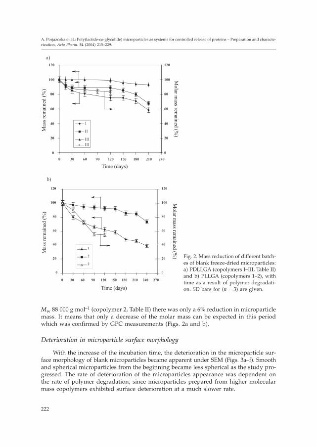
With the increase of the incubation time, the deterioration in the microparticle sur-face morphology of blank microparticles became apparent under SEM (Figs. 3a–f). Smoothand spherical microparticles from the beginning became less spherical as the study pro-gressed. The rate of deterioration of the microparticles appearance was dependent onthe rate of polymer degradation, since microparticles prepared from higher molecularmass copolymers exhibited surface deterioration at a much slower rate.
222
A. Porjazoska et al.: Poly(lactide-co-glycolide) microparticles as systems for controlled release of proteins – Preparation and characte-rization, Acta Pharm. 54 (2004) 215–229.
Fig. 2. Mass reduction of different batch-es of blank freeze-dried microparticles:a) PDLLGA (copolymers I–III, Table II)and b) PLLGA (copolymers 1–2), withtime as a result of polymer degradati-on. SD bars for (n = 3) are given.

223
A. Porjazoska et al.: Poly(lactide-co-glycolide) microparticles as systems for controlled release of proteins – Preparation and characte-rization, Acta Pharm. 54 (2004) 215–229.
Fig. 3. SEM of PDLLGA microparticles (with copolymer III): a) non-degraded, and b) after 7 monthsof degradation; PLLGA microparticles (with copolymer 1): c) non-degraded and d) after 7 monthsof degradation, and PLLGA microparticles (with copolymer 2): e) non-degraded and f) after 12months of degradation (bars stand for 50 �m). (For microparticles see also Table II).

In vitro release of BSA from microparticles
Due to the large molecular size of the proteins and their insolubility in polymers,the rate of BSA release from PLGA microparticles is thought to depend mainly on therate of polymer degradation and BSA diffusion through the microparticle pores. For allbatches of the microparticles studied, there was an initial »burst« release of BSA duringthe first hour of the study. This burst release probably represented the release of poorlyentrapped and surface-associated BSA. For the microparticles prepared from the PLLGA(68/32) copolymers, the BSA release showed a typical triphasic release profile (Fig. 4a),in which the initial release of BSA from microparticles prepared from copolymers withthe higher molar mass of 88 000 g mol–1 (larger mean particle size) was considerably lo-wer (� 18% during 0.5 hours) than that for the 44 050 g mol–1 polymer (� 52% in the
224
A. Porjazoska et al.: Poly(lactide-co-glycolide) microparticles as systems for controlled release of proteins – Preparation and characte-rization, Acta Pharm. 54 (2004) 215–229.
Fig. 4. The effect of the copolymer molarmass on the rate of release of BSA entrap-ped: a) PLLGA microparticles (with copoly-mers 1–2) and b) in PDLLGA microparticles(with copolymers I–III). SD bars for n = 3are given. (For microparticles see also Ta-ble II).

same period) (Table III). The same was true for the PDLLGA (80/20) particles, whichshowed a biphasic release profile (Fig. 4b) where the initial burst of BSA from micro-particles prepared from copolymers with lower molar masses of 21 850 and 44 700 g mol–1
was considerably higher than that for the 83 800 g mol–1 copolymer. Namely, between 31and 63% of BSA was released in the initial period of 0.5 hours, depending on the copoly-mer molar mass and/or particle size and porosity (Table III).
Lower initial release from microparticles prepared from polymers with higher Mw,or with a higher content of GA could be correlated with the already discussed influenceof the viscosity of the polymer solutions (faster hardening of microparticles with a morecompact core) or interactions of the hydrophilic segments with BSA during the encapsu-lation which prevents the BSA from diffusing out. Thus the microparticles with a higherdiameter or prepared from polymers with higher Mw or a higher content of hydrophiliccomponent will show a lower burst effect and slower release.
After the initial burst release from PLLGA (68/32) microparticles, there was a lagphase of low release, followed by a phase of constant BSA release in which, during 25hours, 87% (copolymer 1) or 65% (copolymer 2) BSA was released (Fig. 4a). In 10 daysthe BSA was released completely (data not shown graphically) (Table III).
For PDLLGA (80/20) microparticles after the initial release, there was a phase ofslow and constant release of BSA for a period of 7 days. Data are shown only for the first25 hours. Namely, after 25 hours 88% (copolymer I), 85% (copolymer II) or 75% (copoly-mer III) of BSA was released (Fig. 4b), and in 7 days the BSA was released completely(Table III).
225
A. Porjazoska et al.: Poly(lactide-co-glycolide) microparticles as systems for controlled release of proteins – Preparation and characte-rization, Acta Pharm. 54 (2004) 215–229.
Table III. Kinetic parameters for the BSA release from PLLGA and PDLLGA microparticles
CopolymerInitial »burst«
releaseLag phase oflow release
Third phase ofconstant release
R1a K1 (h–1) R2
b K2 (h–1) R3c K3 (h–1)
PLLGA (68/32)Mw (g mol–1)44 050 (1)88 000 (2)
1.0001.000
1.4920.397
0.9940.984
0.1650.056
0.9740.910
0.0090.006
PDLLGA (80/20)Mw (g mol–1)21 850 (I)44 700 (II)83 800 (III)
1.0001.0001.000
1.9721.6650.753
–––
–––
0.9610.9600.963
0.0160.0220.021
a Exponential dependence of the semilogarithmic plot on the amount of drug remaining vs. time in the initial»burst« release
b Exponential dependence of the semilogarithmic plot on the amount of drug remaining vs. time in the lagphase of release
c Exponential dependence of the semilogarithmic plot on the amount of drug remaining vs. time in the phaseof constant of release
R1–R3 – respective correlation coefficientsK1 – rate constant for the initial »burst« release, determined from the biexponential function.K2 – rate constant for the lag phase release, determined from the biexponential function.K3 – rate constant for the phase of constant release, determined from the biexponential function.

In the early phase including the lag phase, the protein release is governed by a dif-fusion-controlled mechanism through a network of water filled pores and channels. Inthe later phase, erosion of the polymer matrices is considered to control protein releasefrom the core of microparticles (20). This explanation could be correlated with the hy-drophobic/hydrophilic properties of the two series of microparticles, with the micropar-ticle sizes and with the character of molecular mass changes during the blank micro-particles degradation process (Figs. 2a,b).
Another serious problem in delivering protein pharmaceuticals is their inherentphysical and chemical instability. Potential sources of irreversible deactivation of pro-teins encapsulated in PLGA microparticles are the following (12). First, exposure to or-ganic solvents, which is a »stress« factor for proteins, during microparticles formulation;second, increased levels of moisture providing higher protein mobility; third, an acidicenvironment produced by acidic degradation products and carboxylic acid end groupsof PLGA; and fourth, adsorption of the protein to the polymer surface, which may cata-lyze protein conformation changes from �-helix into �-sheet, resulting in its aggregation,and thus irreversible protein activity loss.
The impact of the encapsulation procedure on the secondary structure of BSA waspreliminarily assessed using FTIR spectroscopy, from the amide I region. Technical andtheoretical limitations enable determination of only three regions of vibrations, amide I,II and III, of the existing nine (A, B, I–III). The fact that 90% of IR investigations of thesecondary protein structure are referred to amide I region (C=O, CN, CCN def.) resultedin an analysis of only this spectral region (21, 22). According to Jackson et al. (21), thefree amide I vibrations are expected at 1670 cm–1, while vibrations at 1695 cm–1 are a re-sult of a strong transitional dipole bonding in �-conformations. Thus, vibrations for theprotein amide I region appear between 1700 and 1600 cm–1.
The secondary structure of BSA from the amide I region, according to the methodssuggested by Byler and Susi (23), as well as Prestrelski et al. (24) and Bramanti and Be-nedetti (25), was determined after the Fourier-deconvolution of FTIR spectra, using aniterative curve fitting procedure of the Gauss-Lorenzian function. The content of �-helixwas selected as a solid parameter for structural integrity, and it was calculated using thepeak area in the characteristic frequency region (1660–1653 cm–1). Our preliminary re-sults showed that in the microparticles obtained from all copolymers, the secondarystructure of BSA was preserved up to 30%.
Our further research is focused on the investigation of the influence of various stagesor various procedures for microparticles preparation on the encapsulated protein activity.
CONCLUSIONS
Poly(lactide-co-glycolide) copolymers with different molar masses and a differentmolar ratio of DL-lactide or L-lactide to glycolide were used for the preparation of blankand BSA loaded microparticles by the w/o/w method. It was found that the copolymersmolar mass and composition influence the mean geometric diameter of blank and load-ed microparticles, as well as their loading efficiency. This was correlated with the viscosityof the copolymer solutions and the specific interactions with the hydrophilic segmentsin the copolymer backbone.
226
A. Porjazoska et al.: Poly(lactide-co-glycolide) microparticles as systems for controlled release of proteins – Preparation and characte-rization, Acta Pharm. 54 (2004) 215–229.

The BSA release profiles, triphasic for microparticles of PLLGA and biphasic forthose of PDLLGA, were correlated with the microparticles morphology, size and porosi-ty, as well as with the hydrophobic/hydrophilic properties and molar masses of copoly-mers and their degradation rates (mass loss and molar mass reduction).
Preliminary analyses of the FTIR spectra of BSA-loaded microparticles, in the amideI region, showed that the secondary protein structure was preserved to about 30%, onthe basis of the calculated �-helix content. However, detailed investigations of the mag-nitude of different stages of the procedure-induced protein structural perturbations dur-ing encapsulation are in progress.
Acknowledgements. – This work was supported by the Ministry of Science of the Republic ofMacedonia and the Turkish Scientific and Technical Research Council (TUBITAK)-Marmara Re-search Center, Turkey.
Abbreviations. – DL-LA – DL-lactide, L-LA – L-lactide, PLA – poly(lactide), where lactide is L-or DL, PDLLA – poly(DL-lactide), PLLA – poly(L-lactide), PGA – polyglycolide, PLGA – poly(lac-tide-co-glycolide), where lactide is L- or DL, PDLLGA – poly(DL-lactide-co-glycolide), PLLGA –poly(L-lactide-co-glycolide), PVA – poly(vinyl alcohol)
REFERENCES
1. C. Berkland, K. K. Kim and D. W. Pack, Fabrication of PLG microspheres with precisely con-trolled and monodisperse size distributions, J. Control. Rel. 73 (2001) 59–74.
2. S. Cohen, T. Yoshioka, M. Lucarelli, L. H. Hwang and R. Langer, Controlled delivery systemsfor protein based on poly(lactic/glycolic acid) microspheres, Pharm. Res. 8 (1991) 713–720.
3. S. K. Sahoo, J. Panyam, S. Prabha and V. Labhasetwar, Residual polyvinyl alcohol associatedwith poly(D,L-lactide-co-glycolide) nanoparticles affects their physical properties and cellularuptake, J. Control. Rel. 82 (2002) 105–114.
4. Y. Y. Yang, T. Sh. Chung, X. L. Bai and W. Kh. Chang, Effect of preparation conditions on mor-phology and release profiles of biodegradable polymeric microspheres containing protein fabri-cated by double-emulsion method, Chem. Eng. Sci. 55 (2000) 2223–2236.
5. C. Berkland, M. King, A. Cox, K. K. Kim and D. W. Pack, Precise control of PLG microspheresize provides enhanced control of drug release rate, J. Control. Rel. 82 (2002) 137–147.
6. Sh. Takada, Y. Yamagata, M. Misaki, K. Taira and T. Kurokawa, Sustained release of humangrowth hormone from microcapsules prepared by a solvent evaporation technique, J. Control.Rel. 88 (2003) 229–242.
7. X. L. Bai, Y. Y. Yang, T. Sh. Chung, S. Ng and J. Heller, Effect of polymer composition on the fab-rication of poly(ortho-ester) microspheres for controlled release of protein, J. Appl. Polym. Sci. 80(2001) 1630–1642.
8. F. Boury, H. Marchais, J. E. Proust and J. P. Benoit, Bovine serum albumin release from poly-(�-hydroxy acid) microspheres: effect of polymer molecular weight and surface properties, J.Control. Rel. 45 (1997) 75–86.
9. K. J. Zhu, H. L. Jiang, X. Y. Du, J. Wang, W. X. Xu and S. F. Liu, Preparation and characterizationof hCG-loaded polylactide or poly(lactide-co-glycolide) microspheres using a modified water-in-oil-in-water (w/o/w) emulsion solvent evaporation technique, J. Microencapsul. 18 (2001) 247–260.
10. R. E. Eliaz and J. Kost, Characterization of a polymeric PLGA-injectable implant delivery sys-tem for the controlled release of proteins, J. Biomed. Mater. Res. 50 (2000) 388–396.
227
A. Porjazoska et al.: Poly(lactide-co-glycolide) microparticles as systems for controlled release of proteins – Preparation and characte-rization, Acta Pharm. 54 (2004) 215–229.

11. D. T. O’ Hagan, H. Jefery and S. S. Davis, The preparation and characterization of poly(lacti-de-co-glycolide) microparticles: III. Microparticle/polymer degradation rates and in vitro relea-se of a model protein, Int. J. Pharm. 103 (1994) 37–45.
12. I. J. Castellanos, K. G. Carrasquillo, J. de Jesus Lopez, M. Alvarez and K. Griebnow, Encapsula-tion of bovine serum albumin in poly(lactide-co-glycolide) microspheres by the solid-in-oil-in-water technique, J. Pharm. Pharmacol. 53 (2001) 167–178.
13. A. Porjazoska, N. Kayaman-Apohan, O. Karal-Yilmaz, M. Cvetkovska, K. Baysal and B. M.Baysal, Synthesis and characterization of glycolide, L-lactide, and PDMS-based terpolymers asa support for cell cultures, J. Biomater. Sci. Polym. Ed. 13 (2002) 1119–1134.
14. J. W. Kostanski and P. P. deLuca, A novel in vitro release technique for peptide-containing bio-degradable microspheres, AAPS Pharm. Sci. Tech. 1 (2000) 1–16.
15. I. Soriano, E. Carmen and M. Labres, Preparation and evaluation of insulin-loaded poly(DL-lactide) microspherers using an experimental design, Eur. J. Pharm. 2 (1996) 135–142.
16. K. Mladenovska, Lj. Klisarova, E. I. Janjevic and K. Goracinova, BSA loaded gelatin microspheres:Preparation and drug release rate in the presence of collagenase, Acta Pharm. 52 (2002) 91–101.
17. K. Mladenovska, E. I. Janjevic, M. D. Glavas, E. E. Kumbaradzi and K. Goracinova, Biodistri-bution of 131I-BSA loaded gelatin microspheres after peroral application to BALB/c mice – Par-ticle size study, Acta Pharm. 53 (2003) 187–197.
18. M. I. Ugwoke, N. Verbeke and R. Kinget, Microencapsulation of amorphine HCl with gelatin,Int. J. Pharm. 148 (1997) 23–32.
19. D. K. Gilding and A. M. Reed, Biodegradable polymers for use in surgery-polyglycolic/poly-(lactic acid) homo- and copolymers: 1. Polymer 20 (1979) 1459–1464.
20. T. Morita, Y. Horikiri, T. Suzuki and H. Yoshino, Applicability of various amphiphilic polymersto the modification of protein release kinetics from biodegradable reservoir-type microspheres,Eur. J. Pharm. Biopharm. 51 (2001) 45–53.
21. M. Jackson and H. H. Mantsch, The use and misuse of FTIR spectroscopy in the determinationof protein structure, Crit. Rev. Biochem. Mol. Biol. 30 (1995) 95–120.
22. K. G. Carrasquillo, J. C. Aponte Caro, A. Alejandro, D. Diaz Toro and K. Grebenow, Reductionof structural perturbations in BSA by non-aqueous microencapsulation, J. Pharm. Pharmacol. 53(2001) 115–120.
23. D. M. Byler and H. Susi, Examination of the secondary structure of proteins by deconvolvedFTIR spectra, Biopolymers 25 (1986) 496–487.
24. S. J. Prestrelski, N. Tedischi, T. Arakawa and J. F. Carpenter, Dehydration-induced conformati-onal transitions in proteins and their inhibition by stabilizers, Biophys. J. 65 (1993) 661–671.
25. E. Bramanti and E. Benedetti, Determination of the secondary structure of isomeric forms of hu-man serum albumin by a particular frequency deconvolution procedure applied to Fouriertransform IR analysis, Biopolymers 38 (1996) 639–653.
228
A. Porjazoska et al.: Poly(lactide-co-glycolide) microparticles as systems for controlled release of proteins – Preparation and characte-rization, Acta Pharm. 54 (2004) 215–229.
S A @ E T A K
Poli(laktid-ko-glikolid) mikro~estice kao sustavi za kontroliranoosloba|anje proteina – Priprava i karakterizacija
ALEKSANDRA PORJAZOSKA, KATERINA GORACINOVA, KRISTINA MLADENOVSKA, MARIJA GLAVA[,MAJA SIMONOVSKA, EMILIJA IVANOVSKA JANJEVI] i MAJA CVETKOVSKA
Poli(DL-laktid-ko-glikolid) (PDLLGA) i poli(L-laktid-ko-glikolid) (PLLGA) kopolimeriprire|eni su polimerizacijom laktida i glikolida uz otvaranje prstenova i karakteriziranipomo}u GPC, FTIR, 1H NMR i DSC. Kopolimeri razli~itih molarnih masa i stalnog omje-ra laktida i glikolida upotrebljeni su za pripravu mikro~estica s gove|im serumskimalbuminom (BSA) metodom dvostruke emulzije tipa voda/ulje/voda. Prou~avan je utjecajmolarne mase i sastava kopolimera na oblik, veli~inu, iskori{tenje i stupanj razgradnjemikro~estica, uklapanje i osloba|anje BSA. Za mikro~estice pripravljene s PDLLGA ko-polimerom utvr|en je bifazi~ni profil osloba|anja BSA, a za mikro~estice s PLLGA ko-polimerom trifazi~an profil. Za obje vrste karakteristi~no je brzo po~etno osloba|anje.Razmatrani su mogu}i uzroci takvog pona{anja.
Klju~ne rije~i: poli(laktid-ko-glikolid), mikro~estice, protein, in vitro osloba|anje
Institute of Organic Technology, Faculty of Technology and Metallurgy, University »Ss Cyril andMethodius«, Skopje, R. Macedonia
Institute of Pharmaceutical Technology, Faculty of Pharmacy, University »Ss Cyril and Methodius«,Skopje, R. Macedonia
Institute of Nuclear Medicine and Pathophisiology, Faculty of Medicine, University »Ss Cyril andMethodius«, Skopje, R. Macedonia
229
A. Porjazoska et al.: Poly(lactide-co-glycolide) microparticles as systems for controlled release of proteins – Preparation and characte-rization, Acta Pharm. 54 (2004) 215–229.
Related Documents


