Poly-L-lysines and poly-L-arginines induce leakage of negatively charged phospholipid vesicles and translocate through the lipid bilayer upon electro- static binding to the membrane Marcel Reuter, Christian Schwieger, Annette Meister, G¨ oran Karlsson, Alfred Blume PII: S0301-4622(09)00128-8 DOI: doi: 10.1016/j.bpc.2009.06.002 Reference: BIOCHE 5267 To appear in: Biophysical Chemistry Received date: 4 March 2009 Revised date: 29 May 2009 Accepted date: 2 June 2009 Please cite this article as: Marcel Reuter, Christian Schwieger, Annette Meister, G¨oran Karlsson, Alfred Blume, Poly-L-lysines and poly-L-arginines induce leakage of negatively charged phospholipid vesicles and translocate through the lipid bilayer upon electrostatic binding to the membrane, Biophysical Chemistry (2009), doi: 10.1016/j.bpc.2009.06.002 This is a PDF file of an unedited manuscript that has been accepted for publication. As a service to our customers we are providing this early version of the manuscript. The manuscript will undergo copyediting, typesetting, and review of the resulting proof before it is published in its final form. Please note that during the production process errors may be discovered which could affect the content, and all legal disclaimers that apply to the journal pertain. peer-00562955, version 1 - 4 Feb 2011 Author manuscript, published in "Biophysical Chemistry 144, 1-2 (2009) 27" DOI : 10.1016/j.bpc.2009.06.002

Welcome message from author
This document is posted to help you gain knowledge. Please leave a comment to let me know what you think about it! Share it to your friends and learn new things together.
Transcript

�������� ����� ��
Poly-L-lysines and poly-L-arginines induce leakage of negatively chargedphospholipid vesicles and translocate through the lipid bilayer upon electro-static binding to the membrane
Marcel Reuter, Christian Schwieger, Annette Meister, Goran Karlsson,Alfred Blume
PII: S0301-4622(09)00128-8DOI: doi: 10.1016/j.bpc.2009.06.002Reference: BIOCHE 5267
To appear in: Biophysical Chemistry
Received date: 4 March 2009Revised date: 29 May 2009Accepted date: 2 June 2009
Please cite this article as: Marcel Reuter, Christian Schwieger, Annette Meister, GoranKarlsson, Alfred Blume, Poly-L-lysines and poly-L-arginines induce leakage of negativelycharged phospholipid vesicles and translocate through the lipid bilayer upon electrostaticbinding to the membrane, Biophysical Chemistry (2009), doi: 10.1016/j.bpc.2009.06.002
This is a PDF file of an unedited manuscript that has been accepted for publication.As a service to our customers we are providing this early version of the manuscript.The manuscript will undergo copyediting, typesetting, and review of the resulting proofbefore it is published in its final form. Please note that during the production processerrors may be discovered which could affect the content, and all legal disclaimers thatapply to the journal pertain.
peer
-005
6295
5, v
ersi
on 1
- 4
Feb
2011
Author manuscript, published in "Biophysical Chemistry 144, 1-2 (2009) 27" DOI : 10.1016/j.bpc.2009.06.002
ACC
EPTE
D M
ANU
SCR
IPT
ACCEPTED MANUSCRIPT
Poly-L-lysines and poly-L-arginines induce leakage of negatively
charged phospholipid vesicles and translocate through the lipid
bilayer upon electrostatic binding to the membrane
Marcel Reuter,a, c Christian Schwieger,a,d Annette Meister,a Göran Karlssonb and Alfred
Blumea,*
a Institute of Chemistry, Martin-Luther-University Halle-Wittenberg, Muehlpforte 1, 06108
Halle, Germanyb Department of Physical and Analytical Chemistry, Uppsala University, Box 579, 75123
Uppsala, Swedenc Present address: School of Chemistry, The University of Edinburgh, West Mains Road, EH9
3JJ Edinburgh, Scotland, United Kingdomd Present address: Institute Nationale de la Recherche Agronomique (INRA)
Unité Biopolymers, Interaction, Assemblage, BP 71627, 44316 Nantes Cedex 3, France
E-mail: [email protected]
Title running head: PLL and PLA interactions with negatively charged membranes
peer
-005
6295
5, v
ersi
on 1
- 4
Feb
2011
ACC
EPTE
D M
ANU
SCR
IPT
ACCEPTED MANUSCRIPT
Abstract Poly-L-lysines (PLL) and poly-L-arginines (PLA) of different polymer chain lengths interact
strongly with negatively charged phospholipid vesicles mainly due to their different electrical
charges. 1-Palmitoyl-2-oleoyl-sn-glycero-3-phosphoglycerol (POPG), 1,2-dipalmitoyl-sn-
glycero-3-phosphoglycerol (DPPG) and their mixtures (1/1 mol/mol) with the respective
phosphatidylcholines of equivalent chain length were chosen as model membrane systems
that form at room temperature either the fluid Lα or the gel phase Lβ lipid bilayer membranes,
respectively. Leakage experiments revealed that fluid POPG membranes are more perturbed
compared to gel phase DPPG membranes upon peptide binding. Furthermore, it was found
that pure PG membranes are more prone to release the vesicle contents as a result of pore
formation than the lipid mixtures POPG/POPC and DPPG/DPPC. For the longer polymers (≥
44 amino acids) maximal dye-release was observed when the molar ratio of the concentrations
of amino acid residues to charged lipid molecules reached a value of RP = 0.5, i.e. when the
outer membrane layer was theoretically entirely covered by the polymer. At ratios lower or
higher than 0.5 leakage dropped significantly. Furthermore, PLL and PLA insertion and/or
translocation through lipid membranes were analyzed by using FITC-labelled polymers by
monitoring their fluorescence intensity upon membrane binding. Short PLL molecules and
PLA molecules of all lengths seemed to translocate through both fluid and gel phase lipid
bilayers. Comparison of the PLL and PLA fluorescence assay results showed that PLA
interacts stronger with phospholipid membranes compared to PLL. Isothermal titration
calorimetry (ITC) measurements were performed to give further insight into these
mechanisms and to support the findings obtained by fluorescence assays. Cryo-transmission
electron microscopy (cryo-TEM) was used to visualise changes in the vesicle’s morphology
after addition of the polypeptides.
2
peer
-005
6295
5, v
ersi
on 1
- 4
Feb
2011

ACC
EPTE
D M
ANU
SCR
IPT
ACCEPTED MANUSCRIPT
Keywords
Phospholipid membranes; cationic polypeptides; peptide lipid interactions; dye-release assay
3
peer
-005
6295
5, v
ersi
on 1
- 4
Feb
2011

ACC
EPTE
D M
ANU
SCR
IPT
ACCEPTED MANUSCRIPT
IntroductionSince the 1970ies many publications on lipid interactions with cationic polypeptides have
appeared [1-4]. The major interest in these studies arises from cell biology [5] and from drug
delivery investigations [6]. In pharmaceutical applications poly-L-lysines (PLLs) and poly-L-
arginines (PLAs) serve as model compounds for the distribution of biologically active
substances in organisms and different tissues [7]. It was shown that PLLs and PLAs in high
concentrations have anticarcinogenic properties [8]. Cationic polypeptides also serve as
models for cell toxic and antimicrobial peptides [9,10]. A particularly important function of
arginine-rich peptides is their assistance of the internalisation of the HI-Virus into cells via
Tat protein interactions [11-14].
Binding of these cationic polypeptides to negatively charged phospholipid membranes leads
to the formation of large aggregates [15,16]. Peptide binding results in a perturbation of the
phospholipid membrane, which is an important step for processes like vesicle leakage and in
some cases subsequent lysis, peptide translocation, membrane and vesicle fusion and lipid
phase transformation.
Pore formation and vesicle lysis can be analysed with a dye-release assay based on liposomes
which contain a high amount of a self-quenching fluorescent dye in the inner volume.
Dequenching takes place when the dye is released into solution due to membrane rupture.
With this method it was shown that PLLs cause pore formation in DPPG/DPPC membranes
on a timescale of one week [16]. Using a similar approach Young and Kauss [17] found that
PLLs increase cell membrane permeability with increasing PLL length.
Endocytosis is an important biological mechanism involved in uptake of macromolecules and
particles into cells that is relevant in medical applications. PLL, PLA [18] and their shorter
homologues oligo-arginine [14] and oligo-lysine assist this process according to their abilities
of charge shielding and aggregation when binding to negatively charged membranes and/or
drug molecules [19]. This effect plays a role in the cellular uptake of viruses [11,12] and
oligonucleotides [6]. Also, PLL and PLA are able to translocate through lipid membranes
without being complexed as shown by Shibata et al. [20] and Sakai & Matile [4].
Fuchs & Raines [13,18] analysed and explained the key steps of the process of peptide
translocation through cell membranes: PLA first binds to heparan sulphates exposed on the
outer cell membrane which is followed by endocytotic uptake. PLA is then released into the
cytoplasm after degradation of the binding component with subsequent leakage from the
transferring vesicles.
4
peer
-005
6295
5, v
ersi
on 1
- 4
Feb
2011

ACC
EPTE
D M
ANU
SCR
IPT
ACCEPTED MANUSCRIPT
Membrane binding and destabilisation is also important in membrane and vesicle fusion.
Bondeson & Sundler [21] pointed out that the oligo- and polymers of the three naturally
occurring basic amino acids and ornithine induce membrane fusion of negatively charged
vesicles while acidification of the lipid suspension (pH 5 to 6) acts as a trigger. Similar studies
were carried out by Walter et al. [22] and Gad et al. [2] who found that cross linking of two
negatively charged vesicles is an important step in membrane fusion. Gad et al. proposed that
longer polymers have a stronger tendency to induce the fusion process than shorter ones at the
same (amino acid residue) concentration whereas Walter et al. challenged this proposal. An
excellent review of polymer and cation induced membrane fusion was published by Arnold
[23].
PLL and PLA interactions with phospholipid membranes were studied using a wide range of
different methods: X-ray analyses showed that phospholipid suspensions in high
concentrations form stable multilayers when the negative charge of DPPG molecules (in the
gel phase) is neutralised upon PLL binding (> 44 amino acid residues) which intercalate
between two bilayers as helices [24]. Secondary structure transitions of PLL after binding to
gel phase membranes from coil to alpha-helix or beta-sheet were investigated using FT-IR
[25] and calorimetric methods, such as DSC and ITC. Binding of PLL and PLA to
membranes leads to changes in the lipid main transition temperature and in the order of the
lipid chains [25]. ITC was shown to be ideally suited to follow peptide adsorption on
membrane surfaces [26,27]. Atomic force microscopy [28], dynamic light scattering [15,27],
and electron microscopy [2] revealed in particular aggregation effects and allowed their
quantitative evaluation. Zeta potential measurements were performed to evaluate the surface
charge of vesicles and the changes after peptide binding [15].
Theoretical calculations showed that the dominating contribution to the binding affinity is the
release of counter-ions from the surface when the polymer is bound [29]. It was found that the
charge densities of both macromolecular (polypeptide) and supramolecular (lipid membrane)
entity have to be of the same order to allow efficient adsorption [30]. Despite numerous
studies on the binding of polyelectrolytes to lipid membranes a number of open questions
remain, particularly about the importance of non-electrostatic contributions to the binding
affinity. Another question that needs to be addressed is the efficiency of PLL and PLA to
induce vesicle leakage as a function of polypeptide chain length. In this publication we focus
on the interactions of PLL and PLA of different lengths to phospholipid membranes
composed of either phosphatidylglycerols (PG) molecules or 1/1 mol/mol mixtures of PG and
phosphatidylcholines (PC) in both the fluid and the gel phase. Leakage behaviour of
5
peer
-005
6295
5, v
ersi
on 1
- 4
Feb
2011

ACC
EPTE
D M
ANU
SCR
IPT
ACCEPTED MANUSCRIPT
phospholipid vesicles induced by peptide binding is addressed in the same way as
translocation studies of PLL and PLA through lipid membranes. An intensive kinetic study of
these processes is presented aiming at a comparison and deconvolution of these two effects.
ITC studies were performed to complement the leakage and translocation investigations.
Peptide induced changes in the morphology of the phospholipid vesicles were investigated
using cryo-TEM.
Materials and Methods
Peptides and FITC labelling procedure
Poly-L-lysines (hydrobromide salts) with average molecular weights between 1,000 to 5,000,
4,000 to 15,000, 15,000 to 30,000, 30,000 to 70,000, 70,000 to 150,000 and 150,000 to
300,000 were purchased from Sigma-Aldrich (Steinheim, Germany). FITC-labelled PLL
(hydrobromide salts, the labelling ratio is 1 dye molecule/200 lysine residues) of a molecular
weight between 15,000 to 30,000 and 30,000 to 70,000 and PLAs (hydrochloride salts) of
5,000 to 15,000, 15,000 to 70,000 and higher than 70,000 were also obtained from Sigma.
The average molecular weight which differed slightly from batch to batch was determined by
the supplier using viscosity measurements. Throughout this article the average number of
amino acid residues in one peptide molecule is given.
FITC-PLAs were synthesised according to a method described by Fülöp et al. [31]. 10 mg
(0.0512 mmol) PLA and 0.0997 mg FITC (2.56 x 10⁻⁴ mmol, from a stock solution, from
Sigma-Aldrich) were dissolved in 0.5 ml DMSO (analytical grade, Sigma-Aldrich GmbH,
Deisenhofen, Germany) under nitrogen to obtain a labelling ratio of 0.005 mol FITC/1 mol
arginine residues. This mixture was stirred in the dark for three days at room temperature. The
reaction was followed using thin layer chromatography (TLC) with a 50/50 v/v solvent
mixture of DMSO and distilled water. RF values for FITC and FITC-PLA were 1 and 0,
respectively. DMSO was removed by freeze-drying. For purification the raw product was
dissolved in distilled water and was dialyzed against 100 ml distilled water using a 3.5 kDa
cut-off membrane (SERVA Spectra/Por, Heidelberg, Germany) and a micro dialysis capsule
(Carl Roth GmbH + Co. KG, Karlsruhe, Germany). The distilled water was replaced every 12
hours. Purity checks were performed using TLC. Eventually, the purified product was
obtained by freeze-drying. The yields were between 70 and 80% for both FITC-PLA samples.
1 and 10 mM stock solutions of peptides in 100 mM NaCl solution were prepared, stored in
the refrigerator at 4 °C, and used for a maximum of up to four weeks.
6
peer
-005
6295
5, v
ersi
on 1
- 4
Feb
2011
ACC
EPTE
D M
ANU
SCR
IPT
ACCEPTED MANUSCRIPT
Preparation of vesicles for dye-release, translocation and ITC assays
All lipids (POPG, POPC, DPPG, and DPPC) were purchased from Genzyme (Liestal,
Switzerland) and used without further purification. Phospholipid vesicles with entrapped
calcein (disodium salt) (Fluka Chemie GmbH, Buchs, Switzerland) for dye-release assays
were prepared according to New [32]. 267.53 mg (0.4 mmol) calcein were dissolved in 5 ml
deionised water (SG Wasseraufbereitung und Regenerierstation GmbH, Hamburg-Barsbüttel,
Germany) yielding a 80 mM dye solution. 15.76 mg (0.02 mmol) POPG were suspended in 4
ml 80 mM calcein solution by heating to 50 °C and vortexing (two to three times) giving a 5
mM POPG (dye) suspension. 1 ml aliquots of this suspension were extruded 15 times using
an Avestin extruder (Ottawa, Canada) through two polycarbonate membranes with a pore size
of 100 nm (Avanti Polar Lipids, Inc., Alabaster, AL, USA) to produce large unilamellar
vesicles (LUVs). The extrusion temperature was maintained 10 K above the phase transition
temperature (Tm (POPG) = 4 °C). The external calcein dye was removed by centrifugation of
the suspension through a column filled with Sephadex G-75 gel (Pharmacia, Uppsala,
Sweden). Each suspension was centrifuged twice at 3000 rpm for three minutes using a fresh
column for each centrifugation step (Biofuge 17RS, Heraeus Sepatech GmbH, Osterode,
Germany). Directly after the centrifugation the eluted lipid suspensions were mixed with an
equivalent volume of 100 mM NaCl solution to prevent an osmotic gradient which could lead
to vesicle rupture. Each vesicle suspension was stored at 4 °C and used for up to 4 days. The
vesicle size was controlled by DLS and was found to be in the diameter range of 100 to 120
nm. DLS measurements were carried out using an ALV-NIBS/HPPS (ALV-Laser
Vertriebsgesellschaft mbH, Langen, Germany). Vesicles were checked for dye-release every
day revealing no leakage when stored at 4 °C.
Lipid suspensions for ITC and experiments with FITC-labelled peptides were prepared in a
100 mM NaCl solution and were extruded and characterised as described before. Vesicles
containing different lipids, i.e. PG and PC, were prepared from 5 mM standard solutions in
chloroform and a small amount of methanol (HPLC grade solvents, Carl Roth GmbH + Co.
KG, Germany) to ensure homogenous lipid distribution in the vesicles. Equivalent amounts of
stock solutions were mixed to obtain 1/1 molar ratios of PG and PC. The solvent was
removed by heating the solution to 40 °C and subjecting it to a dry nitrogen stream until all
solvent was evaporated. The lipid film was dried for 12 hours under vacuum and was finally
suspended in 100 mM NaCl. The lipid suspension was then extruded and checked as
described before.
7
peer
-005
6295
5, v
ersi
on 1
- 4
Feb
2011
ACC
EPTE
D M
ANU
SCR
IPT
ACCEPTED MANUSCRIPT
Phosphate determination assay
The lipid content of the used vesicle suspensions was determined based on the phosphate
determination assays by Chen et al. and Fiske and Subbarow [33,34]. The content of each
sample was determined three times.
Fluorescence assays
Fluorescence measurements were carried out using a FluoroMax-2 fluorescence spectrometer
(Instruments S.A. GmbH, Grasbrunn, Germany). Excitation and emission wavelengths for
calcein and FITC are similar with 490 and 520 nm, respectively. Dye-release assays were
performed at 22 °C and experiments with FITC-labelled peptides at 20 °C. All kinetic
experiments in aqueous suspensions were done in PS cuvettes (Sarstedt AG & Co.,
Nümbrecht, Germany). Spectra acquisition in organic solvents was performed in quartz
cuvettes.
For the dye-release experiments a peptide solution, containing either PLL or PLA, with a
salinity of 100 mM was stirred continuously in the cuvette inside the spectrometer while
detecting the fluorescence signal. After an equilibration time of 30 s a well-defined amount of
calcein charged phospholipid vesicles was added to reach a lipid concentration of 100 µM.
Dye-release from the vesicles was followed over a time period of 690 s. Then, 100 µl of a 5%
Triton X-100 solution (Sigma-Aldrich) was added to determine the value of maximal dye-
release. During a sequence of experiments the peptide concentration was varied but the lipid
concentration was kept constant.
Experiments with FITC-labelled polymers were carried out similarly to the dye-release
experiments. Peptide solutions in a cuvette were stirred continually for a minute and then a
well-defined amount of lipid suspension was added to reach a final lipid concentration of 100
µM. FITC is an analogous dye to calcein which changes its quantum yield depending on the
chemical environment. For the three different solvents water, DMSO and chloroform the
relative quantum yields are 0.9, 0.8 and 5*10-3, respectively. When FITC-labelled PLL or
PLA bind to lipid membranes the fluorescence intensity will decrease strongly. If every FITC
moiety would insert into the lipid membrane, the normalised FITC fluorescence intensity
would drop to 0.01.
Isothermal titration calorimetry (ITC)
8
peer
-005
6295
5, v
ersi
on 1
- 4
Feb
2011
ACC
EPTE
D M
ANU
SCR
IPT
ACCEPTED MANUSCRIPT
ITC measurements were performed using a VP-ITC (MicroCal Inc., Northhampton, MA,
USA). The cell with a volume of 1.4 ml contained either a 2 or 4 mM lipid suspension
(average vesicle diameter 110 nm) which was degassed directly before the experiment. 271 µl
of a 20 mM peptide solution was injected with a stirred syringe (stirring speed 270 rpm) with
the following sequence (in brackets the delay time after an injection is given): 1 x 1 µl (300 s)
and 27 x 10 µl (900 s). Processing of the results was done with the Origin software for ITC
data (provided by MicroCal Inc.). The enthalpy value of the first injection was omitted due to
experimental errors.
Cryo-TEM
The electron microscopy investigations were performed with a Zeiss 902A instrument,
operating at 80 kV. Specimens were prepared by a blotting procedure, performed in a
chamber with controlled temperature and humidity. A drop of the sample solution (1 mg ml-1)
was placed onto an EM grid coated with a perforated polymer film. Excess solution was then
removed with a filter paper, leaving a thin film of the solution spanning the holes of the
polymer film on the EM grid. Vitrification of the thin film was achieved by rapid plunging of
the grid into liquid ethane held just above its freezing point. The vitrified specimen was kept
below 108 K during both, transfer to the microscope and investigation.
POPG-polypeptide samples were prepared by adding the desired quantity of 1 mM
polypeptide solution to 40 µl of an extruded 5 mM POPG suspension and completion to 2 ml
with water. For all samples the concentration of NaCl was adjusted to 100 mM.
ResultsDye-release of phospholipid vesicles upon PLL and PLA binding
The dye release experiments were performed as described above. In the figure legends the
ratio of the overall concentration of amino acid residues (lysines or arginines, respectively) to
the concentration of charged lipid molecules, i.e. PG molecules, is given. This molar ratio is
denoted RP. From previous experiments it is well-known that the fluorescence intensity is
directly proportional to the calcein concentration in the concentration range from 0.01 to 10
µM. A further increase in calcein concentration to 100 and 1000 µM leads to a 30 and 10,000
fold drop in fluorescence intensity, respectively, due to a self-quenching of the dye. The
calcein concentrations have been chosen to obtain a maximum fluorescence intensity
difference during the leakage experiments.
9
peer
-005
6295
5, v
ersi
on 1
- 4
Feb
2011

ACC
EPTE
D M
ANU
SCR
IPT
ACCEPTED MANUSCRIPT
Dye-release from POPG and DPPG
Leakage experiments were performed with POPG , DPPG , POPG/POPC and DPPG/DPPC
vesicles (1:1) at room temperature in order to evaluate the influences of both the lipid phase
state (either Lα or Lβ) and the charge density on vesicle lysis and the occurrence of membrane
defects, induced by PLL and PLA of different polymer lengths. Dye-release kinetic curves
from POPG and DPPG vesicles induced by PLL 44 are shown in Fig. 1a and 1b, respectively.
At low and high RP values of 0.13 and 1.28, respectively, PLL 44 does not induce any
significant dye-release from POPG vesicles. At RP values of 0.26, 0.38 and 0.52 a
continuously increasing leakage is observed. The first of the fluorescence intensity curves has
an exponential shape which transforms then into a rather sigmoidal curve form for higher RP
values. At an RP of 0.64 the intensity increase has a sigmoidal form and the kinetics of dye-
release is decreased so that maximally expected dye-release is not reached within the chosen
observation time.
In the case of DPPG vesicles the dye released from vesicles due to PLL 44 interactions
reaches a maximum of 41% at an RP of 1.19. The RP regime where leakage occurs (from 0.36
to 1.19) is larger than in the case of POPG. The curve shapes are either slightly sigmoidal (RP
= 0.64) or exponential (RP = 1.28). At low and high RP values of 0.12 and 3.57 the leakage
reaches a minimum (< 5%).
The kinetic curves for dye-release from POPG vesicles that is induced by PLA 69 are shown
in Fig. 1c. At RP values lower than 0.13 and higher than 1.28 less than 10% of the entrapped
dye molecules are released into solution. At intermediate RP values of 0.38 and 0.64 a leakage
of 33 and 66% is observed, respectively. Both curves appear to be bi-exponential indicating
two processes that originate possibly from membrane disordering and pore formation. For an
RP value of 0.38 the time constants t1 and t2 are approximately 25 s and 300 s, respectively. At
an RP of 0.64 these time constants t1 and t2 are similar with 63 and 278 s, respectively.
--- Insert Fig. 1a-c. ---
Comparison of maximal dye-release of POPG and DPPG vesicles depending on PLL and
PLA of different chain length
Dye-release curves were monitored for PLLs with different lengths interacting with pure
POPG and DPPG vesicles and for PLAs with different lengths binding to pure lipid vesicles
and mixed vesicles of PG and PC. The maximal relative dye-release in % was calculated
according to the formula: dye release (%) = 100 ∙ (I690 sec – I0 sec)/ (cdil*I720 sec – I0 sec) where the
10
peer
-005
6295
5, v
ersi
on 1
- 4
Feb
2011
ACC
EPTE
D M
ANU
SCR
IPT
ACCEPTED MANUSCRIPT
intensity value at 720 s was taken after addition of Triton X 100, thus the introduction of a
dilution factor cdil (2.1/2). The maximum values for each PLL and PLA length are shown in
Fig. 2a-c). The leakage was highest for lipids in the fluid Lα phase and decreased for lipids in
the gel phase Lβ. The PG/PC lipid mixtures showed a further decrease in dye release. The
amount of dye released from POPG vesicles by PLLs of lengths from 19 to 906 varied
between 36 and 56%. The dye-release values increased with increasing PLL length except for
PLL 44. For DPPG gel state vesicles the maximal dye release varied more strongly with
differing polymer length. No clear trend could be observed, except a somewhat higher leakage
for the shorter PLLs. For all longer PLLs (≥ 44) a maximal dye-release is reached at a RP
value of 0.5 ± 0.1. PLL 19 causes an increasing vesicle leakage until it reaches an RP value of
4 with a maximum leakage rate of 36% (Fig. 3a-b).
--- Insert Fig. 2a-c. ---
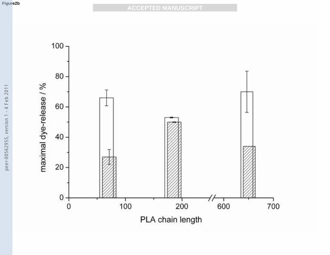
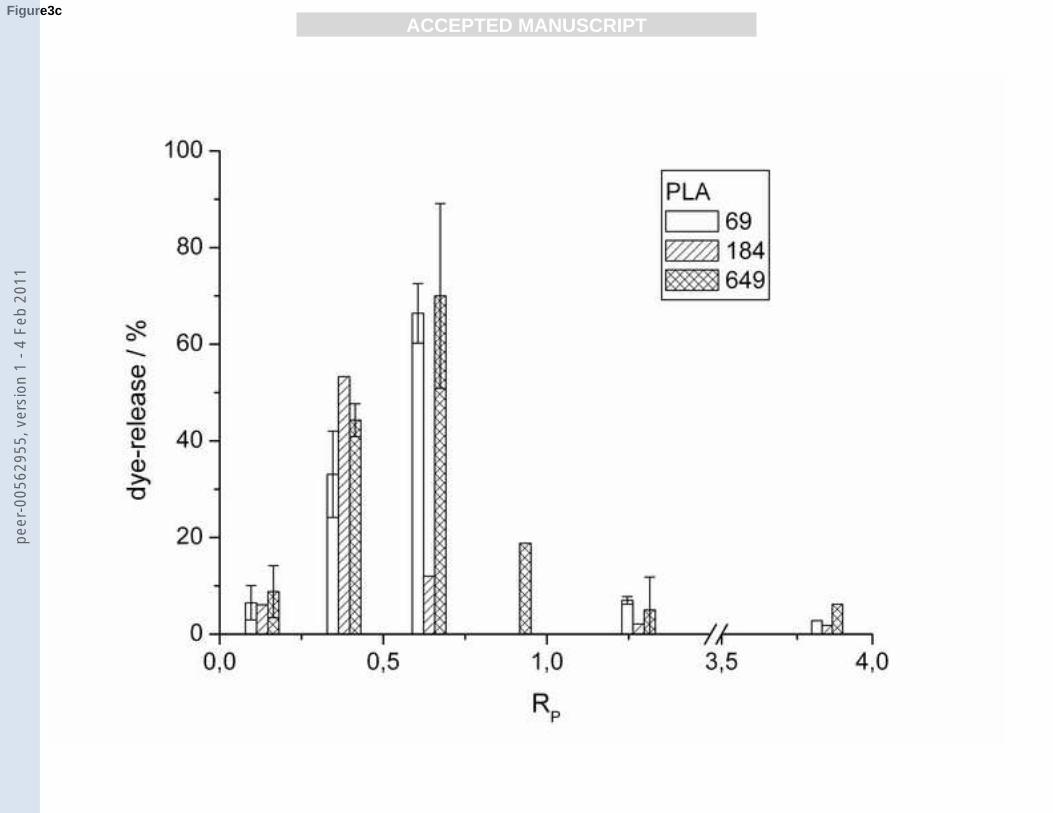
PLA induced maximum leakage for POPG vesicles ranging from 60 to 80% with a minimum
for PLA 184 which has an intermediate chain length. In the case of gel state DPPG vesicles
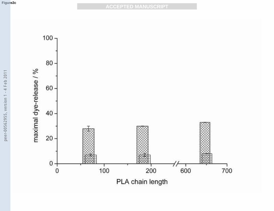
the dye-release was lower without significant trend. In the case of the fluid POPG/POPC and
gel state DPPG/DPPC vesicles no significant differences between the different polymer
lengths were observed. However, dye release was generally lower than for the pure POPG or
DPPG vesicles. All PLAs cause only little leakage (≤ 10%) from POPG vesicles at RP values
either smaller than 0.13 or higher than 1.28. Maximal dye-release occurs at an RP of 0.38 (for
PLA 184) and 0.64 (for PLA 69 and 649) (Fig. 3c).
--- Insert Fig. 3a-c. ---
Binding of FITC-labelled PLL 106 and 319 to DPPG membranes
Binding studies of FITC-labelled PLL 106 and 319 to DPPG membranes were performed to
check whether the different PLLs are binding to negatively charged DPPG membranes. FITC-
PLL 106 and FITC-PLL 319 solutions of different concentrations were chosen to match RP
values of 0.4, 0.5, 0.8, 1 and 2. When DPPG vesicles are injected into these peptide solutions
(Fig. 4a-b) the first step of peptide binding to the phospholipid membranes occurs very fast.
Depending on the actual peptide and the RP value a second much slower process occurs on a
time scale as observed in the dye-release experiments.
11
peer
-005
6295
5, v
ersi
on 1
- 4
Feb
2011

ACC
EPTE
D M
ANU
SCR
IPT
ACCEPTED MANUSCRIPT
--- Insert Fig. 4a-b. ---
At an RP value of 0.4 and 0.5 FITC-PLL 106 binds in two steps to the DPPG membrane: The
first step is very fast and leads to a drop in the normalised fluorescence intensity to 0.15
whereas the second step requires 300 s and more until a constant fluorescence intensity value
of 0.1 is reached. For the higher RP values of 0.8 and 1 the first binding step shows a smaller
drop in fluorescence intensity to 0.2 and 0.25. The second step is now characterised by a
sigmoidal decay of the normalised fluorescence intensity which ends at a slightly lower value
of 0.07 and 0.09. At an even higher RP value of 2 the sudden drop in the fluorescence intensity
occurs only to 0.55 and then increases again to a value of 0.6.
FITC-PLL 319 shows principally similar kinetic binding curves. However, there is a
characteristic shift in the final normalised fluorescence intensities for the same RP values
compared to the experiments with FITC-PLL 106. Particularly for the RP values of 0.4 and 0.5
the kinetic curves obtained with the two PLLs are very similar. After lipid injection, both
curves show a fast drop in fluorescence intensity to 0.15 followed by a slow exponential
decrease to a value of 0.05. For the RP values 0.8, 1 and 2 only a single binding process is
observed for FITC-PLL 319 ending at fluorescence intensity values of 0.3, 0.5 and 0.8.
The final values of normalised FITC-fluorescence intensity reveal to which extent PLL is
binding to and penetrating the lipid membranes. A final value of 0.5 indicates that half of the
amino acid residues are bound to lipid vesicles.
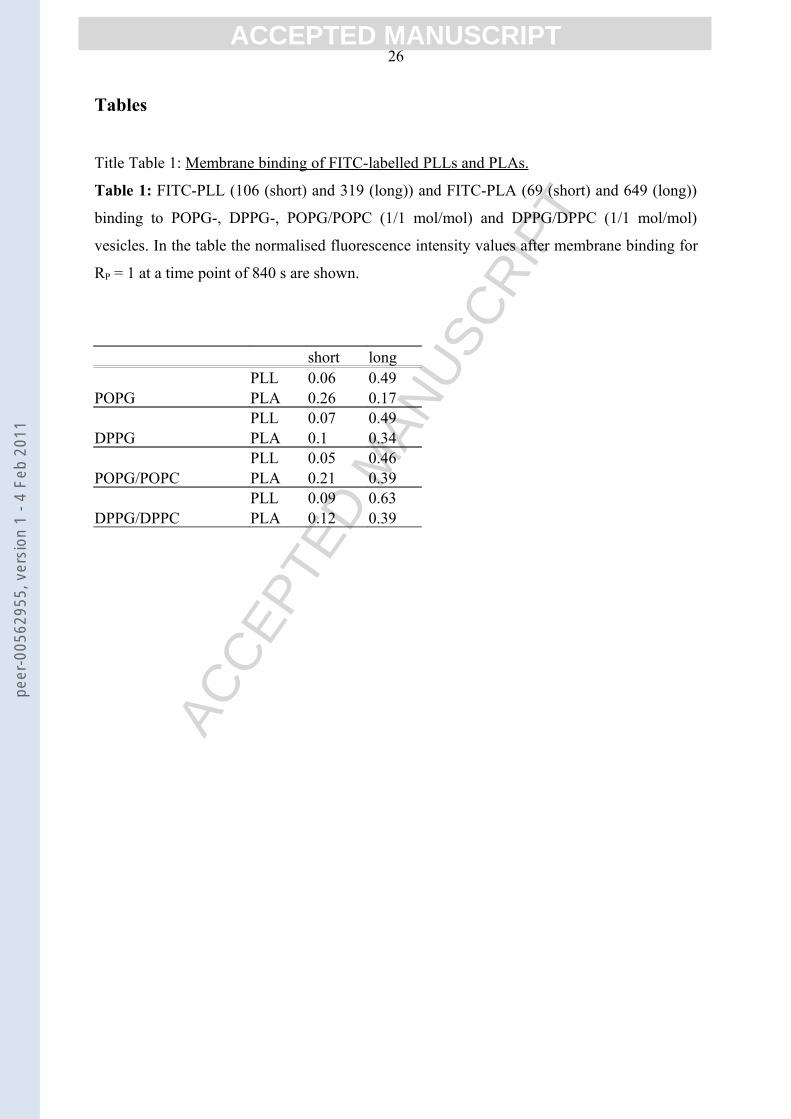
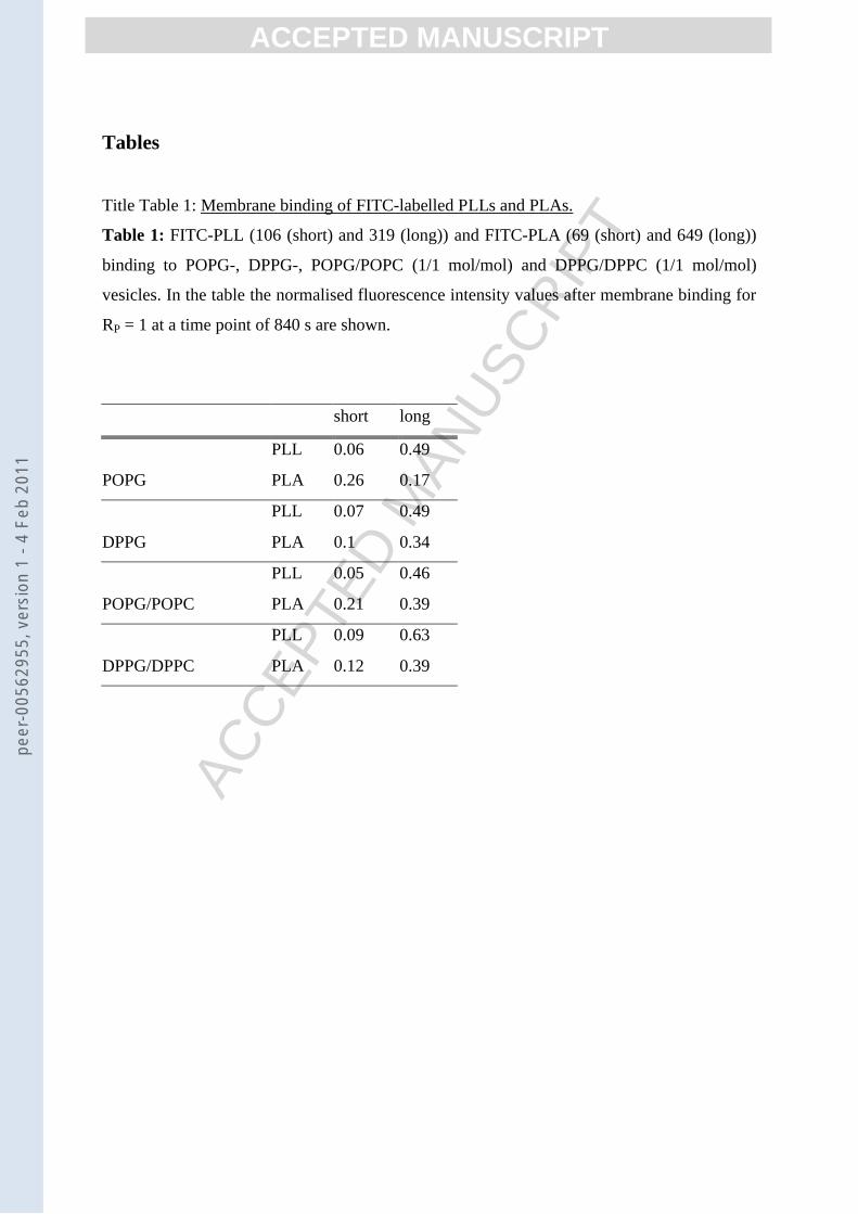
In table 1 all final values for the normalised FITC-fluorescence intensity (at RP = 1) after
binding of the labelled peptides are given. In case of the short FITC-PLL 106 all normalised
FITC-fluorescence intensity values lie in the range of 0.05 to 0.09 once binding is complete.
The longer FITC-PLL 319, on the other hand, has final values between 0.46 and 0.63 for all
membranes.
Binding of FITC-labelled PLA 69 and PLA 649
Binding of FITC-PLA 69 and FITC-PLA 649 to PG membranes leads to more diverse results.
The shorter FITC-PLA 69 reaches for fluid membranes of POPG and POPG/POPC
fluorescence intensity values of 0.26 and 0.21, respectively. In case of the gel phase
membranes of DPPG and DPPG/DPPC fluorescence intensity values of 0.1 and 0.12 are
reached, respectively.
When FITC-PLA 649 binds to the fluid POPG membrane, a final value for the normalised
fluorescence intensity of 0.17 is reached which is surprising as one would expect for the
longer PLA that less side chains are bound to the membrane compared to its shorter
12
peer
-005
6295
5, v
ersi
on 1
- 4
Feb
2011
ACC
EPTE
D M
ANU
SCR
IPT
ACCEPTED MANUSCRIPT
homologue FITC-PLA 69, because of sterical reasons. In case of the gel state DPPG- and
both the fluid and gel-phase PG/PC-membranes, final fluorescence intensity values between
0.34 and 0.39 are reached, which are very similar compared to the values for the shorter PLA.
Cryo-TEM
As revealed by the dynamic light scattering and EM measurements (see inset of Fig. 5), the
extruded suspension of 5 mM POPG without added polypeptide contains a homogeneous
population of vesicles with an average diameter of 100-120 nm. No multilamellar structures
were observed before polypeptide addition. The addition of the polypeptides PLA and PLL
induced drastic changes in the vesicle’s morphology. The formation of POPG-polypeptide
aggregates is shown in Fig. 5 for PLA649, PLL19 and PLL803 for all investigated POPG-
polypeptide ratios. In the case of PLA649 the aggregates are composed of several flattened
vesicles that stick to each other (Fig. 5a-c). Some of these vesicles are no longer unilamellar
but multilamellar. The thickness of the unilamellar PLA-coated vesicles reaches values of up
to 15 nm. The PLL-coated vesicles also formed clusters of 10 to 100 vesicles. The vesicles
were also deformed or flattened in the contact region (Fig. 5).
--- Insert Fig. 5. ---
Isothermal titration calorimetry (ITC)
ITC experiments were performed to study the thermodynamics of PLL and PLA binding to
lipid membranes, but generic differences to the fluorescence assays in the experimental
parameters have to be taken into account. The first difference is that for ITC measurements
higher lipid and peptide concentrations are needed to obtain a sufficient signal to noise ratio.
Results of preliminary experiments (not shown) by fluorescence spectroscopy showed that the
dye-release rate is increased with increasing peptide and lipid concentrations. The second
difference is that in ITC-experiments a peptide solution of high concentration was injected
into a lipid vesicle suspension, whereas in the fluorescence assays a well-defined amount of
lipid vesicles was injected only once into a peptide solution. Furthermore, in ITC repeated
addition of small amounts of peptide solution into a lipid suspension takes place.
The ITC experiments were carried out with DPPG and DPPG/DPPC membranes, either above
or below the main transition temperature Tm of ca. 41 °C.
PLL 402 binding to DPPG and DPPG/DPPC
13
peer
-005
6295
5, v
ersi
on 1
- 4
Feb
2011

ACC
EPTE
D M
ANU
SCR
IPT
ACCEPTED MANUSCRIPT
The binding of PLL 402 to DPPG membranes at 20 and 60 °C (Fig. 6a) reveals a
characteristic difference in binding to either gel phase or fluid membranes, respectively. The
curve at 20 °C shows endothermic binding enthalpies with a maximum at RP = 0.29 and falls
to very low enthalpy values after RP = 0.5. At 60 °C binding enthalpies are exothermic until a
RP value of 0.8 is reached. Thereafter, the binding enthalpy rises to endothermic values with a
maximum at RP = 1 and then decreases at RP = 1.3 to values below 0.1 kcal mol-1.
The binding to a mixed DPPG/DPPC membrane should be similar, only that the electrostatic
attraction to the membrane surface due to the dilution of the charges should be lower. The
binding curves of PLL 402 to the mixed membranes DPPG/DPPC (Fig. 6b) at the same
temperatures resemble in their shape indeed pretty much those for the pure DPPG
membranes. Overall the binding enthalpies are smaller in the case of the mixed membranes
and the data show more scatter for measurements at 60 °C.
--- Insert Fig. 6a-b. ---
PLL 803 binding to DPPG and DPPG/DPPC
Binding curves of the longer PLL 803 to DPPG and PG/PC membranes are shown in Fig. 7a-
b. The curves for binding to the pure and the mixed membranes in the gel phase (at 20 °C)
resemble each other very closely. The two curves are characterised by exothermic binding
enthalpies for RP smaller than 0.25. An increase of the binding enthalpies to ca. 0 kcal mol-1
(at RP = 0.29) indicates the termination of PLL 803 binding to the gel phase membranes.
In case of the pure fluid DPPG membrane at 60 °C PLL 803 binding occurs at least until an
RP value of 1. During initial binding (RP = 0 to 0.8) of PLL 803 binding enthalpies range
mainly between -0.6 and -0.44 kcal mol-1. In the final stages of this initial binding the binding
enthalpy values become larger (ca. -1 kcal mol-1) but are quite scattered. In the second part of
the curve the binding enthalpy rises to endothermic values and then falls to 0.1 kcal mol-1 (at
RP = 1). The titration curve of PLL 803 to fluid, mixed DPPG/DPPC membranes shows only
small heat effects. Either there is no binding or the binding enthalpy becomes very low.
--- Insert Fig. 7a-b. ---
PLA 649 binding to DPPG and DPPG/DPPC
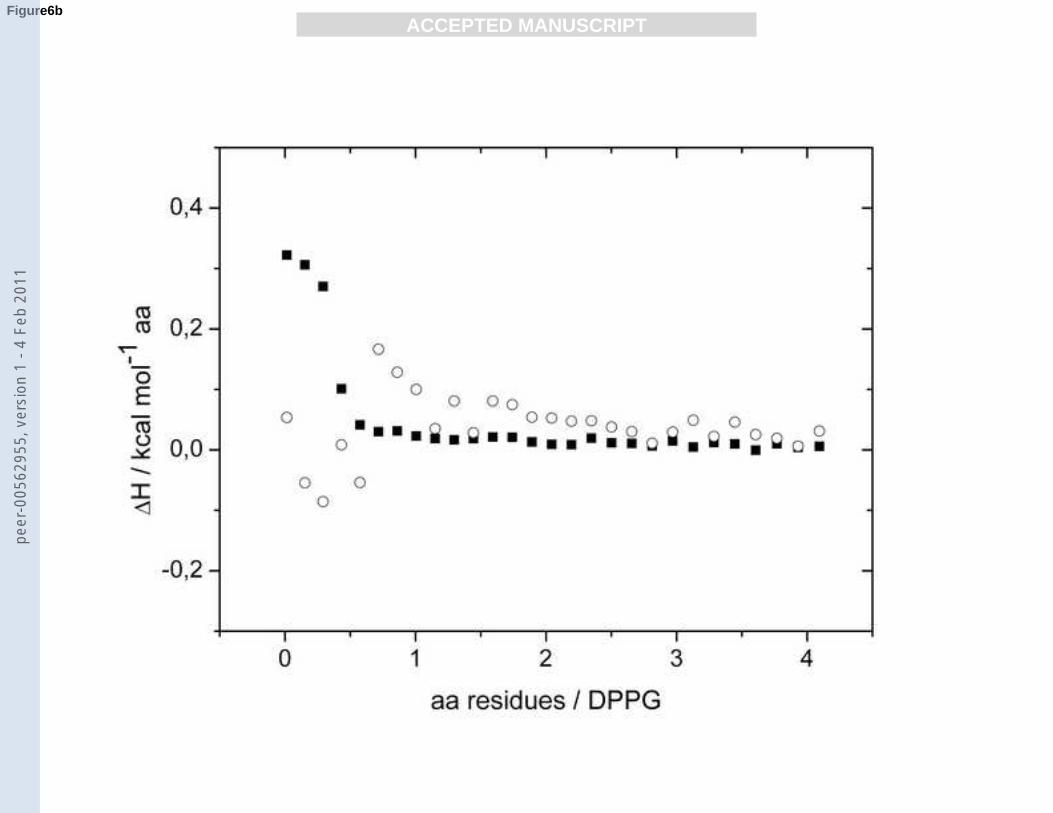
In case of the pure gel-phase DPPG membrane at 30 °C PLA 649 binding to the membrane is
first endothermic with a binding enthalpy of up to 0.45 kcal mol-1. The binding enthalpy
14
peer
-005
6295
5, v
ersi
on 1
- 4
Feb
2011

ACC
EPTE
D M
ANU
SCR
IPT
ACCEPTED MANUSCRIPT
changes sign and becomes exothermic. The binding curve shows a characteristic minimum at
an RP value of 0.5 indicating peptide binding mainly to the outer membrane leaflet. Between
RP values of 0.5 and 1 the binding enthalpy vanishes and becomes essentially zero at an RP
value of 1.
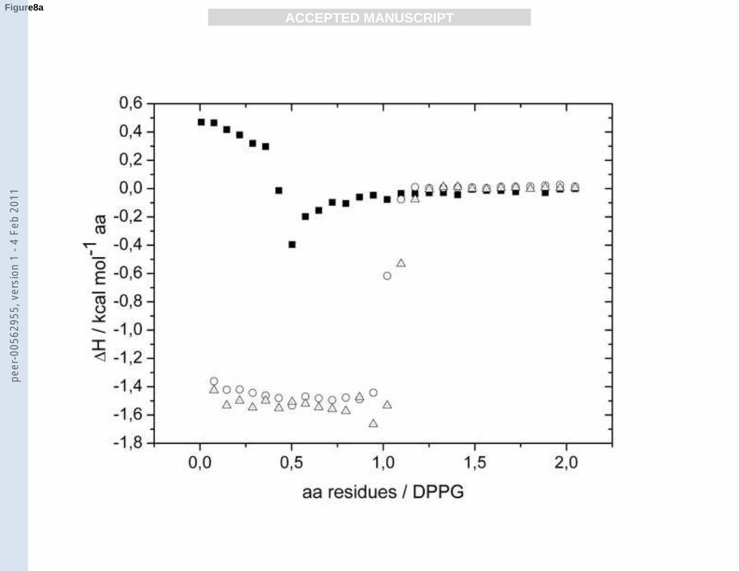
Binding of PLA 649 to DPPG membranes in the fluid phase at 50 and 60 °C occurs with
constant exothermic enthalpies ΔH of -1.5 kcal mol-1 until an RP of 1 is reached (Fig. 8a).
Between an RP of 1 and 1.25 ΔH becomes zero. These two isotherms indicate a complete
binding of PLA to both inner and the outer leaflet of lipid membranes in the fluid phase Lα.
The binding curves of PLA 649 to the mixed DPPG/DPPC membranes (Fig. 8b) have more or
less a sigmoid shape, as expected for weaker binding due to reduced surface charge density of
the lipid vesicles. All binding enthalpies are exothermic. For the gel phase membrane (30 °C)
the binding enthalpies are somewhat smaller and the heat signals disappear at a ratio RP of ca.
0.5. This indicates that only outside binding can take place in the gel phase. The titration
curves for the fluid DPPG/DPPC membranes show also exothermic heats of binding. Here,
the RP values are shifted to higher values indicating that now the PLA 649 can reach the inner
monolayer of the vesicles. The binding enthalpy seems to become more endothermic with
increasing temperature.
--- Insert Fig. 8a-b. ---
DiscussionThe positively charged polypeptides PLL and PLA adsorb to negatively charged phospholipid
vesicles which leads to the formation of large aggregates around the isoelectric point [15,35].
In this paper we tried to analyse the processes that occur during peptide binding to lipid
membranes and within the aggregate. In particular, leakage of an entrapped dye from lipid
vesicles, translocation behaviour of the charged polypeptides PLL and PLA and vesicle fusion
was assayed using suitable fluorescence and microscopy methods, respectively. Isothermal
titration calorimetry was applied to extend and confirm results obtained with the previous
methods, in particular ITC was used to clarify whether the polypeptides can cross the
membranes and bind to the inner monolayer of the vesicles.
Dye-release from phospholipid vesicles
Electrostatic interactions have a dominating influence in the adsorption of cationic
polypeptides to phospholipid membranes [36]. Therefore, the molar ratio of amino acid
15
peer
-005
6295
5, v
ersi
on 1
- 4
Feb
2011

ACC
EPTE
D M
ANU
SCR
IPT
ACCEPTED MANUSCRIPT
residues to charged lipid molecules (PGs) RP is an important quantity for the analysis of
leakage and translocation processes. Besides pure electrostatic interactions, hydrophobic and
van-der-Waals interactions and hydrogen bonds can play a role in membrane binding
processes. Lipid bilayers are composed of an outer and an inner leaflet where the latter one is
not accessible for peptides during the initial binding event at low RP values. However, during
the binding to the membrane pores or defects may be formed which locally disrupt the barrier
function so that an exchange of internal and external vesicle medium can take place. For most
of the amphipathic peptides that have been extensively studied this exchange follows an all-
or-none mechanism where a complete mixing of inner and outer medium occurs [37,11,38].
In case of the less often observed graded mechanism [39] only a well-defined amount of dye-
solution is released from the vesicles. It is not known which mechanism PLL and PLA follow.
Experiments such as those presented by Heerklotz & Seelig [39] could not be performed,
because the aggregates formed are too large to be purified by size-exclusion chromatography.
Leakage from lipid vesicles can be due to both temporary pore formation, which is normally
reversible, and complete vesicle lysis. If no leakage is observed, the vesicles are embedded in
a peptide-lipid aggregate with non-disrupted (intact) membranes.
PLL induced dye-release from POPG and mixed PG/PC vesicles has been probed by several
groups [16,2]. These groups either focused on a long-term kinetic investigation (over several
days) and/or used completely different experimental conditions. In our paper a kinetic
description of vesicle leakage on a short time scale is given for both PLL and PLA of different
polymer lengths yielding a quantitative evaluation of membrane stability. Still, a reasonable
number of interactions between phospholipid membranes and peptides, such as leakage,
peptide adsorption, insertion and translocation and secondary structure changes, make the
whole kinetic process very complex and do not allow to resolve the different influences on
vesicle leakage from each other. Therefore, the analysis is limited to a qualitative level of
understanding.
POPG membranes in the fluid Lα phase have a maximal leakage rate upon peptide-binding
that enables a proper observation of the induced processes (Fig. 1a and c for PLL 44 and PLA
69, resp.). At very low peptide concentrations (RP = 0.1), both PLL and PLA adsorb to the
membranes without an induction of pore formation. At an elevated peptide concentration (RP
= 0.2 to 0.4) kinetic curves have either an exponential shape in the case of PLL 44 indicating
that only a single process contributes to leakage or a bi-exponential shape revealing two
processes that induce dye-release in case of PLL 69. For RP values between 0.4 and 0.7
kinetic curves for PLL 44 adopt a sigmoidal shape which suggests that leakage is decelerated
16
peer
-005
6295
5, v
ersi
on 1
- 4
Feb
2011

ACC
EPTE
D M
ANU
SCR
IPT
ACCEPTED MANUSCRIPT
when peptide binding starts, presumably due to hindered binding on the membrane surface at
peptide excess. In case of PLA 69 the biexponential leakage mechanism persists manifesting
the superior binding capability of arginines over lysines [40].
At an RP around 0.5 dye-release from POPG vesicles is maximal for all longer PLL and PLA
(≥44), only PLL 19 shows an increasing dye-release until RP = 4. Theoretically, at an RP = 0.5
the outer monolayers are entirely covered with peptide molecules leading also to a maximal
cross linking of vesicles. This cross linking of vesicles could be visualised for POPG vesicles
covered with PLA 649. The POPG vesicles are deformed at the regions of contact with
adjacent vesicles, but do not rupture. This strong deformation of the polypeptide-coated
unilamellar vesicles indicates that the POPG-polypeptide interaction is either very strong or
very asymmetric, or that the vesicles are very flexible [41].
When there is an excess of peptide molecules no leakage is observed because vesicles are
fully covered with peptide that have a protection function on the surface [15]. Final values for
the dye-release after finished kinetics provide a measure for the extent of pore formation and
vesicle lysis which cannot be distinguished from each other. Thus, the term “membrane
defects” is used to describe the present results.
PLAs induce more membrane defects than PLLs with a maximum of 70% for PLA 649 and
60% for PLL 44 (for POPG), respectively. A possible explanation for this finding is that the
amino acid arginine with a protonated side chain can act as a hydrogen bond donor with five
possible H-bonds to H-bond acceptors (like the ones present in the lipid head groups) whereas
the protonated side chain of lysine can only form three which makes PLA a stronger binding
partner [42]. Both for PLL and PLA the occurrence of membrane defects is decreased in the
gel phase Lβ (DPPG vesicles) proving that these membranes are more stable against peptide
adsorption because of a less dynamic structure. For PLL 44 and 402 and PLA 184 there is a
maximum in leakage from DPPG vesicles compared to both shorter and longer polymers.
This finding can be explained on the basis of two counteracting effects: With increasing
polymer length, the binding constant KD is increased leading to a raised leakage rate.
However, sterical effects gain weight with increasing polymer length that produce a decrease
in dye-release. In case of the mixed PG/PC membranes leakage is decreased to 30% in case of
the fluid membranes and it drops to less than 10% in case of the gel phase membranes.
Mainly, PCs are assumed to stabilise the membranes [22], while also reducing the membrane
charge which explains the drop in leakage due to less close peptide binding on the membrane
[30]. The mixed membranes have a maximal dye-release observed at RP values of 1.3 (PLL 44
und PLA 69, data not shown). As a matter of fact, the reduced membrane charge requires
17
peer
-005
6295
5, v
ersi
on 1
- 4
Feb
2011

ACC
EPTE
D M
ANU
SCR
IPT
ACCEPTED MANUSCRIPT
more cationic polypeptide to reach substantial cross linking which is the prerequisite for
membrane leakage.
Isothermal titration calorimetry confirmed that membranes in the fluid phase (DPPG at 60 °C)
are more sensitive to leakage than gel phase membranes (DPPG at 20 °C) upon PLL (402 and
803) and PLA (649) binding (Fig. 6a, 7a and 8a). In the fluid phase binding detected by
enthalpy processes occurs until an RP ~ 1 is reached which indicates that both inner and outer
membrane leaflet are accessible to the cationic polypeptides, whereas in the gel phase peptide
binding nearly stops when an RP ~ 0.5 is reached.
For PLA 649 ITC also shows the strong difference in peptide binding to either DPPG or
DPPG/DPPC membranes in the fluid phase. In the latter case the binding constant is much
smaller, as a result of the decreased slope of binding enthalpy values. Peptide binding is also
complete at an RP of 1 revealing that the inner membrane leaflet is accessible for peptide
binding, but not as a result of total vesicle lysis or pore formation which would have been
detected by leakage. The ITC binding curve of PLA 649 and DPPG/DPPC in the gel phase (at
30 °C) which resembles the curve in the fluid phase is not suitable to explain the low dye-
release, leaving open the possibility of membrane binding via alternative ways as for example
peptide translocation into the inner membrane leaflet.
Peptide translocation through phospholipid membranes
FITC that is covalently bound to the PLL and PLA molecules serves as an ideal probe for
peptide adsorption to phospholipid membranes and, furthermore, allows the evaluation of
translocation properties of these peptides. The quantum yield of FITC strongly depends on the
chemical environment which was examined by taking FITC spectra in different solvents.
Roughly the quantum yield of FITC decreases several orders of magnitude with decreasing
dielectric constant. As PLL or PLA is adsorbed to a phospholipid membrane FITC is
intercalated between lipid molecules with their hydrophobic tails. FITC integrated into the
membrane is then in an apolar environment and its normalised fluorescence intensity should
drop to 0.5%. In the case of small RP values (0.1 to 0.4) the normalised fluorescence intensity
for the FITC-PLLs falls only to 5% which is probably due to the fact that not every FITC
probe will be intercalated into the outer membrane leaflet. Another reason might be that FITC
does not penetrate the membrane deep enough, i.e. is located in an environment with slightly
higher dielectric constant. The reason why the fluorescence intensity values of FITC-labelled
PLAs drop only to values of 0.2 when binding to an excess of fluid (!) membranes is not fully
18
peer
-005
6295
5, v
ersi
on 1
- 4
Feb
2011

ACC
EPTE
D M
ANU
SCR
IPT
ACCEPTED MANUSCRIPT
clear but is also probably due to the location of the probe in the interface. We assume,
however that at an RP of 0.1 PLA is completely bound to the membrane.
The ability of PLL and PLA to penetrate and translocate through a lipid membrane was
assayed depending on the state of the membrane, i.e. fluid or gel phase, and for both a short
and a long polymer. Table 1 shows all obtained normalised fluorescence intensity values at an
RP of 1 after an incubation time of 840 sec where both the processes of adsorption and
translocation have finished. This RP value was chosen because equal amounts of amino acid
residues and lipids are present and enabled us to investigate to which extent PLL and PLA are
bound to inner and outer leaflet of the whole membrane. From the leakage experiments it was
known that membranes suffer from PLL and PLA induced defects particularly in the fluid
phase, but resist temporary pore formation better in the gel phase. PG/PC membranes
exhibited even less membrane defects upon cationic polypeptide binding. A striking feature of
the short FITC-PLL 106 upon binding to all examined membrane types is that always
normalised fluorescence intensity values of less than 0.1 were observed which is in the same
region as for the smallest investigated RP value (0.1). Contrary to FITC-PLL 106, FITC-PLL
319 ends up at normalised fluorescence intensity values around 0.5 when probed with the four
membrane types. These results suggest that the short FITC-PLL 106 is able to translocate
through both fluid and gel phase membranes whereas the longer FITC-PLL 319 is not.
Similar findings were reported earlier by Shibata et al. [20] who probed the translocation
ability of FITC-PLL 106 (from the same supplier) through soybean phospholipid membranes
with confocal microscopy.
The translocation ability of (oligo)peptides with high arginine content was described by
several groups [14,13,43]. The short FITC-PLA 69 shows overall the same effect as the short
FITC-PLL 106, so short PLA molecules translocate through lipid bilayers in a similar manner
independent of membrane phase and composition. FITC-PLA 649 is two times longer than
the investigated PLL counterpart, but seems to translocate through the fluid POPG membrane
which is remarkable for a polymer of this length. At the same time one has to be aware that
PLA 649 also induces a high leakage from phospholipid vesicles which may eventually result
in lysis that would also make the inner leaflet easily accessible for peptide binding. An
unambiguous interpretation of FITC-PLA 649 penetration through the fluid POPG bilayer is
therefore not possible. Considering the low leakage rate of DPPG/DPPC membranes upon
binding of this peptide (8%) and a final value of the normalised fluorescence intensity of 0.39
suggests that at least 7% of the peptide molecules translocate (percentage of accessible inner
lipid molecules = total value – inaccessible value measured in the assay – leakage rate/2 =
19
peer
-005
6295
5, v
ersi
on 1
- 4
Feb
2011

ACC
EPTE
D M
ANU
SCR
IPT
ACCEPTED MANUSCRIPT
50% - 39% - (8%/2)) on the basis of total vesicle lysis and assuming an all-or-none
mechanism. For both POPG/POPC and DPPG vesicles such an estimate is more difficult
because there are maximal leakage rates of 30% which would mean that there is no
translocation observed if vesicles that leak are totally lysed. 34% (DPPG) and 39%
(POPG/POPC) of the initial fluorescence intensity remain at the end of peptide binding and
translocation. ITC measurements confirm that there is a complete binding of PLA 649 to both
inner and outer leaflet (until RP = 1) of a DPPG membrane at 50 and 60 °C. As there is no
total dye-release (only 70%) it can be confirmed that PLA 649 translocates through a fluid
phospholipid membrane. The results for the gel phase and the mixed PG/PC membranes
indicate that there is a decreased binding of PLA 649 to these membranes. However, heats of
binding can be at least detected until an RP of 1 is reached. Again, the problem that we cannot
fully distinguish between possible but not known vesicle lysis and translocation does not
permit a clear interpretation.
The ITC curves for PLL 402 which serves as a good equivalent to FITC-PLL 319 indicate a
complete binding to both the inner and outer leaflet of the fluid phospholipid membrane
(DPPG at 60 °C). This process was not observed in the FITC-assay raising the question for
the reason for this difference. As mentioned above the concentration is much higher in the
ITC experiment. This can enhance dye-release from phospholipid vesicles, particularly in the
fluid phase (data not shown), and possibly can lead to increased peptide translocation ability
through phospholipid membranes. Binding of PLL 402 to gel phase and mixed PG/PC
membranes, however, confirms unambiguously that peptide binding takes only place on the
outer membrane leaflet.
ConclusionsWe have shown using fluorescence spectroscopy, isothermal titration calorimetry, and
electron microscopy that the binding of poly-L-lysines and poly-L-arginines to negatively
charged lipid vesicles is a process occurring in several steps which, depending on the charge
ratio of lipid charges to amino acid side chain charges, can lead to the formation of membrane
defects, to disruption of the vesicles and/or translocation of the polypeptides to the vesicle
interior. The behaviour of the system also depends on the total concentration of the binding
partners and on the experimental procedures, i.e. whether the polypeptides are added to the
lipid vesicles or vice versa. Fluid lipid vesicles are more easily penetrated by the polypeptides
than gel state vesicles and comparing PLL and PLA the latter can more easily translocate
through the membrane. The results show that only the combination of different techniques can
shed light onto the binding and translocation process of cationic polypeptides.
20
peer
-005
6295
5, v
ersi
on 1
- 4
Feb
2011

ACC
EPTE
D M
ANU
SCR
IPT
ACCEPTED MANUSCRIPT
References
[1] H.K. Kimelberg, D. Papahadjopoulos, Interaction of basic proteins with phospholipid
membranes. Binding and changes in the sodium permeability of phosphatidylserine
vesicles, J. Biol. Chem. 246 (1971) 1142.
[2] A.E. Gad, B.L. Silver, G.D. Eytan, Polycation-induced fusion of negatively-charged
vesicles, Biochim. Biophys. Acta 690 (1982) 124.
[3] K. Fukushima, T. Noguchi, R. Shimozawa, Conformations of poly(L-lysine) induced
by dimyristoylphosphatidylglycerol vesicles in alkaline buffer solutions, Fukuoka
Daigaku Rigaku Shuho 22 (1992) 167.
[4] N. Sakai, S. Matile, Anion-Mediated Transfer of Poly-L-Arginine across Liquid and
Bilayer Membranes, J. Am. Chem. Soc. 125 (2003) 14348.
[5] B. Alberts , A. Johnson, J. Lewis, M. Raff, K. Roberts, P. Walter, Molecular Biology
Of The Cell (Garland Science, New York, 2008).
[6] D. Lochmann, E. Jauk, A. Zimmer, Drug delivery of oligonucleotides by peptides,
Eur. J. Pharm. Biopharm. 58 (2004) 237.
[7] T.P. Johnston, K.R. Kuchimanchi, H. Alur, M. Chittchang, A.K. Mitra, Inducing a
change in the pharmacokinetics and biodistribution of poly-L-lysine in rats by
complexation with heparin, J. Pharm. Pharmacol. 55 (2003) 1083.
[8] L. Arnold, A. Dagan, J. Gutheil, N. Kaplan, Antineoplastic activity of poly(L-lysine)
with some ascites tumor cells, Proc. Natl. Acad. Sci. U.S.A. 76 (1979) 3246.
[9] K. Matsuzaki, O. Murase, K. Miyajima, Kinetics of Pore Formation by an
Antimicrobial Peptide, Magainin 2, in Phospholipid Bilayers, Biochemistry 34 (1995)
12553.
21
peer
-005
6295
5, v
ersi
on 1
- 4
Feb
2011

ACC
EPTE
D M
ANU
SCR
IPT
ACCEPTED MANUSCRIPT
[10] A. Pokorny, T.H. Birkbeck, P.F. Almeida, Mechanism and Kinetics of δ-Lysin
Interaction with Phospholipid Vesicles, Biochemistry 41 (2002) 11044.
[11] J.L. Nieva, S. Nir, A. Muga, F.M. Goni, J. Wilschut, Interaction of the HIV- 1 Fusion
Peptide with Phospholipid Vesicles: Different Structural Requirements for Fusion and
Leakage, Biochemistry 33 (1994) 3201.
[12] A.D. Frankel, C.O. Pabo, Cellular uptake of the tat protein from human
immunodeficiency virus, Cell 55 (1988) 1189.
[13] S.M. Fuchs, R.T. Raines, Pathway for Polyarginine Entry into Mammalian Cells,
Biochemistry 43 (2004) 2438.
[14] E. Goncalves, E. Kitas, J. Seelig, Binding of Oligoarginine to Membrane Lipids and
Heparan Sulfate: Structural and Thermodynamic Characterization of a Cell-
Penetrating Peptide, Biochemistry 44 (2005) 2692.
[15] D. Volodkin, V. Ball, P. Schaaf, J.-C. Voegel, H. Mohwald, Complexation of
phosphocholine liposomes with polylysine. Stabilization by surface coverage versus
aggregation, Biochim. Biophys. Acta 1768 (2007) 280.
[16] D. Volodkin, H. Mohwald, J.-C. Voegel, V. Ball, Coating of negatively charged
liposomes by polylysine: Drug release study, J. Control. Release 117 (2007) 111.
[17] D.H. Young, H. Kauss, Release of Calcium from Suspension-Cultured Glycine max
Cells by Chitosan, Other Polycations, and Polyamines in Relation to Effects on
Membrane Permeability, Plant Physiol. 73 (1983) 698.
[18] S.M. Fuchs, R.T. Raines, Internalization of cationic peptides: the road less (or more?)
traveled, Cell. Mol. Life Sci. 63 (2006) 1819.
[19] H.E. Davis, M. Rosinski, J.R. Morgan, M.L. Yarmush, Charged polymers modulate
retrovirus transduction via membrane charge neutralization and virus aggregation,
Biophys. J. 86 (2004) 1234.
22
peer
-005
6295
5, v
ersi
on 1
- 4
Feb
2011

ACC
EPTE
D M
ANU
SCR
IPT
ACCEPTED MANUSCRIPT
[20] A. Shibata, S. Murata, S. Ueno, S. Liu, S. Futaki, Y. Baba, Synthetic copoly(Lys/Phe)
and poly(Lys) translocate through lipid bilayer membranes, Biochim. Biophys. Acta
1616 (2003) 147.
[21] J. Bondeson, R. Sundler, Promotion of acid-induced fusion by basic peptides. Amino
acid and phospholipid specificities, Biochim. Biophys. Acta 1026 (1990) 186.
[22] A. Walter, C.J. Steer, R. Blumenthal, Polylysine induces pH-dependent fusion of
acidic phospholipid vesicles: a model for polycation-induced fusion, Biochim.
Biophys. Acta 861 (1986) 319.
[23] K. Arnold, in: Handbook of Biological Physics, Vol. 1: Structure and Dynamics of
Membranes, ed. R. Lipowsky, E. Sackmann, Cation-Induced Vesicle Fusion
Modulated by Polymers and Proteins, Chapter 19 (Elsevier, 1995) p. 903.
[24] G. Förster, C. Schwieger, F. Faber, T. Weber, A. Blume, Influence of poly(L-lysine)
on the structure of dipalmitoylphosphatidylglycerol/water dispersions studied by X-ray
scattering, Eur. Biophys. J. 36 (2007) 425.
[25] C. Schwieger, A. Blume, Interaction of poly(L-lysines) with negatively charged
membranes: an FT-IR and DSC study, Eur. Biophys. J. 36 (2007) 437.
[26] J. Seelig, Titration calorimetry of lipid-peptide interactions., Biochim. Biophys. Acta
1331 (1997) 103.
[27] K. Kawakami, Y. Nishihara, K. Hirano, Effect of Hydrophilic Polymers on Physical
Stability of Liposome Dispersions, J. Phys. Chem. B 105 (2001) 2374.
[28] I. Tsogas, D. Tsiourvas, G. Nounesis, C.M. Paleos, Interaction of Poly-L-arginine with
Dihexadecyl Phosphate/Phosphatidylcholine Liposomes, Langmuir 21 (2005) 5997.
[29] K. Wagner, D. Harries, S. May, V. Kahl, J.O. Rädler, A. Ben-Shaul, Direct Evidence
for Counterion Release upon Cationic Lipid-DNA Condensation, Langmuir 16 (2000)
303.
23
peer
-005
6295
5, v
ersi
on 1
- 4
Feb
2011
ACC
EPTE
D M
ANU
SCR
IPT
ACCEPTED MANUSCRIPT
[30] C. Russ, T. Heimburg, H. von Grünberg, The Effect of Lipid Demixing on the
Electrostatic Interaction of Planar Membranes across a Salt Solution, Biophys. J. 84
(2003) 3730.
[31] L. Fülöp, B. Penke, M. Zarandi, Synthesis and Fluorescent Labelling of Beta-Amyloid
Peptides, J. Peptide Sci. 7 (2001) 397.
[32] R.R.C. New, Liposomes a practical approach (IRL Press at Oxford University Press
Oxford - New York - Tokyo, 1990).
[33] P.S. Chen, T.Y. Toribara, H. Warner, Microdetermination of Phosphorus, Anal. Chem.
28 (1956) 1756.
[34] C.H. Fiske, Y. Subbarow, The Colorimetric Determination of Phosphorus, J. Biol.
Chem. 66 (1925) 375.
[35] M. Reuter, Diploma thesis, Martin-Luther-University Halle-Wittenberg, Germany
(2006).
[36] A.H. Juffer, C.M. Shepherd, H.J. Vogel, Protein membrane electrostatic interactions:
Application of the Lekner summation technique, J. Phys. Chem. 114 (2001) 1892.
[37] R. Blumenthal, P.J. Millard, M.P. Henkart, C.W. Reynolds, P.A. Henkart, Liposomes
as targets for granule cytolysin from cytotoxic large granular lymphocyte tumors,
Proc. Natl. Acad. Sci. U.S.A. 81 (1984) 5551.
[38] J.E. Cummings, D.P. Satchell, Y. Shirafuji, A.J. Ouellette, T.K. Vanderlick,
Electrostatically controlled Interactions of Mouse Paneth Cell α-Defensins with
Phospholipid Membranes, Aust. J. Chem. 56 (2003) 1031.
[39] H. Heerklotz, J. Seelig, Leakage and lysis of lipid membranes induced by the
lipopeptide surfactin, Eur. Biophys. J. 36 (2007) 305.
24
peer
-005
6295
5, v
ersi
on 1
- 4
Feb
2011

ACC
EPTE
D M
ANU
SCR
IPT
ACCEPTED MANUSCRIPT
[40] D.J. Mitchell, D.T. Kim, L. Steinman, C.G. Fathman, J.B. Rothbard, Polyarginine
enters cells more efficiently than other polycationic homopolymers, J. Peptide Res. 56
(2000) 318.
[41] S. Huebner, B.J. Battersby, R. Grimm, G. Cevc, Lipid-DNA complex formation:
Reorganization and rupture of lipid vesicles in the presence of DNA as observed by
cryoelectron microscopy, Biophys. J. 76 (1999) 3158.
[42] M.G. Hutchings, M.C. Grossel, D.A.S. Merckel, A.M. Chippendale, M. Kenworthy,
G. McGeorge, The structure of m-xylylenediguanidinium sulfate: a putative molecular
tweezer ligand for anion chelation., Cryst. Growth Des. 1 (2001) 339.
[43] S. Futaki, T. Suzuki, W. Ohashi, T. Yagami, S. Tanaka, K. Ueda, Y. Sugiura,
Arginine-rich Peptides: An Abundant Source Of Membrane-permeable Peptides
having Potential As Carriers for Intracellular Protein Delivery, J. Biol. Chem. 276
(2001) 5836.
25
peer
-005
6295
5, v
ersi
on 1
- 4
Feb
2011
ACC
EPTE
D M
ANU
SCR
IPT
ACCEPTED MANUSCRIPT
Tables
Title Table 1: Membrane binding of FITC-labelled PLLs and PLAs.
Table 1: FITC-PLL (106 (short) and 319 (long)) and FITC-PLA (69 (short) and 649 (long))
binding to POPG-, DPPG-, POPG/POPC (1/1 mol/mol) and DPPG/DPPC (1/1 mol/mol)
vesicles. In the table the normalised fluorescence intensity values after membrane binding for
RP = 1 at a time point of 840 s are shown.
short long
POPGPLL 0.06 0.49PLA 0.26 0.17
DPPGPLL 0.07 0.49PLA 0.1 0.34
POPG/POPCPLL 0.05 0.46PLA 0.21 0.39
DPPG/DPPCPLL 0.09 0.63PLA 0.12 0.39
26
peer
-005
6295
5, v
ersi
on 1
- 4
Feb
2011

ACC
EPTE
D M
ANU
SCR
IPT
ACCEPTED MANUSCRIPT
Figures (with caption and title)
Title 1: Dye-release kinetic curves of PLLs and PLAs with PG vesicles.
Fig. 1
a): Dye-release curves for PLL 44 interaction with POPG vesicles. The PLL concentration
was varied, with RP values of 0.13 (-), 0.26 (-), 0.38 (-), 0.51 (-), 0.64 (-) and 1.28 (-). After an
equilibration time of 30 s a defined amount of calcein-containing vesicles (final concentration
80 µM) were added (time point of 0 s in the graph). The reaction temperature was 22 - 23 °C.
b): Dye-release curves for PLL 44 interaction with DPPG vesicles with RP values of 0.12 (-),
0.6 (-), 1.19 (-), and 3.57 (-) . After an equilibration time of 30 s a defined amount of calcein-
containing vesicles (final concentration 85 µM) were added (time point of 0 s in the graph).
The reaction temperature was 22 - 23 °C.
c): Dye-release curves for PLA 69 interaction with POPG vesicles with RP values of 0.13 (-),
0.38 (-), 0.64 (-), 1.28 (-) and 3.85 (-). After an equilibration time of 30 s a defined amount of
calcein-containing vesicles (final concentration 75 µM) were added (time point of 0 s in the
graph). The reaction temperature was 22 - 23 °C.
Title 2: Maximal dye-release from POPG and DPPG and mixed vesicles induced by PLLs and
PLAs.
Fig. 2
a): Maximal dye-release from POPG (empty bar) and DPPG (diagonally dashed bar) vesicles
induced by PLLs of the average lengths 19, 44, 123, 220, 402 and 906.
b): Maximal dye-release from POPG (empty bar) and DPPG (diagonally dashed bar) vesicles
induced by PLAs of the average lengths 69, 184 and 649.
c): Maximal dye-release from POPG/POPC (1/1, mol/mol) (patterned bar) and DPPG/DPPC
(1/1, mol/mol) (horizontally dashed bar) vesicles induced by PLAs of the average lengths 69,
184 and 649.
Title 3: POPG dye-release dependence on RP (PLLs and PLAs).
Fig. 3
a): Dye-release from POPG vesicles as a function of RP for PLLs 19, 44 and 123.
b): Dye-release from POPG vesicles as a function of RP for PLLs 220, 402 and 906.
c): Dye-release from POPG vesicles as a function of RP for PLAs 69, 184 and 649.
27
peer
-005
6295
5, v
ersi
on 1
- 4
Feb
2011
ACC
EPTE
D M
ANU
SCR
IPT
ACCEPTED MANUSCRIPT
Title 4: Binding of FITC-labelled PLLs to DPPG membranes.
Fig. 4: FITC-PLL 106 (upper panel) and FITC-PLL 319 (lower panel) binding to DPPG
vesicles. The FITC-PLL concentration was varied in different experiments. DPPG vesicles at
a constant lipid concentration of 100 µM were injected at a time point of 0 s and the binding
of FITC-PLL was monitored with time. Final RP values are: 0.4 (-), 0.5 (-), 0.8 (-), 1 (-) and 2
(-). The reaction temperature was 22 - 23 °C.
Title 5: Cryo-TEM images of PLA and PLL coated POPG vesicles.
Fig. 5: Cryo-TEM images of polypeptide-coated unilamellar POPG vesicles with PLA 649 at
different RP values (a-c), PLL 19 at RP = 0.05 (d,e), and PLL 803 at RP = 0.25 (f) prepared at
20°C.
Title 6: ITC binding curves for PLL 402 to PG membranes.
Fig. 6
a): Reaction heats for the titration of 20 mM PLL 402 into a 2 mM DPPG suspension at 20
(■) and 60 (○) °C.
b): Reaction heats for the titration of 20 mM PLL 402 into a 2 mM DPPG/DPPC (1/1
mol/mol) suspension at 20 (■) and 60 (○) °C.
Title 7: ITC binding curves for PLL 803 to PG membranes.
Fig. 7
a): Reaction heats for the titration of 20 mM PLL 803 into a 2 mM DPPG suspension at 20
(■) and 60 (○) °C.
b): Reaction heats for the titration of 20 mM PLL 803 into a 4 mM DPPG/DPPC (1/1
mol/mol) suspension at 20 (■) and 60 (○) °C.
Title 8: ITC binding curves for PLA 649 to PG membranes.
Fig. 8
a): Reaction heats for the titration of 20 mM PLA 649 into a 2 mM DPPG suspension at 30
(■), 50 (○) and 60 (∆) °C.
b): Reaction heats for the titration of 20 mM PLA 649 into a 2 mM DPPG/DPPC (1/1
mol/mol) suspension at 30 (■), 50 (○) and 60 (∆) °C.
28
peer
-005
6295
5, v
ersi
on 1
- 4
Feb
2011

ACC
EPTE
D M
ANU
SCR
IPT
ACCEPTED MANUSCRIPTFigure1a
peer
-005
6295
5, v
ersi
on 1
- 4
Feb
2011

ACC
EPTE
D M
ANU
SCR
IPT
ACCEPTED MANUSCRIPTFigure1b
peer
-005
6295
5, v
ersi
on 1
- 4
Feb
2011

ACC
EPTE
D M
ANU
SCR
IPT
ACCEPTED MANUSCRIPTFigure1c
peer
-005
6295
5, v
ersi
on 1
- 4
Feb
2011

ACC
EPTE
D M
ANU
SCR
IPT
ACCEPTED MANUSCRIPTFigure2a
peer
-005
6295
5, v
ersi
on 1
- 4
Feb
2011
ACC
EPTE
D M
ANU
SCR
IPT
ACCEPTED MANUSCRIPTFigure2b
peer
-005
6295
5, v
ersi
on 1
- 4
Feb
2011
ACC
EPTE
D M
ANU
SCR
IPT
ACCEPTED MANUSCRIPTFigure2c
peer
-005
6295
5, v
ersi
on 1
- 4
Feb
2011

ACC
EPTE
D M
ANU
SCR
IPT
ACCEPTED MANUSCRIPTFigure3a
peer
-005
6295
5, v
ersi
on 1
- 4
Feb
2011

ACC
EPTE
D M
ANU
SCR
IPT
ACCEPTED MANUSCRIPTFigure3b
peer
-005
6295
5, v
ersi
on 1
- 4
Feb
2011
ACC
EPTE
D M
ANU
SCR
IPT
ACCEPTED MANUSCRIPTFigure3c
peer
-005
6295
5, v
ersi
on 1
- 4
Feb
2011

ACC
EPTE
D M
ANU
SCR
IPT
ACCEPTED MANUSCRIPTFigure4
peer
-005
6295
5, v
ersi
on 1
- 4
Feb
2011

ACC
EPTE
D M
ANU
SCR
IPT
ACCEPTED MANUSCRIPTFigure5
peer
-005
6295
5, v
ersi
on 1
- 4
Feb
2011

ACC
EPTE
D M
ANU
SCR
IPT
ACCEPTED MANUSCRIPTFigure6a
peer
-005
6295
5, v
ersi
on 1
- 4
Feb
2011
ACC
EPTE
D M
ANU
SCR
IPT
ACCEPTED MANUSCRIPTFigure6b
peer
-005
6295
5, v
ersi
on 1
- 4
Feb
2011

ACC
EPTE
D M
ANU
SCR
IPT
ACCEPTED MANUSCRIPTFigure7a
peer
-005
6295
5, v
ersi
on 1
- 4
Feb
2011

ACC
EPTE
D M
ANU
SCR
IPT
ACCEPTED MANUSCRIPTFigure7b
peer
-005
6295
5, v
ersi
on 1
- 4
Feb
2011
ACC
EPTE
D M
ANU
SCR
IPT
ACCEPTED MANUSCRIPTFigure8a
peer
-005
6295
5, v
ersi
on 1
- 4
Feb
2011

ACC
EPTE
D M
ANU
SCR
IPT
ACCEPTED MANUSCRIPTFigure8b
peer
-005
6295
5, v
ersi
on 1
- 4
Feb
2011
ACC
EPTE
D M
ANU
SCR
IPT
ACCEPTED MANUSCRIPT
Tables
Title Table 1: Membrane binding of FITC-labelled PLLs and PLAs.
Table 1: FITC-PLL (106 (short) and 319 (long)) and FITC-PLA (69 (short) and 649 (long))
binding to POPG-, DPPG-, POPG/POPC (1/1 mol/mol) and DPPG/DPPC (1/1 mol/mol)
vesicles. In the table the normalised fluorescence intensity values after membrane binding for
RP = 1 at a time point of 840 s are shown.
short long
POPG
PLL 0.06 0.49
PLA 0.26 0.17
DPPG
PLL 0.07 0.49
PLA 0.1 0.34
POPG/POPC
PLL 0.05 0.46
PLA 0.21 0.39
DPPG/DPPC
PLL 0.09 0.63
PLA 0.12 0.39
peer
-005
6295
5, v
ersi
on 1
- 4
Feb
2011
Related Documents