Crystal structure and activities of EXPB1 (Zea m 1), a -expansin and group-1 pollen allergen from maize Neela H. Yennawar †‡ , Lian-Chao Li ‡§ , David M. Dudzinski ¶ , Akira Tabuchi § , and Daniel J. Cosgrove §†† † Huck Institutes of the Life Sciences and Departments of § Biology and ¶ Biochemistry and Molecular Biology, Pennsylvania State University, University Park, PA 16802 This contribution is part of the special series of Inaugural Articles by members of the National Academy of Sciences elected on May 3, 2005. Contributed by Daniel J. Cosgrove, July 15, 2006 Expansins are small extracellular proteins that promote turgor- driven extension of plant cell walls. EXPB1 (also called Zea m 1) is a member of the -expansin subfamily known in the allergen literature as group-1 grass pollen allergens. EXPB1 induces exten- sion and stress relaxation of grass cell walls. To help elucidate expansin’s mechanism of wall loosening, we determined the struc- ture of EXPB1 by x-ray crystallography to 2.75-Å resolution. EXPB1 consists of two domains closely packed and aligned so as to form a long, shallow groove with potential to bind a glycan backbone of 10 sugar residues. The structure of EXPB1 domain 1 resembles that of family-45 glycoside hydrolase (GH45), with conservation of most of the residues in the catalytic site. However, EXPB1 lacks a second aspartate that serves as the catalytic base required for hydrolytic activity in GH45 enzymes. Domain 2 of EXPB1 is an Ig-like -sandwich, with aromatic and polar residues that form a potential surface for polysaccharide binding in line with the glycan binding cleft of domain 1. EXPB1 binds to maize cell walls, most strongly to xylans, causing swelling of the cell wall. Tests for hydrolytic activity by EXPB1 with various wall polysaccharides proved negative. Moreover, GH45 enzymes and a GH45-related protein called ‘‘swol- lenin’’ lacked wall extension activity comparable to that of ex- pansins. We propose a model of expansin action in which EXPB1 fa- cilitates the local movement and stress relaxation of arabinoxylan– cellulose networks within the wall by noncovalent rearrangement of its target. plant cell wall B efore maturation plant cells typically experience a period of prolonged cell enlargement, often resulting in a 10 3 -fold increase in volume. The impressive height of trees, some ex- ceeding 100 m, depends on such enlargement, which entails massive vacuolar expansion and irreversible yielding of the cellulosic cell wall. In physical terms, the rate-limiting process for cell enlargement resides within the cell wall, which must be loosened so as to allow wall stress relaxation and consequent water uptake for vacuole enlargement and stretching of the wall (1, 2). Currently, the only plant proteins shown to cause cell wall relaxation are expansins (3, 4), although xyloglucan endotrans- glucosylase, pectate lyase, cellulase, and other enzymes partic- ipate in cell wall restructuring during cell growth (5–8). Expansins were originally discovered in a ‘‘fishing expedition’’ for catalysts of cell wall extension (9, 10). When walls are clamped in tension and incubated in acidic buffer, these proteins rapidly induce wall extension and enhance wall stress relaxation. Their biological role in promoting cell enlargement is amply supported by in vitro and in vivo experiments, as well as by studies of gene expression, gene silencing, and ectopic expression (3, 11–13). In addition to cell enlargement, expansins are also implicated in other developmental processes where wall loosen- ing occurs, such as in fruit softening, organ abscission, seed germination, and pollen tube invasion of the grass stigma (14–17). Two expansin families with wall-loosening activity have been identified, named -expansins (EXPA) and -expansins (EXPB); both are found in all groups of land plants, from mosses to flowering plants (3, 18). Although they have only 20% amino acid identity, EXPA and EXPB proteins are of similar size (27 kDa), their sequences align well with one another, and they contain a number of conserved residues and characteristic motifs distributed throughout the length of the protein. EXPA and EXPB appear to act on different cell wall components, but their native targets have not yet been well defined. A subset of EXPBs is known in the immunological literature as group-1 grass pollen allergens (19–21). These EXPBs are abundantly and specifically expressed in grass pollen, causing hay fever and seasonal asthma in an estimated 200–400 million humans (22, 23). The extraordinary abundance of group-1 allergens [comprising up to 4% of the protein extracted from grass pollen (24)] is unique (as far as we know) in the world of expansins, which are typically found in very low abundance and tightly bound to the cell wall. The abundance of group-1 allergens in grass pollen bespeaks a unique biological role, namely to loosen the cell walls of the grass stigma and style, thereby aiding pollen tube penetration and assisting delivery of its two sperm cells to the ovule, where a double fertilization occurs, forming the diploid zygote and the triploid endosperm. Seed development follows, and, because cereal grasses provide the largest food source for humanity (e.g., rice, maize, wheat, and barley, to name but a few), the importance of these events for human welfare is hard to overestimate. Other genes in the EXPB family are expressed in a variety of other tissues in the plant body and in general lack the specific allergenic epitopes characteristic of group-1 allergens (24, 25). These so-called ‘‘vegetative EXPBs’’ are thought to have cell wall loosening activity and substrate specificity similar to the group-1 allergens, but these inferences have yet to be demonstrated experimentally. The mechanism by which expansins loosen cell walls has not yet been worked out in molecular detail. Plant cell walls consist of a scaffold of long cellulose microfibrils 4 nm in diameter embedded in a matrix of cellulose-binding glycans, such as xyloglucan and arabinoxylan, and gel-forming pectic polysaccha- Conflict of interest statement: No conflicts declared. Abbreviations: EXPA, -expansin; EXPB, -expansin; GH45, family-45 glycoside hydrolase; D1, domain 1 of EXPB1; D2, domain 2 of EXPB1; CBM, carbohydrate-binding module; PDB, Protein Data Bank. Data deposition: The atomic coordinates have been deposited in the Protein Data Bank, www.pdb.org (PDB ID code 2HCZ). See accompanying Profile on page 14661. ‡ N.H.Y. and L.-C.L. contributed equally to this work. Present address: Department of Medicine, Harvard Medical School and Massachusetts General Hospital, Boston, MA 02114. †† To whom correspondence should be addressed at: Pennsylvania State University, 208 Mueller Laboratory, University Park, PA 16802. E-mail: [email protected]. © 2006 by The National Academy of Sciences of the USA 14664 –14671 PNAS October 3, 2006 vol. 103 no. 40 www.pnas.orgcgidoi10.1073pnas.0605979103

Welcome message from author
This document is posted to help you gain knowledge. Please leave a comment to let me know what you think about it! Share it to your friends and learn new things together.
Transcript
Crystal structure and activities of EXPB1 (Zea m 1), a�-expansin and group-1 pollen allergen from maizeNeela H. Yennawar†‡, Lian-Chao Li‡§, David M. Dudzinski¶�, Akira Tabuchi§, and Daniel J. Cosgrove§††
†Huck Institutes of the Life Sciences and Departments of §Biology and ¶Biochemistry and Molecular Biology, Pennsylvania State University,University Park, PA 16802
This contribution is part of the special series of Inaugural Articles by members of the National Academy of Sciences elected on May 3, 2005.
Contributed by Daniel J. Cosgrove, July 15, 2006
Expansins are small extracellular proteins that promote turgor-driven extension of plant cell walls. EXPB1 (also called Zea m 1) isa member of the �-expansin subfamily known in the allergenliterature as group-1 grass pollen allergens. EXPB1 induces exten-sion and stress relaxation of grass cell walls. To help elucidateexpansin’s mechanism of wall loosening, we determined the struc-ture of EXPB1 by x-ray crystallography to 2.75-Å resolution. EXPB1consists of two domains closely packed and aligned so as to forma long, shallow groove with potential to bind a glycan backboneof �10 sugar residues. The structure of EXPB1 domain 1 resemblesthat of family-45 glycoside hydrolase (GH45), with conservation ofmost of the residues in the catalytic site. However, EXPB1 lacks asecond aspartate that serves as the catalytic base required forhydrolytic activity in GH45 enzymes. Domain 2 of EXPB1 is an Ig-like�-sandwich, with aromatic and polar residues that form a potentialsurface for polysaccharide binding in line with the glycan bindingcleft of domain 1. EXPB1 binds to maize cell walls, most strongly toxylans, causing swelling of the cell wall. Tests for hydrolytic activityby EXPB1 with various wall polysaccharides proved negative.Moreover, GH45 enzymes and a GH45-related protein called ‘‘swol-lenin’’ lacked wall extension activity comparable to that of ex-pansins. We propose a model of expansin action in which EXPB1 fa-cilitates the local movement and stress relaxation of arabinoxylan–cellulose networks within the wall by noncovalent rearrangementof its target.
plant cell wall
Before maturation plant cells typically experience a period ofprolonged cell enlargement, often resulting in a �103-fold
increase in volume. The impressive height of trees, some ex-ceeding 100 m, depends on such enlargement, which entailsmassive vacuolar expansion and irreversible yielding of thecellulosic cell wall. In physical terms, the rate-limiting process forcell enlargement resides within the cell wall, which must beloosened so as to allow wall stress relaxation and consequentwater uptake for vacuole enlargement and stretching of the wall(1, 2). Currently, the only plant proteins shown to cause cell wallrelaxation are expansins (3, 4), although xyloglucan endotrans-glucosylase, pectate lyase, cellulase, and other enzymes partic-ipate in cell wall restructuring during cell growth (5–8).
Expansins were originally discovered in a ‘‘fishing expedition’’for catalysts of cell wall extension (9, 10). When walls areclamped in tension and incubated in acidic buffer, these proteinsrapidly induce wall extension and enhance wall stress relaxation.Their biological role in promoting cell enlargement is amplysupported by in vitro and in vivo experiments, as well as by studiesof gene expression, gene silencing, and ectopic expression (3,11–13). In addition to cell enlargement, expansins are alsoimplicated in other developmental processes where wall loosen-ing occurs, such as in fruit softening, organ abscission, seedgermination, and pollen tube invasion of the grass stigma(14–17).
Two expansin families with wall-loosening activity have beenidentified, named �-expansins (EXPA) and �-expansins(EXPB); both are found in all groups of land plants, from mossesto flowering plants (3, 18). Although they have only �20%amino acid identity, EXPA and EXPB proteins are of similar size(�27 kDa), their sequences align well with one another, and theycontain a number of conserved residues and characteristic motifsdistributed throughout the length of the protein. EXPA andEXPB appear to act on different cell wall components, but theirnative targets have not yet been well defined.
A subset of EXPBs is known in the immunological literatureas group-1 grass pollen allergens (19–21). These EXPBs areabundantly and specifically expressed in grass pollen, causing hayfever and seasonal asthma in an estimated 200–400 millionhumans (22, 23). The extraordinary abundance of group-1allergens [comprising up to 4% of the protein extracted fromgrass pollen (24)] is unique (as far as we know) in the world ofexpansins, which are typically found in very low abundance andtightly bound to the cell wall. The abundance of group-1allergens in grass pollen bespeaks a unique biological role,namely to loosen the cell walls of the grass stigma and style,thereby aiding pollen tube penetration and assisting delivery ofits two sperm cells to the ovule, where a double fertilizationoccurs, forming the diploid zygote and the triploid endosperm.Seed development follows, and, because cereal grasses providethe largest food source for humanity (e.g., rice, maize, wheat, andbarley, to name but a few), the importance of these events forhuman welfare is hard to overestimate.
Other genes in the EXPB family are expressed in a variety ofother tissues in the plant body and in general lack the specificallergenic epitopes characteristic of group-1 allergens (24, 25).These so-called ‘‘vegetative EXPBs’’ are thought to have cell wallloosening activity and substrate specificity similar to the group-1allergens, but these inferences have yet to be demonstratedexperimentally.
The mechanism by which expansins loosen cell walls has notyet been worked out in molecular detail. Plant cell walls consistof a scaffold of long cellulose microfibrils �4 nm in diameterembedded in a matrix of cellulose-binding glycans, such asxyloglucan and arabinoxylan, and gel-forming pectic polysaccha-
Conflict of interest statement: No conflicts declared.
Abbreviations: EXPA, �-expansin; EXPB, �-expansin; GH45, family-45 glycoside hydrolase;D1, domain 1 of EXPB1; D2, domain 2 of EXPB1; CBM, carbohydrate-binding module; PDB,Protein Data Bank.
Data deposition: The atomic coordinates have been deposited in the Protein Data Bank,www.pdb.org (PDB ID code 2HCZ).
See accompanying Profile on page 14661.
‡N.H.Y. and L.-C.L. contributed equally to this work.
�Present address: Department of Medicine, Harvard Medical School and MassachusettsGeneral Hospital, Boston, MA 02114.
††To whom correspondence should be addressed at: Pennsylvania State University, 208Mueller Laboratory, University Park, PA 16802. E-mail: [email protected].
© 2006 by The National Academy of Sciences of the USA
14664–14671 � PNAS � October 3, 2006 � vol. 103 � no. 40 www.pnas.org�cgi�doi�10.1073�pnas.0605979103
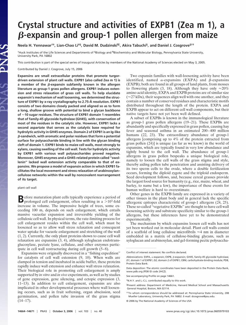
rides (Fig. 1). The cellulose-binding glycans form a stablenetwork with the cellulose microfibrils by binding to their surfacevia hydrogen bonds between hydroxyl groups and via van derWaals forces between the sugar rings; the network is furtherstabilized by calcium ions and borate diesters that link pecticpolysaccharides together. Cell walls also contain small amountsof structural proteins with a reinforcing role (26, 27). Wallexpansion entails rearrangement or modification of the matrix toallow turgor-driven movement or slippage of cellulose microfi-brils within the matrix (1).
Most of the biochemical work on expansins to date has focusedon EXPAs, which do not hydrolyze the major structural poly-saccharides of the wall and indeed are devoid of every enzymeactivity assayed to date (28). Our current model proposes thatEXPAs disrupt the polysaccharide complexes that link cellulosemicrofibrils together. The pollen EXPBs (group-1 allergens)have a marked loosening action on cell walls from grasses, butnot from dicots, whereas the reverse is true for EXPAs; there-fore, it seems that the two forms of expansin target differentcomponents of the cell wall (21, 24). Grass cell walls are notablefor containing relatively small amounts of xyloglucan and pectin,which are replaced with �-(133),(134)-D-glucan and glucu-ronoarabinoxylan (29), two potential targets of EXPBs in theirwall-loosening activity.
Sequence analysis suggests that expansins consist of twodomains (2, 3). The putative N-terminal domain [domain 1 ofEXPB1 (D1)] has distant sequence similarity (�20% identity) tothe catalytic domain of family-45 glycoside hydrolases (GH45)(http:��afmb.cnrs-mrs.fr�CAZY). Despite this resemblance,EXPAs do not hydrolyze wall polysaccharides, and so thesequence similarity is enigmatic. The C-terminal domain [do-main 2 of EXPB1 (D2)] has sequence similarity (from 35% to�10% identity) to another class of allergens, the group-2�3 grasspollen allergens, whose biological function is unknown (30).
In this study we present the crystal structure of a native EXPBpurified from maize pollen. In the allergen field it is designatedZea m 1 isoform d, whereas by expansin nomenclature it is calledEXPB1 (GenBank accession no. AAO45608). The allergenname ‘‘Zea m 1’’ encompasses a group of at least four pollenproteins (EXPB1, EXPB9, EXPB10, and EXPB11) in two rather
divergent sequence classes (24). EXPB1 is the most abundant ofthe maize group-1 allergens. We also test EXPB1 for binding andactivity on cell walls. At the end we discuss a molecular modelof expansin action that is consistent with its structure and knownbiophysical and biochemical activities.
ResultsEXPB1 Has Two Closely Packed Domains. Native EXPB1 was purifiedfrom maize pollen and crystallized in 15% (wt�vol) polyethyleneglycol 4000 with 0.1 or 0.2 M ammonium sulfate. Two crystalswere analyzed, yielding x-ray diffraction patterns consistent withthe monoclinic C2 space group. EXPB1 structure was solved andrefined to 2.75-Å resolution (see Materials and Methods) with acrystallographic R-factor of 0.233 and an Rfree of 0.291 (Table 1,which is published as supporting information on the PNAS website).
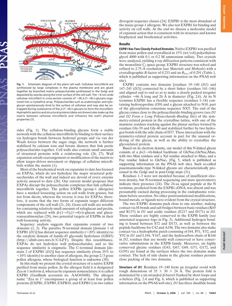
EXPB1 contains two domains [residues 19–140 (D1) and147–245 (D2)] connected by a short linker (residues 141–146)and aligned end to end so as to make a closely packed irregularcylinder �66 Å long and 26 Å in diameter (Fig. 2A). At its Nterminus EXPB1 has a flexible sequence (residues 1–18) con-taining hydroxyproline (O9) and a glycan attached to N10, partof the glycosylation consensus sequence NXT. The end of theglycan comes close to the polysaccharide-binding groove (see D1and D2 Form a Long Polysaccharide-Binding Site) of the sym-metry-related protein in the crystalline lattice, with one of themannose residues stacking against the planar surface formed byresidues Gly-39 and Gly-40 and stabilized further by two hydro-gen bonds with the side chain of D37. These interactions with thesymmetry-related protein account in part for the unusual or-dering of the glycan, as well as the ability to crystallize theglycosylated protein.
Based on its electron density, our model of this N-linked glycanconsists of a �-(134)-linked backbone of GlcNac1GlcNac2Man3with two Man residues and a Xyl residue attached to Man3 and aFuc residue linked to GlcNac1 (Fig. 5, which is published assupporting information on the PNAS web site). Such so-calledpaucimannosidic-type N-linked glycans are characteristically pro-cessed in the Golgi and in post-Golgi steps (31).
Residues 1–3 were not modeled because of insufficient elec-tron density, but N-terminal sequencing and mass spectrometryindicate their presence (24). The 24-aa signal peptide at the Nterminus, predicted from the EXPB1 cDNA, was absent and waspresumably excised during processing in the endoplasmic retic-ulum before secretion. No other posttranslational modifications,bound metals, or ligands were evident from the crystal structure.
The two EXPB1 domains pack close to one another, makingcontact via H-bonds and salt bridges between basic residues (K65and R137) in D1 and acidic residues (E217 and D171) in D2.These residues are highly conserved in the EXPB family (seeannotated sequence logo in Fig. 3). Additional hydrogen bond-ing is found between S72 and D173, as well as between thepeptide backbone for C42 and A196. The two domains also makecontact via a hydrophobic patch consisting of I44, P51, Y52, andY92 in D1 and L164, Y167, and the hydrocarbon chain of K166in D2, residues that are mostly well conserved or have conser-vative substitutions in the EXPB family. Moreover, six highlyconserved glycine residues (G43, G67, G69, G71, G172, andG195) are found at the surfaces where the two domains makecontact. The lack of side chains in the glycine residues permitsclose packing of the two domains.
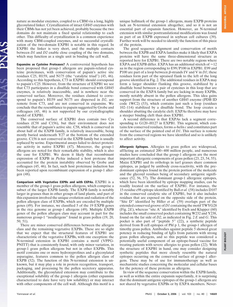
Structure of D1. Residues 19–140 form an irregular ovoid withrough dimensions of 35 � 30 � 24 Å. The protein fold isdominated by a six-stranded �-barrel f lanked by short loops and�-helices (Fig. 2 A and Fig. 6, which is published as supportinginformation on the PNAS web site). D1 has three disulfide bonds
Fig. 1. Schematic diagram of the plant cell wall. Cellulose microfibrils aresynthesized by large complexes in the plasma membrane and are gluedtogether by branched matrix polysaccharides synthesized in the Golgi anddeposited by vesicles along the inner surface of the cell wall. The �4-nm-widecellulose microfibril in cross-section consists of �36 �-(134)-D-glucans orga-nized into a crystalline array. Polysaccharides such as arabinoxylan and xylo-glucan spontaneously bind to the surface of cellulose and may also be en-trapped during coalescence of the �-(134)-D-glucans to form the microfibril.Hydrophilic pectins and structural proteins (data not shown) also make up thematrix between cellulose microfibrils and influence the wall’s physicalproperties (7).
Yennawar et al. PNAS � October 3, 2006 � vol. 103 � no. 40 � 14665
PLA
NT
BIO
LOG
YIN
AU
GU
RAL
ART
ICLE
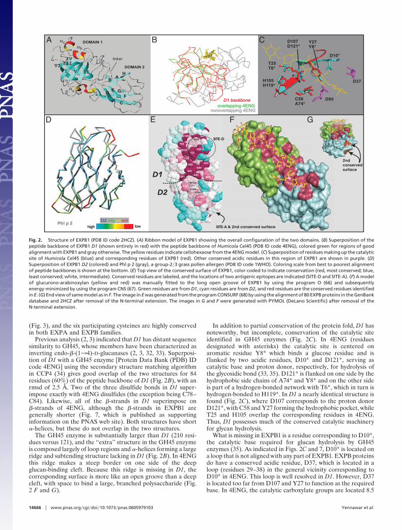
(Fig. 3), and the six participating cysteines are highly conservedin both EXPA and EXPB families.
Previous analysis (2, 3) indicated that D1 has distant sequencesimilarity to GH45, whose members have been characterized asinverting endo-�-(134)-D-glucanases (2, 3, 32, 33). Superposi-tion of D1 with a GH45 enzyme [Protein Data Bank (PDB) IDcode 4ENG] using the secondary structure matching algorithmin CCP4 (34) gives good overlap of the two structures for 84residues (60%) of the peptide backbone of D1 (Fig. 2B), with anrmsd of 2.5 Å. Two of the three disulfide bonds in D1 super-impose exactly with 4ENG disulfides (the exception being C78–C84). Likewise, all of the �-strands in D1 superimpose on�-strands of 4ENG, although the �-strands in EXPB1 aregenerally shorter (Fig. 7, which is published as supportinginformation on the PNAS web site). Both structures have short�-helices, but these do not overlap in the two structures.
The GH45 enzyme is substantially larger than D1 (210 resi-dues versus 121), and the ‘‘extra’’ structure in the GH45 enzymeis composed largely of loop regions and �-helices forming a largeridge and subtending structure lacking in D1 (Fig. 2B). In 4ENGthis ridge makes a steep border on one side of the deepglucan-binding cleft. Because this ridge is missing in D1, thecorresponding surface is more like an open groove than a deepcleft, with space to bind a large, branched polysaccharide (Fig.2 F and G).
In addition to partial conservation of the protein fold, D1 hasnoteworthy, but incomplete, conservation of the catalytic siteidentified in GH45 enzymes (Fig. 2C). In 4ENG (residuesdesignated with asterisks) the catalytic site is centered onaromatic residue Y8* which binds a glucose residue and isf lanked by two acidic residues, D10* and D121*, serving ascatalytic base and proton donor, respectively, for hydrolysis ofthe glycosidic bond (33, 35). D121* is f lanked on one side by thehydrophobic side chains of A74* and Y8* and on the other sideis part of a hydrogen-bonded network with T6*, which in turn ishydrogen-bonded to H119*. In D1 a nearly identical structure isfound (Fig. 2C), where D107 corresponds to the proton donorD121*, with C58 and Y27 forming the hydrophobic pocket, whileT25 and H105 overlap the corresponding residues in 4ENG.Thus, D1 possesses much of the conserved catalytic machineryfor glycan hydrolysis.
What is missing in EXPB1 is a residue corresponding to D10*,the catalytic base required for glucan hydrolysis by GH45enzymes (35). As indicated in Figs. 2C and 7, D10* is located ona loop that is not aligned with any part of EXPB1. EXPB proteinsdo have a conserved acidic residue, D37, which is located in aloop (residues 29–38) in the general vicinity corresponding toD10* in 4ENG. This loop is well resolved in D1. However, D37is located too far from D107 and Y27 to function as the requiredbase. In 4ENG, the catalytic carboxylate groups are located 8.5
A
D
B C
E F G
Fig. 2. Structure of EXPB1 (PDB ID code 2HCZ). (A) Ribbon model of EXPB1 showing the overall configuration of the two domains. (B) Superposition of thepeptide backbone of EXPB1 D1 (shown entirely in red) with the peptide backbone of Humicola Cel45 (PDB ID code 4ENG), colored green for regions of goodalignment with EXPB1 and gray otherwise. The yellow residues indicate cellohexaose from the 4ENG model. (C) Superposition of residues making up the catalyticsite of Humicola Cel45 (blue) and corresponding residues of EXPB1 (red). Other conserved acidic residues in this region of EXPB1 are shown in purple. (D)Superposition of EXPB1 D2 (colored) and Phl p 2 (gray), a group-2�3 grass pollen allergen (PDB ID code 1WHO). Coloring scale from best to poorest alignmentof peptide backbones is shown at the bottom. (E) Top view of the conserved surface of EXPB1, color-coded to indicate conservation (red, most conserved; blue,least conserved; white, intermediate). Conserved residues are labeled, and the locations of two antigenic epitopes are indicated (SITE-D and SITE-A). (F) A modelof glucurono-arabinoxylan (yellow and red) was manually fitted to the long open groove of EXPB1 by using the program O (66) and subsequentlyenergy-minimized by using the program CNS (67). Green residues are from D1, cyan residues are from D2, and red residues are the conserved residues identifiedin E. (G) End view of same model as in F. The image in E was generated from the program CONSURF (68) by using the alignment of 80 EXPB proteins in the GenBankdatabase and 2HCZ after removal of the N-terminal extension. The images in G and F were generated with PYMOL (DeLano Scientific) after removal of theN-terminal extension.
14666 � www.pnas.org�cgi�doi�10.1073�pnas.0605979103 Yennawar et al.
Å apart, which is sufficient distance to accommodate a watermolecule needed for hydrolysis (35). In D1, the carboxylates forD107 and D37 are 15 Å apart, too distant for this catalyticmechanism. Moreover, simple lateral movement of the loop tobring D37 into a correct position seems unlikely because the loopresidues following D37 are rigidly held in place by severalstabilizing interactions. Thus, a key part of the catalytic machin-ery required for hydrolytic activity of GH45 enzymes is lackingin EXPB1.
Inspection of the EXPB1 structure revealed another acidicresidue, D95, which is close to D107 (the carboxylate groups are8.5 Å away). D95 is highly conserved in group-1 allergens, as wellas in EXPBs in general (Fig. 3), but not in EXPA. However, D95is not correctly positioned relative to the D107�Y27 site and thepresumed position of the glycan backbone to serve as thecatalytic base for hydrolysis. D95 and D37 have an appropriatedistance from each other to potentially serve in hydrolysis of asugar residue, which might be bound to the planar hydrophobicsurface made up of G39, G40, and A41 backbone atoms, butnone of these residues are part of the site that is conserved withGH45 enzymes.
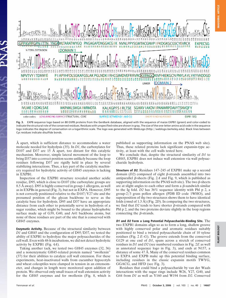
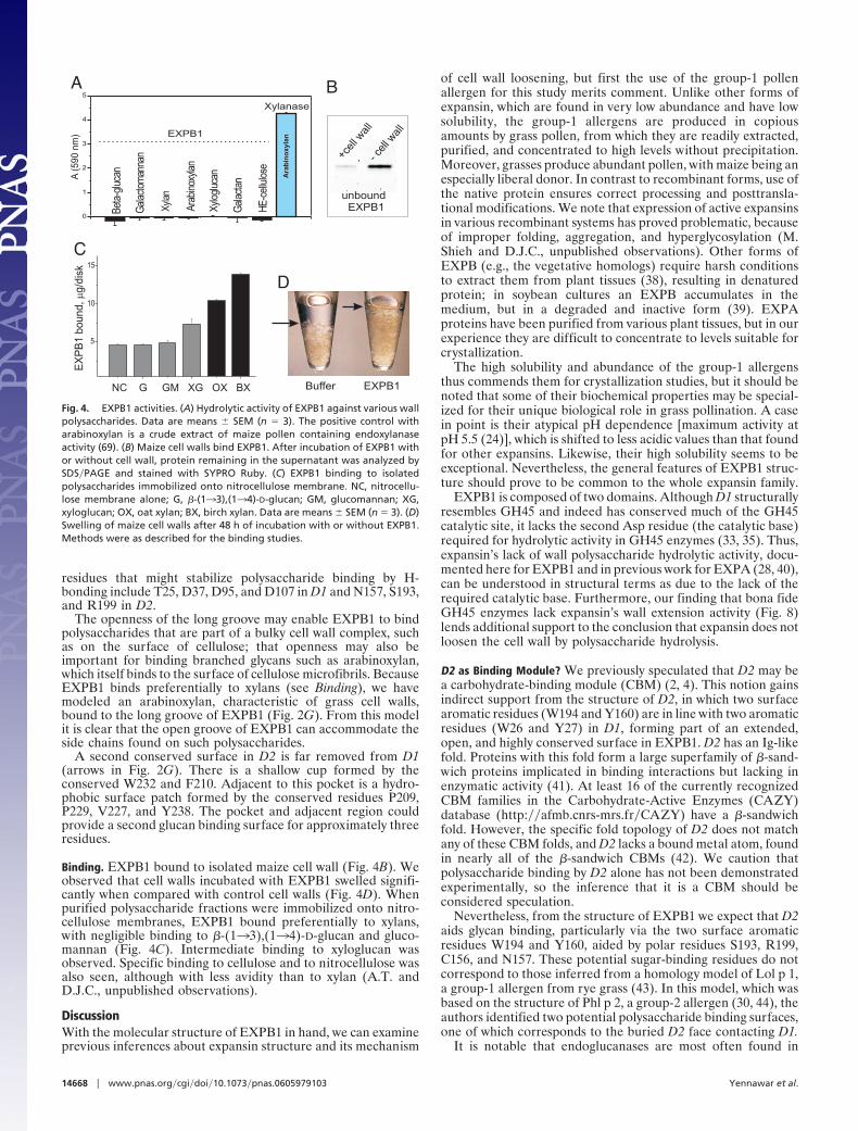
Enzymatic Activity. Because of the structural similarity betweenD1 and GH45 and the configuration of D95�D37, we tested theability of EXPB1 to hydrolyze the major polysaccharides of thecell wall. Even with 48-h incubations, we did not detect hydrolyticactivity by EXPB1 (Fig. 4A).
Taking another tack, we tested two GH45 enzymes (32, 36)and a nonenzymatic GH45-related protein named ‘‘swollenin’’(37) for their abilities to catalyze cell wall extension. For theseexperiments, heat-inactivated walls from cucumber hypocotylsand wheat coleoptiles were clamped in tension in an extensom-eter, and changes in length were monitored upon addition ofprotein. We observed only small traces of wall extension activityfor the GH45 enzymes and for swollenin (Fig. 8, which is
published as supporting information on the PNAS web site).Thus, these related proteins lack significant expansin-type ac-tivity, at least with the cell walls tested here.
We conclude that, despite the structural similarity of D1 toGH45, EXPB1 does not induce wall extension via wall polysac-charide hydrolysis.
Structure of D2. Residues 147–245 of EXPB1 make up a seconddomain (D2) composed of eight �-strands assembled into twoantiparallel �-sheets (Fig. 2 A and Fig. 9, which is published assupporting information on the PNAS web site). The two �-sheetsare at slight angles to each other and form a �-sandwich similarto the Ig fold. D2 has 36% sequence identity with Phl p 2, agroup-2�3 grass pollen allergen (PDB ID code 1WHO), andsuperposition of the two structures shows them to have identicalfolds (rmsd of 1.3 Å) (Fig. 2D). In comparing the two structures,we find that D2 tends to have shorter �-strands compared withPhl p 2, and the two proteins deviate slightly in the loop regionsconnecting the �-strands.
D1 and D2 Form a Long Potential Polysaccharide-Binding Site. Thetwo EXPB1 domains align so as to form a long, shallow groovewith highly conserved polar and aromatic residues suitablypositioned to bind a twisted polysaccharide chain of 10 xyloseresidues (Fig. 2 E–G). The groove extends from the conservedG129 at one end of D1, spans across a stretch of conservedresidues in D1 and D2 (see numbered residues in Fig. 2E as wellas annotated sequence logo in Fig. 3), and ends at N157, adistance of some 47 Å. Many of the conserved residues commonto EXPA and EXPB make up this potential binding surface,including residues in the classic expansin motifs TWYG,GGACG, and HFD (see Fig. 3).
Residues that could bind a polysaccharide by van der Waalsinteractions with the sugar rings include W26, Y27, G40, andG44 from D1 as well as Y160 and W194 from D2. Conserved
Fig. 3. EXPB sequence logo based on 80 EXPB proteins from the GenBank database, aligned with the sequence of maize EXPB1 (green) and color-coded toindicate the structural role of the conserved residues. Residues with unspecified roles are shown in gray. The size of the one-letter amino acid code in the sequencelogo indicates the degree of conservation on a logarithmic scale. The logo was generated with WebLogo (http:��weblogo.berkeley.edu). Black lines betweenCys residues indicate disulfide bonds.
Yennawar et al. PNAS � October 3, 2006 � vol. 103 � no. 40 � 14667
PLA
NT
BIO
LOG
YIN
AU
GU
RAL
ART
ICLE
residues that might stabilize polysaccharide binding by H-bonding include T25, D37, D95, and D107 in D1 and N157, S193,and R199 in D2.
The openness of the long groove may enable EXPB1 to bindpolysaccharides that are part of a bulky cell wall complex, suchas on the surface of cellulose; that openness may also beimportant for binding branched glycans such as arabinoxylan,which itself binds to the surface of cellulose microfibrils. BecauseEXPB1 binds preferentially to xylans (see Binding), we havemodeled an arabinoxylan, characteristic of grass cell walls,bound to the long groove of EXPB1 (Fig. 2G). From this modelit is clear that the open groove of EXPB1 can accommodate theside chains found on such polysaccharides.
A second conserved surface in D2 is far removed from D1(arrows in Fig. 2G). There is a shallow cup formed by theconserved W232 and F210. Adjacent to this pocket is a hydro-phobic surface patch formed by the conserved residues P209,P229, V227, and Y238. The pocket and adjacent region couldprovide a second glucan binding surface for approximately threeresidues.
Binding. EXPB1 bound to isolated maize cell wall (Fig. 4B). Weobserved that cell walls incubated with EXPB1 swelled signifi-cantly when compared with control cell walls (Fig. 4D). Whenpurified polysaccharide fractions were immobilized onto nitro-cellulose membranes, EXPB1 bound preferentially to xylans,with negligible binding to �-(133),(134)-D-glucan and gluco-mannan (Fig. 4C). Intermediate binding to xyloglucan wasobserved. Specific binding to cellulose and to nitrocellulose wasalso seen, although with less avidity than to xylan (A.T. andD.J.C., unpublished observations).
DiscussionWith the molecular structure of EXPB1 in hand, we can examineprevious inferences about expansin structure and its mechanism
of cell wall loosening, but first the use of the group-1 pollenallergen for this study merits comment. Unlike other forms ofexpansin, which are found in very low abundance and have lowsolubility, the group-1 allergens are produced in copiousamounts by grass pollen, from which they are readily extracted,purified, and concentrated to high levels without precipitation.Moreover, grasses produce abundant pollen, with maize being anespecially liberal donor. In contrast to recombinant forms, use ofthe native protein ensures correct processing and posttransla-tional modifications. We note that expression of active expansinsin various recombinant systems has proved problematic, becauseof improper folding, aggregation, and hyperglycosylation (M.Shieh and D.J.C., unpublished observations). Other forms ofEXPB (e.g., the vegetative homologs) require harsh conditionsto extract them from plant tissues (38), resulting in denaturedprotein; in soybean cultures an EXPB accumulates in themedium, but in a degraded and inactive form (39). EXPAproteins have been purified from various plant tissues, but in ourexperience they are difficult to concentrate to levels suitable forcrystallization.
The high solubility and abundance of the group-1 allergensthus commends them for crystallization studies, but it should benoted that some of their biochemical properties may be special-ized for their unique biological role in grass pollination. A casein point is their atypical pH dependence [maximum activity atpH 5.5 (24)], which is shifted to less acidic values than that foundfor other expansins. Likewise, their high solubility seems to beexceptional. Nevertheless, the general features of EXPB1 struc-ture should prove to be common to the whole expansin family.
EXPB1 is composed of two domains. Although D1 structurallyresembles GH45 and indeed has conserved much of the GH45catalytic site, it lacks the second Asp residue (the catalytic base)required for hydrolytic activity in GH45 enzymes (33, 35). Thus,expansin’s lack of wall polysaccharide hydrolytic activity, docu-mented here for EXPB1 and in previous work for EXPA (28, 40),can be understood in structural terms as due to the lack of therequired catalytic base. Furthermore, our finding that bona fideGH45 enzymes lack expansin’s wall extension activity (Fig. 8)lends additional support to the conclusion that expansin does notloosen the cell wall by polysaccharide hydrolysis.
D2 as Binding Module? We previously speculated that D2 may bea carbohydrate-binding module (CBM) (2, 4). This notion gainsindirect support from the structure of D2, in which two surfacearomatic residues (W194 and Y160) are in line with two aromaticresidues (W26 and Y27) in D1, forming part of an extended,open, and highly conserved surface in EXPB1. D2 has an Ig-likefold. Proteins with this fold form a large superfamily of �-sand-wich proteins implicated in binding interactions but lacking inenzymatic activity (41). At least 16 of the currently recognizedCBM families in the Carbohydrate-Active Enzymes (CAZY)database (http:��afmb.cnrs-mrs.fr�CAZY) have a �-sandwichfold. However, the specific fold topology of D2 does not matchany of these CBM folds, and D2 lacks a bound metal atom, foundin nearly all of the �-sandwich CBMs (42). We caution thatpolysaccharide binding by D2 alone has not been demonstratedexperimentally, so the inference that it is a CBM should beconsidered speculation.
Nevertheless, from the structure of EXPB1 we expect that D2aids glycan binding, particularly via the two surface aromaticresidues W194 and Y160, aided by polar residues S193, R199,C156, and N157. These potential sugar-binding residues do notcorrespond to those inferred from a homology model of Lol p 1,a group-1 allergen from rye grass (43). In this model, which wasbased on the structure of Phl p 2, a group-2 allergen (30, 44), theauthors identified two potential polysaccharide binding surfaces,one of which corresponds to the buried D2 face contacting D1.
It is notable that endoglucanases are most often found in
A B
C
D
Fig. 4. EXPB1 activities. (A) Hydrolytic activity of EXPB1 against various wallpolysaccharides. Data are means � SEM (n � 3). The positive control witharabinoxylan is a crude extract of maize pollen containing endoxylanaseactivity (69). (B) Maize cell walls bind EXPB1. After incubation of EXPB1 withor without cell wall, protein remaining in the supernatant was analyzed bySDS�PAGE and stained with SYPRO Ruby. (C) EXPB1 binding to isolatedpolysaccharides immobilized onto nitrocellulose membrane. NC, nitrocellu-lose membrane alone; G, �-(133),(134)-D-glucan; GM, glucomannan; XG,xyloglucan; OX, oat xylan; BX, birch xylan. Data are means � SEM (n � 3). (D)Swelling of maize cell walls after 48 h of incubation with or without EXPB1.Methods were as described for the binding studies.
14668 � www.pnas.org�cgi�doi�10.1073�pnas.0605979103 Yennawar et al.
nature as modular enzymes, coupled to a CBM via a long, highlyglycosylated linker. Crystallization of intact GH45 enzymes withtheir CBMs has not yet been achieved, probably because the twodomains do not maintain a fixed spatial relationship to eachother. This difficulty of crystallization is a common experiencewith many CBM-coupled enzymes, and so successful crystalli-zation of the two-domain EXPB1 is notable in this regard. InEXPB1 the linker is very short, and the multiple contactsbetween D1 and D2 enable close coupling of the two domains,which may function as a single unit in binding the cell wall.
Expansins as Cysteine Proteases? A controversial hypothesis hasbeen proposed that group-1 allergens are papain-related cys-teine proteinases, with conservation of papain’s active siteresidues C25, H159, and N175 (the ‘‘catalytic triad’’) (45, 46).According to this hypothesis, C73 in EXPB1 should correspondto papain’s C25. However, from the structure of EXPB1 we seethat C73 participates in a disulfide bond conserved with GH45enzymes, is relatively inaccessible, and is nowhere near theconserved surface. Moreover, the residues claimed to corre-spond to papain’s H159 and N175 are dispersed in D2, areremote from C73, and are not conserved in expansins. Weconclude that the resemblance to papain suggested by Grobe andcolleagues (45, 46) is not supported by our crystallographicmodel of EXPB1.
The conserved surface of EXPB1 does contain two Cysresidues (C58 and C156), but their environment does notresemble that of papain’s active site. C58, which is conserved inabout half of the EXPB family, is relatively inaccessible, beingmostly buried underneath Y27 at the bottom of the extendedgroove. C156 is not conserved in the EXPB family but is usuallyreplaced by serine. Experimental assays failed to detect protein-ase activity in native EXPB1 (47). Moreover, the group-1allergens are noted for their remarkable stability, which is alsothe case for EXPB1. We deem it likely that recombinantexpression of EXPB in Pichia induced a host protease thataccounted for the protein instability observed by Grobe andcolleagues (45, 46). In fact, such host proteinase induction hasbeen reported upon recombinant expression of a group-1 aller-gen (48).
Comparison with Vegetative EXPBs and with EXPAs. EXPB1 is amember of the group-1 grass pollen allergens, which comprise asubset of the larger EXPB family. The EXPB family is notablylarger in grasses than in other groups of land plants, and part ofthis expansion involved the unique evolution and radiation of thepollen allergen class of EXPBs, which are encoded by multiplegenes (49). For instance, we classified 5 of the 19 EXPB genesin the rice genome as group-1 allergens (49). Multiple EXPBgenes of the pollen allergen class may account in part for thenumerous group-1 ‘‘isoallergens’’ found in grass pollen (19, 20,50, 51).
There are minor conserved differences between the allergenclass and the remaining vegetative EXPBs. These are so slightthat we expect that the structural features of EXPB1 arecharacteristic of the vegetative EXPBs, with one exception: theN-terminal extension in EXPB1 contains a motif (VPPG-PNITT) that is consistently found, with only minor variation, ingroup-1 grass pollen allergens but not in other EXPBs. Thismotif contains one or more hydroxyprolines and a glycosylatedasparagine, features common to the pollen allergen class ofEXPB (52). The function of this N-terminal extension is un-known, but it may play a role in protein recognition, transport,packaging, and processing by the pollen secretory apparatus.Additionally, the glycosylated extension may contribute to theexceptional solubility of the group-1 allergens (other expansinscharacterized to date have very low solubility) or may interactwith other components of the cell wall. Although this motif is a
unique hallmark of the group-1 allergens, many EXPB proteinslack an N-terminal extension altogether, and so it is not anessential part of expansin function. However, an N-terminalextension with similar posttranslational modifications was foundas part of an EXPB expressed in soybean cell cultures (39).Further work will be needed to identify the function of this pieceof the protein.
The good sequence alignment and conservation of motifsbetween the EXPB and EXPA families make it likely that EXPAproteins will have the same three-dimensional structure asreported here for EXPB1. There are two notable regions whereEXPA and EXPB differ. EXPA has an additional stretch of �12aa in the region corresponding to E99�P100 in EXPB1. E99 andP100 are part of a loop between �-strands IV and V in D1; theseresidues form part of the upraised flank to the left of the longgroove identified in Fig. 2. The additional residues in EXPA mayform a larger shoulder flanking this groove, stabilized by adisulfide bond between a pair of cysteines in this loop that areconserved in the EXPA family but are lacking in many EXPBs,mostly notably absent in the pollen allergens. This idea gainssupport from the structure of another GH45 enzyme (PDB IDcode 1WC2) (53), which contains just such a loop (residues102–114) stabilized by a disulfide bond. The loop creates ashoulder abutting the catalytic cleft. EXPAs therefore may havea steeper binding cleft than does EXPB1.
A second difference is that EXPAs lack a segment corre-sponding to G120–H127 in EXPB1. This segment, which con-tains few conserved residues, forms �-helix c and constitutes partof the surface of the pointed end of D1. This surface is remotefrom the conserved regions we have identified and so is unlikelyto affect activity.
Allergenic Epitopes. Allergies to grass pollen are widespread,aff licting an estimated 200–400 million people, and numerousstudies have concluded that the group-1 allergens are the mostimportant allergenic components of grass pollen (23, 23, 54, 55).Maize EXPB1 and its orthologs in turf grasses share commonepitopes, as judged by antibody cross-reactivity, with the pre-dominant epitopes found in the protein portion of the moleculeand the glycosyl residues being of secondary antigenic signifi-cance (52, 56, 57). The dominant group-1 allergenic epitopes,which have been identified by epitope mapping studies, can bereadily located on the surface of EXPB1. For instance, the15-residue c98 epitope identified by Ball et al. (58) includes D107in the conserved catalytic site of EXPB1, but it also includesresidues that are exposed on the opposite side of the protein.‘‘Site D’’ identified by Hiller et al. (59) overlaps part of theextended conserved groove of D1 containing the motif TWYG28(Fig. 2E), whereas ‘‘site A’’ identified by Esch and Klapper (60)includes the small conserved pocket containing W232 and Y238,found on the far side of D2, as indicated in Fig. 2 E and G. Thispocket is also part of ‘‘peptide 5’’ (22), a synthetic peptidederived from B cell epitopes of Phl p 1, the group-1 allergen oftimothy grass pollen. Antibodies against peptide 5 showed greatpotency in reducing binding of IgEs from patients with stronggrass pollen allergens, and so this peptide was considered apotentially useful component of an epitope-based vaccine fortreating patients with severe allergies to grass pollen (22). Withthe structure of EXPB1 in hand, one may consider designingsynthetic peptides that more closely resemble the naturalepitopes occurring on the conserved surface of group-1 aller-gens. These may be of use for immunotherapy as well asmechanistic studies concerning the molecular and cellular basesfor the potency of these proteins as allergens.
In view of the sequence conservation within the EXPB family,as well as within the entire expansin superfamily, it is surprisingthat the dominant antigenic epitopes of the group-1 allergens arenot shared by vegetative EXPBs or by EXPA members. Never-
Yennawar et al. PNAS � October 3, 2006 � vol. 103 � no. 40 � 14669
PLA
NT
BIO
LOG
YIN
AU
GU
RAL
ART
ICLE
theless, this seems to be the case because antibodies raisedagainst the group-1 allergens do not recognize other forms ofexpansin. This is indeed fortunate, because otherwise personswith strong allergies to grass pollen would also be allergic to freshfruits, vegetables, grains, and other plant tissues that expressmembers of this large gene family that is ubiquitous in plants.
A Molecular Model of Wall Loosening by Expansins. Expansin actionmay be summarized as follows: the protein binds one or morewall polysaccharides and within seconds induces wall stressrelaxation followed by wall extension, without hydrolysis of thewall polymers. There is no requirement for ATP or anothersource of chemical energy, and the wall continues to extend solong as the wall bears sufficient tension and expansin is present(that is, expansin acts catalytically, not stoichiometrically).
In the case of EXPB1, we imagine that stress relaxation beginswhen it binds a taut arabinoxylan tethered to a cellulose micro-fibril, causing local release of the arabinoxylan from the cellulosesurface. Movement of the EXPB along the arabinoxylan–cellulose junction would enable it to unzip the hydrogen bondsbetween the polysaccharides, relaxing the taut tether and allow-ing turgor-driven displacement of cellulose and arabinoxylan,which may then reassociate in a relaxed state to restore wallstrength. During this movement, the two expansin domainsmight shift in a hinge-like manner, binding and letting go of thearabinoxylan independent of each other, leading to an inch-worm-like movement along the polysaccharide. We estimate thatas little as a 10° shift in angle between domains could cause aone-residue dislocation of the polysaccharide along the bindingsurface.
To assess the feasibility of such interdomain movement, weestimated the buried surface area between the two domainsusing CCP4 (34). The value is 589 Å2, which is indicative of aweak interdomain interaction (61), consistent with domainmovements as imagined above. A potential source of energy forthese movements is the mechanical strain energy stored by thetaut polysaccharide in a turgor-stretched cell wall. In this model,expansin acts as molecular device that uses the strain energystored in a taut cellulose-binding glycan to help dissociate theglycan from the surface of cellulose.
Materials and MethodsProtein Purification, Crystallization, and Data Collection. Native Zeam 1 was extracted from pollen of field-grown maize plants at 4°Cin 0.125 M sodium carbonate and then purified to electro-phoretic homogeneity in the presence of 5 mM DTT by using twochromatographic steps as described (24). With this method, fourZea m 1 isoforms were readily distinguished, and we used themost abundant isoform, Zea m 1d (EXPB1), for crystallizationand activity assays. For the binding experiments, EXPB1 wasfurther purified by HPLC on a reverse-phase column (DiscoveryC8, 15 cm � 4.6 mm i.d., 5 �m; Supelco, Bellefonte, PA)preequilibrated with 10% acetonitrile containing 0.1% trif lu-oroacetic acid. Bound protein was eluted at 1 ml�min�1 with alinear gradient of 22–90% acetonitrile in the same solution for20 min at a flow rate of 1 ml�min�1 at 25°C. We confirmed wallextension activity of EXPB1 purified in this way.
Crystals were grown at 21°C for 9 days by using EXPB1 at 10.5mg�ml in 100 mM Na acetate (pH 4.6) in 5-�l hanging drops,with addition of 5 �l of precipitant [15% (wt�vol) polyethyleneglycol 4000 with 0.1 or 0.2 M ammonium sulfate] and with a 1-mlreservoir volume. Two crystals were analyzed, yielding diffrac-tion patterns consistent with the monoclinic C2 space group. Crystal1 had unit cell dimensions of a � 113.7 Å, b � 45.2 Å, and c � 70.3Å with angles � � 90.0°, � � 124.6°, and � � 90.0°; crystal 2 hadunit cell dimensions of a � 112.6 Å, b � 44.4 Å, and c � 69.6Å with angles � � 90.0°, � � 124.4°, and � � 90.0°.
Data were collected by using a RU200 rotating anode x-ray
generator (Rigaku) with CuK� radiation, operating at 5 kW ofpower (50 kV, 100 mA) (Molecular Structure, The Woodlands,TX). Three-degree oscillation frames, each exposed for 120 min,were collected on an R-AXIS IV detector. The two crystals wereused to get a 93% complete data set. DENZO and SCALE-PACK software suites (62) were used for data processing.
Structure Solution and Refinement. Our final model of EXPB1structure was based on the native crystal data set and was solvedby molecular replacement calculations by using the programAMoRe (63) with the structure of Phl p 1 (PDB ID code 1N10),which has 58% amino acid identity with EXPB1 over 240residues. EXPB1 has four more residues at its C terminus. Thebest molecular replacement solution in AMoRE was obtained bydeleting the first 13 residues of the N terminus (attempts thatincluded this stretch did not yield a solution) and by including allof the side chains for the rest of the protein (attempts with justthe backbone atoms did not yield a good solution as well) andincluding all of the available data to 2.75 Å. The correlationcoefficient and the R-factor for the best solution were 55.1% and51.0%, respectively. The next best solution had an inferiorcorrelation coefficient and R-factor of 49.3% and 53.9%, en-abling us to proceed with further refinement and model buildingwith confidence. For further refinement details and comparisonwith the 1N10 structure, see Supporting Text, which is publishedas supporting information on the PNAS web site. Coordinatesand structure factors of the structure have been deposited in theProtein Data Bank [PDB ID code 2HCZ (64)]. A summary ofthe refinement results is given in Table 1.
Polysaccharide Hydrolysis. Two milligrams of dye-coupled insolu-ble polysaccharides (AZCL polysaccharides; Megazyme, Wick-low, Ireland) were suspended in 100 �l of buffer [50 mM sodiumacetate (pH 4.5) with 1 mM NaN3 and 10 mM DTT] andincubated with shaking at 30°C for 48 h with or without 30 �gof EXPB1. At the end of the incubation, 300 �l of 2.5% Trizmabase was added to each tube to stop reaction, the suspension wascentrifuged, and the absorbance (590 nm) of the supernatant wasmeasured.
Binding. Cell walls were collected from maize silks, cleaned byphenol�acetic acid�water washes (65), and lyophilized. EXPB1was purified on a CM-Sepharose Fast Flow (Amersham Bio-sciences) column in a LP system (Bio-Rad) (24). EXPB1 (10 �g)was incubated with 1 mg of cell wall in 400 �l of 50 mM sodiumacetate (pH 5.5) for 1 h at 25°C with agitation. After incubation,protein remaining in the supernatant was analyzed by SDS�PAGE (12% polyacrylamide) and stained with SYPRO Rubyprotein gel stain (Bio-Rad).
Commercial polysaccharides [200 �g each, consisting of oatspelts xylan (Sigma), birch wood xylan (Fluka), barley �-glucan(catalog no. G-6513; Sigma), konjac glucomanna (Megazyme),or tamarind xyloglucan (Megazyme)] were dissolved in 10 �l of20 mM sodium acetate (pH 4.5) and applied to Protran BA83nitrocellulose membrane disks (diameter, �7 mm; pore size, 0.2�m; Whatman). The disks were dried at 80°C overnight. Thecoated disks were incubated with blocking reagent (Roche)dissolved in 0.1 M maleic acid buffer for 1 h at room temperatureto reduce nonspecific binding of EXPB1. After the blocking, thedisks were washed with 20 mM Na acetate five times for 3 mineach and then incubated with EXPB1 (20 �g per tube; purifiedby reverse-phase chromatography; see Protein Purification, Crys-tallization, and Data Collection) in 400 �l of 20 mM sodiumacetate (pH 5.5) at 25°C for 1 h. After the incubation, thesupernatant (unbound protein) was analyzed by reverse-phasechromatography (see Protein Purification, Crystallization, andData Collection). The amount of EXPB1 bound to the coatednitrocellulose membrane disks was calculated from the reduc-
14670 � www.pnas.org�cgi�doi�10.1073�pnas.0605979103 Yennawar et al.
tion in the amount of unbound protein assessed by reverse-phaseHPLC of the supernatant.
In memory of Paul B. Green (1921–1998). We thank Dr. Greg Farber forinestimable advice and assistance with growing the EXPB1 crystals; Dr.Javier Sampedro for useful discussions; Daniel M. Durachko, Edward
Wagner, and Dr. Hemant Yennawar for expert technical assistance; Dr.Colin Mitchison (Genencor International, Palo Alto, CA) for the gift ofthe swollenin sample; Dr. Ines Munoz (Uppsala University, Uppsala,Sweden) for the gift of the TrCel45 sample; and Dr. Jan-Christer Janson(Uppsala University) for the gift of the MeCel45 sample. This work wassupported by Department of Energy Grant FG02-84ER13179 andNational Institutes of Health Grant 5R01GM60397 (to D.J.C.).
1. Cosgrove DJ (2005) Nat Rev Mol Cell Biol 6:850–861.2. Cosgrove DJ (1997) Plant Cell 9:1031–1041.3. Sampedro J, Cosgrove DJ (2005) Genome Biol 6:242.1–242.11.4. Cosgrove DJ (2000) Nature 407:321–326.5. Rose JK, Braam J, Fry SC, Nishitani K (2002) Plant Cell Physiol 43:1421–1435.6. Domingo C, Roberts K, Stacey NJ, Connerton I, Ruiz-Teran F, McCann MC
(1998) Plant J 13:17–28.7. Carpita NC, Gibeaut DM (1993) Plant J 3:1–30.8. Park YW, Tominaga R, Sugiyama J, Furuta Y, Tanimoto E, Samejima, M,
Sakai F, Hayashi T (2003) Plant J 33:1099–1106.9. McQueen-Mason S, Durachko DM, Cosgrove DJ (1992) Plant Cell 4:1425–
1433.10. Li Z-C, Durachko DM, Cosgrove DJ (1993) Planta 191:349–356.11. Darley CP, Forrester AM, McQueen-Mason SJ (2001) Plant Mol Biol 47:179–
195.12. Choi DS, Lee Y, Cho HT, Kende H (2003) Plant Cell 15:1386–1398.13. Pien S, Wyrzykowska J, McQueen-Mason S, Smart C, Fleming A (2001) Proc
Natl Acad Sci USA 98:11812–11817.14. Belfield EJ, Ruperti B, Roberts JA, McQueen-Mason SJ (2005) J Exp Bot
56:817–823.15. Cho HT, Cosgrove DJ (2000) Proc Natl Acad Sci USA 97:9783–9788.16. Rose JK, Lee HH, Bennett AB (1997) Proc Natl Acad Sci USA 94:5955–5960.17. Chen F, Dahal P, Bradford KJ (2001) Plant Physiol 127:928–936.18. Li Y, Darley CP, Ongaro V, Fleming A, Schipper O, Baldauf SL, McQueen-
Mason SJ (2002) Plant Physiol 128:854–864.19. Andersson K, Lidholm J (2003) Int Arch Allergy Immunol 130:87–107.20. Ansari AA, Kihara TK, Marsh DG (1987) J Immunol 12:4034–4041.21. Cosgrove DJ, Bedinger P, Durachko DM (1997) Proc Natl Acad Sci USA
94:6559–6564.22. Focke M, Mahler V, Ball T, Sperr WR, Majlesi Y, Valent P, Kraft D, Valenta
R (2001) FASEB J 15:2042–2044.23. Ball T, Edstrom W, Mauch L, Schmitt J, Leistler B, Fiebig H, Sperr WR,
Hauswirth AW, Valent P, Kraft D, et al. (2005) FEBS J 272:217–227.24. Li LC, Bedinger PA, Volk C, Jones AD, Cosgrove DJ (2003) Plant Physiol
132:2073–2085.25. Wu Y, Meeley RB, Cosgrove DJ (2001) Plant Physiol 126:222–232.26. Hall Q, Cannon MC (2002) Plant Cell 14:1161–1172.27. Ribeiro JM, Pereira CS, Soares NC, Vieira AM, Feijo JA, Jackson PA (2006)
J Exp Bot 57:2025–2035.28. McQueen-Mason SJ, Cosgrove DJ (1995) Plant Physiol 107:87–100.29. Carpita NC (1996) Annu Rev Plant Physiol Plant Mol Biol 47:445–476.30. De Marino S, Morelli MA, Fraternali F, Tamborini E, Musco G, Vrtala S,
Dolecek C, Arosio P, Valenta R, Pastore A (1999) Struct Folding Des7:943–952.
31. Lerouge P, Cabanes-Macheteau M, Rayon C, Fischette-Laine AC, Gomord V,Faye L (1998) Plant Mol Biol 38:31–48.
32. Xu B, Hellman U, Ersson B, Janson JC (2000) Eur J Biochem 267:4970–4977.33. Davies GJ, Tolley SP, Henrissat B, Hjort C, Schulein M (1995) Biochemistry
34:16210–16220.34. Collaborative Computational Project, Number 4 (1994) Acta Crystallogr D
50:760–763.35. Davies GJ, Dodson GG, Hubbard RE, Tolley SP, Dauter Z, Wilson KS, Hjort
C, Mikkelsen JM, Rasmussen G, Schulein M (1993) Nature 365:362–364.
36. Saloheimo A, Henrissat B, Hoffren AM, Teleman O, Penttila M (1994) MolMicrobiol 13:219–228.
37. Saloheimo M, Paloheimo M, Hakola S, Pere J, Swanson B, Nyyssonen E, BhatiaA, Ward M, Penttila M (2002) Eur J Biochem 269:4202–4211.
38. Lee Y, Choi D (2005) Mol Cells 20:119–126.39. Downes BP, Steinbaker CR, Crowell DN (2001) Plant Physiol 126:244–252.40. McQueen-Mason S, Cosgrove DJ (1994) Proc Natl Acad Sci USA 91:6574–
6578.41. Bork P, Holm L, Sander C (1994) J Mol Biol 242:309–320.42. Boraston AB, Bolam DN, Gilbert HJ, Davies GJ (2004) Biochem J 382:769–
781.43. Barre A, Rouge P (2002) Biochem Biophys Res Commun 296:1346–1351.44. Fedorov AA, Ball T, Valenta R, Almo SC (1997) Int Arch Allergy Immunol
113:109–113.45. Grobe K, Becker WM, Petersen A (1999) Eur J Biochem 263:33–40.46. Grobe K, Poppelmann M, Becker WM, Petersen A (2002) Eur J Biochem
269:2083–2092.47. Li LC, Cosgrove DJ (2001) Eur J Biochem 268:4217–4226.48. Poppelmann M, Becker WM, Petersen A (2002) Electrophoresis 23:993–997.49. Sampedro J, Lee Y, Carey RE, dePamphilis C, Cosgrove DJ (2005) Plant J
44:409–419.50. Petersen A, Becker WM, Schlaak M (1993) J Allergy Clin Immunol 92:789–796.51. Suphioglu C, Singh MB, Knox RB (1993) Int Arch Allergy Appl Immunol
102:144–151.52. Petersen A, Becker WM, Moll H, Blumke M, Schlaak M (1995) Electrophoresis
16:869–875.53. Xu B, Janson JC, Sellos D (2001) Eur J Biochem 268:3718–3727.54. Marsh DG, Milner FH, Johnson P (1966) Int Arch Allergy Appl Immunol
29:521–535.55. Ball T, Fuchs T, Sperr WR, Valent P, Vangelista L, Kraft D, Valenta R (1999)
FASEB J 13:1277–1290.56. Howlett BJ, Clarke AE (1981) Biochem J 197:707–714.57. Petersen A, Schramm G, Schlaak M, Becker WM (1998) Clin Exp Allergy
28:315–321.58. Ball T, Vrtala S, Sperr WR, Valent P, Susani M, Kraft D, Valenta R (1994)
J Biol Chem 269:28323–28328.59. Hiller KM, Esch RE, Klapper DG (1997) J Allergy Clin Immunol 100:335–340.60. Esch RE, Klapper DG (1989) Mol Immunol 26:557–561.61. Nooren IM, Thornton JM (2003) J Mol Biol 325:991–1018.62. Otwinowski Z, Minor W (1997) Methods Enzymol 276:307–326.63. Navaza J (1994) Acta Crystallogr A 50:157–163.64. Berman HM, Westbrook J, Feng Z, Gilliland G, Bhat TN, Weissig H,
Shindyalov IN, Bourne PE (2000) Nucleic Acids Res 28:235–242.65. Fry SC (1988) The Growing Plant Cell Wall: Chemical and Metabolic Analysis
(Longman Scientific and Technical, London).66. Jones TA, Zou JY, Cowan SW, Kjeldgaard M (1991) Acta Crystallogr A
47:110–119.67. Brunger AT, Adams PD, Clore GM, DeLano WL, Gros P, Grosse-Kunstleve
RW, Jiang JS, Kuszewski J, Nilges M, Pannu NS, et al. (1998) Acta CrystallogrD 54:905–921.
68. Glaser F, Pupko T, Paz I, Bell RE, Bechor-Shental D, Martz E, Ben-Tal N(2003) Bioinformatics 19:163–164.
69. Wu SS, Suen DF, Chang HC, Huang AH (2002) J Biol Chem 277:49055–49064.
Yennawar et al. PNAS � October 3, 2006 � vol. 103 � no. 40 � 14671
PLA
NT
BIO
LOG
YIN
AU
GU
RAL
ART
ICLE
Related Documents