Pneumoconiosis A disease of the lungs characterized by fibrosis and caused by the chronic inhalation of mineral dusts, especially silica and asbestos. Helen Lang Dept. Geology & Geography West Virginia University Geol 484 – Minerals & the Environment

Welcome message from author
This document is posted to help you gain knowledge. Please leave a comment to let me know what you think about it! Share it to your friends and learn new things together.
Transcript
Pneumoconiosis
A disease of the lungs characterized by fibrosis and caused by the chronic inhalation of mineral dusts, especially silica and asbestos.
Helen LangDept. Geology & GeographyWest Virginia UniversityGeol 484 – Minerals & the Environment
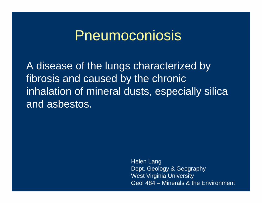
Particle Size and Dust Inhalation
• MMAD = mass median aerodynamic diameter• MMAD > 15 μm, deposited in outer portion of
nasal passages• 15 μm > MMAD > 10 μm, deposited in nasal
turbinates and pharynx• 10 μm > MMAD > 5 μm, inhalable, can descend
as far as the major airways, trachea and main stem bronchi
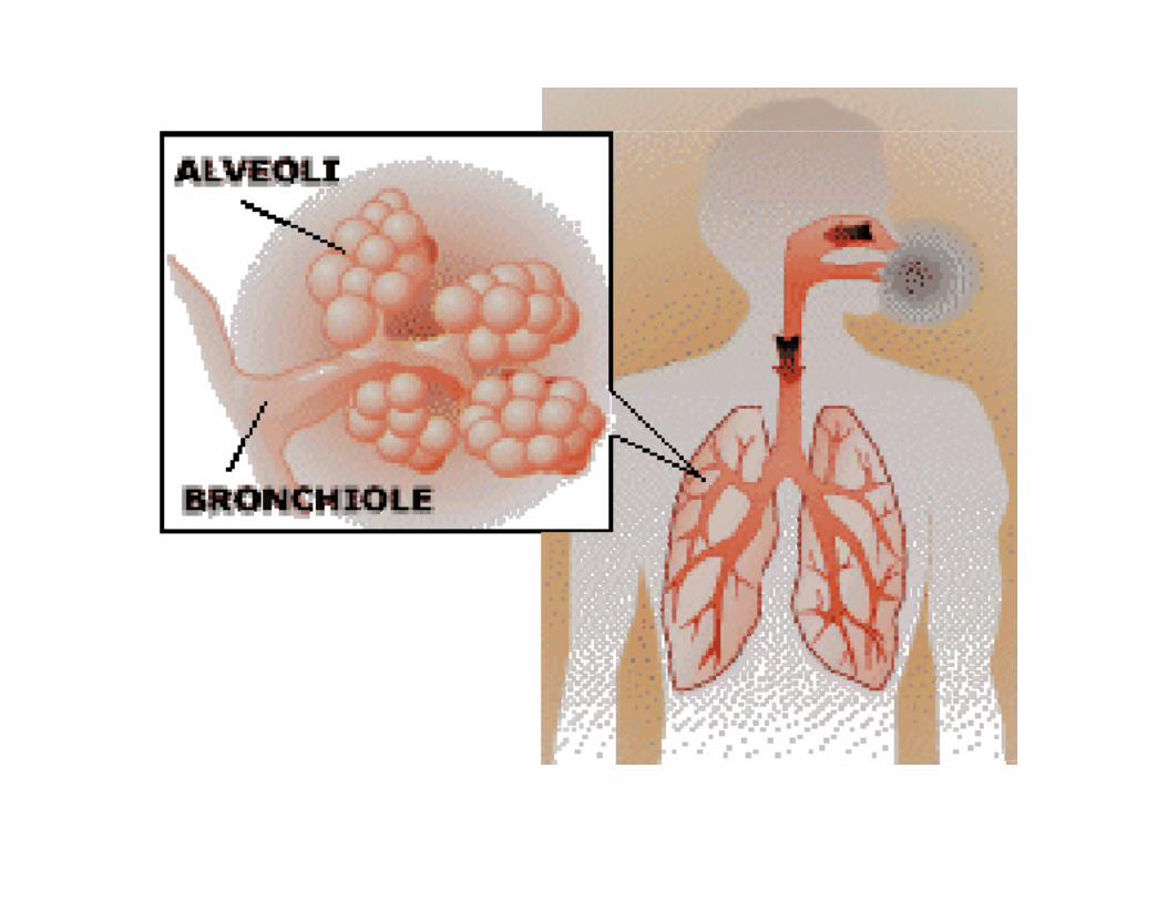
• 5 μm > MMAD, respirable, can penetrate as far as the terminal bronchioles and alveoli (these cause pneumonoconiosis)
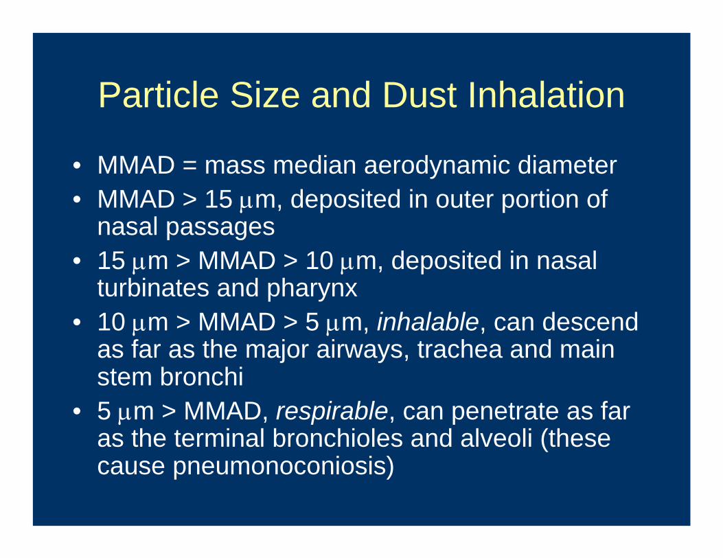
Nose and throat
Pharynx describes the part of the throat that begins from behind the nose to the beginning of the voice box and the esophagus.
The nasal turbinates are shelf-like structures in the nasal cavity (which begins where the inside of your nose enters your head). They serve to provide moisture, warmth, and airflow for breathing, and many of the body's natural defenses against infection.
http://en.wikipedia.org/wiki/Image:Illu01_head_neck.jpghttp://www.seattledoctors.com/images/sidenose.jpg

The Human Lung – Gray’s Anatomy
FIG. 962– Bronchi and bronchioles. The lungs have been widely separated and tissue cut away to expose the air-tubes.
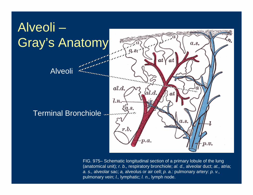
Alveoli –Gray’s Anatomy
FIG. 975– Schematic longitudinal section of a primary lobule of the lung (anatomical unit); r. b., respiratory bronchiole; al. d., alveolar duct; at., atria; a. s., alveolar sac; a, alveolus or air cell; p. a.: pulmonary artery: p. v.,pulmonary vein; l., lymphatic; l. n., lymph node.
Terminal Bronchiole
Alveoli

Cardiovascular System on the Web
• Human Anatomy Online: an educational website
• www.innerbody.com/htm/anim.htmlView– Circulatory System– Heart (cut view)– Lungs (may not work)
When Insoluble Inorganic Material (like silica and asbestos) enters the lungs,
• It experiences little or no metabolic breakdown and dissolves VERY slowly
• Respirable (<5μm) particles accumulate in the alveoli
• Particles must be physically removed or they stay in the lungs and cause inflammation and disease
Dust removal mechanisms
• Some particles are removed from larger airways by mucociliary clearance mechanisms
• Some particles are engulfed by scavenger cells called macrophages
• Some are taken up by the pulmonary lymphatic system and transported to the lymph nodes for ultimate excretion
Definitions
• Macrophage –– any of several phagocytic cells in connective
tissue, lymphatic tissue, and bone marrow– a cell in the immune system that helps the
body fight infection and disease– a type of white blood cell
• Phagocytic cells –– those cells that ingest and destroy other cells,
microorganisms or other foreign matter in blood and tissues
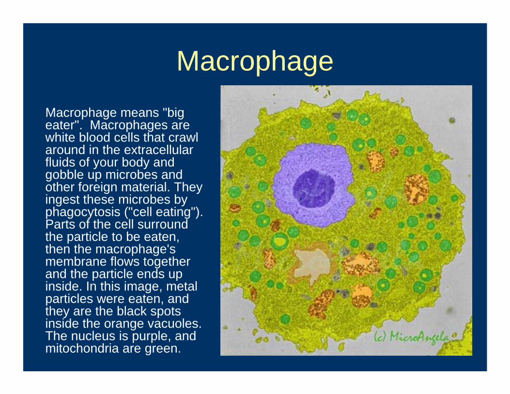
MacrophageMacrophage means "big eater". Macrophages are white blood cells that crawl around in the extracellular fluids of your body and gobble up microbes and other foreign material. They ingest these microbes by phagocytosis ("cell eating"). Parts of the cell surround the particle to be eaten, then the macrophage's membrane flows together and the particle ends up inside. In this image, metal particles were eaten, and they are the black spots inside the orange vacuoles. The nucleus is purple, and mitochondria are green.

Schematic of Phagocytosis
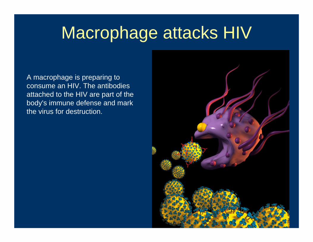
Macrophage attacks HIV
A macrophage is preparing to consume an HIV. The antibodies attached to the HIV are part of the body's immune defense and mark the virus for destruction.
If particulate matter is not removed, it causes chronic inflammation
• Particles engulfed by macrophages may be transported upward and removed from lungs - OK
• If not, they are retained in the lung and initiate a pathway of chronic inflammation, which leads to pneumoconiosis (called “frustrated phagocytosis”)
“Frustrated Phagocytosis”
Causes a “cascade of toxic effects” which eventually leads to lesions in the lungs and lung diseases called pneumoconioses
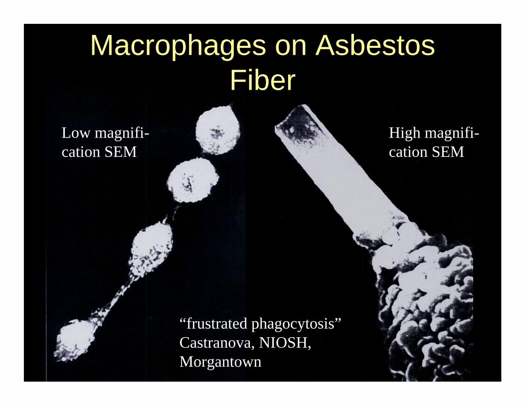
Low magnifi-cation SEM
High magnifi-cation SEM
“frustrated phagocytosis”Castranova, NIOSH, Morgantown
Macrophages on Asbestos Fiber
“Cascade of Toxic Effects”• Death of the macrophage and release of
its toxic contents• Re-ingestion of dust particles by newly
recruited macrophages and other cells• Stimulation of local factors in an attempt to
wall off the inflammatory process• Eventual formation of a fibrotic lesion and
fibrosis in the lung tissue• Concomitant impairment of the cellular
immunity at the local level
Silica-related diseases
• Chronic silicosis• Accelerated silicosis• Acute silicosis
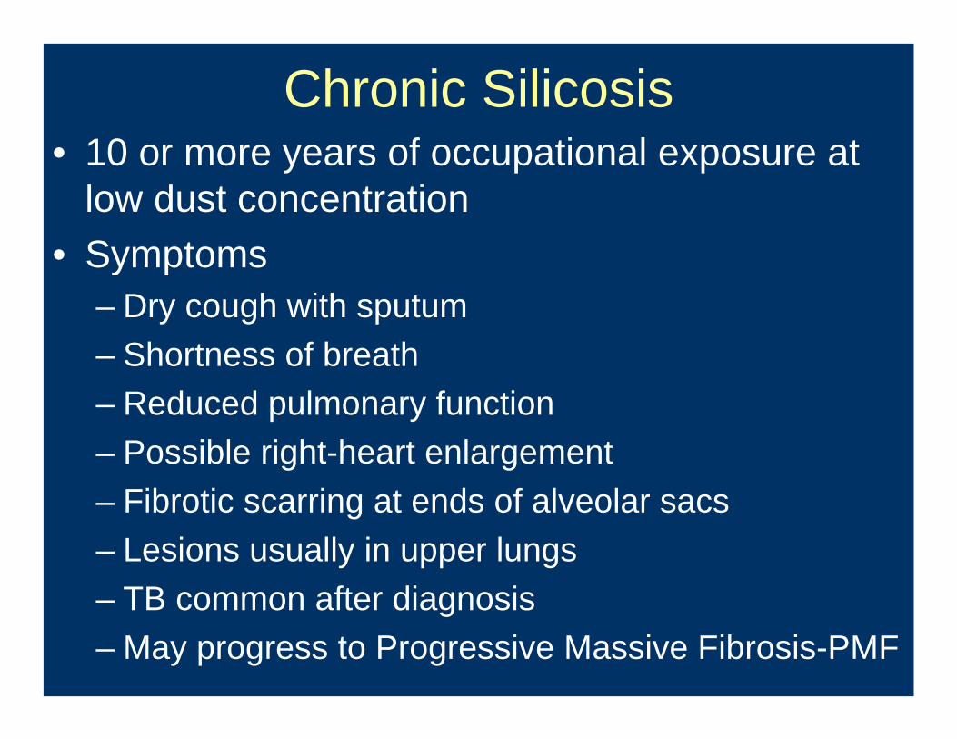
Chronic Silicosis• 10 or more years of occupational exposure at
low dust concentration• Symptoms
– Dry cough with sputum– Shortness of breath– Reduced pulmonary function– Possible right-heart enlargement– Fibrotic scarring at ends of alveolar sacs– Lesions usually in upper lungs– TB common after diagnosis– May progress to Progressive Massive Fibrosis-PMF
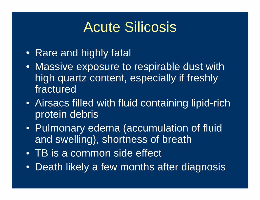
Acute Silicosis
• Rare and highly fatal• Massive exposure to respirable dust with
high quartz content, especially if freshly fractured
• Airsacs filled with fluid containing lipid-rich protein debris
• Pulmonary edema (accumulation of fluid and swelling), shortness of breath
• TB is a common side effect• Death likely a few months after diagnosis
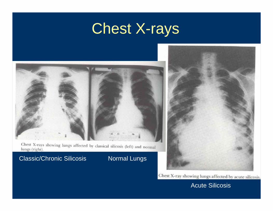
Chest X-rays
Normal LungsClassic/Chronic Silicosis
Acute Silicosis
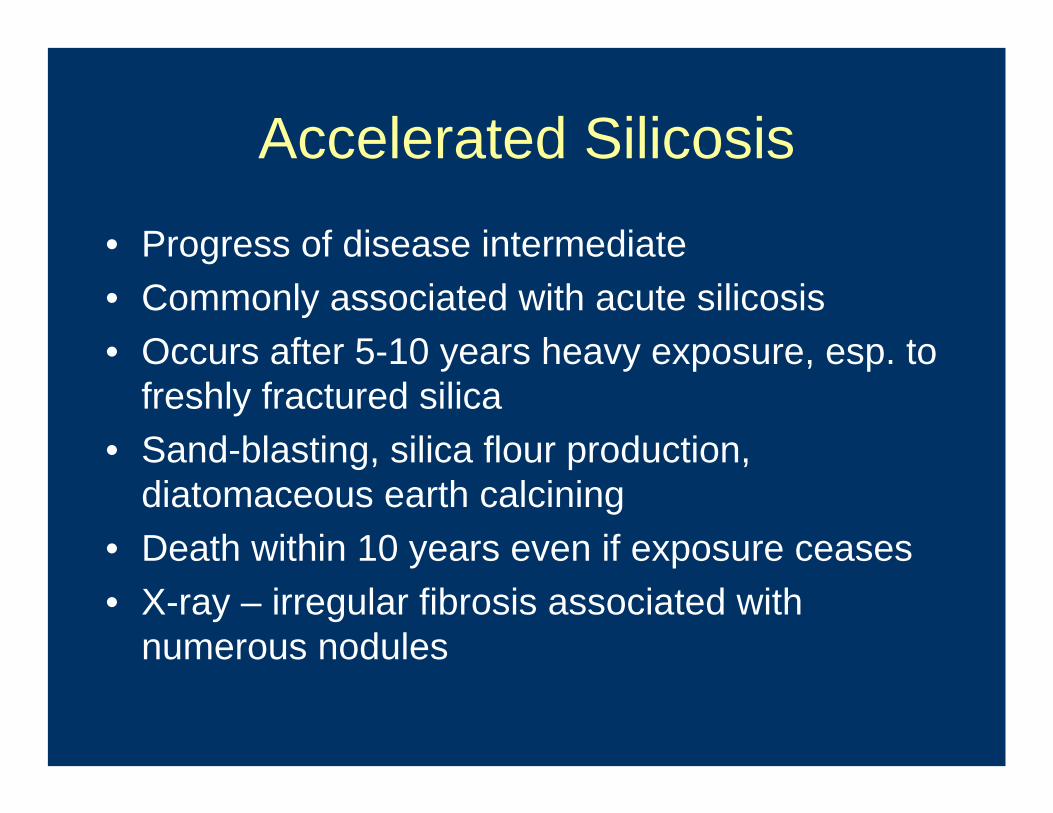
Accelerated Silicosis
• Progress of disease intermediate• Commonly associated with acute silicosis• Occurs after 5-10 years heavy exposure, esp. to
freshly fractured silica• Sand-blasting, silica flour production,
diatomaceous earth calcining• Death within 10 years even if exposure ceases• X-ray – irregular fibrosis associated with
numerous nodules
Asbestosis and Coal Workers’Pneumoconiosis (“black-lung”) are
similar diseases• More about asbestos-related diseases
later
Related Documents