Associate Editor: P. Madeddu Pluripotency rush! Molecular cues for pluripotency, genetic reprogramming of adult stem cells, and widely multipotent adult cells Antonio Paolo Beltrami ⁎, Daniela Cesselli, Carlo Alberto Beltrami Interdepartmental Center for Regenerative Medicine (CIME), Department of Pathology, University of Udine, Italy abstract article info Keywords: Pluripotent stem cell Embryonic stem cell Induced pluripotent stem cell Adult stem cell OCT4 Nanog Sox2 In the last few years, pluripotent stem cells have been the objective of intense investigation efforts. These cells are of paramount therapeutic interest, since they could be utilized: as in vitro models of disease, for pharmaceutical screening purposes, and for the regeneration of damaged organs. Over the years, pluripotent cells have been cultured from teratomas, the inner cell mass, and primordial germ cells. Accumulating informations have partially decrypted the molecular machinery responsible for the maintenance of a very primitive state, permitting the reprogramming of differentiated cells. Although the debate is still open, an extreme excitement is arising from two strictly related possibilities: pluripotent cells could be obtained from adult tissues with minimal manipulations or very rare pluripotent cells could be identified in adult tissues. This intriguing option will trigger new researches aimed both at identifying the possible biological role of pluripotent adult stem cells and at exploiting their potential clinical use. The present review article will summarize current knowledge of the molecular cues for pluripotency but also discusses whether pluripotent stem cells could be obtained from adult tissues. © 2009 Elsevier Inc. All rights reserved. Contents 1. Introduction . . . . . . . . . . . . . . . . . . . . . . . . . . . . . . . . . . . . . . . . . . . . . . 23 2. Molecular cues for pluripotency . . . . . . . . . . . . . . . . . . . . . . . . . . . . . . . . . . . . . 24 3. Pluripotent adult stem cells . . . . . . . . . . . . . . . . . . . . . . . . . . . . . . . . . . . . . . . 4. Induced pluripotent stem cells (iPS) . . . . . . . . . . . . . . . . . . . . . . . . . . . . . . . . . . . 5. Conclusion . . . . . . . . . . . . . . . . . . . . . . . . . . . . . . . . . . . . . . . . . . . . . . . References . . . . . . . . . . . . . . . . . . . . . . . . . . . . . . . . . . . . . . . . . . . . . . . . . . 1. Introduction In the last years, the scientific community has been increasingly interested in pluripotent stem cells, as testified by the large number of scientific publications dealing with this field of investigation. Plu- ripotent cells are of paramount therapeutic interest, since they could be utilized: as in vitro models of disease, for pharmaceutical screening purposes, and for the regeneration of damaged organs. Stem cell biology has been evolving so rapidly that different researchers utilize the same terms with different meanings. For this reason, it was recently published a glossary of stem-cell related terms aimed at creating a common language to be used by every scientist working in this field (Smith, 2006). In this introduction, we will discuss how stem cells, stem cell plasticity, and the differentiation potential of a stem cell are defined, remarking where disagreement exists among researchers. Stem cells are considered to be entities that “can continuously pro- duce unaltered daughter cells together with daughter cells that have different, more restricted properties” (Smith, 2006). Stem cell plasticity, instead, is believed to be an “unproven notion that tissue stem cells may broaden potency in response to physiological demands or insults” (Smith, 2006). These definitions reflect the classical and quite static Pharmacology & Therapeutics 124 (2009) 23–30 Abbreviations: ES, embryonic stem cells; (h), human; (m), mouse; HSC, hemato- poietic stem cells; ICM, inner cell mass; iPS, induced pluripotent stem cells; MAPC, multipotent adult progenitor cells; MASC, multipotent adult stem cells; MIAMI, marrow-isolated adult multilineage inducible cells; MSC, mesenchymal stem cells; NSC, neural stem cells; PGC, primordial germ cells; SSC, spermatogonial stem cells; SKP, skin-derived precursor cells; VSEL, very small embryonic like cells. ⁎ Corresponding author. Cattedra di Anatomia Patologica, Università degli Studi di Udine; p.zzle S. Maria della Misericordia, 33100 Udine, Italy. Tel.: +39 0432 559477; fax: +39 0432 559420. E-mail address: [email protected] (A.P. Beltrami). 26 27 27 27 0163-7258/$ – see front matter © 2009 Elsevier Inc. All rights reserved. doi:10.1016/j.pharmthera.2009.06.003 Contents lists available at ScienceDirect Pharmacology & Therapeutics journal homepage: www.elsevier.com/locate/pharmthera

Welcome message from author
This document is posted to help you gain knowledge. Please leave a comment to let me know what you think about it! Share it to your friends and learn new things together.
Transcript
Pharmacology & Therapeutics 124 (2009) 23–30
Contents lists available at ScienceDirect
Pharmacology & Therapeutics
j ourna l homepage: www.e lsev ie r.com/ locate /pharmthera
Associate Editor: P. Madeddu
Pluripotency rush! Molecular cues for pluripotency, genetic reprogramming of adultstem cells, and widely multipotent adult cells
Antonio Paolo Beltrami ⁎, Daniela Cesselli, Carlo Alberto BeltramiInterdepartmental Center for Regenerative Medicine (CIME), Department of Pathology, University of Udine, Italy
Abbreviations: ES, embryonic stem cells; (h), humapoietic stem cells; ICM, inner cell mass; iPS, inducedmultipotent adult progenitor cells; MASC, multipotemarrow-isolated adult multilineage inducible cells; MNSC, neural stem cells; PGC, primordial germ cells; SSC, sskin-derived precursor cells; VSEL, very small embryoni⁎ Corresponding author. Cattedra di Anatomia Patolo
Udine; p.zzle S. Maria della Misericordia, 33100 Udine,fax: +39 0432 559420.
E-mail address: [email protected] (A.P. Belt
0163-7258/$ – see front matter © 2009 Elsevier Inc. Aldoi:10.1016/j.pharmthera.2009.06.003
a b s t r a c t
a r t i c l e i n f oKeywords:
Pluripotent stem cellEmbryonic stem cellInduced pluripotent stem cellAdult stem cellOCT4NanogSox2In the last few years, pluripotent stem cells have been the objective of intense investigation efforts. Thesecells are of paramount therapeutic interest, since they could be utilized: as in vitro models of disease, forpharmaceutical screening purposes, and for the regeneration of damaged organs. Over the years, pluripotentcells have been cultured from teratomas, the inner cell mass, and primordial germ cells. Accumulatinginformations have partially decrypted the molecular machinery responsible for the maintenance of a veryprimitive state, permitting the reprogramming of differentiated cells.Although the debate is still open, an extreme excitement is arising from two strictly related possibilities:pluripotent cells could be obtained from adult tissues with minimal manipulations or very rare pluripotentcells could be identified in adult tissues. This intriguing option will trigger new researches aimed both atidentifying the possible biological role of pluripotent adult stem cells and at exploiting their potential clinicaluse. The present review article will summarize current knowledge of the molecular cues for pluripotency butalso discusses whether pluripotent stem cells could be obtained from adult tissues.
© 2009 Elsevier Inc. All rights reserved.
Contents
1. Introduction . . . . . . . . . . . . . . . . . . . . . . . . . . . . . . . . . . . . . . . . . . . . . . 232. Molecular cues for pluripotency . . . . . . . . . . . . . . . . . . . . . . . . . . . . . . . . . . . . . 243. Pluripotent adult stem cells . . . . . . . . . . . . . . . . . . . . . . . . . . . . . . . . . . . . . . . 244. Induced pluripotent stem cells (iPS) . . . . . . . . . . . . . . . . . . . . . . . . . . . . . . . . . . . 245. Conclusion . . . . . . . . . . . . . . . . . . . . . . . . . . . . . . . . . . . . . . . . . . . . . . . 24
262727
References . . . . . . . . . . . . . . . . . . . . . . . . . . . . . . . . . . . . . . . . . . . . . . . . . . 2527
1. Introduction
In the last years, the scientific community has been increasinglyinterested in pluripotent stem cells, as testified by the large numberof scientific publications dealing with this field of investigation. Plu-ripotent cells are of paramount therapeutic interest, since they could
n; (m), mouse; HSC, hemato-pluripotent stem cells; MAPC,nt adult stem cells; MIAMI,SC, mesenchymal stem cells;permatogonial stem cells; SKP,c like cells.gica, Università degli Studi diItaly. Tel.: +39 0432 559477;
rami).
l rights reserved.
be utilized: as in vitro models of disease, for pharmaceutical screeningpurposes, and for the regeneration of damaged organs.
Stem cell biology has been evolving so rapidly that differentresearchers utilize the same terms with different meanings. For thisreason, it was recently published a glossary of stem-cell related termsaimed at creating a common language to be used by every scientistworking in thisfield (Smith, 2006). In this introduction,wewill discusshow stem cells, stem cell plasticity, and the differentiation potential ofa stem cell are defined, remarking where disagreement exists amongresearchers.
Stem cells are considered to be entities that “can continuously pro-duce unaltered daughter cells together with daughter cells that havedifferent, more restricted properties” (Smith, 2006). Stem cell plasticity,instead, is believed to be an “unproven notion that tissue stem cellsmay broaden potency in response to physiological demands or insults”(Smith, 2006). These definitions reflect the classical and quite static
24 A.P. Beltrami et al. / Pharmacology & Therapeutics 124 (2009) 23–30
view of stem cells, that regards these latter as very rare, tissue residentelements endowed with peculiar properties (i.e. self-renewal and mul-tipotency) inherently different from those of the mature cells (Zipori,2005). As a consequence, a hierarchical model has been very successfulin describing the hematopoietic stem cell (HSC) system, where rare,multipotent, HSCs are present within the bone marrow, capable oflong term engraftment in irradiated recipients. Nonetheless, this modelis valid only ifwe assume that: 1) stemcells generate progenitors,whichhave a less pronounced self-renewal capability and a bias in their dif-ferentiation potential, and 2) every differentiation step is irreversible.However, several exceptions could be mentioned to the concepts ofcompartmentalization and unidirectionality of this model, and in ad-dition the organization of the hemopoietic system may not represent ageneral structure of stemcell systems (Randall &Weissman,1998; Cerny& Quesenberry, 2004; Kirkland, 2004). For this reason, three differentmodels have been proposed: 1) the chiaroscuro stem cell model; 2) thephase space model; and 3) the stem state model. In the chiaroscuro stemcell model, there is “no progenitor/stem cell hierarchy, but rather areversible continuum. This may, in turn, be dependent on shiftingchromatin structure and gene expressionwith cell cycle transit” (Cerny& Quesenberry, 2004). Following this model, the transition of a prim-itive cell through the cell cycle alters the expression of surface antigens,cytokine receptors, adhesion molecules, and gene expression, thusmodulating complex functions, such as progenitor forming capacity,tissue homing, and engraftment (Cerny & Quesenberry, 2004). Thephase space model originates from the consideration that, in stem cellbiology, “a formof uncertainty principle is inplay,which can be stated asfollows: it is not possible to simultaneously measure the proliferativeand the differentiative capabilities of a cell, as the act of measuring oneproperty irrevocably alters the other” (Kirkland, 2004). A consequenceof this alternative model, based on continuous variables, is that “theoutcome of stem cell divisionmust be described by a probability densityfunction, rather than by a discrete probability distribution”. Therefore,the daughter of a stem cell division could have degrees of stemness evenhigher than the parent cell (dedifferentiation). Furthermore, in thismodel the self-renewal concept is replaced by population renewal,while stem cell plasticity is not only possible but a predictable andfundamental trait of stemness (Zipori, 2005). In the stem state modelcellsmay enter a stem state reversibly, implying that stemnesswould bea state rather than a cell entity (Blau, et al., 2001; Zipori, 2004).
Cells can be classified according to their differentiation potential; atotipotent cell can form an entire organism, while pluripotent cellshave the ability to form all the body's cell lineages, including germcells and some, or even all, extra-embryonic cell types. Multipotentcells, instead, have the ability to formmultiple lineages that constitutean entire tissue (Smith, 2006). Therefore, the difference betweentotipotency and pluripotency is the ability to form an entire organismin vivo, while germline competence is the main difference betweentoti/pluripotent cells and multipotent cells. Consequently, if we ad-here to this definition, embryonic stem cells (ES) are pluripotent,while fetal and adult stem cells can be considered, with the exceptionof spermatogonial stem cells (Guan, et al., 2006; Conrad, et al., 2008),multipotent. Although the ability of mouse ES (mES) to generate fullyfunctional germ cells can be assayed in vivo, human ES (hES) cannotbe tested for this function without raising ethical concerns (Lensch,et al., 2007). Consequently, mouse ES pluripotency is tested evaluatingblastocyst chimerism or tetraploid aggregation followed by gestation,whereas human ES pluripotency is generally tested evaluating theability to form teratomas and teratocarcinomas (i.e. tumors containingan array of differentiated cell types, and possibly undifferentiated cellsresembling embryonic cells of the inner cell mass) upon injection invivo (Lensch, et al., 2007). Nonetheless, the ability to form teratomasdoes not imply germline competence. Regarding extra-embryonicderivatives, while human ES can differentiate into trophoblast cells,when mES are reintroduced into early embryos they rarely do so(Beddington & Robertson, 1989; Nagy, et al., 1990). The reduced
trophoectodermal differentiation potential of mES has been consid-ered for a long time as one of the major differences between mES andhES (Ginis, et al., 2004), but it has been recently demonstrated thatmES can differentiate into trophoectoderm in vitro (He, et al., 2008).
The above-mentioned disagreements in models and definitionstestify to the complexity of the stem cell research and demonstrate theneed to define pluripotency at a molecular level. The present reviewarticle will summarize current knowledge of the molecular cues forpluripotency, but also discusses whether pluripotent stem cells couldbe obtained from adult tissues. A more comprehensive description ofthe history of ES research is beyond the scope of this review, but canbe found in recent articles (Yu & Thomson, 2008).
2. Molecular cues for pluripotency
The identification of the molecular determinants required tomaintain stem cell pluripotency, in particular both the extrinsic andintrinsic regulators of human and mouse ES pluripotency, has been amajor challenge in stem cell biology for many years.
2.1. Extrinsic factors
As illustrated in Table 1, several signal transduction pathways havebeen associated with the preservation of ES pluripotency (Lensch,et al., 2006). However, hES and mES rely on different mechanisms toachieve this purpose; in fact, while mES require a combination of LIFand BMP4, hES necessitate a complex balance of Smad2/3 activation,Smad1/5/8 inhibition, bFGF-, and IGF-II-signaling (Smith, et al., 1988;Vallier, et al., 2005; Stewart, et al., 2008). Accumulating knowledge onhES growth factor requirements has allowed to design serum-free,feeder-free culturemedia (Ludwig, et al., 2006), however cultured hESgenerate hES-derived fibroblast-like cells that, in concert with theprovided extracellular matrix (i.e. Matrigel) and culture medium, actas an in vitro niche (Stewart, et al., 2008). This latter provides cell–cellcontacts, extracellular matrix components, and growth factors thatact together to promote stem cell self-renewal. In response to bFGF,feeder cells produce Activin A, TGFβ1, chordin, GDF3, Gremlin, andIGF-II, thus maintaining ES pluripotency (Stewart, et al., 2008).
2.2. Intrinsic factors
Extrinsic factors activate in ES both (a) a transcription factor circuitryand (b) epigenetic modifications able to maintain pluripotency.
2.2.1. Transcription factor circuitryThe existence of factors present in primitive cells that can trans-
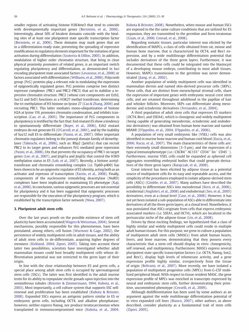
fer pluripotency in a dominant fashion was first established perform-ing somatic cell nuclear transfer or fusing ES with differentiated cells(Ying, et al., 2002; Do & Scholer, 2004; Cowan, et al., 2005). Threeare the main candidate molecules responsible for the preservationor transfer of pluripotency; the transcription factors OCT4, Nanog, andSox2 (Table 2). OCT4 deficient embryos die at the time of implanta-tion, and, although trophoblast is formed, inner cell mass (ICM) is not(Nichols, et al., 1998). OCT4 levels need to be tightly controlled inorder to promote pluripotency, since increased concentrations of thisfactor induce differentiation (Niwa, et al., 2000). Nanog too is crucialfor pluripotency; it is highly expressed in ES, primordial germ cells(PGCs), and ICM, while it is down-regulated in mature cells andsomatic tissues (Chambers, et al., 2003; Mitsui, et al., 2003). Further-more, it is able to confer LIF independence to mES and to inhibitdifferentiation of hES (Mitsui, et al., 2003; Hyslop, et al., 2005; Pan& Thomson, 2007). Last, Sox2 was shown to cooperate with OCT4to regulate several pluripotency related genes, such as Nanog, Fgf4(Rodda, et al., 2005), osteopontin (Botquin, et al., 1998), and Lefty1(Nakatake, et al., 2006). The disruption of Sox2 function leads todifferentiation along trophoectoderm lineage and polyploidization of
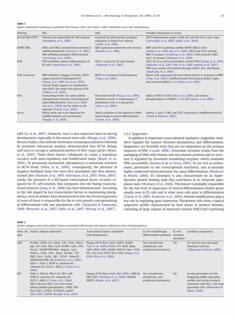
Table 1Signal transduction pathways associated with human (hES) and mouse (mES) embryonic stem cells pluripotency.
Pathway hES mES Probable mechanism of action
IL6/LIF-JAK-STAT3 Present, but dispensable for self-renewal(Humphrey, et al., 2004).
Essential for self-renewal; promotesexpansion in feeder free cultures(Smith, et al., 1988).
STAT3 downstream targets: Lefty, Id1 and Id2, Aes1, and c-myc(Cartwright, et al., 2005; Sekkai, et al., 2005).
MAPK/ERK ERK1 and ERK2 constitutively activated inundifferentiated ES (Dvorak, et al., 2005).ERK inhibition promotes differentiation(Armstrong, et al., 2006).
ERK suppression promotes self-renewal(Burdon, et al., 1999).
BMP and PI3-K pathways inhibit MAPK/ERK in mice(Paling, et al., 2004; Qi, et al., 2004). bFGF and PI3-K activateERK in humans (Armstrong, et al., 2006). FGF4 activates ERK1and 2 in humans (Mayshar, et al., 2008).
PI3K PI3K inhibition induces differentiation ofES cells (Armstrong, et al., 2006).
PI3K is crucial for ES self-renewal(Takahashi, et al., 2005).
IGFII, LIF, Eras, and neurotrophin activate PI3K (Paling, et al., 2004;Takahashi, et al., 2005; Pyle, et al., 2006; Bendall, et al., 2007).PI3K may control self-renewal through GSK3β, p53, and Nanog(Takahashi, et al., 2005).
TGFB SUPERFAMILY BMP inhibitors (Noggin, Gremlin, GDF3)support growth of pluripotent ES(Wang, et al., 2005; Xu, et al., 2005).Activin/Nodal support, in combinationwith bFGF, the feeder-free growth of ES(Vallier, et al., 2005).
BMP2 or 4 preserve ES pluripotency(Ying, et al., 2003).
Mouse cells upregulate Id transcription factors in response to BMP(Ying, et al., 2003). Undifferentiated hES express Nodal, Cripto,and activated SMADs 2/3 (Vallier, et al., 2005).
WNT Contrasting results: for some authorsendogenously activated, downregulatedupon differentiation (Sato, et al., 2004;Sato et al., 2004), but for others is theopposite (Dravid, et al., 2005).
Restricted to ICM (Wang, et al., 2004),activation results in maintenance ofpluripotency over a 5 day period(Sato, et al., 2004).
Induces STAT3 in mES (Hao, et al., 2006), and inducesphosphorilation of SMADs 2/3 in hES (James, et al., 2005).
NOTCH Notch seems not to be important forundifferentiated cell maintenance(Noggle, et al., 2006).
Notch activation in the absence of otherstimuli leads to neural differentiation(Lowell, et al., 2006).
Notch1, 2, and 3, CBF1 and TLE1 expressed in undifferentiated ES(Walsh & Andrews, 2003).
25A.P. Beltrami et al. / Pharmacology & Therapeutics 124 (2009) 23–30
mES (Li, et al., 2007). However, Sox2 is also expressed later on duringdevelopment, especially in the neural stem cells (Miyagi, et al., 2006).Recent studies, that utilized chromatin immunoprecipitation followedby promoter microarray analysis, demonstrated that OCT4, Nanog,and Sox2 co-occupy a substantial portion of their target genes (Boyer,et al., 2005). These three factors collaborate to create a regulatorycircuitry with auto-regulatory and feedforward loops (Boyer, et al.,2005). As previously mentioned, pluripotency is extremely sensitiveto OCT4 levels (Niwa, et al., 2000), consequently, the presence ofnegative feedback loops has been first postulated and then demon-strated (Ben-Shushan, et al.,1995; Fuhrmann, et al., 2001;Niwa, 2007).Lately, the presence of a Klf-based transcription factor circuitry, re-quired for ES self-renewal, that integrates into the Nanog transcrip-tional network (Jiang, et al., 2008) has been demonstrated . Accordingto the role played by key transcription factors in maintaining plurip-otency, several authors have demonstrated that the forced expressionof some of these is responsible for the in vitro genetic reprogrammingof differentiated cells into pluripotent cells (Takahashi & Yamanaka,2006; Meissner, et al., 2007; Okita, et al., 2007; Wernig, et al., 2007).
Table 2Surface antigens and transcription factors associated with human and mouse embryonic ste
Stem celltype
Surface antigen expression Transcription factors associatedwith pluripotency
mES VCAM1, CD44 , Cav , Emp1 , Csf1 , Osmr , Abca1 ,Jag1 , Kit , Fzd5 , Il6st , Ly75 , ICAM1, Gjb3 , CD9 ,Slc2a3 , 9430079M16Rik , Ppap2a , Gja7 ,Abcb1a , Enah , Ptch , Akp2 , Slc29a1 , Trfr ,Fgfr2, Utrn , Ptch2 , Ak1 , CD1d1 , Adam19 ,1600025H15Rik, ltb (Palmqvist, et al., 2005),SSEA-1, SSEA-3, FGFR-4, connexin-43,connexin-45, GLUT-1, ABCG-2 (Ginis, et al.,2004).
Nanog, OCT4, Rex1, Sox2, FoxD3(Loh, et al., 2006), Prmt1, YY1, RCdk1, NF45, Sall4, Zfp281, BAF1Rif1, Elys, Err2, REST, Dax1, Pelo2006; Kim, et al., 2008).
hES SSEA-3, SSEA-4, TRA-1-81, TRA-1-60,FGFR-4, connexin-43, connexin-45,GLUT-1, ABCG-2 (Ginis, et al., 2004),TRA-2-49 and TRA-2-54 (liver/bone/kidney alkaline phosphatase), CD90, CD9,HLA-A,B,C, GCTM2, GCTM343, podxl1(ISCI, 2007), IGF1R (Bendall, et al., 2007).
Nanog, OCT4, Rex1, Sox2 (ISCI,ERH, BTF3 (Richards, et al., 200NR2F2 (Babaie, et al., 2007).
2.2.2. EpigeneticsIn addition to important transcriptional regulators, epigenetic mod-
ifiers regulate the balance between pluripotency and differentiation.Epigenetics are heritable traits that are not dependent on the primarysequence of DNA (Cavalli, 2006). Chromatin structure results from thepackaging of DNAwith histone and non-histone proteins and its struc-ture is regulated by chromatin remodeling enzymes, which modulateDNA accessibility (Keenen & de la Serna, 2009). ES are rich in euchro-matin, permissive to the transcriptional machinery, and accumulatehighly condensed heterochromatic foci upon differentiation (Meshorer& Misteli, 2006). ES chromatin is also characterized by an hyper-dynamic protein binding state that contributes to its relatively open,plastic state (Meshorer, et al., 2006). This feature is probably responsiblefor the low level of expression of several differentiation-related genesusually seen in ES cells and in other stem cells prior to differentiation(Carter, et al., 2005; Szutorisz, et al., 2005). Histonemodifications play akey role in regulating gene expression. Pluripotent cells show a typicalepigenetic profile characterized by dual marks or bivalent domains,consisting of large regions of repressive histone H3K27me3 harboring
m cell pluripotency.
In vitro multilineagedifferentiation potential
In vivoteratomaformation
Germline competence
, Setdb1nf2, Rybp,
55, Nac1, Tif1b,(Wang, et al.,
Yes (mesodermic,endodermic, andectodermic derivatives).
Yes Yes both in vitro and uponblastocyst injection(Aflatoonian & Moore, 2006).
2007), HMGA1,4), HMGB2,
Yes (mesodermic,endodermic, andectodermic derivatives).
Yes In vitro generation of cellsdisplaying mRNA expressionprofiles and surface markersconsistent with PGCs, and earlyspermatid cells (Aflatoonian &Moore, 2006).
26 A.P. Beltrami et al. / Pharmacology & Therapeutics 124 (2009) 23–30
smaller regions of activating histone H3K4me3 that tend to coincidewith developmentally important genes (Bernstein, et al., 2006).Interestingly, about 50% of bivalent domains coincide with the bind-ing sites of at least one pluripotent state specific transcription factor(Bernstein, et al., 2006). These domains may mark genes that arein a differentiation-ready state, preventing the spreading of repressivemodifications to regulatoryelements important for the initiation of geneactivation during differentiation (Szutorisz & Dillon, 2005). In addition,modulation of higher order chromatin structure, that bring in closephysical proximity promoters of related genes, is an important switchregulating pluripotency and differentiation, as demonstrated in lociencoding pluripotent state associated factors (Levasseur, et al., 2008) orfactors associatedwith differentiation (Williams, et al., 2006). Polycombgroup (PcG) proteins play a relevant role in modulating the expressionof epigenetically regulated genes. PcG proteins comprise two distinctrepressor complexes (PRC1 and PRC2–PRC3) that act to stabilize a re-pressive chromatin structure. PRC2 (whose core components are Ezh2,Suz12 and EeD) functions as a H3K27 methyl transferase, determiningthe tri-methylation of H3 histone on lysine 27 (Cao & Zhang, 2004) andrecruiting PRC1. This latter mediates mono-ubiquitination of histoneH2A at lysine 119, promotes chromatin condensation, and inhibits tran-scription (Cao, et al., 2005). The importance of PcG components inpluripotency is testified by the fact that: Eedmutant ES showa tendencyto spontaneously differentiate (Boyer, et al., 2006), Ezh2 mutantembryos do not generate ES (O'Carroll, et al., 2001), and by the inabilityof Suz12 null ES to differentiate (Pasini, et al., 2007). Other importantchromatin regulators belong to the jumonji domain family of demethy-lases (Takeuchi, et al., 2006), such as: Rbp2 (Jarid1a) that can recruitPRC2 to its target genes and enhances PcG mediated gene repression(Pasini, et al., 2008), Utx that prevents the occupancy of PRC1 on targetgenes (Lee, et al., 2007), and Jmjd1a and Jmjd2c that control the H3K9methylation status in ES (Loh, et al., 2007). Recently, a histone acetyl-transferase and chromatin remodeling complex (i.e. Tip60-p400) hasbeen shown tobe anepigenetic regulator of ES identity, actingboth as anactivator and repressor of transcription (Fazzio, et al., 2008). Finally,components of the nucleosome remodeling deacetylase (NuRD)complexes have been implicated in maintaining ES phenotype (Kaji,et al., 2006). In conclusion, various epigenetic processes are not essentialfor pluripotency and it has been suggested that epigenetic processesare responsible for the execution of the pluripotency program, which isestablished by the transcription factor network (Niwa, 2007).
3. Pluripotent adult stem cells
Over the last years proofs on the possible existence of stem cellplasticity have been accumulated(Wagers &Weissman, 2004). Severalmechanisms, possibly responsible for this phenomenon, have beenpostulated, among others, cell fusion (Wurmser & Gage, 2002), thepersistence of widely multipotent cells in adult tissues, and the abilityof adult stem cells to de-differentiate, acquiring higher degrees ofstemness (Kirkland, 2004; Zipori, 2005). Taking into account theselatter two possibilities, scientists have investigated whether adultmammalian tissues could host broadly multipotent cells whose di-fferentiation potential was not restricted to the germ layer of theirorigin.
In line with the close relationship between ES and germ cells, aspecial place among adult stem cells is occupied by spermatogonialstem cells (SSCs). The latter was first identified in the adult murinetestis for its ability to repopulate sterile gonads upon injection into theseminiferous tubules (Brinster & Zimmermann, 1994; Kubota, et al.,2003). Most importantly, a cell culture system that supports SSC self-renewal and proliferation has been established (Kubota & Brinster,2008). Expanded SSCs express an antigenic pattern similar to ES orembryonic germ cells, including OCT4, and alkaline phosphatase;however, neither express Nanog, nor produce teratocarcinomas whentransplanted in immunocompromised mice (Kubota, et al., 2004;
Kubota & Brinster, 2008). Nonetheless, when mouse and human SSCsare exposed to the the same culture conditions that are utilized for ESexpansion, they are transmitted to the germline and form teratomas(Guan, et al., 2006; Conrad, et al., 2008).
Regarding somatic tissues, particular interest was elicited by theidentification of MAPCs, a class of cells obtained from rat, mouse andhuman bone marrow, that is characterized by OCT4, and Rex1 ex-pression, and by a wide multilineage differentiation potential thatincludes derivatives of the three germ layers. Furthermore, it wasdocumented that these cells could be integrated into the blastocystof a developing mouse embryo, contributing to most adult tissues.However, MAPCs transmission to the germline was never demon-strated (Jiang, et al., 2002).
Another population of widely multipotent cells was identified inmammalian dermis and named skin-derived precursor cells (SKPs).These cells, that are distinct from mesenchymal stromal cells, sharethe expression of important genes with embryonic neural crest stemcells, and are retained throughout adulthood in the papillae of hairand whisker follicles. Moreover, SKPs can differentiate along meso-dermic and ectodermic derivatives (Fernandes, et al., 2004).
A third population of adult stem cells that expresses ES markers(OCT4, Rex1, and SSEA4), which is clonogenic and widely multipotent(being capable of generating mesodermic, ectodermic and endoder-mic derivatives) was obtained from human bone marrow and namedMIAMI (D'Ippolito, et al., 2004; D'Ippolito, et al., 2006).
A population of very small embryonic like (VSEL) cells was alsoidentified inmurine bonemarrowand human cord blood (Kucia, et al.,2006; Kucia, et al., 2007). The main characteristics of these cells are:their extremely small dimensions (3–5 µm), and the expression of atypical immunophenotype (i.e.: CXCR4+ AC133+ CD34+ lin− CD45−).Furthermore, murine VSEL cells could be expanded as spheroid cellaggregates resembling embryoid bodies that could generate deriva-tives of the three germ layers (Ratajczak, et al., 2008).
Great interest has been elicitied by adipose tissue as a potentialsource of multipotent cells for its easy and repeatable access, and thesimplicity of the procedures employed to isolate adipose-derived stemcells (ASCs) (Gimble, et al., 2007). Many authors have described thepossibility to differentiate ASCs into mesodermal (Kern, et al., 2006),ectodermal (Anghileri, et al., 2008) and endodermal (Seo, et al., 2005)derivatives, even at a clonal level (Case, et al., 2008). However, it hasnot yet been isolated a sub-population of ASCs able to differentiate intoderivatives of all the three germ layers, at a clonal level. Nonetheless, itwas postulated that ASCs originate from cells that express embryonicassociated markers (i.e. SSEA1, and OCT4), which are localized in theperivascular niche of the adipose tissue (Lin, et al., 2008).
Driven by these exciting findings, we hypothesized that a class ofhighly similar and widely multipotent cells could reside in multipleadult human tissues. For this purpose, we grew in culture a populationof multipotent adult stem cells (MASCs) from adult human hearts,livers, and bone marrow, demonstrating that they possess everycharacteristic that a stem cell should display in vitro: clonogenicity,self renewal, and multipotency. Furthermore, MASCs express severalpluripotent state specific transcription factors (i.e. OCT4, Nanog, Sox2,and Rex1), display high levels of telomerase activity, and a geneexpression profile highly similar, irrespectively from the tissueof origin (Beltrami, et al., 2007). More recently, we have isolated apopulation of multipotent progenitor cells (MPCs) from G-CSF mobi-lized peripheral blood. With respect to tissue resident MASC, the geneexpression profile of MPCs was enriched in transcripts expressed byneural and embryonic stem cells, further demonstrating their prim-itive, uncommitted phenotype (Cesselli, et al., 2009).
Although dedifferentiation has been used by some authors as anargument against the wide multilineage differentiation potential ofin vitro expanded cell lines (Bianco, 2007), other authors, as abovedescribed, consider plasticity as a fundamental trait of stem cells(Zipori, 2005).
27A.P. Beltrami et al. / Pharmacology & Therapeutics 124 (2009) 23–30
4. Induced pluripotent stem cells (iPS)
Advancing knowledge on the molecular determinants of pluripo-tency has allowed to convert in vitro somatic cells into induced plurip-otent stemcells. To this end,fibroblastswerefirst retrovirally transducedwith OCT4, Sox2, Klf4, and c-myc and then selected utilizing differentapproaches: an Fbx15, an OCT4 or a Nanog driven drug resistance mar-ker, or employing purely morphological features (Takahashi & Yama-naka, 2006; Meissner, et al., 2007; Okita, et al., 2007; Wernig, et al.,2007). The first report, based upon Fbx15 activation, generated cells thatwere multipotent, able to form teratomas in vivo, but unable to con-tribute to the germline once injected into a blastocyst (Takahashi &Yamanaka, 2006). Furthermore, the epigenetic state of these cells wasintermediate between the fibroblast and the ES one (Takahashi &Yamanaka, 2006). In contrast, selection for OCT4, Nanog or utilizingmorphological criteria led to the generation of cells that had a DNAmethylation, a gene expression, and a chromatin state similar to those ofES cells (Meissner, et al., 2007;Wernig, et al., 2007). Furthermore, theselatter iPS can contribute to the germ-line (Meissner, et al., 2007;Wernig,et al., 2007). Two major points were raised in these works: geneticreprogramming is a slow process that takes several weeks in vitroto reach its maximum (Meissner, et al., 2007); moreover, the fourtransgenes are initially needed in order to start the process, but theybecome strongly silenced in established germline-competent iPS (Okita,et al., 2007; Wernig, et al., 2007). An important safety concern relatedto this procedure regards the observation that mice generated throughthe microinjection of iPS into blastocysts display a high prevalence oftumor formation, likely to be associated with the reactivation of c-myctransgene expression (Okita, et al., 2007). In order to circumvent thedangers associated with retroviral-mediated gene transfer and with thepermanent integration of activated oncogenes into the genome, geneticreprogramming was recently obtained through the repeated transfec-tion of cells with two expression plasmids (Okita, et al., 2008) or withthe excision of transgenes after reprogramming (Kaji, et al., 2009).Otherauthors took into consideration the possibility that stem cells, andspecifically neural stem cells (NSCs), could be reprogrammed with abetter efficiency than fibroblasts (Kim, et al., 2009). Accordingly, theyobserved a faster appearance of reprogrammed cells following infectionwith the four factors. Therefore, they considered NSCs as a more ad-vanced stage in the reprogramming process, through which moredifferentiated cells should go in order to acquire a pluripotent state. Theendogenous expression of Sox2, c-myc, and Klf-4, alkaline phosphatase,and SSEA1 by NSCs is in line with this observation. Following thisstrategy, the same group recently demonstrated that OCT4 alone issufficient to induce NSC pluripotency (Kim, et al., 2009). Altogetherthese results indicate that OCT4, Sox2, Klf4, c-myc, and endogenous Na-nog are responsible for the genetic reprogramming of adult cells toan embryonic phenotype and that epigenetic regulation is required inorder to execute the program.
5. Conclusion
Pluripotent cells are of paramount therapeutic interest, since theycould be utilized: as in vitro models of disease, for pharmaceuticalscreening purposes, and for the regeneration of damaged organs.The possibility to obtain pluripotent cells from adult patients wouldcircumvent ethical issues related to the disruption of human embryos,and would offer unique advantages, allowing researchers to performdrug screening on cells obtained from patients with genetic diseases,and cell therapy with autologous-derived, immune-compatible cells(Chien, 2008). Consequently, to reach this end, two major areas ofinvestigation have been developed aimed at: identifying the mole-cular machinery responsible for pluripotency, and verifying if plu-ripotent stem cells could be obtained from adult tissues.
Molecular and cellular biology studies have revealed both plu-ripotent stem cell growth factor requirements, and the transcription
factor network that are responsible for themaintenance of pluripotency.The principal components of this latter (i.e. OCT4, Nanog, and Sox2)cooperate to control a cascade of interconnected pathways regulatingself-renewal, genomic surveillance, and cell fate determination (Kim,et al., 2008). These factors act through epigenetic regulators, such as theJumonji domain familyof demethylases, and thePcGproteins, to executethe pluripotency program (Keenen & de la Serna, 2009). Of note, all ofthese molecules may be targeted to induce pluripotency (Xu, et al.,2008), as recently obtained forcing their expression into somatic cells(Takahashi & Yamanaka, 2006). Although very promising, iPS derivationis a slow process that occurswith low frequency (Maherali, et al., 2008).In addition, pluripotent cells are tumor initiating cells.
Until now, the possibility that adult human tissues could host eithera population of widely multipotent cells (i.e. stem cells able to crosslineage boundaries) or pluripotent stem cells has been considered asan heresy (Anderson, et al., 2001). Nonetheless, the number of reportson widely multipotent cells isolated from postnatal organisms hasgrown so much that it is now starting to be regarded as a possibility(Slack, 2008). Utilizing stringent pluripotency criteria, the majorityof the adult stem cell types described so far cannot be considered astruly pluripotent, with the exception of the spermatogonial stem cells(Conrad, et al., 2008). Regarding widely multipotent adult stemcells, one of the major issues against their presence in vivo is thatthey would be artifacts due to a so extensive in vitro manipulationthat determines dedifferentiation events similar to those seen in thegeneration of iPS (Bianco, 2007; Slack, 2008). The observations thatiPS colony formation are rare events, while the generation of somewidely multipotent cells, such as SKPs and MASCs, are quicker andreproducible (Fernandes, et al., 2004; D'Aurizio, et al., 2007), goagainst this argument. As above described, the transcription factorOCT4 is at the center of a gene regulatory network that maintains ESpluripotency. This transcription factor is epigenetically silenced insomatic tissues, while it remains active in the germline (Lengner, et al.,2008). A series of provocative articles has shown that OCT4+ cells maypersist in adult tissues (Zangrossi, et al., 2007; Lin, et al., 2008) andthat these cells could be expanded in culture (Beltrami, et al., 2007).However, these findings have been recently criticized (Lengner, et al.,2007). A possible explanation for these discrepancies is that adult stemcells may express OCT4 at low levels (beyond those required to inducepluripotency); in addition, its overexpression in adult tissues is as-sociated with an arrest in cell differentiation and with tumorigenicity(Ben-Porath, et al., 2008; Hochedlinger, et al., 2005). Moving ourattention from the analysis of just one transcription factor to the entiretranscriptome, we have recently demonstrated that widely multi-potent adult stem cells are enriched in genes expressed by ES (Cesselli,et al., 2009). However, the role played by OCT4 in the maintenance ofwidely multipotent adult stem cells is still an open question.
In conclusion, somatic cells can be reprogrammed to a pluripotentstatewith fouror less transcription factors, andwidelymultipotent cells,sharing some properties with pluripotent stem cells, can be isolatedfrom adult tissues. Both cell types offer some advantages; pluripotentstem cells can proliferate extensively in vitro and differentiate into fullymature cell types, while widely multipotent stem cells are producedwith high efficiency and possess a low tumorigenic potential.
It is conceivable, that in the next future research will progress inparallel both on pluripotent stem cells and widely multipotent adultstem cells and each one of these two fields will benefit from dis-coveries made in the other.
References
Aflatoonian, B., &Moore, H. (2006). Germ cells frommouse and human embryonic stemcells. Reproduction 132(5), 699–707.
Anderson, D. J., Gage, F. H., & Weissman, I. L. (2001). Can stem cells cross lineageboundaries? Nat Med 7(4), 393–395.
Anghileri, E., Marconi, S., Pignatelli, A., Cifelli, P., Galie, M., Sbarbati, A., Krampera, M.,Belluzzi, O., & Bonetti, B. (2008). Neuronal differentiation potential of humanadipose-derived mesenchymal stem cells. Stem Cells Dev 17(5), 909–916.
28 A.P. Beltrami et al. / Pharmacology & Therapeutics 124 (2009) 23–30
Armstrong, L., Hughes, O., Yung, S., Hyslop, L., Stewart, R., Wappler, I., Peters, H., Walter,T., Stojkovic, P., Evans, J., Stojkovic, M., & Lako, M. (2006). The role of PI3K/AKT,MAPK/ERK and NFkappabeta signalling in the maintenance of human embryonicstem cell pluripotency and viability highlighted by transcriptional profiling andfunctional analysis. Hum Mol Genet 15(11), 1894–1913.
Babaie, Y., Herwig, R., Greber, B., Brink, T. C., Wruck, W., Groth, D., Lehrach, H., Burdon, T.,& Adjaye, J. (2007). Analysis of Oct4-dependent transcriptional networks regulatingself-renewal and pluripotency in human embryonic stem cells. Stem Cells 25(2),500–510.
Beddington, R. S., & Robertson, E. J. (1989). An assessment of the developmental potentialof embryonic stem cells in the midgestation mouse embryo. Development 105(4),733–737.
Beltrami, A. P., Cesselli, D., Bergamin, N., Marcon, P., Rigo, S., Puppato, E., D'Aurizio, F.,Verardo, R., Piazza, S., Pignatelli, A., Poz, A., Baccarani, U., Damiani, D., Fanin, R.,Mariuzzi, L., Finato, N., Masolini, P., Burelli, S., Belluzzi, O., Schneider, C., & Beltrami,C. A. (2007). Multipotent cells can be generated in vitro from several adult humanorgans (heart, liver and bone marrow). Blood 110(9), 3438–3446.
Ben-Porath, I., Thomson, M.W., Carey, V. J., Ge, R., Bell, G. W., Regev, A., &Weinberg, R. A.(2008). An embryonic stem cell-like gene expression signature in poorlydifferentiated aggressive human tumors. Nat Genet 40(5), 499–507.
Ben-Shushan, E., Sharir, H., Pikarsky, E., & Bergman, Y. (1995). A dynamic balancebetween ARP-1/COUP-TFII, EAR-3/COUP-TFI, and retinoic acid receptor: Retinoid Xreceptor heterodimers regulates Oct-3/4 expression in embryonal carcinoma cells.Mol Cell Biol 15(2), 1034–1048.
Bendall, S. C., Stewart, M. H., Menendez, P., George, D., Vijayaragavan, K., Werbowetski-Ogilvie, T., Ramos-Mejia, V., Rouleau, A., Yang, J., Bosse, M., Lajoie, G., & Bhatia, M.(2007). IGF and FGF cooperatively establish the regulatory stem cell niche ofpluripotent human cells in vitro. Nature 448(7157), 1015–1021.
Bernstein, B. E., Mikkelsen, T. S., Xie, X., Kamal, M., Huebert, D. J., Cuff, J., Fry, B., Meissner,A.,Wernig,M., Plath, K., Jaenisch, R.,Wagschal, A., Feil, R., Schreiber, S. L., & Lander, E.S. (2006). A bivalent chromatin structure marks key developmental genes inembryonic stem cells. Cell 125(2), 315–326.
Bianco, P. (2007). Life in plastic is fantastic. Blood 110(9), 3090.Blau, H. M., Brazelton, T. R., & Weimann, J. M. (2001). The evolving concept of a stem
cell: Entity or function? Cell 105(7), 829–841.Botquin, V., Hess,H., Fuhrmann,G., Anastassiadis, C., Gross,M. K., Vriend, G., & Scholer, H. R.
(1998).NewPOUdimer configurationmediates antagonistic control of anosteopontinpreimplantation enhancer by Oct-4 and Sox-2. Genes Dev 12(13), 2073–2090.
Boyer, L. A., Lee, T. I., Cole, M. F., Johnstone, S. E., Levine, S. S., Zucker, J. P., Guenther, M. G.,Kumar, R. M., Murray, H. L., Jenner, R. G., Gifford, D. K., Melton, D. A., Jaenisch, R., &Young, R. A. (2005). Core transcriptional regulatory circuitry in human embryonicstem cells. Cell 122(6), 947–956.
Boyer, L. A., Plath, K., Zeitlinger, J., Brambrink, T., Medeiros, L. A., Lee, T. I., Levine, S. S.,Wernig, M., Tajonar, A., Ray, M. K., Bell, G. W., Otte, A. P., Vidal, M., Gifford, D. K.,Young, R. A., & Jaenisch, R. (2006). Polycomb complexes repress developmentalregulators in murine embryonic stem cells. Nature 441(7091), 349–353.
Brinster, R. L., & Zimmermann, J. W. (1994). Spermatogenesis following male germ-celltransplantation. Proc Natl Acad Sci U S A 91(24), 11298–11302.
Burdon, T., Stracey, C., Chambers, I., Nichols, J., & Smith, A. (1999). Suppression of SHP-2and ERK signalling promotes self-renewal of mouse embryonic stem cells. Dev Biol210(1), 30–43.
Cao, R., Tsukada, Y., & Zhang, Y. (2005). Role of Bmi-1 and Ring1A in H2A ubiquitylationand Hox gene silencing. Mol Cell 20(6), 845–854.
Cao, R., & Zhang, Y. (2004). The functions of E(Z)/EZH2-mediated methylation of lysine27 in histone H3. Curr Opin Genet Dev 14(2), 155–164.
Carter, M. G., Sharov, A. A., VanBuren, V., Dudekula, D. B., Carmack, C. E., Nelson, C., & Ko,M. S. (2005). Transcript copy number estimation using a mouse whole-genomeoligonucleotide microarray. Genome Biol 6(7), R61.
Cartwright, P., McLean, C., Sheppard, A., Rivett, D., Jones, K., & Dalton, S. (2005). LIF/STAT3controls ES cell self-renewal and pluripotency by a myc-dependent mechanism. De-velopment 132(5), 885–896.
Case, J., Horvath, T. L., Ballas, C. B., March, K. L., & Srour, E. F. (2008). In vitro clonalanalysis of murine pluripotent stem cells isolated from skeletal muscle and adiposestromal cells. Exp Hematol 36(2), 224–234.
Cavalli, G. (2006). Chromatin and epigenetics in development: Blending cellular memorywith cell fate plasticity. Development 133(11), 2089–2094.
Cerny, J., & Quesenberry, P. J. (2004). Chromatin remodeling and stem cell theory ofrelativity. J Cell Physiol 201(1), 1–16.
Cesselli, D., Beltrami, A. P., Rigo, S., Bergamin, N., D'Aurizio, F., Verardo, R., Piazza, S.,Klaric, E., Fanin, R., Toffoletto, B., Marzinotto, S., Mariuzzi, L., Finato, N., Pandolfi, M.,Leri, A., Schneider, C., Beltrami, C. A., & Anversa, P. (2009). Multipotent progenitorcells are present in human peripheral blood. Circ Res 104(10), 1225–1234.
Chambers, I., Colby, D., Robertson, M., Nichols, J., Lee, S., Tweedie, S., & Smith, A. (2003).Functional expression cloning of Nanog, a pluripotency sustaining factor in embryonicstem cells. Cell 113(5), 643–655.
Chien, K. R. (2008). Regenerative medicine and human models of human disease. Na-ture 453(7193), 302–305.
Conrad, S., Renninger, M., Hennenlotter, J., Wiesner, T., Just, L., Bonin, M., Aicher, W.,Buhring, H. J., Mattheus, U.,Mack, A.,Wagner, H. J., Minger, S., Matzkies, M., Reppel,M.,Hescheler, J., Sievert, K. D., Stenzl, A., & Skutella, T. (2008). Generation of pluripotentstem cells from adult human testis. Nature 456(7220), 344–349.
Cowan, C. A., Atienza, J., Melton, D. A., & Eggan, K. (2005). Nuclear reprogramming ofsomatic cells after fusion with human embryonic stem cells. Science 309(5739),1369–1373.
D'Aurizio, F., Marcon, P., Bergamin, N., Martini, S., Puppato, E., Toffoletto, B., Finato, N.,Livi, U., Poz, A., Cesselli, D., Beltrami, A. P., & Beltrami, C. A. (2007). Dynamic
evaluation of cardiac stem cell (CSC) outgrowth from human primary explants[Late-Breaking Basic Science Abstract]. Circ Res 101(11), 1210.
D'Ippolito, G., Diabira, S., Howard, G. A., Menei, P., Roos, B. A., & Schiller, P. C. (2004).Marrow-isolated adult multilineage inducible (MIAMI) cells, a unique populationof postnatal young and old human cells with extensive expansion and differentia-tion potential. J Cell Sci 117(Pt 14), 2971–2981.
D'Ippolito,G.,Howard,G.A., Roos, B.A., & Schiller, P. C. (2006). Isolationandcharacterizationof marrow-isolated adult multilineage inducible (MIAMI) cells. Exp Hematol 34(11),1608–1610.
Do, J. T., & Scholer, H. R. (2004). Nuclei of embryonic stem cells reprogram somatic cells.Stem Cells 22(6), 941–949.
Dravid, G., Ye, Z., Hammond, H., Chen, G., Pyle, A., Donovan, P., Yu, X., & Cheng, L. (2005).Defining the role of Wnt/beta-catenin signaling in the survival, proliferation, andself-renewal of human embryonic stem cells. Stem Cells 23(10), 1489–1501.
Dvorak, P., Dvorakova, D., Koskova, S., Vodinska, M., Najvirtova, M., Krekac, D., & Hampl,A. (2005). Expression and potential role of fibroblast growth factor 2 and itsreceptors in human embryonic stem cells. Stem Cells 23(8), 1200–1211.
Fazzio, T. G., Huff, J. T., & Panning, B. (2008). An RNAi screen of chromatin proteinsidentifies Tip60-p400 as a regulator of embryonic stem cell identity. Cell 134(1),162–174.
Fernandes, K. J., McKenzie, I. A., Mill, P., Smith, K. M., Akhavan, M., Barnabe-Heider, F.,Biernaskie, J., Junek, A., Kobayashi, N. R., Toma, J. G., Kaplan, D. R., Labosky, P. A.,Rafuse, V., Hui, C. C., & Miller, F. D. (2004). A dermal niche for multipotent adultskin-derived precursor cells. Nat Cell Biol 6(11), 1082–1093.
Fuhrmann, G., Chung, A. C., Jackson, K. J., Hummelke, G., Baniahmad, A., Sutter, J.,Sylvester, I., Scholer, H. R., & Cooney, A. J. (2001). Mouse germline restriction of Oct4expression by germ cell nuclear factor. Dev Cell 1(3), 377–387.
Gimble, J.M., Katz, A. J., & Bunnell, B. A. (2007). Adipose-derived stemcells for regenerativemedicine. Circ Res 100(9), 1249–1260.
Ginis, I., Luo, Y., Miura, T., Thies, S., Brandenberger, R., Gerecht-Nir, S., Amit, M., Hoke, A.,Carpenter, M. K., Itskovitz-Eldor, J., & Rao, M. S. (2004). Differences between humanand mouse embryonic stem cells. Dev Biol 269(2), 360–380.
Guan, K., Nayernia, K., Maier, L. S., Wagner, S., Dressel, R., Lee, J. H., Nolte, J., Wolf, F., Li,M., Engel, W., & Hasenfuss, G. (2006). Pluripotency of spermatogonial stem cellsfrom adult mouse testis. Nature 440(7088), 1199–1203.
Hao, J., Li, T. G., Qi, X., Zhao, D. F., & Zhao, G. Q. (2006). WNT/beta-catenin pathway up-regulates Stat3 and converges on LIF to prevent differentiation of mouse embryonicstem cells. Dev Biol 290(1), 81–91.
He, S., Pant, D., Schiffmacher, A., Meece, A., & Keefer, C. L. (2008). Lymphoid enhancerfactor 1-mediated Wnt signaling promotes the initiation of trophoblast lineagedifferentiation in mouse embryonic stem cells. Stem Cells 26(4), 842–849.
Hochedlinger, K., Yamada, Y., Beard, C., & Jaenisch, R. (2005). Ectopic expression of Oct-4blocks progenitor-cell differentiation and causes dysplasia in epithelial tissues. Cell121(3), 465–477.
Humphrey, R. K., Beattie, G. M., Lopez, A. D., Bucay, N., King, C. C., Firpo, M. T., Rose-John,S., & Hayek, A. (2004). Maintenance of pluripotency in human embryonic stem cellsis STAT3 independent. Stem Cells 22(4), 522–530.
Hyslop, L., Stojkovic, M., Armstrong, L., Walter, T., Stojkovic, P., Przyborski, S., Herbert,M., Murdoch, A., Strachan, T., & Lako, M. (2005). Downregulation of Nanog inducesdifferentiation of human embryonic stem cells to extraembryonic lineages. StemCells 23(8), 1035–1043.
ISCI. (2007). Characterization of human embryonic stem cell lines by the InternationalStem Cell Initiative. Nat Biotechnol 25(7), 803–816.
James, D., Levine, A. J., Besser, D., & Hemmati-Brivanlou, A. (2005). TGFbeta/activin/nodal signaling is necessary for the maintenance of pluripotency in humanembryonic stem cells. Development 132(6), 1273–1282.
Jiang, J., Chan, Y. S., Loh, Y. H., Cai, J., Tong, G. Q., Lim, C. A., Robson, P., Zhong, S., & Ng,H. H. (2008). A core Klf circuitry regulates self-renewal of embryonic stem cells.Nat Cell Biol 10(3), 353–360.
Jiang, Y., Jahagirdar, B. N., Reinhardt, R. L., Schwartz, R. E., Keene, C. D., Ortiz-Gonzalez, X. R.,Reyes, M., Lenvik, T., Lund, T., Blackstad, M., Du, J., Aldrich, S., Lisberg, A., Low, W. C.,Largaespada, D. A., & Verfaillie, C. M. (2002). Pluripotency of mesenchymal stem cellsderived from adult marrow. Nature 418(6893), 41–49.
Kaji, K., Caballero, I. M., MacLeod, R., Nichols, J., Wilson, V. A., & Hendrich, B. (2006). TheNuRD component Mbd3 is required for pluripotency of embryonic stem cells. NatCell Biol 8(3), 285–292.
Kaji, K., Norrby, K., Paca, A., Mileikovsky, M., Mohseni, P., & Woltjen, K. (2009). Virus-free induction of pluripotency and subsequent excision of reprogramming factors.Nature 458(7239), 771–775.
Keenen, B., & de la Serna, I. L. (2009). Chromatin remodeling in embryonic stem cells:Regulating the balance between pluripotency and differentiation. J Cell Physiol 219(1), 1–7.
Kern, S., Eichler, H., Stoeve, J., Kluter, H., & Bieback, K. (2006). Comparative analysis ofmesenchymal stem cells from bonemarrow, umbilical cord blood, or adipose tissue.Stem Cells 24(5), 1294–1301.
Kim, J., Chu, J., Shen, X., Wang, J., & Orkin, S. H. (2008). An extended transcriptionalnetwork for pluripotency of embryonic stem cells. Cell 132(6), 1049–1061.
Kim, J. B., Sebastiano, V., Wu, G., Arauzo-Bravo, M. J., Sasse, P., Gentile, L., Ko, K., Ruau, D.,Ehrich, M., van den Boom, D., Meyer, J., Hubner, K., Bernemann, C., Ortmeier, C.,Zenke, M., Fleischmann, B. K., Zaehres, H., & Scholer, H. R. (2009). Oct4-inducedpluripotency in adult neural stem cells. Cell 136(3), 411–419.
Kirkland, M. A. (2004). A phase space model of hemopoiesis and the concept of stemcell renewal. Exp Hematol 32(6), 511–519.
Kubota, H., Avarbock, M. R., & Brinster, R. L. (2003). Spermatogonial stem cells sharesome, but not all, phenotypic and functional characteristics with other stem cells.Proc Natl Acad Sci U S A 100(11), 6487–6492.
29A.P. Beltrami et al. / Pharmacology & Therapeutics 124 (2009) 23–30
Kubota, H., Avarbock, M. R., & Brinster, R. L. (2004). Growth factors essential for self-renewal and expansion of mouse spermatogonial stem cells. Proc Natl Acad Sci U S A101(47), 16489–16494.
Kubota, H., & Brinster, R. L. (2008). Culture of rodent spermatogonial stem cells, malegermline stem cells of the postnatal animal. Methods Cell Biol 86, 59–84.
Kucia, M., Halasa, M., Wysoczynski, M., Baskiewicz-Masiuk, M., Moldenhawer, S., Zuba-Surma, E., Czajka, R., Wojakowski, W., Machalinski, B., & Ratajczak, M. Z. (2007).Morphological and molecular characterization of novel population of CXCR4+SSEA-4+ Oct-4+ very small embryonic-like cells purified from human cord blood:Preliminary report. Leukemia 21(2), 297–303.
Kucia,M., Reca, R., Campbell, F. R., Zuba-Surma, E.,Majka,M., Ratajczak, J., & Ratajczak,M. Z.(2006). A population of very small embryonic-like (VSEL) CXCR4(+)SSEA-1(+)Oct-4(+) stem cells identified in adult bone marrow. Leukemia 20(5), 857–869.
Lee,M.G., Villa, R., Trojer, P., Norman, J., Yan, K. P., Reinberg, D., Di Croce, L., & Shiekhattar, R.(2007). Demethylation of H3K27 regulates polycomb recruitment and H2Aubiquitination. Science 318(5849), 447–450.
Lengner, C. J., Camargo, F. D., Hochedlinger, K., Welstead, G. G., Zaidi, S., Gokhale, S.,Scholer, H. R., Tomilin, A., & Jaenisch, R. (2007). Oct4 expression is not required formouse somatic stem cell self-renewal. Cell Stem Cell 1(4), 403–415.
Lengner, C. J., Welstead, G. G., & Jaenisch, R. (2008). The pluripotency regulator Oct4: Arole in somatic stem cells? Cell Cycle 7(6), 725–728.
Lensch, M. W., Daheron, L., & Schlaeger, T. M. (2006). Pluripotent stem cells and theirniches. Stem Cell Rev 2(3), 185–201.
Lensch, M. W., Schlaeger, T. M., Zon, L. I., & Daley, G. Q. (2007). Teratoma formationassays with human embryonic stem cells: A rationale for one type of human-animalchimera. Cell Stem Cell 1(3), 253–258.
Levasseur, D. N., Wang, J., Dorschner, M. O., Stamatoyannopoulos, J. A., & Orkin, S. H.(2008). Oct4 dependence of chromatin structure within the extended Nanog locusin ES cells. Genes Dev 22(5), 575–580.
Li, J., Pan, G., Cui, K., Liu, Y., Xu, S., & Pei, D. (2007). A dominant-negative form of mouseSox2 induces trophectoderm differentiation and progressive polyploidy in mouseembryonic stem cells. J Biol Chem 282(27), 19481–19492.
Lin, G., Garcia, M., Ning, H., Banie, L., Guo, Y. L., Lue, T. F., & Lin, C. S. (2008). Defining stemand progenitor cells within adipose tissue. Stem Cells Dev 17(6), 1053–1063.
Loh, Y.H.,Wu,Q., Chew, J. L., Vega, V. B., Zhang,W., Chen, X., Bourque,G., George, J., Leong, B.,Liu, J., Wong, K. Y., Sung, K.W., Lee, C.W., Zhao, X. D., Chiu, K. P., Lipovich, L., Kuznetsov,V. A., Robson, P., Stanton, L. W.,Wei, C. L., Ruan, Y., Lim, B., & Ng, H. H. (2006). The Oct4and Nanog transcription network regulates pluripotency in mouse embryonic stemcells. Nat Genet 38(4), 431–440.
Loh, Y. H., Zhang, W., Chen, X., George, J., & Ng, H. H. (2007). Jmjd1a and Jmjd2c histoneH3 Lys 9 demethylases regulate self-renewal in embryonic stem cells. Genes Dev 21(20), 2545–2557.
Lowell, S., Benchoua, A., Heavey, B., & Smith, A. G. (2006). Notch promotes neurallineage entry by pluripotent embryonic stem cells. PLoS Biol 4(5), e121.
Ludwig, T. E., Bergendahl, V., Levenstein, M. E., Yu, J., Probasco, M. D., & Thomson, J. A.(2006). Feeder-independent culture of human embryonic stem cells. Nat Methods 3(8), 637–646.
Maherali, N., Ahfeldt, T., Rigamonti, A., Utikal, J., Cowan, C., & Hochedlinger, K. (2008). Ahigh-efficiency system for the generation and study of human induced pluripotentstem cells. Cell Stem Cell 3(3), 340–345.
Mayshar, Y., Rom, E., Chumakov, I., Kronman, A., Yayon, A., & Benvenisty, N. (2008).Fibroblast growth factor 4 and its novel splice isoform have opposing effects on themaintenance of human embryonic stem cell self-renewal. Stem Cells 26(3), 767–774.
Meissner, A., Wernig, M., & Jaenisch, R. (2007). Direct reprogramming of geneticallyunmodified fibroblasts into pluripotent stem cells. Nat Biotechnol 25(10), 1177–1181.
Meshorer, E., & Misteli, T. (2006). Chromatin in pluripotent embryonic stem cells anddifferentiation. Nat Rev Mol Cell Biol 7(7), 540–546.
Meshorer, E., Yellajoshula, D., George, E., Scambler, P. J., Brown, D. T., & Misteli, T. (2006).Hyperdynamic plasticity of chromatin proteins in pluripotent embryonic stem cells.Dev Cell 10(1), 105–116.
Mitsui, K., Tokuzawa, Y., Itoh, H., Segawa, K., Murakami, M., Takahashi, K., Maruyama, M.,Maeda, M., & Yamanaka, S. (2003). The homeoprotein Nanog is required formaintenance of pluripotency in mouse epiblast and ES cells. Cell 113(5), 631–642.
Miyagi, Satoru, Nishimoto, Masazumi, Saito, Tetsuichiro, Ninomiya, Mikiko, Sawamoto,Kazunobu, Okano, Hideyuki, Muramatsu, Masami, Oguro, Hideyuki, Iwama, Atsushi,& Okuda, Akihiko (2006). The Sox2 regulatory region 2 functions as a neural stemcell-specific enhancer in the telencephalon. J Biol Chem 281(19), 13374–13381.
Nagy, A., Gocza, E., Diaz, E. M., Prideaux, V. R., Ivanyi, E., Markkula, M., & Rossant, J.(1990). Embryonic stem cells alone are able to support fetal development in themouse. Development 110(3), 815–821.
Nakatake, Y., Fukui, N., Iwamatsu, Y., Masui, S., Takahashi, K., Yagi, R., Yagi, K., Miyazaki,J., Matoba, R., Ko, M. S., & Niwa, H. (2006). Klf4 cooperates with Oct3/4 and Sox2 toactivate the Lefty1 core promoter in embryonic stem cells. Mol Cell Biol 26(20),7772–7782.
Nichols, J., Zevnik, B., Anastassiadis, K., Niwa, H., Klewe-Nebenius, D., Chambers, I., Scholer,H., & Smith, A. (1998). Formation of pluripotent stem cells in themammalian embryodepends on the POU transcription factor Oct4. Cell 95(3), 379–391.
Niwa, H. (2007). How is pluripotency determined and maintained? Development 134(4), 635–646.
Niwa, H., Miyazaki, J., & Smith, A. G. (2000). Quantitative expression of Oct-3/4 definesdifferentiation, dedifferentiation or self-renewal of ES cells.Nat Genet 24(4), 372–376.
Noggle, S. A., Weiler, D., & Condie, B. G. (2006). Notch signaling is inactive but induciblein human embryonic stem cells. Stem Cells 24(7), 1646–1653.
O'Carroll, D., Erhardt, S., Pagani, M., Barton, S. C., Surani, M. A., & Jenuwein, T. (2001). Thepolycomb-group gene Ezh2 is required for early mouse development. Mol Cell Biol21(13), 4330–4336.
Okita, K., Ichisaka, T., & Yamanaka, S. (2007). Generation of germline-competentinduced pluripotent stem cells. Nature 448(7151), 313–317.
Okita, K., Nakagawa, M., Hyenjong, H., Ichisaka, T., & Yamanaka, S. (2008). Generation ofmouse induced pluripotent stem cells without viral vectors. Science 322(5903),949–953.
Paling, N. R., Wheadon, H., Bone, H. K., &Welham, M. J. (2004). Regulation of embryonicstem cell self-renewal by phosphoinositide 3-kinase-dependent signaling. J BiolChem 279(46), 48063–48070.
Palmqvist, L., Glover, C. H., Hsu, L., Lu, M., Bossen, B., Piret, J. M., Humphries, R. K., &Helgason, C. D. (2005). Correlation of murine embryonic stem cell gene expressionprofiles with functional measures of pluripotency. Stem Cells 23(5), 663–680.
Pan, G., & Thomson, J. A. (2007). Nanog and transcriptional networks in embryonic stemcell pluripotency. Cell Res 17(1), 42–49.
Pasini, D., Bracken, A. P., Hansen, J. B., Capillo, M., & Helin, K. (2007). The polycombgroup protein Suz12 is required for embryonic stem cell differentiation. Mol CellBiol 27(10), 3769–3779.
Pasini, D., Hansen, K. H., Christensen, J., Agger, K., Cloos, P. A., & Helin, K. (2008).Coordinated regulation of transcriptional repression by the RBP2 H3K4 demethy-lase and polycomb-repressive complex 2. Genes Dev 22(10), 1345–1355.
Pyle, A. D., Lock, L. F., & Donovan, P. J. (2006). Neurotrophins mediate human embryonicstem cell survival. Nat Biotechnol 24(3), 344–350.
Qi, X., Li, T. G., Hao, J., Hu, J., Wang, J., Simmons, H., Miura, S., Mishina, Y., & Zhao, G. Q.(2004). BMP4 supports self-renewal of embryonic stem cells by inhibitingmitogen-activated protein kinase pathways. Proc Natl Acad Sci U S A 101(16), 6027–6032.
Randall, T. D., & Weissman, I. L. (1998). Characterization of a population of cells in thebone marrow that phenotypically mimics hematopoietic stem cells: Resting stemcells or mystery population? Stem Cells 16(1), 38–48.
Ratajczak, M. Z., Zuba-Surma, E. K., Shin, D. M., Ratajczak, J., & Kucia, M. (2008). Verysmall embryonic-like (VSEL) stem cells in adult organs and their potential role inrejuvenation of tissues and longevity. Exp Gerontol 43(11), 1009–1017.
Richards, M., Tan, S. P., Tan, J. H., Chan, W. K., & Bongso, A. (2004). The transcriptomeprofile of human embryonic stem cells as defined by SAGE. Stem Cells 22(1), 51–64.
Rodda, D. J., Chew, J. L., Lim, L. H., Loh, Y. H., Wang, B., Ng, H. H., & Robson, P. (2005).Transcriptional regulation of nanog by OCT4 and Sox2. J Biol Chem 280(26),24731–24737.
Sato, N., Meijer, L., Skaltsounis, L., Greengard, P., & Brivanlou, A. H. (2004). Maintenance ofpluripotency in human and mouse embryonic stem cells through activation of Wntsignaling by a pharmacological GSK-3-specific inhibitor. Nat Med 10(1), 55–63.
Sekkai, D., Gruel, G., Herry, M., Moucadel, V., Constantinescu, S. N., Albagli, O., Tronik-LeRoux, D., Vainchenker,W., & Bennaceur-Griscelli, A. (2005).Microarray analysis of LIF/Stat3 transcriptional targets in embryonic stem cells. Stem Cells 23(10), 1634–1642.
Seo, M. J., Suh, S. Y., Bae, Y. C., & Jung, J. S. (2005). Differentiation of human adiposestromal cells into hepatic lineage in vitro and in vivo. Biochem Biophys Res Commun328(1), 258–264.
Slack, J. M. (2008). Origin of stem cells in organogenesis. Science 322(5907), 1498–1501.Smith, Austin (2006). A glossary for stem cell biology. Nature 441(4954), 1060.Smith, A. G., Heath, J. K., Donaldson, D. D., Wong, G. G., Moreau, J., Stahl, M., & Rogers, D.
(1988). Inhibition of pluripotential embryonic stem cell differentiation by purifiedpolypeptides. Nature 336(6200), 688–690.
Stewart, M. H., Bendall, S. C., & Bhatia, M. (2008). Deconstructing human embryonicstem cell cultures: Niche regulation of self-renewal and pluripotency. J Mol Med 86(8), 875–886.
Szutorisz, H., Canzonetta, C., Georgiou, A., Chow, C. M., Tora, L., & Dillon, N. (2005).Formation of an active tissue-specific chromatin domain initiated by epigeneticmarking at the embryonic stem cell stage. Mol Cell Biol 25(5), 1804–1820.
Szutorisz, H., &Dillon,N. (2005). Theepigeneticbasis for embryonic stemcell pluripotency.Bioessays 27(12), 1286–1293.
Takahashi, K., Murakami, M., & Yamanaka, S. (2005). Role of the phosphoinositide 3-kinase pathway in mouse embryonic stem (ES) cells. Biochem Soc Trans 33(Pt 6),1522–1525.
Takahashi, K., & Yamanaka, S. (2006). Induction of pluripotent stem cells from mouseembryonic and adult fibroblast cultures by defined factors. Cell 126(4), 663–676.
Takeuchi, T., Watanabe, Y., Takano-Shimizu, T., & Kondo, S. (2006). Roles of jumonji andjumonji family genes in chromatin regulation and development. Dev Dyn 235(9),2449–2459.
Vallier, L., Alexander, M., & Pedersen, R. A. (2005). Activin/nodal and FGF pathwayscooperate tomaintainpluripotencyof humanembryonic stemcells. J Cell Sci 118(Pt 19),4495–4509.
Wagers, A. J., & Weissman, I. L. (2004). Plasticity of adult stem cells. Cell 116(5), 639–648.Walsh, J., & Andrews, P. W. (2003). Expression of Wnt and Notch pathway genes in a
pluripotent human embryonal carcinoma cell line and embryonic stem cell. Apmis111(1), 197–210 discussion 210-191.
Wang, Q. T., Piotrowska, K., Ciemerych, M. A., Milenkovic, L., Scott, M. P., Davis, R. W., &Zernicka-Goetz, M. (2004). A genome-wide study of gene activity reveals develop-mental signaling pathways in the preimplantation mouse embryo. Dev Cell 6(1),133–144.
Wang, J., Rao, S., Chu, J., Shen, X., Levasseur, D. N., Theunissen, T. W., & Orkin, S. H.(2006). A protein interaction network for pluripotency of embryonic stem cells.Nature 444(7117), 364–368.
Wang, G., Zhang, H., Zhao, Y., Li, J., Cai, J., Wang, P., Meng, S., Feng, J., Miao, C., Ding, M., Li,D., & Deng, H. (2005). Noggin and bFGF cooperate to maintain the pluripotency ofhuman embryonic stem cells in the absence of feeder layers. Biochem Biophys ResCommun 330(3), 934–942.
Wernig, M., Meissner, A., Foreman, R., Brambrink, T., Ku, M., Hochedlinger, K., Bernstein,B. E., & Jaenisch, R. (2007). In vitro reprogramming of fibroblasts into a pluripotentES-cell-like state. Nature 448(7151), 318–324.

30 A.P. Beltrami et al. / Pharmacology & Therapeutics 124 (2009) 23–30
Williams, R. R., Azuara, V., Perry, P., Sauer, S., Dvorkina, M., Jorgensen, H., Roix, J., McQueen,P., Misteli, T., Merkenschlager, M., & Fisher, A. G. (2006). Neural induction promoteslarge-scale chromatin reorganisation of the Mash1 locus. J Cell Sci 119(Pt 1), 132–140.
Wurmser, A. E., & Gage, F. H. (2002). Stem cells: Cell fusion causes confusion. Nature 416(6880), 485–487.
Xu, R. H., Peck, R. M., Li, D. S., Feng, X., Ludwig, T., & Thomson, J. A. (2005). Basic FGF andsuppression of BMP signaling sustain undifferentiated proliferation of human EScells. Nat Methods 2(3), 185–190.
Xu, Y., Shi, Y., & Ding, S. (2008). A chemical approach to stem-cell biology and regenerativemedicine. Nature 453(7193), 338–344.
Ying, Q. L., Nichols, J., Chambers, I., & Smith, A. (2003). BMP induction of Id proteinssuppresses differentiation and sustains embryonic stem cell self-renewal in collabora-tion with STAT3. Cell 115(3), 281–292.
Ying, Q. L., Nichols, J., Evans, E. P., & Smith, A. G. (2002). Changing potency by spontaneousfusion. Nature 416(6880), 545–548.
Yu, J., & Thomson, J. A. (2008). Pluripotent stem cell lines. Genes Dev 22(15), 1987–1997.Zangrossi, S., Marabese, M., Broggini, M., Giordano, R., D'Erasmo, M., Montelatici, E.,
Intini, D., Neri, A., Pesce, M., Rebulla, P., & Lazzari, L. (2007). Oct-4 expression inadult human differentiated cells challenges its role as a pure stem cell marker. StemCells 25(7), 1675–1680.
Zipori, D. (2004). The nature of stem cells: State rather than entity. Nat Rev Genet 5(11),873–878.
Zipori, D. (2005). The stem state: Plasticity is essential, whereas self-renewal andhierarchy are optional. Stem Cells 23(6), 719–726.
Related Documents

![Review Article Pluripotency of Stem Cells from Human ...downloads.hindawi.com/journals/sci/2016/5957806.pdf · teeth and stem cells from human exfoliated deciduous teeth (SHED) [].](https://static.cupdf.com/doc/110x72/5ffe612d378b6f32f5730534/review-article-pluripotency-of-stem-cells-from-human-teeth-and-stem-cells-from.jpg)