Disruption of State Estimation in the Human Lateral Cerebellum R. Chris Miall 1* , Lars O. D. Christensen 2 , Owen Cain 1 , James Stanley 1 1 School of Psychology, University of Birmingham, Birmingham, United Kingdom, 2 Department of Experimental Psychology, University of Oxford, Oxford, United Kingdom The cerebellum has been proposed to be a crucial component in the state estimation process that combines information from motor efferent and sensory afferent signals to produce a representation of the current state of the motor system. Such a state estimate of the moving human arm would be expected to be used when the arm is rapidly and skillfully reaching to a target. We now report the effects of transcranial magnetic stimulation (TMS) over the ipsilateral cerebellum as healthy humans were made to interrupt a slow voluntary movement to rapidly reach towards a visually defined target. Errors in the initial direction and in the final finger position of this reach-to-target movement were significantly higher for cerebellar stimulation than they were in control conditions. The average directional errors in the cerebellar TMS condition were consistent with the reaching movements being planned and initiated from an estimated hand position that was 138 ms out of date. We suggest that these results demonstrate that the cerebellum is responsible for estimating the hand position over this time interval and that TMS disrupts this state estimate. Citation: Miall RC, Christensen LOD, Cain O, Stanley J (2007) Disruption of state estimation in the human lateral cerebellum. PLoS Biol 5(11): e316. doi:10.1371/journal.pbio. 0050316 Introduction The central nervous system (CNS) can never know exactly the current state of the peripheral motor apparatus—the limbs and muscles that are under CNS control—because of unavoidable delays in conduction of sensory afferent signals from the periphery, as well as in their central neural processing. Hence the sensed state of the system (the set of variables including limb segment positions and velocities that capture its behaviour) always lags behind its true state [1]. These delays vary with the sensory modality but can be substantial, and estimates of the delay involved in using visual feedback to control and correct ongoing movements vary from about 100–300 ms [2–5]. In addition, any physiological sensor will have some inaccuracies, compounded by neural noise, that lead to errors in the measurements. Furthermore, the parameters that the CNS might aim to control, such as the position or velocity of the peripheral motor system, are often hidden from the CNS by indirect relationships between these peripheral variables (muscle lengths or joint angles) and the sensory encoders. For example, vertebrate joint angles are encoded mainly in information carried by muscle spindles, which can only provide a mixed signal that is proportional to muscle length and its rate of change. To measure and control the kinematics of a movement requires decoding these afferent signals to estimate joint angles from muscle lengths. In addition, combining an independent prediction of the state of the peripheral apparatus with afferent measurements of its state can provide an estimate that is more accurate than that of either predictor or sensors alone [6]. For these various reasons, it is widely assumed that the brain generates an estimate of the true state of the peripheral motor system, by integration of the latest afferent sensory information with an efferent copy of motor commands using prior knowledge of the relationships between efferent signals and the subsequent sensory reafference [7–10]. The process of translating an efferent copy of a motor command into predicted sensory reafference is encapsulated by the idea of a forward model [7,11]. A forward model receives efferent copies of the motor commands and also receives sensory inputs that describe the motor state. The output of the model is a prediction of the sensory consequences of the motor command, i.e., a prediction of the change in motor state. State estimation must be a predictive process because of central delays in processing of the motor command, in peripheral conduction of the efferent signal, and in neuromuscular excitation- contraction coupling. Hence the true motor state of the motor periphery lags behind the central (CNS) changes in motor commands. The state estimation process is therefore inseparably coupled to the process of forward modelling [11]. Forward modelling has been proposed to be a key function of the cerebellum [7,12,13], and the cerebellum has been specifically linked to state estimation [14,15], possibly in conjunction with the superior parietal cortex [4,16–18]. The cerebellum receives appropriate ascending proprioceptive inputs and the efferent copies of descending motor com- mands, and it outputs to cortical and brain stem motor nuclei [12]. It also has the necessary adaptive mechanisms to support this hypothesised role, because the forward model predic- tions must be refined and maintained by experience-based motor learning [11,12]. However, to date, there has been no direct experimental evidence of this cerebellar contribution to state estimation; indirect evidence has been derived from brain imaging [19–22] and from studies of cerebellar patients with chronic lesions [23–25]. Academic Editor: James Ashe, University of Minnesota, United States of America Received July 11, 2007; Accepted September 28, 2007; Published November 27, 2007 Copyright: Ó 2007 Miall et al. This is an open-access article distributed under the terms of the Creative Commons Attribution License, which permits unrestricted use, distribution, and reproduction in any medium, provided the original author and source are credited. Abbreviations: CNS, central nervous system; PPC, posterior parietal cortex; TMS, transcranial magnetic stimulation * To whom correspondence should be addressed. E-mail: [email protected] PLoS Biology | www.plosbiology.org November 2007 | Volume 5 | Issue 11 | e316 2733 P L o S BIOLOGY

Welcome message from author
This document is posted to help you gain knowledge. Please leave a comment to let me know what you think about it! Share it to your friends and learn new things together.
Transcript
Disruption of State Estimationin the Human Lateral CerebellumR. Chris Miall
1*, Lars O. D. Christensen
2, Owen Cain
1, James Stanley
1
1 School of Psychology, University of Birmingham, Birmingham, United Kingdom, 2 Department of Experimental Psychology, University of Oxford, Oxford, United Kingdom
The cerebellum has been proposed to be a crucial component in the state estimation process that combinesinformation from motor efferent and sensory afferent signals to produce a representation of the current state of themotor system. Such a state estimate of the moving human arm would be expected to be used when the arm is rapidlyand skillfully reaching to a target. We now report the effects of transcranial magnetic stimulation (TMS) over theipsilateral cerebellum as healthy humans were made to interrupt a slow voluntary movement to rapidly reach towardsa visually defined target. Errors in the initial direction and in the final finger position of this reach-to-target movementwere significantly higher for cerebellar stimulation than they were in control conditions. The average directional errorsin the cerebellar TMS condition were consistent with the reaching movements being planned and initiated from anestimated hand position that was 138 ms out of date. We suggest that these results demonstrate that the cerebellum isresponsible for estimating the hand position over this time interval and that TMS disrupts this state estimate.
Citation: Miall RC, Christensen LOD, Cain O, Stanley J (2007) Disruption of state estimation in the human lateral cerebellum. PLoS Biol 5(11): e316. doi:10.1371/journal.pbio.0050316
Introduction
The central nervous system (CNS) can never know exactlythe current state of the peripheral motor apparatus—thelimbs and muscles that are under CNS control—because ofunavoidable delays in conduction of sensory afferent signalsfrom the periphery, as well as in their central neuralprocessing. Hence the sensed state of the system (the set ofvariables including limb segment positions and velocities thatcapture its behaviour) always lags behind its true state [1].These delays vary with the sensory modality but can besubstantial, and estimates of the delay involved in using visualfeedback to control and correct ongoing movements varyfrom about 100–300 ms [2–5]. In addition, any physiologicalsensor will have some inaccuracies, compounded by neuralnoise, that lead to errors in the measurements. Furthermore,the parameters that the CNS might aim to control, such as theposition or velocity of the peripheral motor system, are oftenhidden from the CNS by indirect relationships between theseperipheral variables (muscle lengths or joint angles) and thesensory encoders. For example, vertebrate joint angles areencoded mainly in information carried by muscle spindles,which can only provide a mixed signal that is proportional tomuscle length and its rate of change. To measure and controlthe kinematics of a movement requires decoding theseafferent signals to estimate joint angles from muscle lengths.In addition, combining an independent prediction of thestate of the peripheral apparatus with afferent measurementsof its state can provide an estimate that is more accurate thanthat of either predictor or sensors alone [6]. For these variousreasons, it is widely assumed that the brain generates anestimate of the true state of the peripheral motor system, byintegration of the latest afferent sensory information with anefferent copy of motor commands using prior knowledge ofthe relationships between efferent signals and the subsequentsensory reafference [7–10]. The process of translating anefferent copy of a motor command into predicted sensoryreafference is encapsulated by the idea of a forward model
[7,11]. A forward model receives efferent copies of the motorcommands and also receives sensory inputs that describe themotor state. The output of the model is a prediction of thesensory consequences of the motor command, i.e., aprediction of the change in motor state. State estimationmust be a predictive process because of central delays inprocessing of the motor command, in peripheral conductionof the efferent signal, and in neuromuscular excitation-contraction coupling. Hence the true motor state of themotor periphery lags behind the central (CNS) changes inmotor commands. The state estimation process is thereforeinseparably coupled to the process of forward modelling [11].Forward modelling has been proposed to be a key function
of the cerebellum [7,12,13], and the cerebellum has beenspecifically linked to state estimation [14,15], possibly inconjunction with the superior parietal cortex [4,16–18]. Thecerebellum receives appropriate ascending proprioceptiveinputs and the efferent copies of descending motor com-mands, and it outputs to cortical and brain stem motor nuclei[12]. It also has the necessary adaptive mechanisms to supportthis hypothesised role, because the forward model predic-tions must be refined and maintained by experience-basedmotor learning [11,12]. However, to date, there has been nodirect experimental evidence of this cerebellar contributionto state estimation; indirect evidence has been derived frombrain imaging [19–22] and from studies of cerebellar patientswith chronic lesions [23–25].
Academic Editor: James Ashe, University of Minnesota, United States of America
Received July 11, 2007; Accepted September 28, 2007; Published November 27,2007
Copyright: � 2007 Miall et al. This is an open-access article distributed under theterms of the Creative Commons Attribution License, which permits unrestricteduse, distribution, and reproduction in any medium, provided the original authorand source are credited.
Abbreviations: CNS, central nervous system; PPC, posterior parietal cortex; TMS,transcranial magnetic stimulation
* To whom correspondence should be addressed. E-mail: [email protected]
PLoS Biology | www.plosbiology.org November 2007 | Volume 5 | Issue 11 | e3162733
PLoS BIOLOGY

A loss of state estimation would lead to inaccuracies inmotor control, because control signals would be based onout-of-date information. Thus a rapid reaching action madewithout state estimation of the moving hand would tend toovershoot its target, because information that the desiredtarget had been reached would only arrive at the CNS afterthe hand had passed beyond. This would result in movementerrors analogous to the hypometria of cerebellar patients[12]. State estimation is also important for the synchronousand coordinated activation of different motor effectors. If thefuture state of one effector can be predicted, then controlsignals to the other can be issued to produce simultaneousactions, which are a key feature of coordinated action.Without these predictions, the two effectors could only becontrolled reactively [23,24], after measurement of theoutcome of each command. The loss of coordination andasynchrony of joint actions that would be expected from afailure of state estimation are again similar to the poorlycoordinated and ataxic movements of cerebellar subjects[12,26,27]. Thus, there is theoretical and experimentalevidence to suggest that the cerebellum is involved in stateestimation. To date, we are aware of no studies that havedirectly tested this hypothesis by experimental disruption ofthe cerebellum.
So, to further test the hypothesis that the humancerebellum is involved in the generation of a state estimate,we have used transcranial magnetic stimulation (TMS) overthe ipsilateral cerebellum during voluntary arm actions tobriefly disturb its function. We used a task in which humanswere required to make a slow, lateral, but untargetedmovement with their arm before being suddenly cued tomake a rapid pointing movement towards a static target.Accurate reaching in these circumstances requires up-to-dateknowledge of the arm’s moving position at the moment of thego cue. Any failure to estimate the arm’s initial state causedby the cerebellar TMS should be evident as inaccuratemovement. However, because of its location, the humancerebellum is difficult to stimulate with transcranial coils, andTMS targeted at the lateral cerebellum can also directly
stimulate neck muscles, the brachial plexus, muscles in theneck or shoulder, and is sufficiently loud that it can provide astartling stimulus affecting speed of movement onset. Wehave used a series of control conditions to separate non-specific effects from a specific change in initial movementdirection and in terminal error, which were seen only withcerebellar TMS.
Results
Participants viewed a virtual image of a static target inthree-dimensional (3-D) space ahead of them, and startedeach trial by lifting their right index finger from a start keyand moving steadily towards their right (Figure 1A). Liquidcrystal device (LCD) goggles blocked the view of their handand of the target as soon at the start key was released. Anauditory go cue, 500-1500 ms after trial onset, instructedthem to make a rapid upwards- and leftwards- pointingmovement to the virtual target. Their index finger hadtypically moved laterally 10–40 cm from its original positionwhen the go cue was delivered (Figure 1B). Final positionalerrors on control trials were small (Figure 1B) and averaged4.2 cm across all conditions. Thus, participants were normallyable to compensate for their initial lateral arm motion andreach the target despite the lack of visual feedback. Visionwas allowed after the reach-to-target motion was complete,avoiding any slow drifting of accuracy across trials. However,on a random 50% of trials in each block, TMS was deliveredwithin their reaction time after the auditory go cue, in orderto disrupt the planning and initiation of the reach-to-targetmovement. Reaction times for control trials without TMSaveraged 265 ms (discussed later), but were reduced to 170 msduring TMS trials; the three TMS pulses were delivered at 50,100, and 150 ms during this interval.
Cerebellar TMS Increases Final ErrorThe short train of three TMS pulses delivered over the
lateral cerebellum caused a significant within-subject increasein mean error for TMS trials compared with non-TMS trials.In our initial experiments, we tested eight participants withTMS over the right ipsilateral cerebellum, the contralateral(left) motor cortex, and the ipsilateral neck, using separaterecording sessions separated by at least one day. The meanincrease in end-point errors with the cerebellar stimulationsite was 36% (2.26 cm 6 0.37 standard error of the mean[SEM], n ¼ 8, t(7) ¼ 5.72, p , 0.0001), and was significantlyhigher than the other two conditions (repeated measuresanalysis of variance [ANOVA], F(2,14) ¼ 4.468, p ¼ 0.032).However, this reduction in pointing accuracy could have
been a nonspecific effect of the TMS stimulation, which canbe uncomfortable and even startling. To include othercontrol conditions, we then expanded our cerebellar testgroup to a total of 32 participants, testing each participant inthis main condition of interest as well as in one or more otherconditions. Because the extra participants were not tested inall other stimulation conditions, the following analyses arereported as between-group comparisons.With this expanded dataset (Figure 2A), the increase in
mean terminal errors in cerebellar TMS trials compared tonon-TMS trials was reduced from 36% to 23.7% (or 1.71 cm6 0.144 SEM, n ¼ 32, t(31) ¼ 3.80, p , 0.001). However, thisTMS-induced error was still significantly higher when
PLoS Biology | www.plosbiology.org November 2007 | Volume 5 | Issue 11 | e3162734
State Estimation in the Cerebellum
Author Summary
Motor control depends on the brain’s awareness of the current stateof the body. Knowing the current position and movement of thearm, for example, allows one to reach rapidly and accurately towardsa target. However, sensory information reaches the brain only after ashort delay, and the arm may already be in motion. Therefore, it hasbeen proposed that the brain must calculate a ‘‘state estimate’’—bycombining sensory information about the last known position of thearm with predictions of its responses to recent movementcommands—which it uses to accurately plan and control a reachingmovement. To test this idea, we used transcranial magneticstimulation to briefly disrupt several separate areas in the brain asparticipants reached to a target. We show that stimulation over thecerebellum caused reaching errors consistent with movementsplanned on the arm’s position about 140 ms previously, whereasstimulation of other brain areas did not disrupt reaching direction.These results add weight to the hypothesis that the cerebellumpredicts the state of the motor system. This hypothesis can explainthe loss of movement control experienced by cerebellar patientsand supports computational theories that the cerebellum is apredictive model of the motor system.
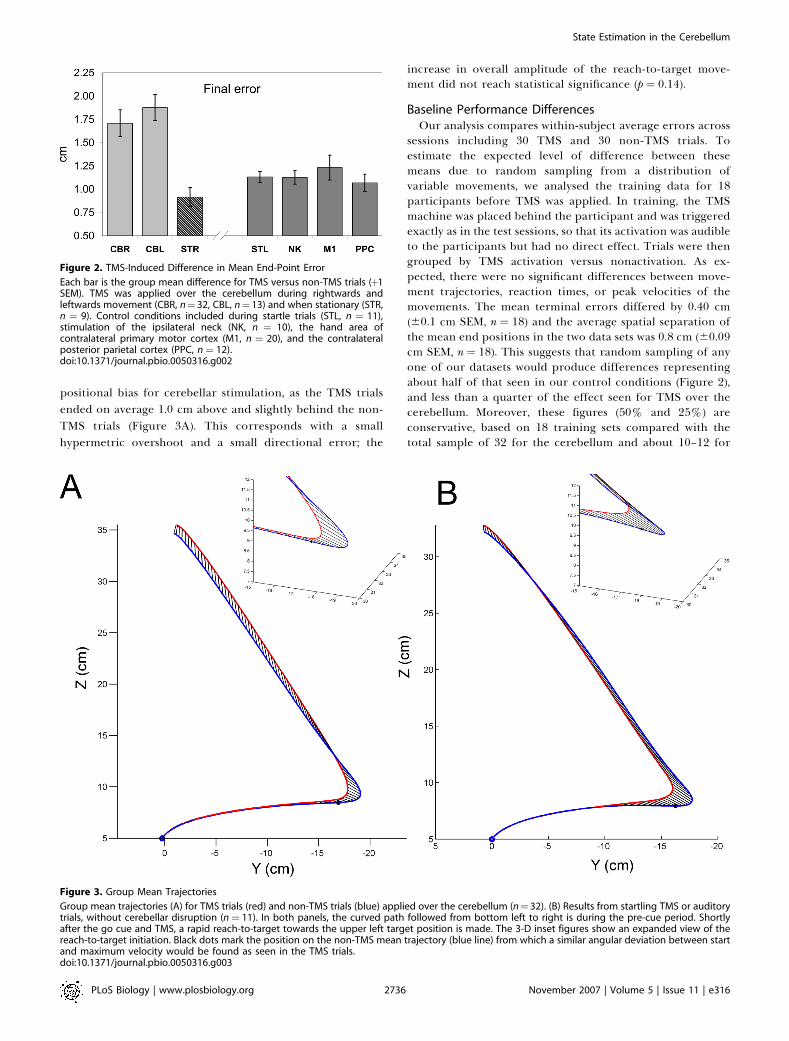
compared with stimulation lower on the neck (1.12 cm, n ¼11), or over the hotspot in the primary motor cortex forinducing visible twitches in the first dorsal interosseousmuscle in the hand (1.23 cm, n ¼ 21), and higher than whenstartling auditory clicks were presented either using the TMScoil over the ear or using ear phones without TMS (1.18 cm, n¼ 4 and 7, respectively). It was also higher than withstimulation over the contralateral posterior parietal cortex(1.07 cm, n¼ 12), which was targeted using the coordinates ofthe P3 electrode in the 10–20 electroencephalogram (EEG)electrode positioning scheme [28]. A one-way ANOVA withfive conditions (cerebellum, neck, startle, parietal, and motorcortical stimulation) was significant (F(4,78)¼ 3.79, p¼ 0.007,and post-hoc comparisons of the cerebellar condition with
the other four conditions were all significant, p , 0.025). Thedifference from stimulation over the hand area of thecontralateral motor cortex was smallest (p¼ 0.025); the otherfour control conditions were not significantly separable fromeach other (p . 0.27)]. Thus, whereas each of these sitesinduced some increase in end-point error, presumably due tothe nonspecific effects of the stimulation, the effects causedby stimulation over the ipsilateral cerebellum were mostpronounced and statistically reliableThe increase in error was partly due to a 14% increase in
end-point variability across trials. However, the RMS end-point standard deviation measured across all three dimen-sions was not significantly different for any of the fiveconditions (p . 0.103). There was also a significant end-point
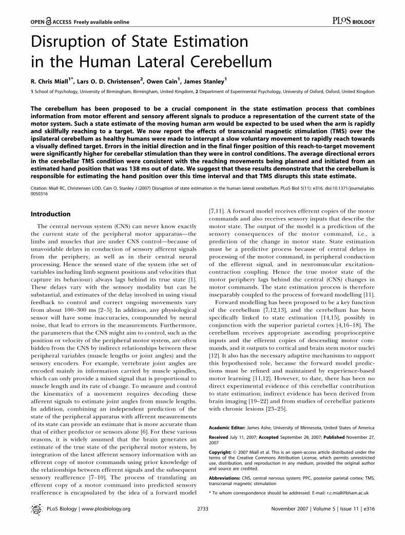
Figure 1. The Experimental Task and Typical Single-Participant Data
The experimental task (A), individual trial data (B and C), and session averaged data (D and E) (n¼ 30 trials) from one typical participant as TMS wereapplied over the right lateral cerebellum. (B and D) show the finger trajectory viewed from behind and from the right of the subject (C and E). In allpanels, TMS trials are plotted in red and non-TMS trials are in blue. Clockwise rotation in (B and D) is defined as increasing azimuth angle; clockwiserotation in (C and E) is defined as decreasing elevation angle.doi:10.1371/journal.pbio.0050316.g001
PLoS Biology | www.plosbiology.org November 2007 | Volume 5 | Issue 11 | e3162735
State Estimation in the Cerebellum
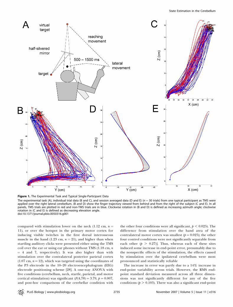
positional bias for cerebellar stimulation, as the TMS trialsended on average 1.0 cm above and slightly behind the non-TMS trials (Figure 3A). This corresponds with a smallhypermetric overshoot and a small directional error; the
increase in overall amplitude of the reach-to-target move-ment did not reach statistical significance (p ¼ 0.14).
Baseline Performance DifferencesOur analysis compares within-subject average errors across
sessions including 30 TMS and 30 non-TMS trials. Toestimate the expected level of difference between thesemeans due to random sampling from a distribution ofvariable movements, we analysed the training data for 18participants before TMS was applied. In training, the TMSmachine was placed behind the participant and was triggeredexactly as in the test sessions, so that its activation was audibleto the participants but had no direct effect. Trials were thengrouped by TMS activation versus nonactivation. As ex-pected, there were no significant differences between move-ment trajectories, reaction times, or peak velocities of themovements. The mean terminal errors differed by 0.40 cm(60.1 cm SEM, n ¼ 18) and the average spatial separation ofthe mean end positions in the two data sets was 0.8 cm (60.09cm SEM, n ¼ 18). This suggests that random sampling of anyone of our datasets would produce differences representingabout half of that seen in our control conditions (Figure 2),and less than a quarter of the effect seen for TMS over thecerebellum. Moreover, these figures (50% and 25%) areconservative, based on 18 training sets compared with thetotal sample of 32 for the cerebellum and about 10–12 for
Figure 3. Group Mean Trajectories
Group mean trajectories (A) for TMS trials (red) and non-TMS trials (blue) applied over the cerebellum (n¼ 32). (B) Results from startling TMS or auditorytrials, without cerebellar disruption (n¼ 11). In both panels, the curved path followed from bottom left to right is during the pre-cue period. Shortlyafter the go cue and TMS, a rapid reach-to-target towards the upper left target position is made. The 3-D inset figures show an expanded view of thereach-to-target initiation. Black dots mark the position on the non-TMS mean trajectory (blue line) from which a similar angular deviation between startand maximum velocity would be found as seen in the TMS trials.doi:10.1371/journal.pbio.0050316.g003
Figure 2. TMS-Induced Difference in Mean End-Point Error
Each bar is the group mean difference for TMS versus non-TMS trials (þ1SEM). TMS was applied over the cerebellum during rightwards andleftwards movement (CBR, n¼ 32, CBL, n¼ 13) and when stationary (STR,n ¼ 9). Control conditions included during startle trials (STL, n ¼ 11),stimulation of the ipsilateral neck (NK, n ¼ 10), the hand area ofcontralateral primary motor cortex (M1, n ¼ 20), and the contralateralposterior parietal cortex (PPC, n¼ 12).doi:10.1371/journal.pbio.0050316.g002
PLoS Biology | www.plosbiology.org November 2007 | Volume 5 | Issue 11 | e3162736
State Estimation in the Cerebellum
other conditions. The larger datasets would be less affected byrandom sampling.
TMS Effect Is Exposed by Dynamic State ChangeTMS took place during the reaction time between the go
cue and the start of the reach-to-target movement, while thehand was being actively moved towards the right. Thus, theTMS-induced error for the cerebellar stimulation conditionis, we hypothesize, due to the disruption of the stateestimation process within the ipsilateral cerebellum andwould affect the estimation during the current rightwardsmovement. If true, then the effect should not be seen if theneed for state estimation was minimized. We thereforecompared TMS stimulation at the same cerebellar locationbut with the participant holding their arm stationary at themoment of cue onset. In this control condition, the startingbutton was shifted 20 cm laterally, to be coincident with themean start position of the hand in other conditions, and theparticipant was instructed to lift the finger from the startbutton but to then remain stationary until the auditory gocue. Hence the starting position was known and static priorto cue onset. A reach-to-target from this fixed position wouldnot require renewed state-estimation because the state wasconstant and up to date throughout the reaction time period.End point errors were significantly lower in this conditionthan with cerebellar stimulation (0.92 cm, n¼ 9) and were not
significantly different from the other control conditions(Figure 2). Hence the TMS-induced effect is specific to thoseconditions in which the initial state of the arm is dynamicallychanging, when its true state must be estimated,
Cerebellar TMS Causes Initial Aiming ErrorThe duration of the cued reach-to-target movement was
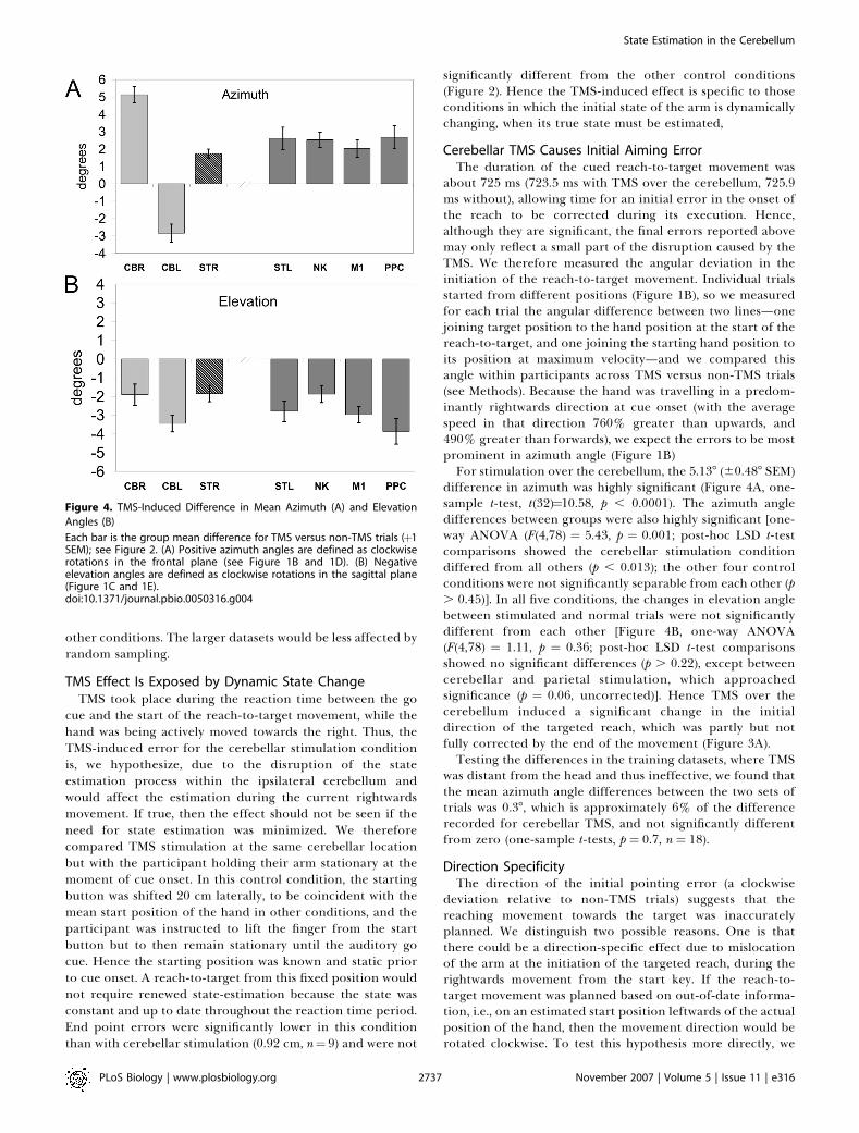
about 725 ms (723.5 ms with TMS over the cerebellum, 725.9ms without), allowing time for an initial error in the onset ofthe reach to be corrected during its execution. Hence,although they are significant, the final errors reported abovemay only reflect a small part of the disruption caused by theTMS. We therefore measured the angular deviation in theinitiation of the reach-to-target movement. Individual trialsstarted from different positions (Figure 1B), so we measuredfor each trial the angular difference between two lines—onejoining target position to the hand position at the start of thereach-to-target, and one joining the starting hand position toits position at maximum velocity—and we compared thisangle within participants across TMS versus non-TMS trials(see Methods). Because the hand was travelling in a predom-inantly rightwards direction at cue onset (with the averagespeed in that direction 760% greater than upwards, and490% greater than forwards), we expect the errors to be mostprominent in azimuth angle (Figure 1B)For stimulation over the cerebellum, the 5.138 (60.488 SEM)
difference in azimuth was highly significant (Figure 4A, one-sample t-test, t(32)¼10.58, p , 0.0001). The azimuth angledifferences between groups were also highly significant [one-way ANOVA (F(4,78) ¼ 5.43, p ¼ 0.001; post-hoc LSD t-testcomparisons showed the cerebellar stimulation conditiondiffered from all others (p , 0.013); the other four controlconditions were not significantly separable from each other (p. 0.45)]. In all five conditions, the changes in elevation anglebetween stimulated and normal trials were not significantlydifferent from each other [Figure 4B, one-way ANOVA(F(4,78) ¼ 1.11, p ¼ 0.36; post-hoc LSD t-test comparisonsshowed no significant differences (p . 0.22), except betweencerebellar and parietal stimulation, which approachedsignificance (p ¼ 0.06, uncorrected)]. Hence TMS over thecerebellum induced a significant change in the initialdirection of the targeted reach, which was partly but notfully corrected by the end of the movement (Figure 3A).Testing the differences in the training datasets, where TMS
was distant from the head and thus ineffective, we found thatthe mean azimuth angle differences between the two sets oftrials was 0.38, which is approximately 6% of the differencerecorded for cerebellar TMS, and not significantly differentfrom zero (one-sample t-tests, p ¼ 0.7, n ¼ 18).
Direction SpecificityThe direction of the initial pointing error (a clockwise
deviation relative to non-TMS trials) suggests that thereaching movement towards the target was inaccuratelyplanned. We distinguish two possible reasons. One is thatthere could be a direction-specific effect due to mislocationof the arm at the initiation of the targeted reach, during therightwards movement from the start key. If the reach-to-target movement was planned based on out-of-date informa-tion, i.e., on an estimated start position leftwards of the actualposition of the hand, then the movement direction would berotated clockwise. To test this hypothesis more directly, we
Figure 4. TMS-Induced Difference in Mean Azimuth (A) and Elevation
Angles (B)
Each bar is the group mean difference for TMS versus non-TMS trials (þ1SEM); see Figure 2. (A) Positive azimuth angles are defined as clockwiserotations in the frontal plane (see Figure 1B and 1D). (B) Negativeelevation angles are defined as clockwise rotations in the sagittal plane(Figure 1C and 1E).doi:10.1371/journal.pbio.0050316.g004
PLoS Biology | www.plosbiology.org November 2007 | Volume 5 | Issue 11 | e3162737
State Estimation in the Cerebellum

include one additional group of participants, who were testedwith TMS over the cerebellum during movements made froma far-right position, such that the arm at the go cue was, onaverage, at the same position, but was moving leftwards(Figure 5). This group showed end-point error amplitudes(mean 1.88 cm, n ¼ 13) inseparable from the originalcerebellar group (1.71 cm, n¼ 32), and as before, significantlygreater than all the other control conditions (Figure 2, one-way ANOVA, F(5,69) ¼ 3.57, p ¼ 0.006, post-hoc t-tests p ,
0.005). The mean azimuth angle was �2.848, in other words,rotated counterclockwise, and hence statistically differentfrom all other conditions (Figure 4A, p , 0.025); the meanelevation angle did not differ from other conditions (Figure4B, p . 0.19).
The second possibility is that the TMS caused theparticipants to mislocate the target, and they were thereforeaccurately reaching to the wrong location. We can dismissthis with the stationary start position data. In this case, therewas very little azimuth error (1.748, not significantly differentfrom all other control conditions), and the end point errorswere actually the smallest observed. In addition, we corre-lated the directional errors in azimuth and elevation for bothgroups (cerebellar TMS during rightwards and leftwardsmovements) against the end-point errors in the x-, y-, and z-axis (Figure 1). We hypothesize that if TMS caused targetmislocation, then the initial angular deviations would becorrelated with the final positional errors. However, we foundno evidence to support this: there was only one near-
significant correlation between change in azimuth angleand change in x error during leftwards movement condition(p ¼ 0.061, Bonferroni adjusted); this is not the axis in whichchanges in azimuth angle would be most prominent, given thenear-vertical plane of movements (Figure 1).Thus we interpret this as further evidence that the initial
direction was inaccurate and that some but not all of thiserror was corrected during the reach. We find no evidencethat the final position was mislocated and that the initialangles were altered to reach this final location.
Estimating the Internal Error in Hand StateTo estimate the hand state used to plan the reach-to-target
action, we could backtrack along the mean trajectory of non-TMS trials, participant by participant, to find a point at whichthe angle towards the maximum velocity position was equalto the mean angular deviation seen for that participant inTMS trials. In other words, by assuming that the angular errorin aiming to the target was due to a failure of the cerebellarstate estimation of the hand position, during the initial slowmovement, we found that the prior position of the hand—before the go cue—at which the angular difference betweenstart and maximum velocity points would be the same as wasfound between TMS and non-TMS trials. This position was3.97 cm (6 1.01 SEM) leftwards of the actual reach-to-targetstart position (black dot, Figure 3A). Converting frompositional differences along their mean path between TMSand non-TMS trials, and given the mean hand speed at thecue onset, measured subject-by-subject, the distance of 3.97cm suggests that the reach-to-target was planned based on thehand’s position 138 ms (619 ms SEM) previously. This ‘‘state-estimation interval’’ is significantly greater than zero (one-sample t-test, t(18)¼ 7.33, p , 0.0001) and is also greater thanthe change in reaction time caused by the TMS (one-tailedpaired-sample t-test, t(18)¼ 2.011, p¼ 0.030; see ‘‘nonspecificeffects’’ below). For the group tested making initially left-wards arm movement, the ‘‘state-estimation interval’’ wasalmost exactly the same, 134 ms (617 ms SEM). Thisprediction interval cannot be calculated for the stationary-start condition, because backtracking on a stationary trajec-tory is not possible.
Other Nonspecific Stimulation EffectsThe velocity profiles of the reach-to-target movements
were significantly altered by cerebellar TMS (Figure 6A–6C).The effect was to reduce the reaction time for the rapidreach-to-target movement by about 80 ms and to increase thepeak velocity by about 15%. For the main condition ofinterest, with TMS over the lateral cerebellum duringongoing movement, the mean reaction time for TMS versusnon-TMS trials was 172.5 ms versus 265.9 ms. TMS-inducedstartle effects have been reported previously [29] because ofthe noise and cutaneous stimulation. TMS over the cerebel-lum using the large double-cone coils does cause noticeableauditory stimulation, as well as cutaneous and muscularstimulation. Very similar effects on the movement profileswere seen in a control group (Figure 6D) tested with TMSstimulation over the right ear (n¼4) or with startling auditorywhite noise bursts played over headphones (n¼7), confirmingthat the reduction in reaction time and increase in peakvelocity is likely to be due to a startle response. For this startlecontrol group (n¼ 11), the mean reaction time was 175.2 ms
Figure 5. Group Mean Trajectories for TMS Trials (Red) and Non-TMS
Trials (Blue) Applied over the Cerebellum (n¼ 13)
Solid lines indicate stimulation during initial rightwards movement;dotted lines show stimulation during initial leftwards movement. Thedeviation between between TMS and non-TMS trajectories at the start ofthe reach towards the final target is reversed between the twoconditions, while final errors are similar. The insert at top right is theterminal portion of the trajectories, rotated into the frontal plane. Thisemphasises the greater overshoot in the z-axis for rightwards TMS trials(red solid lines) compared to leftwards TMS trials (red dotted lines),which mainly overshot in depth (x-axis).doi:10.1371/journal.pbio.0050316.g005
PLoS Biology | www.plosbiology.org November 2007 | Volume 5 | Issue 11 | e3162738
State Estimation in the Cerebellum
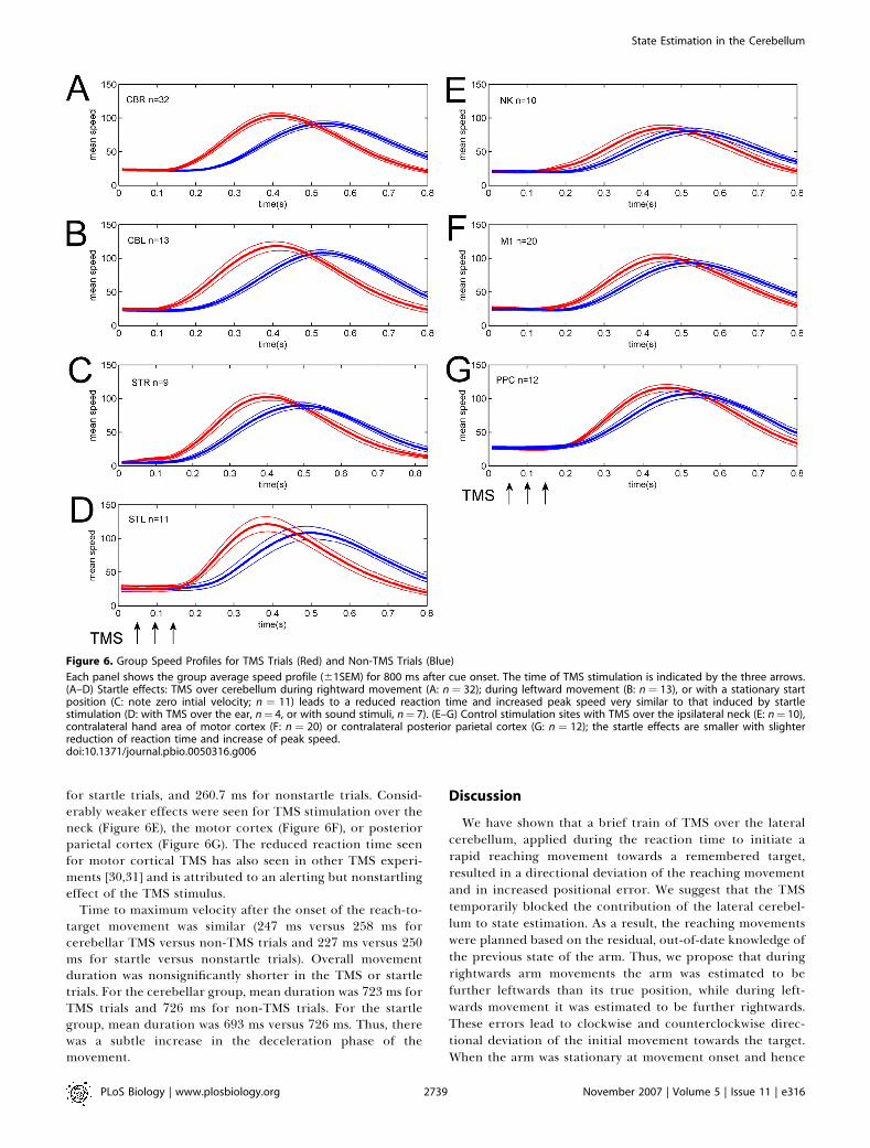
for startle trials, and 260.7 ms for nonstartle trials. Consid-erably weaker effects were seen for TMS stimulation over theneck (Figure 6E), the motor cortex (Figure 6F), or posteriorparietal cortex (Figure 6G). The reduced reaction time seenfor motor cortical TMS has also seen in other TMS experi-ments [30,31] and is attributed to an alerting but nonstartlingeffect of the TMS stimulus.
Time to maximum velocity after the onset of the reach-to-target movement was similar (247 ms versus 258 ms forcerebellar TMS versus non-TMS trials and 227 ms versus 250ms for startle versus nonstartle trials). Overall movementduration was nonsignificantly shorter in the TMS or startletrials. For the cerebellar group, mean duration was 723 ms forTMS trials and 726 ms for non-TMS trials. For the startlegroup, mean duration was 693 ms versus 726 ms. Thus, therewas a subtle increase in the deceleration phase of themovement.
Discussion
We have shown that a brief train of TMS over the lateralcerebellum, applied during the reaction time to initiate arapid reaching movement towards a remembered target,resulted in a directional deviation of the reaching movementand in increased positional error. We suggest that the TMStemporarily blocked the contribution of the lateral cerebel-lum to state estimation. As a result, the reaching movementswere planned based on the residual, out-of-date knowledge ofthe previous state of the arm. Thus, we propose that duringrightwards arm movements the arm was estimated to befurther leftwards than its true position, while during left-wards movement it was estimated to be further rightwards.These errors lead to clockwise and counterclockwise direc-tional deviation of the initial movement towards the target.When the arm was stationary at movement onset and hence
Figure 6. Group Speed Profiles for TMS Trials (Red) and Non-TMS Trials (Blue)
Each panel shows the group average speed profile (61SEM) for 800 ms after cue onset. The time of TMS stimulation is indicated by the three arrows.(A–D) Startle effects: TMS over cerebellum during rightward movement (A: n ¼ 32); during leftward movement (B: n ¼ 13), or with a stationary startposition (C: note zero intial velocity; n ¼ 11) leads to a reduced reaction time and increased peak speed very similar to that induced by startlestimulation (D: with TMS over the ear, n¼ 4, or with sound stimuli, n¼ 7). (E–G) Control stimulation sites with TMS over the ipsilateral neck (E: n¼ 10),contralateral hand area of motor cortex (F: n ¼ 20) or contralateral posterior parietal cortex (G: n ¼ 12); the startle effects are smaller with slighterreduction of reaction time and increase of peak speed.doi:10.1371/journal.pbio.0050316.g006
PLoS Biology | www.plosbiology.org November 2007 | Volume 5 | Issue 11 | e3162739
State Estimation in the Cerebellum
the state estimate was unchanging, then the direction of thereaching movements was largely unaffected by the stimula-tion.
Our results therefore provide further evidence for acontribution by the cerebellum to state estimation and, byinference, to forward modelling the sensory consequences ofaction [12,22,32,33]. These forward model predictions, com-bined with independent sensory information from theperiphery, provide an optimal estimate of the current stateof the arm. However, there is considerable debate about therole of the cerebellum in motor control [14,20,34,35] andother functions, such as timing, motor learning, predictivecontrol, and inverse dynamic modelling, have all beenproposed. We believe the present data are best understoodin the context of state estimation. First, predictive control[36,37] is very closely related to state estimation, because theconsequences of motor commands must be predicted inorder to update and control subsequent actions. Hence stateestimation is a subset of predictive control [1]. Next, thecerebellum appears important in timing, especially duringdiscrete motor tasks [38,39]. The timing component of ourtask is minimal as movements were initiated after a randominterval, eliminating a timing strategy and participants wereinstructed to reach as fast as possible after the go cue.Furthermore, the target was static, and so the directionaldifferences in reaching behaviour in TMS versus non-TMStrials are difficult to justify by timing alone.
Another important postulated function is in inversedynamic modelling, with a functional role for the cerebellumin generating motor commands rather than in predictingtheir outcome [7,33,40]. Again, the pattern of results we haveshown is not easily fit by this hypothesis. In particular, thereach-to-target from a stationary position was not differentfrom control conditions, whereas disruption of an inversemodel would affect all movement for which the model wasused. These data also argue against an explanation that theTMS impaired control of interjoint dynamics. Failure tocompensate for interjoint dynamics, which has been pro-posed as an explanation for cerebellar ataxia [27], mightcontribute to our results, as limb dynamics would be differentfor the movements made from left and right. However, theinitial movements were relatively slow (mean velocity 27.9 cm/s, Figure 6) and so these dynamic effects would be small incomparison to their effects during the much faster reach-to-target action. Indeed, the reach-to-target made from thestatic position was as rapid (peak velocities of about 100 cm/s,Figure 6C) as in other conditions and, we assume, was fastenough to expose weak interjoint coordination. Its trajectorywas not different from the control conditions. But whetherthese effects contribute more subtly to our overall results willrequire further work, perhaps using faster baseline move-ments so that both the dynamic effects and the positionalmisestimation are larger.
It is also thought that state estimation is required tominimize timing differences between effectors during coor-dinated actions [41,42]. Loss of coordination is one of thecardinal symptoms of cerebellar dysfunction [43] and isevident as gross ataxia as well as in more subtle measures suchas the failure to coordinate grip and lift forces during objectmanipulation [23,24,44]. Recently, Deidrichsen et al. testedcoordination of a reach and a button press and were able toseparate experimentally time-dependent and state-depend-
ent behavioural strategies. They found that functionalactivation changes in the lateral cerebellum were betterexplained by state estimation than by timing [45], consistentwith the present results.Our estimates of the angular deviations imply that the
rapid reaching movements were planned on information thatis about 138 ms out of date, an interval that is in theappropriate order of magnitude of sensory reafference[2,3,5]. From this result, we would predict that cerebellarpathology would lead to movement control based on thearm’s state about 138 ms less advanced than actual. Thiswould result in direction- and speed-specific deviations in theinitial segment of any rapid movement, especially those madeduring ongoing action, as we have shown here. It would alsolead to hypermetria, as the state estimate would be disturbedthroughout the reaching movement, rather than just at itsinitiation. Thus, even the final stages of reaching to a targetwould be affected more obviously than we have seen in ourexperiments, where, we suspect, the TMS-induced disruptionof state estimate was brief and may have largely recovered bythe end of the reaching action. The consequences ofexperimentally delaying visual feedback, which effectivelymakes the state estimate inaccurate with respect to actualfeedback, are similar to that of cerebellar inactivation [2,46].Delayed state estimation would therefore explain loss ofcoordination and ataxia.The state estimation process is likely to be iterative [8], with
the current state being updated by an optimised weighting ofafferent proprioceptive and visual information and byefferent motor commands. An iterative calculation isoptimal, because previous state estimates, even if inaccurate,provide an additional source of information to be used in thenew estimate. This is particularly true for physiologicalsystems in which the state cannot change instantly; theremust be a strong correlation between previous state and newstates. We cannot tell from the present results to whether the138 ms interval reflects ‘‘freezing’’ of the state estimate, untilthe cerebellum generates a new estimate, or whether itreflects the fall-back use of out-of-date proprioceptive andvisual information. Possible methods to address this questionwould involve using longer trains of TMS, to stretch the timethat it was perturbed, or to use repetitive TMS to induce atemporary ‘‘virtual lesion’’, coupled with adaptation todelayed visual feedback.However another important issue is whether the state
estimate is localized entirely within the cerebellum or isdistributed across this and other areas. One obviouscandidate, given its well-documented role in spatial repre-sentations, is the posterior parietal cortex [47–49]. It has alsobeen proposed as a locus of the state estimate [16,17,50] andhas been recently implicated in the sense of agency and themental representation of [51,52]; agency is also dependent onforward modelling [53]. Our data argue against the possibilitythat the state estimations are generated exclusively by theparietal cortex, because of the disruption caused by cer-ebellar TMS, but it seems plausible that both areas areinvolved. One possibility is that the parietal cortex maintainsa body representation or body schema [50,54] that is updatedduring movements [16]. We would argue that this update iscalculated by the cerebellum.Anatomical connections to the cerebellum are consistent
with this, as it receives powerful projections from posterior
PLoS Biology | www.plosbiology.org November 2007 | Volume 5 | Issue 11 | e3162740
State Estimation in the Cerebellum
parietal cortex [55], which may hold a representation of thecurrent state estimate [16], as well as from cortical motorareas, sending an efferent copy of descending commands [56].It also receives visual and proprioceptive afferents, althoughin our task, visual feedback was blocked during the action.The output of the cerebellar processing, which we proposeconstitutes an estimation of the change in the motor statecaused by the efferent signals, may then return to posteriorparietal cortex [57–60] to update its representation, or bedirected to motor areas to contribute to the control of theactions [56]. In this framework, one might expect that TMS ofposterior parietal cortex would also disrupt reach-to-targetactions in this task. Targeting the superior parietal cortexusing the P3 electroencephalogram electrode coordinates didnot cause significant effects. However, this negative resultshould be taken with caution, because we may have missed acritical locus within the posterior parietal cortex (PPC)responsible for maintaining the state representation.
The PPC also contributes to the representation of targetpositions [61], and hence it is important to distinguish loss-of-state estimation from errors in localizing the rememberedtarget. This might be a result of TMS-perturbed input to thePPC, or it may be possibly due to a cerebellar role in targetlocalization. However, we saw no evidence for mislocalizationof the target in any of our conditions. In particular, we founddirection-specific changes in initial movement direction withcerebellar TMS that were uncorrelated with changes in endposition of the reach-to-target movement. Moreover, theseeffects were significantly different from the errors caused bycerebellar TMS when the hand was initially stationary,although one might expect any effect of target mislocaliza-tion to be common across all these three conditions. There islimited published evidence of a role for the cerebellum inlocalization of a visual target [62], and several opposingresults [63–65]. Thus, it seems unlikely that the changes indirection were a sign of movements planned towards aperturbed position,
However, comparing the TMS data from the stationarystart condition to the two active movement conditions doesraise another concern, because it is well known that the TMSthresholds in motor cortex are lower during active movementthan during rest. We did not test thresholds for activationover the cerebellum; however, we note that the arm was not atrest in this static position but was actively held in the air justabove the start key; this is a motor task in which thecerebellum is actively engaged [66–68]. Others have usedsingle-shock stimulation levels of 55% with the same double-cone coil and have seen brief changes in excitability of thecontralateral motor cortex that are consistent with activationof the cerebellar cortex [69]; we have seen the same effects onmuscle-evoked potential (MEP) amplitude (unpublishedresults) using triplets of 20-Hz pulses as used in the presentexperiments, even at stimulation levels of as low as 35% andwith the arm genuinely at rest. So although it is possible thatour TMS protocol was less effective during the stationarycondition, we do not think this likely to have influenced theseresults.
Control Experiments and Other ConsiderationsThe TMS pulse train is expected to lead to a temporary
disruption of the neural processing in underlying targettissue. We targeted the hand area of the ipsilateral cerebellar
cortex at a site at which we have previously caused disruptionof visually guided action [70], and at which TMS is known toaffect cerebellar–cerebral projections leading to measurablechanges in motor cortical excitation in the contralateral handarea [69]. One important control condition was therefore totest the same TMS protocol applied directly to the contrala-teral motor cortex, to rule out indirect effects of thecerebellar stimulation at this remote site. Motor corticalTMS did raise terminal errors lead to some directionaldeviation of the reaching movement, but both of these effectswere of a significantly smaller magnitude than those seenafter cerebellar stimulation (Figures 2 and 3).At the same time, the cerebellar TMS stimulation caused a
noticeable change in movement kinematics, with a significantreduction in movement onset latency and an increase in peakvelocity. Similar but weaker effects were also generated bystimulation over the neck, at a site 3 cm below the cerebellarstimulation site. We reproduced the cerebellar effect onreaction times using a startling stimulus that did not involvefunctional TMS, either by using one wing of the double-conecoil placed over the participants’ right ear to induce the noiseand possible auditory nerve stimulation caused by the TMSstimulation over the cerebellum or neck, or by playing loudwhite noise bursts through headphones without any activeTMS. Thus TMS aimed at the lateral cerebellum can startlethe participant and lead to changes in the velocity profile ofthe movement, regardless of its effect on the cerebellum.However, even though these control conditions could inducea similar magnitude shift in reaction time and increase inpeak velocity, they induced neither the terminal errors northe initial directional errors that were caused by cerebellarTMS. This confirms that the initial directional deviation andthe final positional errors were not a result of the startleeffect. Furthermore, we also tested cerebellar stimulationfrom a static starting position, a condition that minimizes theneed for a dynamic update of the state estimation. Again, wesaw change in movement onset and velocity attributable tostartle, but we saw no directional deviation or terminal error.It is also possible that TMS applied over the lateral
cerebellum could cause movement errors due to directstimulation of muscles in the neck, which might lead toshoulder or upper arm deviation or cause arm movement bystimulation of the brachial plexus [69]. We discounted bothpossibilities by testing TMS stimulation over the neck at a sitemore likely to activate the brachial plexus, and thatstimulation generated visible twitches in the neck musclesbut without inducing the directional or terminal errors. TMSat the level used (45% of machine output) is unlikely to causecortico-spinal stimulation [69,71]. Another control involvedmeasuring the total deviation of the hand that was held staticover the start position, without any active reaching task,during TMS of the cerebellum. This would expose anyinvoluntary hand motion induced either by the TMS,including activation of the cortico-spinal collaterals [71], orby its startling effect. On TMS trials (three participants), theindex finger was briefly deviated laterally by less than 5 mm,and within 200 ms had returned to within 1 mm of its initialposition, within the normal reaction time period (266 ms).Hence our results are unlikely to be due to TMS-inducedperipheral effects.A final methodological consideration is that the signifi-
cantly reduced reaction times seen after cerebellar TMS
PLoS Biology | www.plosbiology.org November 2007 | Volume 5 | Issue 11 | e3162741
State Estimation in the Cerebellum

mean that the reach-to-target starts from a position near tothe mean hand position at which the angular differencebetween TMS and non-TMS trials is equal (Figure 3A). Inother words, one could argue that the TMS has merely shiftedthe mean start position leftwards in accordance with thereduction in reaction time, and has not affected the internalstate estimate of the hand. However, the interval of 138 msestimated from comparing directional errors on TMS versusnon-TMS trials is significantly greater than the change inreaction time (93 ms). Furthermore, reduction in reactiontime alone should not necessarily cause a directional error. Ifthe reach-to-target movements on TMS trials were plannedusing an accurate state estimate, then their initial directionshould be towards the target, despite their reduced latency.This result was clearly seen for the startle trials (Figure 3B), inwhich the reaction times were advanced by 85 ms, but theinitial direction was unchanged. Thus the startling stimulusdoes not affect the movement direction, whereas cerebellarTMS does, and this dissociates the effects of reduction inreaction time from the loss of state estimation of the hand.
In conclusion, we suggest that these results indicate that thelateral cerebellum is responsible for estimating the true stateof the peripheral motor system over a short time interval. Weassume this estimation is based on forward modelling of theexpected consequences of outgoing motor commands andthat the updated estimation is sent from the cerebellum tocerebral areas responsible for planning and controlling thereaching action. These experiments do not tell us how thecerebellum generates these signals, whether the TMS protocolhas any influence on cerebellar learning, or whether stateestimates are topographically organized in the cerebellarcortex or nuclei. Experiments using methods with finerspatial resolution than TMS will be needed to address theseimportant questions.
Materials and Methods
Participants. Forty-five right-handed participants (age range 22–48y, 13 male) received TMS, after providing informed written consent,and with approval from the Central Office for Research EthicsCommittees. Two of these were authors of this article: RCM andLODC. Seven participants (age range 22–50, 6 male) were tested withauditory stimulation, including the authors RCM and JS.
Tasks. Participants sat at a table with their head supported by achin rest and wearing Plato LCD goggles (Translucent Tech). A TMScoil was held in position using a Magic-Arm (Adaptivation). Theposition of the right index finger was recorded using a PolhemusFastrak at 120 Hz. Trials were timed by a computer running underDOS. Each trial began with the index finger depressing a start key onthe table top in front of the right shoulder. Cued by a set of threerising tones at 500-ms intervals, the participant was required torelease the start key on the third tone, and to begin to move the righthand towards the right side. The Plato goggles were switched toopaque as soon as the start key was released. Early or late release ofthe key led to the trial being aborted. At a uniform random time 500–1,500 ms after the key was released, the onset of a fourth continuoustone cued the participant to make a rapid forwards and upwardsreach to place the index finger on the position of a virtual targetimage, reflecting a 1-cm target in a mirror. The target wasapproximately 28 cm above and 15 cm in front of the start key.One second after this final go cue, the Plato goggles were switched totransparent, allowing terminal vision of the static finger and virtualtarget. The subject then returned to the start key at their own pace.
On each session, participants were given 60 practice trials, on arandom 50% of which a series of three TMS clicks were heard, at 50,100, and 150 ms after the onset of the go cue. During training, the coilwas held about 1 m from the head; training data were only recordedfor 18 participants. Immediately after the practise, the TMS coil was
positioned against the scalp, and another 60 trials collected, withactive TMS on half the trials.
Nine participants were also tested in a condition is which the startkey was moved 20 cm laterally, to the average position at which thereach to the target started (Figure 1B). Subjects were instructed to liftthe index finger off the start key but to remain stationary until the gocue signalled the rapid reach to the target. All other aspects of thetask remained the same.
TMS. Repetitive stimulation was delivered as three biphasic pulsestriggered at 20 Hz (50 ms) by the experimental control computer, at45% of machine output, using a Magstim Rapid (Magstim Co.). Forstimulation of the lateral cerebellum (n¼32), a 90-mm radius double-cone coil was centred 3 cm lateral and 1 cm below the inion [69,70]. Inthis position, one wing of the coil normally overlaps the participant’sright ear. Ear plugs were provided.
To test the effects of TMS noise and its possible stimulation of theright ear, the orientation of the double cone coil was reversed in fourparticipants so that one coil surrounded the right ear while the otherwas approximately normal to the scalp. Biphasic stimulation was setat 45% of the machine output. These data were combined with thatof a group of seven participants in which the TMS trigger pulses wereused to trigger brief white noise bursts (100 dB, 20-ms duration),played through binaural headphones. This white noise was suffi-ciently loud to evoke observable reflexive blinks in all participants,while remaining within safety limits. Comparison of the data from thetwo groups (ear TMS versus auditory stimulation) revealed nosignificant differences, and the two datasets were combined.
For stimulation of the neck (n¼ 11) a flat, 70-mm radius figure-of-eight coil was used, with the coil centre 3 cm below the site used forcerebellar stimulation (3 cm lateral and 4 cm below the inion).Stimulator output was set at 45% of machine output.
For stimulation of the motor cortex (n¼ 20) the flat, 70-mm radiusfigure-of-eight coil was positioned at a site where an observabletwitch of the right first dorsal interoseus muscle was seen. Stimulatoroutput was set at the resting threshold. For posterior parietal cortex,all participants (n¼ 12) were also tested with M1 stimulation and thesame stimulator intensity was used. The P3 electrode position wasmeasured using standard landmarks.
Data analysis. Index finger trajectories were analysed in Matlabversion R2007a. A Polhemus Fastrak receiver was taped above theright index finger, and before the experiment began, each participantheld the index finger stationary in the position of the virtual target,under full vision. The recorded marker position was then taken as thetarget position in all subsequent analysis, accounting for the 1–1.5 cmpositional offset of the marker from the index finger pad. Fingerposition was recorded in three axes at 120 Hz; angular rotations ofthe hand that would invalidate this positional offset were minimaland estimated at less than 1 mm; the relative difference between TMSand non-TMS trials is less than 10% of this (0.1 mm).
TMS artefacts. TMS magnetic pulses can generate a significantone-sample (8 ms) artefact in the Polhemus motion tracking data,which uses magnetic field technology. These artefacts were detectedin the first-differenced time series data and removed by interpolationacross neighbouring data points.
To assess the impact of these artefacts, we recorded the apparentmarker position of a static marker placed at the average start positionof the reach-to-target movement, with the TMS coil placed inapproximate similar position as when testing a participant. Artefactremoval was successful and the apparent residual motion of themarker was under 0.1 cm or 1 cm/s. The duration of the artefact wasalso restricted mainly to within the typical reaction time, so anyresidual error did not affect analysis of the reach trajectory. Testingwith the marker attached to a participant’s finger held stationarywhile TMS was applied to the cerebellum (three participants, 20 trialseach) showed that stimulation of the cerebellum caused minor fingermotion that was recovered within 200 ms of TMS termination.
The cleaned positional data were then low-pass filtered (8th orderzero phase 7.5 Hz Butterworth filter). Low-pass filtered index fingertrajectories were differentiated to velocity, and the tangential speedwas averaged across all TMS (n¼ 30) and non-TMS trials (n¼ 30) persubject for each condition. The subject mean velocities were thenaveraged across the subject group.
Mean speed and jerk (second derivative of speed) profiles wereexamined before and after the removal of the TMS-artefacts in thePolhemus data to confirm that the artefact removal was effective. Alltrials (TMS and non-TMS) were processed and filtered identically.
Detection of terminal and directional error. The following stepswere taken for analysis of each trial. First, the time-point ofmaximum velocity was detected between the go cue and the end ofrecording (open circles, Figure 1B and 1C). Termination of the reach-
PLoS Biology | www.plosbiology.org November 2007 | Volume 5 | Issue 11 | e3162742
State Estimation in the Cerebellum
to-target was then taken as the time point at which movementvelocity fell below 5% of the maximum (filled circles and trialnumber, Figure 1B and 1C).
Reach-to-target movement onset was detected as the point ofmaximum curvature between the go cue and the point of maximumvelocity (asterisks, Figure 1B and 1C). To find this, each trajectoryfrom go cue to termination was spatially re-sampled to 100 uniformlyspaced points, the rate of change of these spatial positions thenrecorded as curvature, and the maximum curvature spatial positionfound. The time point of original data sample the within the originaltime series closest to this spatial position was then recorded as thetime point at which the reach movement initiated.
End point error was measured as the Cartesian distance of thefinger from the target. Directional errors were measured as theazimuth or elevation differenced between lines joining the start pointand target, versus the start point and maximum velocity point foreach trial. Mean angular differences in azimuth and elevationbetween all TMS (n ¼ 30) and all non-TMS trials (n ¼ 30) werecalculated per subject for each experimental session.
Estimation of predictive interval. To estimate the positional offsetthat corresponded to the directional error measured, the anglebetween the start position and the position of maximum velocity wascalculated for the mean trajectory of all non-TMS trials (n ¼ 30) foreach subject in each condition. Trajectories were spatially re-sampledbefore averaging (e.g., Figure 1D and 1E). Then, by iterativelyrecalculating this angle for each data position before the startposition, we found the first position at which the angular differenceexceeded the mean angular difference between TMS and non-TMStrials (black dots, Figure 3). The distance along the mean trajectorybetween this position and the start position was found. To estimatethe time interval that corresponded to this spatial offset, the meanvelocity of the hand was found at cue onset, for each subject. Dividingthe estimated offset, subject-by-subject by the mean velocity, weestimated the time interval of the state estimation.
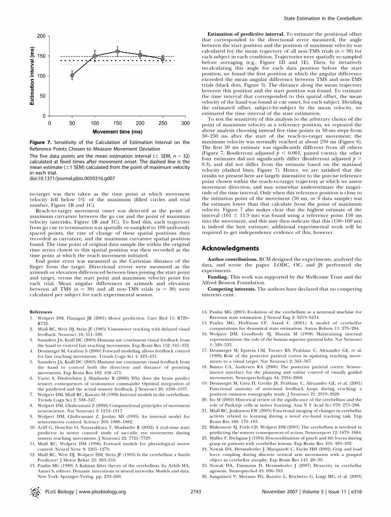
To test the sensitivity of this analysis to the arbitrary choice of thepoint of maximum velocity as a reference position, we repeated theabove analysis choosing instead five time points in 50-ms steps from50–250 ms after the start of the reach-to-target movement; themaximum velocity was normally reached at about 250 ms (Figure 6).The first 50 ms estimate was significantly different from all others(Figure 7, Bonferroni adjusted p , 0.001, paired t-tests); the otherfour estimates did not significantly differ (Bonferroni adjusted p .0.3), and did not differ from the estimate based on the maximalvelocity (dashed lines, Figure 7). Hence, we are satisfied that theresults we present here are largely insensitive to the precise referencepoint chosen within the reach-to-target trajectory at which we assessmovement direction, and may somewhat underestimate the magni-tude of the time interval. Only when this reference position is close tothe initiation point of the movement (50 ms, or 6 data sample) wasthe estimate lower than that calculate from the point of maximumvelocity. Figure 7 also makes clear that the highest estimate of theinterval (161 6 11.9 ms) was found using a reference point 150 msinto the movement, and this may then indicate that this (150–160 ms)is indeed the best estimate; additional experimental work will berequired to get independence evidence of this, however.
Acknowledgments
Author contributions. RCM designed the experiments, analysed thedata, and wrote the paper. LODC, OC, and JS performed theexperiments.
Funding. This work was supported by the Wellcome Trust and theAlfred Benson Foundation.
Competing interests. The authors have declared that no competinginterests exist.
References1. Wolpert DM, Flanagan JR (2001) Motor prediction. Curr Biol 11: R729–
R732.2. Miall RC, Weir DJ, Stein JF (1985) Visuomotor tracking with delayed visual
feedback. Neurosci 16: 511–520.3. Saunders JA, Knill DC (2003) Humans use continuous visual feedback from
the hand to control fast reaching movements. Exp Brain Res 152: 341–352.4. Desmurget M, Grafton S (2000) Forward modeling allows feedback control
for fast reaching movements. Trends Cogn Sci 4: 423–431.5. Saunders JA, Knill DC (2005) Humans use continuous visual feedback from
the hand to control both the direction and distance of pointingmovements. Exp Brain Res 162: 458–473.
6. Vaziri S, Diedrichsen J, Shadmehr R (2006) Why does the brain predictsensory consequences of oculomotor commands? Optimal integration ofthe predicted and the actual sensory feedback. J Neurosci 26: 4188–4197.
7. Wolpert DM, Miall RC, Kawato M (1998) Internal models in the cerebellum.Trends Cogn Sci 2: 338–347.
8. Wolpert DM, Ghahramani Z (2000) Computational principles of movementneuroscience. Nat Neurosci 3: 1212–1217.
9. Wolpert DM, Ghahramani Z, Jordan MI (1995) An internal model forsensorimotor control. Science 269: 1880–1882.
10. Ariff G, Donchin O, Nanayakkara T, Shadmehr R (2002) A real-time statepredictor in motor control: study of saccadic eye movements duringunseen reaching movements. J Neurosci 22: 7721–7729.
11. Miall RC, Wolpert DM (1996) Forward models for physiological motorcontrol. Neural Netw 9: 1265–1279.
12. Miall RC, Weir DJ, Wolpert DM, Stein JF (1993) Is the cerebellum a SmithPredictor? J Motor Behav 25: 203–216.
13. Paulin MG (1989) A Kalman filter theory of the cerebellum. In: Arbib MA,Amari S, editors. Dynamic interations in neural networks: Models and data.New York: Springer-Verlag. pp. 239–260.
14. Paulin MG (2005) Evolution of the cerebellum as a neuronal machine forBayesian state estimation. J Neural Eng 2: S219–S234.
15. Paulin MG, Hoffman LF, Assad C (2001) A model of cerebellarcomputations for dynamical state estimation. Auton Robots 11: 279–284.
16. Wolpert DM, Goodbody SJ, Husain M (1998) Maintaining internalrepresentations: the role of the human superior parietal lobe. Nat Neurosci1: 529–533.
17. Desmurget M, Epstein CM, Turner RS, Prablanc C, Alexander GE, et al.(1999) Role of the posterior parietal cortex in updating reaching move-ments to a visual target. Nat Neurosci 2: 563–567.
18. Buneo CA, Andersen RA (2006) The posterior parietal cortex: Sensor-imotor interface for the planning and online control of visually guidedmovements. Neuropsychologia 44: 2594–2606
19. Desmurget M, Grea H, Grethe JS, Prablanc C, Alexander GE, et al. (2001)Functional anatomy of nonvisual feedback loops during reaching: apositron emission tomography study. J Neurosci 21: 2919–2928.
20. Ito M (2002) Historical review of the significance of the cerebellum and therole of Purkinje cells in motor learning. Ann N Y Acad Sci 978: 273–288.
21. Miall RC, Jenkinson EW (2005) Functional imaging of changes in cerebellaractivity related to learning during a novel eye-hand tracking task. ExpBrain Res 166: 170–183.
22. Blakemore SJ, Frith CD, Wolpert DM (2001) The cerebellum is involved inpredicting the sensory consequences of action. Neuroreport 12: 1879–1884.
23. Muller F, Dichgans J (1994) Dyscoordination of pinch and lift forces duringgrasp in patients with cerebellar lesions. Exp Brain Res 101: 485–492.
24. Nowak DA, Hermsdorfer J, Marquardt C, Fuchs HH (2002) Grip and loadforce coupling during discrete vertical arm movements with a graspedobject in cerebellar atrophy. Exp Brain Res 145: 28–39.
25. Nowak DA, Timmann D, Hermsdorfer J (2007) Dexterity in cerebellaragenesis. Neuropsychol 45: 696–703.
26. Sanguineti V, Morasso PG, Baratto L, Brichetto G, Luigi MG, et al. (2003)
Figure 7. Sensitivity of the Calculation of Estimation Interval on the
Reference Points Chosen to Measure Movement Deviation
The five data points are the mean estimation interval (6 SEM, n ¼ 32)calculated at fixed times after movement onset. The dashed line is themean estimate (61 SEM) calculated from the point of maximum velocityin each trial.doi:10.1371/journal.pbio.0050316.g007
PLoS Biology | www.plosbiology.org November 2007 | Volume 5 | Issue 11 | e3162743
State Estimation in the Cerebellum

Cerebellar ataxia: Quantitative assessment and cybernetic interpretation.Hum Mov Sci 22: 189–205.
27. Bastian AJ, Martin TA, Keating JG, Thach WT (1996) Cerebellar ataxia:Abnormal control of interaction torques across multiple joints. J Neuro-physiol 76: 492–509.
28. Herwig U, Satrapi P, Schonfeldt-Lecuona C (2003) Using the international10–20 EEG system for positioning of transcranial magnetic stimulation.Brain Topogr 16: 95–99.
29. Leocani L, Cohen LG, Wassermann EM, Ikoma K, Hallett M (2000) Humancorticospinal excitability evaluated with transcranial magnetic stimulationduring different reaction time paradigms. Brain 123: 1161–1173.
30. Sawaki L, Okita T, Fujiwara M, Mizuno K (1999) Specific and non-specificeffects of transcranial magnetic stimulation on simple and go/no-goreaction time. Exp Brain Res 127: 402–408.
31. Pascual-Leone A, Valls-Sole J, Wassermann EM, Brasil-Neto J, Cohen LG, etal. (1992) Effects of focal transcranial magnetic stimulation on simplereaction time to acoustic, visual and somatosensory stimuli. Brain 115:1045–1059.
32. Ito M (1970) Neurophysiological aspects of the cerebellar motor controlsystem. Int J Neurol 7: 162–176.
33. Kawato M, Gomi H (1992) A computational model of four regions of thecerebellum based on feedback-error-learning. Biol Cybern 68: 95–103.
34. Nixon PD (2003) The role of the cerebellum in preparing responses topredictable sensory events. Cerebellum 2: 114–122.
35. Ohyama T, Nores WL, Murphy M, Mauk MD (2003) What the cerebellumcomputes. Trends Neurosci 26: 222–227.
36. Bastian AJ (2006) Learning to predict the future: the cerebellum adaptsfeedforward movement control. Curr Opin Neurobiol 16: 645–649.
37. Vilis T, Hore J (1980) Central neural mechanisms contributing to cerebellartremor produced by limb perterbations. J Neurophysiol 43: 279–291.
38. Ivry RB (1996) The representation of temporal information in perceptionand motor control. Curr Opin Neurobiol 6: 851–857.
39. Spencer RM, Ivry RB, Zelaznik HN (2005) Role of the cerebellum inmovements: control of timing or movement transitions? Exp Brain Res 161:383–396.
40. Kawato M (1999) Internal models for motor control and trajectoryplanning. Curr Opin Neurobiol 9: 718–727.
41. Miall RC (1998) The cerebellum, predictive control and motor coordina-tion. Novartis Found Symp 218: 272–290.
42. Miall RC, Reckess GZ, Imamizu H (2001) The cerebellum coordinates eyeand hand tracking movements. Nat Neurosci 4: 638–644.
43. Holmes G (1939) The cerebellum of man. Brain 62: 1–30.44. Monzee J, Drew T, Smith AM (2004) Effects of muscimol inactivation of the
cerebellar nuclei on precision grip. J Neurophysiol 91: 1240–1249.45. Diedrichsen J, Criscimagna-Hemminger SE, Shadmehr R (2007) Dissociat-
ing timing and coordination as functions of the cerebellum. J Neurosci 27:6291–6301.
46. Miall RC, Weir DJ, Stein JF (1987) Visuo-motor tracking during reversibleinactivation of the cerebellum. Exp Brain Res 65: 455–464.
47. Buneo CA, Andersen RA (2006) The posterior parietal cortex: sensorimo-tor interface for the planning and online control of visually guidedmovements. Neuropsychologia 44: 2594–2606.
48. Pellijeff A, Bonilha L, Morgan PS, McKenzie K, Jackson SR (2006) Parietalupdating of limb posture: an event-related fMRI study. Neuropsychologia44: 2685–2690.
49. Blakemore SJ, Sirigu A (2003) Action prediction in the cerebellum and inthe parietal lobe. Exp Brain Res 153: 239–245.
50. Sirigu A, Duhamel JR, Cohen L, Pillon B, Dubois B, et al. (1996) The mentalrepresentation of hand movements after parietal cortex damage. Science273: 1564–1568.
51. MacDonald PA, Paus T (2003) The role of parietal cortex in awareness ofself-generated movements: a transcranial magnetic stimulation study.Cereb Cortex 13: 962–967.
52. Sirigu A, Daprati E, Pradat-Diehl P, Franck N, Jeannerod M (1999)Perception of self-generated movement following left parietal lesion. Brain122: 1867–1874.
53. Frith CD, Blakemore SJ, Wolpert DM (2000) Abnormalities in the awarenessand control of action. Philos Trans R Soc Lond (Biol) 355: 1771–1788.
54. Critchley M (1953) Disorders of the body image. In: Critchley M, editors.The parietal lobe. New York: Hafner Press. pp. 225–255.
55. Stein JF, Glickstein M (1992) The role of the cerebellum in the visualguidance of movement. Physiol Rev 72: 967–1017.
56. Kelly RM, Strick PL (2003) Cerebellar loops with motor cortex andprefrontal cortex of a nonhuman primate. J Neurosci 23: 8432–8444.
57. Amino Y, Kyuhou S, Matsuzaki R, Gemba H (2001) Cerebello-thalamo-cortical projections to the posterior parietal cortex in the macaquemonkey. Neurosci Lettr 309: 29–32.
58. Schmahmann JD, Pandya DN (1990) Anatomical investigation of projec-tions from thalamus to posterior parietal cortex in the rhesus monkey: aWGA-HRP and fluorescent tracer study. J Comp Neurol 295: 299–326.
59. Clower DM, West RA, Lynch JC, Strick PL (2001) The inferior parietallobule is the target of output from the superior colliculus, hippocampus,and cerebellum. J Neurosci 21: 6283–6291.
60. Clower DM, Dum RP, Strick PL (2005) Basal ganglia and cerebellar inputsto ‘AIP’. Cereb Cortex 15: 913–920.
61. Colby CL, Goldberg ME (1999) Space and attention in parietal cortex.Annu Rev Neurosci 22: 319–49.
62. Kitazawa S, Kimura M, Yin P-B (1998) Cerebellar complex spikes encodeboth destinations and errors in arm movements. Nature 392: 494–497.
63. Nixon PD, Passingham RE (1999) The cerebellum and cognition: cerebellarlesions do not impair spatial working memory or visual associative learningin monkeys. Eur J Neurosci 11: 4070–4080.
64. Golla H, Thier P, Haarmeier T (2005) Disturbed overt but normal covertshifts of attention in adult cerebellar patients. Brain 128: 1525–1535
65. Diedrichsen J, Hashambhoy Y, Rane T, Shadmehr R (2005) Neuralcorrelates of reach errors. J Neurosci 25: 9919–9931.
66. Vaillancourt DE, Thulborn KR, Corcos DM (2003) Neural basis for theprocesses that underlie visually guided and internally guided force controlin humans. J Neurophysiol 90: 3330–3340.
67. Suminski AJ, Rao SM, Mosier KM, Scheidt RA (2007) Neural andelectromyographic correlates of wrist posture control. J Neurophysiol 97:1527–1545.
68. Monzee J, Smith AM (2004) Responses of cerebellar interpositus neurons topredictable perturbations applied to an object held in a precision grip. JNeurophysiol 91: 1230–1239.
69. Werhahn KJ, Taylor J, Ridding M, Meyer BU, Rothwell JC (1996) Effect oftranscranial magnetic stimulation over the cerebellum on the excitabilityof human motor cortex. Electroenceph Clin Neurophysiol 101: 58–66.
70. Miall RC, Christensen LO (2004) The effect of rTMS over the cerebellum innormal human volunteers on peg-board movement performance. NeurosciLett 371: 185–189.
71. Lai M, Baker MR, Fisher KM, Baker SN (2005) Inhibition of motor cortexfollowing magnetic stimulation over the contralateral occiput may have acorticospinal origin. Soc Neurosci 179.14.
PLoS Biology | www.plosbiology.org November 2007 | Volume 5 | Issue 11 | e3162744
State Estimation in the Cerebellum
Related Documents