COPI Complex Is a Regulator of Lipid Homeostasis Mathias Beller 1,2* , Carole Sztalryd 1,3* , Noel Southall 4 , Ming Bell 3 , Herbert Ja ¨ ckle 2 , Douglas S. Auld 4 , Brian Oliver 1* 1 Laboratory of Cellular and Developmental Biology, National Institute of Diabetes and Digestive and Kidney Diseases, National Institutes of Health, Bethesda, Maryland, United States of America, 2 Max-Planck-Institut fu ¨ r biophysikalische Chemie, Abteilung fu ¨ r Molekulare Entwicklungsbiologie, Go ¨ ttingen, Germany, 3 GRECC/Geriatrics, Veterans Affairs Medical Center, Department of Medicine, School of Medicine, University of Maryland, Baltimore, Maryland, United States of America, 4 NIH Chemical Genomics Center, National Institutes of Health, Bethesda, Maryland, United States of America Lipid droplets are ubiquitous triglyceride and sterol ester storage organelles required for energy storage homeostasis and biosynthesis. Although little is known about lipid droplet formation and regulation, it is clear that members of the PAT (perilipin, adipocyte differentiation related protein, tail interacting protein of 47 kDa) protein family coat the droplet surface and mediate interactions with lipases that remobilize the stored lipids. We identified key Drosophila candidate genes for lipid droplet regulation by RNA interference (RNAi) screening with an image segmentation-based optical read-out system, and show that these regulatory functions are conserved in the mouse. Those include the vesicle-mediated Coat Protein Complex I (COPI) transport complex, which is required for limiting lipid storage. We found that COPI components regulate the PAT protein composition at the lipid droplet surface, and promote the association of adipocyte triglyceride lipase (ATGL) with the lipid droplet surface to mediate lipolysis. Two compounds known to inhibit COPI function, Exo1 and Brefeldin A, phenocopy COPI knockdowns. Furthermore, RNAi inhibition of ATGL and simultaneous drug treatment indicate that COPI and ATGL function in the same pathway. These data indicate that the COPI complex is an evolutionarily conserved regulator of lipid homeostasis, and highlight an interaction between vesicle transport systems and lipid droplets. Citation: Beller M, Sztalryd C, Southall N, Bell M, Ja ¨ckle H, et al. (2008) COPI complex is a regulator of lipid homeostasis. PLoS Biol 6(11): e292. doi:10.1371/journal.pbio. 0060292 Introduction Lipid homeostasis is critical in health and disease, but remains poorly understood (for review see [1]). Non-esterified free fatty acid (NEFA) is used for energy generation in beta- oxidation, membrane phospholipid synthesis, signaling, and in regulation of transcription factors such as the peroxisome proliferator-activated receptors (PPARs). Essentially all cells take up excess NEFA and convert it to energy-rich neutral lipids in the form of triglycerides (TG). TG is packaged into specialized organelles called lipid droplets. NEFA is regen- erated from lipid droplet stores to meet metabolic and energy needs, and lipid droplets protect cells against lip- otoxicity by sequestering excess NEFA. Lipid droplets are the main energy storage organelles and are thus central to our understanding of energy homeostasis. Despite their impor- tance, we know very little about the ontogeny and regulation of these organelles. Lipid droplets are believed to form in the ER membrane by incorporating a growing TG core between the leaflets of the bilayer, and ultimately are released surrounded by a phospholipid monolayer. Cytosolic lipid droplets possess a protein coat and grow by synthesis of TG at the lipid droplet surface [2] and by fusion with other lipid droplets [3]. Formation of nascent droplets and aggregation of existing droplets is likely to require a dynamic exchange of lipids and proteins from and to the droplet. Indeed, the range of proteins identified in lipid droplet proteomic studies suggests extensive trafficking between lipid droplets and other cellular compartments, including the endoplasmic reticulum (ER) [4– 6]. Additionally, lipid droplet-associated proteins translocate between the cytosol and lipid droplets [7]. For example, tail interacting protein of 47 kDa (TIP47) associates with small, putative nascent, lipid droplets [8–10], but is not found on larger droplets, which are coated by other members of the perilipin, adipocyte differentiation related protein (ADRP), TIP47 (PAT) protein family. Intriguingly, TIP47 mediates mannose 6-phosphate receptor trafficking between the lysosome and Golgi [11], raising the possibility that trafficking is involved in lipid droplet ontogeny or fate. However, unlike the well-studied Golgi trafficking system, the routes to and from the lipid droplet are unknown. Once lipid droplets are formed, stored TG is mobilized in a regulated manner. Triglyceride, diglyceride (DG), and mono- glyceride lipases convert TG back into NEFA. Most of our knowledge concerning lipolysis is based on extensively studied adipocytes in which at least two lipolytic enzymes Academic Editor: Pierre Leopold, CNRS-Universite de Nice Parc Valrose, France Received May 12, 2008; Accepted October 14, 2008; Published November 25, 2008 This is an open-access article distributed under the terms of the Creative Commons Public Domain declaration which stipulates that, once placed in the public domain, this work may be freely reproduced, distributed, transmitted, modified, built upon, or otherwise used by anyone for any lawful purpose. Abbreviations: ADRP, adipocyte differentiation related protein; ATGL, adipocyte triglyceride lipase; BFA, Brefeldin A; CGI-58, Comparative Gene Identification-58; COPI, Coat Protein Complex I; DG, diglyceride; DRSC, Drosophila RNAi Screening Center; dsRNA, double-stranded RNA; ER, endoplasmic reticulum; FDR, false discovery rate; GEF, guanine nucleotide exchange factor; GO, Gene Ontology; HSL, hormone sensitive lipase; LSD, lipid storage droplet; NEFA, non-esterified free fatty acid; PAT, perilipin, adipocyte differentiation related protein, tail interacting protein of 47 kDa; RNAi, RNA interference; TG, triglyceride; TIP47, tail interacting protein of 47 kDa * To whom correspondence should be addressed. E-mail: [email protected] (M. Beller); [email protected] (C. Sztalryd); [email protected] (B. Oliver) PLoS Biology | www.plosbiology.org November 2008 | Volume 6 | Issue 11 | e292 2530 P L o S BIOLOGY

Welcome message from author
This document is posted to help you gain knowledge. Please leave a comment to let me know what you think about it! Share it to your friends and learn new things together.
Transcript
COPI Complex Is a Regulator of LipidHomeostasisMathias Beller
1,2*, Carole Sztalryd
1,3*, Noel Southall
4, Ming Bell
3, Herbert Jackle
2, Douglas S. Auld
4, Brian Oliver
1*
1 Laboratory of Cellular and Developmental Biology, National Institute of Diabetes and Digestive and Kidney Diseases, National Institutes of Health, Bethesda, Maryland,
United States of America, 2 Max-Planck-Institut fur biophysikalische Chemie, Abteilung fur Molekulare Entwicklungsbiologie, Gottingen, Germany, 3 GRECC/Geriatrics,
Veterans Affairs Medical Center, Department of Medicine, School of Medicine, University of Maryland, Baltimore, Maryland, United States of America, 4 NIH Chemical
Genomics Center, National Institutes of Health, Bethesda, Maryland, United States of America
Lipid droplets are ubiquitous triglyceride and sterol ester storage organelles required for energy storage homeostasisand biosynthesis. Although little is known about lipid droplet formation and regulation, it is clear that members of thePAT (perilipin, adipocyte differentiation related protein, tail interacting protein of 47 kDa) protein family coat thedroplet surface and mediate interactions with lipases that remobilize the stored lipids. We identified key Drosophilacandidate genes for lipid droplet regulation by RNA interference (RNAi) screening with an image segmentation-basedoptical read-out system, and show that these regulatory functions are conserved in the mouse. Those include thevesicle-mediated Coat Protein Complex I (COPI) transport complex, which is required for limiting lipid storage. Wefound that COPI components regulate the PAT protein composition at the lipid droplet surface, and promote theassociation of adipocyte triglyceride lipase (ATGL) with the lipid droplet surface to mediate lipolysis. Two compoundsknown to inhibit COPI function, Exo1 and Brefeldin A, phenocopy COPI knockdowns. Furthermore, RNAi inhibition ofATGL and simultaneous drug treatment indicate that COPI and ATGL function in the same pathway. These dataindicate that the COPI complex is an evolutionarily conserved regulator of lipid homeostasis, and highlight aninteraction between vesicle transport systems and lipid droplets.
Citation: Beller M, Sztalryd C, Southall N, Bell M, Jackle H, et al. (2008) COPI complex is a regulator of lipid homeostasis. PLoS Biol 6(11): e292. doi:10.1371/journal.pbio.0060292
Introduction
Lipid homeostasis is critical in health and disease, butremains poorly understood (for review see [1]). Non-esterifiedfree fatty acid (NEFA) is used for energy generation in beta-oxidation, membrane phospholipid synthesis, signaling, andin regulation of transcription factors such as the peroxisomeproliferator-activated receptors (PPARs). Essentially all cellstake up excess NEFA and convert it to energy-rich neutrallipids in the form of triglycerides (TG). TG is packaged intospecialized organelles called lipid droplets. NEFA is regen-erated from lipid droplet stores to meet metabolic andenergy needs, and lipid droplets protect cells against lip-otoxicity by sequestering excess NEFA. Lipid droplets are themain energy storage organelles and are thus central to ourunderstanding of energy homeostasis. Despite their impor-tance, we know very little about the ontogeny and regulationof these organelles.
Lipid droplets are believed to form in the ER membrane byincorporating a growing TG core between the leaflets of thebilayer, and ultimately are released surrounded by aphospholipid monolayer. Cytosolic lipid droplets possess aprotein coat and grow by synthesis of TG at the lipid dropletsurface [2] and by fusion with other lipid droplets [3].Formation of nascent droplets and aggregation of existingdroplets is likely to require a dynamic exchange of lipids andproteins from and to the droplet. Indeed, the range ofproteins identified in lipid droplet proteomic studies suggestsextensive trafficking between lipid droplets and other cellularcompartments, including the endoplasmic reticulum (ER) [4–6]. Additionally, lipid droplet-associated proteins translocatebetween the cytosol and lipid droplets [7]. For example, tail
interacting protein of 47 kDa (TIP47) associates with small,putative nascent, lipid droplets [8–10], but is not found onlarger droplets, which are coated by other members of theperilipin, adipocyte differentiation related protein (ADRP),TIP47 (PAT) protein family. Intriguingly, TIP47 mediatesmannose 6-phosphate receptor trafficking between thelysosome and Golgi [11], raising the possibility that traffickingis involved in lipid droplet ontogeny or fate. However, unlikethe well-studied Golgi trafficking system, the routes to andfrom the lipid droplet are unknown.Once lipid droplets are formed, stored TG is mobilized in a
regulated manner. Triglyceride, diglyceride (DG), and mono-glyceride lipases convert TG back into NEFA. Most of ourknowledge concerning lipolysis is based on extensivelystudied adipocytes in which at least two lipolytic enzymes
Academic Editor: Pierre Leopold, CNRS-Universite de Nice Parc Valrose, France
Received May 12, 2008; Accepted October 14, 2008; Published November 25,2008
This is an open-access article distributed under the terms of the Creative CommonsPublic Domain declaration which stipulates that, once placed in the public domain,this work may be freely reproduced, distributed, transmitted, modified, built upon,or otherwise used by anyone for any lawful purpose.
Abbreviations: ADRP, adipocyte differentiation related protein; ATGL, adipocytetriglyceride lipase; BFA, Brefeldin A; CGI-58, Comparative Gene Identification-58;COPI, Coat Protein Complex I; DG, diglyceride; DRSC, Drosophila RNAi ScreeningCenter; dsRNA, double-stranded RNA; ER, endoplasmic reticulum; FDR, falsediscovery rate; GEF, guanine nucleotide exchange factor; GO, Gene Ontology; HSL,hormone sensitive lipase; LSD, lipid storage droplet; NEFA, non-esterified free fattyacid; PAT, perilipin, adipocyte differentiation related protein, tail interacting proteinof 47 kDa; RNAi, RNA interference; TG, triglyceride; TIP47, tail interacting protein of47 kDa
* To whom correspondence should be addressed. E-mail: [email protected] (M.Beller); [email protected] (C. Sztalryd); [email protected] (B. Oliver)
PLoS Biology | www.plosbiology.org November 2008 | Volume 6 | Issue 11 | e2922530
PLoS BIOLOGY

have been identified: adipocyte triglyceride lipase (ATGL)[12–14] and hormone sensitive lipase (HSL) [15]. Due to thehydrophobic properties of the lipid droplet TG core, lipasesare likely to act at the surface of lipid droplets [16], wheremembers of the PAT protein family regulate lipase access tothe TG core. Mammalian genomes encode at least five PAT-proteins. Whereas perilipin is the dominant PAT protein inadipocytes, ADRP is the dominant PAT protein in non-adipose tissues in which it is tightly associated with the lipiddroplet surface [17]. PAT members appear to have ahierarchical affinity for the lipid droplet surface. In non-mammalian genomes, there are fewer PAT proteins. Forexample, two PAT proteins termed lipid storage droplet 1and 2 (LSD-1 and LSD-2) are found in Drosophila melanogaster[10]. The crucial role of PAT proteins is evolutionaryconserved as the absence of perilipin in mice [18,19], orLSD-2 in flies [20,21] results in lean animals. Overexpressionof LSD-2 results in obese flies [20]. These data indicate theconserved PAT proteins at the lipid droplet surface areimportant regulators of energy storage.
It seems likely that PAT proteins protect lipid fromlipolysis, but the role of PAT proteins may not be limited topassive steric hindrance of lipase access to the TG core, asillustrated by perilipin. Unphosphorylated perilipin protectsthe lipid droplet from lipase activity. Following stimulationby protein kinase A (PKA), however, phospho-perilipin actsas a docking site for HSL [22,23], which translocates fromthe cytosol to the droplet surface [24]. Whereas phospho-perilipin promotes massive NEFA release from the droplet,this is not mediated exclusively by HSL, as mice lacking HSLfunction show a relatively mild phenotype marked by theaccumulation of DG, thus demonstrating that HSL acts as aDG lipase in vivo [25]. The TG lipase functioning in HSLnull mice is ATGL. In the current view of adipocyte lipolysis,ATGL is responsible for the first step in TG hydrolysis,liberating DG and NEFA, whereas HSL acts as a DG lipase.We know very little about how ATGL is targeted to the lipiddroplet.
In contrast to the lean phenotype in animals lackingperilipin (mouse) or LSD-2 (fly), both mice and flies lackingATGL are obese. In mice, the absence of ATGL results inexcessive TG accumulation in liver and muscle [12,14].Similarly, human patients suffering from neutral lipid storagedisease carry mutations resulting in truncated ATGL iso-
forms [26]. ATGL function is evolutionary conserved, as flieslacking the Drosophila ATGL ortholog, Brummer, accumulatecopious amounts of body fat [13]. The lipid droplet-associated protein Comparative Gene Identification-58(CGI-58) acts as an ATGL colipase [27]. Mutations in theCGI-58 gene result in ectopic fat accumulation in patientssuffering from Chanarin Dorfman Syndrome (CDS, [28]),supporting the idea that both ATGL and CGI-58 are requiredfor mobilizing lipid stores in nonadipose tissue. Interestingly,CGI-58 physically interacts with perilipin as demonstrated byboth coimmunoprecipitation and fluorescence resonanceenergy transfer (FRET) studies [22,29,30]. In addition, thereare other lipases and probably many more cofactors encodedin the genome. Understanding which ones act at the lipiddroplet surface and how their localization is regulated will beimportant.Drosophila is a powerful model for pathway discovery due to
well-developed genetics. Additionally, greater than 60% ofthe genes associated with human disease have clear orthologsin Drosophila [31]. Drosophila is highly relevant to lipid dropletstudy, as lipid droplets in Drosophila and mammals areassociated with many of the same proteins [4–6,32–35].Finally, the emerging model of lipid storage and endocrineregulation are similar in humans and Drosophila [36], suggest-ing that Drosophila will be a good genetic model for lipidstorage and lipid storage diseases in humans. We thereforeutilized genome-wide RNA interference (RNAi) screening inDrosophila tissue culture cells to identify and characterizenovel regulators of lipid storage. We then tested for thefunction of these regulators in mouse lipid droplet regulationby directed RNAi studies. We identified 318 Drosophila genesrequired to limit lipid storage and 208 Drosophila genesrequired to promote lipid storage. These genes encodeknown regulators of lipid storage as well as genes notpreviously associated with lipid storage regulation.Because the protein composition of the lipid droplet
surface is so critical for lipid droplet function, and becausevery little is known about how lipid droplet decoration isregulated, we focused on the exciting finding that theretrograde vesicle-trafficking machinery, utilizing the CoatProtein Complex I (COPI) and COPI regulators, was requiredto utilize lipid stores. COPI subunit knockdown by RNAi, aswell as COPI inhibition with compounds, resulted in increasedlipid storage both in Drosophila and mouse tissue culture cells,demonstrating evolutionary conservation of our findings.COPI and COPII vesicles are essential components of the
trafficking machinery cycling between the ER and Golgi(reviewed in, e.g., [37]). COPI vesicles mediate cargo transportfrom the Golgi back to the ER, including escaped ER-residentproteins. The anterograde counterpart, COPII, mediatestransport of proteins and lipids from the ER to the Golgi.Whereas interference with either COPI or COPII complexesdisrupts Golgi function [38,39], only COPI was required forlipid droplet utilization, clearly demonstrating that COPI andnot general Golgi function is required for TG utilization.Although we certainly do not rule out communicationbetween the Golgi and lipid droplet, we suggest that thereis a novel ER/lipid droplet trafficking system using a subset ofthe ER/Golgi transport machinery.We found that the basis for lipid overstorage following
COPI knockdown was a decreased lipolytic rate. Using ourexisting knowledge of the PAT family members and lipases in
PLoS Biology | www.plosbiology.org November 2008 | Volume 6 | Issue 11 | e2922531
COPI and Lipid Storage
Author Summary
Fat cells, and cells in general, convert fatty acids into triglyceridesthat are stored in droplets for future use. Despite the enormousimportance of lipid droplets in obesity and other disease processes,we know very little about how lipid reserves in droplets are formedand how those reserves are drawn down. We have used the modelfruit fly Drosophila to identify candidate regulators of lipid storageand utilization, and have shown that many of these candidates havefunctions that are conserved in mammals. We focused our attentionon a vesicle-trafficking pathway that we show is required for themodulation of the types of regulatory and enzymatic proteins foundon the lipid droplet surface. Interfering with the function of thistrafficking system with either RNA interference or small-moleculecompounds alters lipid storage. The understanding of this newpathway, as well as the specific reagents we used, may ultimatelylead to new therapeutics.
the regulation of lipolysis, we examined changes in proteincomposition at the lipid droplet surface. Interestingly, wefound that interfering with the COPI pathway results inectopic accumulation of TIP47 at the lipid droplet surface.Furthermore, ATGL at the lipid droplet surface was greatlyreduced. Combining the effects of ATGL knockdown andcompounds affecting COPI function did not elicit a strongerdecrease in lipolysis, indicating that ATGL and COPI areboth part of the same lipolytic pathway. Thus, our studiesprovide a functional link between COPI retrograde traffick-ing and the proteins at the lipid droplet surface. Moregenerally, these results indicate that Drosophila RNAi screen-ing is suited to detect uncharted pathways affecting NEFAregulation and to achieve a deeper understanding of cellularlipid droplet regulation.
Results
Genome-Wide RNAi to Identify Regulators of LipidStorage in Drosophila
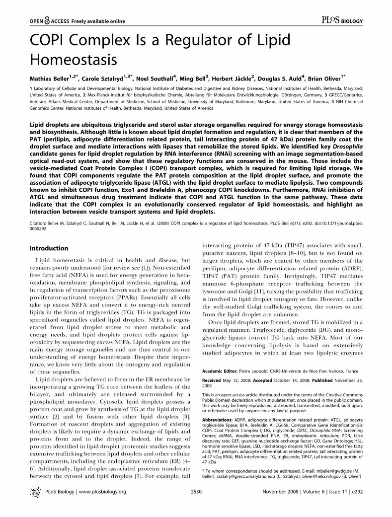
Lipid droplets are well studied in mammalian cells, butDrosophila cells have not been extensively used in lipid dropletstudies. Lipid droplets are ubiquitous organelles, and wefound that Drosophila S2 and SL2 (unpublished data), as well asS3 and Kc167 cells (this study) accumulated TG in lipiddroplets in the presence of excess NEFA. Kc167 cells, forexample, stored little lipid when grown on standard media(Figure 1A), whereas in the presence of NEFA (400 lM oleicacid), they readily (within 12 h) accumulated TG packaged indroplets (Figure 1B), which we visualized with the lipid-specific dye BODIPY493/503 [40].
Treatment of Drosophila cells with double-stranded RNA(dsRNA) decreases, or ‘‘knocks down,’’ transcript levels forgenes sharing the dsRNA sequence, a process known as RNAi[41]. To help determine whether Drosophila tissue culture is agood model for lipid droplet function, we used RNAi totarget genes encoding known lipid droplet regulators. Flies ormice lacking ATGL store more TG than wild type (‘‘over-storage’’) [12–14], whereas those lacking diacylglycerol acyltransferase1 (Dgat1), a key enzyme in TG synthesis [42,43],store less lipid (‘‘understorage’’). Knockdown of bmm, whichencodes Drosophila ATGL, increased lipid storage as expected(Figure 1C and 1D). Conversely, treating cells with dsRNAtargeting midway (mdy), which encodes Drosophila Dgat1,decreased lipid storage (Figure 1E and 1F). Thus, Drosophilacells can be used to analyze gene functions necessary toincrease as well as decrease lipid storage.
Although differences in lipid storage are often obvious, wewere interested in generating a fully quantitative dataset tosupport future meta-analysis. To systematically identify andcharacterize the genes involved in lipid storage, we developeda microscopy-based quantification method based on imagesegmentation and measurement of nuclear to lipid dropletcross-sectional area (see Figure 2A–2D and Materials andMethods). This technique allowed us to detect lipid storagedifferences caused by the different feeding conditions andcontrol dsRNA treatments (Figure 2E). We used this imagingmethod to perform a genome-wide RNAi screen with thewell-characterized dsRNA library of the Harvard DrosophilaRNAi Screening Center (DRSC). This collection covered morethan 95% of the predicted Drosophila genes [44]. dsRNAsagainst bmm and mdy were included in each screening plate as
controls. We also included wells with no dsRNA and with orwithout oleic acid as controls.As a screening cell line, we used Kc167 cells, which showed
the best balance of lipid droplet deposition, RNAi suscept-ibility characteristics, and adhesion during assay development(unpublished data). Following dsRNA treatment of oleic acid-fed cells and image analysis, ratiometric data were normalizedwithin plates and across the entire screening collection usinglinear models, B-score, Z-score/median absolute deviation(MAD), and strictly standardized mean difference (SSMD)[45–48], all of which gave similar results. B-score normal-ization [46] across the entire screen marginally out-per-formed other methods (see Materials and Methods, Table S1).B-score results were used for all analyses reported here.Rank-order analysis of the genome-wide screening results
demonstrated that the majority of dsRNAs had no effect onlipid storage. However, two cohorts of dsRNAs resulted inlipid overstorage, as expected for genes required forpromoting lipid utilization, or understorage, as expectedfor genes required for promoting lipid storage (Figure 2F).Thresholds for determining whether a particular dsRNAresulted in a phenotype were selected to balance falsenegatives and false positives based on the results for bmm,mdy, and no oleic acid controls. At B-scores � 2.0 and ��1.7,greater than 89% of wells treated with dsRNA targeting bmmor without oleic acid resulted in the correct overstorage orunderstorage call, respectively (Figure 2G). Using thesecutoffs, we identified 208 candidate genes required forincreasing lipid storage (understorage on knockdown, B-score ��1.7, Tables S2 and S9) and 318 required for reducing
Figure 1. Drosophila Cells as a Model of Lipid Storage
(A–F) Drosophila Kc167 cells grown in standard medium (A, C, and E) ormedium supplemented with 400 lM oleic acid complexed to 0.4% BSA(B, D, and F). Cells were treated with dsRNA targeting brummer (bmm; [Cand D]) or midway (mdy; [E and F]). The corresponding mouse genes areAtgl and Dgat-1. Nuclei are shown in blue (stained with Hoechst 33342),and lipid droplets in green (stained with BODIPY493/503). Scale bar in (A)represents 10 lm.doi:10.1371/journal.pbio.0060292.g001
PLoS Biology | www.plosbiology.org November 2008 | Volume 6 | Issue 11 | e2922532
COPI and Lipid Storage

lipid storage or lipid utilization (overstorage on knockdown,B-score � 2.0, Tables S3 and S9). These data suggest thatabout 3% of the Drosophila genome is directly or indirectlyinvolved in lipid storage. All data are available in thesupplement (Table S4) and at http://lipofly.mpibpc.mpg.de/and http://flyrnai.org.
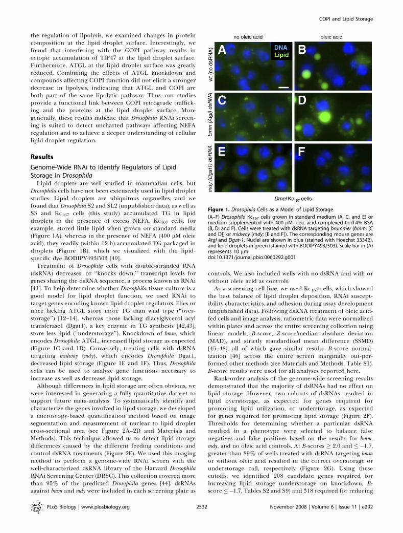
The most critical test of screen performance is coherenceas measured by the identification of multiple genes in amultisubunit complex or a known pathway [49]. Suchcoherent gene sets are also the best candidates for moredetailed analysis. To categorize the dsRNA phenotypesaccording to molecular networks, we analyzed the identifiedgenes using Gene Ontology (GO) [50] terms with the VLADtool [51]. This analysis allows for the detection of statisticallyoverrepresented GO terms among a set of genes and projectsthose enrichments onto the GO-term hierarchy. Genes with apossible function in lipid storage regulation as detected bythe RNAi screen were tested against the complete Drosophilagene set for enrichment of GO terms associated withbiological process, molecular function, and cellular compo-nent. Identified, enriched terms were structured in hierarch-
ical networks (Figures 3–5; the results are also tabulated inTable S5). We also took advantage of data from a concurrentlipid storage screen using an independent dsRNA library andDrosophila S2 cells [52]. This allows us to develop a robustoverview of lipid droplet storage.Duplication of extensive RNAi screens using different
libraries on different cell types provides a cross-validatingfunction that is extremely useful in the analysis of compre-hensive datasets. The overlap (25%, 57 genes) between the S2cell screen (227 genes identified; Table S6) and our genome-wide study on Kc167 cells (526 genes identified) was highlysignificant (p , 1e�14, Wilcox test). More importantly, the GOterm networks were quite similar and suggest that keypathways have been identified (Figure 3). For example, bothscreens show that interfering with translation factors andribosomes result in lipid storage defects (GO:0022613,GO:0006412). Additionally, genes resulting in lipid storagedefects are enriched for transcriptional regulators in bothscreens (GO:0010467) and trafficking (GO:0006911,GO:0006890). The only major differences between the screenswere that genes involved in pre-mRNA processing were
Figure 2. RNAi-Mediated Genome-Wide Screening of Drosophila Cells for Lipid Storage Phenotypes
(A and B) Regions of original images from the screen of cells stained with DAPI (A) or the lipid droplet-specific dye BODIPY 493/503 (B).(C and D) Image segmentation results of the images shown in (A and B). Nuclei and lipid droplet boundaries are depicted.(E) Sample off-line lipid droplet quantification results by image segmentation. Cells were grown with or without oleic acid and treated with theindicated dsRNAs. Significant differences between dsRNA treated and untreated cells at p , 0.05 (two-sided t-test) are shown (indicated with an asterisk[*]). Error bars indicate standard deviation.(F) Rank-order plot of the normalized screen results where positive B-scores indicate overstorage and negative B-scores indicate understorage.(G) Performance of controls incorporated in the on-line screening plates. Black bars represent overstorage (B-score . 2.0), and light grey bars representunderstorage phenotypes (B-score , �1.7).doi:10.1371/journal.pbio.0060292.g002
PLoS Biology | www.plosbiology.org November 2008 | Volume 6 | Issue 11 | e2922533
COPI and Lipid Storage
PLoS Biology | www.plosbiology.org November 2008 | Volume 6 | Issue 11 | e2922534
COPI and Lipid Storage
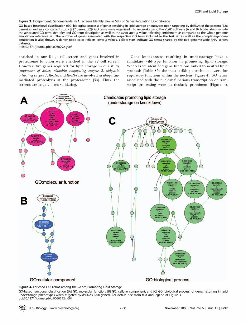
enriched in our Kc167 cell screen and genes involved inproteasome function were enriched in the S2 cell screen.However, five genes required for lipid storage in our study(suppressor of deltex, ubiquitin conjugating enzyme 2, ubiquitinactivating enzyme 1, Roc1a, and Roc1b) are involved in ubiquitin-mediated proteolysis at the proteasome [53]. Thus, thescreens are largely cross-validating.
Gene knockdowns resulting in understorage have acandidate wild-type function in promoting lipid storage.Whereas we identified gene functions linked to neutral lipidsynthesis (Table S5), the most striking enrichments were forregulatory functions within the nucleus (Figure 4). GO termsassociated with the nuclear functions transcription or tran-script processing were particularly prominent (Figure 4).
Figure 4. Enriched GO Terms among the Genes Promoting Lipid Storage
GO-based functional classification ([A] GO: molecular function; [B] GO: cellular component, and [C] GO: biological process) of genes resulting in lipidunderstorage phenotypes when targeted by dsRNAs (208 genes). For details, see main text and legend of Figure 3.doi:10.1371/journal.pbio.0060292.g004
Figure 3. Independent, Genome-Wide RNAi Screens Identify Similar Sets of Genes Regulating Lipid Storage
GO-based functional classification (GO: biological process) of genes resulting in lipid storage phenotypes upon targeting by dsRNAs of the present (526genes) as well as a concurrent study (227 genes; [52]). GO terms were organized into networks using the VLAD software (A and B). Node labels includethe associated GO-term identifier and GO-term description as well as the associated p-value reflecting enrichment as compared to the whole-genomeannotation reference set. The number of genes associated with the respective GO term included in the test set as well as the complete-genomeannotation is also shown. A darker node color reflects lower p-values. Yellow stars indicate GO-terms shared by the two genome-wide RNAi screendatasets.doi:10.1371/journal.pbio.0060292.g003
PLoS Biology | www.plosbiology.org November 2008 | Volume 6 | Issue 11 | e2922535
COPI and Lipid Storage

PLoS Biology | www.plosbiology.org November 2008 | Volume 6 | Issue 11 | e2922536
COPI and Lipid Storage
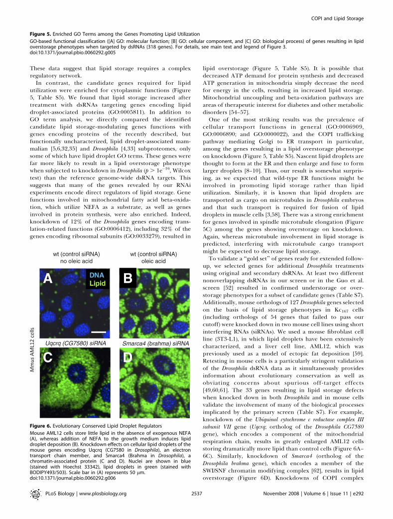
These data suggest that lipid storage requires a complexregulatory network.
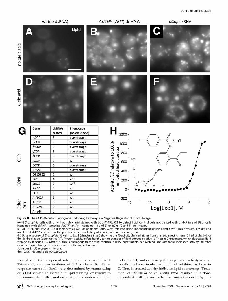
In contrast, the candidate genes required for lipidutilization were enriched for cytoplasmic functions (Figure5, Table S5). We found that lipid storage increased aftertreatment with dsRNAs targeting genes encoding lipiddroplet-associated proteins (GO:0005811). In addition toGO term analysis, we directly compared the identifiedcandidate lipid storage-modulating genes functions withgenes encoding proteins of the recently described, butfunctionally uncharacterized, lipid droplet-associated mam-malian [5,6,32,35] and Drosophila [4,33] subproteomes, onlysome of which have lipid droplet GO terms. These genes werefar more likely to result in a lipid overstorage phenotypewhen subjected to knockdown in Drosophila (p . 1e�16, Wilcoxtest) than the reference genome-wide dsRNA targets. Thissuggests that many of the genes revealed by our RNAiexperiments encode direct regulators of lipid storage. Genefunctions involved in mitochondrial fatty acid beta-oxida-tion, which utilize NEFA as a substrate, as well as genesinvolved in protein synthesis, were also enriched. Indeed,knockdown of 12% of the Drosophila genes encoding trans-lation-related functions (GO:0006412), including 32% of thegenes encoding ribosomal subunits (GO:0033279), resulted in
lipid overstorage (Figure 5, Table S5). It is possible thatdecreased ATP demand for protein synthesis and decreasedATP generation in mitochondria simply decrease the needfor energy in the cells, resulting in increased lipid storage.Mitochondrial uncoupling and beta-oxidation pathways areareas of therapeutic interest for diabetes and other metabolicdisorders [54–57].One of the most striking results was the prevalence of
cellular transport functions in general (GO:0006909,GO:0006890; and GO:0000022), and the COPI traffickingpathway mediating Golgi to ER transport in particular,among the genes resulting in a lipid overstorage phenotypeon knockdown (Figure 5, Table S5). Nascent lipid droplets arethought to form at the ER and then enlarge and fuse to formlarger droplets [8–10]. Thus, our result is somewhat surpris-ing, as we expected that wild-type ER functions might beinvolved in promoting lipid storage rather than lipidutilization. Similarly, it is known that lipid droplets aretransported as cargo on microtubules in Drosophila embryosand that such transport is required for fusion of lipiddroplets in muscle cells [3,58]. There was a strong enrichmentfor genes involved in spindle microtubule elongation (Figure5C) among the genes showing overstorage on knockdown.Again, whereas microtubule involvement in lipid storage ispredicted, interfering with microtubule cargo transportmight be expected to decrease lipid storage.To validate a ‘‘gold set’’ of genes ready for extended follow-
up, we selected genes for additional Drosophila treatmentsusing original and secondary dsRNAs. At least two differentnonoverlapping dsRNAs in our screen or in the Guo et al.screen [52] resulted in confirmed understorage or over-storage phenotypes for a subset of candidate genes (Table S7).Additionally, mouse orthologs of 127 Drosophila genes selectedon the basis of lipid storage phenotypes in Kc167 cells(including orthologs of 54 genes that failed to pass ourcutoff) were knocked down in two mouse cell lines using shortinterfering RNAs (siRNAs). We used a mouse fibroblast cellline (3T3-L1), in which lipid droplets have been extensivelycharacterized, and a liver cell line, AML12, which waspreviously used as a model of ectopic fat deposition [59].Retesting in mouse cells is a particularly stringent validationof the Drosophila dsRNA data as it simultaneously providesinformation about evolutionary conservation as well asobviating concerns about spurious off-target effects[49,60,61]. The 33 genes resulting in lipid storage defectswhen knocked down in both Drosophila and in mouse cellsvalidate the involvement of many of the biological processesimplicated by the primary screen (Table S7). For example,knockdown of the Ubiquinol cytochrome c reductase complex IIIsubunit VII gene (Uqcrq; ortholog of the Drosophila CG7580gene), which encodes a component of the mitochondrialrespiration chain, results in greatly enlarged AML12 cellsstoring dramatically more lipid than control cells (Figure 6A–6C). Similarly, knockdown of Smarca4 (ortholog of theDrosophila brahma gene), which encodes a member of theSWI/SNF chromatin modifying complex [62], results in lipidoverstorage (Figure 6D). Knockdowns of COPI complex
Figure 6. Evolutionary Conserved Lipid Droplet Regulators
Mouse AML12 cells store little lipid in the absence of exogenous NEFA(A), whereas addition of NEFA to the growth medium induces lipiddroplet deposition (B). Knockdown effects on cellular lipid droplets of themouse genes encoding Uqcrq (CG7580 in Drosophila), an electrontransport chain member, and Smarca4 (Brahma in Drosophila), achromatin-associated protein (C and D). Nuclei are shown in blue(stained with Hoechst 33342), lipid droplets in green (stained withBODIPY493/503). Scale bar in (A) represents 50 lm.doi:10.1371/journal.pbio.0060292.g006
Figure 5. Enriched GO Terms among the Genes Promoting Lipid Utilization
GO-based functional classification ([A] GO: molecular function; [B] GO: cellular component, and [C] GO: biological process) of genes resulting in lipidoverstorage phenotypes when targeted by dsRNAs (318 genes). For details, see main text and legend of Figure 3.doi:10.1371/journal.pbio.0060292.g005
PLoS Biology | www.plosbiology.org November 2008 | Volume 6 | Issue 11 | e2922537
COPI and Lipid Storage

members resulted in overstorage in Drosophila S2 and Kc167cells, and in mouse 3T3-L1 and AML12 cells (Table S7).Although there is much to be gleaned from the screen, wefocused our attention on the Golgi to ER trafficking COPIcomplex.
COPI Complex Is a Regulator of Lipid StorageOverrepresentation of genes encoding ER/Golgi vesicle-
associated proteins among the genes showing a lipid over-storage phenotype on knockdown suggests that vesicletrafficking proteins participate in lipid utilization. Moststrikingly, six out of the seven genes encoding COPI subunits(Figure 7) that mediate retrograde transport from the Golgito the ER, showed dramatically increased lipid storagefollowing dsRNA treatment in the genome-wide RNAi screen
(B-score¼ 4.6 to 11.1, false discovery rate [FDR]-corrected p¼1e�5 to 1e�34). Enrichment for members of such multisubunitcomplexes in RNAi screens has outstanding predictive value[49]. Our observed enrichment for essentially all the COPI-associated factors among the knockdowns resulting in lipidoverstorage, strongly suggests that COPI is required forlimiting lipid storage (FDR-corrected p , 1e�6). In addition,dsRNAs targeting ADP ribosylation factor at 79F (Arf79F) had thesame effect as COPI knockdown. Arf79F encodes a small Gprotein homologous to mammalian Arf1, the key regulator ofCOPI vesicle formation at the Golgi [63]. Surprisingly, eCOPwas the only COPI subunit repeatedly failing to produce alipid storage phenotype following RNAi in both the S2 [52]and our Kc167 cell screens. Although this is a negative result,we suggest that this subunit is not involved in lipid storageregulation (see Discussion). Interestingly, none of the sevenCOPII members required for anterograde transport from theER to the Golgi [37,38] showed a lipid accumulationphenotype following RNAi (Figure 7B; B-score ¼ 0.0 to 1.4,FDR-p ¼ 0.99 to 0.78), strongly suggesting that lipid over-storage due to COPI knockdown is not a general consequenceof disrupted trafficking between the ER and Golgi.In organisms, cells are exposed to differing NEFA levels due
to feeding and fasting. Therefore, to test for the function ofthe COPI complex in physiological conditions withoutelevated NEFA, we also performed new RNAi experimentswith or without supplementing the media with oleic acid(Figure 8A–8G; additional data not shown). Even in theabsence of oleic acid, knockdowns of all the members of theCOPI complex that promoted lipid droplet deposition underfed conditions also promoted accumulation without feeding(Figure 8A–8G; additional data not shown). Thus, the lipidstorage phenotype was also independent of the nutritionalstatus of the cells.To further investigate whether the observed lipid storage
phenotype after the loss of COPI-subunit function is due to aspecific pathway or a more general effect of interference withGolgi and ER integrity, we also tested additional dsRNAstargeting transcripts encoding the COPII-associated proteinsCG10882, Sar1, Sec23, Sec31, and PLD (Figure 8G). Further-more, the Drosophila genome encodes five Arf proteins [64],which we also reinvestigated in additional RNAi experiments.Arf79F encodes ARF1, which is required for COPI function,but Arf51F, Arf72A, Arf84F, and Arf102F are not known to berequired for COPI-mediated transport [65]. Only Arf79Fresulted in a mutant lipid droplet phenotype upon RNAiknockdown (Figure 8G). These experiments demonstratedthat the lipid overstorage phenotype is specific to COPI lossof function and raise the possibility that the lipid overstoragephenotype is Golgi independent.Although multiple dsRNAs verified the phenotypic effect of
COPI knockdown, we sought to further validate those resultswith an independent technique, to rule out effects based onthe RNAi treatment, or the prolonged incubation time (4 d)due to the knockdown procedure. Therefore, we also testedpharmacologically for COPI involvement in lipid storage. Wetreated Drosophila S3 cells for 18 h with 24 differentconcentrations of Exo1, a selective inhibitor of Arf1 activity[66], and determined the dose response (Figure 8H). Lipiddroplets were stained with the same dye as for the RNAiexperiments. As in the RNAi experiments, we used internalcontrols, including cells with no oleic acid feeding, cells
Figure 7. COPI Is a Regulator of Lipid Storage
(A) Schematic representation of the COPI trafficking pathway (center)directing traffic from the Golgi (right) to the ER (left). COPII members (notdetailed) direct traffic from the ER to the Golgi. Members showingincreased lipid storage following knockdown in the Drosophila primaryscreen are highlighted (red).(B) Primary screen results for COPI and COPII components are tabulated.The average B-score for the given gene (higher B-scores indicate morestored lipid, see Figure 2) as well as the average false discovery rate(FDR)-corrected p-value are given.doi:10.1371/journal.pbio.0060292.g007
PLoS Biology | www.plosbiology.org November 2008 | Volume 6 | Issue 11 | e2922538
COPI and Lipid Storage
treated with the compound solvent, and cells treated withTriacsin C, a known inhibitor of TG synthesis [67]. Dose-response curves for Exo1 were determined by enumeratingcells that showed an increase in lipid staining (or relative tothe enumerated cells based on a cytosolic counterstain; inset
in Figure 8H) and expressing this as per cent activity relativeto cells incubated in oleic acid and full inhibited by TriacsinC. Thus, increased activity indicates lipid overstorage. Treat-ment of Drosophila S3 cells with Exo1 resulted in a dose-dependent (half maximal effective concentration [EC50] ¼ 5
Figure 8. The COPI-Mediated Retrograde Trafficking Pathway Is a Negative Regulator of Lipid Storage
(A–F) Drosophila cells with or without oleic acid stained with BODIPY493/503 to detect lipid. Control cells not treated with dsRNA (A and D) or cellsincubated with dsRNAs targeting Arf79F (an Arf1 homolog) (B and E) or aCop (C and F) are shown.(G) All COPI, and several COPII members as well as additional Arfs, were retested using independent dsRNAs and gave similar results. Results andnumber of dsRNAs present in the primary screen (including oleic acid) and retests are given.(H) Dose response of Drosophila S3 cells to Exo1 (structure inset) showing the %-activity derived either from the lipid specific signal (filled circles [�]) orthe lipid/cell ratio (open circles [*]). Percent activity refers hereby to the changes of lipid storage relative to Triacsin C treatment, which decreases lipidstorage by blocking TG synthesis (this is analogous to the mdy controls in RNAi experiments, see Material and Methods). Increased activity indicatesincreased lipid storage, which increased with concentration.Scale bar in (A) represents 10 lm.doi:10.1371/journal.pbio.0060292.g008
PLoS Biology | www.plosbiology.org November 2008 | Volume 6 | Issue 11 | e2922539
COPI and Lipid Storage
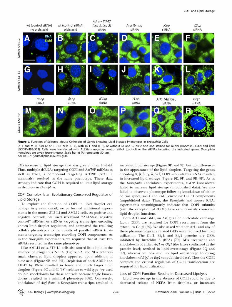
lM) increase in lipid storage that was greater than 10-fold.Thus, multiple dsRNAs targeting COPI and Arf79F mRNAs aswell as Exo1, a compound targeting Arf79F (Arf1 inmammals), resulted in the same phenotype. These datastrongly indicate that COPI is required to limit lipid storagein droplets in Drosophila.
COPI Complex Is an Evolutionary Conserved Regulator ofLipid Storage
To explore the function of COPI in lipid droplet cellbiology in greater detail, we performed additional experi-ments in the mouse 3T3-L1 and AML12 cells. As positive andnegative controls, we used irrelevant ‘‘ALLStars negativecontrol’’ siRNAs, or siRNAs targeting transcripts encodingknown lipid droplet regulators, and compared the resultingcellular phenotypes to the results of parallel siRNA treat-ments targeting transcripts encoding COPI components. Asin the Drosophila experiments, we required that at least twosiRNAs resulted in the same phenotype.
Like AML12 cells, 3T3-L1 cells also stored little lipid in theabsence of exogenous NEFA (Figure 9A and 9G), whereassmall, clustered lipid droplets appeared upon addition ofoleic acid (Figure 9B and 9H). Depletion of both ADRP andTIP47 by RNAi resulted in fewer and much larger lipiddroplets (Figure 9C and 9I [68]) relative to wild type (we useddouble knockdowns for these controls because single knock-downs resulted in a minimal phenotype [68]). Conversely,knockdown of Atgl (bmm in Drosophila) transcripts resulted in
increased lipid storage (Figure 9D and 9J), but no differencesin the appearance of the lipid droplets. Targeting the genesencoding a, b, b9, c, d, or f COPI subunits by siRNAs resultedin increased lipid storage (Figure 9E, 9F, and 9K–9P). As inthe Drosophila knockdown experiments, eCOP knockdownfailed to increase lipid storage (unpublished data). We alsofailed to observe a phenotype following knockdown of eitherof two genes, sec24 and Pld1, encoding COPII components(unpublished data). Thus, the Drosophila and mouse RNAiexperiments unambiguously indicate that COPI subunits(with the exception of eCOP) have evolutionarily conservedlipid droplet functions.Both Arf1 and Gbf1, an Arf guanine nucleotide exchange
factor (GEF), are required for COPI recruitment from thecytosol to Golgi [69]. We also asked whether Arf1 and any ofthree pharmacologically related GEFs were required for lipidutilization. The Gbf1, Big1, and Big2 proteins are GEFsinhibited by Brefeldin A (BFA) [70]. BFA treatment andknockdowns of either Arf1 or Gbf1 (the latter confirmed at theprotein level) resulted in lipid overstorage (Figure 9Q and9R), whereas we observed no lipid overstorage followingknockdown of Big1 or Big2 (unpublished data). Thus the COPIcomplex and critical regulators of COPI translocation arerequired for lipid utilization.
Loss of COPI Function Results in Decreased LipolysisLipid overstorage in the absence of COPI could be due to
decreased release of NEFA from droplets, or increased
Figure 9. Function of Selected Mouse Orthologs of Genes Showing Lipid Storage Phenotypes in Drosophila Cells
(A–F and M–R) AML12 or 3T3-L1 cells (G–L), with (B–F and H–R), or without (A and G) oleic acid and stained for nuclei (Hoechst 33342) and lipid(BODIPY493/503). Cells were transfected with ALLStars negative control siRNA (control) or the siRNAs targeting the indicated genes. Drosophilahomologs are given (parentheses). Scale bar in (A) represents 50 lm.doi:10.1371/journal.pbio.0060292.g009
PLoS Biology | www.plosbiology.org November 2008 | Volume 6 | Issue 11 | e2922540
COPI and Lipid Storage

synthesis of TG for storage, or both. In order to explorewhether COPI is required for one or both of these generalfunctions, we measured both NEFA release and esterificationof NEFA into TG in AML12 cells (Figure 10A). As expected,we observed increased release of NEFA from cells treatedwith control siRNAs targeting Adrp and Tip47 transcripts,which is mediated by increased amounts of lipid droplet-associated ATGL [68]. In contrast, NEFA release decreasedwhen Atgl lipase transcripts were targeted as controls.Additionally, we observed increased incorporation of NEFAinto TG following Atgl knockdown, suggesting that thetremendous increase in TG seen in those cells is due todecreased NEFA release and continued synthesis of TGdespite the reduced efflux. The modest increase in incorpo-ration of NEFA into TG following COPI knockdown wasinsignificant. However, we observed approximately 40% ofwild-type NEFA release in cells treated with siRNAs targetingeither cCOPI or fCOPI transcripts—in the same range asafter Atgl knockdown (Figure 10A). In separate experiments,we also observed decreased NEFA release following Gbf1knockdown, but not following Big1 or Big2 knockdown (Table
S8). These data indicate that COPI is a novel regulator oflipolysis.We also asked whether short-term pharmacological inhib-
ition of COPI trafficking phenocopies the COPI knockdownphenotype in mouse cells, as we noted in Drosophila cells. Weused COPI inhibitors Exo1 and BFA [39,66], both of whichresult in increased lipid storage. Both compounds reducedNEFA release to the same extent as the siRNAs targetingCOPI subunit mRNAs (Figure 10B). To dissect the role ofCOPI in lipolysis, we used a combination of siRNAs targetingdifferent genes in the lipolytic pathway, and Exo1 or BFAtreatment, to mimic genetic epistasis experiments (a proventool for dissecting functional relationships between membersof the same or different pathways [71]). Combining siRNA-mediated knockdown of COPI members and BFA or Exo1treatment did not enhance the decreased lipolysis phenotype(Figure10B), indicating that the observed effects followingdrug treatment are only COPI mediated. Additionally, thesedata suggest that there are no serious compound-based sideeffects vis-a-vis lipid droplets, even for the broad-spectruminhibitor BFA (also note that other BFA-sensitive GEFs, Big1and Big2, did not result in a lipid storage phenotype onknockdown). Decreased lipolysis could be due to decreasedlipase activity at the lipid droplet. To determine whether thatlipase was ATGL, we combined siRNAs targeting ATGLtranscripts and BFA or Exo1 drug treatment (Figure 10B). IfATGL were responsible, then ATGL knockdown would haveno effect on BFA- or Exo1-treated cells. Indeed, the lipolysisrate was not further decreased, suggesting that COPI-mediated lipolysis effects are mediated by ATGL. Thisconclusion is further supported by experiments in which wetreated cells with siRNAs targeting ADRP and TIP47 tran-scripts in combination with either BFA or Exo1. In theabsence of ADRP and TIP47, more ATGL is found at the lipiddroplet surface [68]. We also found that Exo1 or BFAtreatment rescues the effect of ADRP and TIP47 knockdown.This, along with the finding that COPI and ATGL are in thesame pathway, suggests that COPI is an important positiveregulator of ATGL.
Loss of COPI Function Results in Altered Lipid DropletProtein CompositionWild-type COPI could mediate release of NEFA from lipid
droplets by altering the heterogeneous and dynamic collec-tion of lipid droplet-associated proteins found in differentcell types and conditions [72]. To further explore whathappens to lipid droplets following COPI knockdown, weexamined the distribution of TIP47 and ADRP on the lipiddroplet surface. These are the only PAT proteins expressed inAML12 cells [68]. In control cells incubated with oleic acid,and control siRNAs, ADRP was associated with the lipiddroplet surface whereas TIP47 was mostly found in smallerpunctate cytoplasmic inclusions and more ill-defined cyto-plasmic locations ([68] and Figure 11A). TIP47 and ADRPwere not colocalized in untreated cells. Following siRNAtreatments targeting a, b, b9, c, and f COPI subunit or Gbf1transcripts, both ADRP and TIP47 were observed on the samelipid droplets (Figure 11B–11H). Treating the cells with BFAhad the same effect on TIP47 localization (Figure 12A and12B). These data indicate that COPI is required for a wild-type pattern of PAT localization to the lipid droplet.PAT proteins are tightly associated with the lipid droplet
Figure 10. NEFA Incorporation and NEFA Release Measured in AML12
Cells after RNAi-Mediated Gene Knockdown and Drug Treatment
(A) Relative activity [(experimental/ALLStars negative control (control))�1] in radiolabel assays for TG esterification (nM) (grey bar) and NEFArelease (nM) (black) relative to total protein concentration in cells treatedwith siRNAs targeting the indicated transcripts. Significance at p , 0.01,impaired t-test, is shown (indicated by an asterisk [*]). Standard error isindicated by the bars.(B) Relative activity [(experimental/ALLStars negative control (control)and DMSO) � 1] in radiolabel assays for NEFA release (nM) relative tototal protein concentration in cells treated with siRNAs targeting theindicated transcripts in the presence of DMSO only (open bar), BFA (5lM) in DMSO (light-grey bar), or Exo1 (5lM) in DMSO (dark-grey bar).Significance at p , 0.01, impaired t-test, is shown (indicated by anasterisk [*]). Standard error is indicated by the bars.doi:10.1371/journal.pbio.0060292.g010
PLoS Biology | www.plosbiology.org November 2008 | Volume 6 | Issue 11 | e2922541
COPI and Lipid Storage

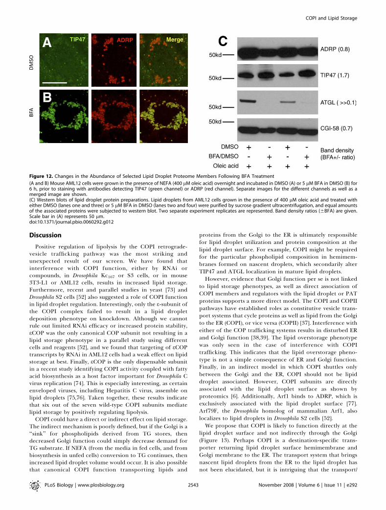
surface. In order to distinguish localization to the region ofthe lipid droplet from true localization to the lipid dropletsurface, we treated cells with BFA after oleic acid feeding, andisolated lipid droplets by sucrose gradient ultracentrifuga-tion. This treatment separates the lipid droplets from cytosoland other membrane fractions. To determine what proteinswere on the lipid droplets, western blots were probed withantibodies detecting ADRP, TIP47, and ATGL, as well as theATGL cofactor CGI-58. Whereas ADRP and CGI-58 remainedquantitatively unchanged after BFA treatment, TIP47 proteinlevels in the lipid droplet fraction increased nearly 2-fold(Figure 12C). There was no change in TIP47 in the cytosolicfraction (unpublished). The cell-staining experiments showeda more dramatic increase in TIP47 at the ADRP-positive lipid
droplets than we observed in the western blots, butimportantly, both cell staining and western blotting showincreased TIP47 on COPI inhibition. Strikingly, ATGL levelsdecreased to near or below the detection limit, suggestingthat BFA treatment drives ATGL off the lipid droplet surface,or prevents ATGL association with the lipid droplet (Figure12C). Thus, both cell staining and analysis of isolated dropletsindicate that wild-type COPI limits abundance of TIP47 atthe lipid droplet surface and is required for ATGL local-ization to the droplet surface. Taken together with theepistasis results demonstrating that COPI and ATGL functionin the same pathway, these results indicate that COPI-mediated targeting of ATGL to the lipid droplet is requiredfor lipolysis.
Figure 11. TIP47 Translocates to the Lipid Droplet Surface upon Down-Regulation of COPI
Mouse AML12 cells were treated with ALLStars negative control siRNA (control) (A) or siRNAs targeting cCOP (B) for 4 d, respectively. Cells wereincubated with 400 lM oleic acid for 12 h prior to staining with antibodies detecting TIP47 (green channel) or ADRP (red channel). Images of theseparate channels as well as a merged image are shown. (C–H) Merged channel images of similarly treated mouse AML12 cells. Targeted genes areindicated. Scale bar in (A) represents 50 lm.doi:10.1371/journal.pbio.0060292.g011
PLoS Biology | www.plosbiology.org November 2008 | Volume 6 | Issue 11 | e2922542
COPI and Lipid Storage
Discussion
Positive regulation of lipolysis by the COPI retrograde-vesicle trafficking pathway was the most striking andunexpected result of our screen. We have found thatinterference with COPI function, either by RNAi orcompounds, in Drosophila Kc167 or S3 cells, or in mouse3T3-L1 or AML12 cells, results in increased lipid storage.Furthermore, recent and parallel studies in yeast [73] andDrosophila S2 cells [52] also suggested a role of COPI functionin lipid droplet regulation. Interestingly, only the e-subunit ofthe COPI complex failed to result in a lipid dropletdeposition phenotype on knockdown. Although we cannotrule out limited RNAi efficacy or increased protein stability,eCOP was the only canonical COP subunit not resulting in alipid storage phenotype in a parallel study using differentcells and reagents [52], and we found that targeting of eCOPtranscripts by RNAi in AML12 cells had a weak effect on lipidstorage at best. Finally, eCOP is the only dispensable subunitin a recent study identifying COPI activity coupled with fattyacid biosynthesis as a host factor important for Drosophila Cvirus replication [74]. This is especially interesting, as certainenveloped viruses, including Hepatitis C virus, assemble onlipid droplets [75,76]. Taken together, these results indicatethat six out of the seven wild-type COPI subunits mediatelipid storage by positively regulating lipolysis.
COPI could have a direct or indirect effect on lipid storage.The indirect mechanism is poorly defined, but if the Golgi is a‘‘sink’’ for phospholipids derived from TG stores, thendecreased Golgi function could simply decrease demand forTG substrate. If NEFA (from the media in fed cells, and frombiosynthesis in unfed cells) conversion to TG continues, thenincreased lipid droplet volume would occur. It is also possiblethat canonical COPI function transporting lipids and
proteins from the Golgi to the ER is ultimately responsiblefor lipid droplet utilization and protein composition at thelipid droplet surface. For example, COPI might be requiredfor the particular phospholipid composition in hemimem-branes formed on nascent droplets, which secondarily alterTIP47 and ATGL localization in mature lipid droplets.However, evidence that Golgi function per se is not linked
to lipid storage phenotypes, as well as direct association ofCOPI members and regulators with the lipid droplet or PATproteins supports a more direct model. The COPI and COPIIpathways have established roles as constitutive vesicle trans-port systems that cycle proteins as well as lipid from the Golgito the ER (COPI), or vice versa (COPII) [37]. Interference witheither of the COP trafficking systems results in disturbed ERand Golgi function [38,39]. The lipid overstorage phenotypewas only seen in the case of interference with COPItrafficking. This indicates that the lipid overstorage pheno-type is not a simple consequence of ER and Golgi function.Finally, in an indirect model in which COPI shuttles onlybetween the Golgi and the ER, COPI should not be lipiddroplet associated. However, COPI subunits are directlyassociated with the lipid droplet surface as shown byproteomics [6]. Additionally, Arf1 binds to ADRP, which isexclusively associated with the lipid droplet surface [77].Arf79F, the Drosophila homolog of mammalian Arf1, alsolocalizes to lipid droplets in Drosophila S2 cells [52].We propose that COPI is likely to function directly at the
lipid droplet surface and not indirectly through the Golgi(Figure 13). Perhaps COPI is a destination-specific trans-porter returning lipid droplet surface hemimembrane andGolgi membrane to the ER. The transport system that bringsnascent lipid droplets from the ER to the lipid droplet hasnot been elucidated, but it is intriguing that the transport/
Figure 12. Changes in the Abundance of Selected Lipid Droplet Proteome Members Following BFA Treatment
(A and B) Mouse AML12 cells were grown in the presence of NEFA (400 lM oleic acid) overnight and incubated in DMSO (A) or 5 lM BFA in DMSO (B) for6 h, prior to staining with antibodies detecting TIP47 (green channel) or ADRP (red channel). Separate images for the different channels as well as amerged image are shown.(C) Western blots of lipid droplet protein preparations. Lipid droplets from AML12 cells grown in the presence of 400 lM oleic acid and treated witheither DMSO (lanes one and three) or 5 lM BFA in DMSO (lanes two and four) were purified by sucrose gradient ultracentrifugation, and equal amountsof the associated proteins were subjected to western blot. Two separate experiment replicates are represented. Band density ratios (6BFA) are given.Scale bar in (A) represents 50 lm.doi:10.1371/journal.pbio.0060292.g012
PLoS Biology | www.plosbiology.org November 2008 | Volume 6 | Issue 11 | e2922543
COPI and Lipid Storage
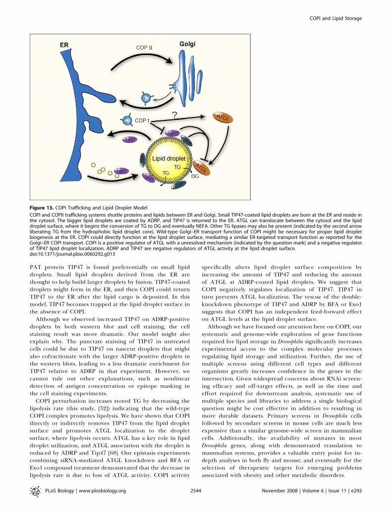
PAT protein TIP47 is found preferentially on small lipiddroplets. Small lipid droplets derived from the ER arethought to help build larger droplets by fusion. TIP47-coateddroplets might form in the ER, and then COPI could returnTIP47 to the ER after the lipid cargo is deposited. In thismodel, TIP47 becomes trapped at the lipid droplet surface inthe absence of COPI.
Although we observed increased TIP47 on ADRP-positivedroplets by both western blot and cell staining, the cellstaining result was more dramatic. Our model might alsoexplain why. The punctate staining of TIP47 in untreatedcells could be due to TIP47 on nascent droplets that mightalso cofractionate with the larger ADRP-positive droplets inthe western blots, leading to a less dramatic enrichment forTIP47 relative to ADRP in that experiment. However, wecannot rule out other explanations, such as nonlineardetection of antigen concentration or epitope masking inthe cell staining experiments.
COPI perturbation increases stored TG by decreasing thelipolysis rate (this study, [52]) indicating that the wild-typeCOPI complex promotes lipolysis. We have shown that COPIdirectly or indirectly removes TIP47 from the lipid dropletsurface and promotes ATGL localization to the dropletsurface, where lipolysis occurs. ATGL has a key role in lipiddroplet utilization, and ATGL association with the droplet isreduced by ADRP and Tip47 [68]. Our epistasis experimentscombining siRNA-mediated ATGL knockdown and BFA orExo1 compound treatment demonstrated that the decrease inlipolysis rate is due to loss of ATGL activity. COPI activity
specifically alters lipid droplet surface composition byincreasing the amount of TIP47 and reducing the amountof ATGL at ADRP-coated lipid droplets. We suggest thatCOPI negatively regulates localization of TIP47. TIP47 inturn prevents ATGL localization. The rescue of the double-knockdown phenotype of TIP47 and ADRP by BFA or Exo1suggests that COPI has an independent feed-forward effecton ATGL levels at the lipid droplet surface.Although we have focused our attention here on COPI, our
systematic and genome-wide exploration of gene functionsrequired for lipid storage in Drosophila significantly increasesexperimental access to the complex molecular processesregulating lipid storage and utilization. Further, the use ofmultiple screens using different cell types and differentorganisms greatly increases confidence in the genes in theintersection. Given widespread concerns about RNAi screen-ing efficacy and off-target effects, as well as the time andeffort required for downstream analysis, systematic use ofmultiple species and libraries to address a single biologicalquestion might be cost effective in addition to resulting inmore durable datasets. Primary screens in Drosophila cellsfollowed by secondary screens in mouse cells are much lessexpensive than a similar genome-wide screen in mammaliancells. Additionally, the availability of mutants in mostDrosophila genes, along with demonstrated translation tomammalian systems, provides a valuable entry point for in-depth analyses in both fly and mouse; and eventually for theselection of therapeutic targets for emerging problemsassociated with obesity and other metabolic disorders.
Figure 13. COPI Trafficking and Lipid Droplet Model
COPI and COPII trafficking systems shuttle proteins and lipids between ER and Golgi. Small TIP47-coated lipid droplets are born at the ER and reside inthe cytosol. The bigger lipid droplets are coated by ADRP, and TIP47 is returned to the ER. ATGL can translocate between the cytosol and the lipiddroplet surface, where it begins the conversion of TG to DG and eventually NEFA. Other TG lipases may also be present (indicated by the second arrowliberating TG from the hydrophobic lipid droplet core). Wild-type Golgi–ER transport function of COPI might be necessary for proper lipid dropletbiogenesis at the ER. COPI could directly function at the lipid droplet surface, mediating a similar ER-targeted transport function as reported for theGolgi–ER COPI transport. COPI is a positive regulator of ATGL with a unresolved mechanism (indicated by the question mark) and a negative regulatorof TIP47 lipid droplet localization. ADRP and TIP47 are negative regulators of ATGL activity at the lipid droplet surface.doi:10.1371/journal.pbio.0060292.g013
PLoS Biology | www.plosbiology.org November 2008 | Volume 6 | Issue 11 | e2922544
COPI and Lipid Storage
Materials and Methods
Genome-wide Drosophila RNAi screen wet-bench procedures. Weused the Harvard Drosophila RNAi Screening Center (DRSC, http://www.flyrnai.org) dsRNA collection, which covers more than 95% ofthe transcriptome (Release 3.2 BDGP) with a total of 17,076 dsRNAs[44] in duplicate. We seeded 1.5 3 104 Kc167 cells (DRSC) in 10 ll ofserum-free Schneider’s medium (GIBCO) in each well of microscopy-quality 384-well plates containing the pre-aliquoted dsRNAs (ap-proximately 250 ng of dsRNA/well). Plates were spun at 1,200 rpm for1 min and incubated for 45 min at 25 8C. We then added 40 ll ofcomplete Schneider’s medium supplemented with 10% FCS (JRHBiosciences), 50 units penicillin; and 50 lg of streptomycin/ml(GIBCO) and 6400 lM oleic acid (Calbiochem) complexed to 0.4%BSA (Sigma). Plates were sealed and incubated in a humidifiedincubator at 25 8C for 4 d. The cells were subsequently fixed for 10min in 4% formaldehyde in PBS followed by a 10-min permeabiliza-tion step in PBS including 0.1% Triton X-100. For lipid dropletvisualization and cell counting (nuclei), we incubated for 1 h with PBSincluding 5 lg/ml BODIPY493/503 (Molecular Probes) and 5 lg/mlDAPI or 5 lg/ml Hoechst33342 (Molecular Probes). After two washeswith PBS including 0.01% Tween-20, cells were kept in 40 ll of PBSand visualized with a 203 objective on a Discovery1 automatedmicroscope system (Molecular Devices).
Secondary RNAi screen wet-bench procedures. A subset of 276genes of the primary screen library were targeted by 362 additionaldsRNAs (Table S10) generated from PCR products obtained from theDrosophila RNAi screening center of Harvard (DRSC). PCR fragmentswere reamplified using a modified T7 oligonucleotide (59-GTA ATACGA CTC ACT ATA GG-39) and a touchdown PCR protocol. PCRproducts were subsequently used for in vitro transcription reactionsusing T7 RNA polymerase (Fermentas). Following DNAse-mediateddigestion of the PCR template, dsRNAs were purified with Multi-screen PCR purification filter plates (Millipore). RNAi treatment wasperformed either as described for the primary screen in optical-quality 96-well plates (BD) with adjusted dsRNA and cell numbers, induplicate (approximately 1 lg of dsRNA and 5 3 104 cells/well).Imaging was performed either with a BD Pathway 855 Bioimagerautomated microscope (BD) or with a Zeiss Axiovert200M (Carl Zeiss)and the OpenLab software (Improvision).
For the secondary mouse siRNA screen (Table S10), we usedAML12 murine liver cells (Steven Farmer, Boston University) and3T3-L1 fibroblast cells (ATCC) grown according to protocols of theAmerican Type Culture Collection (ATCC). Assays were done in 96-multiwell plates (Fisher Scientific) at a density of 0.25 3 104 cells/wellon growth medium supplemented with 200 lM oleic acid, which wasadded 18 h prior to fixation of the cells. Cells were transfected withHiperfect transfection reagent (0.75 ll/well) (Qiagen) and exper-imental or ALLStars negative control siRNA oligonucleotides (10nM), according to the manufacturer’s instructions (Qiagen). Fourdays after transfection, cells were fixed and stained as describedabove for the Drosophila cells and imaged with a BD Pathway 855Bioimager automated microscope system (BD).
Image analysis. Images of Drosophila cells (two sites/well in theprimary screen; six sites/well for the secondary screen) wereprocessed with a custom image segmentation algorithm (availablefrom M. Beller upon request) written for the ImageJ software package[78]. After a sharpening and a brightness/contrast adjustment (for theBODIPY images; equal values for all images) or a gamma correction(for the DAPI images; same values for all images), a backgroundsubtraction followed by an Otsu thresholding step was run (Figure1A–1D). Watershed processing to identify solitary particles followed.Finally, lipid droplets or nuclei were identified with the generic‘‘analyze particles’’ function of the ImageJ software with the followingsettings: (1) settings for the nuclei: size from 10 to 10,000 pixels, 256bins, outlines as well as measurement results displayed, measurementson the edges excluded, clear results, flood, and summary of theresults; and (2) settings for the lipid droplets: identical parametersexcept size ranging from one to 200 pixels and a circularity from zeroto one. For each detected particle, the size and area were measured.For each image, the total numbers of particles (‘‘counts’’) orcumulative measured area for all particles (‘‘area’’) are reported. Acustom Perl script concatenated the summarized measurements, andthe obtained information was used to calculate the ratio of lipiddroplets per cells as a measure of lipid storage (‘‘lipid droplet/nuclei(area)’’ or ‘‘lipid droplet/nuclei (counts)’’).
Mammalian cell image analysis (four sites/well) was performed asdescribed above with some adjusted settings reflecting the largermammalian cell size as well as differences in imaging equipment (nobrightness or contrast adjustments were applied). The generic
‘‘analyze particles’’ function of the ImageJ software was used withthe following settings: (1) settings for the nuclei: size from 80 to10,000 pixels, 256 bins, outlines as well as measurement resultsdisplayed, measurements on the edges excluded, clear results, flood,and summary of the results; and (2) settings for the lipid droplets:identical parameters, except the size ranging from one to 2,000 pixelsand a circularity from zero to one.
Primary screen data analysis. The general thrust of the analysis isgiven below and is followed by a detailed description. Screen data areavailable (Table S4; http://lipofly.mpibpc.mpg.de/). Results wererobust to data handling method (Table S1). Genes passing thresh-olding conditions (Tables S2 and S3) were used for the GO termanalysis (Table S5). B-score p-values can be used to further restrict thegene lists shown in Tables S2 and S3.
Data analysis was performed with custom scripts written in the Rlanguage and packages provided by the Bioconductor project [79].The lipid droplets and nuclei area measurements of the two imagesper well were used to calculate an averaged lipid area per nuclei areavalue per well. Additionally to the primary images, a number of wellsrequired reimaging based on visual inspection (size of the completedataset: N ¼ 48,241 wells). To identify and extract images with badquality, the values for lipid droplet (LD) area and count measure-ments as well as for the corresponding nuclei measurements of thetwo images per well were plotted against each other to look forvariation within wells. In addition, the corresponding ‘‘LD area pernuclei area’’ and ‘‘LD count per nuclei count’’ ratios were plottedagainst each other per well. These plots showed 95 prominentoutliers (segmentation artifacts/‘‘bad’’ wells), which were removed(resulting N ¼ 48,146 wells). The data values of reimaged wells wereaveraged.
The screen dataset was platewise normalized for within-plate andbetween-plate differences by four different algorithms. Because ofthe limited number of controls per plate, 98% of the wells per platewere used as a reference set in the normalization procedure asproposed in [47] in which the largest and smallest 1% values of theplate were removed to generate the reference set. Before datanormalization, LD areas per nuclei area ratios were log-transformed.A classical robust Z-score normalization was performed first [zi¼ (xi�medianj)/madj, where zi is the Z-score of well i; xi is the raw value ofwell i; and medianj and madj are the median and median absolutedeviation (MAD) of the plate j] in addition to the recently proposedstrictly standardized mean difference normalization [SSMDi ¼ (xi �meanj)/square root (2/nj � 2.5 3 ((nj � 1) 3 SDj2))]. Those relatedalgorithms were supplemented with both a fitted linear modelnormalization using the Prada package [45] and by B-score normal-ization [46]. Benjamini and Hochberg FDR-corrected p-values for alldsRNAs were calculated with the complete screen data (without thelargest and smallest 1%) as a reference set. Scoring was done both ona platewise and screenwise manner. For the platewise hit identi-fication, positives were identified by a quartile-based thresholdingalgorithm [48]. For this purpose, the first quartile (Q1), the median(Q2), and the third quartile (Q3) were calculated first. Afterwards,threshold T were calculated [Tupper¼Q3þ c3 (Q3�Q2) and Tlower¼Q1� c 3 (Q2�Q1), where c is a variable depending on the targetederror rate] [48]. The same hit selection strategy was also chosen forthe screen-wide hit identification among the linear model normalizeddataset. For the other normalization algorithms, fixed thresholdswere selected. In all cases, threshold levels (as well as the c in thequartile-based thresholding) were chosen based on the identificationrates of the internal controls brummer dsRNA, midway dsRNA, andwells with no oleic acid, which were present on every screening plate.The highest possible threshold was chosen capable of balancing bothfalse-positive and -negative rates.
Identified Drosophila lipid regulating gene functions (Tables S2 andS3) were subjected to in silico analysis for enriched GO terms. Forthis purpose, we used the standard settings of the VLAD tool (MouseGenome Informatics Web site [51]) using the complete Drosophilagenome as a reference set. Results of the enrichment analyses werevisualized by pruned GO term networks (pruning threshold ¼ 4;collapsing threshold ¼ 5), and results (pruning threshold ¼ 3;collapsing threshold ¼ 6) are additionally tabulated (Table S5).
Detailed lists of the scoring genes were annotated with thefollowing information (Table S9): GO terms from FlyBase [80];orthologs from FlyMine [81]; human disease gene orthologs fromHomophila (http://superfly.ucsd.edu/homophila/, used with a signifi-cance threshold of E , 1 3 10–50, [31]; InParanoid [82] orthologs(http://inparanoid.sbc.su.se/cgi-bin/index.cgi); and Drosophila [4,33]; aswell as mammalian [5,6,32,34] lipid droplet subproteome data.
Secondary screen data analysis. A subset of genes identified in thegenome-wide screen with a potential function in cellular lipid storage
PLoS Biology | www.plosbiology.org November 2008 | Volume 6 | Issue 11 | e2922545
COPI and Lipid Storage
regulation was assayed by at least one additional dsRNA. In total, 276genes were tested by targeting with 362 dsRNA sequences (Table S10).Because we were interested in validating the full range of phenotypesobserved and not just the positives, we sampled across a broad rangeof B-scores. We performed directed retesting on the genes encodingCOPI members. To test for COPI specificity, we used secondarydsRNA sequences targeting Arf family members not involved in COPIfunction as well as COPII vesicle transport encoding transcripts ascontrols. dsRNAs targeting those genes did not result in a phenotypein the primary screen. For a ‘‘positive’’ identification, we requiredthat two independent nonoverlapping dsRNAs or siRNAs give thesame phenotype. In addition, we tested mouse AML12 hepatocytesand mouse 3T3-L1 fibroblasts for an evolutionary conservation of theidentified lipid storage modulators. Assuming that off-target effectsare random, this also minimizes misleading off-target effects, and iscertainly more stringent than the current standard of two positiveRNAi reagents with retesting in the same species and cell type [60]. Intotal, 127 mouse genes covered by 312 siRNAs were tested (TableS10). Genes across the screen that were validated using the image-based analysis with additional RNAi reagents are listed in Table S7.Additional gene and COPI validation comes from small compoundphenocopy, cell staining experiments, and measurements on lipidmetabolism as outlined further below.
Lipid droplet area and nuclei area measurements obtained fromthe image segmentation procedure, which was carried out asdescribed for the primary screen results, was used to express theratio of lipid per cell. For each screen, plate data were mediannormalized. In order to identify genes modulating lipid storage, abasic thresholding of median 6 23MAD was used. Since the datasetswere enriched for modulators of lipid storage, the median as well asMAD was calculated on the basis of control wells incorporated in theassay plates. For the Drosophila, AML12, and 3T3-L1 datasets, thosewells contained no RNAi reagent, but were otherwise treatedidentical to the experimental wells. Screening plates also containedother control dsRNAs/siRNAs wells. The Drosophila secondary screenplates contained wells with dsRNAs targeting bmm or mdy as in theprimary screen. In the case of the 3T3-L1 and AML12 cells, platescontained siRNAs targeting Atgl or a combination of two siRNAstargeting both Adrp and Tip47 transcripts [68]. Median 6 thresholdswere adjusted in order to fulfill the same prerequisites as in theprimary screen, namely a maximum of identified controls with aminimum of false positives. False positives were scored based on thewells lacking RNAi reagent.
Small-molecule compound-based modulation of cellular lipidstorage. Small-molecule compound experiments were performedwith embryonic Drosophila S3 cells (Bloomington Drosophila StockCenter [DGRC]), which showed excellent oleic acid feeding character-istics during RNAi assay development but inferior RNAi character-istics as compared to the Kc167 cells. S3 cells showed superioradherence during automated liquid handling in 1,536-well format.We dispensed 4 ll of cells at 1.25 3 106 cells/ml into LoBase AuroraCOC 1,536-well plates (black walled, clear bottom) with a bottle-valvesolenoid-based dispenser (Aurora) to obtain 5,000 cells/well. A total of23 nl of compound solution of different concentrations weretransferred to the assay plates using a Kalypsis pin tool equippedwith a 1,536-pin array containing 10-nl slotted pins (FP1S10, 0.457-mm diameter, 50.8 mm long; V&P Scientific). One microliter of oleicacid (400 lM) was added, and the plate was lidded with stainless steelrubber gasket-lined lids containing pinholes. After 18–24-h incuba-tion at 24 8C and 95% humidity, BODIPY 493/503 (Molecular Probes)was added to the wells to stain lipid droplets, and the Cell TrackerRed CMTPC dye (Molecular Probes) was added to enumerate cellnumber. Fluorescence was detected by excitation of the fluorophoreswith a 488-nm laser on an Acumen Explorer (TTP Lab Tech). Thetotal intensity in channel 1 (500–530 nm) reflected lipid dropletaccumulation. Cells were detected using channel 3 (575–640 nm) with5-lm width and 100-lm depth filters. The ratio of the total intensityin PMT channel 1 over total intensity of channel 3 was also calculated.Percent activity was computed relative to an internal control (100%inhibited lipid droplet deposition due to the presence of 20 lMTriacsin C), which was added to 32 wells/plate.
Lipolysis and lipogenesis measurements in AML12 cells. Measure-ments of NEFA released from lipid droplets or incorporated into theTG fraction were performed as previously described [23,68,83].Briefly, AML12 cells treated with or without specific siRNAs (10nM) for 4 d were incubated overnight with growth mediumsupplemented with 400 lM oleic acid complexed to 0.4% bovineserum albumin to promote triacylglycerol deposition and [3H] oleicacid, at 1 3 106 dpm/well, was included as a tracer. In lipolysisexperiments, re-esterification of fatty acids in AML12 cells was
prevented by including 10 lM Triacsin C (Biomol), an inhibitor ofacyl coenzyme A synthetase [67], in the medium. Quadruplicate wellswere tested for each condition. Lipolysis was determined bymeasuring radioactivity released into the media in 1 h. For the lipidextraction and thin layer chromatography, the cell monolayer waswashed with ice-cold PBS and scraped into 1 ml of PBS. Lipids wereextracted by the Bligh-Dyer method [84], and 10% of the total lipidwas analyzed by thin layer chromatography [83,85]. AML12 cellstreated with or without specific siRNAs were additionally incubatedwith either vehicle (DMSO), 5 lM of Exo1 (12.5 mg/ml DMSO), or BFA(10 mg/ml DMSO) during the time of radioactivity release into themedia (2 h). NEFA incorporation into the TG fraction and NEFArelease are calculated as nanomoles/milligram protein (Table S8).Protein measurements were performed using a commercial BCAassay kit (Pierce Biotechnology) according to the manufacturer’sinstructions. Statistical significance was tested by impaired Student ttest (GraphPad software).
Antibodies. Rabbit anti-TIP47 and goat anti-ADRP were used aspreviously published [9]. Antibodies targeting mouse ATGL werepurchased from Cell Signaling Technology. The CGI-58 antibody wasa gift from Dr. Osumi [29].
Immunocytochemistry. Cells were plated in four-well Lab-Tekchamber slides (Nunc) and incubated overnight with 400 lM oleicacid. In compound experiments, wells received vehicle (DMSO) or 5lM BFA (10 mg/ml DMSO) treatment for 6 h. RNAi treatment priorto immunocytochemistry is outlined above. For ADRP and TIP47staining, cells were fixed in 3% v/v paraformaldhyde/PBS for 15 minat room temperature. Staining was performed by published methods[9,86]. Cells were viewed with a confocal laser scanning microscope(LSM510; Carl Zeiss MicroImaging) using a 633 oil objective lens.
Fat cake preparation. Eight 100-mm dishes for each condition weretreated with 400 lM oleic acid overnight and further treated withDMSO or BFA (5 lM) for 6 h on the next day. Cells were washed threetimes with phosphate buffered saline (PBS; pH 7.4), scraped into PBS,and then pelleted by low-speed centrifugation. LD isolation was asreported [8]. The lipid fat cake was isolated and resuspended in 150 llof PBS containing 5% SDS before 150 ll of 23 Laemmli samplebuffer were added. For CGI-58 and ATGL western blots, thoseprotein extracts were directly loaded. For ADRP and Tip47, thesamples were diluted 200-fold (ADRP) or 20-fold (TIP47), respec-tively. A total of 35 ll were loaded then on each lane. X-ray films wereused to detect the western blots. Quantification was done with ImageJ[78].
Supporting Information
Table S1. Comparison of Different Primary Screen Analysis Methods
Found at doi:10.1371/journal.pbio.0060292.st001 (34 KB XLS).
Table S2. Genes Showing an Understorage Phenotype in the PrimaryDrosophila RNAi Screen
Found at doi:10.1371/journal.pbio.0060292.st002 (187 KB DOC).
Table S3. Genes Showing an Overstorage Phenotype in the PrimaryDrosophila RNAi Screen
Found at doi:10.1371/journal.pbio.0060292.st003 (273 KB DOC).
Table S4. Drosophila Primary Screen Dataset
Found at doi:10.1371/journal.pbio.0060292.st004 (16 MB XLS).
Table S5. Geneontology-Term Analysis with the VLAD Tool
Found at doi:10.1371/journal.pbio.0060292.st005 (186 KB XLS).
Table S6. Detailed Comparison to the Study of Guo et al. [52]
Found at doi:10.1371/journal.pbio.0060292.st006 (72 KB XLS).
Table S7. Validated RNAi Screen Hits
Found at doi:10.1371/journal.pbio.0060292.st007 (47 KB XLS).
Table S8. AML12 Cell Lipolysis and Lipogenesis Measurements
Found at doi:10.1371/journal.pbio.0060292.st008 (31 KB XLS).
Table S9. Drosophila Primary Screen Hits
Found at doi:10.1371/journal.pbio.0060292.st009 (1 MB XLS).
Table S10. Secondary screen data
Found at doi:10.1371/journal.pbio.0060292.st010 (848 KB XLS).
PLoS Biology | www.plosbiology.org November 2008 | Volume 6 | Issue 11 | e2922546
COPI and Lipid Storage
Accession numbers
Drosophila RNAi screen hits: FBgn0000028, FBgn0000042,FBgn0000114, FBgn0000339, FBgn0000489, FBgn0000547,FBgn0000567, FBgn0001186, FBgn0001204, FBgn0001301,FBgn0002878, FBgn0003048, FBgn0003118, FBgn0003339,FBgn0003380, FBgn0003392, FBgn0003462, FBgn0003557,FBgn0003607, FBgn0003691, FBgn0004167, FBgn0004187,FBgn0004401, FBgn0004587, FBgn0004595, FBgn0004611,FBgn0004652, FBgn0004797, FBgn0004838, FBgn0004856,FBgn0004879, FBgn0005411, FBgn0005626, FBgn0005630,FBgn0010083, FBgn0010215, FBgn0010355, FBgn0010638,FBgn0010750, FBgn0011571, FBgn0011701, FBgn0013746,FBgn0014020, FBgn0015320, FBgn0015818, FBgn0015919,FBgn0016926, FBgn0016940, FBgn0019643, FBgn0020611,FBgn0020908, FBgn0021768, FBgn0022246, FBgn0023143,FBgn0024285, FBgn0024308, FBgn0024555, FBgn0024754,FBgn0025638, FBgn0026206, FBgn0026317, FBgn0026620,FBgn0026722, FBgn0026878, FBgn0027495, FBgn0027589,FBgn0027885, FBgn0027951, FBgn0028360, FBgn0028420,FBgn0028982, FBgn0029123, FBgn0029526, FBgn0029661,FBgn0029731, FBgn0029766, FBgn0029824, FBgn0029850,FBgn0029873, FBgn0029935, FBgn0030075, FBgn0030077,FBgn0030087, FBgn0030093, FBgn0030189, FBgn0030244,FBgn0030390, FBgn0030434, FBgn0030492, FBgn0030608,FBgn0030872, FBgn0030904, FBgn0031008, FBgn0031030,FBgn0031031, FBgn0031074, FBgn0031093, FBgn0031232,FBgn0031390, FBgn0031518, FBgn0031626, FBgn0031673,FBgn0031816, FBgn0031836, FBgn0031888, FBgn0031894,FBgn0032049, FBgn0032340, FBgn0032351, FBgn0032360,FBgn0032363, FBgn0032388, FBgn0032454, FBgn0032622,FBgn0032800, FBgn0032868, FBgn0032945, FBgn0033155,FBgn0033160, FBgn0033541, FBgn0034071, FBgn0034402,FBgn0034646, FBgn0034709, FBgn0034839, FBgn0034946,FBgn0034967, FBgn0035085, FBgn0035136, FBgn0035294,FBgn0035546, FBgn0035569, FBgn0035631, FBgn0036274,FBgn0036374, FBgn0036470, FBgn0036556, FBgn0036734,FBgn0036761, FBgn0036811, FBgn0037024, FBgn0037149,FBgn0037178, FBgn0037250, FBgn0037278, FBgn0037304,FBgn0037568, FBgn0037920, FBgn0037924, FBgn0038168,FBgn0038191, FBgn0038343, FBgn0038359, FBgn0038391,FBgn0038592, FBgn0038633, FBgn0038662, FBgn0039054,FBgn0039941, FBgn0039959, FBgn0039997, FBgn0040279,FBgn0040291, FBgn0040369, FBgn0040534, FBgn0040651,FBgn0040777, FBgn0042693, FBgn0050126, FBgn0050470,FBgn0051313, FBgn0051374, FBgn0051632, FBgn0051814,FBgn0052056, FBgn0052062, FBgn0052112, FBgn0052121,FBgn0052150, FBgn0052202, FBgn0052352, FBgn0052397,FBgn0052440, FBgn0052635, FBgn0052704, FBgn0052710,FBgn0052711, FBgn0052970, FBgn0053207, FBgn0053500,FBgn0053516, FBgn0058413, FBgn0061200, FBgn0083976,FBgn0083992, FBgn0085381, FBgn0086441, FBgn0086674,FBgn0086899, FBgn0243486, FBgn0259162, FBgn0259169,FBgn0259171, FBgn0259217, FBgn0259228, FBgn0259240,FBgn0259243, FBgn0000008, FBgn0000100, FBgn0000116,FBgn0000212, FBgn0000409, FBgn0000492, FBgn0000636,FBgn0000986, FBgn0001133, FBgn0001216, FBgn0001217,FBgn0001218, FBgn0001942, FBgn0002023, FBgn0002590,FBgn0002593, FBgn0002607, FBgn0002906, FBgn0002921,FBgn0003031, FBgn0003060, FBgn0003209, FBgn0003277,FBgn0003279, FBgn0003360, FBgn0003600, FBgn0003687,FBgn0003701, FBgn0003941, FBgn0003942, FBgn0004110,FBgn0004922, FBgn0004926, FBgn0005593, FBgn0005614,FBgn0005630, FBgn0005648, FBgn0008635, FBgn0010078,FBgn0010220, FBgn0010348, FBgn0010352, FBgn0010391,FBgn0010409, FBgn0010410, FBgn0010412, FBgn0010431,FBgn0010612, FBgn0010808, FBgn0011211, FBgn0011272,FBgn0011284, FBgn0011701, FBgn0011726, FBgn0011745,FBgn0011837, FBgn0012034, FBgn0013275, FBgn0013276,FBgn0013277, FBgn0013278, FBgn0013279, FBgn0013325,FBgn0013981, FBgn0014020, FBgn0014857, FBgn0015024,FBgn0015288, FBgn0015393, FBgn0015756, FBgn0015774,FBgn0015778, FBgn0015834, FBgn0016120, FBgn0016694,FBgn0016926, FBgn0017397, FBgn0017545, FBgn0017566,FBgn0017579, FBgn0019624, FBgn0019886, FBgn0019936,FBgn0020129, FBgn0020386, FBgn0020439, FBgn0020910,FBgn0022343, FBgn0022935, FBgn0023170, FBgn0023171,FBgn0023213, FBgn0023531, FBgn0024150, FBgn0024330,FBgn0024733, FBgn0024939, FBgn0025286, FBgn0025582,FBgn0025724, FBgn0025725, FBgn0026262, FBgn0026666,
FBgn0026741, FBgn0027321, FBgn0027348, FBgn0027615,FBgn0028530, FBgn0028867, FBgn0028968, FBgn0028969,FBgn0029088, FBgn0029161, FBgn0029504, FBgn0029761,FBgn0029799, FBgn0029822, FBgn0029860, FBgn0029897,FBgn0030025, FBgn0030088, FBgn0030174, FBgn0030259,FBgn0030341, FBgn0030384, FBgn0030386, FBgn0030606,FBgn0030610, FBgn0030669, FBgn0030692, FBgn0030696,FBgn0030726, FBgn0030915, FBgn0030951, FBgn0030990,FBgn0031300, FBgn0031392, FBgn0031545, FBgn0031696,FBgn0031771, FBgn0031842, FBgn0031980, FBgn0032053,FBgn0032215, FBgn0032261, FBgn0032330, FBgn0032400,FBgn0032518, FBgn0032587, FBgn0032596, FBgn0032619,FBgn0032656, FBgn0032675, FBgn0032833, FBgn0032987,FBgn0033029, FBgn0033081, FBgn0033085, FBgn0033282,FBgn0033313, FBgn0033341, FBgn0033368, FBgn0033379,FBgn0033403, FBgn0033591, FBgn0033652, FBgn0033699,FBgn0033902, FBgn0033912, FBgn0034020, FBgn0034258,FBgn0034487, FBgn0034488, FBgn0034537, FBgn0034579,FBgn0034649, FBgn0034751, FBgn0034902, FBgn0034948,FBgn0034968, FBgn0034987, FBgn0035276, FBgn0035315,FBgn0035422, FBgn0035562, FBgn0035563, FBgn0035638,FBgn0035699, FBgn0035753, FBgn0035872, FBgn0035976,FBgn0036135, FBgn0036213, FBgn0036288, FBgn0036343,FBgn0036351, FBgn0036360, FBgn0036398, FBgn0036449,FBgn0036462, FBgn0036492, FBgn0036532, FBgn0036534,FBgn0036576, FBgn0036613, FBgn0036728, FBgn0036820,FBgn0036825, FBgn0036895, FBgn0036990, FBgn0037010,FBgn0037028, FBgn0037093, FBgn0037097, FBgn0037098,FBgn0037102, FBgn0037207, FBgn0037249, FBgn0037270,FBgn0037356, FBgn0037415, FBgn0037429, FBgn0037529,FBgn0037546, FBgn0037559, FBgn0037566, FBgn0037610,FBgn0037637, FBgn0037752, FBgn0037813, FBgn0037912,FBgn0037942, FBgn0037955, FBgn0038049, FBgn0038074,FBgn0038131, FBgn0038281, FBgn0038345, FBgn0038538,FBgn0038628, FBgn0038629, FBgn0038734, FBgn0038760,FBgn0038881, FBgn0038996, FBgn0039205, FBgn0039214,FBgn0039302, FBgn0039359, FBgn0039402, FBgn0039404,FBgn0039464, FBgn0039520, FBgn0039580, FBgn0039857,FBgn0040007, FBgn0040010, FBgn0040233, FBgn0040512,FBgn0040529, FBgn0040634, FBgn0040766, FBgn0040793,FBgn0043001, FBgn0043904, FBgn0050007, FBgn0050290,FBgn0050387, FBgn0051158, FBgn0051284, FBgn0051291,FBgn0051302, FBgn0051354, FBgn0051361, FBgn0051450,FBgn0051453, FBgn0051554, FBgn0051613, FBgn0051754,FBgn0051774, FBgn0051847, FBgn0052050, FBgn0052105,FBgn0052179, FBgn0052193, FBgn0052219, FBgn0052311,FBgn0052600, FBgn0052633, FBgn0052720, FBgn0052733,FBgn0052773, FBgn0052778, FBgn0052797, FBgn0053128,FBgn0053147, FBgn0053256, FBgn0053271, FBgn0053300,FBgn0053319, FBgn0058337, FBgn0062412, FBgn0062413,FBgn0083950, FBgn0085392, FBgn0085408, FBgn0085424,FBgn0085436, FBgn0086710, FBgn0086712, FBgn0086758,FBgn0086904, FBgn0250791, FBgn0250814, FBgn0250834,FBgn0250908, FBgn0259113, FBgn0259212, FBgn0259232, andFBgn0259246.
Mouse genes with a confirmed function in lipid storage regulation:MGI:107807, MGI:107851, MGI:1333825, MGI:1334462, MGI:1335073,MGI:1351329 , MGI:1353495 , MGI:1354962 , MGI:1858696 ,MGI:1861607 , MGI:1891824 , MGI:1891829 , MGI:1913585 ,MGI:1914062 , MGI:1914103 , MGI:1914144 , MGI:1914234 ,MGI:1914454 , MGI:1915822 , MGI:1916296 , MGI:1917599 ,MGI:1929063 , MGI:2385261 , MGI:2385656 , MGI:2387591 ,MGI:2388481, MGI:2443241, MGI:3041174, MGI:3694697, MGI:88192,MGI:95301, MGI:98342, and MGI:99431.
Acknowledgments
We thank Patrick Muller, Bernard Mathey-Prevot, and NorbertPerrimon for comments on data analysis, David Sturgill for help inR programming, Ya-Qin Zhang for technical help performing thesmall compound experiments, Katharina Thiel for technical helpgenerating dsRNAs, Matthias Dobbelstein for providing access to theBD Pathway Bioimager System, and Cathrin Hippel for support inoperating the system, Dr. Osumi for reagents, and Dean Londos, AlanKimmel, and Joshua Zimmerberg for discussions and helpful com-ments.
Full datasets and dsRNA sequences can be obtained at the Lipofly(http://lipofly.mpibpc.mpg.de/) or DRSC (http://flyrnai.org) websites,respectively.
PLoS Biology | www.plosbiology.org November 2008 | Volume 6 | Issue 11 | e2922547
COPI and Lipid Storage

Author contributions. M. Beller, C. Sztalryd, and B. Oliver designedthe study. Drosophila RNAi screen was performed by M. Beller and B.Oliver, mammalian siRNA screen by C. Sztalryd, and M. Bell, small-molecule compound experiments by N. Southall and D. S. Auld, anddata analysis was by M. Beller. M. Beller, C. Sztalryd, H. Jackle, and B.Oliver prepared the manuscript.
Funding. The work was funded in part by National Institute ofGeneral Medical Sciences (NIGMS) grant R01 GM067761 (DRSC), bythe National Institute of Diabetes and Digestive and Kidney Diseases(NIDDK) National Institutes of Health (NIH) Intramural researchprogram (M. Beller and B. Oliver), National Human Genome
Research Institute (NHGRI) NIH intramural program, and NIHroadmap for molecular libraries (N. Southall and D. S. Auld), by acareer development award 1–05-CD-17 from the American DiabetesAssociation (to C. Sztalryd), by a grant from NIH RO1 DK 075017–01A2 (to C. Sztalryd), by the Geriatric Research, Education andClinical Center (C. Sztalryd), Baltimore Veterans Affairs Health Careand Center (C. Sztalryd), by the Clinical Nutrition Research Unit ofMaryland (C. Sztalryd), and by the Max Planck Society (M. Beller andH. Jackle).
Competing interests. The authors have declared that no competinginterests exist.
References1. Brasaemle DL (2007) Thematic review series: adipocyte biology. The
perilipin family of structural lipid droplet proteins: stabilization of lipiddroplets and control of lipolysis. J Lipid Res 48: 2547–2559.
2. Kuerschner L, Moessinger C, Thiele C (2008) Imaging of lipid biosynthesis:how a neutral lipid enters lipid droplets. Traffic 9: 338–352.
3. Bostrom P, Andersson L, Rutberg M, Perman J, Lidberg U, et al. (2007)SNARE proteins mediate fusion between cytosolic lipid droplets and areimplicated in insulin sensitivity. Nat Cell Biol 9: 1286–1293.
4. Beller M, Riedel D, Jansch L, Dieterich G, Wehland J, et al. (2006)Characterization of the Drosophila lipid droplet subproteome. Mol CellProteomics 5: 1082–1094.
5. Liu P, Ying Y, Zhao Y, Mundy DI, Zhu M, et al. (2004) Chinese hamster ovaryK2 cell lipid droplets appear to be metabolic organelles involved inmembrane traffic. J Biol Chem 279: 3787–3792.
6. Bartz R, Zehmer JK, Zhu M, Chen Y, Serrero G, et al. (2007) Dynamicactivity of lipid droplets: protein phosphorylation and GTP-mediatedprotein translocation. J Proteome Res 6: 3256–3265.
7. Wolins NE, Quaynor BK, Skinner JR, Schoenfish MJ, Tzekov A, et al. (2005)S3–12, Adipophilin, and TIP47 package lipid in adipocytes. J Biol Chem280: 19146–19155.
8. Wolins NE, Rubin B, Brasaemle DL (2001) TIP47 associates with lipiddroplets. J Biol Chem 276: 5101–5108.
9. Sztalryd C, Bell M, Lu X, Mertz P, Hickenbottom S, et al. (2006) Functionalcompensation for adipose differentiation-related protein (ADFP) by Tip47in an ADFP null embryonic cell line. J Biol Chem 281: 34341–34348.
10. Miura S, Gan JW, Brzostowski J, Parisi MJ, Schultz CJ, et al. (2002)Functional conservation for lipid storage droplet association amongPerilipin, ADRP, and TIP47 (PAT)-related proteins in mammals, Drosophila,and Dictyostelium. J Biol Chem 277: 32253–32257.
11. Diaz E, Pfeffer SR (1998) TIP47: a cargo selection device for mannose 6-phosphate receptor trafficking. Cell 93: 433–443.
12. Haemmerle G, Lass A, Zimmermann R, Gorkiewicz G, Meyer C, et al. (2006)Defective lipolysis and altered energy metabolism in mice lacking adiposetriglyceride lipase. Science 312: 734–737.
13. Gronke S, Mildner A, Fellert S, Tennagels N, Petry S, et al. (2005) Brummerlipase is an evolutionary conserved fat storage regulator in Drosophila. CellMetab 1: 323–330.
14. Zimmermann R, Strauss JG, Haemmerle G, Schoiswohl G, Birner-Gruenberger R, et al. (2004) Fat mobilization in adipose tissue is promotedby adipose triglyceride lipase. Science 306: 1383–1386.
15. Vaughan M, Berger JE, Steinberg D (1964) Hormone-sensitive lipase andmonoglyceride lipase activities in adipose tissue. J Biol Chem 239: 401–409.
16. Pieroni G, Verger R (1979) Hydrolysis of mixed monomolecular films oftriglyceride/lecithin by pancreatic lipase. J Biol Chem 254: 10090–10094.
17. Brasaemle DL, Barber T, Wolins NE, Serrero G, Blanchette-Mackie EJ, et al.(1997) Adipose differentiation-related protein is an ubiquitously expressedlipid storage droplet-associated protein. J Lipid Res 38: 2249–2263.
18. Martinez-Botas J, Anderson JB, Tessier D, Lapillonne A, Chang BH, et al.(2000) Absence of perilipin results in leanness and reverses obesity inLepr(db/db) mice. Nat Genet 26: 474–479.
19. Tansey JT, Sztalryd C, Gruia-Gray J, Roush DL, Zee JV, et al. (2001) Perilipinablation results in a lean mouse with aberrant adipocyte lipolysis, enhancedleptin production, and resistance to diet-induced obesity. Proc Natl AcadSci U S A 98: 6494–6499.
20. Gronke S, Beller M, Fellert S, Ramakrishnan H, Jackle H, et al. (2003)Control of fat storage by a Drosophila PAT domain protein. Curr Biol 13:603–606.
21. Teixeira L, Rabouille C, Rorth P, Ephrussi A, Vanzo NF (2003) DrosophilaPerilipin/ADRP homologue Lsd2 regulates lipid metabolism. Mech Dev 120:1071–1081.
22. Granneman JG, Moore HP, Granneman RL, Greenberg AS, Obin MS, et al.(2007) Analysis of lipolytic protein trafficking and interactions inadipocytes. J Biol Chem 282: 5726–5735.
23. Sztalryd C, Xu G, Dorward H, Tansey JT, Contreras JA, et al. (2003)Perilipin A is essential for the translocation of hormone-sensitive lipaseduring lipolytic activation. J Cell Biol 161: 1093–1103.
24. Su CL, Sztalryd C, Contreras JA, Holm C, Kimmel AR, et al. (2003)Mutational analysis of the hormone-sensitive lipase translocation reactionin adipocytes. J Biol Chem 278: 43615–43619.
25. Haemmerle G, Zimmermann R, Hayn M, Theussl C, Waeg G, et al. (2002)Hormone-sensitive lipase deficiency in mice causes diglyceride accumu-lation in adipose tissue, muscle, and testis. J Biol Chem 277: 4806–4815.
26. Fischer J, Lefevre C, Morava E, Mussini JM, Laforet P, et al. (2007) The geneencoding adipose triglyceride lipase (PNPLA2) is mutated in neutral lipidstorage disease with myopathy. Nat Genet 39: 28–30.
27. Lass A, Zimmermann R, Haemmerle G, Riederer M, Schoiswohl G, et al.(2006) Adipose triglyceride lipase-mediated lipolysis of cellular fat stores isactivated by CGI-58 and defective in Chanarin-Dorfman Syndrome. CellMetab 3: 309–319.
28. Lefevre C, Jobard F, Caux F, Bouadjar B, Karaduman A, et al. (2001)Mutations in CGI-58, the gene encoding a new protein of the esterase/lipase/thioesterase subfamily, in Chanarin-Dorfman syndrome. Am J HumGenet 69: 1002–1012.
29. Yamaguchi T, Omatsu N, Matsushita S, Osumi T (2004) CGI-58 interactswith perilipin and is localized to lipid droplets. Possible involvement ofCGI-58 mislocalization in Chanarin-Dorfman syndrome. J Biol Chem 279:30490–30497.
30. Subramanian V, Rothenberg A, Gomez C, Cohen AW, Garcia A, et al. (2004)Perilipin A mediates the reversible binding of CGI-58 to lipid droplets in3T3-L1 adipocytes. J Biol Chem 279: 42062–42071.
31. Chien S, Reiter LT, Bier E, Gribskov M (2002) Homophila: human diseasegene cognates in Drosophila. Nucleic Acids Res 30: 149–151.
32. Brasaemle DL, Dolios G, Shapiro L, Wang R (2004) Proteomic analysis ofproteins associated with lipid droplets of basal and lipolytically-stimulated3T3-L1 adipocytes. J Biol Chem 279: 46835–46842.
33. Cermelli S, Guo Y, Gross SP, Welte MA (2006) The lipid-droplet proteomereveals that droplets are a protein-storage depot. Curr Biol 16: 1783–1795.
34. Turro S, Ingelmo-Torres M, Estanyol JM, Tebar F, Fernandez MA, et al.(2006) Identification and characterization of associated with lipid dropletprotein 1: a novel membrane-associated protein that resides on hepaticlipid droplets. Traffic 7: 1254–1269.
35. Wu CC, Howell KE, Neville MC, Yates JR 3rd, McManaman JL (2000)Proteomics reveal a link between the endoplasmic reticulum and lipidsecretory mechanisms in mammary epithelial cells. Electrophoresis 21:3470–3482.
36. Gronke S, Muller G, Hirsch J, Fellert S, Andreou A, et al. (2007) Duallipolytic control of body fat storage and mobilization in Drosophila. PLoSBiol 5:: e137. doi:10.1371/journal.pbio.0050137
37. Lee MC, Miller EA, Goldberg J, Orci L, Schekman R (2004) Bi-directionalprotein transport between the ER and Golgi. Annu Rev Cell Dev Biol 20:87–123.
38. Yoshimura S, Yamamoto A, Misumi Y, Sohda M, Barr FA, et al. (2004)Dynamics of Golgi matrix proteins after the blockage of ER to Golgitransport. J Biochem 135: 201–216.
39. Lippincott-Schwartz J, Yuan L, Tipper C, Amherdt M, Orci L, et al. (1991)Brefeldin A’s effects on endosomes, lysosomes, and the TGN suggest ageneral mechanism for regulating organelle structure and membranetraffic. Cell 67: 601–616.
40. Gocze PM, Freeman DA (1994) Factors underlying the variability of lipiddroplet fluorescence in MA-10 Leydig tumor cells. Cytometry 17: 151–158.
41. Caplen NJ, Fleenor J, Fire A, Morgan RA (2000) dsRNA-mediated genesilencing in cultured Drosophila cells: a tissue culture model for the analysisof RNA interference. Gene 252: 95–105.
42. Smith SJ, Cases S, Jensen DR, Chen HC, Sande E, et al. (2000) Obesityresistance and multiple mechanisms of triglyceride synthesis in micelacking Dgat. Nat Genet 25: 87–90.
43. Buszczak M, Lu X, Segraves WA, Chang TY, Cooley L (2002) Mutations inthe midway gene disrupt a Drosophila acyl coenzyme A: diacylglycerolacyltransferase. Genetics 160: 1511–1518.
44. Boutros M, Kiger AA, Armknecht S, Kerr K, Hild M, et al. (2004) Genome-wide RNAi analysis of growth and viability in Drosophila cells. Science 303:832–835.
45. Hahne F, Arlt D, Sauermann M, Majety M, Poustka A, et al. (2006) Statisticalmethods and software for the analysis of high throughput reverse geneticassays using flow cytometry readouts. Genome Biol 7: R77.
46. Malo N, Hanley JA, Cerquozzi S, Pelletier J, Nadon R (2006) Statisticalpractice in high-throughput screening data analysis. Nat Biotechnol 24:167–175.
47. Zhang XD, Ferrer M, Espeseth AS, Marine SD, Stec EM, et al. (2007) The useof strictly standardized mean difference for hit selection in primary RNA
PLoS Biology | www.plosbiology.org November 2008 | Volume 6 | Issue 11 | e2922548
COPI and Lipid Storage
interference high-throughput screening experiments. J Biomol Screen 12:497–509.
48. Zhang XD, Yang XC, Chung N, Gates A, Stec E, et al. (2006) Robuststatistical methods for hit selection in RNA interference high-throughputscreening experiments. Pharmacogenomics 7: 299–309.
49. DasGupta R, Nybakken K, Booker M, Mathey-Prevot B, Gonsalves F, et al.(2007) A case study of the reproducibility of transcriptional reporter cell-based RNAi screens in Drosophila. Genome Biol 8: R203.
50. Gene Ontology Consortium (2006) The Gene Ontology (GO) project in2006. Nucleic Acids Res 34: D322–326.
51. Bult CJ, Eppig JT, Kadin JA, Richardson JE, Blake JA (2008) The MouseGenome Database (MGD): mouse biology and model systems. Nucleic AcidsRes 36: D724–728.
52. Guo Y, Walther TC, Rao M, Stuurman N, Goshima G, et al. (2008)Functional genomic screen reveals genes involved in lipid-dropletformation and utilization. Nature 453: 657–661.
53. Ho MS, Tsai PI, Chien CT (2006) F-box proteins: the key to proteindegradation. J Biomed Sci 13: 181–191.
54. Fong TM (2005) Advances in anti-obesity therapeutics. Expert OpinInvestig Drugs 14: 243–250.
55. Kurtz DM, Rinaldo P, Rhead WJ, Tian L, Millington DS, et al. (1998)Targeted disruption of mouse long-chain acyl-CoA dehydrogenase genereveals crucial roles for fatty acid oxidation. Proc Natl Acad Sci U S A 95:15592–15597.
56. Strauss AW, Powell CK, Hale DE, Anderson MM, Ahuja A, et al. (1995)Molecular basis of human mitochondrial very-long-chain acyl-CoAdehydrogenase deficiency causing cardiomyopathy and sudden death inchildhood. Proc Natl Acad Sci U S A 92: 10496–10500.
57. Yamamoto M, Nakamura Y (1994) Inhibition of beta-oxidation by 3-mercaptopropionic acid produces features of Reye’s syndrome in perfusedrat liver. Gastroenterology 107: 517–524.
58. Welte MA, Cermelli S, Griner J, Viera A, Guo Y, et al. (2005) Regulation oflipid-droplet transport by the perilipin homolog LSD2. Curr Biol 15: 1266–1275.
59. Schadinger SE, Bucher NL, Schreiber BM, Farmer SR (2005) PPARgamma2regulates lipogenesis and lipid accumulation in steatotic hepatocytes. Am JPhysiol Endocrinol Metab 288: E1195–1205.
60. Echeverri CJ, Beachy PA, Baum B, Boutros M, Buchholz F, et al. (2006)Minimizing the risk of reporting false positives in large-scale RNAi screens.Nat Methods 3: 777–779.
61. Kulkarni MM, Booker M, Silver SJ, Friedman A, Hong P, et al. (2006)Evidence of off-target effects associated with long dsRNAs in Drosophilamelanogaster cell-based assays. Nat Methods 3: 833–838.
62. Tamkun JW (1995) The role of brahma and related proteins in tran-scription and development. Curr Opin Genet Dev 5: 473–477.
63. Orci L, Palmer DJ, Ravazzola M, Perrelet A, Amherdt M, et al. (1993)Budding from Golgi membranes requires the coatomer complex of non-clathrin coat proteins. Nature 362: 648–652.
64. Celniker SE, Wheeler DA, Kronmiller B, Carlson JW, Halpern A, et al.(2002) Finishing a whole-genome shotgun: release 3 of the Drosophilamelanogaster euchromatic genome sequence. Genome Biol 3: RE-SEARCH0079.
65. Donaldson JG, Honda A (2005) Localization and function of Arf familyGTPases. Biochem Soc Trans 33: 639–642.
66. Feng Y, Yu S, Lasell TK, Jadhav AP, Macia E, et al. (2003) Exo1: a newchemical inhibitor of the exocytic pathway. Proc Natl Acad Sci U S A 100:6469–6474.
67. Igal RA, Wang P, Coleman RA (1997) Triacsin C blocks de novo synthesis ofglycerolipids and cholesterol esters but not recycling of fatty acid intophospholipid: evidence for functionally separate pools of acyl-CoA.Biochem J 324: 529–534.
68. Bell M, Wang H, Chen H, McLenithan JC, Gong DW, et al. (2008)Consequences of lipid droplet coat protein downregulation in liver cells:abnormal lipid droplet metabolism and induction of insulin resistance.Diabetes 57: 2037–2045.
69. Szul T, Grabski R, Lyons S, Morohashi Y, Shestopal S, et al. (2007)Dissecting the role of the ARF guanine nucleotide exchange factor GBF1 inGolgi biogenesis and protein trafficking. J Cell Sci 120: 3929–3940.
70. Anders N, Jurgens G (2008) Large ARF guanine nucleotide exchange factorsin membrane trafficking. Cell Mol Life Sci. E-pub ahead of print. doi: 10.1007/s00018–008-8227–7
71. Levine AJ, Hu W, Feng Z, Gil G (2007) Reconstructing signal transductionpathways: challenges and opportunities. Ann N Y Acad Sci 1115: 32–50.
72. Granneman JG, Moore HP (2008) Location, location: protein traffickingand lipolysis in adipocytes. Trends Endocrinol Metab 19: 3–9.
73. Szymanski KM, Binns D, Bartz R, Grishin NV, Li WP, et al. (2007) Thelipodystrophy protein seipin is found at endoplasmic reticulum lipiddroplet junctions and is important for droplet morphology. Proc Natl AcadSci U S A 104: 20890–20895.
74. Cherry S, Kunte A, Wang H, Coyne C, Rawson RB, et al. (2006) COPIactivity coupled with fatty acid biosynthesis is required for viralreplication. PLoS Pathog 2: e102. doi:10.1371/journal.ppat.0020102
75. Barba G, Harper F, Harada T, Kohara M, Goulinet S, et al. (1997) HepatitisC virus core protein shows a cytoplasmic localization and associates tocellular lipid storage droplets. Proc Natl Acad Sci U S A 94: 1200–1205.
76. Coffey CM, Sheh A, Kim IS, Chandran K, Nibert ML, et al. (2006) Reovirusouter capsid protein micro1 induces apoptosis and associates with lipiddroplets, endoplasmic reticulum, and mitochondria. J Virol 80: 8422–8438.
77. Nakamura N, Akashi T, Taneda T, Kogo H, Kikuchi A, et al. (2004) ADRP isdissociated from lipid droplets by ARF1-dependent mechanism. BiochemBiophys Res Commun 322: 957–965.
78. Girish V, Vijayalakshmi A (2004) Affordable image analysis using NIHImage/ImageJ. Indian J Cancer 41: 47.
79. Gentleman RC, Carey VJ, Bates DM, Bolstad B, Dettling M, et al. (2004)Bioconductor: open software development for computational biology andbioinformatics. Genome Biol 5: R80.
80. Crosby MA, Goodman JL, Strelets VB, Zhang P, Gelbart WM (2007) FlyBase:genomes by the dozen. Nucleic Acids Res 35: D486–491.
81. Lyne R, Smith R, Rutherford K, Wakeling M, Varley A, et al. (2007) FlyMine:an integrated database for Drosophila and Anopheles genomics. Genome Biol8: R129.
82. O’Brien KP, Remm M, Sonnhammer EL (2005) Inparanoid: a comprehen-sive database of eukaryotic orthologs. Nucleic Acids Res 33: D476–480.
83. Tansey JT, Huml AM, Vogt R, Davis KE, Jones JM, et al. (2003) Functionalstudies on native and mutated forms of perilipins. A role in protein kinaseA-mediated lipolysis of triacylglycerols. J Biol Chem 278: 8401–8406.
84. Bligh EG, Dyer WJ (1959) A rapid method of total lipid extraction andpurification. Can J Biochem Physiol 37: 911–917.
85. Sztalryd C, Levacher C, Picon L (1989) Acceleration by triiodothyronine ofadipose conversion of rat preadipocytes from two adipose localizations.Cell Mol Biol 35: 81–88.
86. Dalen KT, Dahl T, Holter E, Arntsen B, Londos C, et al. (2007) LSDP5 is aPAT protein specifically expressed in fatty acid oxidizing tissues. BiochimBiophys Acta 1771: 210–227.
PLoS Biology | www.plosbiology.org November 2008 | Volume 6 | Issue 11 | e2922549
COPI and Lipid Storage
Related Documents