Pleural Ultrasound
Jul 15, 2015
Welcome message from author
This document is posted to help you gain knowledge. Please leave a comment to let me know what you think about it! Share it to your friends and learn new things together.
Transcript


Pleural Ulrasound
Gamal Rabie Agmy ,MD ,FCCP
Professor of Chest Diseases, Assiut University


• Immediate bedside availability
• Immediate bedside repeatability
• Rapid goal directed application
• Cost saving
• Reduction in radiation exposure
Advantages of Transthoracic
Ultrasonography


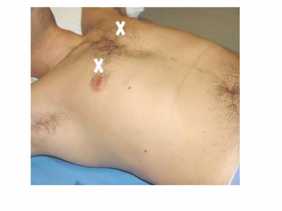
Scanning Positions for Chest Sonography


1-Frequently very high in the supine
critically ill patient
2-Particular caution in post CABG
patients -unilateral diaphragmatic
dysfunction may confuse the examiner
3-Massive edema and obesity may
degrade image quality
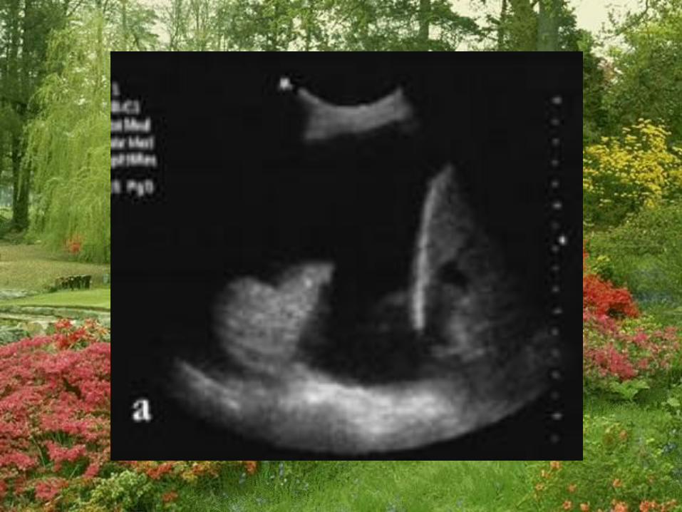
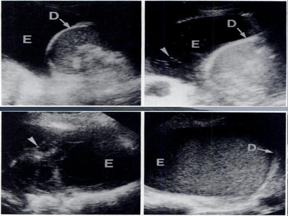
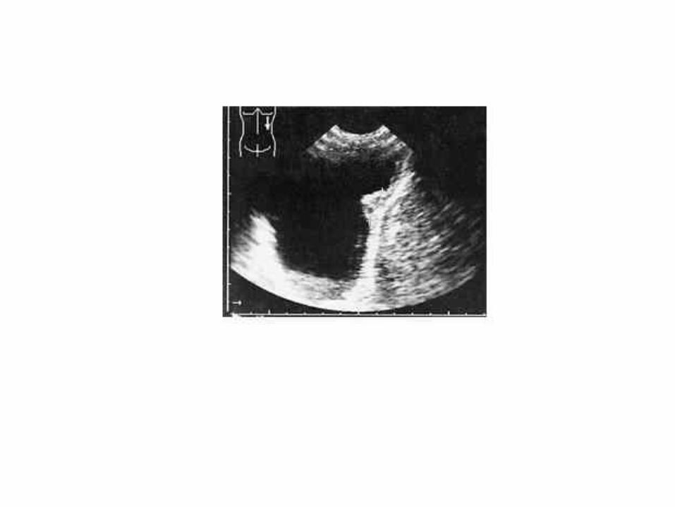
Diaphragm

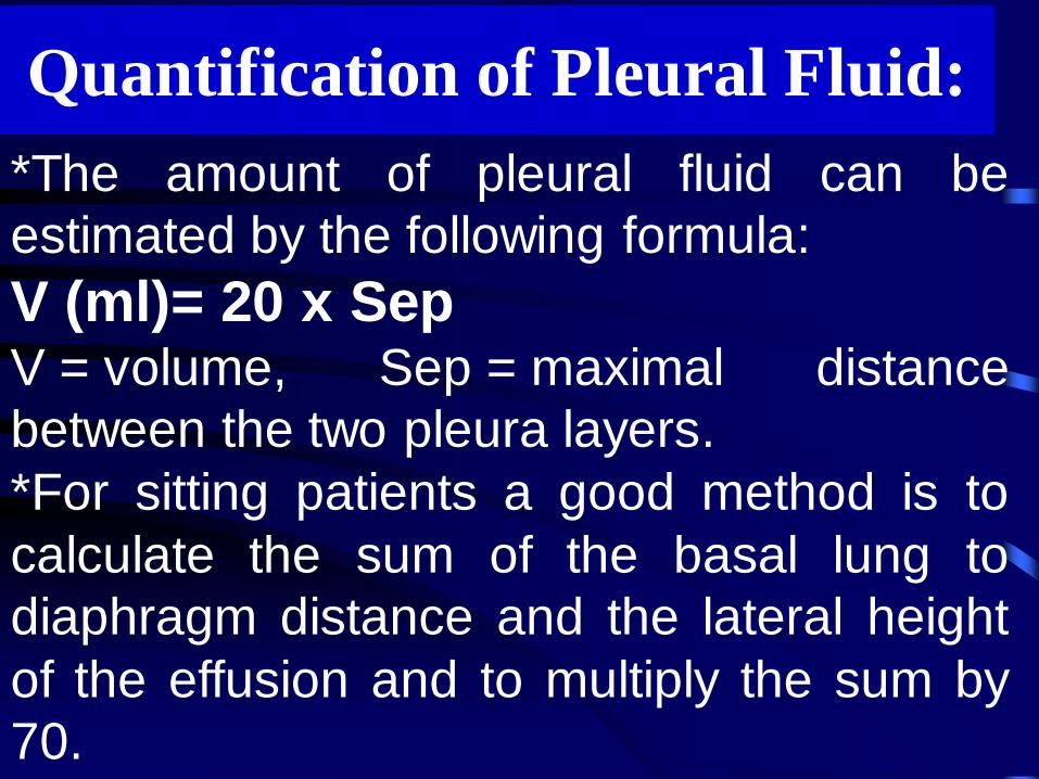
*The amount of pleural fluid can be
estimated by the following formula:
V (ml)= 20 x Sep V = volume, Sep = maximal distance
between the two pleura layers.
*For sitting patients a good method is to
calculate the sum of the basal lung to
diaphragm distance and the lateral height
of the effusion and to multiply the sum by
70.
Quantification of Pleural Fluid:
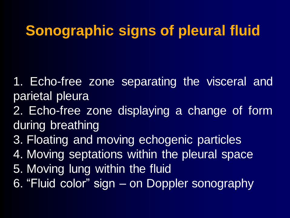
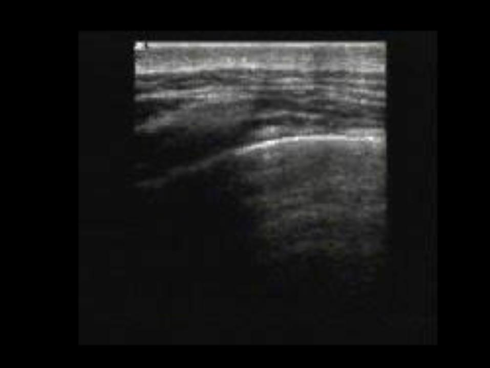
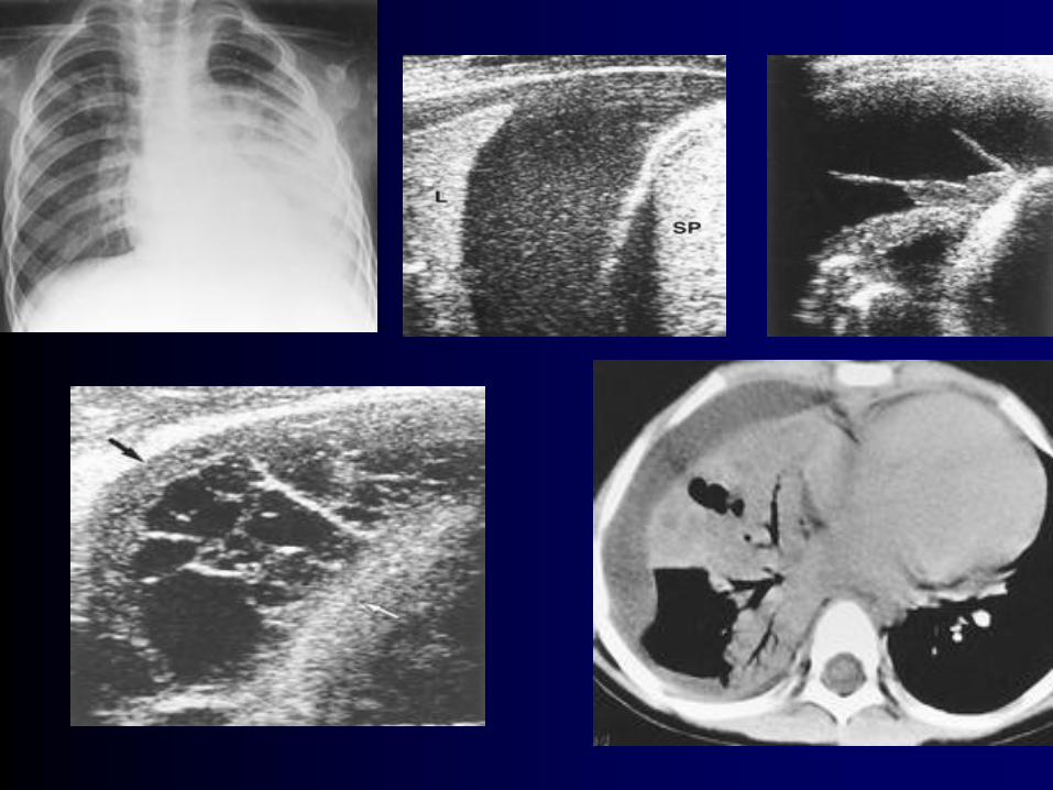
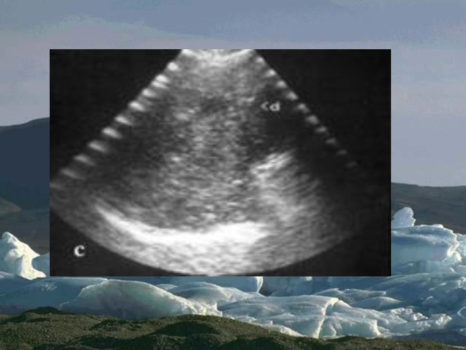
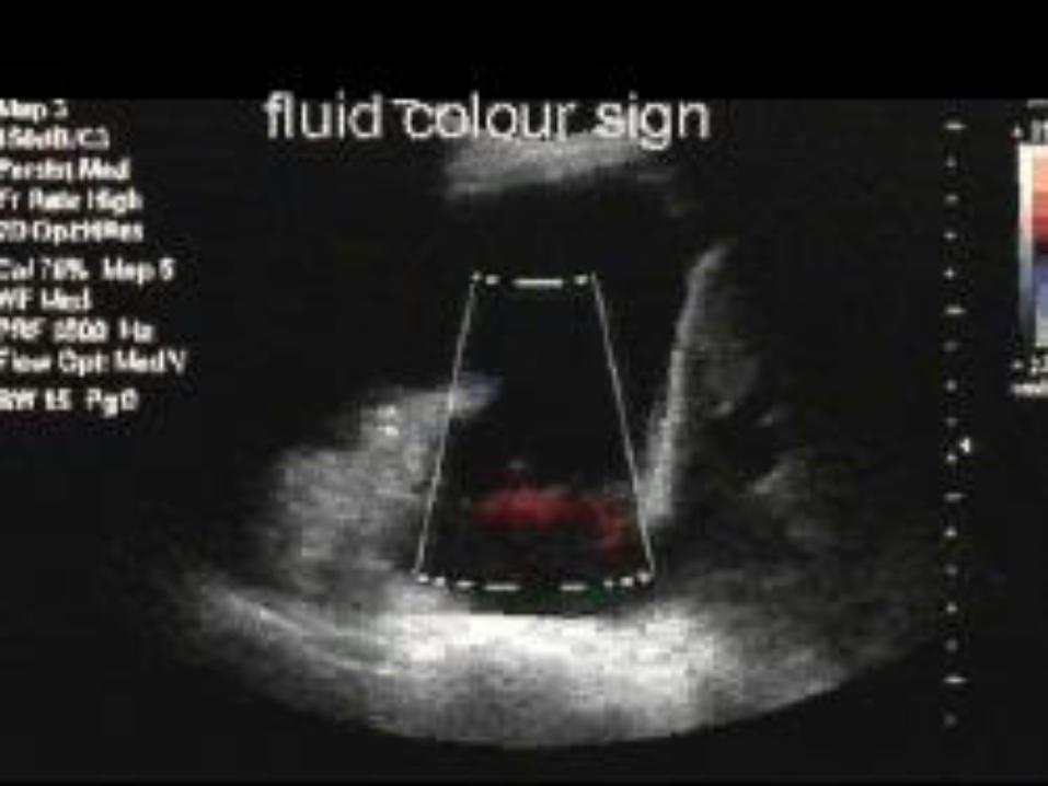
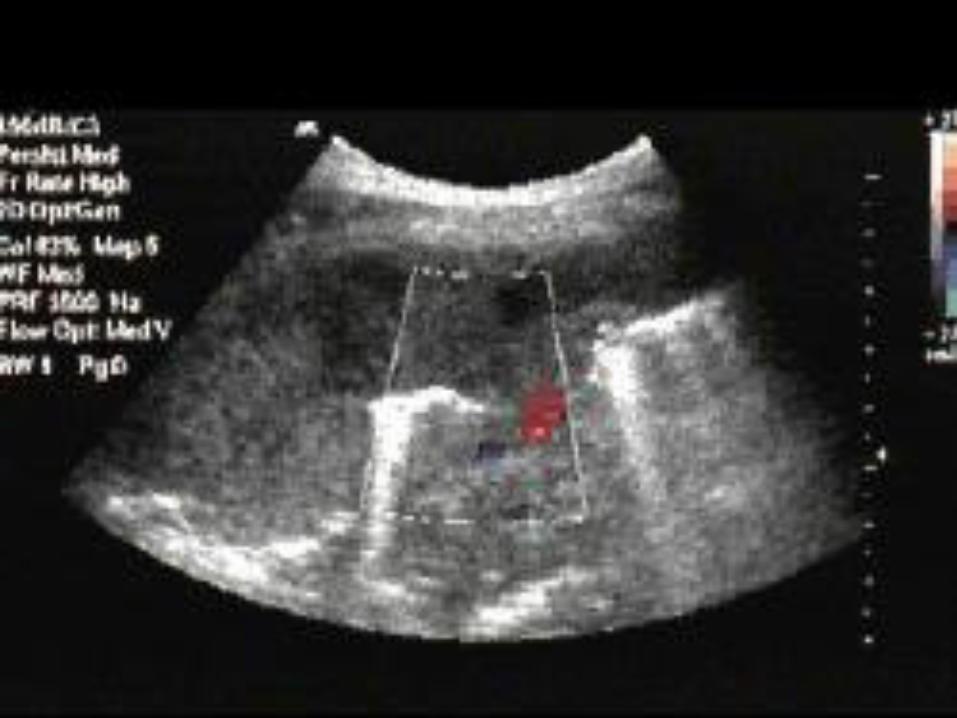
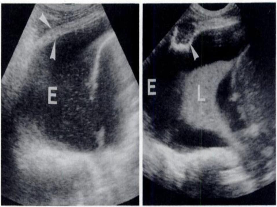
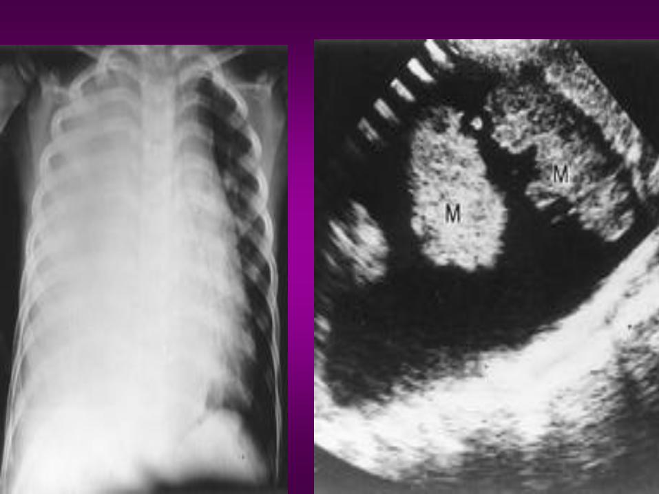
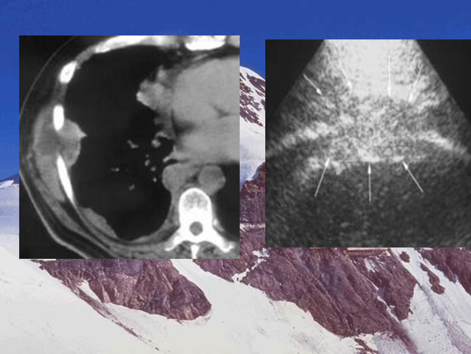
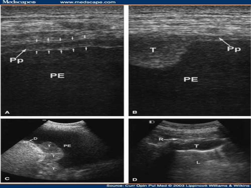
Sonographic signs of pleural fluid
1. Echo-free zone separating the visceral and
parietal pleura
2. Echo-free zone displaying a change of form
during breathing
3. Floating and moving echogenic particles
4. Moving septations within the pleural space
5. Moving lung within the fluid
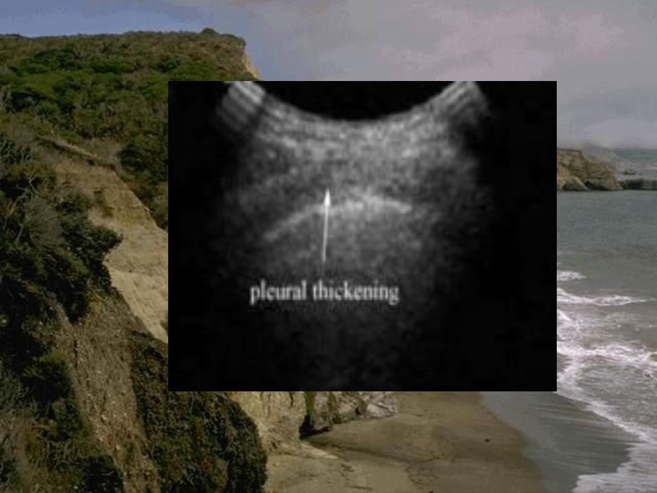
6. “Fluid color” sign – on Doppler sonography
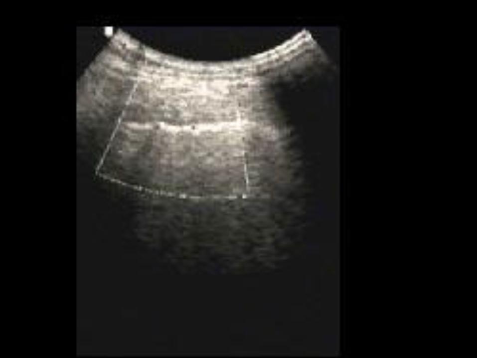

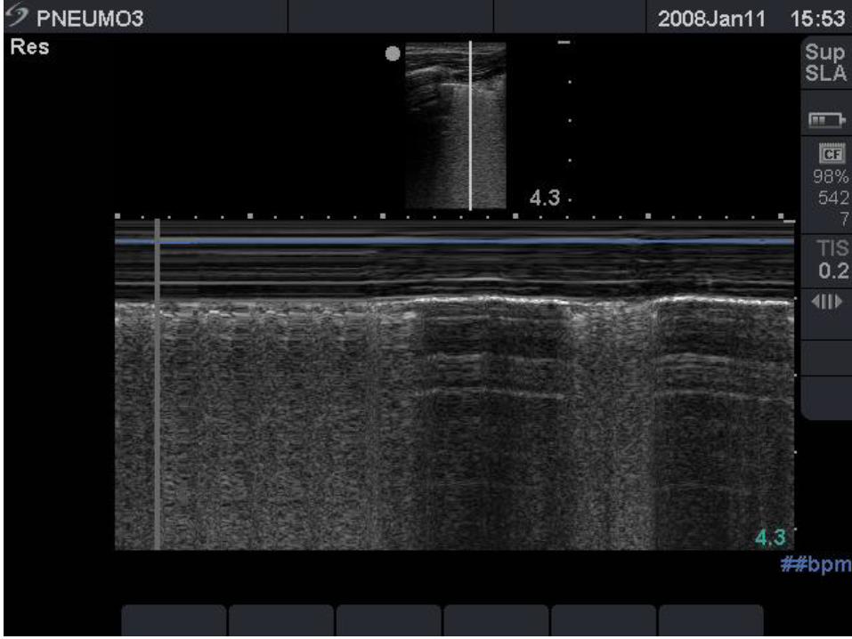
the "seashore sign" (Fig.3).
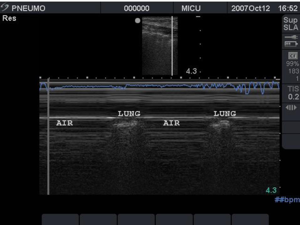

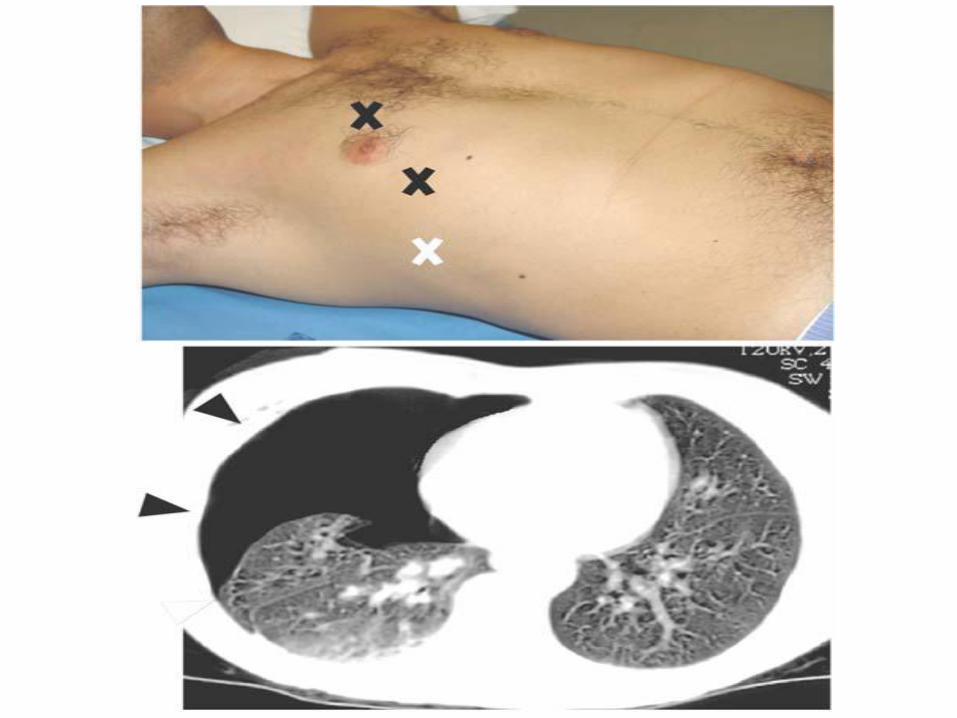
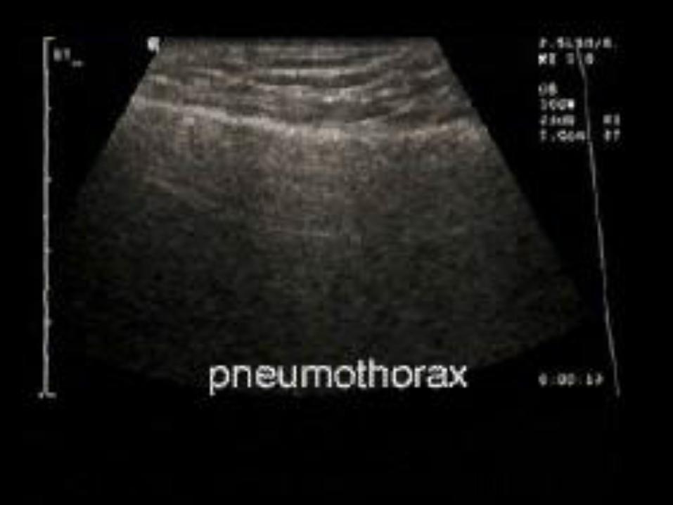
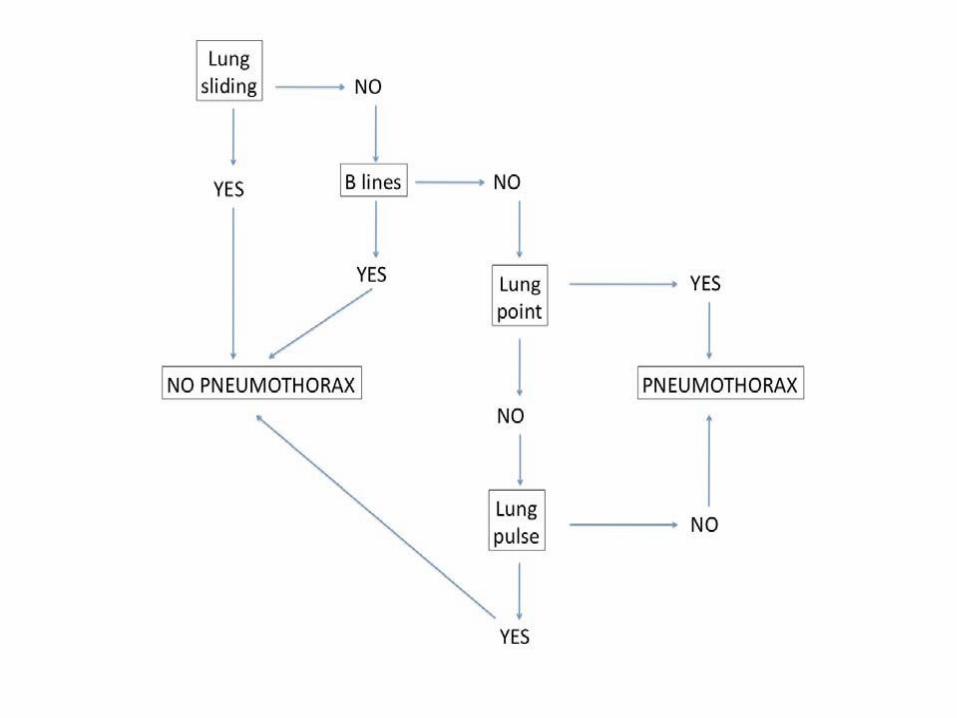
Absent lung sliding
Exaggerated horizontal artifacts
Loss of comet-tail artifacts
Broadening of the pleural line to a band
Lung point
Loss of lung impulse
The key sonographic signs of
Pneumothorax

.]
.]

Related Documents