Platelet-rich plasma may prevent titanium-mesh exposure in alveolar ridge augmentation with anorganic bovine bone Torres J, Tamimi F, Alkhraisat MH, Mancho´n A ´ , Linares R, Prados-Frutos JC, Herna´ndez G, Lo´pez Cabarcos E. Platelet-rich plasma may prevent titanium-mesh exposure in alveolar ridge augmentation with anorganic bovine bone. J Clin Periodontol 2010; 37: 943–951. doi: 10.1111/j.1600-051X.2010.01615.x. Abstract Objective: Bone augmentation with the titanium-mesh (Ti-mesh) technique is susceptible to a large rate of complications such as morbidity of bone graft donor site, and mesh exposure to the oral cavity. The purpose of this study was to evaluate the effectiveness of anorganic bovine bone (ABB) in alveolar bone augmentation with the Ti-mesh technique. In addition, we investigated the effect of platelet-rich plasma (PRP) in preventing mesh exposure by using it to cover the Ti-mesh. Patients and Methods: Patients included in the clinical trial were randomly allocated by a blinded assistant into two groups. The 30 patients recruited for this study underwent 43 alveolar bone augmentation with the Ti-mesh technique using ABB as graft material in all of them. In 15 patients, the Ti-meshes were covered with PRP (PRP group) whereas in the other 15 the Ti-meshes were not (control group). After 6 months, patients were called for clinical, radiographic, and histological evaluation, and implant placement surgery. A total of 97 implants were placed in the augmented bone and their evolution was followed up for a period of 24 months. Results: Significant differences were found between the two study groups in terms of complications and bone formation. In the control group, 28.5% of the cases suffered from mesh exposure, while in the PRP group, no exposures were registered. Radiographic analysis revealed that bone augmentation was higher in the PRP group than in the control group. Overall, 97.3% of implants placed in the control group and 100% of those placed in the PRP group were successful during the monitoring period. We suggest that the positive effect of PRP on the Ti-mesh technique is due to its capacity to improve soft tissue healing, thereby protecting the mesh and graft material secured beneath the gingival tissues. Conclusions: Alveolar bone augmentation using ABB alone in the Ti-mesh technique is sufficient for implant rehabilitation. Besides, covering the Ti-meshes with PRP was a determining factor in avoiding mesh exposure. Ti-mesh exposure provoked significant bone loss, but in most cases it did not affect the subsequent placement of implants. Key words: anorganic bovine bone; bone regeneration; platelet-rich plasma; soft tissue healing; titanium-mesh exposure Accepted for publication 21 July 2010 Jesu ´ s Torres 1,2 , Faleh Tamimi 2,3 , Mohammad Hamdan Alkhraisat 4 , A ´ ngel Mancho ´n 1 , Rafael Linares 1 , Juan Carlos Prados-Frutos 1 , Gonzalo Herna ´ndez 5 and Enrique Lo ´ pez Cabarcos 4 1 Ciencias de la Salud III, Universidad Rey Juan Carlos, Alcorco ´n, Spain; 2 Private practice, Clinica Dental Alcala ´, Madrid, Spain; 3 Faculty of Dentistry, McGill University, Montreal, QC, Canada; 4 Faculty of Pharmacy, Universidad Complutense, Madrid, Spain; 5 Faculty of Dentistry, Universidad Complutense, Madrid, Spain J Clin Periodontol 2010; 37: 943–951 doi: 10.1111/j.1600-051X.2010.01615.x 943 r 2010 John Wiley & Sons A/S

Welcome message from author
This document is posted to help you gain knowledge. Please leave a comment to let me know what you think about it! Share it to your friends and learn new things together.
Transcript
Platelet-rich plasma mayprevent titanium-meshexposure in alveolar ridgeaugmentation with anorganicbovine bone
Torres J, Tamimi F, Alkhraisat MH, Manchon A, Linares R, Prados-Frutos JC,Hernandez G, Lopez Cabarcos E. Platelet-rich plasma may prevent titanium-meshexposure in alveolar ridge augmentation with anorganic bovine bone. J ClinPeriodontol 2010; 37: 943–951. doi: 10.1111/j.1600-051X.2010.01615.x.
AbstractObjective: Bone augmentation with the titanium-mesh (Ti-mesh) technique issusceptible to a large rate of complications such as morbidity of bone graftdonor site, and mesh exposure to the oral cavity. The purpose of this study was toevaluate the effectiveness of anorganic bovine bone (ABB) in alveolar boneaugmentation with the Ti-mesh technique. In addition, we investigated the effect ofplatelet-rich plasma (PRP) in preventing mesh exposure by using it to cover theTi-mesh.
Patients and Methods: Patients included in the clinical trial were randomlyallocated by a blinded assistant into two groups. The 30 patients recruited forthis study underwent 43 alveolar bone augmentation with the Ti-mesh techniqueusing ABB as graft material in all of them. In 15 patients, the Ti-meshes werecovered with PRP (PRP group) whereas in the other 15 the Ti-meshes were not(control group). After 6 months, patients were called for clinical, radiographic, andhistological evaluation, and implant placement surgery. A total of 97 implants wereplaced in the augmented bone and their evolution was followed up for a period of 24months.
Results: Significant differences were found between the two study groups interms of complications and bone formation. In the control group, 28.5% ofthe cases suffered from mesh exposure, while in the PRP group, no exposureswere registered. Radiographic analysis revealed that bone augmentation washigher in the PRP group than in the control group. Overall, 97.3% of implantsplaced in the control group and 100% of those placed in the PRP group weresuccessful during the monitoring period. We suggest that the positive effect ofPRP on the Ti-mesh technique is due to its capacity to improve soft tissue healing,thereby protecting the mesh and graft material secured beneath the gingivaltissues.
Conclusions: Alveolar bone augmentation using ABB alone in the Ti-mesh techniqueis sufficient for implant rehabilitation. Besides, covering the Ti-meshes with PRP wasa determining factor in avoiding mesh exposure. Ti-mesh exposure provokedsignificant bone loss, but in most cases it did not affect the subsequent placement ofimplants.
Key words: anorganic bovine bone; boneregeneration; platelet-rich plasma; soft tissuehealing; titanium-mesh exposure
Accepted for publication 21 July 2010
Jesus Torres1,2, Faleh Tamimi2,3,Mohammad Hamdan Alkhraisat4,Angel Manchon1, Rafael Linares1,Juan Carlos Prados-Frutos1, GonzaloHernandez5 and Enrique LopezCabarcos4
1Ciencias de la Salud III, Universidad Rey
Juan Carlos, Alcorcon, Spain; 2Private
practice, Clinica Dental Alcala, Madrid,
Spain; 3Faculty of Dentistry, McGill
University, Montreal, QC, Canada; 4Faculty
of Pharmacy, Universidad Complutense,
Madrid, Spain; 5Faculty of Dentistry,
Universidad Complutense, Madrid, Spain
J Clin Periodontol 2010; 37: 943–951 doi: 10.1111/j.1600-051X.2010.01615.x
943r 2010 John Wiley & Sons A/S
Several techniques for alveolar boneaugmentation have been described inorder to give a solution for inadequatealveolar bone volume, which often pre-cludes the ideal placement of dentalimplants placement (Adell et al. 1990,Simion et al. 1994a, Buser et al. 1999,Cordaro et al. 2002, Rocchietta et al.2008). Onlay grafts and guided boneregeneration (GBR) are widely usedfor alveolar ridge augmentation beforeor simultaneous to implant placement.Clinical and histological data supportthe use of these approaches (van Steen-berghe et al. 1997, Keller et al. 1999,Parma Benfenati et al. 1999). However,the success of GBR procedures seems tobe highly technique sensitive and there-fore application to a wide community ofoperators and clinical settings remainsunclear (Simion et al. 1994a, Tinti et al.1996, Tinti & Parma-Benfenati 1998,Simion et al. 2007, Rocchietta et al.2008). On the other hand, onlay graftimplies the extraction of an autologousbone block that is often traumatic for thepatient.
GBR presents several controversiesconcerning two aspects: the type ofbarrier and the type of graft used (Boyneet al. 1985, von Arx et al. 1996).Regarding the first issue, two principalbarriers have been proposed: cell-occlu-sive membranes and titanium (Ti)meshes. Cell-occlusive membranes
Conflict of interest and source offunding statement
The authors declare that they have noconflict of interests.This work was supported by the Ministryof Science and Technology (grantMAT2009-14234-C03-01), the postdoctor-al fellow FECYT (F.T), and the BSCH-UCM Program for Research Groups(GR58/08).
showed very good results obtaininggreat quantity of regenerated bone,however, they have demonstrated twomajor inconveniences: (i) low stiffnessfor maintaining the contour of theregenerated sites, and (ii) a high riskof infection after wound dehiscence andbarrier exposure (Simion et al. 1994a–c). A major inconvenience of the Ti-mesh technique concerns the high rateof exposure that may facilitate graftinfection or loss (Table 1). In GBRtechniques, soft tissue closure over theaugmented area plays an important rolein preventing wound dehiscence andbacterial contamination of the exposedmembrane. In addition, the improvedstiffness of GBR using Ti-mesh com-pared with cell-occlusive membranespermits to obtain predictable results inboth lateral and vertical bone augmen-tation (Malchiodi et al. 1998, Maioranaet al. 2001, Artzi et al. 2003, Rocuzzo etal. 2004, 2007, Proussaefs & Lozada2006, Corinaldesi et al. 2007, Pieri et al.2008).
Although GBR technique is muchmore predictable in bone width augmen-tation, an increase of vertical bonevolume has been described, even insevere cases, in a predictable way(Table 1). Early studies advocated theuse of autogenous bone in the augmen-ted space beneath Ti-meshes (Boyne etal. 1985, von Arx et al. 1996). Althoughthe autologous bone is considered thegold standard bone substitute because ofits intrinsic properties, its availability isrestricted by the limited amount ofintraoral grafts, the morbidity associatedto second surgery at the donor site, andthe high cost for bone harvesting fromextraoral sites. Therefore, alternativebiomaterials have been developed tosubstitute this material.
Among the available bone substi-tutes, anorganic bovine bone (ABB)
has received a wealth of reports in theliterature demonstrating its long-termsuccess when used in alveolar boneaugmentation procedures. Bio-Osss
(Geistlich Biomaterials; Wolhusen,Switzerland) is a biocompatible andosteoconductive ABB (Rosen et al.2002) that provides an excellent scaf-fold for new bone formation (Hammerleet al. 1998, Piattelli et al. 1999). It hasbeen extensively used for alveolar boneaugmentation (Zitzmann et al. 2001,Fugazzotto 2003a, b) with high clinicalsuccess rates (Carmagnola et al. 2003).Accordingly, previous studies haveintroduced the use of ABB to the Ti-mesh technique, either alone, or com-bined with autologous bone (Maioranaet al. 2001, Corinaldesi et al. 2007, Pieriet al. 2008) (Table 1).
Platelet-rich plasma (PRP) is an auto-logous fibrin adhesive with high plateletconcentration easily obtained fromwhole blood by centrifugation (Anto-niades 1981, Marx et al. 1998, Anitua1999) Furthermore, PRP has a highconcentration of angiogenic and mito-genic growth factors implicated in softtissue healing, such as TGF (Wikesjo etal. 1998), PDGF, and EGF (Giannobileet al. 1996). Indeed, several studies havesuggested that the application of auto-genous PRP can enhance soft tissuewound healing. (Eppley et al. 2006). Inthis work, we have considered that thehealing of soft tissues over Ti-meshes inalveolar ridge augmentation proceduresmight benefit from local application ofPRP, avoiding subsequent exposure ofthe Ti mesh, and its derived complica-tions.
The recent consensus statements ofthe European Workshop on Perio-dontology 2008 highlighted the factthat bone augmentation procedures canfail and that implants placed in theseareas do not necessarily enjoy the high
Table 1. Summary of clinical studies reporting the amount bone gained and complications rate using the Ti-mesh technique
Pts/BAP (n/n) Type of graft (%) ABW (mm) ABH (mm) ME (%) Impl (n) Survival (%) Success (%) References
20/20 AB (100) ID ID 50 28 ID ID Von Arx et al. (1996)25/25 AB (100) 5.65 ID 0 120 ID 100 Malchiodi et al. (1998)23/23 AB (100) n ID 5 17.3 ID ID ID Rocuzzo et al. (2004)18/18 AB (100) n ID 4.8 22.2 37 100 100 Rocuzzo et al. (2007)14/23 AB/ABB (50/50) ID ID 14.2 59 98.3 ID Maiorana et al. (2001)16/19 AB/ABB (70/30) 4.16 3.71 5.3 44 100 100 Pieri et al. (2008)12/12 AB/ABB (70/30) ID ID 0 35 100 100 Corinaldesi et al. (2007)7/7 AB/ABB (ID) 3.71 2.86 57 ID ID ID Proussaefs & Lozada (2006)10/10 ABB (100) ID 5.2 20 20 100 ID Artzi et al. (2003)
nBlock grafts.
Pts, patients; BAP, bone augmentation procedures; ABW, average bone width gained; ABH, average bone height gained; ME, mesh exposure; Impl,
implants placed; AB, autologous bone; ID, insufficient data.
944 Torres et al.
r 2010 John Wiley & Sons A/S
long-term survival rates of dentalimplants placed in pristine sites. Theconsensus emphasized the researchneed to solve this problem (Tonetti &Hammerle 2008). In the present study, aclinical trial was performed to evaluatetwo aspects regarding the Ti-mesh tech-nique: (i) to examine the outcome ofABB grafting alone, and (ii) the benefitof covering the Ti-mesh with PRP inorder to improve soft tissue healing andprevent exposure. The results wereobtained by means of clinical investiga-tion, radiographs, and histological ana-lysis.
Patient and Methods
Patients
Before commencing this study, approvalwas obtained from the Ethical Commit-tee for Clinical Trials of the ‘‘HospitalSan Carlos’’ (Madrid, Spain) to carryout a pilot clinical study in ‘‘DentalClinic Alcala’’ (Madrid, Spain). Patientswere enrolled in the study on the basisof having insufficient bone height(47 mm), width (43 mm) or both, ineither maxilla or mandible (Fig. 1).Patients who needed simultaneous sinusfloor augmentation or nasal floor aug-mentation were included, while smokers(410 cigarettes per day) and patientswith severe systemic disease [ASA (IIIor IV) – American Society of Anesthe-siology] were excluded. Informed writ-ten consent to participate in this studywas obtained from all patients afterexplaining the objectives and protocolof the study, and the possible sideeffects.
During the study period (from May2003 to September 2008), 209 patientsattended the dental office demand-ing implant treatment. Among thesepatients, 30 fulfill the criteria and wererecruited for this randomized-controlledclinical trial. The study group was con-stituted of 17 females and 13 males withan age range between 48 and 76 yearsold. There was heterogeneity in thesystemic diseases present in some ofthe selected patients such as diabetes,heart failure, and osteoporosis; however,none of these conditions are known tojeopardize the implant’s success (Mom-belli & Cionca 2006).
Clinical assessment of thin or thickbiotype was based on simple visualinspection. However, we are aware thatthe precision of this method to identifygingival biotype is limited and highly
dependent on the clinician criteria (Egh-bali et al. 2009).
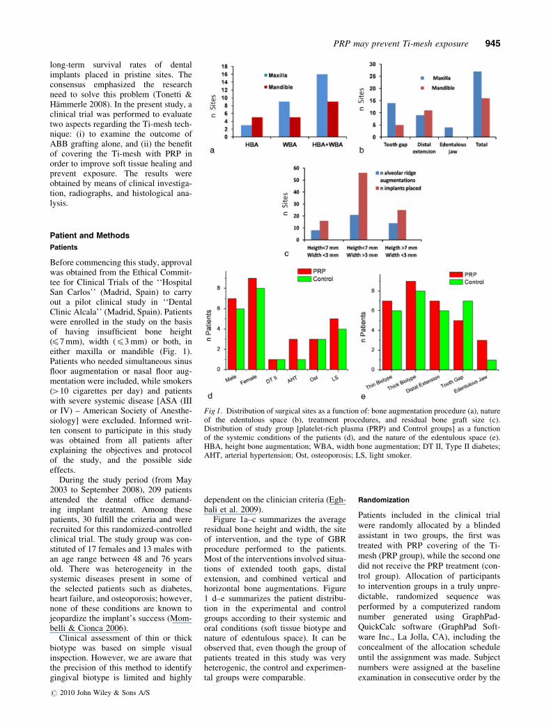
Figure 1a–c summarizes the averageresidual bone height and width, the siteof intervention, and the type of GBRprocedure performed to the patients.Most of the interventions involved situa-tions of extended tooth gaps, distalextension, and combined vertical andhorizontal bone augmentations. Figure1 d–e summarizes the patient distribu-tion in the experimental and controlgroups according to their systemic andoral conditions (soft tissue biotype andnature of edentulous space). It can beobserved that, even though the group ofpatients treated in this study was veryheterogenic, the control and experimen-tal groups were comparable.
Randomization
Patients included in the clinical trialwere randomly allocated by a blindedassistant in two groups, the first wastreated with PRP covering of the Ti-mesh (PRP group), while the second onedid not receive the PRP treatment (con-trol group). Allocation of participantsto intervention groups in a truly unpre-dictable, randomized sequence wasperformed by a computerized randomnumber generated using GraphPad-QuickCalc software (GraphPad Soft-ware Inc., La Jolla, CA), including theconcealment of the allocation scheduleuntil the assignment was made. Subjectnumbers were assigned at the baselineexamination in consecutive order by the
Fig 1. Distribution of surgical sites as a function of: bone augmentation procedure (a), natureof the edentulous space (b), treatment procedures, and residual bone graft size (c).Distribution of study group [platelet-rich plasma (PRP) and Control groups] as a functionof the systemic conditions of the patients (d), and the nature of the edentulous space (e).HBA, height bone augmentation; WBA, width bone augmentation; DT II, Type II diabetes;AHT, arterial hypertension; Ost, osteoporosis; LS, light smoker.
PRP may prevent Ti-mesh exposure 945
r 2010 John Wiley & Sons A/S

principal investigator. The sample sizeused has been usual in previous studiesfor this type of clinical evaluation(Table 1). The presence of systemicdisorders was registered and its distribu-tion was balanced among the two treat-ment groups (Fig. 1d).
Blinding
The surgeon was blinded to the graftmaterial applied over the Ti mesh ineach patient throughout the entire pro-cedures preceding graft implantation.Once the Ti-meshes were screwed tothe alveolar bone by the surgeon, anassistant handled the PRP or nothing.However, a possible bias could occurduring the suturing because at this timethe surgeon was not blinded anymore.Clinicians who made the post-surgicalfollow-up were blinded to study groups.
PRP
PRP was prepared according to Anitua’smethod (Anitua 1999). Blood was col-lected from all patients 30 min. beforestarting the surgery to ensure the blind-ing of the surgeon. In the PRP group,10–20 ml of blood was withdrawn viavenous aspiration into 4.5 ml test tubesand mixed with a 3.8% sodium citratesolution at a ratio of 5/1 (v/v) achievinganticoagulation through calcium bind-ing. The blood was then centrifugedusing a Btis PRGF System II centrifuge(Bti Biotechnology Institute S.L, Vitor-ia, Spain) into three basic components:red blood cells (RBCs), PRP, and plate-let-poor plasma (PPP) (Fig. 2a).Because of the different densities ofthe components, the RBC layer formsat the bottom of the tube, the PRP layerin the middle, and the PPP layer at thetop. A pipette (Gilson Inc., Middleton,
WI, USA) was used to separate thelayers, from the less dense to the denser.Therefore PPP was separated first (about2.25 ml) followed by PRP (about0.9 ml), leaving as residual the RBCslayer (about 2.25 ml) (Fig. 2b).
Surgical protocol
An alveolar ridge augmentation wasperformed in all patients following themethod described by Boyne et al. (1985)and Von Arx et al. (1996). Under localanaesthesia, a mid-crestal with diver-gent buccal incisions was performed toallow the elevation of two mucoperios-teal flaps to the buccal and palatalaspects. Perforations into the marrowspace were produced. In all patients,ABB particles were adapted to the defi-cient ridge and a Ti-mesh that wasindividually trimmed was placed overthe grafts and fixed with microscrews.Subsequently, PRP was used as a mem-brane covering the Ti-meshes in thePRP group, while nothing was addedto cover the Ti-mesh in the controlgroup (Fig. 3). Then, releasing perios-teal incisions were made and a tension-free, tight wound closure was accom-plished. Post-operatively, Amoxicillin750 mg (Clamoxyls, GlaxoSmithKline,Middlesex, UK) was prescribed threetimes a day for 7 days, Ibuprofen600 mg (Espidifens, Zambon Switzer-land Ltd, Barcelona, Spain) three timesa day for 4 days, and clorhexidine0.20% (Perio-aids, Dentaid, Barcelona,Spain) three times a day for 10 days.
Fig 2. (a) Blood samples after centrifugation to obtain platelet-rich plasma (PRP). Note theseparation in two fractions, the red one (containing red blood cells) and the yellow one(containing leucocytes and platelets). (b) PRP as obtained from the blood samples after beingactivated with a 30% CaCl2 solution forms a viscous gel that can be easily manipulated.
Fig 3. (a) Ti-mesh adapted on the maxillary residual ridge. (b) Placement of platelet-rich plasma gel over the Ti-mesh. (c) Healed alveolarridge after 6 months without Ti-mesh exposition. A panoramic radiograph (d), a sagittal CT section (e), and a transverse CT section (f) of thetreated site 6 months after the intervention showing the alveolar ridge augmentation achieved.
946 Torres et al.
r 2010 John Wiley & Sons A/S
Sutures were removed 1 week aftersurgery. A healing time of 6 monthswas allowed before implant placement(Osseotite, Biomet 3i Inc., Palm Beach,FL, USA). During the 6-month healingperiod, patients were instructed not towear their prosthesis to avoid transmu-cosal pressure on the augmented area.They were recommended to follow asoft diet, and were recalled every 2weeks for follow-up. After implant pla-cement, patients were called for follow-up every 6 months until the end of thestudy period (2 years).
The surgical procedures and implantsurgery were performed by the samesurgeon in order to avoid possible bias,while the follow-up assessment wasperformed by another blinded clinician.The exposure of the Ti-meshes wasdetermined by visual inspection, andmeasured using a periodontal probe. Arepresentative case with this reconstruc-tive method is presented in Fig. 3.
Radiographical analysis
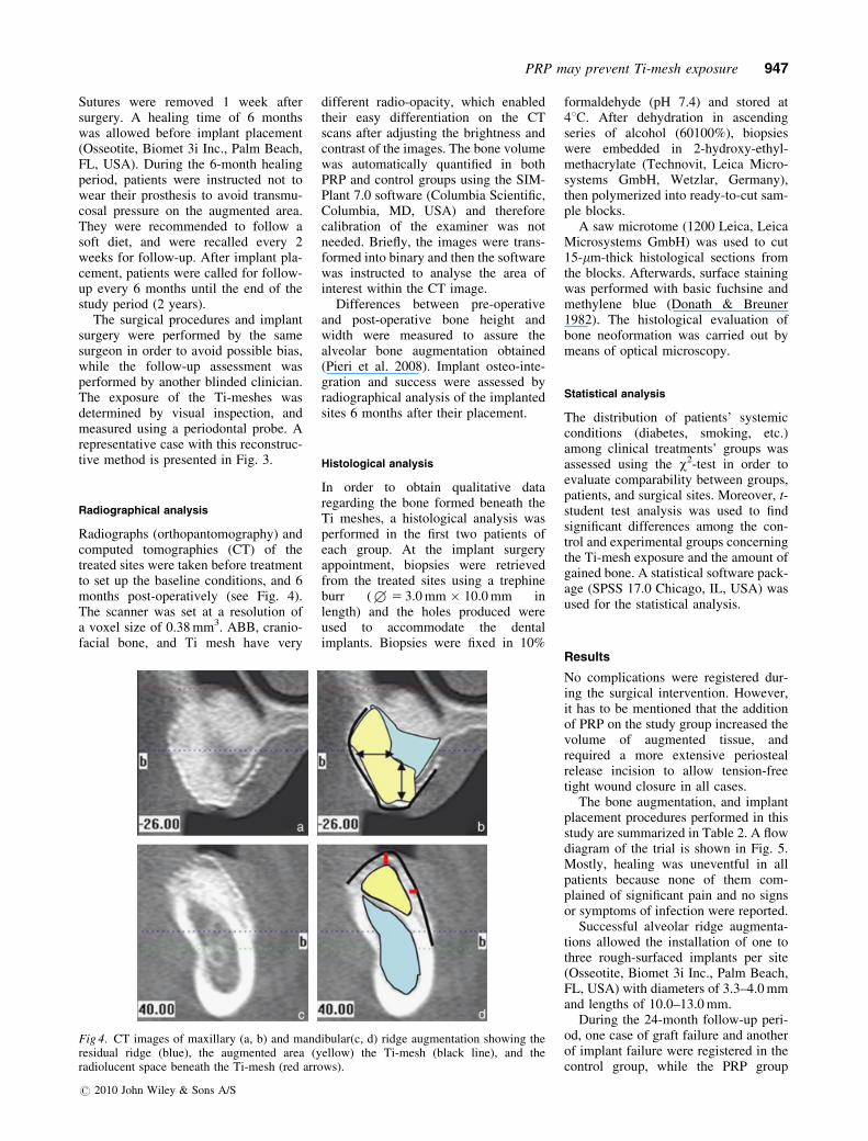
Radiographs (orthopantomography) andcomputed tomographies (CT) of thetreated sites were taken before treatmentto set up the baseline conditions, and 6months post-operatively (see Fig. 4).The scanner was set at a resolution ofa voxel size of 0.38 mm3. ABB, cranio-facial bone, and Ti mesh have very
different radio-opacity, which enabledtheir easy differentiation on the CTscans after adjusting the brightness andcontrast of the images. The bone volumewas automatically quantified in bothPRP and control groups using the SIM-Plant 7.0 software (Columbia Scientific,Columbia, MD, USA) and thereforecalibration of the examiner was notneeded. Briefly, the images were trans-formed into binary and then the softwarewas instructed to analyse the area ofinterest within the CT image.
Differences between pre-operativeand post-operative bone height andwidth were measured to assure thealveolar bone augmentation obtained(Pieri et al. 2008). Implant osteo-inte-gration and success were assessed byradiographical analysis of the implantedsites 6 months after their placement.
Histological analysis
In order to obtain qualitative dataregarding the bone formed beneath theTi meshes, a histological analysis wasperformed in the first two patients ofeach group. At the implant surgeryappointment, biopsies were retrievedfrom the treated sites using a trephineburr (+5 3.0 mm � 10.0 mm inlength) and the holes produced wereused to accommodate the dentalimplants. Biopsies were fixed in 10%
formaldehyde (pH 7.4) and stored at41C. After dehydration in ascendingseries of alcohol (60100%), biopsieswere embedded in 2-hydroxy-ethyl-methacrylate (Technovit, Leica Micro-systems GmbH, Wetzlar, Germany),then polymerized into ready-to-cut sam-ple blocks.
A saw microtome (1200 Leica, LeicaMicrosystems GmbH) was used to cut15-mm-thick histological sections fromthe blocks. Afterwards, surface stainingwas performed with basic fuchsine andmethylene blue (Donath & Breuner1982). The histological evaluation ofbone neoformation was carried out bymeans of optical microscopy.
Statistical analysis
The distribution of patients’ systemicconditions (diabetes, smoking, etc.)among clinical treatments’ groups wasassessed using the w2-test in order toevaluate comparability between groups,patients, and surgical sites. Moreover, t-student test analysis was used to findsignificant differences among the con-trol and experimental groups concerningthe Ti-mesh exposure and the amount ofgained bone. A statistical software pack-age (SPSS 17.0 Chicago, IL, USA) wasused for the statistical analysis.
Results
No complications were registered dur-ing the surgical intervention. However,it has to be mentioned that the additionof PRP on the study group increased thevolume of augmented tissue, andrequired a more extensive periostealrelease incision to allow tension-freetight wound closure in all cases.
The bone augmentation, and implantplacement procedures performed in thisstudy are summarized in Table 2. A flowdiagram of the trial is shown in Fig. 5.Mostly, healing was uneventful in allpatients because none of them com-plained of significant pain and no signsor symptoms of infection were reported.
Successful alveolar ridge augmenta-tions allowed the installation of one tothree rough-surfaced implants per site(Osseotite, Biomet 3i Inc., Palm Beach,FL, USA) with diameters of 3.3–4.0 mmand lengths of 10.0–13.0 mm.
During the 24-month follow-up peri-od, one case of graft failure and anotherof implant failure were registered in thecontrol group, while the PRP group
Fig 4. CT images of maxillary (a, b) and mandibular(c, d) ridge augmentation showing theresidual ridge (blue), the augmented area (yellow) the Ti-mesh (black line), and theradiolucent space beneath the Ti-mesh (red arrows).
PRP may prevent Ti-mesh exposure 947
r 2010 John Wiley & Sons A/S
presented no complications (see Tables2 and 3). Moreover, the amount of boneheight and width gained was higher inpatients treated with PRP (Table 2).Successful implants were uncoveredfor fixed prosthetic rehabilitation andno implant complications were regis-tered beyond this point.
Histological observations
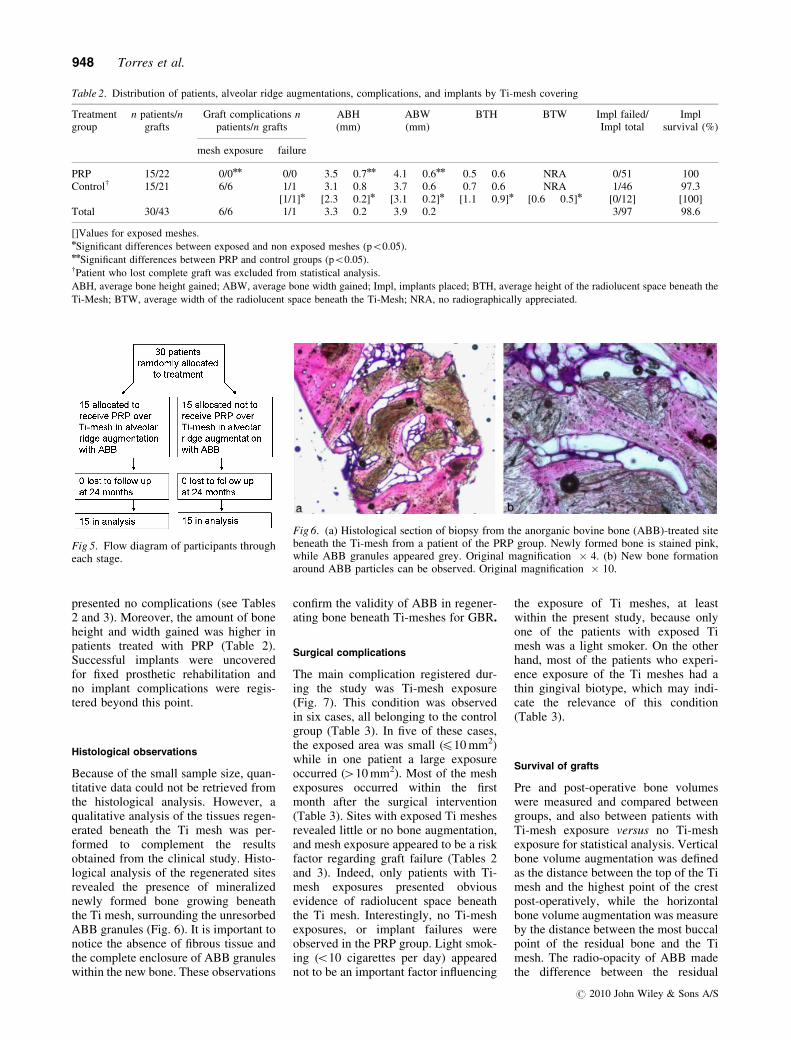
Because of the small sample size, quan-titative data could not be retrieved fromthe histological analysis. However, aqualitative analysis of the tissues regen-erated beneath the Ti mesh was per-formed to complement the resultsobtained from the clinical study. Histo-logical analysis of the regenerated sitesrevealed the presence of mineralizednewly formed bone growing beneaththe Ti mesh, surrounding the unresorbedABB granules (Fig. 6). It is important tonotice the absence of fibrous tissue andthe complete enclosure of ABB granuleswithin the new bone. These observations
confirm the validity of ABB in regener-ating bone beneath Ti-meshes for GBR.
Surgical complications
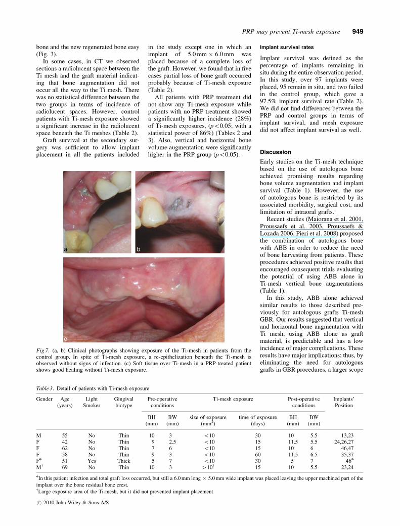
The main complication registered dur-ing the study was Ti-mesh exposure(Fig. 7). This condition was observedin six cases, all belonging to the controlgroup (Table 3). In five of these cases,the exposed area was small (410 mm2)while in one patient a large exposureoccurred (410 mm2). Most of the meshexposures occurred within the firstmonth after the surgical intervention(Table 3). Sites with exposed Ti meshesrevealed little or no bone augmentation,and mesh exposure appeared to be a riskfactor regarding graft failure (Tables 2and 3). Indeed, only patients with Ti-mesh exposures presented obviousevidence of radiolucent space beneaththe Ti mesh. Interestingly, no Ti-meshexposures, or implant failures wereobserved in the PRP group. Light smok-ing (o10 cigarettes per day) appearednot to be an important factor influencing
the exposure of Ti meshes, at leastwithin the present study, because onlyone of the patients with exposed Timesh was a light smoker. On the otherhand, most of the patients who experi-ence exposure of the Ti meshes had athin gingival biotype, which may indi-cate the relevance of this condition(Table 3).
Survival of grafts
Pre and post-operative bone volumeswere measured and compared betweengroups, and also between patients withTi-mesh exposure versus no Ti-meshexposure for statistical analysis. Verticalbone volume augmentation was definedas the distance between the top of the Timesh and the highest point of the crestpost-operatively, while the horizontalbone volume augmentation was measureby the distance between the most buccalpoint of the residual bone and the Timesh. The radio-opacity of ABB madethe difference between the residual
Table 2. Distribution of patients, alveolar ridge augmentations, complications, and implants by Ti-mesh covering
Treatmentgroup
n patients/ngrafts
Graft complications npatients/n grafts
ABH(mm)
ABW(mm)
BTH BTW Impl failed/Impl total
Implsurvival (%)
mesh exposure failure
PRP 15/22 0/0nn 0/0 3.5 � 0.7nn 4.1 � 0.6nn 0.5 � 0.6 NRA 0/51 100Controlw 15/21 6/6 1/1 3.1 � 0.8 3.7 � 0.6 0.7 � 0.6 NRA 1/46 97.3
[1/1]n [2.3 � 0.2]n [3.1 � 0.2]n [1.1 � 0.9]n [0.6 � 0.5]n [0/12] [100]Total 30/43 6/6 1/1 3.3 � 0.2 3.9 � 0.2 3/97 98.6
[]Values for exposed meshes.nSignificant differences between exposed and non exposed meshes (po0.05).nnSignificant differences between PRP and control groups (po0.05).wPatient who lost complete graft was excluded from statistical analysis.
ABH, average bone height gained; ABW, average bone width gained; Impl, implants placed; BTH, average height of the radiolucent space beneath the
Ti-Mesh; BTW, average width of the radiolucent space beneath the Ti-Mesh; NRA, no radiographically appreciated.
Fig 5. Flow diagram of participants througheach stage.
Fig 6. (a) Histological section of biopsy from the anorganic bovine bone (ABB)-treated sitebeneath the Ti-mesh from a patient of the PRP group. Newly formed bone is stained pink,while ABB granules appeared grey. Original magnification � 4. (b) New bone formationaround ABB particles can be observed. Original magnification � 10.
948 Torres et al.
r 2010 John Wiley & Sons A/S
bone and the new regenerated bone easy(Fig. 3).
In some cases, in CT we observedsections a radiolucent space between theTi mesh and the graft material indicat-ing that bone augmentation did notoccur all the way to the Ti mesh. Therewas no statistical difference between thetwo groups in terms of incidence ofradiolucent spaces. However, controlpatients with Ti-mesh exposure showeda significant increase in the radiolucentspace beneath the Ti meshes (Table 2).
Graft survival at the secondary sur-gery was sufficient to allow implantplacement in all the patients included
in the study except one in which animplant of 5.0 mm � 6.0 mm wasplaced because of a complete loss ofthe graft. However, we found that in fivecases partial loss of bone graft occurredprobably because of Ti-mesh exposure(Table 2).
All patients with PRP treatment didnot show any Ti-mesh exposure whilepatients with no PRP treatment showeda significantly higher incidence (28%)of Ti-mesh exposures, (po0.05; with astatistical power of 86%) (Tables 2 and3). Also, vertical and horizontal bonevolume augmentation were significantlyhigher in the PRP group (po0.05).
Implant survival rates
Implant survival was defined as thepercentage of implants remaining insitu during the entire observation period.In this study, over 97 implants wereplaced, 95 remain in situ, and two failedin the control group, which gave a97.5% implant survival rate (Table 2).We did not find differences between thePRP and control groups in terms ofimplant survival, and mesh exposuredid not affect implant survival as well.
Discussion
Early studies on the Ti-mesh techniquebased on the use of autologous boneachieved promising results regardingbone volume augmentation and implantsurvival (Table 1). However, the useof autologous bone is restricted by itsassociated morbidity, surgical cost, andlimitation of intraoral grafts.
Recent studies (Maiorana et al. 2001,Proussaefs et al. 2003, Proussaefs &Lozada 2006, Pieri et al. 2008) proposedthe combination of autologous bonewith ABB in order to reduce the needof bone harvesting from patients. Theseprocedures achieved positive results thatencouraged consequent trials evaluatingthe potential of using ABB alone inTi-mesh vertical bone augmentations(Table 1).
In this study, ABB alone achievedsimilar results to those described pre-viously for autologous grafts Ti-meshGBR. Our results suggested that verticaland horizontal bone augmentation withTi mesh, using ABB alone as graftmaterial, is predictable and has a lowincidence of major complications. Theseresults have major implications; thus, byeliminating the need for autologousgrafts in GBR procedures, a larger scope
Fig 7. (a, b) Clinical photographs showing exposure of the Ti-mesh in patients from thecontrol group. In spite of Ti-mesh exposure, a re-epithelization beneath the Ti-mesh isobserved without signs of infection. (c) Soft tissue over Ti-mesh in a PRP-treated patientshows good healing without Ti-mesh exposure.
Table 3. Detail of patients with Ti-mesh exposure
Gender Age(years)
LightSmoker
Gingivalbiotype
Pre-operativeconditions
Ti-mesh exposure Post-operativeconditions
Implants’Position
BH(mm)
BW(mm)
size of exposure(mm2)
time of exposure(days)
BH(mm)
BW(mm)
M 55 No Thin 10 3 o10 30 10 5.5 13,23F 42 No Thin 9 2.5 o10 15 11.5 5.5 24,26,27F 62 No Thin 7 6 o10 15 10 6 46,47F 58 No Thin 9 3 o10 60 11.5 6.5 35,37Fn 51 Yes Thick 5 7 o10 30 5 7 46n
Mw 69 No Thin 10 3 410w 15 10 5.5 23,24
nIn this patient infection and total graft loss occurred, but still a 6.0 mm long � 5.0 mm wide implant was placed leaving the upper machined part of the
implant over the bone residual bone crest.wLarge exposure area of the Ti-mesh, but it did not prevented implant placement
PRP may prevent Ti-mesh exposure 949
r 2010 John Wiley & Sons A/S
of patients may be treated. Moreover,unlike autologous grafts, ABB graftsproved to be dimensionally stable dur-ing the 2-year follow-up period. This isattributed to the combination of biocom-patibility, osteoconductivity, and lowresorption properties of ABB in vivo(Schlegel et al. 2003, Zitzmann et al2001).
Ti-mesh exposure has been correlatedto subsequent complications, such asgraft resorption and loss, often impair-ing implant treatment (Von Arx 1996).These facts were confirmed in this studybecause graft resorption and lossoccurred only in cases were exposureof the Ti-mesh occurred. The patientsof the control group suffered from arate of complications similar to thatreported previously for GBR with auto-logous graft and Ti meshes (Tables 1and 2). On the other hand, PRP mayprevent the incidence of Ti-mesh expo-sure (Table 2).
It was observed that soft tissue heal-ing was better when PRP was appliedover the Ti-mesh compared with con-trols without PRP coverage. This waslikely to translate into an improvedgingival biotype and subsequent impor-tant resistance to Ti-mesh exposure.
There is a large controversy regardingthe usefulness of PRP in bone regenera-tion procedures (Torres et al. 2009).Many studies have shown that PRP isunable to influence bone growth incavities and defects. However, most ofthe dental literature has been focused onevaluating its effect on hard tissues,ignoring the potential benefits on sur-rounding soft tissues. PRP may enhancesoft tissue healing by concentrating thelarge amounts of fibrin and growthfactors secreted by platelets thatincrease both angiogenesis and fibro-blast cell differentiation (Tamimi et al.2007). PRP increases early woundstrength by reducing the inflammatoryphase of wound healing allowing earlydeposition of collagen, glycosaminogly-can, and fibronectin. Moreover, PRP hasalso been found to decrease patientmorbidity and pain (Bashutski & Wang2008).
Interestingly, gingival healingseemed to have an effect on the under-lying bone formation. Bone grafts in thecontrol group experienced resorptionbeneath the Ti mesh, while in thePRP group the amount of augmentedbone was higher and no graft resorptionwas observed. We believe this phenom-enon occurs mainly due to soft tissue
protection rather than by direct effect onbone formation.
Conclusion
The results of the present study demon-strated that ABB alone may be used asgraft material in the Ti-mesh technique,obtaining predictable results in localizedridge augmentation procedures. More-over, applying PRP over the Ti mesh,may prevent complications such asmesh exposure, and graft failure.
Acknowledgements
Authors acknowledge Mrs. AscensionMarin and the staff of ‘‘Clinica DentalAlcala’’ for their help and support ofthis study.
References
Adell, R., Lekholm, U., Grondahl, K., Branemark, P.
I., Lindstrom, J. & Jacobsson, M. (1990) Recon-
struction of severely resorbed edentulous maxillae
using osseointegrated fixtures in immediate auto-
genous bone grafts. International Journal of Oral
and Maxillofacial Implants 5, 233–246.
Anitua, E. (1999) Plasma rich in growth factors:
preliminary results of use in the preparation of
future sites for implants. International Journal of
Oral and Maxillofacial Implants 14, 429–535.
Artzi, Z., Dayan, D., Alpern, Y. & Nemcovsky, C. E.
(2003) Vertical ridge augmentation using xeno-
genic material supported by a configured titanium
mesh: clinicohistophatologic and histochemical
study. International Journal of Oral and Maxillo-
facial Implants 18, 440–446.
Bashutski, J. D. & Wang, H. L. (2008) Role of platelet-
rich plasma in soft tissue root-coverage procedures:
a review. Quintessence International 39, 473–483.
Boyne, P J., Cole, M. D., Stringer, D. & Shafqat, J. P.
(1985) A technique for osseous restoration of
deficient edentulous maxillary ridges. Journal of
Oral and Maxillofacial Surgery 43, 87–91.
Buser, D., Dula, K., Hess, D., Hirt, H. P. & Belser, U.
C. (1999) Localized ridge augmentation with auto-
grafts and barrier membranes. Periodontology 2000
17, 22–35.
Carmagnola, D., Adriaens, P. & Berglundh, T. (2003)
Healing of human extraction sockets filled with
Bio-Oss. Clinical Oral Implants Research 14,
137–143.
Cordaro, L., Amade, D. S. & Cordaro, M. (2002)
Clinical results of alveolar ridge augmentation with
mandibular block bone grafts in partially edentu-
lous patients prior to implant placement. Clinical
Oral Implants Research 13, 103–111.
Corinaldesi, G., Pieri, F., Marchetti, C., Fini, M.,
Aldini, N. N. & Giardino, R. (2007) Histologic
and histomorphometric evaluation of alveolar
ridge augmentation using bone grafts and titanium
micromesh in humans. Journal of Periodontology
78, 1477–1484.
Donath, K. & Breuner, G. (1982) A method for the
study of undecalcified bones and teeth with attached
soft tissues. The Sage-Schliff (sawing and grinding)
technique. Journal of Oral Pathology 11, 318–326.
Eghbali, A., De Rouck, T., De Bruyn, H. & Cosyn, J.
(2009) The gingival biotype assessed by experi-
enced and inexperienced clinicians. Journal of
Clinical Periodontology 36, 958–963.
Eppley, B. L., Pietrzak, W. S. & Blanton, M. (2006)
Platelet-rich plasma: a review of biology and appli-
cations in plastic surgery. Plastic Reconstructive
Surgery 118, 147e–159.
Fugazzotto, P. A. (2003a) GBR using bovine bone
matrix and resorbable an onresorbable membranes.
Part 1: histologic results. International Journal
of Periodontics and Restorative Dentistry 23,
361–369.
Fugazzotto, P. A. (2003b) GBR using bovine bone
matrix and resorbable an onresorbable membranes.
Part 2: clinical results. International Journal
of Periodontics and Restorative Dentistry 23,
599–605.
Giannobile, W. V., Hernandez, R. A., Finkelman, R.
D., Ryan, S., Kiritsy, C. P., D’Andrea, M. & Lynch,
S. E. (1996) Comparative effects of platelet-derived
growth factor-BB and insulin-like growth factor-I,
individually and in combination, on periodontal
regeneration in Macaca fascicularis. Journal of
Periodontal Research 31, 301–312.
Hammerle, C. H., Chiantella, G. C., Karring, T. &
Lang, N. P. (1998) The effect of a deproteinized
bovine bone material on bone regeneration around
titanium dental implants. Clinical Oral Implants
Research 9, 151–162.
Keller, E. E., Tolman, D. E. & Eckert, S. (1999)
Surgical-prosthodontic reconstruction of advanced
maxillary bone compromise with autogenous onlay
block bone grafts and osseointegrated endosseous
implants: a 12-year study of 32 consecutive
patients. International Journal of Oral and Max-
illofacial Implants 14, 197–209.
Maiorana, C., Santoro, F., Rabagliati, M. & Salina, S.
(2001) Evaluation of the use of iliac cancellous
bone and anorganic bovine bone in the reconstruc-
tion of the atrophic maxilla with titanium mesh: a
clinical and histologic investigation. International
Journal of Oral and Maxillofacial Implants 16,
427–432.
Malchiodi, L., Scarano, A., Quaranta, M. & Piattelli,
A. (1998) Rigid fixation by means of titanium mesh
in edentulous ridge expansion for horizontal ridge
augmentation in the maxilla. International Journal
of Oral and Maxillofacial Implants 13, 701–705.
Marx, R. E., Carlson, E. R., Eichstaedt, R. M.,
Schimmele, S. R., Strauss, J. E. & Georgeff, K. R.
(1998) Platelet-rich plasma. Growth factors
enhancement for bone grafts. Oral Surgery Oral
Medicine Oral Pathology Oral Radiology and
Endodontics 85, 638–646.
Mombelli, A. & Cionca, N. (2006) Systemic diseases
affecting osseointegration therapy. Clinical Oral
Implants Research 17, 97–103.
Parma-Benfenati, S., Tinti, C., Albrektsson, T. &
Johansson, C. (1999) Histologic evaluation of
guided vertical ridge augmentation around implants
in humans. International Journal of Periodontics
and Restorative Dentistry 19, 424–437.
Piattelli, M., Favero, G. A., Scarano, A., Orsini, G. &
Piattelli, A. (1999) Bone reactions to anorganic
bovine bone (Bio-Oss) used in sinus augmentation
procedures: a histologic long-term report of 20
cases in humans. International Journal of Oral
and Maxillofacial Implants 14, 835–840.
Pieri, F., Corinaldesi, G., Fini, M., Aldini, N. N.,
Giardino, R. & Marchetti, C. (2008) Alveolar ridge
augmentation with titanium mesh and a combina-
tion of autogenous bone and anorganic bovine bone:
a 2-year prospective study. Journal of Perio-
dontology 79, 2093–2103.
Proussaefs, P. & Lozada, J. (2006) Use of titanium
mesh for staged localized alveolar ridge augmenta-
tion: clinical and histologic-histomorphometric
950 Torres et al.
r 2010 John Wiley & Sons A/S
evaluation. Journal of Oral Implantology 32, 237–
247.
Proussaefs, P., Lozada, J., Kleinman, A., Rohrer, M.
D. & McMillan, P J. (2003) The use of titanium
mesh in conjunction with autogenous bone graft and
inorganic bovine bone mineral (bio-oss) for loca-
lized alveolar ridge augmentation: a human study.
International Journal of Periodontics and Restora-
tive Dentistry 23, 185–195.
Rocchietta, I., Fontana, F. & Simion, M. (2008)
Clinical outcomes of vertical bone augmentation
to enable dental implant placement: a systematic
review. Journal of Clinical Periodontology 35,
203–215.
Roccuzzo, M., Ramieri, G., Bunino, M. & Berrone, S.
(2007) Autogenous bone graft alone or associated
with titanium mesh for vertical alveolar ridge
augmentation: a controlled clinical trial. Clinical
Oral Implants Research 18, 286–294.
Roccuzzo, M., Ramieri, G., Spada, M. C., Bianchi, S.
D. & Berrone, S. (2004) Vertical alveolar ridge
augmentation by means of a titanium mesh and
autogenous bone grafts. Clinical Oral Implants
Research 15, 73–81.
Rosen, B. V., Hobbs, L. W. & Spector, M. (2002) The
ultrastructure of anorganic bovine bone and
selected synthetic hydroxyapatites used as bone
graft substitute materials. Biomaterials 23, 921–
928.
Schlegel, K. A., Fichtner, G., Schultze-Mosgau, S. &
Wiltfang, J. (2003) Histologic findings in sinus
augmentation with autogenous bone chips versus a
bovine bone substitute. International Journal of
Oral and Maxillofacial Implants 18, 53–58.
Simion, M., Baldoni, M., Rossi, P. & Zaffe, D. (1994a)
A comparative study of the effectiveness of e-PTFE
membranes with and without early exposure
during the healing period. International Journal
of Periodontics and Restorative Dentistry 14,
166–180.
Simion, M., Fontana, F., Rasperini, G. & Maiorana, C.
(2007) Vertical ridge augmentation by expanded-
polytetrafluoroethylene membrane and a combina-
tion of intraoral autogenous bone graft and depro-
teinized anorganic bovine bone (Bio Oss). Clinical
Oral Implants Research 18, 620–629.
Simion, M., Trisi, P., Maglione, M. & Piattelli, A.
(1994b) A preliminary report on a method for
studying the permeability of expanded polytetra-
fluoroethylene membrane to bacteria in vitro: a
scanning electron microscopic and histological
study. Journal of Periodontology 65, 755–761.
Simion, M., Trisi, P. & Piattelli, A. (1994c) Vertical
ridge augmentation using a membrane technique
associated with osseointegrated implants. Interna-
tional Journal of Periodontics and Restorative
Dentistry 14, 496–511.
Tamimi, F. M., Montalvo, S., Tresguerres, I. & Blanco
Jerez, L. (2007) A comparative study of 2 methods
for obtaining platelet-rich plasma. Journal of Oral
and Maxillofacial Surgery 65, 1084–1093.
Tinti, C. & Parma-Benfenati, S. (1998) Vertical ridge
augmentation: surgical protocol and retrospective
evaluation of 48 consecutively inserted implants.
International Journal of Periodontics and Restora-
tive Dentistry 18, 434–443.
Tinti, C., Parma-Benfenati, S. & Polizzi, G. (1996)
Vertical ridge augmentation: what is the limit?
International Journal of Periodontics and Restora-
tive Dentistry 16, 220–229.
Tonetti, M. S. & Hammerle, C. H. F. (2008) Advances
in bone augmentation to enable dental implant
placement: consensus report of the sixth European
workshop on periodontology. Journal of Clinical
Periodontology 35, 168–172.
Torres, J., Tamimi, F., Martinez, P. P., Alkhraisat, M.
H., Linares, R., Hernandez, G., Torres-Macho, J. &
Lopez-Cabarcos, E. (2009) Effect of platelet-rich
plasma on sinus lifting: a randomized-controlled
clinical trial. Journal of Clinical Periodontology 36,
677–687.
Van Steenberghe, D., Naert, I., Bossuyt, M.,
De Mars, G., Calberson, L., Ghyselen, J. & Brane-
mark, P. I. (1997) The rehabilitation of the severely
resorbed maxilla by simultaneous placement of
autogenous bone grafts and implants: a 10-year
evaluation. Clinical Oral Investigations 1, 102–
108.
Von Arx, T., Hardt, N. & Wallkamm, B. (1996) The
TIME technique: a new method for localized alveo-
lar ridge augmentation prior to placement of dental
implants. International Journal of Oral and Max-
illofacial Implants 11, 387–394.
Wikesjo, U. M., Razi, S. S., Sigurdsson, T. J., Tatakis,
D. N., Lee, M. B., Ongpipattanakul, B., Nguyen, T.
& Hardwick, R. (1998) Periodontal repair in dogs:
effect of recombinant human transforming growth
factor-beta1 on guided tissue regeneration. Journal
of Clinical Periodontology 25, 475–481.
Zitzmann, N. U., Scharer, P., Marinello, C. P., Schup-
bach, P. & Berglundh, T. (2001) Alveolar ridge
augmentation with Bio-Oss: a histologic study in
humans. International Journal of Periodontics
Restorative Dentistry 21, 288–295.
Address:
Jesus Torres Garcıa-Denche
Department of Health Sciences III
Faculty of Health Sciences
Rey Juan Carlos Univertsity
28922 Alcorcon
Madrid
Spain
E-mail: [email protected]
Clinical Relevance
Scientific rationale for the study:Osteoconductive properties of ABBhave been described in many boneaugmentation procedures with goodresults, and hence it seemed reason-able to expect similar outcomeswhen applied in the Ti-mesh techni-que. Although the use of PRP in boneregeneration is a moot question, its
effects over soft tissue seems to beclearer. In this study, we comparedboth the efficacy of ABB alone andthe effect of PRP over soft tissues inthe Ti-mesh technique.Principal findings: ABB alone pro-duces sufficient bone volume aug-mentation for implant rehabilitation,and the use of PRP covering the Timesh can improve the soft tissue
healing over the Ti mesh preventingits exposure.Practical implications: ABB alone isan excellent graft material for the Ti-mesh technique that achieves alveo-lar bone augmentation without theneed of autologous graft. Moreover,PRP can be an excellent tool forpreventing mesh exposure in the Ti-mesh technique.
PRP may prevent Ti-mesh exposure 951
r 2010 John Wiley & Sons A/S
Related Documents