Platelet G i protein Gα i2 is an essential mediator of thrombo-inflammatory organ damage in mice Vasudharani Devanathan a,b,1 , Ina Hagedorn c,d,1 , David Köhler e,1 , Katja Pexa f , Deya Cherpokova c,d , Peter Kraft g , Madhurendra Singh f , Peter Rosenberger e , Guido Stoll g , Lutz Birnbaumer h,2 , Roland P. Piekorz f , Sandra Beer-Hammer a,b,1 , Bernhard Nieswandt c,d,1 , and Bernd Nürnberg a,b,1,2 Department of a Pharmacology and Experimental Therapy and b Interfaculty Center of Pharmacogenomics and Drug Research, University of Tübingen, 72074 Tubingen, Germany; e Department of Anesthesiology and Intensive Care Medicine, University of Tübingen, 72076 Tubingen, Germany; c Department of Experimental Biomedicine, University Hospital, d Rudolf Virchow Center, and g Department of Neurology, University of Würzburg, 97080 Wurzburg, Germany; f Institute of Biochemistry and Molecular Biology II, Düsseldorf University, 40225 Dusseldorf, Germany; and h Neurobiology Laboratory, National Institute of Environmental Health Sciences, National Institutes of Health, Research Triangle Park, NC 27009 Contributed by Lutz Birnbaumer, March 25, 2015 (sent for review July 31, 2014); reviewed by Sean P. Colgan, Emilio Hirsch, and Finn Olav Levy Platelets are crucial for hemostasis and thrombosis and exacerbate tissue injury following ischemia and reperfusion. Important reg- ulators of platelet function are G proteins controlled by seven transmembrane receptors. The G i protein Gα i2 mediates platelet activation in vitro, but its in vivo role in hemostasis, arterial throm- bosis, and postischemic infarct progression remains to be deter- mined. Here we show that mice lacking Gα i2 exhibit prolonged tail-bleeding times and markedly impaired thrombus formation and stability in different models of arterial thrombosis. We thus generated mice selectively lacking Gα i2 in megakaryocytes and platelets (Gnai2 fl/fl /PF4-Cre mice) and found bleeding defects com- parable to those in global Gα i2 -deficient mice. To examine the im- pact of platelet Gα i2 in postischemic thrombo-inflammatory infarct progression, Gnai2 fl/fl /PF4-Cre mice were subjected to experimental models of cerebral and myocardial ischemia/reperfusion injury. In the model of transient middle cerebral artery occlusion stroke Gnai2 fl/fl /PF4-Cre mice developed significantly smaller brain infarcts and fewer neurological deficits than littermate controls. Following myocardial ischemia, Gnai2 fl/fl /PF4-Cre mice showed dramatically reduced reperfusion injury which correlated with diminished for- mation of the ADP-dependent platelet neutrophil complex. In con- clusion, our data provide definitive evidence that platelet Gα i2 not only controls hemostatic and thrombotic responses but also is crit- ical for the development of ischemia/reperfusion injury in vivo. G proteins | platelets | ischemia reperfusion injury | P2Y 12 receptor | thrombosis P latelet activation at sites of vascular injury is essential for normal hemostasis but also is a major pathomechanism un- derlying acute ischemic disease states such as stroke or myo- cardial infarction, which represent leading causes of death and severe disability worldwide (1–3). Upon vascular injury, exposed extracellular matrix constitu- ents of the damaged vessel wall allow initial adhesion of plate- lets, initiating intracellular signaling cascades that result in platelets’ firm adhesion and aggregation (2, 3). Activated plate- lets deliver diffusible local mediators, such as ADP or throm- boxane A 2 , to recruit and activate additional platelets into the growing thrombus. Hence, ADP potentiates the aggregatory ef- fects of other stimuli such as thrombin and collagen and thereby contributes to stable thrombus formation. These mediators or- chestrate platelet signaling by activating G protein-coupled re- ceptors (GPCRs) (4). In particular, platelet activation by ADP is mediated by two GPCRs, P2Y 1 , which couples to the hetero- trimeric G protein G q , and P2Y 12 , which couples to G i proteins (5). Deficiency of either P2Y 1 or P2Y 12 receptors leads to a reduced aggregation response following ADP stimulation, sug- gesting a complementary function of the two types of G proteins, G q and G i , in the induction of platelet activation (6–9). Only P2Y 12 receptors are therapeutically targeted by antagonists, which inhibit platelet aggregation in patients (10, 11). Correspond- ingly, mice lacking the P2Y 12 receptor exhibit a profound defect in platelet activation (7, 8) with prolonged bleeding times which cor- relate with impaired formation and stability of thrombi. However, the impact of this pathway on the progression of thrombo-inflam- matory infarcts in the postischemic brain and heart is unknown. The P2Y 12 receptor is reported to signal selectively through the G pro- tein G i2 , although biochemical reconstitution experiments suggest that it interacts with other G i isoforms such as G i3 (11–13). Platelet G i2 , however, may interact not only with P2Y 12 but also with ad- ditional GPCRs present in platelets. Regardless of these consider- ations, and unlike murine platelets deficient in P2Y 12 receptors, Gα i2 -deficient platelets show only a moderate inhibition of platelet aggregation in vitro, and the translation of this defect into the in vivo situation has not been reported thus far (14, 15). A different approach to study how Gα i2 affects in vivo throm- botic activity of platelets came from a knockin mouse line in which regulator of G-protein signaling (RGS)-insensitive Gα i2 (G184S) was expressed (16). However, the complexity and severity of the phenotype is likely to limit further studies on the progression of platelet-dependent thrombo-inflammatory infarcts (16). Here, we show that Gα i3 only partially compensates for the loss of Gα i2 , revealing that platelet Gα i2 plays a dual role in both thrombus formation in injured vessels and in progression of tissue damage after focal cerebral ischemia or myocardial infarction. Significance Platelet activation is crucial for hemostasis and thrombosis but also contributes to inflammation and progression of tissue dam- age following ischemia/reperfusion injury. Here we demonstrate that platelet activation through the G i protein Gα i2 not only controls hemostatic responses but also thrombo-inflammatory tissue damage following cerebral and cardiac ischemia. Our report on a dual role of G i proteins in platelet function opens new op- tions for pharmaco-therapeutic strategies fighting ischemic dis- eases such as heart attack and stroke. Author contributions: P.R., G.S., L.B., R.P.P., S.B.-H., B. Nieswandt, and B. Nürnberg de- signed research; V.D., I.H., D.K., K.P., D.C., P.K., M.S., and S.B.-H. performed research; L.B. contributed new reagents/analytic tools; V.D., I.H., D.K., K.P., D.C., P.K., P.R., G.S., R.P.P., S.B.-H., B. Nieswandt, and B. Nürnberg analyzed data; and V.D., I.H., D.K., D.C., L.B., R.P.P., S.B.-H., B. Nieswandt, and B. Nürnberg wrote the paper. Reviewers: S.P.C., University of Colorado Health Sciences Center; E.H., University of Tor- ino; and F.O.L., University of Oslo. The authors declare no conflict of interest. 1 D.V., I.H., D.K., and S.B.-H., B. Nieswandt, and B. Nürnberg contributed equally to this work as first or as senior authors, respectively. 2 To whom correspondence may be addressed. Email: [email protected] or bernd. [email protected]. This article contains supporting information online at www.pnas.org/lookup/suppl/doi:10. 1073/pnas.1505887112/-/DCSupplemental. www.pnas.org/cgi/doi/10.1073/pnas.1505887112 PNAS | May 19, 2015 | vol. 112 | no. 20 | 6491–6496 PHARMACOLOGY Downloaded by guest on February 22, 2022

Welcome message from author
This document is posted to help you gain knowledge. Please leave a comment to let me know what you think about it! Share it to your friends and learn new things together.
Transcript
Platelet Gi protein Gαi2 is an essential mediator ofthrombo-inflammatory organ damage in miceVasudharani Devanathana,b,1, Ina Hagedornc,d,1, David Köhlere,1, Katja Pexaf, Deya Cherpokovac,d, Peter Kraftg,Madhurendra Singhf, Peter Rosenbergere, Guido Stollg, Lutz Birnbaumerh,2, Roland P. Piekorzf,Sandra Beer-Hammera,b,1, Bernhard Nieswandtc,d,1, and Bernd Nürnberga,b,1,2
Department of aPharmacology and Experimental Therapy and bInterfaculty Center of Pharmacogenomics and Drug Research, University of Tübingen, 72074Tubingen, Germany; eDepartment of Anesthesiology and Intensive Care Medicine, University of Tübingen, 72076 Tubingen, Germany; cDepartment ofExperimental Biomedicine, University Hospital, dRudolf Virchow Center, and gDepartment of Neurology, University of Würzburg, 97080 Wurzburg,Germany; fInstitute of Biochemistry and Molecular Biology II, Düsseldorf University, 40225 Dusseldorf, Germany; and hNeurobiology Laboratory, NationalInstitute of Environmental Health Sciences, National Institutes of Health, Research Triangle Park, NC 27009
Contributed by Lutz Birnbaumer, March 25, 2015 (sent for review July 31, 2014); reviewed by Sean P. Colgan, Emilio Hirsch, and Finn Olav Levy
Platelets are crucial for hemostasis and thrombosis and exacerbatetissue injury following ischemia and reperfusion. Important reg-ulators of platelet function are G proteins controlled by seventransmembrane receptors. The Gi protein Gαi2 mediates plateletactivation in vitro, but its in vivo role in hemostasis, arterial throm-bosis, and postischemic infarct progression remains to be deter-mined. Here we show that mice lacking Gαi2 exhibit prolongedtail-bleeding times and markedly impaired thrombus formationand stability in different models of arterial thrombosis. We thusgenerated mice selectively lacking Gαi2 in megakaryocytes andplatelets (Gnai2fl/fl/PF4-Cre mice) and found bleeding defects com-parable to those in global Gαi2-deficient mice. To examine the im-pact of platelet Gαi2 in postischemic thrombo-inflammatory infarctprogression, Gnai2fl/fl/PF4-Cre mice were subjected to experimentalmodels of cerebral and myocardial ischemia/reperfusion injury. Inthe model of transient middle cerebral artery occlusion strokeGnai2fl/fl/PF4-Cre mice developed significantly smaller brain infarctsand fewer neurological deficits than littermate controls. Followingmyocardial ischemia, Gnai2fl/fl/PF4-Cre mice showed dramaticallyreduced reperfusion injury which correlated with diminished for-mation of the ADP-dependent platelet neutrophil complex. In con-clusion, our data provide definitive evidence that platelet Gαi2 notonly controls hemostatic and thrombotic responses but also is crit-ical for the development of ischemia/reperfusion injury in vivo.
G proteins | platelets | ischemia reperfusion injury | P2Y12 receptor |thrombosis
Platelet activation at sites of vascular injury is essential fornormal hemostasis but also is a major pathomechanism un-
derlying acute ischemic disease states such as stroke or myo-cardial infarction, which represent leading causes of death andsevere disability worldwide (1–3).Upon vascular injury, exposed extracellular matrix constitu-
ents of the damaged vessel wall allow initial adhesion of plate-lets, initiating intracellular signaling cascades that result inplatelets’ firm adhesion and aggregation (2, 3). Activated plate-lets deliver diffusible local mediators, such as ADP or throm-boxane A2, to recruit and activate additional platelets into thegrowing thrombus. Hence, ADP potentiates the aggregatory ef-fects of other stimuli such as thrombin and collagen and therebycontributes to stable thrombus formation. These mediators or-chestrate platelet signaling by activating G protein-coupled re-ceptors (GPCRs) (4). In particular, platelet activation by ADP ismediated by two GPCRs, P2Y1, which couples to the hetero-trimeric G protein Gq, and P2Y12, which couples to Gi proteins(5). Deficiency of either P2Y1 or P2Y12 receptors leads to areduced aggregation response following ADP stimulation, sug-gesting a complementary function of the two types of G proteins,Gq and Gi, in the induction of platelet activation (6–9).
Only P2Y12 receptors are therapeutically targeted by antagonists,which inhibit platelet aggregation in patients (10, 11). Correspond-ingly, mice lacking the P2Y12 receptor exhibit a profound defect inplatelet activation (7, 8) with prolonged bleeding times which cor-relate with impaired formation and stability of thrombi. However,the impact of this pathway on the progression of thrombo-inflam-matory infarcts in the postischemic brain and heart is unknown. TheP2Y12 receptor is reported to signal selectively through the G pro-tein Gi2, although biochemical reconstitution experiments suggestthat it interacts with other Gi isoforms such as Gi3 (11–13). PlateletGi2, however, may interact not only with P2Y12 but also with ad-ditional GPCRs present in platelets. Regardless of these consider-ations, and unlike murine platelets deficient in P2Y12 receptors,Gαi2-deficient platelets show only a moderate inhibition of plateletaggregation in vitro, and the translation of this defect into the in vivosituation has not been reported thus far (14, 15).A different approach to study how Gαi2 affects in vivo throm-
botic activity of platelets came from a knockin mouse line in whichregulator of G-protein signaling (RGS)-insensitive Gαi2 (G184S)was expressed (16). However, the complexity and severity of thephenotype is likely to limit further studies on the progression ofplatelet-dependent thrombo-inflammatory infarcts (16).Here, we show that Gαi3 only partially compensates for the loss of
Gαi2, revealing that platelet Gαi2 plays a dual role in both thrombusformation in injured vessels and in progression of tissue damageafter focal cerebral ischemia or myocardial infarction.
Significance
Platelet activation is crucial for hemostasis and thrombosis butalso contributes to inflammation and progression of tissue dam-age following ischemia/reperfusion injury. Here we demonstratethat platelet activation through the Gi protein Gαi2 not onlycontrols hemostatic responses but also thrombo-inflammatorytissue damage following cerebral and cardiac ischemia. Our reporton a dual role of Gi proteins in platelet function opens new op-tions for pharmaco-therapeutic strategies fighting ischemic dis-eases such as heart attack and stroke.
Author contributions: P.R., G.S., L.B., R.P.P., S.B.-H., B. Nieswandt, and B. Nürnberg de-signed research; V.D., I.H., D.K., K.P., D.C., P.K., M.S., and S.B.-H. performed research; L.B.contributed new reagents/analytic tools; V.D., I.H., D.K., K.P., D.C., P.K., P.R., G.S., R.P.P.,S.B.-H., B. Nieswandt, and B. Nürnberg analyzed data; and V.D., I.H., D.K., D.C., L.B., R.P.P.,S.B.-H., B. Nieswandt, and B. Nürnberg wrote the paper.
Reviewers: S.P.C., University of Colorado Health Sciences Center; E.H., University of Tor-ino; and F.O.L., University of Oslo.
The authors declare no conflict of interest.1D.V., I.H., D.K., and S.B.-H., B. Nieswandt, and B. Nürnberg contributed equally tothis work as first or as senior authors, respectively.
2To whom correspondence may be addressed. Email: [email protected] or [email protected].
This article contains supporting information online at www.pnas.org/lookup/suppl/doi:10.1073/pnas.1505887112/-/DCSupplemental.
www.pnas.org/cgi/doi/10.1073/pnas.1505887112 PNAS | May 19, 2015 | vol. 112 | no. 20 | 6491–6496
PHARM
ACO
LOGY
Dow
nloa
ded
by g
uest
on
Feb
ruar
y 22
, 202
2

Materials and MethodsGαi-Deficient Mouse Strains. The generation and basal phenotypic charac-terization of Gαi2-deficient and Gαi3-deficient mice have been describedelsewhere (17–20). All mice were maintained in isolated ventilated cages orunder specific pathogen-free conditions according to national guidelines foranimal care at the animal facilities of the Universities of Düsseldorf, Tübingen,and Würzburg. Animal experiments used 10- to 14-wk-old mice of either sexand were conducted in accordance with current laws in combination with theregulations of the local authorities Regierungspräsidium of Dusseldorf, Tubingen,or Wurzburg.
Immunoblot Analysis of Gαi Proteins. Preparation of cell membranes andimmunoblot analysis has been described previously (21, 22).
Platelet Preparation and Aggregation. Platelet preparation and aggregationwas measured as described previously (23).
Tail-Vein Bleeding Assay. Mice were anesthetized, and tail bleeding wasassessed using either the filter paper method or the PBS method as de-scribed elsewhere (23, 24).
Aorta Occlusion Model. As described previously, thrombus formation wasinduced by a single firm compression of the vessel with a forceps downstreamof the flow probe (25).
Intravital Microscopy of Thrombus Formation in FeCl3-Injured MesentericArterioles. This thrombus model was carried out as detailed elsewhere (26).
Transient Middle Cerebral Artery Occlusion. Focal cerebral ischemia was in-duced in mice by 1 h of transient middle cerebral artery occlusion (tMCAO)as described in ref. 25.
Assessment of Functional Outcome and Determination of Infarct Size. Neuro-logical function was assessed by three independent investigators 24 h aftertMCAO in a blinded manner. A modified Bederson score was used to de-termine global neurological function. Motor function and coordination weregraded using the grip test (25, 27). The animals were killed immediatelythereafter, and brains were sliced and stained with 2,3,5-triphenylte-trazolium chloride (TTC) (25).
Murine Model of Myocardial Ischemia. Murine model of myocardial ischemia/reperfusion injury was performed as described previously (28).
Determination of Platelet–Neutrophil Complex Formation. Platelet–neutrophilcomplex (PNC) formation was measured by flow cytometry as describedpreviously (28).
Statistics. Statistical evaluation was performed as indicated in figure legends.A value of P ≤ 0.05 was considered to be statistically significant.
Additional detailed information onmaterials andmethods is provided in SIMaterials and Methods.
ResultsGαi2 Plays a Critical Role in Hemostasis. Earlier in vitro studies withplatelets from Gαi KO mice had indicated a selective role forGαi2 in ADP-induced platelet aggregation (14, 15). Here, weobserved that Gαi2-deficient platelets exhibited a moderatelyreduced aggregation response not only to ADP but also to otherstimuli such as collagen or collagen-related peptide (CRP) (Fig.S1A). Mechanistically, a defect in integrin αIIbβ3 activation(Fig. S1C) and degranulation-dependent P-selectin exposure(Fig. S1B) following various stimuli was evident, but the stim-ulated rise of intracellular calcium was unaffected (Fig. S2).To assess the role of Gαi in platelet function further, we ex-
amined the ability of platelets from Gαi-deficient mice to formaggregates on collagen under flow (Fig. S3). WT and Gαi3-deficient platelets adhered to collagen fibers and formed ag-gregates that consistently grew into large thrombi. In contrast,Gαi2-deficient platelets initially adhered to collagen and wereable to form smaller aggregates, but these aggregates sub-sequently failed to propagate into stable thrombi (Fig. S3 B and
C). These results show that Gαi2 has a significant role in formingstable aggregates under high-shear flow conditions. To test whetherthis defect translated into a bleeding diathesis, we measuredtail-bleeding times in Gαi2-deficient mice using the filter papermethod (Fig. 1). We found markedly prolonged bleeding timesin Gαi2-deficient mice compared with Gαi3-deficient and WTmice, and bleeding had to be stopped manually in 50% of theGαi2-deficient animals to avoid excess blood loss (Fig. 1A). Inaddition, Gαi2-deficient mice showed considerable variationsin tail-bleeding intensity (Fig. 1B). Together, these data show thatGαi2 deficiency results in a moderate defect in platelet aggregationin vitro which translates into a severe hemostatic defect in vivo.
Gαi2 Deficiency in Mice Alters the in Vivo Thrombotic Profile. Toassess the effect of Gαi2 deficiency on occlusive thrombus for-mation in vivo, we used two well-established models of arterialthrombosis. In the first model, vascular injury was induced in theabdominal aorta mechanically by a single firm compression witha forceps, and blood flow was monitored with an ultrasonic flowprobe (25). In WT and Gαi3-deficient mice, injury-provokedthrombosis resulted in complete and irreversible occlusion of thevessels within 10 min (Fig. 2 A and B). In contrast, platelets fromGαi2-deficient mice did not form occlusive thrombi within theobservation time of 30 min (Fig. 2 A and B). In a second model,we monitored thrombus formation in FeCl3-injured mesentericarterioles using intravital microscopy (26). In all WT mice ex-amined, the formation of small platelet aggregates started within10 min after injury (Fig. 2 C and D), resulting in complete vesselocclusion within 20 min (Fig. 1E and Movie S1). In contrast,although the initial adhesion and formation of small aggregateswas similar in arterioles from WT and Gαi2-deficient mice (Fig. 2C and D), the formation of stable and occlusive thrombi did notoccur in the majority of arterioles during the observation periodof 40 min (Fig. 2E and Movie S2), revealing a pivotal role forGαi2 in occlusive thrombus formation in vivo.
Up-Regulated Gαi3 Partially Compensates for the Missing Gαi2. De-letion of one Gαi isoform in mice can provoke an up-regulationof the expression levels of the remaining isoforms, which maysubstitute for some but not all functions (17, 19, 29–33). Becausewe observed a clear-cut phenotype in the Gαi2 KO mice, weanalyzed the expression of Gαi isoforms in their platelets. As inhuman platelets, we detected two Gαi proteins, i.e., Gαi2 andGαi3, by immunoblotting (Fig. S4 A and B). Importantly, a sig-nificant up-regulation of platelet Gαi3 in the absence of Gαi2 was
A B wt
1 2 3 4
*
**
*
**
1 2 3 4 5 6 7 8 9 10 11 12
400
800
1200
Ble
edin
g tim
e [s
ec]
>1200
Gαi2-/-wt
0Gαi3
-/-
Gαi2-/-
Fig. 1. Prolonged bleeding times in global Gαi2-deficient mice. (A) Tail-bleeding times of WT (n = 10; 7.9 ± 2.0 min), Gαi2-deficient (Gαi2−/−; n = 9),and Gαi3-deficient (Gαi3−/−; n = 8; 7.9 ± 2.3 min) mice. Each symbol representsone individual mouse. (B) Filter papers showing 4 and 12 rows of consecutivebleeding spots after tail-tip amputation from a WT and a Gαi2-deficientmouse, respectively. Each spot represents one time point of 20-s intervals. Thesingle asterisk indicates the first bleeding spot, and the double asterisks in-dicate the last bleeding spot after tail-tip amputation. Note that initially thesize of the bleeding spots from the Gαi2-deficient mouse decreased continu-ously (rows 1–7) but then increased (rows 8–10). Eventually, the bleeding spotsdecreased again but had not stopped at the end of the test (rows 11–12).
6492 | www.pnas.org/cgi/doi/10.1073/pnas.1505887112 Devanathan et al.
Dow
nloa
ded
by g
uest
on
Feb
ruar
y 22
, 202
2

evident. A similar picture emerged from analysis of pertussis toxin(PTX)-catalyzed [32P]ADP ribosylation of Gαi proteins (Fig.S4C). From these data one may conclude that the up-regulatedand functionally intact Gαi3 cannot rescue defects caused by themissing Gαi2. Nonetheless, an ADP-induced platelet aggregationstill occurred in the absence of Gαi2, suggesting that Gαi3 maypartially compensate for Gαi2. To explore this possibility in moredetail, we administered PTX, a selective pan-Gαi inhibitor, intoWTmice (Fig. S5) and analyzed platelet aggregation and thrombusformation ex vivo and in vivo (Figs. S1, S3, and S6). In summary,the results demonstrated a much stronger PTX-mediated inhibitionof platelet activation, aggregation, and thrombus formation thanseen with Gαi2 deficiency, suggesting a partial compensatory rolefor Gαi3 in these processes.
Selective Gαi2 Deficiency in Platelets Is Accompanied by Gαi3 Up-Regulation. Next, the specific role of Gαi2 in platelets was studiedin a mouse line that was generated by breeding conditionalGαi2 mice (Gnai2fl/fl) with mice expressing Cre recombinaseunder the control of the platelet factor 4 (PF4) promoter (19, 34).The efficient and platelet-specific deletion of Gαi2 was confirmed,and an increased expression of Gαi3 became evident by immu-noblotting (Fig. S7 A and B). Like the global Gαi2-deficientmice, Gnai2fl/fl/PF4-Cre mice displayed unaltered platelet countsand volume as well as expression of prominent platelet surfacereceptors (Tables S1 and S2). Platelet aggregation measurements(Fig. S7C), flow cytometric analysis of integrin αIIbβ3 activation(Fig. S7D), and degranulation-dependent P-selectin exposure(Fig. S7E) presented defects caused by a selective deficiency ofGαi2 in platelets in response to various stimuli. Interestingly, inthe presence of a specific P2Y12 antagonist, ARC69931, ADP-induced aggregation of Gαi2-deficient platelets was reducedfurther (Fig. S7F), indicating that they still responded to thie-nopyridines and further supporting our data that Gαi3 is a potential
downstream regulator of P2Y12. In addition, we ruled out thepossibility that reduced ATP release is responsible for thesedefects (Fig. S7G).
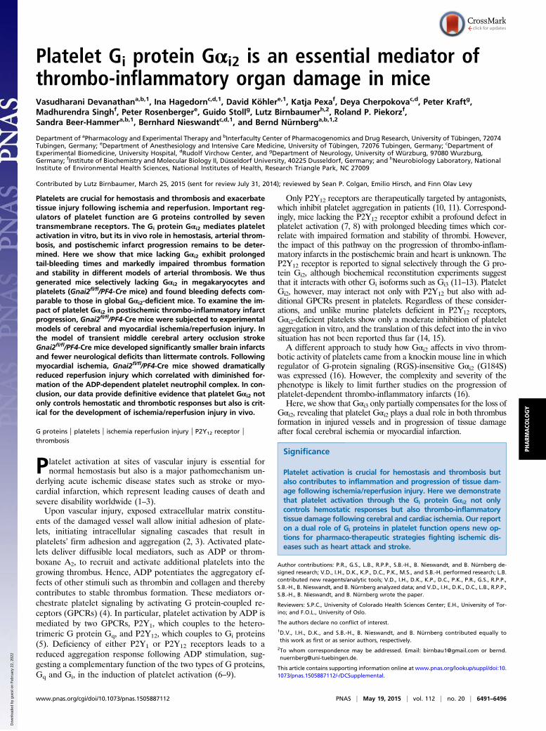
Selective Deficiency of Gαi2 in Platelets Impairs Hemostasis.Next, we as-sessed role of platelet Gαi2 in hemostasis by performing bleeding-time assays. In WT (Gnai2+/+) mice and mostGnai2fl/fl control mice,tail bleeding stopped within 10 min (Fig. S7H). In contrast, themajority of Gnai2fl/fl/PF4-Cre+ mice showed greatly prolongedtail bleeding similar to that in global Gαi2-deficient mice. A secondbleeding assay in which the tip-amputated tail was inserted into atube with PBS confirmed prolonged bleeding (Fig. 3B) and fluc-tuations in bleeding intensity (Movies S3 and S4). In conclusion,selective deletion of Gαi2 in platelets and megakaryocytes is suf-ficient to produce a pronounced bleeding defect.
Platelet-Specific Gαi2 Deficiency Results in Neuroprotection FollowingCerebral Ischemia/Reperfusion Injury. Platelets are thought tointegrate pathways that orchestrate both thrombotic and in-flammatory processes, resulting in a thrombo-inflammatorycascade in acute ischemic disease states in which tissue damageoccurs despite successful recanalization of occluded vessels, aprocess referred to as “reperfusion injury” (3, 35, 36). To eval-uate the impact of platelet Gαi2 on infarct progression, wechallenged Gnai2fl/fl/PF4-Cre mice in the tMCAO model inwhich a filament reversibly occludes the middle cerebral arteryand reduces the regional cerebral flow by >90% (25). After 1 h thefilament is removed to allow reperfusion for 24 h. Subsequently,mice are tested for their motor and coordination functions.Interestingly, assessment of the global neurologic function(Bederson score) (Fig. 4A) revealed that Gnai2fl/fl/PF4-Cre micedeveloped significantly fewer neurologic deficits and had signifi-cantly better (higher) scores in motor function tests (Fig. 4B) thanlittermate controls. These findings were accompanied by a re-duction in infarct volumes by >60% in Gnai2fl/fl/PF4-Cre mice
0 300 600 900 1200 1500 1800
Blo
od fl
ow [m
in]
00.20.40.60.81.01.21.41.6
Time [sec]
wtGαi2
-/-Gαi3
-/-
Gαi2-/- Gαi3
-/-
A
Tim
e to
occ
lusi
on [s
ec]
0
400
800
1200
1600
>1800
wt
B
0 min 10 min 20 min 40 min
wt *
C
D
E
Beg
inni
ng o
f thr
ombu
s fo
rmat
ion
[min
]
0
5
10
15
20
wt Gαi2-/-
Tim
e to
ves
sel
occl
usio
n [m
in]
0
10
20
30
40>40
wt Gαi2-/-Gαi2
-/-
Fig. 2. Defective thrombus formation in global Gαi2-deficient mice. (A and B) The abdominal aorta of WT (n = 10), Gαi2-deficient (n = 7), and Gαi3-deficientmice (n = 6) was injured by tight compression with a forceps, and blood flow was monitored for 30 min. (A) Representative blood-flow recordings. (B) Time tostable vessel occlusion. Each symbol represents one mouse. Mean times to occlusion were 6.1 ± 3.4 min for WT mice and 4.5 ± 1.0 min for Gαi3−/− mice (P >0.05; Welch test). Gαi2−/− mice showed no occlusion within the observation time. (C) Thrombus formation after FeCl3-induced injury of mesenteric arterioleswas monitored with intravital fluorescence microscopy (see also Movies S1 and S2). Representative images were taken at the indicated time points. (Scale bar:100 μm.) The asterisk indicates stable occlusion of the vessel. (D) Thrombus formation began at 7.2 ± 1.7 min for arterioles from WT mice (n = 8) and at 8.9 ±2.9 min for arterioles from Gαi2−/− mice (n = 9); P > 0.05; Welch test. (E) Time to stable occlusion was 17.9 ± 3.0 min for arterioles fromWTmice (n = 9), but 7 of10 arterioles from Gαi2−/− mice showed no occlusion within the observation time. Each symbol represents one vessel.
Devanathan et al. PNAS | May 19, 2015 | vol. 112 | no. 20 | 6493
PHARM
ACO
LOGY
Dow
nloa
ded
by g
uest
on
Feb
ruar
y 22
, 202
2
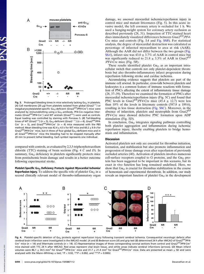
compared with controls, as evaluated by 2,3,5-triphenyltetrazoliumchloride (TTC) staining of brain sections (Fig. 4 C and D). Insummary, Gαi2 deficiency in platelets significantly protects micefrom postischemic brain damage and results in a better outcomefollowing experimental stroke.
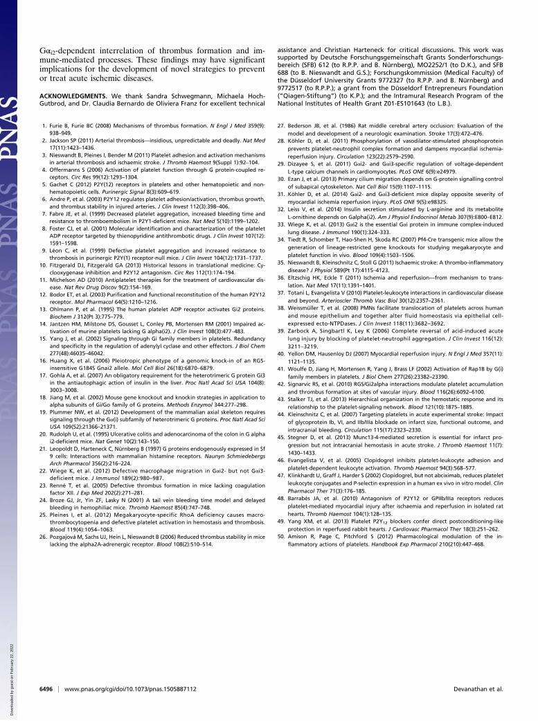
Platelet-Specific Gαi2 Deficiency Protects Against Myocardial Ischemia/Reperfusion Injury. To address the specific role of platelet Gαi2 in asecond clinically relevant model of thrombo-inflammatory organ
damage, we assessed myocardial ischemia/reperfusion injury incontrol mice and mutant littermates (Fig. 5). In this acute in-vasive model, the left coronary artery is occluded for 1 h. Weused a hanging-weight system for coronary artery occlusion asdescribed previously (28, 31). Inspection of TTC-stained heartdiscs immediately visualized differences between Gnai2fl/fl/PF4-Cre mice and controls (Fig. 5A and Fig. S8B). For statisticalanalysis, the degree of myocardial destruction was calculated aspercentage of infarcted myocardium to area at risk (AAR).Although the AAR did not differ between the two groups (Fig.S8A), infarct size was 43.0 ± 3.7% of AAR in control mice butwas significantly reduced to 21.8 ± 3.3% of AAR in Gnai2fl/fl
/PF4-Cre mice (Fig. 5B).These results identified platelet Gαi2 as an important intra-
cellular switch that controls not only platelet-dependent throm-bosis but also thrombo-inflammatory infarct progression duringreperfusion following stroke and cardiac ischemia.Accumulating evidence suggests that platelets are part of the
immune cell arsenal. In particular, cross-talk between platelets andleukocytes is a common feature of immune reactions with forma-tion of PNCs affecting the extent of inflammatory tissue damage(28, 37–39). Therefore we examined the formation of PNCs aftermyocardial ischemia/reperfusion injury (Fig. 5C) and found thatPNC levels in Gnai2fl/fl/PF4-Cre mice (45.4 ± 12.7) were lessthan 10% of the levels in littermate controls (587.8 ± 189.6),resulting in less tissue destruction (Fig. S8C). Moreover, in theabsence of infarction, platelets and neutrophils from Gnai2fl/fl
/PF4-Cre mice showed defective PNC formation upon ADPstimulation (Fig. 5D).In conclusion, Gαi2 integrates signaling pathways controlling
both platelet aggregation and inflammation during ischemia/reperfusion injury, thereby enabling platelets to bridge hemo-stasis and inflammation.
DiscussionActivated platelets not only are essential for thrombus initiation,formation, and stabilization but also promote inflammation andprogression of tissue damage even after reperfusion of previouslyoccluded arteries (40). Activation of platelets involves numerouscell-surface receptors coupled to G proteins, and the Gαi2 pro-tein has been suggested to be important in this scenario, but itsexact in vivo function has long remained undefined. Here, weshow that Gαi2 is crucial for thrombus stabilization in the courseof hemostasis and experimental thrombosis. In addition, our studyreveals an important function of platelet Gαi2 in the development
B
A mouse platelets
Ponceau S
43 kDa
+/+ fl/fl -/- fl/fl- - - +
Gnai2Cre
Gαi2
anti-Gαi2
+/+ -/- fl/fl fl/fl- - - +
Gnai2Cre
Ble
edin
gtim
e[s
ec]
400
800
>1200
50100
1200
0
Fig. 3. Prolonged bleeding times in mice selectively lacking Gαi2 in platelets.(A) Cell membranes (20 μg) from platelets isolated from global (Gnai2−/−) ormegakaryocyte/platelet-specific Gαi2-deficient (Gnai2fl/fl/PF4-Cre+) mice wereanalyzed by immunoblotting using a Gαi2-antibody. PF4-Cre–negative litter-mates (Gnai2fl/fl/PF4-Cre−) and WT animals (Gnai2+/+) were used as controls.Equal loading was controlled by staining with Ponceau S. (B) Tail-bleedingtimes of WT (Gnai2+/+) (n = 5), Gαi2-deficient (Gnai2 −/−) (n = 4), Gnai2fl/fl/PF4-Cre− (n = 5), and Gnai2fl/fl/PF4-Cre+ (n = 4) mice measured with the PBSmethod. Mean bleeding time was 42.4 ± 4.5 s for WTmice and 60.2 ± 9.9 s forGnai2fl/fl/PF4-Cre− mice, but in three of four global Gαi2-deficient mice and inall Gnai2fl/fl/PF4-Cre+ mice the bleeding had to be stopped manually after20 min to prevent lethal bleeding. Each symbol represents one individual.
BA
Gnai2 fl/fl fl/fl Cre - +
∗ ∗
Bed
erso
n sc
ore
1
2
3
4
5
0
Grip
test
∗
1
2
3
4
5
0
6
Gnai2 fl/fl fl/fl Cre - +
C
Gnai2 fl/fl fl/flCre - +
D
Infa
rct v
olum
e [m
m3 ]
0
20
40
60
80
100
120
140
Gnai2 fl/fl fl/fl Cre - +
∗ ∗∗
Fig. 4. Platelet-specific deletion of Gαi2 protects against reperfusion injury following transient cerebral ischemia. Consequential neurologic defects aftercerebral brain infarction were investigated in the tMCAO model. (A and B) Bederson score (A) and grip test (B) determined 24 h after tMCAO of Gnai2fl/fl/PF4-Cre+ mice (n = 14) and littermate controls (n = 14). (C) Representative images of three corresponding coronal sections from control and Gnai2fl/fl/PF4-Cre+
mice stained with TTC 24 h after tMCAO. Red areas represent vital brain tissue, and white areas indicate cerebral infarctions (arrows). (D) Mean infarctvolumes were 86.1 ± 34.5 mm3 for Gnai2fl/fl/PF4-Cre− mice and 31.7 ± 21.4 mm3 for Gnai2fl/fl/PF4-Cre+ mice. Data are presented as mean ± SD and wereanalyzed with the Mann–Whitney u test; *P < 0.02, **P < 0.002, and ***P = 0.0002.
6494 | www.pnas.org/cgi/doi/10.1073/pnas.1505887112 Devanathan et al.
Dow
nloa
ded
by g
uest
on
Feb
ruar
y 22
, 202
2

of cerebral and myocardial ischemia/reperfusion injury. Thesefindings emphasize a thrombo-inflammatory role of platelets andput platelet Gαi2 in the center of controlling both hemostasis andinflammation.Previous studies focused on acute responses of isolated
platelets from Gαi KO mice to stimuli, leaving the question onthe in vivo relevance of Gi signaling unanswered (14, 15, 41). Weshow that global Gαi2 but not Gαi3 deficiency produces a markedincrease in bleeding time, although spontaneous hemorrhagewas not evident. The same profound bleeding phenotype wasseen in Gnai2fl/fl/PF4-Cre mice, demonstrating a central role ofplatelet Gαi2 in hemostasis. Of note, a similar primary hemo-static defect is seen in mice lacking the Gi-coupled ADP receptorP2Y12 (5, 6, 8).Initially, we confirmed previous data that Gαi3 deficiency did
not result in a platelet-specific phenotype. However, PTX treat-ment of mice produced a much stronger reduction of plateletfunction than Gαi2 deficiency. Interestingly, and in contrast toprevious reports, we noticed an up-regulation of Gαi3 expressionin Gαi2 KO mice which we identified to be the only PTX sub-strates in platelets (12–15, 41). Therefore, we assume that Gαi3partially compensates for the missing Gαi2. In fact, Gαi3 has beenshown to interact with P2Y12 in a purified reconstituted system(12). Further support for this conclusion comes from the findingthat the specific P2Y12 receptor antagonist ARC69931 blunts theresidual response to ADP in Gαi2-deficient platelets. Taken to-gether, the platelet P2Y12 receptor signals through two Gαi iso-forms with Gαi2 being the pathophysiologically dominant one.In tail-bleeding assays we observed variability in bleeding in-
tensity in Gαi2-deficient mice which paralleled defective forma-tion and stabilization of arterial thrombi seen in two arterialthrombosis models. These findings agree with impaired throm-bus stability seen in P2Y12 receptor-deficient mice (5, 6) and arecomplementary to reports from a heterozygous RGS-insensitiveGαi2 mutant (+/G184S) demonstrating increased platelet accu-mulation and aggregation at vascular injury sites in vivo, al-though the effects on bleeding times in this mouse model werenot reported (42, 43). Thus, Gαi2 represents the dominant sig-naling entity downstream of the ADP-binding P2Y12 receptorwhere either its absence or its uncontrolled increased signalingactivity has a profound impact on thrombus formation and sta-bility, indicating that balanced Gαi2 activity assures normal he-mostasis and thrombosis.
Platelets also contribute to secondary infarct progression byinflammatory mechanisms that may differ from those involved inthrombus formation (3, 36). Interestingly, Gnai2fl/fl/PF4-Cre micedisplayed a significant protection against cerebral ischemia, andthis protection was associated with a marked reduction in neu-rological deficits and better performance in motor functions,highlighting the pathophysiological importance of Gαi2 in plate-lets during reperfusion injury. Interestingly, microscopic analysisof brain slices showed no increased incidence of intracranialhemorrhage in Gnai2fl/fl/PF4-Cre mice following tMCAO, in-dicating that inhibition of Gαi2 signaling may represent a safeapproach to prevent or treat acute stroke. This assumption iscorroborated by data from a second model in whichGnai2fl/fl/PF4-Cre mice also were protected from myocardial ischemia/reper-fusion injury. This finding supports and extends our view thatearly steps in platelet activation and amplification mechanismsare critical factors in infarct development (35, 44, 45).It is noteworthy that the data presented here argue for a
concerted action of activated platelets and leukocytes to ag-gravate infarct progression. Moreover, in both cell types Gαi2seems to represent a central switch of regulation. Although thecentral role of Gαi2 in leukocyte function is well established,our study also indicates a corresponding role for Gαi2 inplatelets. Previously, pharmacological inhibition of the Gi-coupled ADP P2Y12 receptors has been reported to inhibitPNCs and platelet-dependent leukocyte activation (46, 47),whereas other ex vivo or in situ studies suggested that P2Y12receptor blockers decrease cardiac reperfusion injury bymechanisms other than inhibition of intravascular coagulation(48, 49). Our data clearly show that lack of the P2Y12 signal-transducing Gαi2 in platelets results in the reduced formation ofADP-dependent PNCs which correlates with a significant re-duction in cardiac reperfusion injury. Because platelets expressalso other GiPCRs relevant for putative platelet immunefunctions, a future aim will be to dissect the pathophysiologicalrole of Gi proteins for these signaling pathways (50). In thiscontext, it is tempting to speculate about roles for other Gαiproteins, such as Gαi3, which is expressed in platelets, but itsfunction remains to be elucidated.Taken together, our findings reveal that the thrombotic and
inflammatory functions of platelets converge at the level ofthe signal transducer Gαi2. As a consequence, the severity ofstroke or cardiac infarcts is determined by a platelet-intrinsic
A B C D
Gnai2fl/flCre+
Gnai2fl/flCre-
Infa
rct s
ize
[% o
f AAR
]
10
20
30
40
50
60
0
Gnai2 fl/fl fl/fl Cre - +
∗ ∗
Num
ber o
f PN
Cs/
10.0
00 c
ells
200
400
600
800
1000
0
∗∗
Gnai2 fl/fl fl/fl Cre - +
∗∗ ∗∗∗
Gnai2 fl/fl fl/fl fl/fl fl/flCre - - +ADP - + - +
50
100
150
200
0Num
ber o
f PN
Cs/
10.0
00 c
ells
+
Fig. 5. Platelet-specific deletion of Gαi2 protects against reperfusion injury following myocardial ischemia. (A and B) Gnai2fl/fl/PF4-Cre+ mice (n = 7) and controlmice (n = 5) were exposed to 1 h of ischemia and 2 h of reperfusion. Hearts were stained with Evans Blue to determine the AAR and with TTC to mark vital tissue(red) and necrotic tissue (white). (A) Representative heart slices of a Gnai2fl/fl/PF4-Cre+ mouse and a littermate control with infarcted areas of 22% and 43%,respectively. Images were contrasted for better visibility. The original images of these heart slices are shown in Fig. S8B. (B) Infarct size was calculated as per-centage of AAR. Data are shown as mean ± SEM; **P < 0.01 (t test). (C) Number of PNCs after 1 h of ischemia detected in whole blood by flow cytometry inGnai2fl/fl/PF4-Cre+ mice (n = 10) and littermates (n = 9). (D) Number of PNCs per 10,000 cells at basal levels (Gnai2fl/fl/PF4-Cre−: 38.3 ± 12.5; Gnai2fl/fl/PF4-Cre+: 28.4 ±17.7) and after ADP stimulation (Gnai2fl/fl/PF4-Cre−: 146.1 ± 34.6; Gnai2fl/fl/PF4-Cre+: 45.6 ± 13.3). Data are shown as mean ± SEM and were analyzed with ANOVAfollowed by Dunnett’s test; *P < 0.05 and **P < 0.005 compared with Gnai2fl/fl/PF4-Cre− +ADP.
Devanathan et al. PNAS | May 19, 2015 | vol. 112 | no. 20 | 6495
PHARM
ACO
LOGY
Dow
nloa
ded
by g
uest
on
Feb
ruar
y 22
, 202
2
Gαi2-dependent interrelation of thrombus formation and im-mune-mediated processes. These findings may have significantimplications for the development of novel strategies to preventor treat acute ischemic diseases.
ACKNOWLEDGMENTS. We thank Sandra Schwegmann, Michaela Hoch-Gutbrod, and Dr. Claudia Bernardo de Oliviera Franz for excellent technical
assistance and Christian Harteneck for critical discussions. This work wassupported by Deutsche Forschungsgemeinschaft Grants Sonderforschungs-bereich (SFB) 612 (to R.P.P. and B. Nürnberg), MO2252/1 (to D.K.), and SFB688 (to B. Nieswandt and G.S.); Forschungskommission (Medical Faculty) ofthe Düsseldorf University Grants 9772327 (to R.P.P. and B. Nürnberg) and9772517 (to R.P.P.); a grant from the Düsseldorf Entrepreneurs Foundation(“Qiagen-Stiftung”) (to K.P.); and the Intramural Research Program of theNational Institutes of Health Grant Z01-ES101643 (to L.B.).
1. Furie B, Furie BC (2008) Mechanisms of thrombus formation. N Engl J Med 359(9):938–949.
2. Jackson SP (2011) Arterial thrombosis—insidious, unpredictable and deadly. Nat Med17(11):1423–1436.
3. Nieswandt B, Pleines I, Bender M (2011) Platelet adhesion and activation mechanismsin arterial thrombosis and ischaemic stroke. J Thromb Haemost 9(Suppl 1):92–104.
4. Offermanns S (2006) Activation of platelet function through G protein-coupled re-ceptors. Circ Res 99(12):1293–1304.
5. Gachet C (2012) P2Y(12) receptors in platelets and other hematopoietic and non-hematopoietic cells. Purinergic Signal 8(3):609–619.
6. Andre P, et al. (2003) P2Y12 regulates platelet adhesion/activation, thrombus growth,and thrombus stability in injured arteries. J Clin Invest 112(3):398–406.
7. Fabre JE, et al. (1999) Decreased platelet aggregation, increased bleeding time andresistance to thromboembolism in P2Y1-deficient mice. Nat Med 5(10):1199–1202.
8. Foster CJ, et al. (2001) Molecular identification and characterization of the plateletADP receptor targeted by thienopyridine antithrombotic drugs. J Clin Invest 107(12):1591–1598.
9. Léon C, et al. (1999) Defective platelet aggregation and increased resistance tothrombosis in purinergic P2Y(1) receptor-null mice. J Clin Invest 104(12):1731–1737.
10. Fitzgerald DJ, Fitzgerald GA (2013) Historical lessons in translational medicine: Cy-clooxygenase inhibition and P2Y12 antagonism. Circ Res 112(1):174–194.
11. Michelson AD (2010) Antiplatelet therapies for the treatment of cardiovascular dis-ease. Nat Rev Drug Discov 9(2):154–169.
12. Bodor ET, et al. (2003) Purification and functional reconstitution of the human P2Y12receptor. Mol Pharmacol 64(5):1210–1216.
13. Ohlmann P, et al. (1995) The human platelet ADP receptor activates Gi2 proteins.Biochem J 312(Pt 3):775–779.
14. Jantzen HM, Milstone DS, Gousset L, Conley PB, Mortensen RM (2001) Impaired ac-tivation of murine platelets lacking G alpha(i2). J Clin Invest 108(3):477–483.
15. Yang J, et al. (2002) Signaling through Gi family members in platelets. Redundancyand specificity in the regulation of adenylyl cyclase and other effectors. J Biol Chem277(48):46035–46042.
16. Huang X, et al. (2006) Pleiotropic phenotype of a genomic knock-in of an RGS-insensitive G184S Gnai2 allele. Mol Cell Biol 26(18):6870–6879.
17. Gohla A, et al. (2007) An obligatory requirement for the heterotrimeric G protein Gi3in the antiautophagic action of insulin in the liver. Proc Natl Acad Sci USA 104(8):3003–3008.
18. Jiang M, et al. (2002) Mouse gene knockout and knockin strategies in application toalpha subunits of Gi/Go family of G proteins. Methods Enzymol 344:277–298.
19. Plummer NW, et al. (2012) Development of the mammalian axial skeleton requiressignaling through the Gα(i) subfamily of heterotrimeric G proteins. Proc Natl Acad SciUSA 109(52):21366–21371.
20. Rudolph U, et al. (1995) Ulcerative colitis and adenocarcinoma of the colon in G alphai2-deficient mice. Nat Genet 10(2):143–150.
21. Leopoldt D, Harteneck C, Nürnberg B (1997) G proteins endogenously expressed in Sf9 cells: Interactions with mammalian histamine receptors. Naunyn SchmiedebergsArch Pharmacol 356(2):216–224.
22. Wiege K, et al. (2012) Defective macrophage migration in Gαi2- but not Gαi3-deficient mice. J Immunol 189(2):980–987.
23. Renné T, et al. (2005) Defective thrombus formation in mice lacking coagulationfactor XII. J Exp Med 202(2):271–281.
24. Broze GJ, Jr, Yin ZF, Lasky N (2001) A tail vein bleeding time model and delayedbleeding in hemophiliac mice. Thromb Haemost 85(4):747–748.
25. Pleines I, et al. (2012) Megakaryocyte-specific RhoA deficiency causes macro-thrombocytopenia and defective platelet activation in hemostasis and thrombosis.Blood 119(4):1054–1063.
26. Pozgajová M, Sachs UJ, Hein L, Nieswandt B (2006) Reduced thrombus stability in micelacking the alpha2A-adrenergic receptor. Blood 108(2):510–514.
27. Bederson JB, et al. (1986) Rat middle cerebral artery occlusion: Evaluation of themodel and development of a neurologic examination. Stroke 17(3):472–476.
28. Köhler D, et al. (2011) Phosphorylation of vasodilator-stimulated phosphoproteinprevents platelet-neutrophil complex formation and dampens myocardial ischemia-reperfusion injury. Circulation 123(22):2579–2590.
29. Dizayee S, et al. (2011) Gαi2- and Gαi3-specific regulation of voltage-dependentL-type calcium channels in cardiomyocytes. PLoS ONE 6(9):e24979.
30. Ezan J, et al. (2013) Primary cilium migration depends on G-protein signalling controlof subapical cytoskeleton. Nat Cell Biol 15(9):1107–1115.
31. Köhler D, et al. (2014) Gαi2- and Gαi3-deficient mice display opposite severity ofmyocardial ischemia reperfusion injury. PLoS ONE 9(5):e98325.
32. Leiss V, et al. (2014) Insulin secretion stimulated by L-arginine and its metaboliteL-ornithine depends on Galpha(i2). Am J Physiol Endocrinol Metab 307(9):E800–E812.
33. Wiege K, et al. (2013) Gαi2 is the essential Gαi protein in immune complex-inducedlung disease. J Immunol 190(1):324–333.
34. Tiedt R, Schomber T, Hao-Shen H, Skoda RC (2007) Pf4-Cre transgenic mice allow thegeneration of lineage-restricted gene knockouts for studying megakaryocyte andplatelet function in vivo. Blood 109(4):1503–1506.
35. Nieswandt B, Kleinschnitz C, Stoll G (2011) Ischaemic stroke: A thrombo-inflammatorydisease? J Physiol 589(Pt 17):4115–4123.
36. Eltzschig HK, Eckle T (2011) Ischemia and reperfusion—from mechanism to trans-lation. Nat Med 17(11):1391–1401.
37. Totani L, Evangelista V (2010) Platelet-leukocyte interactions in cardiovascular diseaseand beyond. Arterioscler Thromb Vasc Biol 30(12):2357–2361.
38. Weissmüller T, et al. (2008) PMNs facilitate translocation of platelets across humanand mouse epithelium and together alter fluid homeostasis via epithelial cell-expressed ecto-NTPDases. J Clin Invest 118(11):3682–3692.
39. Zarbock A, Singbartl K, Ley K (2006) Complete reversal of acid-induced acutelung injury by blocking of platelet-neutrophil aggregation. J Clin Invest 116(12):3211–3219.
40. Yellon DM, Hausenloy DJ (2007) Myocardial reperfusion injury. N Engl J Med 357(11):1121–1135.
41. Woulfe D, Jiang H, Mortensen R, Yang J, Brass LF (2002) Activation of Rap1B by G(i)family members in platelets. J Biol Chem 277(26):23382–23390.
42. Signarvic RS, et al. (2010) RGS/Gi2alpha interactions modulate platelet accumulationand thrombus formation at sites of vascular injury. Blood 116(26):6092–6100.
43. Stalker TJ, et al. (2013) Hierarchical organization in the hemostatic response and itsrelationship to the platelet-signaling network. Blood 121(10):1875–1885.
44. Kleinschnitz C, et al. (2007) Targeting platelets in acute experimental stroke: Impactof glycoprotein Ib, VI, and IIb/IIIa blockade on infarct size, functional outcome, andintracranial bleeding. Circulation 115(17):2323–2330.
45. Stegner D, et al. (2013) Munc13-4-mediated secretion is essential for infarct pro-gression but not intracranial hemostasis in acute stroke. J Thromb Haemost 11(7):1430–1433.
46. Evangelista V, et al. (2005) Clopidogrel inhibits platelet-leukocyte adhesion andplatelet-dependent leukocyte activation. Thromb Haemost 94(3):568–577.
47. Klinkhardt U, Graff J, Harder S (2002) Clopidogrel, but not abciximab, reduces plateletleukocyte conjugates and P-selectin expression in a human ex vivo in vitro model. ClinPharmacol Ther 71(3):176–185.
48. Barrabés JA, et al. (2010) Antagonism of P2Y12 or GPIIb/IIIa receptors reducesplatelet-mediated myocardial injury after ischaemia and reperfusion in isolated rathearts. Thromb Haemost 104(1):128–135.
49. Yang XM, et al. (2013) Platelet P2Y12 blockers confer direct postconditioning-likeprotection in reperfused rabbit hearts. J Cardiovasc Pharmacol Ther 18(3):251–262.
50. Amison R, Page C, Pitchford S (2012) Pharmacological modulation of the in-flammatory actions of platelets. Handbook Exp Pharmacol 210(210):447–468.
6496 | www.pnas.org/cgi/doi/10.1073/pnas.1505887112 Devanathan et al.
Dow
nloa
ded
by g
uest
on
Feb
ruar
y 22
, 202
2
Related Documents