HAL Id: hal-00585781 https://hal.archives-ouvertes.fr/hal-00585781 Submitted on 14 Apr 2011 HAL is a multi-disciplinary open access archive for the deposit and dissemination of sci- entific research documents, whether they are pub- lished or not. The documents may come from teaching and research institutions in France or abroad, or from public or private research centers. L’archive ouverte pluridisciplinaire HAL, est destinée au dépôt et à la diffusion de documents scientifiques de niveau recherche, publiés ou non, émanant des établissements d’enseignement et de recherche français ou étrangers, des laboratoires publics ou privés. Platelet function in rheumatoid arthritis: arthritic and cardiovascular implications Armen Yuri Gasparyan, Antonios Stavropoulos-Kalinoglou, Dimitri P. Mikhailidis, Karen M. J. Douglas, George D. Kitas To cite this version: Armen Yuri Gasparyan, Antonios Stavropoulos-Kalinoglou, Dimitri P. Mikhailidis, Karen M. J. Dou- glas, George D. Kitas. Platelet function in rheumatoid arthritis: arthritic and cardiovascular implica- tions. Rheumatology International, Springer Verlag, 2010, 31 (2), pp.153-164. 10.1007/s00296-010- 1446-x. hal-00585781

Welcome message from author
This document is posted to help you gain knowledge. Please leave a comment to let me know what you think about it! Share it to your friends and learn new things together.
Transcript
HAL Id: hal-00585781https://hal.archives-ouvertes.fr/hal-00585781
Submitted on 14 Apr 2011
HAL is a multi-disciplinary open accessarchive for the deposit and dissemination of sci-entific research documents, whether they are pub-lished or not. The documents may come fromteaching and research institutions in France orabroad, or from public or private research centers.
L’archive ouverte pluridisciplinaire HAL, estdestinée au dépôt et à la diffusion de documentsscientifiques de niveau recherche, publiés ou non,émanant des établissements d’enseignement et derecherche français ou étrangers, des laboratoirespublics ou privés.
Platelet function in rheumatoid arthritis: arthritic andcardiovascular implications
Armen Yuri Gasparyan, Antonios Stavropoulos-Kalinoglou, Dimitri P.Mikhailidis, Karen M. J. Douglas, George D. Kitas
To cite this version:Armen Yuri Gasparyan, Antonios Stavropoulos-Kalinoglou, Dimitri P. Mikhailidis, Karen M. J. Dou-glas, George D. Kitas. Platelet function in rheumatoid arthritis: arthritic and cardiovascular implica-tions. Rheumatology International, Springer Verlag, 2010, 31 (2), pp.153-164. �10.1007/s00296-010-1446-x�. �hal-00585781�
1
Platelet Function in Rheumatoid Arthritis: Arthritic and Cardiovascular
Implications
1 Armen Yuri Gasparyan MD, PhD, Research Fellow
1 Antonios Stavropoulos-Kalinoglou PhD, Research Fellow
2 Dimitri P Mikhailidis, MD, FFPM, FRCP, FRCPath, Academic Head, Dept. of
Clinical Biochemistry
1,3 Tracey E Toms MBChB, MRCP, Research Fellow
1 Karen MJ Douglas BSc, MBChB, MD, MRCP, Consultant Rheumatologist
1,3 George D Kitas MD, PhD, FRCP, Professor of Rheumatology
1 Department of Rheumatology, Clinical Research Unit, Russells Hall Hospital, Dudley
Group of Hospitals NHS Foundation Trust, Dudley DY1 2HQ, West Midlands, United
Kingdom
2 Department of Clinical Biochemistry (Vascular Prevention Clinic), Royal Free Hospital,
University College London Medical School, University College London (UCL), London,
United Kingdom
3 Arthritis Research Campaign (ARC) Epidemiology Unit, University of Manchester,
Manchester, United Kingdom
Correspondence to:
Armen Yuri Gasparyan, MD, PhD
Dudley Group of Hospitals NHS Foundation Trust (Teaching)
Russells Hall Hospital, Dudley, West Midlands DY1 2HQ
United Kingdom
Tel. No. +44-1384-244842
Fax No. +44-1384-244808
E-mail: [email protected]
2
Abstract
Patients with rheumatoid arthritis (RA) are at high risk of cardiovascular events. Platelet
biomarkers are involved in inflammation, atherosclerosis and thrombosis. Cardiovascular
and RA-associated factors can alter the structure and function of platelets, starting from
megakaryocytopoiesis. Reactive megakaryocytopoiesis increases circulating platelets count
and triggers hyperactivity. Hyperactive platelets target synovial membranes with subsequent
local rheumatoid inflammation. Hyperactive platelets interact with other cells, and target the
vascular wall. Accumulating evidence suggests that disease modifying anti-rheumatic drugs
(DMARD) decrease platelet activity.
Key words: Rheumatoid arthritis, Platelet function, Biomarkers, Inflammation,
Cardiovascular risk.
3
INTRODUCTION
Rheumatoid Arthritis (RA) is the most common inflammatory polyarthritis. RA affects
approximately 1% of the adult western populations [1] and is characterized by inflammation
of varying intensity with progressive destruction of synovial joints and physical disability. It
has become evident that patients with RA are burdened with excessive cardiovascular
disease (CVD). Chronic inflammation is implicated in the high prevalence of CVD risk
factors (e.g., hypertension, dyslipidemia, diabetes), premature atherosclerosis and altered
coagulation [2]. Atherosclerotic CVD in RA is being increasingly recognized as an extra-
articular manifestation and as a model for research, diagnosis and treatment of
atherothrombosis [3].
Considerable evidence indicates that patients with RA are prone to premature ischaemic
heart disease (IHD), myocardial infarction (MI), and heart failure. There are multiple
pathways that link these conditions, one being thrombosis [4-6]. A large cross-sectional
study, which compared the prevalence of CVD and cerebrovascular disease in 9093 RA and
2479 osteoarthritis (OA) patients in the USA, revealed that RA was associated with an
increased lifetime risk for MI (odds ratio [OR] 1.28, 95% confidence interval [95% CI] 1.24,
1.33) and heart failure (OR 1.43, 95% CI 1.28, 1.59) [7]. The Rochester Epidemiology
Project, a large population-based inception cohort, followed 603 RA patients for almost 40
years, and demonstrated that CVD, the leading cause of morbidity and mortality in RA,
accounted for 49.7% of deaths [2]. Throughout follow-up, mortality rates were significantly
higher in subjects with RA than without (39.0 vs. 29.2/1,000 person-years) [8]. It also
revealed a striking difference in the cumulative incidence of heart failure between RA and
non-RA populations at 30 years of follow-up (37.1% vs. 27.7%, p<0.001), emphasizing how
heart failure, a prothrombotic state [9-10], contributes to the excess mortality in RA.
The high prevalence of established CVD risk factors, such as smoking, hypertension,
diabetes, only play a part in the increase in CVD [11]. Inflammatory mediators, endothelial
dysfunction, and coagulation have also been extensively studied in RA [12].
Pathogenic factors that associate with active synovitis, bone and cartilage destruction in RA
are present in high-grade inflammatory conditions and atherosclerosis [13-16]. Such factors
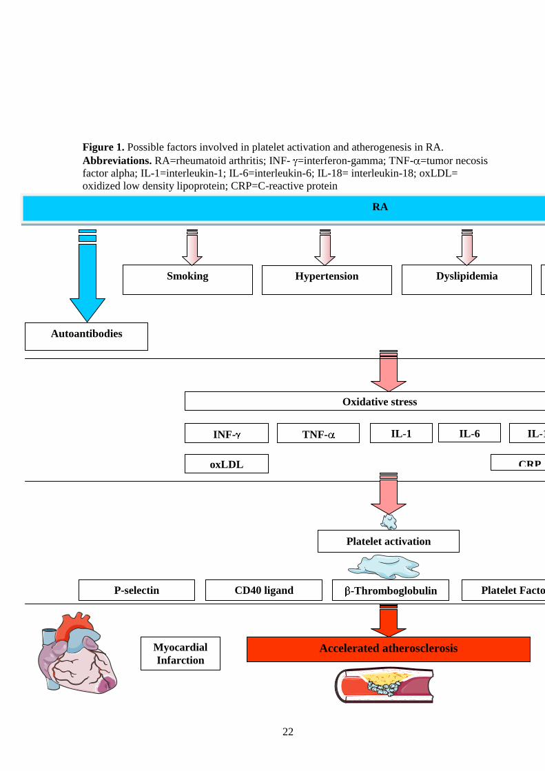
may create an environment in which platelet activation amplifies CVD risk. A few of the
known potential ‘platelet agonists’ include: oxidative stress, hyperinsulinemia, oxidized low
density lipoprotein (oxLDL), C-reactive protein (CRP), tumor necrosis factor alpha (TNFα),
4
interleukins -1, -6, -18, RANK ligand, CD40 ligand, matrix metalloproteinases, monocyte
chemotactic protein-1, fractalkine, and adipocytokines (Fig. 1) [2-5, 12-16].
In this review we discuss platelet function in RA and how this relates to CVD pathology.
SEARCH STRATEGY
The literature search was carried out in MEDLINE for English-language original research
papers published from 1981 to 2009 using the following search terms related to the markers
of platelet activation and RA: platelet function, atherothrombosis, cardiovascular disease,
cardiovascular risk, rheumatoid arthritis, synovial fluid, rheumatoid synovium, mean platelet
volume, P-selectin, platelet aggregation, platelet count, platelet microparticles, gene
polymorphisms, CD40 signaling, platelet chemokines, disease modifying antirheumatic
drugs. The reference lists of the selected articles were also hand searched to identify
important and highly cited reviews on platelet function in inflammation.
PLATELET ACTIVATION WITHIN JOINTS IN RA
Though there is little evidence that platelets are directly involved in joint inflammation in
RA, studies have shown an increased number of platelets and platelet-derived proteins
(growth factors) within the synovium and synovial fluid [17-20]. High platelet counts in
synovial fluid associate with rheumatoid factor (RF) and markers of synovial leukocyte
activation in inflammatory arthritis, but not osteoarthritis [18, 21].
In RA, activated platelets, alone or together with other inflammatory cells and mediators,
may play a significant role in thrombus formation, synovial microcirculation, and destruction
of cartilage [22-25]. In a murine model of knee joint arthritis, platelets were found to interact
with and adhere to endothelial cells (ECs) and leukocytes in the inflamed synovial vessels.
Subsequent platelet aggregation leads to thrombus formation and alteration of the synovial
microcirculation [22]. In a series of experiments in antigen-induced arthritis, P-selectin, an
adhesion molecule produced by platelets and ECs, was shown to be crucial for the
interaction of platelets, leukocytes, and ECs in the inflamed joints [23-24]. Other platelet-
derived proteins also exhibited pro-inflammatory and joint destructive actions. In fact,
platelet-derived growth factor, a potent angiogenic agent, was shown to induce synovitis and
pannus-like hyperplasia in a rabbit model of RA [25].

5
Further evidence on platelet involvement in rheumatoid synovitis relates to the presence of
platelet factor 4 in the synovial fluid, a chemokine and thrombotic agent with capacity to
bind to antithrombin III and neutralize heparin [19]. Compared with circulating platelets, the
surface of platelets in the synovial fluid contains higher levels of platelet factor 4; this
suggests migration of circulating platelets and targeted action against rheumatoid joints [19].
Alternatively, constituents of rheumatoid synovial fluid may recruit platelets from the
circulation and facilitate their prothrombotic and proinflammatory effects within the
synovium [26-27]. Evidence suggests an association between platelet reactive IgG antibodies
in the synovial fluid and heightened platelet activity [27].
To sum up, alpha granules and dense bodies of platelets, activated by systemic rheumatoid
inflammation, may release their own inflammatory and immune mediators, facilitating
initiation and propagation of synovitis. Though precise mechanisms of platelet activation are
not fully understood, it is plausible that inhibition of platelets with subsequent decrease of
platelet-derived inflammatory markers may have beneficial effect on the course of arthritis.
ACTIVATION OF CIRCULATING PLATELETS AND POTENTIAL
IMPLICATIONS IN RA
Platelets are an important component of thrombogenesis and are involved in inflammation,
endothelial dysfunction and atherogenesis [28].
A normal blood platelet count ranges from 150-400×109/L. A platelet’s lifespan is 8-10 days,
and normal daily release from the bone marrow is about 1011
(this may increase 10-fold in
conditions of increased platelet turnover) [29]. Although megakaryocytes contain a nucleus
and the whole biosynthetic apparatus, their offspring, newly formed ‘reticulated platelets’ are
anucleate. However, for at least 24 hours after the release from the bone marrow, young
platelets contain messenger RNA (mRNA), facilitating synthesis of platelet proteins [30]. An
intensive stimulation of the bone marrow and increased platelet turnover may occur in
response to an excessive production of inflammatory cytokines with the resultant increase of
reticulated platelets. These platelets produce proteins, which lead to clot formation [31].
During activation, aggregation, adhesion to other cells and thrombotic plug formation
platelets undergo shape and volume change. Aggregation is associated with the release of
platelet-derived vasoactive and hemostatic substances [32].

6
Mean platelet volume (MPV)
The complexity of platelet physiology and its inter-relations with other biomarkers make it
difficult to assess links between platelet activation, inflammation and atherogenesis. Even
interpretation of changes in MPV [33] is not always straightforward in RA.
Availability of automated blood cell analyzers has made the measurement of platelet count
and morphology common practice. MPV is emerging as an indicator of platelet reactivity,
which could estimate cardiovascular risk [34]. In physiological conditions, an increased
MPV reflects the predominance of young reticulated platelets in the circulation due to
increased platelet turnover. MPV has been viewed as a reflection of activation when they
transform from normal discoid to spheric shape with protrusion of pseudopodia and increase
in the size [35-37]. Large platelets are associated with higher levels of IgG antibodies against
platelet membrane glycoproteins IIb/IIIa, Ib and V [38] and an increased release of
thromboxane A2, beta-thromboglobulin and P-selectin [35,36,39]. Elevated MPV has been
linked with heightened risk in patients with hypertension, obesity, diabetes, smoking, and
hypercholesterolemia [40,41]. Furthermore, increased MPV is related to acute vascular
events such as destabilization of atherosclerotic plaque, unstable angina, MI and paroxysmal
atrial fibrillation [40,42]. MPV is an independent risk factor and predictor of MI in
predisposed subjects [43,44]. In a large prospective cohort of patients with established
cerebrovascular disease (n=3134), MPV predicted stroke within 4 years of follow-up with an
11% increased relative risk of stroke with each femtoliter increment of MPV [45]. A recent
study identified significantly high MPV in patients with familial Mediterranean fever [46],
suggesting a link between inflammation, platelet activation, and prothrombotic state.
MPV changes have been observed in some but not all studies in RA [47-48] (Table 1). In one
study, MPV of 32 active RA patients was significantly lower than that of patients with
osteoarthritis and healthy subjects. This finding was accompanied by increased disease
activity, measured by Disease Activity Score 28 (DAS28), platelet count and biomarkers of
inflammation, which suggested that platelet activation in RA is associated with reactive
megakaryocytopoiesis [48] as part of active inflammation [49-50]. Small MPV may also
reflect accelerated maturation and short lifespan of platelets in active RA. In contrast, in
another prospective study [47], MPV significantly decreased alongside CRP, IL-6 and
platelet count in response to a 2-year anti-rheumatic treatment, questioning the inverse
correlation between MPV and thrombocytosis, observed by others in RA [48] and
inflammatory bowel disease [51]. These results emphasize the need for larger studies to

7
clarify the discrepancies. An additional challenge relates to the methodological issues of
MPV measurement, which is not a static variable and ranges widely with changing profiles
of endogenous platelet agonists, treatment modalities, blood sampling, and storing.
Measuring platelet aggregation
A relatively simple test of platelet function is platelet aggregometry. This can be performed
using platelet-rich plasma or whole blood and different platelet agonists (e.g., arachidonic
acid, adenosine diphosphate [ADP], collagen, thrombin, epinephrine and modified
immunoglobulins) [52]. The agonist is added to the suspension, and a dynamic measure of
platelet clumping is recorded. Platelet aggregometry is widely considered as a ‘gold
standard’ of platelet function assessment. As an in-vitro test, it has limitations (e.g., absence
of interaction with other blood cells, artefacts occurring during sampling, centrifugation and
platelets separation). Whole blood platelet count-based aggregometry overcomes some of
these limitations [52].
In spite of the limitations, some studies have proven platelet aggregometry to be useful for
detecting hyperactivity of platelets in RA [53] and assessing efficiency of anti-rheumatic
drugs [54,55].
Platelet aggregometry studies identified triggers of excessive platelet aggregation. Examples
are rheumatoid seropositivity, antibodies against beta-2-microglobulin and circulating
immune complexes (another link between autoimmune reactions and prothrombotic state in
RA) [53,56,57]. Interestingly, increased in-vitro platelet sensitivity to agonists and
autoimmune factors has also been found in other rheumatic diseases (e.g., polymyalgia
rheumatica, systemic sclerosis, gout), where common mechanisms of immune complex
formation and accelerated atherogenesis can be inhibited by antiplatelets [58].
Thrombocytosis (>400×109/L), characteristic for active RA, has been associated with
increased sensitivity to platelet agonists, such as collagen and epinephrine, suggesting a
pathological link between thrombocytosis and the arachidonic acid cascade [59]. A crucial
role of arachidonic acid metabolites is also evident in the light of normal sensitivity to ADP,
observed in platelet aggregation studies in RA [50].
Soluble P-selectin
8
The concentration of soluble P-selectin in plasma reflects in-vivo platelet activation [60-62].
Platelets, ECs and macrophages are all sources of this glycoprotein [63]. Its high plasma
concentrations are largely associated with excessive release from alpha granules of platelets
rather than from other sources [64,65]. Soluble P-selectin facilitates interaction of platelets
with T-lymphocytes, neutrophils, monocytes and ECs at the sites of rheumatoid
inflammation [23]. Cellular interactions require active participation of another adhesion
molecule, P-selectin Glycoprotein Ligand-1 (PSGL-1), expressed on activated platelets,
lymphocytes, monocytes and leukocytes [66]. By binding PSGL-1, P-selectin mediates
adhesion of platelets and formation of complexes with leukocytes or monocytes. A further
action of bound P-selectin is the up-regulation of other adhesion molecules, and the tethering
and rolling of leukocytes into the endothelium [67]. In collagen-induced murine arthritis,
disruption of P-selectin/PSGL-1 complex by PSGL-1 binding antibodies markedly
suppressed leukocyte recruitment into the inflamed synovium and down-regulation of TNF
synthesis by synoviocytes [68].
Not surprisingly, elevated levels of soluble P-selectin correlate with acute phase reactants
and reflect the intensity of systemic inflammation in RA [60-62,69,70]. In fact, of a number
of adhesion molecules, only P-selectin significantly correlates with RA activity [61].
Additionally, P-selectin associates with MPV [47] and platelet count [69], which implicates
the role of circulating platelets in the elevation of P-selectin concentrations in RA. The role
of P-selectin expression may vary widely, depending on the presence of articular and extra-
articular rheumatoid manifestations. This is why in some observations soluble P-selectin
levels were close to normal [71] or there were no associations with shifts of immune markers
(e.g., soluble IL-2 receptor, a modulating protein of T-lymphocyte activity) [61].
Several studies have failed to provide evidence of direct involvement of soluble P-selectin in
vasculitis, atherosclerotic disease or myocardial dysfunction in RA [33,62,70]. However,
these studies had some limitations, such as inappropriate case-control design, small number
of patients, lack of representative population of patients, neglect of confounding effects of
acute-phase reactants interacting with P-selectin, or lack of correction required in the case of
therapy with DMARDs and cardiovascular drugs, known to inhibit platelet function.
Flow cytometry analysis of platelet membrane-bound proteins
9
Flow cytometry has expanded the opportunities for comprehensive assessment of platelet
activity [72-74]. Using specific monoclonal antibodies against different proteins of platelets
and other cells, flow cytometry assesses markers of degranulation of alpha granules
(CD62P), lysosomes, dense bodies (CD63) of platelets, conformational changes of platelet
receptors GP IIb/IIIa and Ib, the presence of circulating complexes of platelets with
leukocytes and monocytes and platelet-derived microparticles in autoimmune diseases [75].
Flow cytometry analysis is instrumental in monitoring long-term effects of drugs on platelet
function [76]. The number of P-selectin (CD62P) positive cells detected by flow cytometry
strongly correlates with RA activity and inflammatory markers [33,77]. This reciprocates the
results of ELISA tests, implying overproduction of soluble P-selectin due to platelet
involvement in rheumatoid inflammation.
Platelet-bound P-selectin overexpression has been associated with hypertension, diabetes and
heart failure [78-80]. A retrospective cohort study with 517 subjects with diabetes,
hypertension and hyperlipidemia revealed positive correlation of platelet-bound P-selectin
with intimal-medial thickness (IMT), atherosclerotic plaques and stiffness of the carotid
arteries [81]. Similar associations might be expected with cardiovascular co-morbidities in
RA.
Platelet-derived microparticles (PDM)
P-selectin is intimately related to the functioning of PDM in RA [82]. PDM have been
increasingly recognized as markers of platelet activation and potent prothrombotic agents.
PDM increase in quantity and activity in conditions associated with oxidative stress,
autoimmunity and thrombosis (e.g., antiphospholipid syndrome, acute coronary syndromes,
venous thromboembolism, and sepsis) [83].
Flow cytometry can track the origin of microparticles. These are tiny vesicles (0.1-1 µm)
with a complex array of proteins on the surface, which originate from platelets, ECs,
monocytes, lymphocytes, and leukocytes. Sequestration of microparticles occurs due to
platelet activation, aggregation, interaction with leukocytes, monocytes, Ecs, and
spontaneously. High concentrations of PDM have been found in active RA independent of
platelet count, suggesting a relation with activating factors other than thrombocytosis [82].
An interesting hypothesis was proposed that circulating PDM play a more important
pathogenic role than the same PDM within the inflamed rheumatoid joints [84]. Actually,

10
PDM from rheumatoid platelets are predominantly found in the plasma, while more
microparticles in the rheumatoid synovial fluid originate from leukocytes or monocytes [85].
It is likely that inflammatory and thrombogenic targets of PDM differ at various stages of
RA (i.e., migration and rolling of circulating microparticles into the inflamed synovium at
the initial active stage, and opposite migration toward systemic circulation with advancing
joint inflammation). This might partly explain why despite a significant elevation and
correlation of circulating PDM with DAS28 in some studies [82], normal levels of the same
PDM are observed in others, where high levels of CD62P, platelet-monocyte complexes and
soluble CD40L suggested active RA [33].
CD40 Ligand/CD40 complex
CD40 ligand (CD40L), a member of the TNF family, is expressed on the platelet membrane
via arachidonic acid-mediated activation [85]. More than 90% of soluble CD40L is produced
by activated platelets [85]. CD40L binds to platelet GPIIb/IIIa to stabilize arterial thrombus.
Platelet-derived soluble CD40L also binds to its cognate receptor CD40 constitutively
expressed on T-, B-lymphocytes and monocytes, thereby facilitating inflammation [66]. The
CD40L/CD40 complex stimulates the release of chemokines, such as Regulated upon
Activation, Normal T cell Expressed and Secreted (RANTES) and Monocyte
Chemoattractant Protein-1 (MCP-1) from platelets and through this link reinforces T-
lymphocyte-mediated immune reactions [86-88]. In experimental studies, CD40L/CD40
interaction was shown to up-regulate IgG RF production by B-lymphocytes, which was
blocked by administration of CD40L neutralizing antibodies [89]. A strong correlation
between soluble CD40L and IgM/IgG RF was also found in RA patients [90]. CD40L
through the recruitment of leukocytes and other inflammatory cells to the sites of vascular
injury may also be involved in endothelial dysfunction. Although increased levels of soluble
CD40L were observed in active RA [33] and rheumatoid vasculitis [90], interactions of
CD40L/CD40 with other markers of platelet activation, inflammation, autoimmunity,
thrombosis and vascular risk in RA remain obscure.
GENETIC MARKERS OF PLATELET HYPERACTIVITY IN RA
Genetics may be relevant to platelet hyperactivity in RA. Platelet membrane GP IIIa
polymorphism with antigen Ib positivity is a likely genetic factor to predispose to
11
atherothrombotic events. Positive Ib allele was found in one third of RA patients [91]. It is
well known that the threshold of platelet activation in healthy persons and patients carrying
Ib allele is significantly decreased. RA patients carrying this allele have significantly more
circulating CD41a (GPIIb/IIIa) positive platelet aggregates and exhibit an enhanced platelet
response to ADP, compared with patients without this allele. Moreover, the difference in
response to different concentrations of ADP (0.5, 1 and 2 µMol) between Ib allele-carriers
and non-carriers was confined to the 0.5 µMol concentration, which represents the ADP
concentration inducing platelet aggregation not only through the activation of ADP
receptors, but also via the arachidonic acid cascade. Thus, many RA patients, even those
taking anti-platelet drugs, may exhibit platelet hyperactivity. The studies on GPIIIa
polymorphism in RA may provide a useful tool for stratifying patients at high risk of
cardiovascular events and selecting candidates for aggressive anti-platelet therapy.
Polymorphism of another protein from alpha granules of platelets, transforming growth
factor-beta1 (TGF-beta1), also merits consideration. Many cells release TGF-beta1 into the
circulation but platelets are the most important contributors. This cytokine possesses anti-
inflammatory properties, preserving endothelial integrity, avoiding excessive destruction of
the connective tissue and progression of atherosclerosis [92]. TGF-beta1 deficiency
accelerates atherosclerosis. High concentrations of TGF-beta1 are observed in inflammatory
and prothrombotic states and may, through negative feedback, suppress further activation of
platelets. Recently, it was shown that platelet-derived TGF-beta1 enhances osteoclastic
activity of the bone by activating Receptor Activator of NF-kappaB Ligand (RANKL),
bridging platelet function with rheumatoid osteopathy [93].
The T-allele of T869C polymorphism is associated with reduced TGF-beta and high risk of
MI in the general population. This single nucleotide polymorphism is also linked with the
risk of RF positivity, RA development, joint damage, hypertension and mortality in RA [94].
Based on the obtained data, genetically determined platelet dysfunction is a probable
pathophysiological link between rheumatoid and cardiovascular pathology.
ANTI-RHEUMATIC TREATMENT AND PLATELET FUNCTION
Methotrexate has proven efficacy to slow progression of RA, and it is largely recommended
as the drug of choice for patients with early and advanced RA. In a case-control study of 613
RA patients with or without CVD, those who had ever received methotrexate had
12
significantly reduced risk of CVD (OR 0.11, 95%CI 0.02-0.56; p<0.05). Similar trends were
observed in those who had ever received sulfasalazine and/or hydroxychloroquine in
combination with methotrexate [95]. In a landmark prospective study of 1240 RA patients,
191 fatal events were observed over 6 years of follow-up, of which 44% were due to CVD.
Weighted Cox regression analysis revealed substantial benefits of low-dose methotrexate in
terms of CVD mortality reduction (Hazard Ratio [HR] 0.3, 95%CI 0.2-0.7, compared with
no methotrexate) [96]. This significant decrease of CVD mortality risk is not observed with
other traditional DMARDs, such as sulfasalazine (HR 1.3, 95%CI 0.7-2.5 for those
prescribed DMARDs other than methotrexate). The beneficial effect of methotrexate was
independent of dose and folic acid supplementation. This study raises several questions:
whether the reduction in CVD mortality is a result of a direct or indirect antithrombotic
effect; and whether the same beneficial effect is achievable in patients with established CVD,
in whom the risk of thrombotic events is greater.
The available data on the effect of methotrexate on thrombotic markers are inconclusive. In
vitro, methotrexate suppresses expression of PSGL-1 by antigen-stimulated monocytes and
through this may disrupt cellular interactions described within the frames of T-lymphocyte-
mediated inflammation. However, in a clinical scenario, 6 weeks methotrexate therapy
improved RA disease activity but failed to sufficiently suppress platelet hyperactivity,
expressed by soluble levels of P-selectin, beta-thromboglobulin, platelet-leukocyte
complexes, glycoprotein IIb/IIIa conformational change and binding of PAC-1 monoclonal
antibody to platelets [72]. It was also shown that the plasma of RA patients receiving
methotrexate, but not methotrexate plus diclofenac, induced significantly higher in vitro
platelet aggregation than that of healthy controls (p<0.05) [97].
Over the past decade, anti-TNF-alpha therapies (etanercept, infliximab and adalimumab)
have proven to be pivotal in the treatment of RA. The effects of these drugs are mediated via
reduction of chemokines (e.g., RANTES, MCP-1). Successful down-regulation of
rheumatoid inflammation with anti-TNF-alpha agents is hoped to reduce associated CVD
and thrombotic events.
A few short-term studies with infliximab in RA have revealed alterations in prothrombin
fragment 1+2 and D-dimer [98], platelet count, adhesion molecules [99], with the most
significant effect on soluble P-selectin; this suggests a reduction of thrombotic risk.
Nevertheless, the benefits of anti-TNF-alpha treatment should be weighted against possible
progression of heart failure in some patients, particularly those with CVD and advanced
13
heart failure. It is speculated that the suppression of TNF-alpha may interfere with
homeostasis and may up-regulate anticardiolipin and other autoantibodies, leading toward
platelet hyperactivity and thrombosis. Interestingly, a recent study with RA patients treated
with infliximab over a period of 30 weeks proved that clinical non-responsiveness to
infliximab was associated with high levels of platelet factor 4 [100]. Finally, the risk of
adverse thrombotic effects of TNF-alpha blockade may further increase in those with high
disease activity, taking high dose glucocorticoids, cyclooxygenase-2 (COX-2) inhibitors
(coxibs) and heart failure medications.
Platelet effects have also been studied with other DMARDs. Sulfasalazine significantly
reduced platelet count [101] and soluble P-selectin [102]. Hydroxychloroquine therapy is
believed to suppress GPIIb/IIIa, GPIIIa platelet receptors and to decrease platelet count in
RA [103].
Despite some adverse effects of glucocorticoids on CVD risk factors in RA (e.g., insulin
resistance, obesity, hypertension), these drugs are still viewed as essential for anti-
rheumatoid therapy. Through the suppression of inflammation steroids may also suppress
platelets and, thus, reduce CVD. It is likely that adverse cardiovascular effects of steroids are
dose- and time-dependent [104].
The arachidonic acid cascade plays a key role in platelet activation, which favours
therapeutic potential of NSAIDs. Of these, non-selective NSAIDs (naproxen) but not
selective COX-2 inhibitors (meloxicam) have both anti-inflammatory and anti-platelet
effects [55]. Indomethacin was shown to suppress platelet activation-dependant
osteoclastopoiesis, similarly to the action of osteoprotegerin, a natural decoy receptor of
RANKL [105].
CONCLUDING REMARKS
It is now recognized that RA patients are prone to accelerated atherosclerosis and premature
CVD. It is also obvious that established CVD risk factors alone do not fully account for
increased cardiovascular mortality in these patients.
Evidence suggests that platelets are involved in inflammation, endothelial dysfunction and
thrombosis, and are potential targets for anti-rheumatoid and cardiovascular therapy in RA
(Fig. 2). Systemic rheumatoid inflammation mediated by numerous primary (IL-1) and
secondary cytokines (TNF-alpha, IL-6, IL-8), growth factors, and autoantibodies stimulate
14
platelet turnover in the bone marrow in RA. As a product of megakaryocytopoiesis, platelets
are anucleate cells with a lifespan of 8-10 days. Within this relatively short period, especially
over the first 24 hours, platelets can synthesize proteins on their mRNA and can produce
microparticles. Platelets may exceed leukocytes, monocytes and other cells in the production
of P-selectin, CD40L, platelet-derived growth factor, and, thereby, can take a leading
position in the process of systemic rheumatoid inflammation. Platelets are also known to
produce large amounts of TGF-beta1, which suppresses excessive platelet activation and
destruction of the connective tissue, but may fail to exert its beneficial action in RA due to
several reasons, genetic polymorphisms being the most probable.
Platelets, platelet factor 4, platelet-derived growth factors, serotonin and microparticles have
been found in the synovial fluid of patients with RA, where these agents may disturb
microcirculation and fuel synovitis. However, it is highly likely that circulating platelets,
including those originating from the synovial fluid, possess more important vasculopathic
function. The latter merits consideration in specifically designed prospective studies on
cardiovascular risk in RA.
Recent attempts to associate a single marker of platelet function (e.g., P-selectin, MPV) with
accelerated atherosclerosis and CVD in RA have failed [70]. It is, therefore, important to
further investigate shifts of several markers of platelet function in response to DMARDs and
anti-platelets in RA.
REFERENCES
1. Alamanos Y, Voulgari PV, Drosos AA. Incidence and prevalence of rheumatoid arthritis,
based on the 1987 American College of Rheumatology criteria: a systematic review.
Semin Arthritis Rheum 2006;36:182-8.
2. Gabriel SE. Cardiovascular morbidity and mortality in rheumatoid arthritis. Am J Med
2008;121(10 Suppl 1):S9-14.
3. Van Doornum S, McColl G, Wicks IP. Accelerated atherosclerosis: an extraarticular
feature of rheumatoid arthritis? Arthritis Rheum 2002;46:862-73.
4. DeMaria AN. Relative risk of cardiovascular events in patients with rheumatoid arthritis.
Am J Cardiol 2002;89(6A):33D-38D.
5. Kitas GD, Erb N. Tackling ischaemic heart disease in rheumatoid arthritis.
Rheumatology (Oxford) 2003;42:607-13.
6. Gasparyan AY, Stavropoulos-Kalinoglou A, Mikhailidis DP, Toms TE, Douglas KM,
Kitas GD. The Rationale for Comparative Studies of Accelerated Atherosclerosis in
Rheumatic Diseases. Curr Vasc Pharmacol. 2010 Jan 1. [Epub ahead of print]
7. Wolfe F, Freundlich B, Straus WL. Increase in cardiovascular and cerebrovascular
disease prevalence in rheumatoid arthritis. J Rheumatol 2003;30:36-40.
15
8. Nicola PJ, Crowson CS, Maradit-Kremers H, Ballman KV, Roger VL, Jacobsen SJ,
Gabriel SE. Contribution of congestive heart failure and ischemic heart disease to excess
mortality in rheumatoid arthritis. Arthritis Rheum 2006;54:60-7.
9. Lip GY, Gibbs CR. Does heart failure confer a hypercoagulable state? Virchow's triad
revisited. J Am Coll Cardiol 1999;33:1424-6.
10. Ahnert AM, Freudenberger RS. What do we know about anticoagulation in patients with
heart failure? Curr Opin Cardiol 2008;23:228-32.
11. del Rincón ID, Williams K, Stern MP, Freeman GL, Escalante A. High incidence of
cardiovascular events in a rheumatoid arthritis cohort not explained by traditional cardiac
risk factors. Arthritis Rheum 2001;44:2737-45.
12. Szekanecz Z, Koch AE. Vascular involvement in rheumatic diseases: 'vascular
rheumatology'. Arthritis Res Ther 2008;10:224.
13. Sattar N, McCarey DW, Capell H, McInnes IB. Explaining how "high-grade" systemic
inflammation accelerates vascular risk in rheumatoid arthritis. Circulation
2003;108:2957-63.
14. Stevens RJ, Douglas KM, Saratzis AN, Kitas GD. Inflammation and atherosclerosis in
rheumatoid arthritis. Expert Rev Mol Med 2005;7:1-24.
15. Bacon PA, Stevens RJ, Carruthers DM, Young SP, Kitas GD. Accelerated atherogenesis
in autoimmune rheumatic diseases. Autoimmun Rev 2002;1:338-47.
16. Montecucco F, Mach F. Common inflammatory mediators orchestrate
pathophysiological processes in rheumatoid arthritis and atherosclerosis. Rheumatology
(Oxford) 2009;48:11-22.
17. Palmer DG, Hogg N, Revell PA. Lymphocytes, polymorphonuclear leukocytes,
macrophages and platelets in synovium involved by rheumatoid arthritis. A study with
monoclonal antibodies. Pathology 1986;18:431-7.
18. Farr M, Wainwright A, Salmon M, Hollywell CA, Bacon PA. Platelets in the synovial
fluid of patients with rheumatoid arthritis. Rheumatol Int 1984;4:13-7.
19. Endresen GK. Evidence for activation of platelets in the synovial fluid from patients with
rheumatoid arthritis. Rheumatol Int 1989;9:19-24.
20. Endresen GK, Førre O. Human platelets in synovial fluid. A focus on the effects of
growth factors on the inflammatory responses in rheumatoid arthritis. Clin Exp
Rheumatol 1992;10:181-7.
21. Endresen GK. Investigation of blood platelets in synovial fluid from patients with
rheumatoid arthritis. Scand J Rheumatol 1981;10:204-8.
22. Schmitt-Sody M, Klose A, Gottschalk O, Metz P, Gebhard H, Zysk S, Eichhorn ME,
Hernandez-Richter TM, Jansson V, Veihelmann A. Platelet-endothelial cell interactions
in murine antigen-induced arthritis. Rheumatology (Oxford) 2005;44:885-9.
23. Schmitt-Sody M, Metz P, Gottschalk O, Birkenmaier C, Zysk S, Veihelmann A, Jansson
V. Platelet P-selectin is significantly involved in leukocyte-endothelial cell interaction in
murine antigen-induced arthritis. Platelets 2007;18:365-72.
24. Schmitt-Sody M, Metz P, Klose A, Gottschalk O, Zysk S, Hausdorf J, Veihelmann A,
Jansson V. In vivo interactions of platelets and leucocytes with the endothelium in
murine antigen-induced arthritis: the role of P-selectin. Scand J Rheumatol 2007;36:311-
9.
25. Waguri-Nagaya Y, Otsuka T, Sugimura I, Matsui N, Asai K, Nakajima K, Tada T,
Akiyama S, Kato T. Synovial inflammation and hyperplasia induced by
gliostatin/platelet-derived endothelial cell growth factor in rabbit knees. Rheumatol Int
2000;20:13-9.
16
26. Shapleigh C, Valone FH, Schur PH, Goetzl EJ, Austen KF. Platelet-activating activity in
synovial fluids of patients with rheumatoid arthritis, juvenile rheumatoid arthritis, gout,
and noninflammatory arthropathies. Arthritis Rheum 1980;23:800-7.
27. Weissbarth E, Baruth B, Mielke H, Liman W, Deicher H. Platelets as target cells in
rheumatoid arthritis and systemic lupus erythematosus: a platelet specific
immunoglobulin inducing the release reaction. Rheumatol Int 1982;2:67-73.
28. Jagroop IA, Kakafika AI, Mikhailidis DP. Platelets and vascular risk: an option for
treatment. Curr Pharm Des 2007; 13: 1669-83.
29. Kaushansky K. The molecular mechanisms that control thrombopoiesis. J Clin Invest
2005;115:3339-47.
30. Harrison P, Goodall AH. "Message in the platelet"--more than just vestigial mRNA!
Platelets 2008;19:395-404.
31. Lerkevang Grove E, Hvas AM, Dalby Kristensen S. Immature platelets in patients with
acute coronary syndromes. Thromb Haemost 2009;101:151-6.
32. Davì G, Patrono C. Platelet activation and atherothrombosis. N Engl J Med
2007;357:2482-94.
33. Pamuk GE, Vural O, Turgut B, Demir M, Pamuk ON, Cakir N. Increased platelet
activation markers in rheumatoid arthritis: are they related with subclinical
atherosclerosis? Platelets 2008;19:146-54.
34. Yetkin E. Mean platelet volume not so far from being a routine diagnostic and prognostic
measurement. Thromb Haemost 2008;100:3-4.
35. Bath PM, Butterworth RJ. Platelet size: measurement, physiology and vascular disease.
Blood Coagul Fibrinolysis 1996;7:157-61.
36. Boos CJ, Lip GY. Assessment of mean platelet volume in coronary artery disease - what
does it mean? Thromb Res 2007;120:11-3.
37. Jagroop IA, Clatworthy I, Lewin J, Mikhailidis DP. Shape change in human platelets:
measurement with a channelyzer and visualisation by electron microscopy. Platelets
2000; 11: 28-32.
38. Javela K, Kekomäki R. Mean platelet size related to glycoprotein-specific autoantibodies
and platelet-associated IgG. Int J Lab Hematol 2007;29:433-41.
39. Tsiara S, Elisaf M, Jagroop IA, Mikhailidis DP. Platelets as predictors of vascular risk: is
there a practical index of platelet activity? Clin Appl Thromb Hemost 2003;9:177-90.
40. Avramakis G, Papadimitraki E, Papakonstandinou D, Liakou K, Zidianakis M,
Dermitzakis A, Mikhailidis DP, Ganotakis ES. Platelets and white blood cell
subpopulations among patients with myocardial infarction and unstable angina. Platelets
2007; 18: 16-23.
41. Muscari A, De Pascalis S, Cenni A, Ludovico C, Castaldini N, Antonelli S, Bianchi G,
Magalotti D, Zoli M. Determinants of mean platelet volume (MPV) in an elderly
population: relevance of body fat, blood glucose and ischaemic electrocardiographic
changes. Thromb Haemost 2008;99:1079-84.
42. Colkesen Y, Acil T, Abayli B, Yigit F, Katircibasi T, Kocum T, Demircan S, Sezgin A,
Ozin B, Muderrisoglu H. Mean platelet volume is elevated during paroxysmal atrial
fibrillation: a marker of increased platelet activation? Blood Coagul Fibrinolysis
2008;19:411-4.
43. Endler G, Klimesch A, Sunder-Plassmann H, Schillinger M, Exner M, Mannhalter C,
Jordanova N, Christ G, Thalhammer R, Huber K, Sunder-Plassmann R. Mean platelet
volume is an independent risk factor for myocardial infarction but not for coronary artery
disease. Br J Haematol 2002;117:399-404.
17
44. Martin JF, Bath PM, Burr ML. Influence of platelet size on outcome after myocardial
infarction. Lancet 1991;338:1409-11.
45. Bath P, Algert C, Chapman N, Neal B; PROGRESS Collaborative Group. Association of
mean platelet volume with risk of stroke among 3134 individuals with history of
cerebrovascular disease. Stroke 2004;35:622-6.
46. Coban E, Adanir H. Platelet activation in patients with Familial Mediterranean Fever.
Platelets 2008;19:405-8.
47. Milovanovic M, Nilsson E, Järemo P. Relationships between platelets and inflammatory
markers in rheumatoid arthritis. Clin Chim Acta 2004;343:237-40.
48. Kisacik B, Tufan A, Kalyoncu U, Karadag O, Akdogan A, Ozturk MA, Kiraz S, Ertenli
I, Calguneri M. Mean platelet volume (MPV) as an inflammatory marker in ankylosing
spondylitis and rheumatoid arthritis. Joint Bone Spine 2008;75:291-4.
49. Ertenli I, Kiraz S, Oztürk MA, Haznedaroğlu I, Celik I, Calgüneri M. Pathologic
thrombopoiesis of rheumatoid arthritis. Rheumatol Int 2003;23:49-60.
50. Farr M, Scott DL, Constable TJ, Hawker RJ, Hawkins CF, Stuart J. Thrombocytosis of
active rheumatoid disease. Ann Rheum Dis 1983;42:545-9.
51. Kapsoritakis AN, Koukourakis MI, Sfiridaki A, Potamianos SP, Kosmadaki MG,
Koutroubakis IE, Kouroumalis EA. Mean platelet volume: a useful marker of
inflammatory bowel disease activity. Am J Gastroenterol 2001;96:776-81.
52. Barradas MA, Stansby G, Hamilton G, Mikhailidis DP. Diminished platelet yield and
enhanced platelet aggregability in platelet-rich plasma of peripheral vascular disease
patients. Int Angiol 1994; 13: 202-7.
53. Fink PC, Piening U, Fricke PM, Deicher H. Platelet aggregation and aggregation
inhibition by different antiglobulins and antiglobulin complexes from sera of patients
with rheumatoid arthritis. Arthritis Rheum 1979;22:896-903.
54. Saeed SA, Gilani AH, Rasheed H, Bhatti FN, Atiq M, Qureshi A, Jafary R, Connor JD.
Plasma from rheumatoid patients taking low dose methotrexate enhances platelet
aggregation. Res Commun Mol Pathol Pharmacol 2002;111:69-76.
55. Knijff-Dutmer EA, Kalsbeek-Batenburg EM, Koerts J, van de Laar MA. Platelet function
is inhibited by non-selective non-steroidal anti-inflammatory drugs but not by cyclo-
oxygenase-2-selective inhibitors in patients with rheumatoid arthritis. Rheumatology
(Oxford) 2002;41:458-61.
56. Alcalay M, Bontoux D, Peltier A, Vial MC, Vilde JM, Wautier JL. C7 deficiency,
abnormal platelet aggregation, and rheumatoid arthritis. Arthritis Rheum 1981;24:102-3.
57. Falus A, Merétey K, Bagdy D, Diószegi M, Böhm U, Csák E, Bozsóky S. Beta-2-
microglobulin-specific autoantibodies cause platelet aggregation and interfere with ADP-
induced aggregation. Clin Exp Immunol 1982;47:103-9.
58. Riddle JM, Bluhm GB, Pitchford WC, McElroy H, Jimenea C, Leisen J,
Venkatasubramanian K. A comparative study of platelet reactivity in arthritis. Ann N Y
Acad Sci 1981;370:22-9.
59. Colli S, Maderna P, Tremoli E, Colombo F, Canesi B. Platelet function in rheumatoid
arthritis. Scand J Rheumatol 1982;11:139-43.
60. Littler AJ, Buckley CD, Wordsworth P, Collins I, Martinson J, Simmons DL. A distinct
profile of six soluble adhesion molecules (ICAM-1, ICAM-3, VCAM-1, E-selectin, L-
selectin and P-selectin) in rheumatoid arthritis. Br J Rheumatol 1997;36:164-9.
61. Sfikakis PP, Charalambopoulos D, Vaiopoulos G, Mavrikakis M. Circulating P- and L-
selectin and T-lymphocyte activation and patients with autoimmune rheumatic diseases.
Clin Rheumatol 1999;18:28-32.
62. Meyer MF, Schmidt O, Hellmich B, Schatz H, Klein HH, Braun J. Microvascular
dysfunction in rheumatoid arthritis assessed by laser Doppler anemometry: relationship
18
to soluble adhesion molecules and extraarticular manifestations. Rheumatol Int
2007;28:145-52.
63. Li G, Sanders JM, Phan ET, Ley K, Sarembock IJ. Arterial macrophages and
regenerating endothelial cells express P-selectin in atherosclerosis-prone apolipoprotein
E-deficient mice. Am J Pathol 2005;167:1511-8.
64. Frenette PS, Wagner DD. Adhesion molecules: Part II. Blood vessels and blood cells. N
Engl J Med 1996;335:43-5.
65. Chung I, Lip GY. Virchow's triad revisited: blood constituents. Pathophysiol Haemost
Thromb 2003 Sep-2004 Dec;33:449-54.
66. von Hundelshausen P, Weber C. Platelets as immune cells: bridging inflammation and
cardiovascular disease. Circ Res 2007;100:27-40.
67. McNicol A, Israels SJ. Beyond hemostasis: the role of platelets in inflammation,
malignancy and infection. Cardiovasc Hematol Disord Drug Targets 2008;8:99-117.
68. Sumariwalla PF, Malfait AM, Feldmann M. P-selectin glycoprotein ligand 1 therapy
ameliorates established collagen-induced arthritis in DBA/1 mice partly through the
suppression of tumour necrosis factor. Clin Exp Immunol 2004;136:67-75.
69. Ertenli I, Kiraz S, Arici M, Haznedaroglu IC, Calgüneri M, Celik I, Kirazli S. P-selectin
as a circulating molecular marker in rheumatoid arthritis with thrombocytosis. J
Rheumatol 1998;25:1054-8.
70. Bhatia GS, Sosin MD, Patel JV, Grindulis KA, Khattak FH, Davis RC, Lip GY. Plasma
indices of endothelial and platelet activation in Rheumatoid Disease: relationship to
cardiovascular co-morbidity. Int J Cardiol 2009;134:97-103.
71. Ateş A, Kinikli G, Turgay M, Duman M. Serum-soluble selectin levels in patients with
rheumatoid arthritis and systemic sclerosis. Scand J Immunol 2004;59:315-20.
72. Bunescu A, Seideman P, Lenkei R, Levin K, Egberg N. Enhanced Fcgamma receptor I,
alphaMbeta2 integrin receptor expression by monocytes and neutrophils in rheumatoid
arthritis: interaction with platelets. J Rheumatol 2004;31:2347-55.
73. Hagberg IA, Lyberg T. Blood platelet activation evaluated by flow cytometry: optimised
methods for clinical studies. Platelets 2000;11:137-50.
74. Klinkhardt U, Harder S. Flow cytometric measurement of platelet-leukocyte aggregates:
a possible target to monitor platelet function? Semin Thromb Hemost 2005;31:400-3.
75. Joseph JE, Harrison P, Mackie IJ, Isenberg DA, Machin SJ. Increased circulating
platelet-leucocyte complexes and platelet activation in patients with antiphospholipid
syndrome, systemic lupus erythematosus and rheumatoid arthritis. Br J Haematol
2001;115:451-9.
76. Matzdorff A. Platelet function tests and flow cytometry to monitor antiplatelet therapy.
Semin Thromb Hemost 2005;31:393-9.
77. Wang F, Wang NS, Yan CG, Li JH, Tang LQ. The significance of platelet activation in
rheumatoid arthritis. Clin Rheumatol 2007;26:768-71.
78. de Man FH, Nieuwland R, van der Laarse A, Romijn F, Smelt AH, Gevers Leuven JA,
Sturk A. Activated platelets in patients with severe hypertriglyceridemia: effects of
triglyceride-lowering therapy. Atherosclerosis 2000;152:407-14.
79. Preston RA, Coffey JO, Materson BJ, Ledford M, Alonso AB. Elevated platelet P-
selectin expression and platelet activation in high risk patients with uncontrolled severe
hypertension. Atherosclerosis 2007;192:148-54.
80. Chung I, Choudhury A, Patel J, Lip GY. Soluble, platelet-bound, and total P-selectin as
indices of platelet activation in congestive heart failure. Ann Med 2009;41:45-51.
81. Koyama H, Maeno T, Fukumoto S, Shoji T, Yamane T, Yokoyama H, Emoto M, Shoji
T, Tahara H, Inaba M, Hino M, Shioi A, Miki T, Nishizawa Y. Platelet P-selectin
19
expression is associated with atherosclerotic wall thickness in carotid artery in humans.
Circulation 2003;108:524-9.
82. Knijff-Dutmer EA, Koerts J, Nieuwland R, Kalsbeek-Batenburg EM, van de Laar MA.
Elevated levels of platelet microparticles are associated with disease activity in
rheumatoid arthritis. Arthritis Rheum 2002;46:1498-503.
83. Ardoin SP, Shanahan JC, Pisetsky DS. The role of microparticles in inflammation and
thrombosis. Scand J Immunol 2007;66:159-65.
84. Berckmans RJ, Nieuwland R, Tak PP, Böing AN, Romijn FP, Kraan MC, Breedveld FC,
Hack CE, Sturk A. Cell-derived microparticles in synovial fluid from inflamed arthritic
joints support coagulation exclusively via a factor VII-dependent mechanism. Arthritis
Rheum 2002;46:2857-66.
85. Henn V, Slupsky JR, Gräfe M, Anagnostopoulos I, Förster R, Müller-Berghaus G,
Kroczek RA. CD40 ligand on activated platelets triggers an inflammatory reaction of
endothelial cells. Nature 1998;391:591-4.
86. Sallusto F, Schaerli P, Loetscher P, Schaniel C, Lenig D, Mackay CR, Qin S,
Lanzavecchia A. Rapid and coordinated switch in chemokine receptor expression during
dendritic cell maturation. Eur J Immunol 1998;28:2760-9.
87. Sallusto F, Mackay CR. Chemoattractants and their receptors in homeostasis and
inflammation. Curr Opin Immunol 2004;16:724-31.
88. Kim KW, Cho ML, Kim HR, Ju JH, Park MK, Oh HJ, Kim JS, Park SH, Lee SH, Kim
HY. Up-regulation of stromal cell-derived factor 1 (CXCL12) production in rheumatoid
synovial fibroblasts through interactions with T lymphocytes: role of interleukin-17 and
CD40L-CD40 interaction. Arthritis Rheum 2007;56:1076-86.
89. Kyburz D, Corr M, Brinson DC, Von Damm A, Tighe H, Carson DA. Human
rheumatoid factor production is dependent on CD40 signaling and autoantigen. J
Immunol 1999;163:3116-22.
90. Tamura N, Kobayashi S, Kato K, Bando H, Haruta K, Oyanagi M, Kuriyama M, Kipps
TJ, Hashimoto H. Soluble CD154 in rheumatoid arthritis: elevated plasma levels in cases
with vasculitis. J Rheumatol 2001;28:2583-90.
91. McLaren M, Waring A, Galarraga B, Rudd A, Morley K, Belch JJ. Investigation of
platelet glycoprotein IIIa polymorphism using flow cytometry in patients with
rheumatoid arthritis. Scand J Rheumatol 2005;34:437-40.
92. Jackson M, Ahmad Y, Bruce IN, Coupes B, Brenchley PE. Activation of transforming
growth factor-beta1 and early atherosclerosis in systemic lupus erythematosus. Arthritis
Res Ther 2006;8:R81.
93. Weicht B, Maitz P, Kandler B, Fischer MB, Watzek G, Gruber R. Activated platelets
positively regulate RANKL-mediated osteoclast differentiation. J Cell Biochem
2007;102:1300-7.
94. Mattey DL, Nixon N, Dawes PT, Kerr J. Association of polymorphism in the
transforming growth factor {beta}1 gene with disease outcome and mortality in
rheumatoid arthritis. Ann Rheum Dis 2005;64:1190-4.
95. van Halm VP, Nurmohamed MT, Twisk JW, Dijkmans BA, Voskuyl AE. Disease-
modifying antirheumatic drugs are associated with a reduced risk for cardiovascular
disease in patients with rheumatoid arthritis: a case control study. Arthritis Res Ther
2006;8:R151.
96. Choi HK, Hernán MA, Seeger JD, Robins JM, Wolfe F. Methotrexate and mortality in
patients with rheumatoid arthritis: a prospective study. Lancet 2002;359:1173-7.
97. Saeed SA, Gilani AH, Rasheed H, Bhatti FN, Atiq M, Qureshi A, Jafary R, Connor JD.
Plasma from rheumatoid patients taking low dose methotrexate enhances platelet
aggregation. Res Commun Mol Pathol Pharmacol 2002;111:69-76.

20
98. Ingegnoli F, Fantini F, Favalli EG, Soldi A, Griffini S, Galbiati V, Meroni PL, Cugno M.
Inflammatory and prothrombotic biomarkers in patients with rheumatoid arthritis: effects
of tumor necrosis factor-alpha blockade. J Autoimmun 2008;31:175-9.
99. Gonzalez-Gay MA, Garcia-Unzueta MT, Gonzalez-Juanatey C, Miranda-Filloy JA,
Vazquez-Rodriguez TR, De Matias JM, Martin J, Dessein PH, Llorca J. Anti-TNF-alpha
therapy modulates resistin in patients with rheumatoid arthritis. Clin Exp Rheumatol
2008;26:311-6.
100. Trocmé C, Marotte H, Baillet A, Pallot-Prades B, Garin J, Grange L, Miossec P,
Tebib J, Berger F, Nissen MJ, Juvin R, Morel F, Gaudin P. Apolipoprotein A-I and
platelet factor 4 are biomarkers for infliximab response in rheumatoid arthritis. Ann
Rheum Dis 2009; 68: 1328-33.
101. Astbury C, Platt R, Dixon JS, Le Gallez P, Hill J, Bird HA. Optimizing the
assessment of disease activity during treatment with anti-rheumatoid drugs. Br J
Rheumatol 1993;32:467-73.
102. Veale DJ, Maple C, Kirk G, McLaren M, Belch JJ. Soluble cell adhesion molecules--
P-selectin and ICAM-1, and disease activity in patients receiving sulphasalazine for
active rheumatoid arthritis. Scand J Rheumatol 1998;27:296-9.
103. Espinola RG, Pierangeli SS, Gharavi AE, Harris EN. Hydroxychloroquine reverses
platelet activation induced by human IgG antiphospholipid antibodies. Thromb Haemost
2002;87:518-22.
104. Panoulas VF, Douglas KM, Stavropoulos-Kalinoglou A, Metsios GS, Nightingale P,
Kita MD, Elisaf MS, Kitas GD. Long-term exposure to medium-dose glucocorticoid
therapy associates with hypertension in patients with rheumatoid arthritis. Rheumatology
(Oxford) 2008; 47: 72-5.
105. Maitz P, Kandler B, Fischer MB, Watzek G, Gruber R. Activated platelets retain their
potential to induce osteoclast-like cell formation in murine bone marrow cultures.
Platelets 2006;17:477-83.
106. Gasparyan AY, Stavropoulos-Kalinoglou A, Toms TE, Douglas KM, Kitas GD.
Association of Mean Platelet Volume with Hypertension in Rheumatoid Arthritis.
Inflamm Allergy Drug Targets. 2009 Sep 1. [Epub ahead of print]
Table 1. Studies on circulating platelets in RA.
Study N Design Tests Results
[47] 16 Assessments at active stage and after 2 years of treatment
(steroids, methotrexate, NSAIDs). Healthy controls were not
recruited
MPV, platelet count,
thrombopoietin, P-
selectin
Mean MPV decreased from 8.7 to 7.9fL (p<0.001). So did
platelet count, CRP, myeloperoxidase, IL-6. P-selectin did
not change. MPV and P-selectin inversely correlated at
active stage
[48] 32 Assessments at active stage and after 2 months of treatment (not
specified). RA patients were matched with osteoarthritis patients
and healthy subjects
MPV, platelet count Baseline MPV was lower in RA patients, compared with
disease and healthy controls (mean MPV 7.1, 8.3, and 8.5,
respectively; p<0.001).
At follow-up, RA patients’ MPV increased (7.7; p<0.001),
platelet count, CRP and DAS28 decreased (p<0.001) [49] 39 Cross-sectional study P-selectin, platelet
count
P-selectin was markedly higher in those with
thrombocytosis. Positive correlation was found between
P-selectin and CRP, ESR, platelet count, arthritis
symptoms [60] 22 Cross-sectional study P-selectin Significant elevation of adhesion molecules (ICAM-1,
ICAM-3, VCAM-1, L-selectin, P-selectin). Of these, only
P-selectin correlated with RA activity (r=0.46, p<0.05) [61] 25 Cross-sectional study with RA, SLE, SS and healthy control P-selectin P-selectin was the highest in RA group, significantly

21
groups differing from that of healthy controls (p<0.0005). Soluble
interleukin-2 receptor correlated only with L-selectin in
SLE patients [62] 31 RA and osteoarthritis patients were compared. Treatment in RA
group with methotrexate, sulfasalazine, corticosteroids,
leflunomide, etanercept, chloroquine, NSAIDs
P-selectin, cutaneous
capillary blood flow
velocity
Majority of RA patients had extraarticular manifestations.
There was no difference in P-selectin in RA and
osteoarthritis. P-selectin and resting microcircular blood
flow correlated (r=0.52, p=0.001) but there was no
correlation with the velocity in reactive hyperemia [70] 153 Cross-sectional study with healthy controls and 3 subgroups of
RA patients treated with DMARDs, NSAIDs, ACE-I/ARB,
beta-blockers, aspirin: with or without cardiovascular risk
factors, and with left ventricular systolic dysfunction
P-selectin Compared with healthy controls, P-selectin was higher in
RA group (p<0.001). There was no difference in P-
selectin levels between RA subgroups, presumably
because of the use of beta-blockers, suppressing P-selectin
production [33] 27 RA patients with and without disease activity, compared with
healthy controls. Treatment with methotrexate, sulfasalazine,
steroids, leflunomide, etanercept, chloroquine, NSAIDs, COX-2
inhibitors, aspirin
CD62P,
microparticles,
platelet complexes
with monocytes and
neutrophils, soluble
CD40L
P-selectin, complexes with monocytes and sCD40L but
not platelet microparticles were increased in RA. There
was no correlation between sCD40L and ESR, CRP, RF,
and between platelet markers and the carotid artery
intima-media thickness
[77] 28 Comparison between RA patients with and without disease
activity, and healthy controls
CD62P and CD63 Expression of CD62P and CD63 was the highest in active
RA group, where these markers positively correlated with
CRP, ESR
[82] 19 Comparison between RA patients with and without disease
activity, and healthy controls. Patients were treated with
methotrexate, sulfasalazine, hydroxychloroquine and NSAIDs
Platelet count, PMP Significantly increased PMP with normal platelet count in
RA patients. PMP correlated with DAS28
[90] 39 RA patients (9 with vasculitis) were compared with healthy
subjects
Soluble CD40L sCD40L was higher in RA group (p<0.02), especially in
those with vasculitis. sCD40L and IgM, IgG RF strongly
correlated (r=0.64, 0.61, respectively; p<0.001)
Abbreviations. RA: rheumatoid arthritis; NSAIDs: non-steroidal anti-inflammatory drugs;
MPV: mean platelet volume; CRP: C reactive protein; IL-6: interleukin-6; ESR: erythrocyte
sedimentation rate; DAS28: disease activity score 28; ICAM-1: intercellular adhesion
molecule-1; ICAM-3: intercellular adhesion molecule-3; VCAM-1: vascular cell adhesion
molecule-1; SLE: systemic lupus erythematosus; SS: systemic sclerosis; CD62P: platelet
membrane-bound P-selectin; CD40L: CD40 ligand; sCD40L: soluble CD40L; PMP: platelet
microparticles; IgM: immunoglobulin M; IgG: immunoglobulin G; RF: rheumatoid factor.
22
Figure 1. Possible factors involved in platelet activation and atherogenesis in RA.
Abbreviations. RA=rheumatoid arthritis; INF- =interferon-gamma; TNF- =tumor necosis
factor alpha; IL-1=interleukin-1; IL-6=interleukin-6; IL-18= interleukin-18; oxLDL=
oxidized low density lipoprotein; CRP=C-reactive protein
Oxidative stress
CRP
TNF- INF- IL-1 IL-6
oxLDL
IL-18
Hypertension Dyslipidemia Smoking Insulin
resistance
RA
Immune complexes Autoantibodies
P-selectin CD40 ligand -Thromboglobulin Platelet Factor 4 Phospholipase A2
Platelet activation
Myocardial
Infarction Accelerated atherosclerosis Heart Failure

23

Reactive megakaryopoiesis
Rheumatoid immune and inflammatory reactions, CRP↑, TNFa↑, IL-6↑, IL-11↑, thrombopoietin↑
Intrasynovial platelets↑, platelet activation, complexes with outher cells ↑, pannus formation
Figure 2. The role of circulating and synovial rheumatoid platelets
Adhesion of platelets to each other and to endothelial cells through GPIb/IX/V
Secretion of inflammatory and thrombotic agents, alteration of GPIIb/IIIa
Platelet complexes with other cells, aggregation, T-lymphocyte-mediated reactions
Binding of CD40L with platelets GPIIb/IIIa, clot retraction and thrombus stabilization

Megakaryoblast
Megakaryocyte
Platelet
Monocyte
T-lymphocyte
Leukocyte
P-selectin
GPIIbIIIa
GPIb/IX/V
PSGL-1
CD40L
Related Documents