Platelet-derived Growth Factor in Idiopathic Pulmonary Fibrosis Harry N. Antoniades,*" Martin A. Bravo,"§ Rafael E. Avila,* Theofanis Galanopoulos,* Janine Neville-Golden,* Marius Maxwell,* and Moises Selman *The Centerfor Blood Research and *Departments of Cancer Biology and Nutrition, Harvard School ofPublic Health, Boston, Massachusetts 02115; and §Division de Investigacion, Instituto Nacional de Enfermedades Respiratorias, Mexico City, Mexico Abstract Fibrosis is a complex process involving an inflammatory reac- tion, fibroblast proliferation, and abnormal accumulation of interstitial collagens. Mononuclear cells are usually present in lung fibrosis. Activated monocytes and macrophages in culture have been shown to produce several growth factors including platelet-derived growth factor (PDGF). PDGF is a potent mi- togen and chemoattractant for fibroblasts and smooth muscle cells and a stimulator of collagen synthesis. We have studied the expression of c-sis/PDGF-2 mRNA in lung tissues derived from five patients with idiopathic pulmonary fibrosis (IPF) and from four control individuals without IPF. Northern blot analysis of specimens obtained from four patients with IPF revealed the expression of the c-sis/PDGF-2 protooncogene. A control lung tissue without IPF did not express the c-sis pro- tooncogene. In situ hybridization extended these studies dem- onstrating the expression of the c-sis mRNA in the five speci- mens with IPF but not in the four control specimens without IPF. The expression of c-sis mRNA was localized primarily in the epithelial cells. Invading alveolar macrophages also ex- pressed c-sis mRNA. The expression of c-sis mRNA was ac- companied by the expression of PDGF-like proteins in lung specimens with IPF but not in control lung specimens. These findings demonstrate the in vivo expression of the c-sis/ PDGF-2 protooncogene and the production of PDGF-like pro- teins in the epithelial cells and macrophages of the fibrotic tissue. This localized and sustained production of PDGF-like mitogen may constitute an important contributing factor in the abnormal fibroblast proliferation and collagen production, events associated with pulmonary fibrosis. (J. Clin. Invest. 1990. 86:1055-1064.) Key words: PDGF- pulmonary fibrosis - interstitial lung disease * alveolar macrophages * cuboidal epi- thelium * interstitial collagen Introduction The interstitial lung diseases (ILD)' are a heterogeneous group of diffuse inflammatory disorders that affect the pulmonary parenchyma (1-3). Idiopathic pulmonary fibrosis (IPF) is one Address reprint requests to Prof. Harry N. Antoniades, The Center for Blood Research, 800 Huntington Ave., Boston, MA 02115. Receivedfor publication 15 May 1989 and in revisedform 4 June 1990. 1. Abbreviations used in this paper: ILD, interstitial lung disease; IPF, idiopathic pulmonary fibrosis; PDGF, platelet-derived growth factor. of the major ILDs of unknown origin. It is characterized by an increase in the number of inflammatory cells and a progressive alteration of the alveolo-capillary units by a fibrotic process. This process consists of an increase in fibroblast population (2, 3), and an excessive accumulation of interstitial collagens (4), which results in disruption of the gas exchange units causing progressive respiratory failure. The processes mediating the proliferation of fibroblasts and increased collagen synthesis within the alveolar structures are not completely understood. In vitro studies have demon- strated that cultured alveolar macrophages obtained from pa- tients with interstitial lung fibrosis release several growth fac- tors (5, 6) including platelet-derived growth factor (PDGF) (7, 8). PDGF is a potent mitogen and chemoattractant for fibro- blasts and smooth muscle cells and a stimulator of collagen synthesis by fibroblasts. It is normally transported in blood, stored in the alpha-granules of platelets. Human PDGF con- tains two polypeptide chains (PDGF-1 and PDGF-2) linked together by disulfide bonds (9). The PDGF-2, or PDGF-B, chain is encoded by the c-sis/protooncogene localized in chro- mosome 22, and the PDGF-1, or PDGF-A, chain is encoded by a gene localized in chromosome 7 (for review see reference 10). Activated monocytes and macrophages were shown to express the c-sis/PDGF-2 protooncogene and to synthesize and secrete PDGF-like mitogen (7, 8, 1 1). Monocytes/macro- phages are invariably increased in ILD, and it is possible that production of PDGF by these cells contributes to the progres- sion of fibrosis and interstitial collagen accumulation. Support for this possibility has been provided by studies demonstrating that alveolar macrophages obtained from IPF patients express the c-sis/PDGF-2 gene and release PDGF-like mitogen (12). The present studies provide the first direct evidence for the expression of the c-sis protooncogene by human lung tissue obtained from patients with IPF. In situ hybridization has localized the c-sis mRNA both in macrophages and epithelial cells of the fibrotic tissue. Production of PDGF-like proteins was also localized in the macrophages and epithelial cells of patients with IPF but not in normal lung tissue. These studies suggest the possibility that the production of PDGF-like mito- gen by epithelial cells and macrophages in lung tissue may be part of the mechanisms contributing to fibrosis and increased collagen deposition, both characteristics of human pulmonary fibrosis. Methods Tissue collection. Pulmonary specimens from five patients with IPF were collected by open lung biopsy. The patients were hospitalized in the National Institute of Respiratory Diseases, Mexico City, Mexico. They exhibited a typical clinical, radiographic, and respiratory func- tional picture, characterized by a progressive dyspnea, diffuse reticu- lonodular pattern on chest x ray, and a restrictive ventilatory defect with hypoxemia at rest that worsened with exercise. The definitive diagnosis was made by the morphological study of tissue samples ob- Platelet-derived Growth Factor in Idiopathic Pulmonary Fibrosis 1055 J. Clin. Invest. © The American Society for Clinical Investigation, Inc. 002 1-9738/90/10/1055/10 $2.00 Volume 86, October 1990, 1055-1064

Welcome message from author
This document is posted to help you gain knowledge. Please leave a comment to let me know what you think about it! Share it to your friends and learn new things together.
Transcript

Platelet-derived Growth Factor in Idiopathic Pulmonary FibrosisHarry N. Antoniades,*" Martin A. Bravo,"§ Rafael E. Avila,* Theofanis Galanopoulos,* Janine Neville-Golden,*Marius Maxwell,* and Moises Selman*The Center for Blood Research and *Departments of Cancer Biology and Nutrition, Harvard School of Public Health, Boston,Massachusetts 02115; and §Division de Investigacion, Instituto Nacional de Enfermedades Respiratorias, Mexico City, Mexico
Abstract
Fibrosis is a complex process involving an inflammatory reac-tion, fibroblast proliferation, and abnormal accumulation ofinterstitial collagens. Mononuclear cells are usually present inlung fibrosis. Activated monocytes and macrophages in culturehave been shown to produce several growth factors includingplatelet-derived growth factor (PDGF). PDGFis a potent mi-togen and chemoattractant for fibroblasts and smooth musclecells and a stimulator of collagen synthesis. Wehave studiedthe expression of c-sis/PDGF-2 mRNAin lung tissues derivedfrom five patients with idiopathic pulmonary fibrosis (IPF)and from four control individuals without IPF. Northern blotanalysis of specimens obtained from four patients with IPFrevealed the expression of the c-sis/PDGF-2 protooncogene. Acontrol lung tissue without IPF did not express the c-sis pro-tooncogene. In situ hybridization extended these studies dem-onstrating the expression of the c-sis mRNAin the five speci-mens with IPF but not in the four control specimens withoutIPF. The expression of c-sis mRNAwas localized primarily inthe epithelial cells. Invading alveolar macrophages also ex-pressed c-sis mRNA. The expression of c-sis mRNAwas ac-companied by the expression of PDGF-like proteins in lungspecimens with IPF but not in control lung specimens. Thesefindings demonstrate the in vivo expression of the c-sis/PDGF-2 protooncogene and the production of PDGF-like pro-teins in the epithelial cells and macrophages of the fibrotictissue. This localized and sustained production of PDGF-likemitogen may constitute an important contributing factor in theabnormal fibroblast proliferation and collagen production,events associated with pulmonary fibrosis. (J. Clin. Invest.1990. 86:1055-1064.) Key words: PDGF-pulmonary fibrosis -
interstitial lung disease * alveolar macrophages * cuboidal epi-thelium * interstitial collagen
Introduction
The interstitial lung diseases (ILD)' are a heterogeneous groupof diffuse inflammatory disorders that affect the pulmonaryparenchyma (1-3). Idiopathic pulmonary fibrosis (IPF) is one
Address reprint requests to Prof. Harry N. Antoniades, The Center forBlood Research, 800 Huntington Ave., Boston, MA02115.
Receivedfor publication 15 May 1989 and in revisedform 4 June1990.
1. Abbreviations used in this paper: ILD, interstitial lung disease; IPF,idiopathic pulmonary fibrosis; PDGF, platelet-derived growth factor.
of the major ILDs of unknown origin. It is characterized by anincrease in the number of inflammatory cells and a progressivealteration of the alveolo-capillary units by a fibrotic process.This process consists of an increase in fibroblast population (2,3), and an excessive accumulation of interstitial collagens (4),which results in disruption of the gas exchange units causingprogressive respiratory failure.
The processes mediating the proliferation of fibroblastsand increased collagen synthesis within the alveolar structuresare not completely understood. In vitro studies have demon-strated that cultured alveolar macrophages obtained from pa-tients with interstitial lung fibrosis release several growth fac-tors (5, 6) including platelet-derived growth factor (PDGF) (7,8). PDGFis a potent mitogen and chemoattractant for fibro-blasts and smooth muscle cells and a stimulator of collagensynthesis by fibroblasts. It is normally transported in blood,stored in the alpha-granules of platelets. HumanPDGFcon-tains two polypeptide chains (PDGF-1 and PDGF-2) linkedtogether by disulfide bonds (9). The PDGF-2, or PDGF-B,chain is encoded by the c-sis/protooncogene localized in chro-mosome 22, and the PDGF-1, or PDGF-A, chain is encodedby a gene localized in chromosome 7 (for review see reference10). Activated monocytes and macrophages were shown toexpress the c-sis/PDGF-2 protooncogene and to synthesizeand secrete PDGF-like mitogen (7, 8, 1 1). Monocytes/macro-phages are invariably increased in ILD, and it is possible thatproduction of PDGFby these cells contributes to the progres-sion of fibrosis and interstitial collagen accumulation. Supportfor this possibility has been provided by studies demonstratingthat alveolar macrophages obtained from IPF patients expressthe c-sis/PDGF-2 gene and release PDGF-like mitogen (12).The present studies provide the first direct evidence for theexpression of the c-sis protooncogene by human lung tissueobtained from patients with IPF. In situ hybridization haslocalized the c-sis mRNAboth in macrophages and epithelialcells of the fibrotic tissue. Production of PDGF-like proteinswas also localized in the macrophages and epithelial cells ofpatients with IPF but not in normal lung tissue. These studiessuggest the possibility that the production of PDGF-like mito-gen by epithelial cells and macrophages in lung tissue may bepart of the mechanisms contributing to fibrosis and increasedcollagen deposition, both characteristics of human pulmonaryfibrosis.
Methods
Tissue collection. Pulmonary specimens from five patients with IPFwere collected by open lung biopsy. The patients were hospitalized inthe National Institute of Respiratory Diseases, Mexico City, Mexico.They exhibited a typical clinical, radiographic, and respiratory func-tional picture, characterized by a progressive dyspnea, diffuse reticu-lonodular pattern on chest x ray, and a restrictive ventilatory defectwith hypoxemia at rest that worsened with exercise. The definitivediagnosis was made by the morphological study of tissue samples ob-
Platelet-derived Growth Factor in Idiopathic Pulmonary Fibrosis 1055
J. Clin. Invest.© The American Society for Clinical Investigation, Inc.002 1-9738/90/10/1055/10 $2.00Volume 86, October 1990, 1055-1064

tained through open lung biopsy. Biopsies were performed after carefulevaluation by the local medical/surgical committee for confirmation ofdiagnosis and for aiding the course of treatment. Control lung speci-mens were obtained from individuals undergoing lobectomy or wedgeresection for removal of a primary lung tumor. The tissue sections usedin these studies were free of pathologic evidence for malignancy.
In the present studies, we examined five lung specimens from pa-tients with IPF and four control lung specimens without IPF. ForNorthern blot analysis the tissues were immediately snap frozen inliquid nitrogen before being stored in a -80'C freezer. For in situhybridization and immunocytochemistry, tissue specimens were pro-cessed as described below.
Northern blot analysis. Fragments of tissue were immediatelyplaced in ice-cold 4 Mguanidinium isothiocyanate (Fluka AG, Buchs,Switzerland) before being homogenized by a polytron (setting 6 for45-60 s). After being centrifuged for 2 min at 1,000 rpm, the superna-tant fluid was carefully layered on a 5.7 Mcesium chloride cushion andcentrifuged (Beckman Instruments, Inc., Fullerton, CA) in an SW50.1rotor at 35,000 rpm, 20'C for 18 h. Total RNAwas then extracted bystandard ethanol precipitation after phenol extraction. Aliquots ofRNA ranging from 8 to 25 ,ug were heated at 950C for 2 min in asolution containing 50% formamide, 6% formaldehyde, and runningbuffer (20 mMMOPS, pH 7.0, containing 5 mMsodium acetate, 1mMEDTA). The samples were electrophoresed at 35 V overnight on1% agarose gels containing 6% formaldehyde and running buffer.
The RNAwas transferred to Nytran nylon membranes (Schleicher& Schuell, Inc., Keene, NH), using l Ox SSCtransfer buffer, and bakedat 80'C for 1 h in a vacuum oven. The membranes were then hybrid-ized at 42"C for 16 h with 1 X 10⁶ cpm/ml of random-primer labeled(Amersham Corp., Arlington Heights, IL) cDNA probe in a solutioncontaining 50% formamide (Kodak Laboratory and Specialty Chemi-cals, Rochester, NY), 0.1% SDS, 5X SSPE, 5X Denhardt's mixture,and 200 ,g/ml salmon sperm DNA(Sigma Chemical Co., St. Louis,MO). After washing at 65°C with 0.1 X SSC, the membranes weresubjected to autoradiography at -70°C using intensifier screens. Nor-malization of RNAloading was achieved by subsequent hybridizationof the membranes with the cDNA encoding ,B-actin. Positive-controlRNAderived from a human glioblastoma cell line (A 172), and nega-tive-control RNAderived from a human lung fibroblast cell line(MRC-5).
The cDNA probes for these studies include c-sis/PDGF-2 (13),PDGF- I (1 4), and ,B-actin (15).
In situ hybridization. Fresh tissue was cut into 2-mm sections andimmersed in ice-cold 4% paraformaldehyde for 2-8 h and then wasallowed to sink in 30% sucrose/PBS overnight at 4°C to decreasefreezing artifacts. The fixed tissues were then embedded in O.C.T.(Miles Laboratories, Inc., Naperville, IL) for cryostat serial sectioning(8 gm) (30 sections per tissue) and in situ hybridization utilizing 35S-labeled cRNA probes was performed according to the method ofHoffler et al. (16). The specificity of the cRNAprobe for c-sis for in situhybridization was controlled by Northern blot analysis and by hybrid-ization of serial sections with noncomplementary RNAprobes. Tripli-cate sections from each tissue were hybridized with either complemen-tary antisense or noncomplementary sense probes and were developedat weekly intervals for over a period of 3 wk.
In situ hybridization combined with immunocytochemistry. Toidentify the cells expressing the c-sis/PDGF-2 protooncogene, westained the tissues with markers specific for epithelial cells and macro-phages after hybridization, and then counterstained with hematoxylin.For this combined step the dextran sulfate was deleted from the hybrid-ization buffer to avoid background staining during immunocyto-chemistry. For epithelial cells, the tissue was stained with a polyclonalantibody to bovine antikeratin (Sigma Chemical Co.), and for mono-cytes/macrophages with the HAM56monoclonal antibody which alsoreacts with capillary endothelial cells (17). For the staining studies,after hybridization the tissues were washed with PBS, endogenousperoxidase activity was suppressed with 0.3% H202 in methanol, andreacted with the appropriate antibody using the Vectastain ABC kit
(Vector Laboratories, Inc., Burlingame, CA). The tissues were thendehydrated and dipped in NT-B2 emulsion (Kodak Laboratory andSpecialty Chemicals), and processed as described.
In situ hybridization combined with immunostaining for PDGF-like proteins. For the detection of PDGF-like proteins in lung tissuefrom patients with IPF, in situ hybridization was combined with stain-ing with rabbit polyclonal PDGFantisera raised against pure humanPDGF(10). These antisera recognize the PDGFheterodimer and boththe PDGF- I and PDGF-2 homodimers. The procedures for this studyare identical to those described above for the combined steps of in situhybridization and immunocytochemistry. The specificity of the reac-tion with the PDGFantisera was tested by control studies based on thepreincubation of the antisera with excess (50 jig) of purified humanPDGFor recombinant c-sis/PDGF-2 homodimer (Institute of Molec-ular Biology, Boston, MA).
Results
Expression of the c-sis/PDGF-2 protooncogene by lung tissueofpatients with IPF. RNAsamples derived from lung tissue ofa patient with IPF and from control lung tissue without IPFwere subjected to Northern blot analysis. As shown in Fig. 1 A,the c-sis/PDGF-2 protooncogene was expressed by the lungtissue of a patient with IPF and by a positive control glioblas-toma cell line, A- 172. Lung tissue without IPF did not expressthe c-sis protooncogene. In this study 25 ,ug of RNAwas ap-plied in each lane. The RNAblots were rehybridized with af3-actin probe to establish that differences in c-sis protoonco-gene expression were not due to differences in the amount ofRNAused in the individual samples. In a separate studyshown in Fig. 1 B, we examined the expression of the c-sisprotooncogene in three additional specimens of lung tissuewith IPF (lanes 2-4). The small size of the tissue specimensavailable for this study yielded smaller amounts of RNAforNorthern blot analysis. However, all of these specimens ex-
.a- C:D) iLI
LO cmN
0 -
ir 2 3 4
c-s/s
/Q-oc//P7 - s.o -
A
- 1 .9kb
B
Figure 1. Northern blot analysis for the expression of the c-sis/PDGF-2 protooncogene in human lung tissue of a patient with IPFand a human control lung tissue without IPF. Positive control forc-sis expression includes RNAfrom a human glioblastoma cell line.(A-1 72), and negative control from the human MRC-5 fibroblast cellline. In blot 1 A, the amount of RNAper sample is 25 jig. In blot 1B, RNAwas extracted from three additional lung specimens of pa-tients with IPF (lanes 2-4). Total RNAextracted and applied in thisblot is as follows: positive control, glioblastoma cell line A-172, 25jig; negative control MRC-5 fibroblast cell line, 25 jg; lane 2, 8 jig;lane 3, 10 jig; lane 4, 10 jig.
1056 Antoniades, Bravo, Avila, Galanopoulos, Neville-Golden, Maxwell, and Selman
-4.2 k b

4 "S
I
..jA
I"
4 *51St lifrr.A,
-4
C'OuCi
C Q
ZCUd
W0
cis
C.C
Co
CU
U.-
0 .0
C 0
w
'-0
W O
Cso
CUC
.0
CCU
CA
zU~
C;0 o
3 00
to
W _
CU.C
*Zu
'C
*-0C*2;
Platelet-derived Growth Factor in Idiopathic Pulmonary Fibrosis 1057
4.1
.1. 1.I
U

Figure 3. In situ hybridization for c-sis mRNAin lung sections of a patient using complemen-tary (A) or control noncomplementary"sense" (B) RNAprobes. Notice the strong ex-pression of c-sis mRNAwith the complemen-tary probe (A) and the lack of a significant ex-pression with the noncomplementary "sense"probe. The expression was localized in thestained epithelial cells of the alveolar septalsurface. Magnification, 1,000. Labels indicatethe alveolar lumen (arrows) and septum (S).
pressed the c-sis protooncogene, including the specimen inlane 2 which contained < 8 ,gg of RNA. There is no expressionfor c-sis in the negative control lane of MRC-5 fibroblastscontaining 25 ,ug of RNA.
To identify the cells of origin in the tissue expressing thec-sis protooncogene, we proceeded with the following studiesusing in situ hybridization.
Localization of the c-sis/PDGF-2 mRNAin epithelial cellsand macrophages of IPF lung tissue. In situ hybridization wascombined with immunocytochemistry using specific antibod-ies for epithelial cells (anti-keratin antibody) and for macro-phages (HAM56 antibody). Fig. 2 demonstrates the strong ex-pression of c-sis mRNAin the stained epithelial cells of lungtissue from a patient with IPF. Fig. 2 A represents a 630 mag-nification, and Fig. 2, B and C, represents an expanded mag-nification of 1,000. The data shown in Fig. 2 are representativeof the data obtained in the tissue sections derived from the five
lung specimens with IPF examined. The specificity of the hy-bridization data shown in Fig. 2 was tested by hybridization ofthe fibrotic lung sections with control noncomplementary"sense" RNAprobe for c-sis. As shown in Fig. 3 A, hybridiza-tion with complementary "antisense" probe produced a strongexpression of c-sis mRNAin the stained epithelial cells of thefibrotic tissue. Hybridization with noncomplementary "sense"probe did not produce a significant signal (Fig. 3 B). In Figs. 2and 3 A, the autoradiographic grains were localized in thestained epithelial cells of the alveolar septal surface as shownby light microscopy. The stained epithelial cells on the septalsurface can be seen more clearly in the control section of Fig. 3B, in the absence of distortion caused by the path length of 35Sused for the labeling of the cRNAprobe, and in Fig. 6 A, wherethe epithelial cells were counterstained only with hematoxylin.In contrast to the results obtained in IPF tissues, the fournormal lung tissues without IPF did not express c-sis mRNA.
1058 Antoniades, Bravo, Avila, Galanopoulos, Neville-Golden, Maxwell, and Selman

Figure 4. Combined in situ hybridizationfor c-sis mRNAand immunostaining forepithelial cells with anti-keratin antibodyin control human lung tissue without IPF.The magnification in Fig. 4 A is 400, andB is 1,000. Notice the lack of expression ofc-sis mRNAin the control lung tissuewithout IPF.
As shown in Fig. 4, there is no evidence for c-sis mRNAex-pression in the stained epithelial cells of the control tissuewithout IPF.
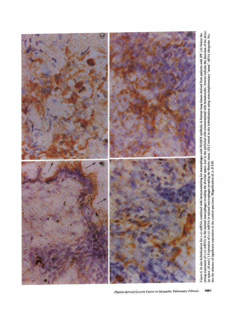
Invading macrophages in IPF lung tissue also expressedc-sis mRNA. This is demonstrated in Fig. 5, which representsa combined in situ hybridization for c-sis mRNAand im-munostaining for macrophages. A weak expression of c-sismRNAwas also observed in the capillary endothelial cells thatwere stained with the HAM56antibody (Fig. 5 B). The ex-pression of c-sis mRNAin macrophages is further demon-strated in Fig. 6. A strong expression of c-sis mRNAcan beseen in both epithelial cells and stained alveolar macrophages(Fig. 6 A). Clusters of macrophages infiltrating the fibrotictissue also expressed c-sis mRNA(Fig. 6, B and C). Controlhybridization with noncomplementary probe did not producea significant signal in the stained macrophages (Fig. 6 D). Thein situ hybridization studies described above demonstrated the
expression of c-sis mRNAin both epithelial cells and macro-phages in IPF lung tissue. Capillary endothelial cells in thesestudies showed much less hybridization.
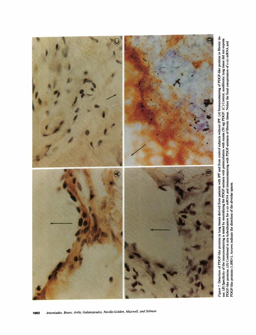
Detection of PDGF-like proteins in lung tissue of patientswith IPF. The expression of c-sis mRNAin the lung tissuesections of patients with IPF was accompanied by the expres-sion of PDGF-like proteins as shown by immunocytochemis-try using specific PDGFantisera. In contrast, control nonfi-brotic lung tissue did not express PDGF-like proteins. Asshown in Fig. 7 A, there is a strong expression of PDGF-likeproteins in fibrotic tissue stained with PDGFantisera. Thespecificity of this immunostaining reaction was tested by neu-tralizing the PDGFantisera with excess purified human PDGFor recombinant c-sis/PDGF-2 homodimer. Fig. 7 B shows thatthere is no immunostaining when the PDGFantisera werepreincubated with excess (50 ng) purified PDGF. Similar re-sults were obtained when the PDGFantisera were preincu-
Platelet-derived Growth Factor in Idiopathic Pulmonary Fibrosis 1059

Figure 5. Combined in situ hybridizationfor c-sis mRNAand immunostaining formacrophages with HAM56antibody inhuman lung tissue derived from a patientwith IPF. The magnification in A is 400,and B is 1,000. Notice the expression ofc-sis mRNAin the stained invading mac-rophages in the IPF tissue (short arrows). Aweak expression is also evident in thestained capillary endothelial cells (longarrow).
bated with excess recombinant PDGF(data submitted, notshown). Control nonfibrotic tissue did not express PDGF-likeproteins when it was immunostained with the PDGFantisera(Fig. 7 C). This is consistent with the lack of expression of c-sismRNAin the control nonfibrotic tissues. Fig. 7 D shows thefocal co-expression of c-sis mRNAand PDGF-like proteins ina lung section of a patient with IPF. In this study we combinedin situ hybridization for c-sis mRNAwith immunostainingwith PDGFantisera. As discussed by Hoffiler et al. (18), thiscombined procedure tends to enhance the intensity of the im-munostaining and weaken the intensity of mRNAdetectionbecause of the added steps in this combined process.
Discussion
The present data provide the first direct evidence for the invivo expression of the c-sis/PDGF-2 protooncogene in lung
tissue derived from patients with idiopathic lung fibrosis (IPF).Lung tissue without IPF did not express the c-sis protoonco-gene. In situ hybridization studies demonstrated a strong ex-pression of c-sis mRNAin epithelial cells and in macrophagesand to a lesser extent in the capillary endothelial cells in thetissues from patients with IPF. Immunostaining with PDGF-antisera detected PDGF-like proteins in lung tissue from pa-tients with IPF but not in control nonfibrotic lung tissue. Fur-thermore, combined in situ hybridization and immunostain-ing with PDGFantisera demonstrated the co-expression ofc-sis mRNAand PDGF-like proteins in the lung tissues ofpatients. The alveolar epithelial cells were the primary cellsexpressing c-sis mRNAand PDGF-like proteins. The in situhybridization data for c-sis mRNAand the immunostainingdata for PDGF-like proteins reported in this manuscript wereuniform and reproducible in all sections derived from the fivelung biopsies of patients.
1060 Antoniades, Bravo, Avila, Galanopoulos, Neville-Golden, Maxwell, and Selman
F6.
C
0Z or 0
3o .0~L
<
_0 r
' :
uoto 0
0o-O00
'0M
CL E =
~ 0
*0
cl0
E0 C
*0U U
D < U
g_ C:n
0
CZCe
- a0._
00
ol.0o ^
a)
oYC
Ce 0.t;
W0 a
o E
~ _M .
Cq *C Ol
e
O~. 0° ~
*I: Q;.; '0 0
-0. .0
* 0*Q
.0
at; 0,Ct.
Platelet-derived Growth Factor in Idiopathic Pulmonary Fibrosis 1061
,0
Z0
o oZ.
.Ca a )
Coa0Z
C 'r waa.-.
-o c
0
o ¢
'0
0 0
Q.X, <.E
CU 06 M
'a 0D~
os e"l *<
C *_
L o
o a. .
,a ao-.a a
a 0L4 Lir.S _ *e
1062 Antoniades, Bravo, Avila, Galanopoulos, Neville-Golden, Maxwell, and Selman
Previous studies have shown that cultured activated mac-rophages express the c-sis protooncogene and produce PDGF-like mitogen (7, 8, 1 1). As shown by Martinet et al. (19), non-activated alveolar macrophages obtained from normalhumans by bronchoalveolar lavage released only smallamounts of PDGF-like mitogen. In contrast, alveolar macro-phages obtained from patients with IPF released much higheramounts (about fourfold) of PDGF-like mitogen. Our findingon the expression of the c-sis protooncogene by macrophagesin lung tissue of patients with IPF are consistent with the ob-servation on the spontaneous release of PDGF-like mitogen byalveolar macrophages obtained from patients with IPF (19). Aspontaneous expression of c-sis mRNAby macrophages indiseased tissue does not appear to be a general phenomenon.For example, studies by Wilcox et al. (20) on the localizationof PDGFmRNAin human atherosclerotic plaques failed todetect expression of c-sis mRNAin macrophages. Similarly,our in situ hybridization data of normal lung tissue failed todemonstrate the expression of c-sis mRNA.Mornex et al. (12)reported that nonactivated alveolar macrophages obtained bybronchoalveolar lavage from normal human smokers ex-pressed c-sis transcripts as judged by Northern blot analysis.-20 Ag of total RNAextracted from the macrophages wasused in that study. Our in situ hybridization in lung specimensof four control subjects without IPF that includes smokers didnot demonstrate expression of c-sis mRNA.It is possible that alow expression of c-sis mRNAby their alveolar macrophageswas not detectable by in situ hybridization and it was onlyapparent in an enriched RNApreparation obtained from mac-rophages (1 1). A weak expression of c-sis mRNAcould be seenin capillary endothelial cells. This low expression in vivo is incontrast to high expression of c-sis mRNAseen in vitro incultured endothelial cells (21, 22). However, our in vivo ob-servation is consistent with the original finding of Barrett et al.(21) who reported a low expression of the c-sis gene in endo-thelial cells in vivo, compared to the high expression seen invitro in cultured endothelial cells.
The studies described above demonstrated a strong expres-sion of c-sis mRNAin epithelial cells in IPF tissue. Normally,epithelial cells do not express the c-sis protooncogene. Forexample, lung tissue without IPF did not express c-sis mRNAas judged by both Northern blot analysis and in situ hybridiza-tion. In contrast, cultured malignant epithelial cell lines de-rived from human patients with breast (23, 24), lung (25), andprostatic (26) cancer were shown to express both PDGFgenesand to secrete PDGF-like mitogen. Because epithelial cells donot display surface receptors to PDGF, it was suggested thatsecretion of PDGF-like mitogen by these cells serves for para-crine functions, such as development of fibrosis associatedwith human breast cancer (23, 24). The present study providesthe first evidence for the spontaneous expression of the c-sisprotooncogene by nonmalignant epithelial cells in lung biopsyspecimen derived from patients with IPF.
In these studies the emphasis is on the expression of thec-sis/PDGF-2 mRNA.A weak expression of the gene encod-ing the PDGF-1 (A) polypeptide chain was detected during a3-wk exposure in both non-IPF- and IPF-associated lung tis-sue, as judged by Northern blot analysis (data not shown).However, the expression of this gene was very weak, and it wasnot possible by in situ hybridization to localize the cells ex-pressing the PDGF-1 mRNAin IPF or non-IPF lung tissue.Similarly, immunostaining with the PDGFantisera failed to
detect the presence of PDGF-like proteins in non-IPF lungtissue (Fig. 7 C).
Fibrosis and excessive collagen synthesis are events asso-ciated with IPF. The unregulated local production of PDGFmay contribute to the expansion of connective tissue cells andcollagen accumulation in lung parenchymal, because PDGFisa potent chemoattractant and mitogen for fibroblasts andsmooth muscle cells (27-29) and a stimulator of collagen syn-thesis by fibroblasts. An increase in the number of fibroblastswithin the alveolar structures, which has been demonstrated inboth animals and humans with chronic interstitial lung disease(30, 31), is critical in the pathogenesis of lung fibrosis. Further-more, PDGFcan stimulate the production of collagenase (32)in fibroblasts, which may explain the progressive derangementin interstitial collagens at least in the initial phases of the dis-ease. In this context, the presence of active collagenase withinthe lower respiratory tract can be responsible for ultrastruc-tural alterations described in patients with IPF like the frag-mentation and fraying of the collagen fibers in the lung inter-stitium (33).
The important new finding in these studies is the recogni-tion of a strong expression of c-sis/PDGF-2 mRNAin theepithelial cells of lung tissue derived from patients with IPF.This "inappropriate" expression may be an important contrib-uting factor in the development of fibrosis. The local constitu-tive production of PDGF-like mitogen by the epithelial cells,could serve for the paracrine recruitment and stimulation oftarget cells such as fibroblasts and smooth muscle cells, and toan exaggerated production and accumulation of interstitialcollagens in patients with IPF.
The initial events leading to IPF are not known. Injury isconsidered to be among the causes of the disease. Recent stud-ies have shown that acute injury in animals induces the revers-ible expression of the c-sis and PDGFreceptor b mRNAsinthe epithelial cells of the injured tissue (Antoniades, H. N., etal., submitted for publication). Normal epithelial cells do notexpress c-sis and PDGFreceptor mRNAs. The expression ofthe mRNAsin the epithelial cells occurred within 1 d of injuryand it was suppressed by day five, which coincides with thetime of reepithelialization of the injured tissue. These findingsprovide direct evidence that acute injury can induce a revers-ible expression of protooncogenes in the epithelium. It is con-ceivable that "chronic" injury can cause an irreversible, inap-propriate gene expression, like the expression of the c-sis pro-tooncogene in the lung epithelium of patients with IPFdescribed in the present studies.
AcknowledgmentsWeexpress our thanks to Dr. Hubert J. Wolfe and Dr. Stephen P.Naber, Department of Pathology, Tufts University-New EnglandMedical Center, for the advice and support in the in situ hybridizationand immunocytochemistry studies. We express our thanks to Mrs.Amal Ghaly and Mrs. Deborah Wilkinson for the preparation of thismanuscript.
Supported by funds from the National Institutes of Health grantsCA3011, HL29583 (H. N. Antoniades), and the Council for TobaccoResearch USA(H. N. Antoniades). Dr. M. Bravo and Dr. R. Avila arePostdoctoral Research Fellows in the Department of Nutrition, Har-vard School of Public Health.
References1. Fulmer, J. D. 1982. The interstitial lung diseases. Chest.
82:172-178.
Platelet-derived Growth Factor in Idiopathic Pulmonary Fibrosis 1063
2. Crystal, R. G., J. E. Gadek, V. J. Ferrans, B. R. Line, and G. W.Hunninghake. 1981. Interstitial lung diseases: current concepts ofpathogenesis, staging and therapy. Am. J. Med. 70:542-568.
3. Crystal, R. G. 1987. Interstitial lung disorders. In Principles ofInternal Medicine. I 1th ed. E. Braunwald et al., editors. McGraw-HillBook Co., NewYork. 1095-1105.
4. Selman, M., M. Montano, C. Ramos, and R. Chapela. 1986.Concentration, biosynthesis and degradation of collagen in idiopathicpulmonary fibrosis. Thorax. 41:355-359.
5. Rennard, S. I., G. W. Hunninghake, P. B. Bitterman, and R. G.Crystal. 1981. Production of fibronectin by the human alveolar macro-phage: mechanism for the recruitment of fibroblasts to sites of tissueinjury in interstitial lung diseases. Proc. Natl. Acad. Sci. USA.78:7147-7151.
6. Bitterman, P., B. S. Adelberg, and R. G. Crystal. 1983. Pulmo-nary fibrosis. Spontaneous release of the alveolar macrophage-derivedgrowth factor in the interstitial lung disorders. J. Clin. Invest.72:1801-1813.
7. Martinet, Y., P. B. Bitterman, J. Mornex, G. R. Grotendorst,and R. M. Crystal. 1986. Activated human monocytes express the c-sisprotooncogene and release a mediator showing PDGF-like activity.Nature (Lond.). 319:158-160.
8. Pantazis, P., E. Sariban, D. Kufe, and H. N. Antoniades. 1986.Induction of c-sis gene expression and synthesis of platelet-derivedgrowth factor in human myeloid leukemia cells during monocyticdifferentiation. Proc. Nadl. Acad. Sci. USA. 83:6455-6459.
9. Antoniades, H. N., and M. W. Hunkapiller. 1983. Humanplate-let-derived growth factor (PDGF): amino-terminal amino acid se-quence. Science (Wash. DC). 220:963-965.
10. Antoniades, H. N., P. Pantazis, and A. J. Owen. 1987. Humanplatelet-derived growth factor and the sis/PDGF-2 gene. In Oncogenes,Genes, and Growth Factors. G. Guroff, editor. John Wiley & Sons,Inc., NewYork. 1-40.
11. Shimokado, K., E. W. Raines, D. K. Madtes, T. B. Barrett, E. P.Benditt, and R. Ross. 1985. A significant part of macrophage-derivedgrowth factor consists of at least two forms of PDGF. Cell. 43:277-286.
12. Mornex, J. F., Y. Martinet, and K. Yamamuchi. 1986. Sponta-neous expression of the c-sis gene and release of a platelet-derivedgrowth factorlike molecule by human alveolar macrophages. J. Clin.Invest. 78:61-66.
13. Collins, T., D. Ginsburg, J. M. Boss, S. H. Orkin, and J. S.Pober. 1985. Cultured human endothelial cells express platelet-derivedgrowth factor B-chain: cDNAcloning and structural analysis. Nature(Lond.). 316:748-750.
14. Betsholtz, C., J. Johnsson, C. H. Heldin, B. Westermark, P.Lind, M. S. Urdea, R. Eddy, T. B. Shows, K. Philpott, A. L. Mellor,T. J. Knott, and J. Scott. 1986. cDNA sequence and chromosomallocalization of human platelet-derived factor A-chain and its expres-sion in tumor cell lines. Nature (Lond.). 320:695-699.
15. Ponte, P., S.-Y. Ng, J. Engel, P. Cunning, and L. Kedes. 1984.Evolutionary conservation in the untranslated regions of actionmRNAs: DNAsequence of a human b-actin cDNA. Nucleic AcidsRes. 12:1687-1696.
16. Hoffler, H., H. Childers, M. R. Monminy, R. M. Lechan, R. H.Goodman, and Y. J. Wolfe. 1986. In situ hybridization methods forthe detection of somatostatin mRNAin tissue sections using antisenseRNAprobes. Histochem. J. 18:587-594.
17. Gown, A. M., T. Tsukada, and R. Ross. 1986. Humanathero-sclerosis II. Immunocytochemical analysis of the cellular compositionof human atherosclerotic lesions. Am. J. Pathol. 25:191-107.
18. Hoffler, H., R. A. DeLellis, and M. J. Wolfe. 1988. In situhybridization and immunocytochemistry. In Advances in Immuno-histochemistry. R. A. DeLellis, editor. Raven Press, NewYork. 47-66.
19. Martinet, Y., W. N. Rom, G. R. Grotendorst, G. R. Martin,and R. G. Crystal. 1987. Exaggerated spontaneous release of platelet-derived growth factor by alveolar macrophages from patients withidiopathic pulmonary fibrosis. N. Engl. J. Med. 317:202-209.
20. Wilcox, J. N., K. M. Smith, L. T. Williams, S. M. Schwartz, andD. Gordon. 1988. Platelet-derived growth factor mRNAdetection inhuman atherosclerotic plaques by in situ hybridization. J. Clin. Invest.82:1134-1143.
21. Barrett, T. B., C. M. Gajducek, S. M. Schwartz, J. K. Swartz,and E. P. Benditt. 1984. Expression of the sis gene by endothelial cellsin culture and in vivo. Proc. Natl. Acad. Sci. USA. 81:6772-6774.
22. Sitaras, N. H., E. Sariban, P. Pantazis, B. Zetter, and H. N.Antoniades. 1987. Human iliac artery endothelial cells express bothgenes encoding the chains of platelet-derived growth factor (PDGF)and synthesize PDGF-like mitogen. J. Cell Physiol. 132:376-380.
23. Rosengurt, E., J. Sinnet-Smith, and J. Taylor-Papadimitrov.1985. Production of PDGF-like growth factor by breast cancer celllines. Int. J. Cancer. 36:247-252.
24. Bronzert, D. A., P. Pantazis, H. N. Antoniades, A. Kasid, N.Davidson, R. B. Dickson, and M. E. Lippman. 1987. Synthesis andsecretion of PDGF-like growth factor by human breast cancer celllines. Proc. Natl. Acad. Sci. USA. 84:5763-5767.
25. Sariban, E., N. M. Sitaras, D. W. Kufe, H. N. Antoniades, andP. Pantazis. 1988. Expression of PDGF-related transcript and synthesisof biologically active PDGF-like proteins by human malignant epithe-lial cell lines. J. Clin. Invest. 82:1157-1164.
26. Sitaras, N., E. Sariban, M. Bravo, P. Pantazis, and H. N. An-toniades. 1988. Constitutive production of PDGF-like proteins byhuman prostate carcinoma cell line. Cancer Res. 48:1930-1935.
27. Grotendorst, G. R., H. E. Seppa, H. K. Kleinman, and G. R.Martin. 1981. Attachment of smooth muscle cells to collagen and theirmigration toward platelet-derived growth factor. Proc. Natl. Acad. Sci.USA. 78:3669-3672.
28. Seppa, H. E., G. R. Grotendorst, S. Seppa, E. Schiffmann, andG. R. Martin. 1982. Platelet-derived growth factor is chemotactic forfibroblasts. J. Cell Biol. 92:584-588.
29. Bernstein, L. R., H. N. Antoniades, and B. R. Zetter. 1982.Migration of cultured vascular cells in response to plasma and plate-let-derived growth factor. J. Cell Sci. 56:71-82.
30. Selman, M., M. Montafio, I. Montfort, and R. Perez Tamayo.1985. A new model of diffuse interstitial pulmonary fibrosis in the rat.Exp. Mol. Pathol. 43:375-387.
31. Greenberg, S. D., R. M. O'Neal, and D. E. Jenkins. 1974. Thepathological findings in diffuse interstitial fibrosis of the lungs. SouthMed. J. 67:571-579.
32. Bauer, E., T. Cooper, J. Huang, J. Altman, and T. Deuel. 1985.Stimulation of in vitro human skin collagenase expression by platelet-derived growth factor. Proc. Natl. Acad. Sci. USA. 82:4132-4136.
33. Crystal, R. G., J. D. Fulmer, W. C. Roberts, M. L. Moss, B. R.Line, and H. Y. Reynolds. 1976. Idiopathic pulmonary fibrosis: clini-cal, histologic, radiographic, physiologic, scintigraphic, cytologic, andbiochemical aspects. Ann. Intern. Med. 85:769-788.
1064 Antoniades, Bravo, Avila, Galanopoulos, Neville-Golden, Maxwell, and Selman
Related Documents