SHORT COMMUNICATION Platelet activating factor stimulates arachidonic acid release in differentiated keratinocytes via arachidonyl non-selective phospholipase A 2 Katarina Mariann Jørgensen • Hanne Solvang Felberg • Rolf K. Berge • Astrid Lægreid • Berit Johansen Received: 3 November 2009 / Revised: 10 December 2009 / Accepted: 14 December 2009 / Published online: 30 December 2009 Ó The Author(s) 2009. This article is published with open access at Springerlink.com Abstract Platelet activating factor (PAF, 1-O-alkyl-2- acetyl-sn-glycero-3-phosphocholine) is known to be pres- ent in excess in psoriatic skin, but its exact role is uncer- tain. In the present study we demonstrate for the first time the role of group VI PLA 2 in PAF-induced arachidonic acid release in highly differentiated human keratinocytes. The group IVa PLA 2 also participates in the release, while secretory PLA 2 s play a minor role. Two anti-inflammatory synthetic fatty acids, tetradecylthioacetic acid and tetra- decylselenoacetic acid, are shown to interfere with sig- nalling events upstream of group IVa PLA 2 activation. In summary, our major novel finding is the involvement of the arachidonyl non-selective group VI PLA 2 in PAF-induced inflammatory responses. Keywords Phospholipase A 2 Á Platelet activating factor Á Arachidonic acid Á Tetradecylthioacetic acid Á Tetradecylselenoacetic acid Abbreviations AA Arachidonic acid AACOCF 3 Arachidonyl trifluoromethyl ketone BEL Bromoenol lactone BSA Bovine serum albumin FCS Fetal calf serum MAFP Methyl arachidonyl fluorophosphonate OA Oleic acid PA Palmitic acid PACOCF 3 Palmitoyl trifluoromethyl ketone PAF Platelet activating factor PAF-R Platelet activating factor receptor PLA 2 Phospholipase A 2 cPLA 2 Cytosolic phospholipase A 2 iPLA 2 Ca 2? -independent phospholipase A 2 sPLA 2 Secretory phospholipase A 2 PPAR Peroxisome proliferator-activated receptor TSA Tetradecylselenoacetic acid TTA Tetradecylthioacetic acid Introduction The lipid mediator platelet activating factor (PAF, 1-O- alkyl-2-acetyl-sn-glycero-3-phosphocholine) is found in excess in psoriatic scale and plasma [15] as well as in other inflammatory skin conditions [34]. Bayerl et al. [6] also reveal increased expression of PAF receptors in psoriatic skin. Phospholipase A 2 enzymes are central mediators of inflammatory responses, including psoriasis [30]. The PLA 2 superfamily of enzymes is often divided into three broader categories: (1) extracellular secretory PLA 2 s (sPLA 2 ), (2) cytosolic calcium-dependent PLA 2 s (cPLA 2 ) and (3) cytosolic calcium-independent PLA 2 s (iPLA 2 ). Recent evidence suggests that all classes of PLA 2 enzymes K. M. Jørgensen Á H. S. Felberg Á B. Johansen (&) Department of Biology, Norwegian University of Science and Technology, NTNU, Trondheim, Norway e-mail: [email protected]; [email protected] A. Lægreid Department of Cancer Research and Molecular Medicine, Norwegian University of Science and Technology, NTNU, Trondheim, Norway R. K. Berge Division of Clinical Biochemistry, Haukeland Hospital, Bergen, Norway 123 Arch Dermatol Res (2010) 302:221–227 DOI 10.1007/s00403-009-1017-8

Welcome message from author
This document is posted to help you gain knowledge. Please leave a comment to let me know what you think about it! Share it to your friends and learn new things together.
Transcript
SHORT COMMUNICATION
Platelet activating factor stimulates arachidonic acid releasein differentiated keratinocytes via arachidonyl non-selectivephospholipase A2
Katarina Mariann Jørgensen •
Hanne Solvang Felberg • Rolf K. Berge •
Astrid Lægreid • Berit Johansen
Received: 3 November 2009 / Revised: 10 December 2009 / Accepted: 14 December 2009 / Published online: 30 December 2009
� The Author(s) 2009. This article is published with open access at Springerlink.com
Abstract Platelet activating factor (PAF, 1-O-alkyl-2-
acetyl-sn-glycero-3-phosphocholine) is known to be pres-
ent in excess in psoriatic skin, but its exact role is uncer-
tain. In the present study we demonstrate for the first time
the role of group VI PLA2 in PAF-induced arachidonic acid
release in highly differentiated human keratinocytes. The
group IVa PLA2 also participates in the release, while
secretory PLA2s play a minor role. Two anti-inflammatory
synthetic fatty acids, tetradecylthioacetic acid and tetra-
decylselenoacetic acid, are shown to interfere with sig-
nalling events upstream of group IVa PLA2 activation. In
summary, our major novel finding is the involvement of the
arachidonyl non-selective group VI PLA2 in PAF-induced
inflammatory responses.
Keywords Phospholipase A2 � Platelet activating factor �Arachidonic acid � Tetradecylthioacetic acid �Tetradecylselenoacetic acid
Abbreviations
AA Arachidonic acid
AACOCF3 Arachidonyl trifluoromethyl ketone
BEL Bromoenol lactone
BSA Bovine serum albumin
FCS Fetal calf serum
MAFP Methyl arachidonyl fluorophosphonate
OA Oleic acid
PA Palmitic acid
PACOCF3 Palmitoyl trifluoromethyl ketone
PAF Platelet activating factor
PAF-R Platelet activating factor receptor
PLA2 Phospholipase A2
cPLA2 Cytosolic phospholipase A2
iPLA2 Ca2?-independent phospholipase A2
sPLA2 Secretory phospholipase A2
PPAR Peroxisome proliferator-activated receptor
TSA Tetradecylselenoacetic acid
TTA Tetradecylthioacetic acid
Introduction
The lipid mediator platelet activating factor (PAF, 1-O-
alkyl-2-acetyl-sn-glycero-3-phosphocholine) is found in
excess in psoriatic scale and plasma [15] as well as in other
inflammatory skin conditions [34]. Bayerl et al. [6] also
reveal increased expression of PAF receptors in psoriatic
skin.
Phospholipase A2 enzymes are central mediators of
inflammatory responses, including psoriasis [30]. The
PLA2 superfamily of enzymes is often divided into three
broader categories: (1) extracellular secretory PLA2s
(sPLA2), (2) cytosolic calcium-dependent PLA2s (cPLA2)
and (3) cytosolic calcium-independent PLA2s (iPLA2).
Recent evidence suggests that all classes of PLA2 enzymes
K. M. Jørgensen � H. S. Felberg � B. Johansen (&)
Department of Biology, Norwegian University of Science
and Technology, NTNU, Trondheim, Norway
e-mail: [email protected]; [email protected]
A. Lægreid
Department of Cancer Research and Molecular Medicine,
Norwegian University of Science and Technology, NTNU,
Trondheim, Norway
R. K. Berge
Division of Clinical Biochemistry, Haukeland Hospital,
Bergen, Norway
123
Arch Dermatol Res (2010) 302:221–227
DOI 10.1007/s00403-009-1017-8

may participate in agonist-induced arachidonic acid (AA)
release [30, 36]. In HaCaT keratinocytes, we previously
detected expression of sPLA2 isoenzymes group IIa, IId, V
and X, as well as the ubiquitous group IVa (cPLA2) and VI
(iPLA2) [4]. PAF is known to activate cPLA2 in several
cell types [14], but its role in activating other PLA2 iso-
enzymes is less well characterized.
The two synthetic fatty acids tetradecylthioacetic acid
(TTA) and tetradecylselenoacetic acid (TSA) [22, 33] have
previously been reported to have anti-inflammatory prop-
erties [35]. To further investigate PLA2-involvement in
inflammatory skin conditions this paper examines and
compares the activation and participation of PLA2 isoen-
zymes in PAF and calcium ionophore (A23187)-induced
AA-release in keratinocytes, and if TTA and TSA interfere
with this.
Results and discussion
Keratinocytes in the outer layer of the epidermis are dif-
ferentiated, as opposed to proliferating keratinocytes
attached to basal laminae. In order to obtain a cell state
resembling mature skin, HaCaT keratinocytes [7] used in
this study (kindly provided by Prof. N. Fusenig, Heidel-
berg, Germany) were differentiated in culture for 3 days
prior to experimentation (protocol as described in refs. [3,
4]). The differentiation state of the cells was documented
by examining the expression of three markers of suprabasal
keratinocytes: Keratin 10 [8], S100A7 (psoriasin) [9] and
sPLA2 IIA [2]. They all show an upregulation of more than
fivefold compared to HaCaT undifferentiated state
(Fig. 1a). [RNA was isolated using a Qiagen RNeasy Mini
kit according to kit protocol, and RT2-PCR performed
using the MX3000 system from Stratagene (primer
sequences shown in Table 1)]. Other studies of HaCaT
cells also demonstrate that upregulation of these genes are
associated with a highly differentiated state [16, 29], thus
the data suggest that the cultivation protocol used, indeed,
differentiates the keratinocytes.
Previous studies of PAF-stimulated AA-release in
keratinocytes have used actively proliferating cells [10, 27].
In order to establish the pro-inflammatory potential of PAF
in differentiated keratinocytes, HaCaT cells were stimu-
lated with PAF-16 (purchased from Calbiochem), and the
subsequent release of [3H]AA and [14C] oleic acid (OA)
(NEN, Perkin Elmer) was measured. Changes in the levels
of these free fatty acids may indicate the involvement of
AA-selective or non-selective phospholipases, respectively
[30]. (Protocol as described previously [3, 4], except that
bovine serum albumin was excluded due to evidence that it
may inhibit PAF activity [13].) At 60 min exposure and 20
lM concentration, PAF typically induces an AA induction
of about twofold in a dose-dependent manner (Fig. 1b). The
fold induction of OA is nearly as large as for AA, which is
especially notable, since OA release is typically a minor
fraction of the fold induction of AA-release [4, 25], and
only rarely equal to it [36]. We may conclude that PAF has a
pro-inflammatory effect in fully differentiated HaCaT, by
induction of AA-release. Furthermore, AA-non-selective
phospholipase A2s, as indicated by the observed OA
release, contribute substantially to this effect.
The physiological relevance of the PAF concentration
used deserves to be further commented upon. HaCaT cells
A B
Fig. 1 PAF induces AA and OA release in differentiated keratino-
cytes. a Initial cell state. Fold induction of differentiation specific
marker genes in proliferating vs post-confluent HaCaT cells. (N = 2,
result shown from one representative experiment). b Dose–response
relationship for PAF stimulation of HaCaT cells. The response is
measured as fold induction of [3H] arachidonic acid and [14C] oleic
acids compared to the unstimulated control. PAF exposure time was
60 min. (All dose–response data in cell culture have been statistically
validated using one way ANOVA at the 95% confidence level, and
the results shown are representative of at least three consecutive
experiments, using at least three parallel samples in each experiment)
222 Arch Dermatol Res (2010) 302:221–227
123
were 100% viable at this PAF concentration as determined
by 3-(4,5-dimethylthiazol-2-yl)-2,5-diphenoltetrazolium
bromide (MTT) dye (Sigma) uptake assay [21] (data not
shown). This study uses natural PAF, a readily degradable
lipid—it has a half life of 0.5–2 s in serum [24]. HaCaT
cells express both sPLA2 GX [4] and PAF-AH II [19],
which are both known to degrade PAF [12, 19]. Addi-
tionally, recent developments in new quantitative methods
for PAF suggest that the PAF concentration in serum in
healthy humans may be as high as 0.2–0.3 lM [26], which
is at least 100 times higher than previously thought. Thus,
the PAF concentration used in this study is non-toxic and
physiologically plausible.
Group IVa is the only truly AA-selective PLA2 isoen-
zyme [30]. In order to examine activation of group IVaPLA2 in PAF-mediated AA-release, HaCaT cells were
treated with PAF in the presence or absence of the group
IVa inhibitor methyl arachidonyl fluorophosphonate
(MAFP) [11] (obtained from Cayman Chemicals), fol-
lowed by lysis and analysis by in vitro group IVa PLA2
enzyme activity assay (for method description see [3, 18]).
[All inhibitors used, their vehicles and the calcium iono-
phore do not affect viability in the concentrations applied
in this study, as verified by MTT assays (not shown).] PAF-
induced activation of group IVa PLA2 enzyme in HaCaT
cells, and this response is reduced by 50% by MAFP
(Fig. 2a). This in vitro assay is highly specific and the
result thus strongly indicates that group IVa PLA2 partic-
ipates in PAF-induced AA-release [18].
In order to determine the importance of the contribution
of group IVa PLA2 in the total PAF-response, PAF-medi-
ated [3H]AA and [14C]OA release in cells treated with PAF
in the presence or absence of MAFP [11] was determined.
We found that MAFP inhibited both [3H]AA and [14C]OA
release in a dose-dependent manner by a maximum of 51
and 28.5% respectively (see Fig. 2b). Since MAFP is also
known to have some effect on group VI PLA2 [25, 28], we
cannot exclude the involvement of PLA2s of this type in
the response. However, our previous studies show that it is
possible to attenuate AA-release in HaCaT cells com-
pletely in the IL-1b pathway using only 10 lM of MAFP
[4]. The partial attenuation of PAF-mediated AA-release
Table 1 Primers for keratinocyte differentiation markers
Entrez ID Symbol Size Forward primer Reverse primer
3858 KRT10 106 TGCACACAGTAGCGACCTTC TGAGACGTAATGTACAAGCTCTGG
6278 S100A7 148 CTCCCAGCAAGGACAGAAAC CTGCTGACGATGATGAAGGA
5320 PLA2G2A 167 GAGCTAGGCCAGTCCATCTG TAACTGAGTGCGGCTTCCTT
A B
C D
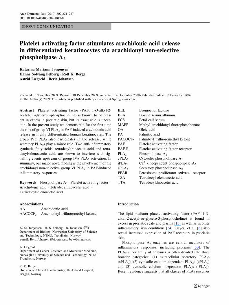
Fig. 2 Different PLA2 inhibitors reduce PAF-induced AA and OA
release. a In vitro cPLA2 activity assay on lysate from HaCaT cells
pre-treated with 25 lM MAFP (90 min) and stimulated by 20 lM
PAF (60 min) prior to lysis. [95% significance indicated by asterisk(compared to unstimulated control) and D (compared to PAF-
stimulated sample) as determined by Student’s t test]. Results shown
are representative of at least three consecutive experiments, using at
least three parallel samples in each experiment. Release assay of [3H]
arachidonic acid and [14C] oleic acids compared to the unstimulated
control (in %). Stimulus is 20 lM PAF for 60 min. b Dose–response
inhibition by MAFP (pretreatment time 90 min.), c inhibition by
indoxam (pretreatment time 90 min), d dose–response inhibition by
PACOCF3 (pretreatment time 15 min). Statistical testing and number
of experiments as for Fig. 1b
Arch Dermatol Res (2010) 302:221–227 223
123

achieved with MAFP therefore suggests a significant con-
tribution to AA-release by AA-nonspecific enzymes in
addition to the contribution by group IVa PLA2. However,
our in vitro enzyme assay clearly shows that group IVaPLA2 is activated by PAF, thus our conclusion is that group
IVa PLA2 participates in PAF-mediated AA-release toge-
ther with AA-nonspecific PLA2 subtypes.
To our knowledge, there are no previous reports of PAF-
induced sPLA2 activation in the literature. Secretory PLA2
enzymes would be candidate enzymes for the OA release
observed; we then examined the role of sPLA2 subtypes in
PAF-mediated AA-release. The sPLA2-selective inhibitor
indoxam [31] (a generous gift from Shionigi Ltd, Japan),
gives a dose-dependent inhibition with a maximum of 33%
of PAF-induced AA-release and 15% of OA release at 25
lM (Fig. 2c). Similar results were obtained using
SB203347 (a kind gift from Lisa Marshall, SmithKline
Beecham, PA, USA), another sPLA2 inhibitor [20, 31]
(results not shown). Interestingly, Fig. 2c thus shows that
the inhibition found with indoxam predominantly affects
AA-release. In several cell types, sPLA2 isoenzymes IIa,
IId and V, but not X, have been shown to be more strongly
arachidonyl-selective when operating intracellularly, a
mechanism which involves sPLA2 isoenzyme selective
caveolin-mediated endocytosis [23]. Our data thus suggest
that one or several of these three sPLA2s (IIa, IId or V) may
participate in PAF-mediated intracellular AA-release in
differentiated HaCaT cells.
Lastly, we examined the possible role of group VI PLA2
in the PAF-induced OA response. We found that the PLA2
subtype VI-specific inhibitor palmitoyl trifluoromethyl
ketone (PACOCF3) [1] (from Calbiochem) dose-depen-
dently reduced the PAF-induced AA-release by 58% and
OA release by 61% at a 25-lM concentration (Fig. 2d).
These results were confirmed with application of bromoenol
lactone (BEL, from Cayman Chemicals) [1], another group
VI inhibitor, which produced comparable levels of maxi-
mum inhibition (data not shown). Although PACOCF3 is
also known to inhibit group IV PLA2 [11], BEL [1] is not
known to do so. As sPLA2 inhibitors were shown to pref-
erentially inhibit AA-release, the incomplete attenuation
achieved with the group IVa/VI inhibitor MAFP, and the
more successful inhibition with PACOCF3, the conclusion
supported is that group VI PLA2 most likely plays a major
role in PAF-mediated AA-release. The group VI PLA2
enzyme is probably at least as important as group IVa PLA2,
judging by its ability to contribute to the high OA release.
The participation as well as the notable significance of group
VI PLA2 in the PAF-mediated response is a novel finding.
Taken together, our data suggest the participation of
both calcium-dependent and -independent cytosolic PLA2
subtypes IVa and VI, as well as of secretory PLA2 sub-
types, in the PAF-induced response in differentiated Ha-
CaT keratinocytes.
The palmitic acid (PA)-derived lipids TTA and TSA
[22, 33] are already shown to exhibit anti-inflammatory
A
B
C
D
E
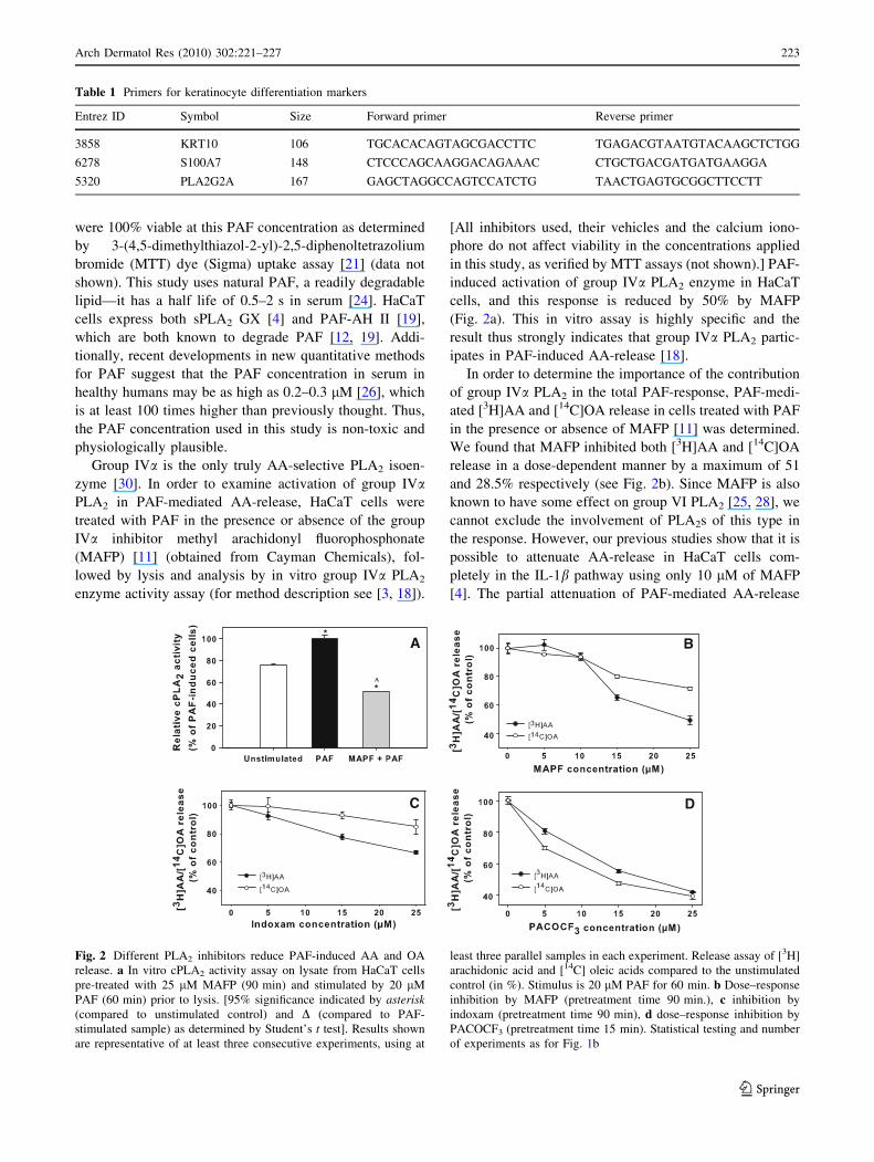
Fig. 3 Tetradecylthioacetic
acid and tetradecylselenoacetic
acid inhibit arachidonic acid
release. The response is
measured as % release of [3H]
arachidonic acid and [14C] oleic
acids compared to the
unstimulated control.
Pretreatment time for all
inhibitors is 90 min. Effects on
the PAF-induced response (left)(20 lM PAF, 60 min). Dose–
response inhibition by a TTA
and b TSA. c Dose–response
result for Palmitic acid. PA acts
as a control, since TTA and
TSA are PA derivatives. Effects
on the calcium ionophore
(A23187)-induced response
(right) (1 lM A23187, 60 min).
d Dose–response inhibition by
MAFP, e dose–response
inhibition by TSA. Statistical
testing and number of
experiments as for Fig. 1b
224 Arch Dermatol Res (2010) 302:221–227
123
properties [35]. Most published studies of TTA and TSA
show their roles as PPAR ligands [35], however, their
potency as anti-inflammatory and anti-apoptotic agents are
not fully explained by this mechanism. It would therefore
be interesting to test whether their anti-inflammatory
properties include inhibition of AA-release. TTA (Fig. 3a)
and TSA (Fig. 3b) show a similar overall trend with
maximum AA inhibition of 60–70% of the PAF-induced
AA-release at 25 lM concentration. Further experiments
have therefore been carried out with only one of these two
inhibitors. TTA and TSA are both derivatives of PA [22],
which was used as a control (Fig. 3c). The data suggests
that the inhibitory effect is specific to TTA and TSA, and
not shared by their common precursor, PA.
We also tested these inhibitors in the calcium ionophore
(A23187) (Sigma) response, to compare with the PAF
response. Calcium ionophore acts by releasing calcium
from both the mitochondrion and the extracellular matrix
[28], and can induce activation of calcium-dependent
enzymes such as group IVa PLA2 [17]. Twenty-five
micromolar of the group IVa PLA2 inhibitor MAFP [11]
was found to strongly inhibit calcium ionophore induced
AA-release (73.5%; Fig. 3d, right). In cells pre-treated with
35 lM of TSA, a near-complete attenuation of A23187-
induced AA-release, 98%, was found (Fig. 3e).
Thus, TTA and TSA give a strong arachidonyl-selective
inhibition in both the PAF and calcium ionophore
responses, suggesting that they affect the group IVa PLA2-
regulated pathway. The data suggest a prominent role for
AA-specific PLA2 subtypes in the response to calcium
ionophore, consistent with our previous studies [32].
We then wished to determine whether TSA actually
acts as inhibitor of the group IVa PLA2 enzyme. The
effect of TSA on a recombinant group IVa PLA2 substrate
was tested using the in vitro enzyme activity assay [18],
and compared to the effects of AACOCF3 and PACOCF3.
AACOCF3 (Cayman Chemicals) is a potent, reversible
group IVa PLA2 inhibitor, with an effect similar to that of
MAFP [11]. Forty nanomolar AACOCF3 gives a 70%
reduction in group IVa PLA2 activity (Fig. 4a). A similar
concentration of the group VI-selective inhibitor PA-
COCF3 gives a 55% reduction in group IVa PLA2 activity,
which is consistent with previous studies showing that
PACOCF3 is a poorer inhibitor of group IVa PLA2 than
AACOCF3 is [11]. TSA shows only a 20% reduction in
group IVa PLA2 activity, suggesting that it has a poor
direct effect on group IVa PLA2. Thus, the observed
strong reduction of cellular AA-release by TSA must
mainly be due to a signalling mechanism leading to the
activation of group IVa PLA2, rather than on the enzyme
itself.
Since PACOCF3 and TTA are both PA derivates, we
lastly wanted to examine whether these inhibitors act
through similar or different mechanisms. TTA and PA-
COCF3 were applied to cells both separately and together,
and AA-release was induced with PAF. Both TTA and
PACOCF3 inhibited AA-release by approx 30%, respec-
tively, while both together inhibited AA-release by almost
60% (Fig. 4b). The effect of TTA and PACOCF3 combined
is clearly additive. This suggests that these inhibitors work
on parallel, separate mechanisms, where TTA acts on
signalling involving group IVa PLA2, while PACOCF3
works on the group VI PLA2 enzyme.
Measuring cellular AA and OA release in the presence
of different PLA2 inhibitors is an established method
within the PLA2 research community [18, 30]. We have
used two structurally and mechanistically different inhibi-
tors against each class of PLA2 in our experiments. Both of
the cPLA2 (MAFP, AACOCF3) [11] and iPLA2 inhibitors
(PACOCF3, BEL) [1, 5] used here are very well
A B
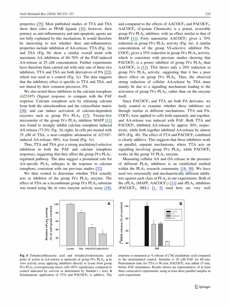
Fig. 4 Tetradecylthioacetic acid and tetradecylselenoacetic acid
point of action in AA-release is upstream of group IVa PLA2. a In
vitro activity assay applying inhibitors directly to lysate from group
IVa PLA2 overexpressing insect cells (95% significance compared to
control indicated by asterisk as determined by Student’s t test). bSimultaneous application of TTA and PACOCF3 is additive. The
response is measured as % release of [3H] arachidonic acid compared
to the unstimulated control. Stimulus is 20 lM PAF for 60 min.
Pretreatment time for TTA is 90 min, PACOCF3 was added 15 min,
before PAF stimulation. Results shown are representative of at least
three consecutive experiments, using at least three parallel samples in
each experiment
Arch Dermatol Res (2010) 302:221–227 225
123
established. The two latter inhibitors display similar
inhibitory properties, and knowing that BEL has high
selectivity for iPLA2 over cPLA2, our conclusions are
strongly supported. The selectivity of the sPLA2 inhibitor
indoxam has been thoroughly characterized [31, 37], and as
a result has become a preferred inhibitor for the secretory
enzyme. The in vitro assay utilizes knowledge on substrate
specificity, enzyme structure and calcium requirements to
positively identify cPLA2: The calcium requirement for
sPLA2 is 100-fold lower than for cPLA2, the assay buffer
contains dithiothreitol which reduces the disulfide bridges
stabilizing the sPLA2 structure, while iPLA2 activity
requires ATP [18]. The total evidence obtained with cell
culture as well as in vitro assays compared with known
information on isoenzyme substrate specificity and inhibi-
tor selectivity against the same isoenzyme thus adds up to a
meaningful overall interpretation. In summary, PAF indu-
ces arachidonic and oleic acid release in differentiated
HaCaT keratinocytes. The main participating PLA2 iso-
enzyme is group VI, with contribution from group IVaPLA2 and to some extent the sPLA2 subtypes. The
important role of group VI PLA2 in PAF-mediated AA-
release is a novel finding, and may therefore represent a
novel intervention point in inflammatory dermatoses.
Acknowledgments This work was funded by the NTNU thematic
priority area for Medical Technology (K.M. Jørgensen). We thank
Randi Sommerfeldt for technical assistance. Prof. Rolf K Berge
wishes to acknowledge the support of Nordic Centre Mitohealth and
the EU-project Athero Remo.
Open Access This article is distributed under the terms of the
Creative Commons Attribution Noncommercial License which per-
mits any noncommercial use, distribution, and reproduction in any
medium, provided the original author(s) and source are credited.
References
1. Ackermann EJ, Conde-Frieboes K, Dennis EA (1995) Inhibition
of macrophage Ca(2 ?)-independent phospholipase A2 by
bromoenol lactone and trifluoromethyl ketones. J Biol Chem
270(1):445–450
2. Andersen S, Sjursen W, Laegreid A, Volden G, Johansen B
(1994) Elevated expression of human nonpancreatic phospholi-
pase A2 in psoriatic tissue. Inflammation 18(1):1–12
3. Anthonsen MW, Andersen S, Solhaug A, Johansen B (2001)
Atypical lambda/iota PKC conveys 5-lipoxygenase/leukotriene
B4-mediated cross-talk between phospholipase A2 s regulating
NF-kappa B activation in response to tumor necrosis factor-alpha
and interleukin-1beta. J Biol Chem 276(38):35344–35351
4. Anthonsen MW, Solhaug A, Johansen B (2001) Functional
coupling between secretory and cytosolic phospholipase A2
modulates tumor necrosis factor-alpha- and interleukin-1beta-
induced NF-kappa B activation. J Biol Chem 276(32):30527–
30536
5. Balsinde J, Dennis EA (1996) Bromoenol lactone inhibits mag-
nesium-dependent phosphatidate phosphohydrolase and blocks
triacylglycerol biosynthesis in mouse P388D1 macrophages.
J Biol Chem 271(50):31937–31941
6. Bayerl C, Brandt H, Niemczyk M, Muller-Decker K, Gretz N
(2003) PAF-receptor in inflammatory versus non inflammatory
human epidermis, cell cultures and embryonal cells. Inflamm Res
52(7):283–286
7. Boukamp P, Petrussevska RT, Breitkreutz D, Hornung J, Mark-
ham A, Fusenig NE (1988) Normal keratinization in a sponta-
neously immortalized aneuploid human keratinocyte cell line.
J Cell Biol 106(3):761–771
8. Candi E, Schmidt R, Melino G (2005) The cornified envelope: a
model of cell death in the skin. Nat Rev Mol Cell Biol 6(4):328–
340
9. Eckert RL, Lee KC (2006) S100A7 (Psoriasin): a story of mice
and men. J Invest Dermatol 126(7):1442–1444
10. Fisher GJ, Talwar HS, Ryder NS, Voorhees JJ (1989) Differential
activation of human skin cells by platelet activating factor:
stimulation of phosphoinositide turnover and arachidonic acid
mobilization in keratinocytes but not in fibroblasts. Biochem
Biophys Res Commun 163(3):1344–1350
11. Ghomashchi F, Loo R, Balsinde J, Bartoli F, Apitz-Castro R,
Clark JD, Dennis EA, Gelb MH (1999) Trifluoromethyl ketones
and methyl fluorophosphonates as inhibitors of group IV and VI
phospholipases A(2): structure–function studies with vesicle,
micelle, and membrane assays. Biochim Biophys Acta 1420(1–
2):45–56
12. Gora S, Lambeau G, Bollinger JG, Gelb M, Ninio E, Karabina
SA (2006) The proinflammatory mediator platelet activating
factor is an effective substrate for human group X secreted
phospholipase A2. Biochim Biophys Acta 1761(9):1093–1099
13. Grigoriadis G, Stewart AG (1992) Albumin inhibits platelet-
activating factor (PAF)-induced responses in platelets and mac-
rophages: implications for the biologically active form of PAF.
Br J Pharmacol 107(1):73–77
14. Ishii S, Shimizu T (2000) Platelet-activating factor (PAF)
receptor and genetically engineered PAF receptor mutant mice.
Prog Lipid Res 39(1):41–82
15. Izaki S, Yamamoto T, Goto Y, Ishimaru S, Yudate F, Kitamura
K, Matsuzaki M (1996) Platelet-activating factor and arachidonic
acid metabolites in psoriatic inflammation. Br J Dermatol
134(6):1060–1064
16. Lemaitre G, Lamartine J, Pitaval A, Vaigot P, Garin J, Bouet S,
Petat C, Soularue P, Gidrol X, Martin MT, Waksman G (2004)
Expression profiling of genes and proteins in HaCaT keratino-
cytes: proliferating versus differentiated state. J Cell Biochem
93(5):1048–1062
17. Lin LL, Lin AY, Knopf JL (1992) Cytosolic phospholipase A2 is
coupled to hormonally regulated release of arachidonic acid. Proc
Natl Acad Sci USA 89(13):6147–6151
18. Lucas KK, Dennis EA (2005) Distinguishing phospholipase A2
types in biological samples by employing group-specific assays in
the presence of inhibitors. Prostaglandins Other Lipid Mediat
77(1–4):235–248
19. Marques M, Pei Y, Southall MD, Johnston JM, Arai H, Aoki J,
Inoue T, Seltmann H, Zouboulis CC, Travers JB (2002) Identi-
fication of platelet-activating factor acetylhydrolase II in human
skin. J Invest Dermatol 119(4):913–919
20. Marshall LA, Hall RH, Winkler JD, Badger A, Bolognese B,
Roshak A, Flamberg PL, Sung CM, Chabot-Fletcher M, Adams
JL et al (1995) SB 203347, an inhibitor of 14 kDa phospholipase
A2, alters human neutrophil arachidonic acid release and
metabolism and prolongs survival in murine endotoxin shock. J
Pharmacol Exp Ther 274(3):1254–1262
21. Mosmann T (1983) Rapid colorimetric assay for cellular growth
and survival: application to proliferation and cytotoxicity assays.
J Immunol Methods 65(1–2):55–63
226 Arch Dermatol Res (2010) 302:221–227
123
22. Muna ZA, Bolann BJ, Chen X, Songstad J, Berge RK (2000)
Tetradecylthioacetic acid and tetradecylselenoacetic acid inhibit
lipid peroxidation and interact with superoxide radical. Free
Radic Biol Med 28(7):1068–1078
23. Murakami M, Koduri RS, Enomoto A, Shimbara S, Seki M,
Yoshihara K, Singer A, Valentin E, Ghomashchi F, Lambeau G,
Gelb MH, Kudo I (2001) Distinct arachidonate-releasing func-
tions of mammalian secreted phospholipase A2 s in human
embryonic kidney 293 and rat mastocytoma RBL-2H3 cells
through heparan sulfate shuttling and external plasma membrane
mechanisms. J Biol Chem 276(13):10083–10096
24. O’Flaherty JT, Surles JR, Redman J, Jacobson D, Piantadosi C,
Wykle RL (1986) Binding and metabolism of platelet-activating
factor by human neutrophils. J Clin Invest 78(2):381–388
25. Oestvang J, Anthonsen MW, Johansen B (2003) Role of secretory
and cytosolic phospholipase A(2) enzymes in lysopho-
sphatidylcholine-stimulated monocyte arachidonic acid release.
FEBS Lett 555(2):257–262
26. Owen JS, Wykle RL, Samuel MP, Thomas MJ (2005) An
improved assay for platelet-activating factor using HPLC-tandem
mass spectrometry. J Lipid Res 46(2):373–382
27. Pei Y, Barber LA, Murphy RC, Johnson CA, Kelley SW, Dy LC,
Fertel RH, Nguyen TM, Williams DA, Travers JB (1998) Acti-
vation of the epidermal platelet-activating factor receptor results
in cytokine and cyclooxygenase-2 biosynthesis. J Immunol
161(4):1954–1961
28. Pressman BC (1976) Biological applications of ionophores. Annu
Rev Biochem 45:501–530
29. Ryle CM, Breitkreutz D, Stark HJ, Leigh IM, Steinert PM, Roop
D, Fusenig NE (1989) Density-dependent modulation of syn-
thesis of keratins 1 and 10 in the human keratinocyte line HA-
CAT and in ras-transfected tumorigenic clones. Differentiation
40(1):42–54
30. Schaloske RH, Dennis EA (2006) The phospholipase A2 super-
family and its group numbering system. Biochim Biophys Acta
1761(11):1246–1259
31. Singer AG, Ghomashchi F, Le Calvez C, Bollinger J, Bezzine S,
Rouault M, Sadilek M, Nguyen E, Lazdunski M, Lambeau G,
Gelb MH (2002) Interfacial kinetic and binding properties of the
complete set of human and mouse groups I, II, V, X, and XII
secreted phospholipases A2. J Biol Chem 277(50):48535–48549
32. Sjursen W, Brekke OL, Johansen B (2000) Secretory and cyto-
solic phospholipase A(2)regulate the long-term cytokine-induced
eicosanoid production in human keratinocytes. Cytokine
12(8):1189–1194
33. Spydevold O, Bremer J (1989) Induction of peroxisomal beta-
oxidation in 7800 C1 Morris hepatoma cells in steady state by
fatty acids and fatty acid analogues. Biochim Biophys Acta
1003(1):72–79
34. Travers JB, Murphy RC, Johnson CA, Pei Y, Morin SM, Clay
KL, Barber LA, Hood AF, Morelli JG, Williams DA (1998)
Identification and pharmacological characterization of platelet-
activating factor and related 1-palmitoyl species in human
inflammatory blistering diseases. Prostaglandins Other Lipid
Mediat 56(5–6):305–324
35. Westergaard M, Henningsen J, Svendsen ML, Johansen C, Jensen
UB, Schroder HD, Kratchmarova I, Berge RK, Iversen L, Bolund
L, Kragballe K, Kristiansen K (2001) Modulation of keratinocyte
gene expression and differentiation by PPAR-selective ligands
and tetradecylthioacetic acid. J Invest Dermatol 116(5):702–712
36. Yellaturu CR, Rao GN (2003) A requirement for calcium-inde-
pendent phospholipase A2 in thrombin-induced arachidonic acid
release and growth in vascular smooth muscle cells. J Biol Chem
278(44):43831–43837
37. Yokota Y, Hanasaki K, Ono T, Nakazato H, Kobayashi T, Arita
H (1999) Suppression of murine endotoxic shock by sPLA2
inhibitor, indoxam, through group IIA sPLA2-independent
mechanisms. Biochim Biophys Acta 1438(2):213–222
Arch Dermatol Res (2010) 302:221–227 227
123
Related Documents