Finally, the genetic map will provide information important to the task of spanning large regions of the chromo- some by physical methods now being developed (14, 15). The defined loci will provide essential reference points for studies that seek to "walk"along the X chromosome. References and Notes 1. D. Botstein, R. White, M. Skolnick, R. Davis, Am. J. Hum. Genet. 32, 314 (1980). 2. D. Hartley, K. Davies, D. Drayna, R. White, R. Wiamson, Nucleic Acids Res. 12, 5277 (1984). 3. J. Murray et al., Nature (London) 300, 69 (1982). 4. R. White et al., ibid. 313, 101 (1985). 5. C. Bridges and T. Morgan, publication of the Carnegie Institution of Washington, Washington D.C. (1923). 6. A. Edwards, Likelihood: An Account of the Statistical Concept of Likelihood and its Appli- cation to Scientific Inferences (Cambridge Univ. Press, Cambridge, England, 1972). 7. G. Lathrop, J. Lalouel, C. Julier, J. Ott, Proc. Natl. Acad. Sci. U.S.A. 81, 3443 (1984). 8. E. Bakker et al., Lancet 1985-I, 655 (1985). 9. S. Bhattacharya et al., Nature (London) 309, 253 (1984). 10. J. Gitschier, D. Drayna, E. Tuddenham, R. White, R. Lawn, ibid. 314, 738 (1985). 11. P. Wieacker et. al., Hum. Genet. 64, 143 (1983). 12. R. Nussbaum, personal communication. 13. V. McKusick, Mendelian Inheritance in Man (University Park Press, Baltimore, Md., ed. 6, 1984). 14. D. Schwartz and C. Cantor, Cell 37, 67 (1984). 15. F. Coffins and S. Weissman, Proc. Natl. Acad. Sci. U.S.A. 81, 6812 (1984). 16. I. Oberle, D. Drayna, G. Camerino, R. White, J. Mandel, ibid. 82, 2824 (1985). 17. D. Drayna et al., ibid. 81, 2836 (1984). 18. G. Camerino et al., ibid., p. 498. 19. J. Aldridge et al., Am. J. Hum. Genet. 36, 546 (1984). 20. R. Nussbaum, W. Crowder, W. Nyhan, C. T. Caskey, Proc. Natl. Acad. Sci. U.S.A. 80, 4035 (1983). 21. D. Page et al., ibid. 79, 5352 (1982). 22. K. Davies et al., Nucleic Acids Res. 11, 2302 (1983). 23. R. Rozen, J. Fox W. Fenton, A. Horw.Ih, L. Rosenberg, Nature (London) 313, 815 (1985). 24. B. Bakker and P. Pearson, personal comrnunica- tion. 25. L. Kurkel, personal communication. 26. R. Race and R. Sanger, Blood Groups in Man (Blackwell, Oxford, ed. 6, 1975), p. 610. 27. Supported by a postdoctoral fellowship from the Muscular Dystrophy Association (to D.D.) and by a Howard Hughes Medical Institute investi- gator award (to R.W.). We thank investigators who generously agreed to share X chromosome DNA probes before publication, including L. Kunke[ and J. Aldridge, P. Pearson and B. Bakker, and J.-L. Mandel and I. Oberle. We also thank F. Ziter for his role in gathering and sampling muscular dystrophy families and M. Hoff for excellent technical assistance. We thank P. Callahan and T. Elsner for" help with linkage analysis and B. Ogden for help in sam- pling muscular dystrophy families. Plasticity of the Differentiated State Helen M. Blau, Grace K. Pavlath, Edna C. Hardeman Choy-Pik Chiu, Laura Silberstein, Steven G. Webster Steven C. Miller, Cecelia Webster Tissue-specific phenotypes result from a sequence of developmental stages. To- tipotent cells in the early embryo give rise to stem cells specific to three distinct layers-endoderm, ectoderm, and meso- derm. Although the lineage, or progres- sion from stem cell to tissue-specific phenotype is not always fixed (1), once a cell is determined, it is generally des- tined for specialization along a specific pathway, such as erythropoiesis or myo- genesis. The option to generate other phenotypes no longer exists for the de- termined vertebrate cell, and its progeny stably inherit its limited potential. The determined cell will give rise to other phenotypes only under unusual experi- mental conditions, such as at sites of regeneration in amphibian limbs where transdifferentiation has occurred or after treatment with a drug such as 5-azacyti- dine; even then, only derivatives of the same embryonic lineage are obtained (2). At some point in development, the deter- mined cell expresses its phenotype, and the genes necessary for its role in the function of a particular tissue are tran- scribed. To obtain tissue-specific phenotypes, a sequence of regulatory mechanisms must exist that determine when in a 758 cell's history specific genes are tran- scribed. The genetic composition of eu- karyotic cells is generally stable and her- itable. Chromosomes are not lost in the course of cell specialization. This is evi- dent since entire frogs can be generated from the transplantation of nuclei of spe- cialized intestine cells into enucleated oocytes and since a diversity of normal tissue-specific cell types can be generat- ed from malignant tumor cells intro- duced into early mouse embryos (3). The current model for the differential expres- sion of genes characteristic of tissues at different points in development requires regulation by DNA sequences on the same chromosome (cis-acting) and on different chromosomes (trans-acting). Cis-acting DNA sequences that impart tissue-specific regulation have been identified from the study of the expres- sion of cloned genes after transfection into cultured cells (4). The diffusible products of trans-acting genes are as- sumed to be negative or positive regula- tors of the cis-acting gene sequences. Although some general mediators of gene transcription and gene-specific binding proteins have been characterized in eukaryotes (5), with the exception of the factors that bind the Drosophila alco- hol dehydrogenase gene (6), no tissue- specific trans-acting regulators have yet been isolated. An understanding of how the expression of tissue-specific genes is activated is not only of fundamental bio- logical interest but also of practical im- portance in implementing genetic engi- peering and possibly gene therapy. Muscle provides a model system for studies of the mechanisms controlling the appearance of tissue-specific func- tions. For a number of species, develop- mentally distinct stages are readily rec- ognized by their morphological and bio- chemical properties, and conversion from one stage to another can be mim- icked under the controlled conditions of tissue culture (7) (Fig. 1). First, a meso- dermal stem cell gives rise to a myoblast, destined for myogenesis. The deter- mined myoblast is capable of recognizing and spontaneously fusing with other myoblasts leading to the production of a differentiated myotube. The multinucle- ated myotube no longer divides or syn- thesizes DNA but produces muscle pro- teins in large quantity. These include constituents of the contractile apparatus and specialized cell-surface conponents essential to neuromuscular transmission. Eventually the differentiated muscle cell exhibits characteristic striations and rhythmic contractions. A further step in this pathway is maturation: the contrac- tile apparatus in muscle at different stages of development contains distinct isoforms of muscle proteins such as my- osin and actin, encoded by different members of multigene families (8, 9). The authors are in the Department of Pharmacolo- gy at Stanford University School of Medicine, Stan- ford, California 94305. The present addresses for C.-P.C. and G.K.P. are the Laboratory of Molecular Carcinogenesis, Dana-Farber Cancer Institute, Har- vard Medical School, Boston, Massachusetts 02115, and the Department of Biological Sciences, Stanford University, respectively. SCIENCE, VOL. 230

Welcome message from author
This document is posted to help you gain knowledge. Please leave a comment to let me know what you think about it! Share it to your friends and learn new things together.
Transcript
Finally, the genetic map will provideinformation important to the task ofspanning large regions of the chromo-some by physical methods now beingdeveloped (14, 15). The defined loci willprovide essential reference points forstudies that seek to "walk"along the Xchromosome.
References and Notes1. D. Botstein, R. White, M. Skolnick, R. Davis,Am. J. Hum. Genet. 32, 314 (1980).
2. D. Hartley, K. Davies, D. Drayna, R. White, R.Wiamson, Nucleic Acids Res. 12, 5277 (1984).
3. J. Murray et al., Nature (London) 300, 69(1982).
4. R. White et al., ibid. 313, 101 (1985).5. C. Bridges and T. Morgan, publication of the
Carnegie Institution of Washington, WashingtonD.C. (1923).
6. A. Edwards, Likelihood: An Account of theStatistical Concept of Likelihood and its Appli-cation to Scientific Inferences (CambridgeUniv. Press, Cambridge, England, 1972).
7. G. Lathrop, J. Lalouel, C. Julier, J. Ott, Proc.Natl. Acad. Sci. U.S.A. 81, 3443 (1984).
8. E. Bakker et al., Lancet 1985-I, 655 (1985).9. S. Bhattacharya et al., Nature (London) 309,
253 (1984).10. J. Gitschier, D. Drayna, E. Tuddenham, R.
White, R. Lawn, ibid. 314, 738 (1985).11. P. Wieacker et. al., Hum. Genet. 64, 143 (1983).12. R. Nussbaum, personal communication.13. V. McKusick, Mendelian Inheritance in Man
(University Park Press, Baltimore, Md., ed. 6,1984).
14. D. Schwartz and C. Cantor, Cell 37, 67 (1984).15. F. Coffins and S. Weissman, Proc. Natl. Acad.
Sci. U.S.A. 81, 6812 (1984).16. I. Oberle, D. Drayna, G. Camerino, R. White, J.
Mandel, ibid. 82, 2824 (1985).17. D. Drayna et al., ibid. 81, 2836 (1984).18. G. Camerino et al., ibid., p. 498.19. J. Aldridge et al., Am. J. Hum. Genet. 36, 546
(1984).20. R. Nussbaum, W. Crowder, W. Nyhan, C. T.
Caskey, Proc. Natl. Acad. Sci. U.S.A. 80, 4035(1983).
21. D. Page et al., ibid. 79, 5352 (1982).22. K. Davies et al., Nucleic Acids Res. 11, 2302
(1983).23. R. Rozen, J. Fox W. Fenton, A. Horw.Ih, L.
Rosenberg, Nature (London) 313, 815 (1985).24. B. Bakker and P. Pearson, personal comrnunica-
tion.25. L. Kurkel, personal communication.26. R. Race and R. Sanger, Blood Groups in Man
(Blackwell, Oxford, ed. 6, 1975), p. 610.27. Supported by a postdoctoral fellowship from the
Muscular Dystrophy Association (to D.D.) andby a Howard Hughes Medical Institute investi-gator award (to R.W.). We thank investigatorswho generously agreed to share X chromosomeDNA probes before publication, including L.Kunke[ and J. Aldridge, P. Pearson and B.Bakker, and J.-L. Mandel and I. Oberle. Wealso thank F. Ziter for his role in gathering andsampling muscular dystrophy families and M.Hoff for excellent technical assistance. Wethank P. Callahan and T. Elsner for" help withlinkage analysis and B. Ogden for help in sam-pling muscular dystrophy families.
Plasticity of the Differentiated StateHelen M. Blau, Grace K. Pavlath, Edna C. Hardeman
Choy-Pik Chiu, Laura Silberstein, Steven G. WebsterSteven C. Miller, Cecelia Webster
Tissue-specific phenotypes result froma sequence of developmental stages. To-tipotent cells in the early embryo giverise to stem cells specific to three distinctlayers-endoderm, ectoderm, and meso-
derm. Although the lineage, or progres-
sion from stem cell to tissue-specificphenotype is not always fixed (1), once a
cell is determined, it is generally des-tined for specialization along a specificpathway, such as erythropoiesis or myo-
genesis. The option to generate otherphenotypes no longer exists for the de-termined vertebrate cell, and its progenystably inherit its limited potential. Thedetermined cell will give rise to otherphenotypes only under unusual experi-mental conditions, such as at sites ofregeneration in amphibian limbs wheretransdifferentiation has occurred or aftertreatment with a drug such as 5-azacyti-dine; even then, only derivatives of thesame embryonic lineage are obtained (2).At some point in development, the deter-mined cell expresses its phenotype, andthe genes necessary for its role in thefunction of a particular tissue are tran-scribed.To obtain tissue-specific phenotypes,
a sequence of regulatory mechanismsmust exist that determine when in a
758
cell's history specific genes are tran-scribed. The genetic composition of eu-
karyotic cells is generally stable and her-itable. Chromosomes are not lost in thecourse of cell specialization. This is evi-dent since entire frogs can be generatedfrom the transplantation of nuclei of spe-
cialized intestine cells into enucleatedoocytes and since a diversity of normaltissue-specific cell types can be generat-ed from malignant tumor cells intro-duced into early mouse embryos (3). Thecurrent model for the differential expres-
sion of genes characteristic of tissues atdifferent points in development requiresregulation by DNA sequences on thesame chromosome (cis-acting) and on
different chromosomes (trans-acting).Cis-acting DNA sequences that imparttissue-specific regulation have beenidentified from the study of the expres-
sion of cloned genes after transfectioninto cultured cells (4). The diffusibleproducts of trans-acting genes are as-
sumed to be negative or positive regula-tors of the cis-acting gene sequences.
Although some general mediators ofgene transcription and gene-specificbinding proteins have been characterizedin eukaryotes (5), with the exception ofthe factors that bind the Drosophila alco-
hol dehydrogenase gene (6), no tissue-specific trans-acting regulators have yetbeen isolated. An understanding of howthe expression of tissue-specific genes isactivated is not only of fundamental bio-logical interest but also of practical im-portance in implementing genetic engi-peering and possibly gene therapy.Muscle provides a model system for
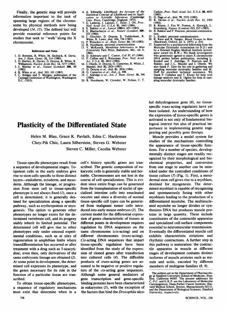
studies of the mechanisms controllingthe appearance of tissue-specific func-tions. For a number of species, develop-mentally distinct stages are readily rec-ognized by their morphological and bio-chemical properties, and conversionfrom one stage to another can be mim-icked under the controlled conditions oftissue culture (7) (Fig. 1). First, a meso-dermal stem cell gives rise to a myoblast,destined for myogenesis. The deter-mined myoblast is capable of recognizingand spontaneously fusing with othermyoblasts leading to the production of adifferentiated myotube. The multinucle-ated myotube no longer divides or syn-thesizes DNA but produces muscle pro-teins in large quantity. These includeconstituents of the contractile apparatusand specialized cell-surface conponentsessential to neuromuscular transmission.Eventually the differentiated muscle cellexhibits characteristic striations andrhythmic contractions. A further step inthis pathway is maturation: the contrac-tile apparatus in muscle at differentstages of development contains distinctisoforms of muscle proteins such as my-osin and actin, encoded by differentmembers of multigene families (8, 9).
The authors are in the Department of Pharmacolo-gy at Stanford University School of Medicine, Stan-ford, California 94305. The present addresses forC.-P.C. and G.K.P. are the Laboratory of MolecularCarcinogenesis, Dana-Farber Cancer Institute, Har-vard Medical School, Boston, Massachusetts 02115,and the Department of Biological Sciences, StanfordUniversity, respectively.
SCIENCE, VOL. 230
Heterokaryons: A Model System forStudying Ceil Specialization
To study the mechanisms regulatingcell specialization, we developed a sys-
tem in which nonmuscle cells can beinduced to express muscle genes predict-ably and stably. We combine entire mus-cle cells with cells of other phenotypesand from other species through the use
of polyethylene glycol. In these fusedcells, or heterokaryons, expression ofpreviously silent muscle genes is activat-ed in response to tissue-specific factorspresent in muscle (10-12). The nuclearcomposition of the heterokaryons andthe activation of genes can be deter-mined by taking advantage of species-specific differences: the nonmuscle cellsused are always human and the musclecells are always mouse. Furthermore,the fusion product is stable; in contrastwith typical interspecific hybrids (syn-karyons), cell division does not occur
and the parental cell nuclei remain intactand retain a full complement of chromo-somes. Finally, changes in gene expres-
sion can be assayed immediately afterfusion and monitored for relatively longperiods thereafter.We have shown that muscle genes are
activated in nonmuscle nuclei upon fu-sion with muscle. This activation must
be mediated -by diffusible, trans-actingmolecules that are transported to nucleithrough the cytoplasm, since the nucleiof the two cell types remain separate anddistinct. These results show that gene
expression by nuclei of highly special-ized cells is remarkably plastic. Thus,the muscle genes in cells with very dif-ferent roles (skin, cartilage, lung, andliver) are receptive to activation by mus-
cle regulatory factors. The differences inthe requirements for gene activation inthese cell types provide insight into themolecular mechanisms that lead to thegeneration and maintenance of pheno-types.
Gene Activation in Heterokaryons
The mouse muscle cell line used toproduce heterokaryons is a diploid sub-clone, C2C12 (13). To test the influence ofhistogenetic state and of in vivo aging onmuscle gene expression, heterokaryonswere produced with mouse muscle andeight different human nonmuscle celltypes, including keratinocytes (ecto-derm), chondrocytes and fibroblasts(mesoderm), and hepatocytes (endo-derm) (14). In addition, the four strainsof fibroblasts used were either from dif-ferent tissues (amniotic fluid, skin, andlung) or from different developmental
Summary. Heterokaryons provide a model system in which to examine how tissue-specific phenotypes arise and are maintained. When muscle cells are fused withnonmuscle cells, muscle gene expression is activated in the nonmuscle cell type.Gene expression was studied either at a single cell level with monoclonal antibodiesor in mass cultures at a biochemical and molecular level. In all of the nonmuscle celltypes tested, including representatives of different embryonic lineages, phenotypes,and developmental stages, muscle gene expression was induced. Differences amongcell types in the kinetics, frequency, and gene dosage requirements for geneexpression provide clues to the underlying regulatory mechanisms. These resultsshow that the expression of genes in the nuclei of differentiated cells is remarkablyplastic and susceptible to modulation by the cytoplasm. The isolation of the genesencoding the tissue-specific trans-acting regulators responsible for muscle geneactivation should now be possible.
Table 1. Cell types in which human muscle genes were activated in heterokaryons.
Biological Muscle gene Assay Cell lineage Phenotype testedfunction product tested
Enzyme Creatine kinase Electrophoresis and Mesoderm Amniotic fibroblastHuman MM enzyme activity Fetal skin fibroblastHuman MB Adult skin fibroblastMouse-human Lung fibroblast
hybrid MM HeLa (malignant)Chondrocyte
Ectoderm KeratinocyteEndoderm Hepatocyte
Contractile Myosin light chains Electrophoresis and Mesoderm Amniotic fibroblastapparatus Fetal monoclonal
Is antibodies2s2f
Actin mRNA's cDNA probesa-cardiac Mesoderm Fetal skin fibroblast
Adult skin fibroblastEctoderm Keratinocyte
a-skeletal Mesoderm Fetal skin fibroblastAdult skin fibroblast
Ectoderm Keratinocyte
Membrane Cell surface antigens Monoclonal antibodiescomponents 24. 1D5 Mesoderm Fetal skin fibroblast
Lung fibroblast5.lHIl Mesoderm Fetal skin fibroblast
Adult skin fibroblastLung fibroblastHeLa (malignant)
Ectoderm KeratinocyteEndoderm Hepatocyte
75915 NOVEMBER 1985
Totipotential (I Stem-cell production
( DeterminationMesodermastem cell Gt t Differentiation
Myoblast 4Maturation
myotube
myotube
stages (fetal skin and adult skin). Onemalignant cell type, HeLa, was also test-ed. To assess the ability of these non-muscle cell types to be reprogrammed toexpress muscle functions in heterokary-ons, it was important to verify theiridentity and determine that they contin-ued to function as specialized cells intissue culture. Accordingly, at the timeoffusion with muscle cells, tissue-specif-ic products were identified in cultures ofchondrocytes, keratinocytes, and hepa-tocytes by immunofluorescence withantibodies to type II collagen, keratin,and albumin, respectively (15). Althoughphenotypic markers are not well charac-terized for fibroblasts, we determinedthat the two strains of skin fibroblastsused in the studies of developmentalstage, from a fetus (14 weeks) and froman adult (66 years), were distinct celltypes: they differed in their proliferativeproperties, cell size, and binding of themitogens, insulin, and insulin-likegrowth factor 1 (16, 17).We took advantage of species differ-
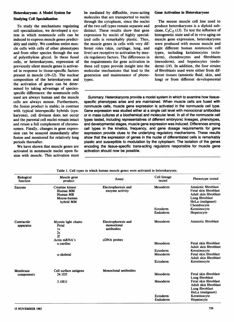
ences to assay the induction of expres-sion of muscle genes in nonmuscle cells.A heterokaryon is readily identified byits mixed nuclear composition: mousenuclei appear punctate and human nucleiare uniformly stained with the fluores-cent dye Hoechst 33258 (Fig. 2). Thenovel activation of a muscle gene isevident by detection of a product specif-ic to human muscle on the heterokaryoncell surface, a product which neither celltype alone produces. This gene product,5.1Hll, is an antigen present in smallamounts on human myoblasts and in large
amounts on myotubes (18). Unfused non-muscle cells grown on the same dish withheterokaryons did not express musclefunctions. Thus, assays of gene productscould be performed at the level of a
single cell in heterokaryons of definednuclear composition. Gene activationwas also analyzed in mass cultures atbiochemical and molecular levels.
In each case tested, human musclegenes were activated (Table 1). Humanmyosin light chains-ls, 2s, 2f, and fe-tal-and human isozymes of creatine ki-nse (CK)-MB, MM, and a functional
Fig. 1. Points of regu-lation in muscle cellspecialization.
mouse-human hybrid MM-were distin-guished from their mouse muscle coun-terparts by their mobility upon gel elec-trophoresis; their identities were con-firmed either by reaction with monoclo-nal antibodies on Western blots or by insitu assays of enzyme activity (10, 19).The human muscle-specific transcriptsof two sarcomeric actin genes, a-cardiacand a-skeletal actin, were detected byNorthern blot and S1 nuclease analysiswith species- and isotype-specific com-plementary DNA (cDNA) and genomicDNA probes (20). Expression of the hu-man muscle-specific cell surface anti-gens, 24.1D5 and 5.1H11, was inducedand could be monitored on single hetero-karyons with monoclonal antibodies (11,
Fig. 2. Activation of human muscle gene for5.lH1l in heterokaryons. Live cells wereincubated with monoclonal antibody to5.lHll followed by biotin conjugated to anantimouse antibody and avidin conjugated toTexas red. Heterokaryons are shown in fluo-rescence microscopy at two different wave-lengths. The binucleate heterokaryon contain-ing one punctate mouse muscle nucleus andone uniformly stained human hepatocyte nu-cleus expresses the antigen, which is uniform-ly distributed on the cell surface. The trinu-cleate heterokaryon (lower center) has notactivated the gene for 5.1H1.
17, 21, 22). Thus, the genes that wereactivated encoded diverse products: en-zymes critical to energy production,structural components of the contractileapparatus, and cell surface components.The relative amounts and sequence ofexpression ofthese different muscle geneproducts in heterokaryons paralleledmyogenesis in pure cultures of humanmuscle cells.
The Kinetics of Gene Expression Differ
We examined the time course and fre-quency of muscle gene expression indifferent cell types contained in hetero-karyons. The proportion of individualheterokaryons expressing the cell sur-face antigen 5. lHI 1 was determined be-tween 1 and 15 days after fusion in 19independent experiments in which morethan 7000 individual heterokaryons wereanalyzed (Fig. 3). The three distinct pat-terns observed differed in the timecourse of gene expression, primarily be-cause of differences in the lag periodbefore 5.1H 1I could be detected. In ad-dition, the ultimate frequency of geneexpression differed among cell types andapproximated 95, 60, and 25 percent forfibroblasts, keratinocytes, and hepato-cytes, respectively. Thus, tissue deriva-tion and possibly embryonic origin havemarked effects: fibroblasts, which arefrom the same embryonic lineage asmuscle (mesoderm), exhibit faster kinet-ics and a higher ultimate frequency of5. IHI I expression than keratinocytes(ectoderm) and hepatocytes (endoderm).In contrast, the kinetic curves for skinfibroblasts from two developmentalstages were indistinguishable (16, 17).To test whether the differences in the
kinetics of 5. IHI I accumulation weredue to phenotypic differences in thetranslation and subsequent processing ofa cell surface protein, the expression ofanother muscle gene was examined atthe transcriptional level. The relativelevels of the messenger RNA's(mRNA's) for the muscle-specific a-car-diac and a-skeletal actins were studied inmass cultures of heterokaryons. Weused species- and isotype-specific cDNAprobes that recognize the transcripts of-the human a-cardiac and a-skeletal actingenes, respectively, but not those ofmouse or of other actin genes (23). Botha-cardiac and a-skeletal actin expressionwas evident in heterokaryons, and thetime course and relative levels of the twotranscripts differed (Fig. 4). These differ-ences in sarcomeric actin expressionwere similar to those observed in purecultures of human muscle cells (20).With this assay we compared the kinet-

ics of accumulation of actin transcripts inheterokaryons produced with keratino-cytes and with fibroblasts. The resultsparalleled those obtained for 5.1Hl1expression. a-Cardiac actin transcriptswere not detectable in keratinocyte het-erokaryons on days 1 and 2 after fusion,when the levels in fibroblast heterokary-ons were marked. By day 5, transcriptswere detectable in keratinocyte hetero-karyons as well (16). Since the differ-ences in kinetics observed at the proteinlevel were also observed at the mRNAlevel, they probably reflect differencesamong cell types in steps necessary forthe activation of gene expression.
Mechanisms for Gene Activation
To examine potential mechanisms re-quired for gene activation, we deter-mined whether DNA replication wasnecessary to reprogram cells from differ-ent stages of development and of differ-ent phenotypes to express muscle genes(11, 16, 17). We exposed nonmuscle cellsto the DNA synthesis inhibitor cytosinearabinoside (Ara-C) before and after fu-sion. The expression of human muscleCK was assayed in extracts of wholecells, and the expression of 5. lHl wasmonitored on individual heterokaryons.The human M-CK gene was activated inall of the seven nonmalignant cell typestested, and three novel CK isozymescontaining this subunit were detected:human MB, human MM, and mouse-human hybrid MM (Fig. 5). In all cases,similar amounts of the human isozymeswere present regardless of whether DNAsynthesis was inhibited. Furthermore,inhibition ofDNA synthesis also had noeffect on the frequency of 5. 1Hl 1expression at the single-cell level. Theseresults suggest that alterations in chro-matin configuration requiring DNA repli-cation are not necessary for the musclegenes in these cell types to be accessibleto and respond to regulatory factors pre-sent in differentiated muscle cells.
In contrast to these results, CK and5. lHl 1 muscle gene expression werenever observed when we formed hetero-karyons with HeLa cells, a malignant,aneuploid cell type (12). We examinedwhether prior treatment with 5-azacyti-dine (SAc), a drug thought to reducegene methylation (24), could alter theresponsiveness of HeLa cells to musclegene regulators in heterokaryons. In-deed, the expression of both 5. lH 1I andCK isozymes containing human subunitswas detected in heterokaryons formedwith SAc-treated HeLa cells, but not incontrol heterokaryons containing un-treated HeLa cells (Fig. 5) or in 5Ac-
treated HeLa cells fused to themselves(12). Therefore gene activation in HeLacells appears to require a mechanisticstep not required by the other cell typestested: first a change induced by SAc isnecessary and then interaction with mus-cle gene regulators.
Gene Dosage Influences Gene Expression
Further differences among cell typesin the activation of muscle genes wereapparent when we examined the effectsof gene dosage, or nuclear ratio, on thefrequency of muscle gene expression
100 Fibroblast Fig. 3. Kinetics of
a: t 5.lHll expression ina 80 heterokaryons con-
taining different cell¢ 60 A Keratinocyte types. Individual het-oI / erokaryons contain-0 1>- 40 - ing nuclei from mus-
Hepatocyte cle and from either2 20 - lung fibroblasts (0),a / keratinocytes (A), or
0 1 hepatocytes (U) were1 2 3 4 6 8 10 12 15 analyzed in replicate
Days after fusion cultures for nuclearcomposition and the
expression of 5.lH11 between 1 and 15 days after polyethylene glycol fusion. Curves arecomputer-derived best fit lines of the data. The size of the symbols includes ± 1 standard errorof the proportion calculated from the standard binomial equation.
H M 1 2 6
Fig. 4. Activation of human muscle-specificactin mRNA's in heterokaryons. Total RNA'swere isolated from differentiated human mus-cle cells (H), differentiated mouse musclecells (M), and mouse muscle-human lungfibroblast heterokaryon cultures on days 1, 2,and 6 after polyethylene glycol-mediated fu-sion. RNA's (5 ,ug from human muscle cellsand 10 ,ug from each of the other samples)were electrophoresed on agarose gels, trans-ferred to nitrocellulose, and hybridized withthe human-specific cDNA probes to either a-cardiac actin (A) or a-skeletal actin (B). Auto-radiograms were exposed to XAR-5 film at-80°C for 6 days.
H M 1 2 6
F(A) F(L) F(FS) F(AS) K C H HeLa Fig. 5. Activation ofhuman creatine ki-nase in different non-
MM muscle cells. Whole-cell extracts wereelectrophoresed on5 percent nonde-
MB naturing polyacrylam-ide gels, and the CKisozymes were de-tected with ultravioletillumination and a
BB coupled enzyme reac-M Hu- + + + -+ - + + -+ tion, yielding the re-
Ara-C 5-Ac duced form ofNADP+ as its end-
product. CK isozymes are shown for mouse (M) muscle cells and human (Hu) muscle cells.Heterokaryons formed between mouse muscle cells and fibroblasts from amniotic fluid F(A),lung F(L), fetal skin F(FS), and adult skin F(AS), keratinocytes (K), chondrocytes (C), andhepatocytes (H) are shown. Cultures marked + were exposed to cytosine arabinoside (Ara-C)for 24 hours before and 24 hours after fusion to ensure inhibition of DNA synthesis. Culturesmarked - were not exposed to Ara-C during this period. Heterokaryons formed between mousemuscle cells and HeLa cells were either untreated (-) or exposed to 5 FM of 5-azacytidine (5-Ac) for 3 days followed by 1 day of drug removal (+). Equivalent enzyme activities were loadedin each lane. Arrows indicate CK isozymes containing human M subunits. Abbreviations: BB isthe nonmuscle isozyme, M-subunit synthesis is initiated early in differentiation, and the dimersMB and MM are characteristic of differentiated muscle.

Fig. 6. Effect of nu-clear ratio on 5.1H1expression in hetero-karyons containingdifferent cell types.Individual hetero-karyons containingnuclei from muscleand from either lungfibroblasts, keratino-cytes, or hepato-cytes were analyzedfor nuclear composi-tion and the expres-sion of 5.1HIl 6days after fusion.Data were groupedinto five ratios ofmuscle: nonmusclenuclei. Error bars in-dicate the standarderror of the propor-tion calculated fromthe standard binomi-al equation.
100
r-
-75
.co0
a 50x0
c
0
25
0o
Fibroblast
(25). Nuclear composition and theexpression of the human muscle cellsurface antigen 5.1Hl1 were monitoredin the same heterokaryons with fluores-cence microscopy at two different wave-lengths (Fig. 2). The results for hetero-karyons of different nuclear compositionwere pooled into five groups accordingto nuclear ratio, or the relative numberof muscle to nonmuscle nuclei they con-
tained (Fig. 6). The proportion in eachgroup that expressed the antigen on a
given day was determined. Examples forthe 6-day time point are shown, beyondwhich the pattern of gene expression didnot change.The effect of gene dosage on 5.1H11
expression differed markedly for thethree cell types. For fibroblasts, a highproportion of heterokaryons (95 percent)expressed 5.lHl1 at all nuclear ratios,except in the increased fibroblast group(1: > 2), in which it was -70 percent.In keratinocyte heterokaryons, gene
expression in the 1:1 nuclear ratio groupnever exceeded 30 percent, whereas in-creased proportions of either muscle or
keratinocyte nuclei resulted in the maxi-mum expression of 70 percent. For hepa-tocyte heterokaryons, the frequency ofgene expression was greatest (50 per-
cent) when the relative number of mus-cle nuclei was increased and lowest (5percent) when the proportion of hepato-cyte nuclei was increased.Although the interaction between two
disparate cell types combined by fusionis likely to be complex, some noteworthyrelationships are apparent from the stud-ies ofgene dosage (16, 25). (i) Even whenthe nonmuscle nuclei of each cell typeoutnumbered the muscle nuclei, muscle
762
Keratinoc yte Hepatocyte
/ I \\1:>2 1:2 1:1 2:1 >2:1
Nuclear ratio(mouse muscle: human nonmuscle)
gene expression was induced, albeit withdifferent frequencies. (ii) The kinetics ofgene expression in fibroblast heterokary-ons with increased proportions of non-
muscle nuclei were slower than when thenuclear ratio was 1:1. These resultscould be due to a requirement for gene
activation of a threshold concentrationof positive regulators: increased timewould be necessary for the progressiveaccumulation of factors that must beshared among fibroblast nuclei. The ratewith a 1:1 nuclear ration exceeded thatwith increased muscle nuclei for fibro-blast heterokaryons, which suggests thatincreased input from muscle was notoptimal; instead a balance offactors con-tributed by each cell type might be re-
quired.In hepatocyte heterokaryons, the fre-
quency of activation of muscle gene
expression was lowest when the propor-tion of hepatocyte nuclei was increasedand highest when the proportion of mus-cle nuclei was increased. That these fre-quencies did not change with time sug-
gests that gene expression was deter-mined by the continued balance betweencomponents contributed by each cell
type. Keratinocytes were more frequent-ly activated to express muscle geneswhen either the proportion of keratino-cyte or of muscle nuclei was increased inheterokaryons. The increased frequencyofgene expression in heterokaryons withexcess keratinocyte nuclei could be dueto the existence of subpopulations ofcells in keratinocyte cultures that dif-fered in their ability to respond to muscleregulatory factors. Accordingly, thechance of obtaining a keratinocyte inwhich activation could occur would in-
crease with the number of keratinocytenuclei. Stratification within colonies ofkeratinocytes was evident in our cul-tures, and although the upper, more ma-ture cells reportedly do not replate well(26), possibly some were incorporatedinto heterokaryons.
Expression of the Nonmuscle Phenotype
To test whether the phenotype of thenonmuscle cell persists in heterokary-ons, we monitored the expression ofalbumin in individual hepatocyte hetero-karyons. Six days after fusion, when themuscle product 5.lH1 1 was expressed atmaximum frequency, albumin was stillpresent in a large proportion of hepato-cyte heterokaryons. By 15 days afterfusion 5.lHlI was still expressed atmaximal frequency, but the proportionof albumin-containing heterokaryonswas reduced. The persistence of albuminat 6 days was not due to the stability ofpreviously synthesized proteins, sincealbumin is secreted within a few hours ofits production. To detect albumin in het-erokaryons, we had to inhibit secretionwith monensin 2.5 hours before fixation(16, 17). Furthermore, the decline inalbumin-containing heterokaryons withtime was not due merely to extinction ofthe liver phenotype in the course of long-term culture, since parallel cultures ofhepatocytes fused to themselves withpolyethylene glycol (PEG) containedalbumin on day 15. From these experi-ments we cannot determine whether theexpression of the genes of one pheno-type precludes the expression of thegenes of the other. This determinationwould require assays of active transcrip-tion for genes characteristic of both non-muscle and muscle phenotypes in singlenuclei. Despite this limitation, our re-sults indicate that the combination ofspecialized cells in a common cytoplasmdoes not result in the immediate degrada-tion of differentiation-specific proteins.Instead, we conclude that the nonmusclephenotype persists for a time in hepato-cyte heterokaryons, but the muscle phe-notype dominates.
Regulatory Circuits Between Nuclei in
Heterokaryons
To examine whether the nonmusclecell influences gene expression in themuscle cell, we examined the accumula-tion of mouse and human mRNA's for a-cardiac actin, a major component of themuscle contractile apparatus. We tookadvantage of the fact that prmary human
SCIENCE, VOL. 230
W M=6

muscle cells and cultured mouse C2C12cells exhibited distinct patterns of a-
cardiac actin transcript accumulationwith time after myotube formation (20,27-29). The amount of a-cardiac actinmRNA in each sample was normalizedto the amount present in a human heartstandard analyzed at the same time. Inthe mouse muscle C2C12 cultures, a-
cardiac actin transcripts accumulatedrapidly, transiently peaked at a levelcomparable to 50 percent of the a-cardi-ac actin mRNA expressed in humanheart within 24 hours, and declined (Ta-ble 2). In contrast, a-cardiac actin tran-scripts accumulated steadily in humanmyotubes over a 6-day period and ex-ceeded the peak reached in the C2C12cell line 16-fold, a level comparable to800 percent that observed in humanheart. In heterokaryon cultures, the timecourse ofhuman transcript accumulationand the amount of transcript per non-muscle nucleus were similar to thoseobserved in pure human, not mouse,muscle cultures. Of greatest surprise wasthe result obtained for mouse a-cardiacactin mRNA in heterokaryons: insteadof continuing to decline, as in puremouse muscle cultures, the amount ofmouse transcripts increased after hetero-karyon formation.These findings suggest that the human
a-cardiac actin gene responds to mousemuscle cytoplasmic factors by producingtranscripts with the time course and atlevels typical ofhuman, not mouse, mus-cle cultures. These differences in tran-script accumulation could arise in partfrom differences in the cis-acting regula-tory regions of the mouse and human a-
cardiac actin genes. In addition, the re-sults are compatible with the hypothesisthat trans-acting mouse muscle factorsactivate human muscle regulatory genesencoding factors that, in turn, act onmouse muscle genes or stabilize mousea-cardiac actin transcripts. Finally, the"activated" phenotype seems to domi-nate, since the usual decline in theamount of mouse a-cardiac actin tran-scripts is overridden by the presence ofthe human nuclei.
Muscle Gene Regulators at DistnctStages of Development
Like the experiments describedabove, the majority of studies of geneexpression in specialized cells have uti-lized the differentiated stage of develop-ment (Fig. 1). This is true primarily be-cause for all cell types, this stage isgenerally the best characterized and.most readily obtained in tissue culture.15 NOVEMBER 1985
Table 2. Relative a-cardiac actin mRNA in pure muscle and in heterokaryon cultures. PEGtreatment occurred after the mouse muscle myotubes had been in fusion medium for 3 days.Thus, to compare the mouse time scale in fusion medium (left) with that after PEG treatment(right), add 3 days.
a-Cardiac actin mRNA Days a-Cardiac actin mRNADays in human heart (%) after in human heart (%)
infusion Mouse Human PEG Mouse in Human inmedium muscle primary treat- hetero- hetero-
C2CI2 muscle ment karyons karyons0 20 1 0 01 50 100 1 10 502 25 200 2 30 2006 20 800 6 40 700
The experiments described below showthat other stages of muscle develop-ment-determination and maturation-are now amenable to analysis.To test whether muscle cells at the
determined stage of development elabo-rate distinct muscle gene regulators, weproduced heterokaryons with fibroblastsand muscle cells at the myoblast (deter-mined) and myotube (differentiated)stages (22). Convenient markers for thisstudy were provided by two cell-surfaceantigens: 5.AH1I and 24.1D5. The5.AHl1 antigen is expressed on culturedmyoblasts to some extent, but markedlyincreases in amount on myotubes. The24. lD5 antigen is detected only on myo-blasts and is no longer present oncemyoblasts fuse to form multinucleateddifferentiated myotubes. The humanforms of both muscle cell-surface com-ponents were recognized with species-specific monoclonal antibodies (17, 20).The 24. 1D5 antigen was detected only inmyoblast heterokaryons (22 percent)(Table 3).
In contrast, 5.1H 1I was expressed in
both myoblast heterokaryons (19 per-cent) and myotube heterokaryons (92percent). Thus, the expression of thesetwo antigens in heterokaryons paralleledtheir expression in human myogenesis.By day 5, a maximum frequency ofexpression was reached in both casesand further increases were not observedwith time. The lower frequency of geneexpression in myoblast than in myotubeheterokaryons may be due to myoblastnuclei that were in different phases of thecell cycle, whereas most of the myotubenuclei were presumably withdrawn fromthe cell cycle. These results suggest thatmuscle cells at myoblast and myotubestages of development differ in the con-centration or type of at least two regula-tory factors.The study of the maturation stage of
muscle development is facilitated by themyosin heavy chains. These proteins aremajor components of the contractile ap-paratus that are encoded by differentgenes in fetal, neonatal, and adult muscletissues (8). Studies of maturation havebeen hindered because these transitions
Table 3. Evidence for two temporally distinct muscle gene regulators. Fetal skin fibroblastswere used except in myotube experiment 3, in which lung fibroblasts were used; S.E.M.,standard error of the mean.
Heterokaryons expressing
24.1D5 5.1HIIExperiment Percentage Percentage
exhibiting Number exhibiting Numberdetectable scored detectable scoredantigen antigen
Fusion partner: myoblast1 34 228 8 2292 13 131 19 593 20 113 30 93.
Mean ± S.E.M. 22 ± 6 472 19 6Total 381
Fusion partner: myotubeI 1 556 90 4072 0 800 98 1663 0 600 88 376
Mean ± S.E.M. 0- 1956 92 ± 3Total 949
763

are not readily obtained in vitro; primari-ly fetal forms of muscle proteins areexpressed in cultured myotubes. Wehave determined that C2CI2 cells expressdevelopmentally distinct myosins. Thesemyosin isoforms were detected with-monoclonal antibodies generated to my-osin purified from human muscle at dif-ferent developmental stages (30). C2CI2cells are stained with two distinct anti-bodies that recognize myosin isozymespresent at fetal and neonatal stages ofdevelopment (Fig. 7). That the intensityof staining differed among myotubes sug-gests that the relative concentrations offetal and neonatal myosins differ. A pro-*ressive increase in the relative amountof neonatal myosin was observed with:tme in culture. The results were not dueto the use of the C2C12 cell line, sincethey have been corroborated with humanprumary muscle cultures (31). Conse-quently, neural contact and complexsubstrates do not seem to be required forcertain steps in muscle maturation. Sincesome of our antibodies to myosin arehuman-specific, it should now be possi-ble to examine the activation of myosinisozymes in heterokaryons and the regu-latory mechanisms underlying the matu-ration stages of muscle development.
Dbcuulen
Several theories have been proposedto account for the development of celldiversity: changes in the genome thataccompany quantal mitoses (32), se-quential repression ofgenes (33) or mod-ulation of gene expression by activatorsand repressors (34) with additional com-plex regulatory effects resulfing from therelative concentration and affinity ofthese factors for specific sequences with-in genes (4-6). Some major questions are
raised by these theories: How fixed andirreversible is the differentiated state or
tissue-specific phenotype of a cell? Whatis required to maintain the differentiatedstate? What underlying molecular mech-anisms regulate the transition from thedetermined to the differentiated to themature cell phenotype? Do these mecha-nisms differ for different specialized cell
types?Our approach to these questions was
inspired by reports by Weiss and othersof experiments with somatic cell hy-brids, in which the expression of genesatypical of a cell's normal phenotypecould be induced (35, 36). A limitation inthe comparative analysis of gene activa-tion in different cell types is that highly
Fig. 7. Expression offetal and neonatal my-osin isozymes inC2C12 muscle cuI-thres. (Top) Reactionof cells with an immu-noglobulin G (IgG)monoclonal antibody(FI.193) that recog-nizes fetal myosin, vi-sualied with fluores-cen-conjugated anti-serum to mouse IgG.(Bottom) Reaction ofthe same ceUs with an
iMMunoglobulin M(ISM) monoclonalantibody (N3.36) thatrecognzes neonatalmyosin, visulizdwith rdamiecon-
jugated antiserum tomouse IgM. Th cells
were fixed with 1 per-cent fomalin 8 daysafter being trans-ferred to differentia-tion medium (2 per-cent horse serum in>lbecco's modified
Eagle's medium).
ancuploid transformed cells were usuallyused. In addition, hybrid cells were fre-quently obtained after proliferation inmitogen-rich media, which led to vari-able retention and rearrangement of ge-netic material (35, 37), both of which arelikely to have influenced gene expression(38). As a result, certain phenotypeswere thought to be incapable of beingactivated, because of either their special-ized state or their developmental stage(37, 39).The activation of muscle gene expres-
sion in all of the eight nonmuscle celltypes we tested is likely to be due toseveral factors. To facilitate the study ofnormal regulatory mechanisms, mostnonmuscle cells were diploid primarycells. To overcome the problems of geneloss and rearrangement, we used a het-erokaryon system that remained stablefor the 2-week life-span of the cells andpermitted a systematic analysis of geneexpression over time. We cannot ruleout the possibility that muscle is particu-larly well suited for intemuclear commu-nication and existence as a heterokary-on, since it is naturally a multinucleatedcell in its differentiated state. However,the use of culture conditions which pro-moted muscle differentiation, not celldivision, was probably critical to thesuccess ofour experiments. Proliferationand differentiation are antagonistic, mu-tually exclusive states in muscle cells(40). Furthermore, specific culture con-ditions that differ markedly among celltypes are required for the optimalexpression of phenotypes in vitro (41).Many investigators using somatic cellgenetics to analyze the expression ofdifferentiated functions used a nonspe-cific mitogen-rich culture medium. Wepropose that with culture conditions thatfavor the differentiation ofthe phenotypeof interest, gene activation in stable het-erokaryons could be achieved with celltypes other than muscle.The ability to activate muscle genes in
heterokaryons is not a peculiarity of theC2CI2 cell line. We have recently beenable to induce human muscle geneexpression with another mouse muscleline, the myogenic derivative of a fibro-blastic lOTl/2 cell tated with 5-azacyti-dine (13). However, the frequency ofgene activation in fibroblast heterokary-ons formed with IOTI/2 is half that ob-served with C2C12 cells. We selected theC2C12 clone for its potential to give riseto extensive diffierentiated contractilemyotubes within a short period, and inthis respect it exceeds all other myogeniclines with which we have had experi-ence. Thus, C2CI2 may be an optimal
SCIENCE, VOL. 230

source of muscle gene regulatory fac-tors. Another myogenic cell line thatmay be enriched for regulators has beenobtained by Wright, who selected formuscle cells resistant to an inhibitor ofdifferentiation, bromodeoxyuridine (42).The selection of cells that undergo rapiddifferentiation should also prove usefulfor characterizing tissue-specific regula-tors in other phenotypes.The plasticity in the function of the
nucleus of specialized cells is remark-able. In each nonmalignant cell type test-ed, the differential state could be alteredin the absence ofDNA replication or celldivision and the cells induced to expresshuman muscle functions after exposureto muscle cytoplasm. Representatives ofdifferent embryonic lineages, pheno-types, and developmental stages weretested, including four strains of fibro-blasts and chondrocytes (mesoderm),keratinocytes (ectoderm), hepatocytes(endoderm), and malignant HeLa cells.Differences among cell types were ob-served in the frequency, kinetics, andgene dosage requirements for the expres-sion of human muscle genes. Cells thatwere more closely related to muscle-mesodermal derivatives-consistentlyexpressed muscle genes sooner and at agreater frequency than the cells of ecto-dermal or endodermal origin.Our experiments suggest that the mod-
ulation of gene expression which accom-panies eukaryotic cell differentiation ismediated by a number of positive andnegative regulatory factors in a concen-tration-dependent manner. When cellsare mixed by fusion, the end result islikely to be due to complex interactions;the following simple interpretation of ourexperiments may nonetheless provide auseful conceptual framework. The de-layed but high frequency of expressionof muscle genes in heterokaryons withone muscle and multiple nonmuscle nu-clei suggests that muscle regulators mustreach a threshold concentration for geneactivation to occur. The observation thatmuscle gene expression in fibroblast het-erokaryons is fastest with a 1:1 ratiosuggests that a balance of factors con-tributed by both cell types is optimal forthat cell type. On the other hand, thefrequency of gene expression in hepato-cytes differs in heterokaryons containingdifferent proportions of muscle and non-muscle nuclei, and the differences persistwith time, suggesting that hepatocytesproduce negative regulators that must betitrated by the positive regulators in mus-cle cells. Muscle cells at different stagesof development seem to synthesize dif-ferent positive regulators. Finally, the15 NOVEMBER 1985
reprogramming of the nonmuscle nucle-us in a heterokaryon results in the syn-thesis by that nucleus of muscle regula-tory factors. Thus, from an analysis ofthe combined effects on gene expressionof two cell types fused to form hetero-karyons, inferences regarding the regula-tors that generate and maintain thesephenotypes can be drawn.Gene expression in cells specialized
for very different functions is not fixedand irreversible. Although under normalcircumstances the pattern of geneexpression of specialized cells is stableand heritable, it can be altered if theregulatory circuits between nucleus andcytoplasm are disrupted. Thus, changesin gene expression during developmentdepend not only on the nucleus, but alsoon the cytoplasm, which plays an essen-tial role as signal transducer. Our resultsextend the observations that nucleargene expression is subject to modulationby the cytoplasm (43) to a system thatmay be particularly amenable to molecu-lar analysis.
Future Prospects
Heterokaryons have utility as experi-mental models for investigating the regu-lation of gene expression in differentiat-ed cells. The cells that are repro-grammed in heterokaryons should pro-vide useful test systems for studying themechanisms of action of the relevantmolecules. Differences among cell typesin the lag period before gene expression,the time course of accumulation of geneproducts, and the frequencies of geneexpression at different nuclear ratiossuggest different mechanistic steps.These could result from differences ingene conformation due to DNA modifi-cations or binding proteins in addition todifferences in the requirement for thetype, concentration, or stoichiometry oftrans-acting factors. We and others havebegun to characterize the cis-acting regu-latory sequences of muscle-specificgenes (44). The isolation of the genes fortrans-acting muscle-regulatory mole-cules at determined, differentiated, andmature stages of muscle developmentshould now also be possible. Approach-es that may prove useful for isolatingmRNA's and genes for uncharacterizedfactors include microinjection of cyto-plasmic components, use of subtractivehybridization to enrich for mRNA's forstage-specific regulators, and transfec-tion of either cDNA's made to the en-riched mRNA's or of genomic DNA intocells in conjunction with selective assays
for gene expression (45). Elucidation ofthe molecular mechanisms underlyingthe reprogramming of nonmuscle celltypes should lead to a better understand-ing of the generation and maintenance ofthese phenotypes and of different myo-genic stages.
Referenc and Notes1. J. R. Whittaker, Dev. Biol. 93, 463 (1982); J. E.
Sulston, E. Schierenberg, J. G. White, J. N.Thomson, ibid. 100, 64 (1983).
2. T. Yamada and D. S. McDevitt, ibid. 38, 104(1974); D. A. Dunis and M. Namenwirth, ibid.56, 97 (1977); S. M. Taylor and P. A. Jones, Cell17, 771 (1979); A. B. Chapman, D. M. Knight,B. S. Dieckmann, G. M. Rigold, J. Biol. Chem.259, 15548 (1984); S. F. Koneczny and C. P.Emerson, Jr., Cell 38, 791 (1984).
3. J. B. Gurdon, J. Embryol. Exp. Morphol. 10,622 (1962); and V. Uehlinger, Nature(London) 210, 1240 (1966); M. J. Dewey, D. W.Martin, Jr., G. R. Martin, B. Mintz, Proc. Natl.Acad. Sci. U.S.A. 74, 5564 (1977).
4. S. L. McKnight and R. Kingsbury, Science 217,316 (1982); D. A. Goldberg, J. W. Posakony andT. Maniatis, Cell 34, 59 (1983); Y. Gluzman andT. Shenk, Eds., Enhancers and EukaryoticGene Expression (Cold Spring Harbor Labora-tory, Cold Spring Harbor, N.Y., 1983); R. Treis-man, M. R. Green, T. Maniatis, Proc. Natl.Acad. Sci. U.S.A. 80, 7428 (1983); D. D. Brown,Cell 37, 359 (1984); M. Mercola, J. Goverman,C. Mirell, K. Calame, Science 227, 266 (1985);A. Ephrussi, G. M. Church, S. Tonegawa, W.Gilbert, ibid., p. 134; R. Grosschedl and D.Baltimore, Cell 41, 885 (1985); C. M. Gorman,P. W. J. Rigby, D. P. Lane, ibid. 42, 519 (1985);K. R. Yamamoto, Annu. Rev. Genet., in press.
5. H. R. Pelham and D. D. Brown, Proc. Nat!.Acad. Sci. U.S.A. 77, 4170 (1980); D. R. En-gelke, S.-Y. Ng, B. S. Shastry, R. G. Roeder,Cell 19, 717 (1980); W. S. Dynan and R. Tjian,Nature (London) 316, 774 (1985); A. B. Lassar,P. L. Martin, R. G. Roeder, Science m, 740(1983); C. S. Parker and J. Topol, Cell 37, 273(1984); C. Wu, Nature (London) 311, 81 (1984).
6. U. Heberlein, B. England and R. Tjian, Cell 41,965 (1985).
7. D. Yaffe, Proc. Natl. Acad. Sci. U.S.A. 61, 477(1968); R. Bischoff and H. Holtzer, J. Cell Biol.41, 188 (1969); I. R. Konigsberg, Dev. Biol. 26,133 (1971); C. P. Emerson and S. K. Beckner, J.Mol. Biol. 93, 431 (1975); J. P. Merlie and F.Gros, Exp. Cell Res. 97, 406 (1976); D. Yaffe and0. Saxel, Nature (London) 270, 725 (1977); B.Nadal-Ginard, Cell 15, 855 (1978); R. V. Storti etal., Cell 13, 589 (1978).
8. G. F. Gauthier and S. Lowey, J. Cell Biol. 74,760 (1977); R. G. Whalen et al., Nature (Lon-don) 292, 805 (1981); V. Mahdavi, M. Peria-samy, B. Nadal-Ginard, ibid. 297, 659 (1982); D.Bader, T. Masaki, D. A. Fischman, J. CeUl Biol.95, 763 (1982); R. Matsuda, D. H. Spector, R. C.Strohman, Dev. Biol. 100, 478 (1983); R. M.Wydro, H. T. Nguyen, R. M. Gubits, B. Nadal-Ginard, J. Biol. Chem. 258, 670 (1983).
9. A. J. Minty, S. Alonso, M. Caravatti, M. E.Buckingham, Cell 30, 185 (1982); B. M. Patersonand J. D. Eldridge, Science 224, 1436 (1984); W.Bains, P. Ponte, H. Blau, L. Kedes, Mol. CellBiol. 4, 1449 (1984); M. Shani et al., Acids Res.9, 579 (1981); E. A. Fyrberg, J. W. Mahaffey, B.J. Bond, N. Davidson, Ceil 33, 115 (1983); P.Gunning, P. Ponte, H. Blau, L. Kedes, Mol.Cell. Biol. 3, 1783 (1983).
10. H. M. Blau, C.-P. Chiu, C. Webster, Cell 32,1171 (1983).
11. C.-P. Chiu and H. M. Blau, ibid. 37, 879 (1984).12. _ ibid. 40, 417 (1985).13. The mouse muscle cell line used in most experi-
ments was originally obtained by Yaffe andSaxel [in (7)] and is a diploid subclone, C2C12,isolated and karyotyped in our laboratory andselected for its ability to differentiate rapidly andproduce extensive contracting myotubes ex-pressing characteristic muscle proteins. Whereindicated, a myogenic derivative of a IOTI/2 cellprovided by A. Lassar and H. Weintraub wasused. Heterokaryons were produced with poly-ethylene glycol (PEG) (10) and maintained in amedium containing the selective agents cytosinearabinoside (Ara-C) and ouabain to eliminateunfused cells.
14. Tissue-specific cell types included chondrocytesisolated in our laboratory from neonatal carti-
765

lage under the tutelage of L. Smith. Keratino-cytes were a strain (NI) obtained from H.Green, isolated from the outer layer of newbornforeskin. Hepatocytes (Hep G2) were a cell lineisolated from a hepatoma, which is nontumori-genic and retains the ability to stably express 17differentiated functions characteristic of liver[D. P. Aden, A. Fogel, S. Plotkin, I. Damjanov,B. S. Knowles, Nature (London) 282, 615(1979); B. B. Knowles, C. C. Howe, D. P. Aden,Science 209, 497 (1980)]. Fibroblasts were ob-tained from the skin of a 14-week fetus and froma 66-year-old adult-by P. Byers and M. Karasek,respectively. Fibroblasts from fetal lung were acell strain (MRC-5) [J. P. Jacobs, C. M. Jones, J.P. Baille, Nature (London) 227, 168 (1970)].
15. Reagents for the detection of tissue-specificproducts were cDNA probes to a-cardiac and a-skeletal actin genes (P. Gunning, A. Minty, andL. Kedes) and monoclonal antibodies to cellsurface antigens 5.1HII and 24.1D5 (F. Walsh),to myosin light chains (F. Stockdale), to colla-gen type II (R. Burgeson), and to keratin (T.-T.Sun). The human-specific antibody to albuminwas generated by us from nonspecific commer-cial antisera obtained from Cappel Labs. Bind-ing of insulin and insulin-like growth factor Iwere assayed with the aid of R. Roth.
16. H. M. Blau, C.-P. Chiu, G. K. Paviath, E. C.Hardeman, J. Cell. Biochem. Suppl. 9B:35(1985).
17. G. K. Pavlath, C.-P. Chiu, H. M. Blau, inpreparation.
18. F. S. Walsh and M. A. Ritter, Nature (London)289, 60 (1981); 0. Hurko and F. S. Walsh,Neurology 33, 737 (1983).
19. H. M. Blau, C.-P. Chiu, G. K. Pavlath, C.Webster, Adv. Exp. Med. Biol. 182, 231 (1985).
20. E. Hardeman, C.-P. Chiu, H. M; Blau, J. CellBiol. Abstr. 101, 207a (1985).
21. F. S. Walsh, S. E. Moore, R. Nayak, Invest.Exploit. Antibody Comb. Sites, in press.
22. G. K. Pavlath and H. M. Blau, J. CeQl Biol.Abstr. 101, 207a (1985).
23. P. Gunning et al., J. Mol. Evol. 20, 202 (1984).24. D. V. Santi, C. E. Garrett, P. J. Barr, Cell 33, 9
(1983).25. G. K. Pavlath and H. M. Blau, J. Cell Biol., in
press.26. Y. Barrandon and H. Green, ibid. 82, 5390
(1985).27. Human primary muscle cells were isolated and
cultured [H. M. Blau and C. Webster, Proc.Natl. Acad. Sci. U.SA. 78, 5623 (1981); ,G. K. Pavlath, ibid. 80, 4856 (1983); H. M. Blauet al., Exp. Cell Res. 144, 495 (1983)].
28. W. Bains, P. Ponte, H. Blau, L. Kedes, Mol.Cell. Biol. 4, 1449 (1984).
29. To assay mouse and human transcripts, threedistinct DNA probes to the al-cardiac actin genewere obtained from P. Gunning, A. Minty, andL. Kedes. To compare actin transcript accumu-lation in mouse and human muscle cultures, acDNA probe capable of detecting both mouseand human cardiac actin transcripts was used.To assay actin transcripts, cDNA probes werelabeled with 32P by nick translation andhybridized on nitroceilulose filters to serial dilu-tions of total RNA isolated from a time course ofmuscle cultures for each species. To facilitatecomparison among expenments, a sample ofRNA from human heart was always included onthe filters. The amount of a-cardiac actin RNAin the samples was quantitated by densitometricscanning of autoradiograms of the filters, andthe results for each were normalized to a heartRNA standard on that autoradiogram. To exam-
ine the accumulation of human a-cardiac actintranscripts in heterokaryons, a second cDNAprobe was used, which was species- and iso-type-specific: at the hybridization stringencyused, only human a-cardiac actin transcriptswere detected (Fig. 4). To examine the expres-sion of the mouse cardiac actin gene in the sameheterokaryons, the RNA from the heterokary-ons was subjected to SI nuclease analysis. Weused a third probe, which shared homology witha longer sequence of human than mouse a-cardiac actin mRNA. Thus, the sequences formouse and human that hybridized well and wereprotected from treatment with S1, an enzymethat digests single-stranded nucleic acids, dif-fered in size and could be distinuished by gelelectrophoresis. The accumulation of humancardiac actin transcripts determined by thismethod paralleled that observed in the slotblots, providing confidence in this method,which requires substantially more manipulationsof the RNA.
30. L. Silberstein, S. G. Webster, H. M. Blau,annual meeting of the American Association forthe Advancement of Science, Los Angeles, May1985; L. Silberstein and H. M. Blau, in Molecu-lar Biology of Muscle Development, E. Emer-son, D. A. Fischman, B. Nadal-Ginard, M. A.Q. Siddiqui, Eds. (Liss, New York, in press).Antibodies were generated to myosin purifiedfrom human muscle at fetal, neonatal, and adultdevelopmental stages. Antibodies were deter-mined to be specific for myosin heavy chain byenzyme-linked solid-phase immunoassay, bytheir reaction in immunoblots with proteins of200,000 molecular weight, and by their distinc-tive staining pattern of striations within culturedmyotubes and within fibers of cross-sectionedmuscle tissues. The myosins recognized bythese antibodies had distinct time courses ofappearance in muscle tissues from mouse andhuman at different stages of development. Theantibodies did not react with myosin in nonmus-cle cells.
31. L. Silberstein, S. G. Webster, D. Arvanitis, H.M. Blau, J. Cell. Biol. Abstr. 101, 45a (1985).
32. H. Holtzer, in Stem CeUs and Tissue Homeosta-sis, B. I. Lord and C. S. Potter, Eds. (Cam-bridge Univ. Press. Cambridge, 1978), p. 1.
33. A. I. Caplan and C. P. Ordah, Science 201, 120(1978).
34. M. Ptashne et al., CeUl 19, 1 (1980).35. M. C. Weiss, Results Prob. Cell Differ. 11, 87
(1980); M. C. Weiss, Somatic Cell Genetics, C.T. Caskey and D. C. Robins, Eds. (Plenum,New York. 1982), p. 169.
36. N. R. Ringertz and R. E. Savage, Eds., CellHybrids (Academic Press, New York, 1976); C.J. Epstein, The Consequences of ChromosomeImbalance: Principles, Mechanisms, and Mod-els (Cambridge Univ Press, Cambridge, inpress), chap. 6.
37. G. J. Darlington, J. K. Rankin, G. Schlanger,Som. Cell Genet. 8, 403 (1982).
38. B. McClintock, Science 226, 792 (1984).39. S.-A. Carlsson, N. R. Ringertz, R. E. Savage,
Exp. Cell Res. 84, 255 (1974); M. Mevel-Ninioand M. C. Weiss, J. Cell. Biol. 90, 339 (1981); S.Junker, J. Cell Sci. 47, 207 (1981); S. Linder, S.H. Zuckerman, N. R. Ringertz, Proc. Natl.Acad. Sci. U.S.A. 78, 6286 (1981); J. B. Law-rence and J. R. Coleman, Dev. Biol. 101, 463(1984); W. Wright, J. Cell Biol. 98, 427 (1984);Exp. Cell Res. 151, 55 (1984).
40. T. A. Linkhart, C. H. Clegg, S. D. Hauschka,Dev. Biol. 86, 19 (1981); B. H. Devlin and I. R.Konigsberg, ibid. 95, 175 (1983); H. T. Nguyen,
R. M. Medford, B. Nadal-Ginard, Cell 34, 281(1983).
41. B. M. Gilfix and H. Green, J. Cell Physiol. 119,1972 (1980); L. M. Reid et al., Ann. N. Y. Acad.Sci. 349, 70 (1980); M. S. Wicha et al., Proc.Natl. Acad. Sci. U.S.A. 79, 3213 (1982); E.Y.-H. Lee, G. Parry, M. T. Bissell, J. Cell Biol. 98,146 (1984).
42. W. E. Wright, J. Cell Biol. 100, 311 (1985).43. J. W. McAvoy, K. E. Dixon, J. A. Marshall,
Dev. Biol. 45, 330 (1975); E. M. De Robertis andJ. B. Gurdon, Proc. Nati. Acad. Sci. U.S.A. 74,2470 (1977); J. R. Whittaker, J. Embryol. Erp.Morphol. 55, 343 (1980); J. C. Gerhart, in Bio-logical Regulation and Development, vol. 2,Molecular Organization and Cell Function, R.F. Goldberger, Ed. (Plenum, New York, 1980),p. 133; M. A. DiBerardino and N. J. Hoffner,Science 219, 862 (1983); J. W. Shay, Mol. Cell.Biochem. 57, 17 (1983); W. B. Wood, E. Schier-enberg, S. Strome, Molecular Biology ofDevel-opment, E. H. Davidson and R. Firtel, Eds.(Liss, New York, 1984), p. 37; J. B. Gurdon, T.J. Mohun, S. Fairman, S. Brennan, Proc. Natl.Acad. Sci. U.S.A. 82, 139 (1985); J. B. Gurdon,S. Fairman, T. J. Mohun, S. Brennan, Cell 41,913 (1985); R. L. Gimlich and J. C. Gerhart,Dev. Biol. 104, 117 (1984).
44. D. Melloul et al., EMBO J. 3, 93 (1984); U.Nudel et al., Proc. Natl. Acad. Sci. U.S.A. 82,3106 (1985); A. Seiler-Tuyns, J. D. Eldridge, B.M. Paterson, ibid. 81, 2980 (1985); S. F. Kon-ieczny and C. P. Emerson, Jr., Mol. Cell Biol. 5,2423 (1985); A. J. Minty, H. M. Blau, L. Kedes,in preparation.
45. P. Kavathas and L. A. Herzenberg, Proc. Natl.Acad. Sci. U.S.A. 80, 524 (1983); K. V. Ander-son and C. Nusslein-Volhard, Nature (London)311, 223 (1984); A. Fainsod, M. Marcus, P.-F.Lin, F. H. Ruddle, Proc. Natl. Acad. Sci.U.S.A. 81, 2393 (1984); T. Yokota et al.,ibid., p. 1070; P. Kavathas, V. P. Sukhatme, L.A. Herzenberg, J. R. Parnes, ibid., p. 7688; V.Episkopou, A. J. M. Murphy, A. Estratiadis,ibid., p. 4657; M. M. Davis et al., ibid., p.2194; L. C. Kuhn, A. McClelland, F. H. Ruddle,Cell 37, 95 (1984); J. Deschatrette, C.Fougere-Deschatrette, L. Corcos, R. T.Schimke, Proc. Natl. Acad. Sci. U.S.A. 82,765(1985).
46. We thank C. Goodman, R. Schimke, D. Spiegel,and K. Yamamoto for critical reading of themanuscript, B. Efron and H. Kraemer for aidwith statistical analyses, A. Pavlath for expertassistance with the computer-derived kineticanalyses, J. Schuster and R. Spudich for techni-cal assistance, and N. K. Williams and R. Jo-seph for helping to prepare the manuscript. Wegratefully acknowledge the gifts of antibodies,cells, and hormones from P. Byers, R. Burge-son, H. Green, M. Karasek, B. Knowles, A.Lassar, R. Roth, T.-T. Sun, F. Stockdale, F.Walsh, H. Weintraub, and D. Yaffe. We extendspecial thaqks.to L. Kedes, A. Minty, and P.Gunning for the different isotype and species-specific actin DNA probes and expert assist-ance in their use. Supported by grants toH.M.B. from the National Institutes of Health(GM26717 and HD18179), the Muscular Dystro-phy Association of America, the March ofDimes Foundation, and the National ScienceFoundation (DCB-8417089), NIH predoctoraltraining grants GM07149 to G.K.P. and S.G.W.and CA 09302 to C.W., NIH postdoctoral fel-lowships to E.C.H. and S.C.M., and a MuscularDystrophy Association postdoctoral fellowshipto L.S. H.M.B. is the recipient of an NIHresearch career development award (HDO0580).
SCIENCE, VOL. 230766
Related Documents