Welcome message from author
This document is posted to help you gain knowledge. Please leave a comment to let me know what you think about it! Share it to your friends and learn new things together.
Transcript
HJÄRNANS PLASTICITETJohan Mårtensson, Fil. Dr.IKVL, Lunds universitet

Bakgrund

• Plasticitetsfältet står i kontrast till en statisk syn på den vuxna hjärnan. Barn utvecklas drastiskt, men även vuxna i viss utsträckning.
• Hjärnans plasticitet kan observeras på flera olika nivåer, från individuella neuron till förändringar på macronivå som vi kan se in vivo med t.ex. MRI.
Nervsystemets kapacitet till förändring

När uppstår förändringar i hjärnan?
• Plasticitet uppkommer när det finns en diskrepans mellan förmåga och krav.
• Om alla krav ledde till neural förändring så vore det troligtvis maladaptivt.
• Det bör således finnas tröskelvärden i form av dos och intensitet som leder till mer beständiga förändringar.
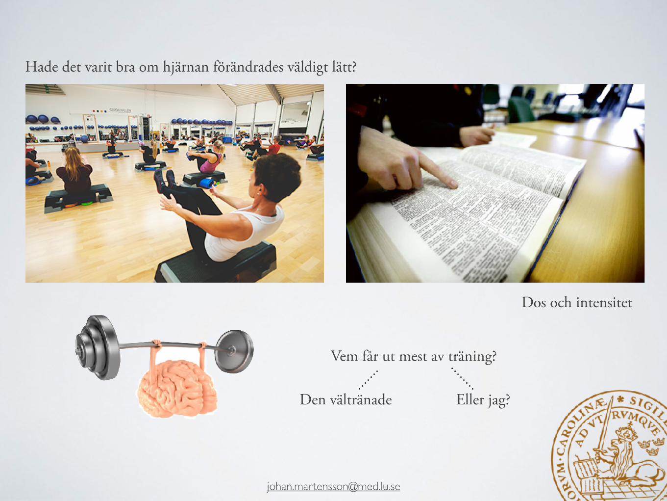
Dos och intensitet
Vem får ut mest av träning?
Den vältränade Eller jag?
Hade det varit bra om hjärnan förändrades väldigt lätt?

Lövdén, Bäckman, Lindenberger, Schafer, & Schmiedek 2010, Psychological Bulletin

Vad händer när det uppstår förändringar?
• Minnen är associerade med förändringar som sker på synaps nivå.
• Hos t.ex. Aplysia sker det förändringar i synapserna vid sensitisering och habituering.

• Chang & Greenough (1982)
• Råttor berövades input från ett öga i form av en ögonlapp.
• Råttorna tränades i en labyrinth.
• Visuella cortex på den tränade hemisfären hade mer dendriter.
Vad händer när det uppstår förändringar?forts.

Nudo et al., 1997
Vad händer när det uppstår förändringar?forts.
(svårt att mäta in vivo..)

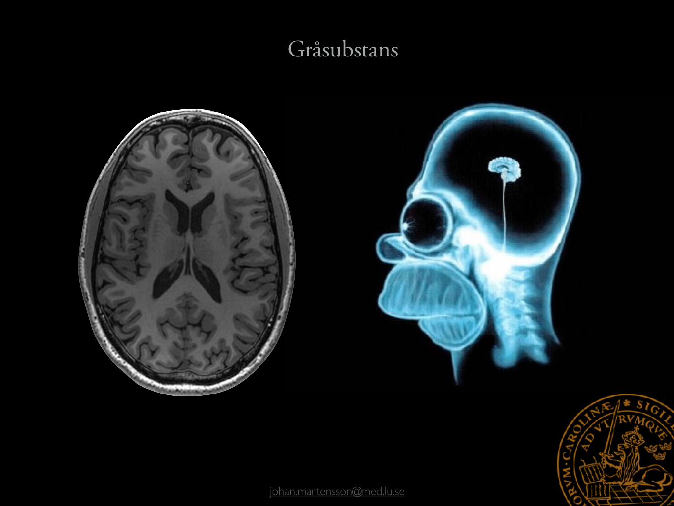
Gråsubstans Nervceller
Vitsubstans Hjälparceller och myelin
a
b
a b
Det finns flera möjliga mekanismer för plasticitet
Och vi kan observera en del av dem via t.ex. MRI

Kortikala förändringar vid djurstudier
En stor del beror på vaskularisering
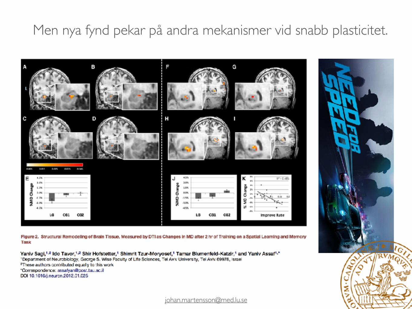
Men nya fynd pekar på andra mekanismer vid snabb plasticitet.

Men nya fynd pekar på andra mekanismer vid snabb plasticitet.
Johansen-Berg et al., 2012
Astrocyter sväller Synapser bildas Dendriter, angio och neurogenes

Hur mäter vi hjärnförändringar?
140000 gånger jordens gravitation
MRI. Men även t.ex. PET eller mått på funktionell plasticitet.

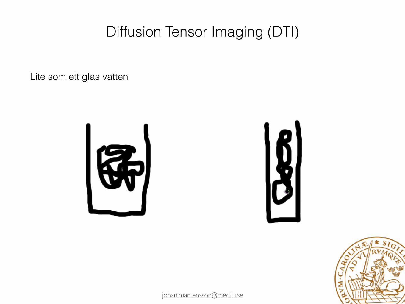


Diffusion Tensor Imaging (DTI)
Och formen ger oss en ledtråd


Studier

Navigation-related structural change in thehippocampi of taxi driversEleanor A. Maguire*†, David G. Gadian‡, Ingrid S. Johnsrude†, Catriona D. Good†, John Ashburner†,Richard S. J. Frackowiak†, and Christopher D. Frith†
†Wellcome Department of Cognitive Neurology, Institute of Neurology, University College London, Queen Square, London WC1N 3BG, United Kingdom;and ‡Radiology and Physics Unit, Institute of Child Health, University College London, London WC1N 1EH, United Kingdom
Communicated by Brenda Milner, McGill University, Montreal, Canada, January 28, 2000 (received for review November 10, 1999)
Structural MRIs of the brains of humans with extensive navigationexperience, licensed London taxi drivers, were analyzed and com-pared with those of control subjects who did not drive taxis. Theposterior hippocampi of taxi drivers were significantly largerrelative to those of control subjects. A more anterior hippocampalregion was larger in control subjects than in taxi drivers. Hippocam-pal volume correlated with the amount of time spent as a taxidriver (positively in the posterior and negatively in the anteriorhippocampus). These data are in accordance with the idea that theposterior hippocampus stores a spatial representation of the en-vironment and can expand regionally to accommodate elaborationof this representation in people with a high dependence onnavigational skills. It seems that there is a capacity for local plasticchange in the structure of the healthy adult human brain inresponse to environmental demands.
One important role of the hippocampus is to facilitate spatialmemory in the form of navigation (1). Increased hippocam-
pal volume relative to brain and body size has been reported insmall mammals and birds who engage in behavior requiringspatial memory, such as food storing (2). In some species,hippocampal volumes enlarge specifically during seasons whendemand for spatial ability is greatest (2, 3). In the healthy human,structural brain differences between distinct groups of subjects(for example, males and females, ref. 4, or musicians andnonmusicians, ref. 5) have been documented. From existingstudies, it is impossible to know whether differences in brainanatomy are predetermined or whether the brain is susceptibleto plastic change in response to environmental stimulation.Furthermore, although lesion work (6, 7) and functional neuro-imaging work (8) confirm the involvement of the human hip-pocampus in spatial memory and navigation, there is still debateabout its precise role. Given the propensity of lower mammali-an!avian hippocampi to undergo structural change in responseto behavior requiring spatial memory (2, 3), the present studyaddressed whether morphological changes could be detected inthe healthy human brain associated with extensive experience ofspatial navigation. Our prediction was that the hippocampuswould be the most likely brain region to show changes.
Taxi drivers in London must undergo extensive training,learning how to navigate between thousands of places in the city.This training is colloquially known as ‘‘being on The Knowledge’’and takes about 2 years to acquire on average. To be licensed tooperate, it is necessary to pass a very stringent set of policeexaminations. London taxi drivers are therefore ideally suitedfor the study of spatial navigation. The use of a group of taxidrivers with a wide range of navigating experience permitted anexamination of the direct effect of spatial experience on brainstructure. In the first instance, we used voxel-based morphom-etry (VBM) to examine whether morphological changes associ-ated with navigation experience were detectable anywhere in thehealthy human brain. VBM is an objective and automaticprocedure that identifies regional differences in relative graymatter density in structural MRI brain scans. It allows everypoint in the brain to be considered in an unbiased way, with no
a priori regions of interest. The data were also analyzed by usinga second and completely independent pixel-counting techniquewithin the hippocampus proper. Comparisons were made be-tween the brain scans of taxi drivers, who had all acquired asignificant amount of large-scale spatial information (as evi-denced by passing the licensing examinations), and those of acomparable group of control subjects who lacked such extensivenavigation exposure.
MethodsSubjects. Right-handed male licensed London taxi drivers (n !16; mean age 44 years; range 32–62 years) participated. All hadbeen licensed London taxi drivers for more than 1.5 years (meantime as taxi driver ! 14.3 years; range ! 1.5–42 years). Theaverage time spent training to be a taxi driver before passing thelicensing tests fully (i.e., time on The Knowledge) was 2 years(range 10 months to 3.5 years; some trained continuously, somepart time). All of the taxi drivers had healthy general medical,neurological, and psychiatric profiles. The scans of controlsubjects were selected from the structural MRI scan database atthe same unit where the taxi drivers were scanned. Thosesubjects below 32 and above 62 years of age were excluded aswere females, left-handed males, and those with any healthproblems. After the application of these exclusion criteria, thescans of 50 healthy right-handed males who did not drive taxiswere included in the analyses for comparison with the taxidrivers. Both the mean age and the age range did not differbetween the taxi driver and control groups. We were also carefulto ensure an even spread of subjects in each decade (for example,41–50 years or 51–60 years) up to the upper limit of the oldesttaxi driver, such that subjects were not clustered at one end ofthe age scale.
Image Acquisition. Structural MRI scans were obtained with a 2.0Tesla Vision system (Siemens GmbH, Erlangen, Germany) by usinga T1-weighted three-dimensional gradient echo sequence (TR 9.7ms; TE 4 ms; flip angle 12°; field of view 256 mm; 108 partitions;partition thickness 1.5 mm; voxel size 1 " 1 " 1.5 mm).
Image Analysis Method 1: VBM. Data were analyzed by using VBMimplemented with Statistical Parametric Mapping (SPM99, Well-come Department of Cognitive Neurology) executed in MATLAB(Mathworks, Sherborn, MA). Detailed descriptions of the tech-nique are given elsewhere (9, 10). Briefly, the subjects’ data werespatially normalized into stereotactic space (11) by registering eachof the images to the same template image by minimizing theresidual sums of squared differences between them. The template
Abbreviations: VBM, voxel-based morphometry; ICV, intracranial volume.
*To whom reprint requests should be addressed. E-mail: [email protected].
The publication costs of this article were defrayed in part by page charge payment. Thisarticle must therefore be hereby marked “advertisement” in accordance with 18 U.S.C.§1734 solely to indicate this fact.
Article published online before print: Proc. Natl. Acad. Sci. USA, 10.1073!pnas.070039597.Article and publication date are at www.pnas.org!cgi!doi!10.1073!pnas.070039597
4398–4403 " PNAS " April 11, 2000 " vol. 97 " no. 8
!
Study
Intervention
Analysis technique
Draganski et al., 2004
Juggling VBM
Colcombe et al., 2006
Aerobics VBM
Draganski et al., 2006
Studying for an exam
VBM
Boyke et al., 2008
Juggling VBM
Driemeyer et al., 2008
Juggling VBM
Ilg et al., 2008
Reading mirrored words
VBM
Teutsch et al., 2008
Pain stimulation
VBM
Ceccarelli et al., 2009
Cognitive learning
TBM
Haier et al., 2009
Tetris CIVET
Scholz et al., 2009
Juggling VBM
Thomas et al., 2009
Visuo-motor VBM
Engvig et al., 2010
Mnemonic training
FreeSurfer cortical
Granert et al., 2010
Motor VBM
Kim et al., 2010
Early postpartum
VBM
Schmidt-Wilcke et al., 2010
Deciphering morse code
VBM
Stein et al., 2010
Language training
VBM
Tang et al., 2010
Meditation VBM
Taubert et al., 2010
Balancing VBM
Erickson et al., 2011
Aerobics FSL-FIRST
Hamzei et al., 2011
Motor task VBM
Hölzel et al., 2011
Meditation VBM
Kwok et al., 2011
Learning color names
VBM
Landi et al., 2011
Visuo-motor VBM
Takeuchi et al., 2011
Working memory
VBM
Wenger et al., 2011
Spatial navigation
FreeSurfer cortical
Woolett & Maguire, 2011
Spatial memory
VBM
Gryga et al., 2012
Motor task VBM
Langer et al., 2012
Motor task Freesurfer cortical
Lövdén et al., 2012
Spatial navigation
Manual HC tracing
Mårtensson et al., 2012
Language training
FreeSurfer cort. & subc.
Development/Plasticity/Repair
Temporal and Spatial Dynamics of Brain Structure Changesduring Extensive Learning
Bogdan Draganski,1 Christian Gaser,2 Gerd Kempermann,3 H. Georg Kuhn,4 Jurgen Winkler,1 Christian Buchel,5 andArne May1,5
1Department of Neurology, University of Regensburg, 93053 Regensburg, Germany2Department of Psychiatry, University of Jena, 07743 Jena, Germany,3Max-Delbruck Center for Molecular Medicine, 13092 Berlin, Germany, 4Institute for Neuroscience and Physiology, Gothenburg University, SE 405 30Gothenburg, Sweden, and 5Department of Systems Neuroscience, University of Hamburg, 20246 Hamburg, Germany
The current view regarding human long-term memory as an active process of encoding and retrieval includes a highly specific learning-induced functional plasticity in a network of multiple memory systems. Voxel-based morphometry was used to detect possible structuralbrain changes associated with learning. Magnetic resonance images were obtained at three different time points while medical studentslearned for their medical examination. During the learning period, the gray matter increased significantly in the posterior and lateralparietal cortex bilaterally. These structural changes did not change significantly toward the third scan during the semester break 3months after the exam. The posterior hippocampus showed a different pattern over time: the initial increase in gray matter during thelearning period was even more pronounced toward the third time point. These results indicate that the acquisition of a great amount ofhighly abstract information may be related to a particular pattern of structural gray matter changes in particular brain areas.
Key words: brain; plasticity; posterior parietal cortex; hippocampus; memory; learning; voxel-based morphometry
IntroductionOne of the most exciting tasks of modern neuroscience is touncover the functional and structural correlates of learning andmemory. Recent theoretical work, as well as neuroimaging andpsychological studies, has used a broad spectrum of stimuli toinvestigate different memory processes. The currently acceptedview regarding memory is that items are first kept in the medialtemporal lobe system followed by a consolidation process basedon changes in the neocortex (Miyashita, 2004) and that regionsknown to be active during perception and encoding are involvedin the subsequent retrieval of learned information (Nyberg et al.,2000; Shannon and Buckner, 2004).
Recent cross-sectional voxel-based morphometry (VBM)studies have demonstrated learning-dependent changes in theadult human brain and suggested anatomical correlates for naviga-tion, arithmetic, linguistic, procedural, and musical learning abilities(Maguire et al., 2000; Golestani et al., 2002; Sluming et al., 2002;Gaser and Schlaug, 2003; Draganski et al., 2004). Given the evidencefrom a recent longitudinal morphometric study showing that learn-ing a complex visuomotor task induced task-specific transient graymatter changes in the adult human brain (Draganski et al., 2004), weaimed to test the hypothesis whether extensive learning of abstract
information can also induce morphological changes in corticalstructures and whether these changes would be transient or longlasting. Based on well established evidence (Eichenbaum, 2004;Squire et al., 2004), we predicted that the medial temporal lobewould show such structural changes.
The German preliminary medical exam, called “Physikum,” isusually taken after 2 years at the end of the preclinical education. Itincludes both oral and written exams in biology, chemistry, bio-chemistry, physics, human anatomy, and physiology. Consequently,the huge amount of new information, demanding a high level ofencoding, retrieval, and usage, requires a 3 month period of dailystudy sessions and represents an ideal group for investigating possi-ble learning-induced structural plasticity of the adult human brain.
Materials and MethodsVolunteersT1-weighted magnetic resonance imaging scans of 38 medical students(21 female, 17 male; mean ! SD age, 24 ! 2.3 years) and 12 age- andsex-matched control subjects (8 female, 4 male; mean ! SD age, 22.1 !1.7 years) were performed at two time points [1.5 Tesla Siemens (Mu-nich, Germany) Symphony scanner, magnetization-prepared rapid-acquisition gradient echo sequence yielding 150 sagittal slices with adefined voxel size of 1 " 1 " 1.08 mm]. In the student group, the firstscan was obtained 3 months before the medical exam, and the secondscan was performed on the first or second day after the exam. In 23 ofthese students, a third scan was performed 3 months later. The averagegrade of our group of volunteers matched the overall average grade of themedical exam that year, which was composed of 7043 medical students,suggesting that our cohort was representative of the population. Thecontrol subjects had no exams in the last 6 months and were not studyingfor any exams at the time. Additionally, they were carefully chosen inregard to educational status (college students for physical therapy) andwere scanned at the same first two time points as the medical students.
Received Oct. 28, 2005; revised March 28, 2006; accepted April 1, 2006.We thank all volunteers for their participation in this study and Gerhard Schuierer, Volker Busch, Michael Rose,
and Eszter Schoell for technical support. A.M. is supported by Deutsche Forschungsgemeinschaft Grant MA 1862/2.C.B. is supported by Volkswagenstiftung and Bundesministerium fur Bildung und Forschung. H.G.K. is supported byVolkswagenStiftung, Vetenskapsrådet, and LUA/ALF Goteborg.
Correspondence should be addressed to Dr. Arne May, Department of Systems Neuroscience, University of Ham-burg, Martinistrasse 52, D-22046 Hamburg, Germany. E-mail: [email protected].
DOI:10.1523/JNEUROSCI.4628-05.2006Copyright © 2006 Society for Neuroscience 0270-6474/06/266314-04$15.00/0
6314 • The Journal of Neuroscience, June 7, 2006 • 26(23):6314 – 6317
Översikt: Lövdén, M., Wenger, E., Mårtensson, J., Lindenberger, U., & Bäckman, L. (2013). Structural brain plasticity in adult learning and development. Neuroscience and Biobehavioral Reviews. doi: 10.1016/j.neubiorev.2013.02.014
Det finns en rik litteratur kring vuxen plasticitet idag
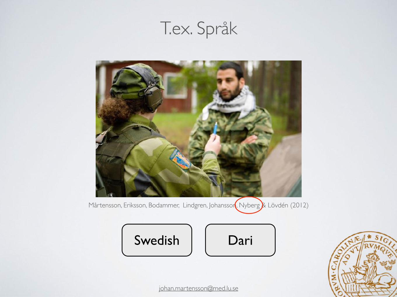
T.ex. Språk
Swedish Dari
Mårtensson, Eriksson, Bodammer, Lindgren, Johansson, Nyberg & Lövdén (2012)
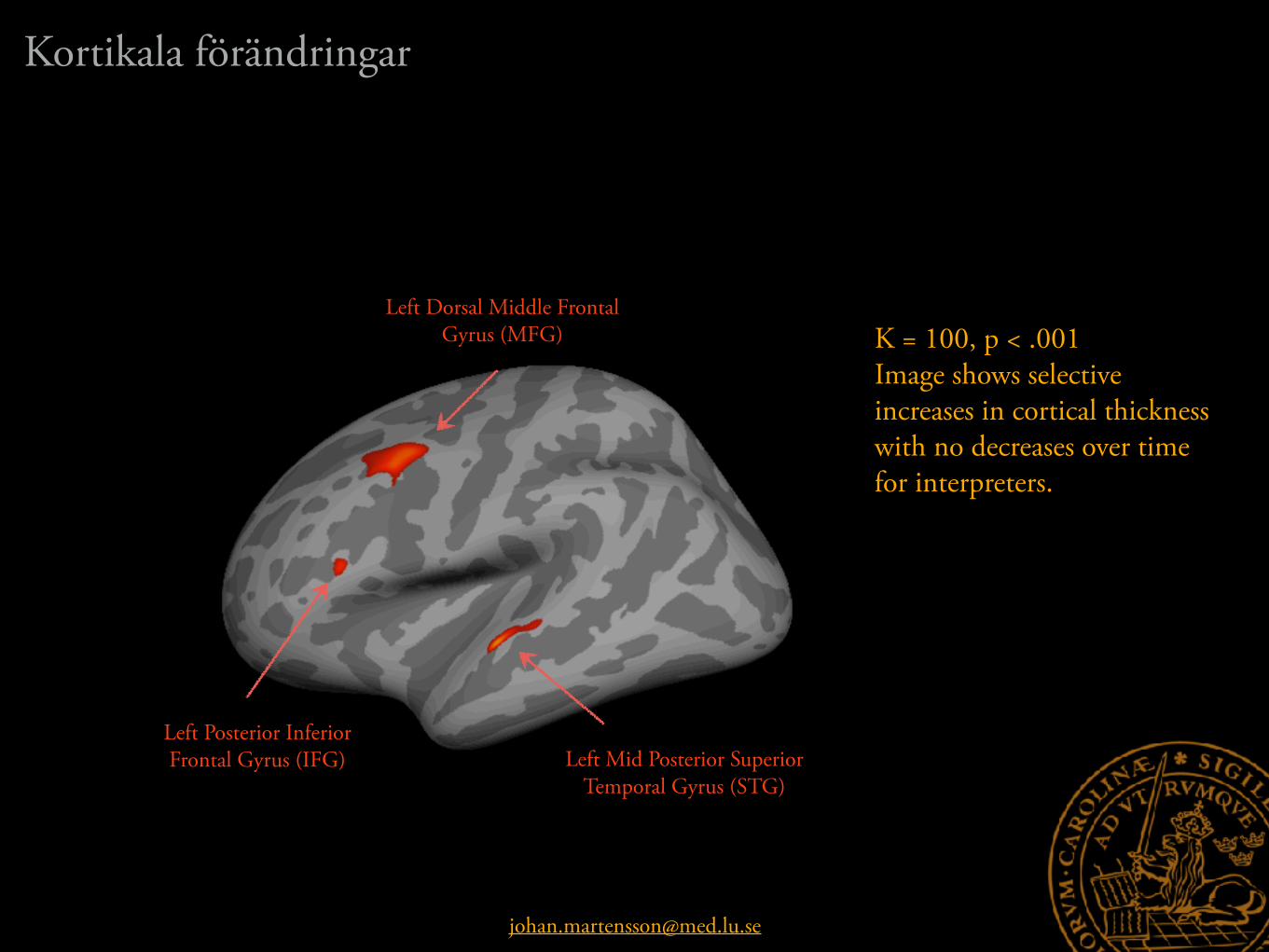
K = 100, p < .001 Image shows selective increases in cortical thickness with no decreases over time for interpreters.
Left Mid Posterior Superior Temporal Gyrus (STG)
Left Dorsal Middle Frontal Gyrus (MFG)
Left Posterior Inferior Frontal Gyrus (IFG)
Kortikala förändringar

Left Mid Posterior Superior Temporal Gyrus (STG)
Left Dorsal Middle Frontal Gyrus (MFG)
Left Posterior Inferior Frontal Gyrus (IFG)
Hickok, G. & Poeppel, D. (2007). The cortical organization of speech processing. Nature reviews, neuroscience.
Kortikala förändringarforts.

Left Hippocampus (LHC)
Size
4300
4400
4500
4600
4700
Pre Post
Interpreters Controls
Right Hippocampus (RHC)
Size
4300
4400
4500
4600
4700
Pre Post
F(1,29) = .90, p = .35 F(1,29) = 8.89, p = .01* denotes p < .05
ns
ns
ns ns
ns
ns
ns*
Subkortikala förändringar

−0,5
−0,4
−0,2
0,0
0,2
0,4
0,5
0,7
Left MFG Left IFG Left HC
Struggle Proficiency
* *
* denotes p < .05
* Teacher rating: ”Bedöm hur stor ansträngning som krävts av varje elev för att uppnå Tolkskolans mål för att få vara kvar vid utbildningen”
1 2
1
2Test Score on their mid year exam. This exam is especially important, if they fail they have to leave the academy.
r
Left STG Right HCStrategy?
Language ability?
Relation till beteendeVi lär oss olika, möjligen med olika neurala system

Vill du lära dig italienska? Delta i vår studie av språkinlärning och hjärnan. Vi står vi för kursavgiften och litteratur!
Som deltagare i studien medverkar du vid 10 undervisningstillfällen i italienska för nybörjare vid Medborgarskolan i Stockholm. Du kommer lära dig en stor mängd glosor varje vecka. Mätningar av minnesförmåga och hjärnstruktur genomförs i en magnetkamera på Karolinska Universitetssjukhuset. Du som vill delta ska vara 18-30 år och ha svenska som modersmål och ej kunna något latinskt språk.Utöver gratis språkundervisning vid medborgarskolan får varje deltagare 500kr per mättillfälle (två). Ytterliggare prestationsbaserad ersättning kan tillkomma
Ansvarig forskare: Martin Lövdén, Docent, Karolinska Institutet
Intresserad? Maila namn och telefonnummer till [email protected] så kontaktar vi dig.
Wisse et al., 2014; Müller 2007
Josef Granqvist Martin Lövdén
Språk, forts.
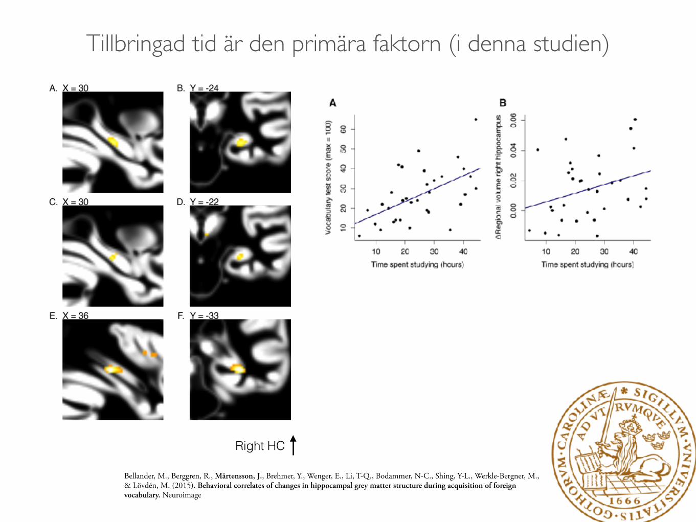
Bellander, M., Berggren, R., Mårtensson, J., Brehmer, Y., Wenger, E., Li, T-Q., Bodammer, N-C., Shing, Y-L., Werkle-Bergner, M., & Lövdén, M. (2015). Behavioral correlates of changes in hippocampal grey matter structure during acquisition of foreign vocabulary. Neuroimage
Right HC
Tillbringad tid är den primära faktorn (i denna studien)

~620 words over 10 weeks; range 86–1150; SD = 324 (!) Average score on an Italian vocabulary test (max = 100) was 27 (range 6–65; SD = 13; skewness = 0.74; kurtosis = 0.61)
Tillbringad tid är den primära faktorn


Lisofsky, N., Mårtensson, J., Eckert, A., Lindenberger, U., Gallinat, J. & Kühn, S. (2015). Hippocampal volume and functional connectivity changes during the female menstrual cycle. Neuroimage.
Structural Changes in Motor Cortex
L"
Gråsubstans ökade under vänsterhandsträning för att sedan delvis normaliseras.
Wenger, E.,*, Kühn, S., Verrel, J., Mårtensson, J., Bodammer, N-C., Lindenberger, U., & Lövdén, M. (2015). The non-linear time course of human gray matter changes in response to motor training. Cerebral cortex.
18 mätpunkter
Menstruationscykeln Motorträning

E.g. Lövdén, M., Wenger, E., Mårtensson, J., Lindenberger, U., & Bäckman, L. (2013). Structural brain plasticity in adult learning and development. Neuroscience and Biobehavioral Reviews. doi: 10.1016/j.neubiorev.2013.02.014
• Tillväxt för att skapa diversitet, följt av selektion, gallring och stabilisering.
• Vi ser liknande mönster i neurogenes, synaptogenes, glia, kortikal plasticitet och utveckling
Plasticitet är inte nödvändigtvis linjär

2,0
3,6
5,2
6,8
8,4
10,0
0,070 0,073 0,076 0,079 0,082 0,085
Prof
icie
ncy
Mean DiffusivitySchlegel, Rudelson and Tse (2012)
Predispositioner spelar roll
Kommer till Lund

Sammanfattning
• Plasticitet kan beskrivas som nervsystemets kapacitet till förändring
• Dessa förändringar kan observeras på micro- och macro-nivå med modern hjärnavbildning
• Förändringarna består av olika effekter, t.ex. vaskularisering, dendritisering, glia celler som sväller
• En rad olika studier visar att plasticitet är möjligt i vuxen ålder, och att den vuxna hjärnan är högst dynamisk

Related Documents