VOL.25 NO.6 June 2020 OFFICIAL PUBLICATION FOR THE FEDERATION OF MEDICAL SOCIETIES OF HONG KONG ISSN 1812 - 1691 Plastic Surgery

Welcome message from author
This document is posted to help you gain knowledge. Please leave a comment to let me know what you think about it! Share it to your friends and learn new things together.
Transcript

VOL.25 NO.6 June 2020
OFFICIAL PUBLICATION FOR THE FEDERATION OF MEDICAL SOCIETIES OF HONG KONG ISSN 1812 - 1691
Plastic Surgery
1
VOL.25 NO.6 JUNE 2020 Contents
Contents
The Cover Shot
Disclaimer All materials published in the Hong Kong Medical Diary represent the opinions of the authors responsible for the articles and do not reflect the official views or policy of the Federation of Medical Societies of Hong Kong, member societies or the publisher.
Publication of an advertisement in the Hong Kong Medical Diary does not constitute endorsement or approval of the product or service promoted or of any claims made by the advertisers with respect to such products or services.
The Federation of Medical Societies of Hong Kong and the Hong Kong Medical Diary assume no responsibility for any injury and/or damage to persons or property arising from any use of execution of any methods, treatments, therapy, operations, instructions, ideas contained in the printed articles. Because of rapid advances in medicine, independent verification of diagnoses, treatment method and drug dosage should be made.
Editorial n Editorial
Dr Walter WK KING2
Medical Bulletinn PICO-second Laser – Is it the Answer?
Dr Vivian Kin-wing LEE4
n MCHK CME Programme Self-assessment Questions 8
n Challenging the Hollow Face: from Volume-based Injection to Tissue-targeted FillingDr Wilson Wai-sun HO
10
n Aesthetic Considerations in the Surgical Treatment of Benign Facial Skin LesionsDr Chun-on MOK & Dr Alec Ho-chuen FUNG
14
n Breast Reconstruction-A Brief Guide for Doctors in Making the DecisionDr Shun-yuen YING
20
n The Making of a Cosmetic SurgeonDr Elvis Wai-ying LEE
26
n A Surgery-first Approach Le Fort I Osteotomy with Simultaneous Alveolar Cleft Grafting for Skeletal Facial Deformities in Adult Cleft Lip and PalateDr Loi-ming CHEUNG
28
CME
To read more aboutThe Federation of MedicalSocieties of Hong Kong
Scan the QR-code
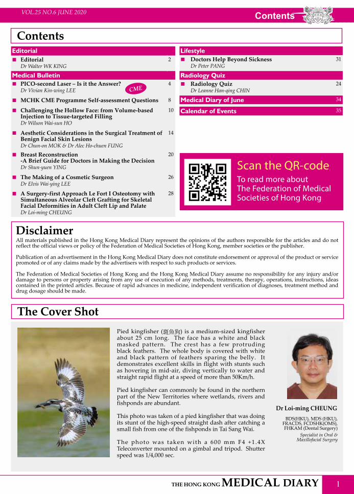
Pied kingfisher (斑魚狗) is a medium-sized kingfisher about 25 cm long. The face has a white and black masked pattern. The crest has a few protruding black feathers. The whole body is covered with white and black pattern of feathers sparing the belly. It demonstrates excellent skills in flight with stunts such as hovering in mid-air, diving vertically to water and straight rapid flight at a speed of more than 50Km/h.
Pied kingfisher can commonly be found in the northern part of the New Territories where wetlands, rivers and fishponds are abundant.
This photo was taken of a pied kingfisher that was doing its stunt of the high-speed straight dash after catching a small fish from one of the fishponds in Tai Sang Wai.
The photo was taken with a 600 mm F4 +1.4X Teleconverter mounted on a gimbal and tripod. Shutter speed was 1/4,000 sec.
Dr Loi-ming CHEUNGBDS(HKU), MDS (HKU),
FRACDS, FCDSHK(OMS), FHKAM (Dental Surgery)
Specialist in Oral & Maxillofacial Surgery
Lifestylen Doctors Help Beyond Sickness
Dr Peter PANG31
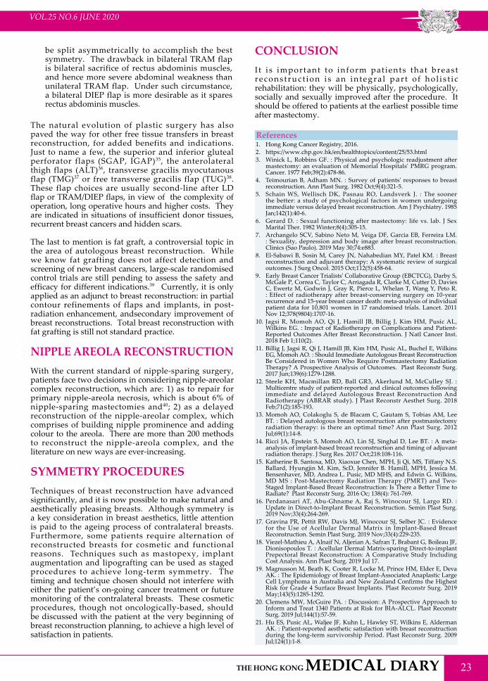
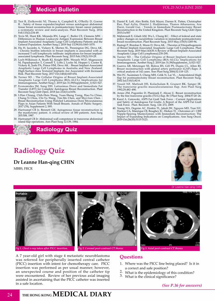
Radiology Quizn Radiology Quiz
Dr Leanne Han-qing CHIN24
Medical Diary of June 34
Calendar of Events 35

2
VOL.25 NO.6 JUNE 2020
Published by The Federation of Medical Societies of Hong Kong
EDITOR-IN-CHIEFDr CHAN Chun-kwong, Jane 陳真光醫生
EDITORSProf CHAN Chi-fung, Godfrey陳志峰教授 (Paediatrics)Dr CHAN Chi-kuen 陳志權醫生 (Gastroenterology & Hepatology) Dr KING Wing-keung, Walter金永強醫生 (Plastic Surgery)Dr LO See-kit, Raymond勞思傑醫生 (Geriatric Medicine)
EDITORIAL BOARDDr AU Wing-yan, Thomas 區永仁醫生 (Haematology and Haematological Oncology)Dr CHAK Wai-kwong 翟偉光醫生 (Paediatrics)Dr CHAN Hau-ngai, Kingsley 陳厚毅醫生 (Dermatology & Venereology) Dr CHAN, Norman 陳諾醫生 (Diabetes, Endocrinology & Metabolism) Dr CHEUNG Fuk-chi, Eric 張復熾醫生 (Psychiatry)Dr CHIANG Chung-seung 蔣忠想醫生 (Cardiology) Prof CHIM Chor-sang, James 詹楚生教授 (Haematology and Haematological Oncology)Dr CHONG Lai-yin 莊禮賢醫生 (Dermatology & Venereology) Dr CHUNG Chi-chiu, Cliff 鍾志超醫生 (General Surgery) Dr FONG To-sang, Dawson 方道生醫生 (Neurosurgery) Dr HSUE Chan-chee, Victor 徐成之醫生 (Clinical Oncology)Dr KWOK Po-yin, Samuel 郭寶賢醫生 (General Surgery) Dr LAM Siu-keung 林兆強醫生 (Obstetrics & Gynaecology)Dr LAM Wai-man, Wendy 林慧文醫生 (Radiology) Dr LEE Kin-man, Philip 李健民醫生 (Oral & Maxillofacial Surgery)Dr LEE Man-piu, Albert 李文彪醫生 (Dentistry) Dr LI Fuk-him, Dominic 李福謙醫生 (Obstetrics & Gynaecology)Prof LI Ka-wah, Michael, BBS李家驊醫生 (General Surgery)Dr LO Chor Man 盧礎文醫生 (Emergency Medicine)Dr LO Kwok-wing, Patrick 盧國榮醫生 (Diabetes, Endocrinology & Metabolism)Dr MA Hon-ming, Ernest 馬漢明醫生 (Rehabilitation)Dr MAN Chi-wai 文志衛醫生 (Urology) Dr NG Wah Shan 伍華山醫生 (Emergency Medicine)Dr PANG Chi-wang, Peter 彭志宏醫生 (Plastic Surgery)Dr TSANG Kin-lun 曾建倫醫生 (Neurology)Dr TSANG Wai-kay 曾偉基醫生 (Nephrology)Dr WONG Bun-lap, Bernard 黃品立醫生 (Cardiology) Dr YAU Tsz-kok 游子覺醫生 (Clinical Oncology)Prof YU Chun-ho, Simon 余俊豪教授 (Radiology) Dr YUEN Shi-yin, Nancy 袁淑賢醫生 (Ophthalmology)
Design and Production
www.apro.com.hk
Editorial
EditorialDr Walter WK KINGMCPS(Manitoba), MD(Vanderbilt, USA) FRCSC(Surgery), FRCSEd, FHKAM(Surgery)Specialist in Plastic SurgeryHonorary Clinical Professor, Department of Surgery, The Chinese University of Hong Kong
Editor
Plastic Surgery is a surgical specialty involving the restoration, reconstruction, regeneration and rejuvenation of the face and body. It consists of two main categories: Reconstructive Surgery and Cosmetic Surgery (from Wikipedia). Reconstructive Surgery includes head and neck surgery, facial plastic surgery, microsurgery, hand surgery and the treatment of congenital abnormalities, facial trauma, burns and skin cancer. Reconstructive Surgery aims to reconstruct or repair a part of the body or improve its functions. Cosmetic Surgery or aesthetic surgery aims at improving and rejuvenating the appearance of the face and body. In essence, plastic surgery techniques form the basis for cosmetic surgery advancement, and in turn, cosmetic or aesthetic surgery is a central component of plastic surgery: Cosmetic Surgery includes eyelid, facial and body aesthetic surgery as well as cosmetic medicine which includes chemical peels, injectables, thread lifts, lasers and other high energy machine treatments. In essence, Plastic surgeons use cosmetic surgery principles and techniques in all reconstructive procedures as well as cosmetic procedures to improve facial and body appearance and function.
In this issue of the Hong Kong Medical Diary on Plastic Surgery, we are honoured to have invited distinguished specialists to enlighten us on selected aspects of Plastic, Reconstructive and Aesthetic Surgery and Medicine:
Dr Vivian LEE, Specialist in Plastic Surgery authored “PICO-second Laser – Is it the Answer”, which is a timely update on the latest advances in the laser treatment of pigmentation including melasma, acne scars and skin pores.
Dr Wilson HO, Specialist in Plastic Surgery and President of the Hong Kong Association of Cosmetic Surgery wrote on “Challenging the Hollow Face: From Volume-based Injection to Tissue-targeted Filling”, which offers insights into the skillful use of fillers as non-surgical approach for rejuvenation of the ageing face.
Dr Chun-on MOK, Specialist in Plastic Surgery and Dr Ho-chuen FUNG, Specialist in Dermatology wrote on “Aesthetic Considerations in the Surgical Treatment of Benign Facial Skin Lesions” in which they outlined the importance of aesthetic approach to the removal of common face moles and skin tumours.
Dr Shun-yuen YING wrote on “Breast Reconstruction – A Brief Guide for Doctors in Making the Decision” to help our breast cancer patients decide on the way forward with breast reconstruction.
Dr Elvis LEE, Specialist in Plastic Surgery and former President, Hong Kong Association of Cosmetic Surgery wrote on “The Making of a Cosmetic Surgeon”, in which he described the training and development of a cosmetic surgeon which normally takes eight or more years upon graduation from medical school.
Dr Peter PANG, Specialist in Plastic Surgery wrote on “Doctors Help Beyond Sickness”, in which he described his valuable experiences
Dr Walter WK KING
3
VOL.25 NO.6 JUNE 2020 Editorial
in doing voluntary plastic surgery work for cleft lip patients in rural China, fundraising and community work for the Rotary and the Lok Sin Tong Benevolent Society Kowloon.
Last but not least, Dr Loi-ming CHEUNG, Specialist in Oral and Maxillofacial Surgery wrote on “A Surgery-first Approach Le Fort I Osteotomy with Simultaneous Alveolar Cleft Grafting for Skeletal Facial Deformities in Adult Cleft Lip and Palate”. He described his elegant and precise work in preparing the bony framework to restoring the facial appearance of cleft lip patients. His work is a testimony to the fact that specialists from multiple specialties can work together as a team to provide the best treatment options to the patient.
On behalf of the readers of The Hong Kong Medical Diary, may I thank all these specialists for their enthusiasm, diligence, cooperation and expertise in producing this issue of The Hong Kong Medical Diary.

Medical Bulletin VOL.25 NO.6 JUNE 2020
4
This article has been selected by the Editorial Board of the Hong Kong Medical Diary for participants in the CME programme of the Medical Council of Hong Kong (MCHK) to complete the following self-assessment questions in order to be awarded 1 CME credit under the programme upon returning the completed answer sheet to the Federation Secretariat on or before 30 June 2020.
Laser technology has been widely implemented in various cutaneous conditions for decades. Nanosecond (NS) Q-switched lasers remain the conventional treatments for cutaneous pigmentation and tattoo removal. Picosecond (PS) laser has rapidly evolved in the market in the past few years. Initially, picosecond laser was used in tattoo removal to optimise efficacy and reduce side effects from nanosecond Q-switched laser. Recently, more and more evidence are emerging to prove its safety and efficacy, which further expand its spectrum of applications. What do we know about picosecond laser so far? This article aims at pointing out some important information about picosecond laser.
MECHANISMThere are some differences between nanosecond Q-Switched laser and picosecond laser. Before discussing the differences between the two lasers, we need to understand two basic theories: Thermal lock-in and Stress lock-in.
THERMAL LOCK-IN (THERMAL RELAXATION THEORY)When a certain structure is heated up to a certain temperature, heat energy will escape to surrounding tissue through conduction. If the heating takes place over a longer period, heat escapes while the structure is being heated and thus the temperature rise of the target structure is limited. This condition is called “heat diffusion”. However, if the structure is heated in a very short time, the temperature rises quickly since there is no time for the heat to diffuse to the surrounding. This condition is called “thermal lock-in”. Once thermal lock-in is achieved selective thermal destruction of the structure becomes possible. Whether or not thermal lock-in is achieved depends on the temporal threshold of the structure which is called the coefficient of thermal relaxation, also known as “thermal relaxation time” (TRT), and the TRT depends on the absorption coefficient and heat diffusion coefficient of the structure, i.e. Chromophore.
STRESS LOCK-IN (STRESS RELAXATION TIME THEORY)When a certain particle is heated, thermal expansion of the particle occurs. The expansion diffuses to the surrounding tissue as vibration, which is called “stress diffusion”. When a particle is heated within an extremely short period, the stress generated within the particle has not enough time to diffuse and stress lock-in is achieved. If the generated stress is high enough, the particle will fracture. The stress relaxation time (SRT) for tattoo pigments is thought to be slightly shorter than one ns. Therefore, by using ns-domain Q-switched lasers, stress lock-in is not achieved since the pulse width of the lasers is longer than the stress relaxation time of the tattoo pigments. However, when a picosecond laser, which can defeat the SRT, is used, stress lock-in is achieved.
Nanosecond laser emits the pulses with a pulse duration at 10-9 of a second. A picosecond laser emits optical pulses with a pulse duration around ten ps, just over 10-12 of a second. When a nanosecond-domain Q-switched laser is used, the major reaction which takes place is photothermal reaction, with a very small photomechanical effect. However, when a picosecond laser is used, stress lock-in occurs, and the major reaction involves the photoacoustic destruction of the particle, with a minor photothermal component. Therefore, more efficient and effective destruction of the particle would take place. Furthermore, it is anticipated that less heat generation will lead to less heat-induced complications such as burn and blister1.
CLINICAL DEVELOPMENT OF PICO SECOND LASERRoss et al. demonstrated the superior efficacy of picosecond pulses over the nanosecond pulses in clearing black tattoos by holding the other laser parameters constant. Picosecond device has become commercially available since 2013 and subsequent clinical trials have confirmed the safety and efficacy of picosecond lasers in treating darkly pigmented, multicoloured, and recalcitrant tattoos. Currently, there are some picosecond laser machines available in the market. PicoSure (755 nm/750 ps), enLIGHTen (1,064 nm, 532 nm/750 ps), PicoWay (1,064 nm, 532 nm/450
PICO-second Laser – Is it the Answer?Dr Vivian Kin-wing LEEMBBS (HK), MRCS(Ed), FCSHK, FHKAM (Surgery)Specialist in Plastic Surgery
Dr Vivian Kin-wing LEE

Medical BulletinVOL.25 NO.6 JUNE 2020
5
ps, 375 ps). These picosecond lasers are superior in the removal of tattoos compared to the nanosecond-domain Q-switched lasers2, and removal of previously difficult tattoo colours with less hyperpigmentation has also been reported3-6.
EFFICACYEvolving evidence has already shown the clinical efficacy of picosecond laser in tattoo removal. Chestnut et al. has published a case series of three patients with refractory naevus of Ota, previous treated by Q-switched lasers, receiving picosecond 755-nm alexandrite laser7. All three patients showed satisfactory responses with a good safety profile. A similar finding was also observed in the local study published by Chan et al. In addition, it is observed that a much lower fluence is used to achieve the required clearance of pigment as compared to Q-switched laser. It was suggested that when the pulse duration is shorter than the thermal relaxation time of the melanosomes, the laser energy would be better absorbed and confined within the target. As a result, the disruption to the surrounding structures and vasculatures would be minimised. Therefore, the fragmentation of the pigment particle would be more specific and confined to pigment itself with less burn to surrounding skin. More and more studies have showed that less fluence is used to achieve acceptable clearance of undesired pigmentation with picosecond laser as compared to Q-switched laser. Besides, evidence have shown better physician and patient satisfaction, less pain score and fewer complications with a picosecond laser.
POST-INFLAMMATORY HYPERPIGNMENTATIONIn the Asian population, complication of post-inflammatory hyperpigmentation (PIH) with the use of Q-switched laser has been reported up to 25%8-10. It was hypothesised that by using high intensity radiation in nanoseconds, both photothermal and photomechanical e f f e c t s w o u l d b e t r i g g e r e d T h e e x c e s s i v e photomechanical effect would produce undesirable damages to the surrounding oxyhemoglobin and melanin, leading to inflammatory responses of the superficial dermal vessels and resulting in PIH. However, Chan et al observed no PIH in his case series with less-than-half energy used by the Alexandrite picosecond laser compared with that of Q-switched11. They hypothesised that the picosecond lasers could have achieved pigmentary removal via a highly effective photomechanical effect. A much lower laser energy would have been required by the picosecond device to achieve the required clearance, as compared to the Q-switched laser. Furthermore, his series has also revealed lower pain scored from patients’ perspectives. Ge et al also found the occurrences of post-inflammatory hyperpigmentation (PS 26% vs QS 34%) was lower in PS than QS group for treatment of naevus of Ota12..
L A S E R - I N D U C E D O P T I C A L BREAKDOWN (LIOB)Traditionally, skin resurfacing is conducted by an ablative laser such as CO2 laser or Er:YAG laser. These lasers induce photo thermolysis in the dermis and result in collagen and elastin production. But they are associated with severe side effects, significant social downtime and risk of complications, particularly in darker skin phototypes. Later, the ablative fractional resurfacing laser has evolved to provide efficacy with few side effects. The array of micro thermal treatment zone is created, but the skin between each micro treatment zone is to remain the untouched. In this way, they found improvement with rapid recovery, shorter downtime and significant less complication rate.
Recently, picosecond laser technology has made a breakthrough in skin rejuvenation. It is a novel minimally invasive non-thermal laser technology resulting in laser-induced optical breakdown (LIOB). It is unlike the current ablative laser devices which are based on selective photothermolysis. LIOB is a non-linear absorption process which occurs only when the irradiance is sufficiently high to produce a critical free-electron density of about 1021 cm-3. When optical breakdown takes place, plasma generation will occur, and it would be followed by explosive vaporisation and mechanical expansion. This highly confined energy leads to cavitation in the dermis. Theoretically, the damage would trigger the neocollagenesis and skin rejuvenation.
Wat et al have shown that the picosecond 755-nm Alexandrite laser with diffractive lens array (DLA) is a safe and effective non-ablative modality for facial skin texture irregularities and dyspigmentation in Chinese skin. Patient tolerance is good with minimal adverse effects of transient erythema and oedema. In his series, no post-inflammatory hyperpigmentation was observed13.
MELASMAMelasma is relatively common in Asian women, and treatment is always challenging. Laser are the third-line treatment for melasma after topical or oral medication and chemical peeling. Nevertheless, lasers are not curing for melasma. Approximately half of the patients suffer from recurrence within 3-6 months after treatment, irrespective of the device used. Recurrence may be recalcitrant to subsequent treatment.
Recently, investigators start to study the role of LIOB in treatment of melasma. Preliminary results are promising, but more data and a longer period of observation are needed.
CONCLUSIONFrom the increasing clinical evidence, this novel minimally invasive non-thermal laser may be an important potential option for benign cutaneous lesions or recalcitrant pigmentary conditions. However, most of the studies are based on small sample size, more well-
Medical Bulletin VOL.25 NO.6 JUNE 2020
6
designed clinical trials with larger sample size should be done to confirm the role of picosecond laser in various cutaneous conditions.
References1. Kasai. Picosecond Laser Treatment for Tattoos and Benign Cutaneous
Pigmented Lesions (Secondary publication).Laser Ther 2017 Dec 31; 26(4): 274–281.
2. Saedi N, Metelitsa A, et al. Treatment of tattoos with a picosecond alexandrite laser: a prospective trial. Arch Dermatol 2012; 148: 1360-3.
3. Brauer JA, Reddy KK, et al. Successful and rapid treatment of blue and green tattoo pigment with a novel picosecond laser. Arch Dermatol 2012; 148:820-3.
4. Alabdulrazzaq H, Brauer JA, et al. Clearance of yellow tattoo ink with a novel 532-nm picoseconds laser. Lasers Surg Med 2015; 47: 285-8.
5. Bernstein EF, Schomacker KT, et al. A novel dual-wavelength, Nd:YAG, picosecond-domain laser safely and effectively removes multicolor tattoos. Lasers Surg Med, 2015
6. Bae YC, Alabdulrazzaq H, et al. Successful treatment of paradoxical darkening. Lasers Surg Med, 2016.
7. Chestnut C, Diehl J, Lask G. Treatment of naevus of ota with a picosecond 755-nm alexandrite laser. Dermatol Surg2015;41:508-510.
8. Ross V, Naseef G, Lin G, Kelly M, Michaud N, Flotte TJ, Raythen J, Anderson RR. Comparison of responses of tattoosto picosecond and nanosecond Q-switched neodymium: YAG lasers. Arch Dermatol 1998;134:167–171.
9. Wang CC, Sue YM, Yang CH, Chen CK. A comparison of Q-switched alexandrite laser and intense pulsed light for the treatment of freckles and lentigines in Asian persons: Arandomised, physician-blinded, split-face comparative trial. J Am Acad Dermatol 2006;54:804–810.
10. Murphy MJ, Huang MY. Q-switched ruby laser treatment of benign pigmented lesions in Chinese skin. Ann Acad Med Singapore 1994;23:60–66.
11. Chan HH, Fung WKK, Ying SY, Kono T. An in vivo trial comparing the use of different types of 532-nm Nd:YAG lasers in the treatment of facial lentigines in oriental patients. Dermatol Surg2000;26:743–749.
12. Ge, Yiping& Yang, Ying & Guo, Lifang & Zhang, Mengli& Wu, Qiuju& Zeng, Rong&Rong, Huizhen& Jia, Gaorong& Shi, Hualing& Fang, Jing & Lin, Tong. (2019). Comparison of a picosecond alexandrite laser versus a Q-switched alexandrite laser for the treatment of nevus of Ota: A randomized, split-lesion, controlled trial. Journal of the American Academy of Dermatology. 10.1016
13. Wat H, Yee-Nam Shek S, Yeung CK, Chan HH. Efficacy and safety of picosecond 755-nm alexandrite laser with diffractive lens array for non-ablative rejuvenation in Chinese skin.Lasers Surg Med. 2019 Jan;51(1):8-13.
Suitable for Meeting / Seminar / Press Conference / Personal Gathering
For enquiry and booking, please contact the Secretariat at 2527 8898.http://www.fmshk.org/rental
ROOM RENTAL PROMOTIONBook now & get FREE 2 hours
FMSHK Member Societies are offered 2 hours FREE rental exclusively.
(Applicable to societies who haven’t used the rental service before)
Well Equipped for Rental:Sound system : microphones / Notebook with LCD projector / 42" TV / Broadband Internet & wifi / Refreshment Ordering, Drinks Ordering / Printing & Photocopy Services
Multi Function Room I Lecture Hall Council Chamber


Medical Bulletin VOL.25 NO.6 JUNE 2020
8
ANSWER SHEET FOR JUNE 2020
Answers to May 2020 Issue
Please return the completed answer sheet to the Federation Secretariat on or before 30 June 2020 for documentation. 1 CME point will be awarded for answering the MCHK CME programme (for non-specialists) self-assessment questions.
Deep Brain Stimulation Beyond the Movement Disorders
1 4 82 5 93 76 10
1. F F T F T T TT T F4. 8.2. 5. 9.3. 7.6. 10.
Name (block letters):____________________________ HKMA No.: __________________ CDSHK No.: _______________
HKID No.: __ __ - __ __ __ __ X X (X) HKDU No.: __________________ HKAM No.: ________________
Contact Tel No.:________________________________ MCHK No. / DCHK No.: __________________(must fill in)
MCHK CME Programme Self-assessment QuestionsPlease read the article entitled “PICO-second Laser – Is it the Answer?” by Dr Vivian Kin-wing LEE and complete the following self-assessment questions. Participants in the MCHK CME Programme will be awarded CME credit under the Programme for returning completed answer sheets via fax (2865 0345) or by mail to the Federation Secretariat on or before 30 June 2020 Answers to questions will be provided in the next issue of The Hong Kong Medical Diary.
Questions 1-10: Please answer T (true) or F (false)
1. Picosecond laser has the same physical property as nanosecond laser.
2. Picosecond laser tends to deliver less heat than nanosecond laser.
3. It needs high fluence to achieve desired clearance of pigmentation with picosecond laser.
4. Mainly photothermal effect is involved in picosecond laser.
5. For picosecond laser, less burn injury is anticipated because of the photoacoustic effect.
6. LIOB is a linear absorption based on selective photothermolysis.
7. Picosecond laser can be used to remove recalcitrant tattoo after Q-switched laser.
8. Picosecond laser never causes post-inflammatory hyperpigmentation.
9. Patients experience more pain with picosecond laser.
10. LIOB causes cavitation in dermis and trigger neocollagenesis and skin rejuvenation.
PICO-second Laser – Is it the Answer?Dr Vivian Kin-wing LEEMBBS (HK), MRCS(Ed), FCSHK, FHKAM (Surgery)Specialist in Plastic Surgery

Comfortable Compression Support for Post-Surgical Recovery
Patented 3D Stretch Fabric
Adjustable to fluctuations in swelling
Easy to put on
Marena Recovery garments are designed to provide comfortable compression support through each step of the recovery journey
Customer Hotline: 2547-5633
Comfortable through extended wear
Soft on the skin
HELP YOUR SCAR FADE TO FEEL YOUR CONFIDENCE GROW
(852) 2547 5633 Hong Wo Pharmaceutical [email protected]
(852) 2547 5633 Hong Wo Pharmaceutical [email protected]
Medical Bulletin VOL.25 NO.6 JUNE 2020
10
Challenging the Hollow Face: from Volume-based Injection to Tissue-targeted FillingDr Wilson Wai-sun HO MBChB, FRCSEd, FCSHK, FHKAM (Surgery)Specialist in Plastic Surgery President, Hong Kong Association of Cosmetic Surgery
Dr Wilson Wai-sun HO
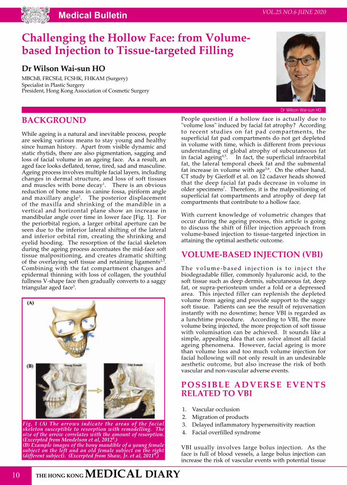
BACKGROUNDWhile ageing is a natural and inevitable process, people are seeking various means to stay young and healthy since human history. Apart from visible dynamic and static rhytids, there are also pigmentation, sagging and loss of facial volume in an ageing face. As a result, an aged face looks deflated, tense, tired, sad and masculine. Ageing process involves multiple facial layers, including changes in dermal structure, and loss of soft tissues and muscles with bone decay1. There is an obvious reduction of bone mass in canine fossa, piriform angle and maxillary angle2. The posterior displacement of the maxilla and shrinking of the mandible in a vertical and horizontal plane show an increase in mandibular angle over time in lower face [Fig. 1]. For the periorbital region, a larger orbital aperture can be seen due to the inferior lateral shifting of the lateral and inferior orbital rim, creating the shrinking and eyelid hooding. The resorption of the facial skeleton during the ageing process accentuates the mid-face soft tissue malpositioning, and creates dramatic shifting of the overlaying soft tissue and retaining ligaments2,3. Combining with the fat compartment changes and epidermal thinning with loss of collagen, the youthful fullness V-shape face then gradually converts to a saggy triangular aged face3.
Fig. 1 (A) The arrows indicate the areas of the facial skeleton susceptible to resorption with remodelling. The size of the arrow correlates with the amount of resorption. (Excerpted from Mendelson et al, 20128.) (B) Example images of the bony mandible of a young female subject on the left and an old female subject on the right (different subject). (Excerpted from Shaw, Jr. et al, 20119.)
People question if a hollow face is actually due to "volume loss" induced by facial fat atrophy? According to recent studies on fat pad compartments, the superficial fat pad compartments do not get depleted in volume with time, which is different from previous understanding of global atrophy of subcutaneous fat in facial ageing4,5. In fact, the superficial infraorbital fat, the lateral temporal cheek fat and the submental fat increase in volume with age5,6. On the other hand, CT study by Gierloff et al. on 12 cadaver heads showed that the deep facial fat pads decrease in volume in older specimens7. Therefore, it is the malpositioning of superficial fat compartments and atrophy of deep fat compartments that contribute to a hollow face.
With current knowledge of volumetric changes that occur during the ageing process, this article is going to discuss the shift of filler injection approach from volume-based injection to tissue-targeted injection in attaining the optimal aesthetic outcome.
VOLUME-BASED INJECTION (VBI)T h e v o l u m e - b a s e d i n j e c t i o n i s t o i n j e c t t h e biodegradable filler, commonly hyaluronic acid, to the soft tissue such as deep dermis, subcutaneous fat, deep fat, or supra-periosteum under a fold or a depressed area. This injected filler can replenish the depleted volume from ageing and provide support to the saggy soft tissue. Patients can see the result of rejuvenation instantly with no downtime; hence VBI is regarded as a lunchtime procedure. According to VBI, the more volume being injected, the more projection of soft tissue with volumisation can be achieved. It sounds like a simple, appealing idea that can solve almost all facial ageing phenomena. However, facial ageing is more than volume loss and too much volume injection for facial hollowing will not only result in an undesirable aesthetic outcome, but also increase the risk of both vascular and non-vascular adverse events.
P O S S I B L E A D V E R S E E V E N T S RELATED TO VBI
1. Vascular occlusion2. Migration of products3. Delayed inflammatory hypersensitivity reaction 4. Facial overfilled syndrome
VBI usually involves large bolus injection. As the face is full of blood vessels, a large bolus injection can increase the risk of vascular events with potential tissue

Medical BulletinVOL.25 NO.6 JUNE 2020
11
necrosis and even blindness. In an intravascular event with small bolus injection (<0.1 ml), a small amount of filler is carried downstream by blood flow. This may cause a limited obstruction that can be bypassed via abundant collateral vessels, unless the region is known to have restricted collaterals like the glabellar region. The clinical effect seen will depend on the presence or absence of adequate collateral circulation in the injected area. With a large bolus of filler material injected into a small or medium-sized vessel, the material may flow retrograde to the normal blood flow direction after it has filled up the distal segment. If the filler bypasses a tributary during its retrograde flow, it may enter this particular pathway and be carried to distant areas [Fig. 2]. This is the possible pathophysiology responsible for injury sites distant to the original injection site postulated by Delorenzin10. In addition, a large volume of filler may lead to more oedema and compression on local blood vessels causing impairment of circulation and possible tissue necrosis.
Van Loghem et al's cadaver study in 2016 comparing cannula with needle injection supported that using needle for large bolus injection can cause product dispersion from the deep targeted placement layer to the non-targeted superficial layer11. It is possible that fillers are more prone to migration in large bolus injection resulting in loss of projection in injected site and lumpiness at other untreated areas. To prevent the incident of vascular occlusion and filler dispersion, small bolus injections with multiple injection points and slow injection are advisable. The hyaluronic acid filler is considered the safest filler as it naturally exists in the human body with low immunogenic i ty . Delayed inf lammatory hypersensitivity reaction arising from hyaluronic acid injection carries a rate of 0.02 %, which is relatively rare12,13. However, more and more case reports in recent few years showed delayed onset inflammatory reactions in both high and low molecular weight hyaluronic acid fillers12-15. Delayed onset inflammatory reaction is an unpredictable adverse reaction and can be resistant
to treatment. It is mediated by T-lymphocytes and characterised by induration, erythema and oedema. It can occur at 48 – 72 hours, several weeks or months after injection and may persist for months16,17. Unlike hypersensitivity due to allergic reaction to filler material, patient with delayed onset inflammatory reaction may develop the reaction after a flu-like illness with unclear mechanism14. Turkmani et al pointed out the possible causes of delayed immune hypersensitivity reaction which included (i) characteristics of the hyaluronic acid filler product as different manufacturers have different cross-linking techniques and manufacturing procedures; (ii) host response; and (iii) large volume of dermal filler in many facial sites and with repeated treatments17.
Repeated large-volume hyaluronic acid injections can cause another horrible outcome: "facial overfilled syndrome". This overfilling syndrome is commonly seen in Hollywood celebrities with the strange "Pillow faces" . The unnatural heaviness in lower mid-face (butt cheeks), the swollen infra-orbital area with loss of normal topography, flawless nasolabial region, puffy and dropping mouth corners, pointy chin, broadened nose (Avatar nose), sausage-like lips, sunset eyes and over-protruding forehead are the characteristics of overfilling syndrome [Fig. 3]4 . The natural facial structure and contour can be permanently destroyed due to the lack of understanding of pathophysiological change in ageing, with too much volume injection.
Fig. 3 Facial overfilled syndrome (photo courtesy of Dr Ting-song LIM MD)
Fig. 2 (A) The effect of small filler bolus on intravascular pathways. In the small bolus condition (<0.1 ml), a small amount of filler is carried downstream by blood flow. This may cause limited obstruction that can be bypassed via abundant collateral vessels, unless the region is known to have restricted collaterals (e.g. the glabellar region). The clinical effect seen will depend on the presence or absence of adequate collateral circulation in the target tissues. (B) The effect of large filler bolus on intravascular pathways. With a large bolus of filler material injected into a small- or medium-sized vessel, the material may flow retrograde to the blood flow's normal direction after it has filled in the distal segment, because there is nowhere else for the filler to go. If the filler bypasses a tributary during its retrograde flow, it may enter this particular pathway and be carried to distant areas. This is the possible pathophysiology responsible for injury sites distant to the original injection site postulated by Delorenzi. (Excerpted from Delorenzi et al, 201410.)

Medical Bulletin VOL.25 NO.6 JUNE 2020
12
TISSUE TARGETED FILLING (TTF)The fundamental principle of performing an aesthetic procedure is to achieve a symmetrical, balanced and harmonious aesthetic result in respect to patient's gender, ethnicity and personal characteristics. The current trend is a subtle progressive change stressing on a natural outcome, especially when patient is having facial expression. Understanding the basic pathophysiological changes of the ageing process is crucial for physicians to make a customised treatment plan and utilise different treatment modalities to optimise the aesthetic outcome. TTF is a rheology-based injection technique. Therefore, in contrast to VBI, TTF focuses on injecting smaller amount of filler which is resilient to repeated facial movements at the correct tissue layer with the filler rheology similar to the tissue that is going to be replaced. In other words, the philosophy of TTF is to replenish "the like with the like" with lower risk of complications. By selecting the right filler, undesirable outcomes such as irregularity, lumpiness and unnatural result can be avoided. Areas like chin and anterior cheeks that need more projection, high viscosity and elasticity hyaluronic acid filler with low cohesivity and good plasticity is suggested. It is because filler placed in the deep layer faces the challenge from both shearing forces by muscles and dynamic soft tissue compression from the top. The high viscosity and elasticity properties can help the filler to resist the spreading and the dynamic compression forces, respectively. Hence, the implanted filler can stay where they are placed and give a high projection capacity with less injection volume. Filler projection is not only defined by the gel hardness (G), but reflected by the balance between both gel hardness and elasticity. A filler with high G which is hard in texture but low in resisting dynamic compression force is easy to fracture and migrate. For superficial injection such as frown lines and tear troughs, a highly cohesive hyaluronic acid gel with low viscosity and good tissue integration should be used. Cohesion is a key factor for dermal integration and creates a consistent, predictable product that adapts well to surrounding tissue. While the highly cohesive product can maintain the gel structure and integrity, good gel-tissue integration can also reduce the Tyndall effect caused by light scattering by hyaluronic acid particles in superficial injection18.
TTF is commonly combined with other treatment modalities to tackle different tissue ageing problems. This holistic approach often combines hyaluronic acid fillers with neuromodulator botulinum toxin type A to relax the hyperactive facial muscles for wrinkle removal and mild degree of lifting. Other treatment modalities like energy-based medical devices (e.g. micro-focused ultrasound device with visualisation to strengthen the superficial musculoaponeurotic system) or threads for lifting the ptotic facial tissues, biostimulators such as calcium hydroxyapatite and poly-l-lactic-acid (PLLA) for neocollagensis and improve skin laxity, lasers and intense pulsed light for skin pigmentation and radiance can also be incorporated into the treatment plan.
SUMMARYAn oval face with good projection and proportion is regarded as the ideal youthful face across all nations. Filling a hollow face as a result of ageing is a challenging work for the physician who needs to bring together sound anatomical knowledge, detailed patient assessment and holistic treatment plan, artistic sense, safety consideration and right product selection. It should be customised for each patient in regard to gender, age, ethnicity, cultural background, medical history and patient's desire in order to achieve a safe and balanced result. Compare to VBI, not only is TTF safer, TTF also gives better and more natural result. TTF in combination with other non-invasive treatment modalities is the benchmark of current aesthetic medicine.
ACKNOWLEDGEMENTThe author would like to express his gratitude to Ms Alice Kar-lai Yang PhD for her assistance in preparing this manuscript.
References1. Wan D, Amirlak B, Rohrich R, Davis K. The clinical importance of the
fat compartments in midfacial aging. Plast Reconstr Surg Glob Open. 2013;1(9): e92.
2. Jeon A, Sung KH, Kim SD, Lee UY, Lee JH, Han SH, Sui HJ. Anatomical changes in the East Asian midface skeleton with aging. Folia Morphol. 2017; 76(4): 730-35.
3. Farkas JP, Pessa JE, Hubbard B, Rohrich RJ. The science and theory behind facial aging. Plast Reconstr Surg Glob Open. 2013 Apr; 1(1): e8-e15.
4. Lim TS. Facial overfilled syndrome complications of inappropriate filler delivery. Prime Journal. 2018 Jan.
5. Gosain AK, Klein MH, Sudhakar PV, Prost RW. A volumetric analysis of soft-tissue changes in the aging midface using high-resolution MRI: implications for facial rejuvenation. Plast Reconstr Surg. 2005 Apr;115(4):1143-52; discussion 1153-5.
6. Rohrich RJ, Arbique GM, Wong C, Brown S, Pessa JE. The anatomy of suborbicularis fat: implications for periorbital rejuvenation. Plast Reconstr Surg. 2009; 124:946–951.
7. Gierloff M, Stöhring C, Buder T, Gassling V, Açil Y, Wiltfang J. Aging changes of the midfacial fat compartments: a computed tomographic study. Plast Reconstr Surg. 2012;129:263–273.
8. Mendelson B, Wong CH. Changes in the facial skeleton with aging: implications and clinical applications in facial rejuvenation. Aesthetic Plast Surg. 2012 Aug; 36(4): 753–760.
9. Shaw RB, Jr. Katzel EB, Koltz PF, Yaremchuk MJ, Girotto JA, Kahn DM, Langstein HN. Aging of the facial skeleton: aesthetic implications and rejuvenation strategies. Plast. Reconstr. Surg. 2011; 127: 374 - 83.
10. Delorenzi C. Complications of injectable fillers, part 2: vascular complications. Aesthetic Surgery Journal. 2014 May; 34(4): 584–600.
11. van Loghem JAJ , Humzah D, Kerscher M. Cannula versus sharp needle for placement of soft tissue fillers: an observational cadaver study. Aesthetic Surgery Journal. 2016 Oct; 1–16.
12. Artzi O, Loizides C, Verner I, Landau M. Resistant and recurrent late reaction to hyaluronic acid-based gel. Dermatol Surg. 2016 Jan; 42(1):31-7.
13. Pérez-Pérez L, García-Gavín J, Wortsman X, Santos-Briz Á. Delayed adverse subcutaneous reaction to a new family of hyaluronic acid dermal fillers with clinical, ultrasound, and histological correction. Dermatol Surg. 2017 Apr; 43(4): 605-08.
14. Goodman GJ. An interesting reaction to a high- and low- molecular weight combination hyaluronic acid. Dermatol Surg. 2015 Apr; 41: S164-66.
15 Homsy A, Rüegg EM, Jandus P, Pittet-Cuénod B, Modarressi A. Immunological reaction after facial hyaluronic acid injection: case reports. Plast Surg Hand Surg. 2017; 4(1): 68-72.
16 De Boulle K. Management of complications after implantations of fillers. J Cosmet Dermatol. 2004;3(1):2–15.
17. Turkmani MG, De Boulle K, Philipp-Dormston WG. Delated hypersensitivity reaction to hyaluronic acid dermal filler following influenza-like illness. Clin Cosmet Investig Dermatol. 2019 Jul; 12: 277-83.
18. Molliard SG, Bon Bétemps J, Hadjab B, Topchian D, Micheels P, Salomon D. Key rheological properties of hyaluronic acid fillers: from tissue integration to product degradation. Plast Aesthet Res 2018;5:17.
C
M
Y
CM
MY
CY
CMY
K
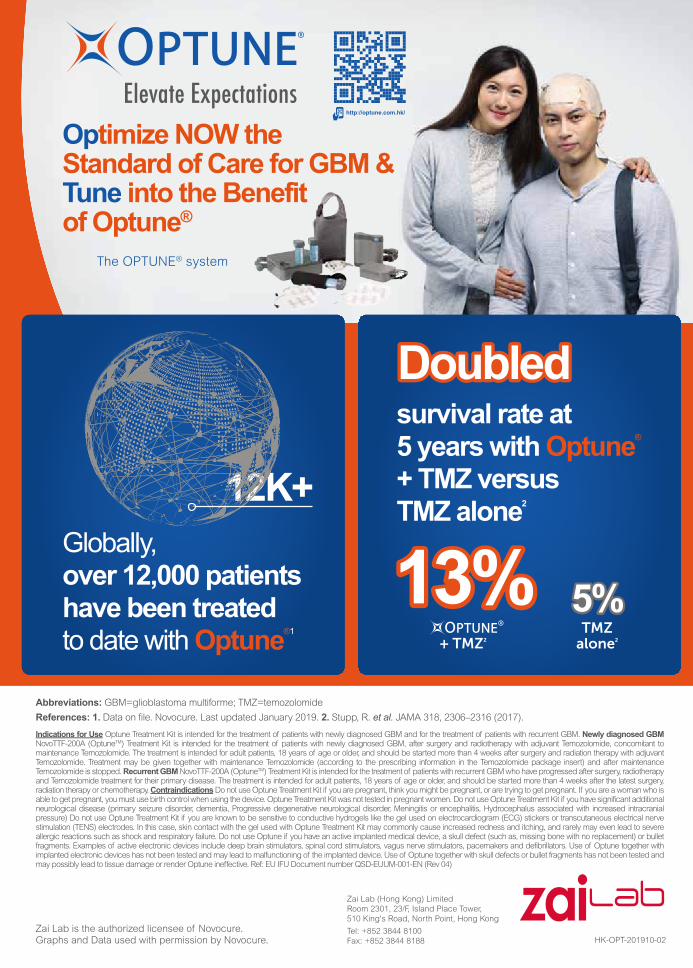
output-Optune-advert infographics-191121-v8.pdf 1 25/11/2019 11:44 AM

Medical Bulletin VOL.25 NO.6 JUNE 2020
14
Aesthetic Considerations in the Surgical Treatment of Benign Facial Skin LesionsDr Chun-on MOK
Dr Alec Ho-chuen FUNG
Specialist in Plastic Surgery
Specialist in Dermatology
Dr Alec Ho-chuen FUNGDr Chun-on MOK
Facial skin lesions are common and are mostly benign. Most benign facial skin lesions pose little health risks to patients unless they are suspicious of malignancy or having a diagnostic problem. Some may also be symptomatic such as progressive growth, irritation, infection or ulceration.
The aesthetic requirement on facial surgery is high as patients would not prefer to have a conspicuous wound or scar on the face. This is particularly demanding in the case of asymptomatic lesions as patients would not be satisfied with a wound or scar that is worse than the original benign facial lesions.
The surgical treatment of benign facial skin lesions would include biopsy/excision of lesions to establish tissue diagnosis in doubtful cases and to rule out malignancy, to treat the infections (abscess or infected tissue) or to remove the symptomatic lesions and a lot of the time, to remove the unsightly asymptomatic lesions on the facial skin.
BIOPSY
Shave BiopsyShave biopsy is done with a no. 15 surgical blade for lesions limited to the epidermis and papillary dermis. The underlying residual tissue may be removed at the same time or later by electrodessication with a hyfrecator or with an Er-YAG Laser.
Punch or Incision BiopsyA small piece of full thickness skin tissue can be obtained in a larger skin lesion by a small 2mm skin punch (Fig. 1) or a small incision for the evaluation of dermal skin pathology. The small punched wound or incision can be closed with a fine nylon stitch.
Fig. 1 A 2mm skin punch (Photo from personal collection)
Excisional BiopsySmall-sized skin lesions including the dermal layer can be completely excised for pathological evaluation as well as surgical treatment. These can be done with a surgical blade (Fig. 2) or a 2-4mm skin punch (Fig. 3). The wound can be closed nicely with fine nylon stitches.
Fig. 2 A small compound naevus excised for histology (Photos from personal collection)
Fig. 3 Multiple facial and nasal angiofibromata excised with a small skin punch and closed with 7/0 nylon stitches (Photos from personal collection)
Excision SurgeryLarger skin lesions involving the dermis or deeper layer can be excised completely along the skin crease direction. The surgical wound can heal with nice scar.
The aesthetic outcome is particularly good over the eyelid (Fig. 4), forehead (Fig. 5), temporal and pre-auricular areas (Fig. 6). In some deeper lesions, incisions along or within the hairline can camouflage the wound nicely (Fig. 7, 8).

Medical BulletinVOL.25 NO.6 JUNE 2020
15
Fig. 4 Complete excision of a lower eyelid intradermal naevus (Photos from personal collection)
Fig. 5 A large forehead lipoma excised completely with a small skin crease incision (Photos from personal collection)
Fig. 6 Accessory auricles of a newborn excised soon after birth (Photos from personal collection)
Fig. 7 & 8 An osteoma of the frontal bone removed via an incision within the hairline (Photos from personal collection)
EPIDERMAL/SUPERFICIAL SKIN LESIONS WITH HYFRECATOR, LASERS OR CRYOTHERAPY?
CryotherapyI would not recommend to treat facial skin lesions, be they superficial or dermal, with cryotherapy. It is often imprecise, requires multiple treatments, heals slowly and is often complicated with erythema, pigmentary changes and sometimes hypertrophic scaring.
Hyfrecator – ElectrodessicationElectrodessication with a hyfrecator is useful in removing epidermal skin tissue but the thermal changes are more prominent than Eribium: YAG Laser. Hyfrecator is often used on the face to swiftly remove thick epidermal lesions (Fig. 9). Superficial thin lesions should still be best treated with Eribium: YAG Laser as the pigmentary changes would be less. Alternatively, with a fine needle electrode, very fine skin lesions (<1 mm) can be nicely cauterized and removed with little skin changes (Fig. 10, 11).
Fig. 9 Thick seborrhoeic keratosis on the cheek cauterised(Photos from personal collection)
Fig. 10 & 11 Multiple small keratosis cauterised with a fine needle electrode (Photos from personal collection)
Medical Bulletin VOL.25 NO.6 JUNE 2020
16
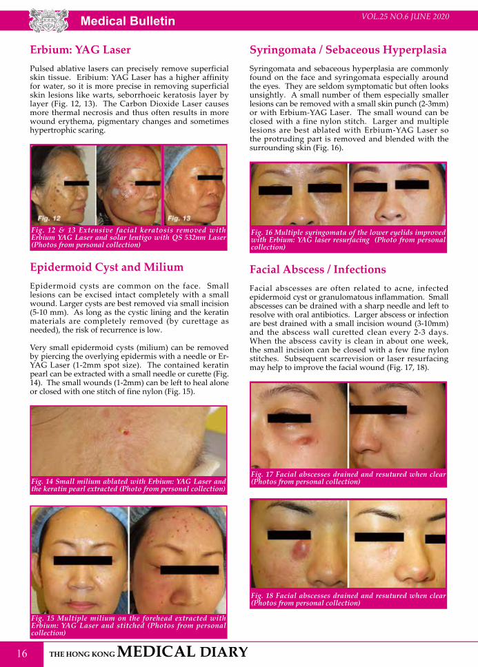
Erbium: YAG LaserPulsed ablative lasers can precisely remove superficial skin tissue. Eribium: YAG Laser has a higher affinity for water, so it is more precise in removing superficial skin lesions like warts, seborrhoeic keratosis layer by layer (Fig. 12, 13). The Carbon Dioxide Laser causes more thermal necrosis and thus often results in more wound erythema, pigmentary changes and sometimes hypertrophic scaring.
Fig. 12 & 13 Extensive facial keratosis removed with Erbium YAG Laser and solar lentigo with QS 532nm Laser (Photos from personal collection)
Epidermoid Cyst and MiliumEpidermoid cysts are common on the face. Small lesions can be excised intact completely with a small wound. Larger cysts are best removed via small incision (5-10 mm). As long as the cystic lining and the keratin materials are completely removed (by curettage as needed), the risk of recurrence is low.
Very small epidermoid cysts (milium) can be removed by piercing the overlying epidermis with a needle or Er-YAG Laser (1-2mm spot size). The contained keratin pearl can be extracted with a small needle or curette (Fig. 14). The small wounds (1-2mm) can be left to heal alone or closed with one stitch of fine nylon (Fig. 15).
Fig. 14 Small milium ablated with Erbium: YAG Laser and the keratin pearl extracted (Photo from personal collection)
Fig. 15 Multiple milium on the forehead extracted with Erbium: YAG Laser and stitched (Photos from personal collection)
Syringomata / Sebaceous HyperplasiaSyringomata and sebaceous hyperplasia are commonly found on the face and syringomata especially around the eyes. They are seldom symptomatic but often looks unsightly. A small number of them especially smaller lesions can be removed with a small skin punch (2-3mm) or with Erbium-YAG Laser. The small wound can be closed with a fine nylon stitch. Larger and multiple lesions are best ablated with Erbium-YAG Laser so the protruding part is removed and blended with the surrounding skin (Fig. 16).
Fig. 16 Multiple syringomata of the lower eyelids improved with Erbium: YAG laser resurfacing (Photo from personal collection)
Facial Abscess / InfectionsFacial abscesses are often related to acne, infected epidermoid cyst or granulomatous inflammation. Small abscesses can be drained with a sharp needle and left to resolve with oral antibiotics. Larger abscess or infection are best drained with a small incision wound (3-10mm) and the abscess wall curetted clean every 2-3 days. When the abscess cavity is clean in about one week, the small incision can be closed with a few fine nylon stitches. Subsequent scarrevision or laser resurfacing may help to improve the facial wound (Fig. 17, 18).
Fig. 17 Facial abscesses drained and resutured when clear (Photos from personal collection)
Fig. 18 Facial abscesses drained and resutured when clear (Photos from personal collection)

Medical BulletinVOL.25 NO.6 JUNE 2020
17
Facial MolesFacial mole removal is common in our practice. Some moles are excised for the suspicion of malignancy. All dysplastic nevus and thick pigmented moles should be excised completely. Smaller and superficial pigmented moles like lentiginous lesions, junctional nevus, intradermal nevus can be removed with a combination of Eribium - YAG Laser ablation and QS 532mm Laser treatment. Eribium - YAG Laser removes the superficial tissue and pigment and the subsequent QS 532mm Laser helps to clear out the deep-seated pigmentation. This treatment regimen is best suited for patients with multiple small pigmented nevus and may take several sessions at bi-monthly interval for the pigmented nevus to disappear (Fig. 19, 20).
Fig. 19 & 20 Multiple small pigmented moles removed with excision as well as laser treatment (Photos from personal collection)

Medical Bulletin VOL.25 NO.6 JUNE 2020
20
Breast Reconstruction-A Brief Guide for Doctors in Making the DecisionDr Shun-yuen YINGMBChB (CUHK), FRCS (Edin), FCSHK, FHKAM (Surgery)Plastic Surgeon
Dr Shun-yuen YING
Breast cancer is the commonest cancer in females of Hong Kong. It accounted for 26.6% of all new cancers in females in 2016.1 This high incidence and the growing demand for breast reconstruction, along with evolving knowledge of breast cancer treatment, continue to pose challenges to plastic surgeons as well as allied breast surgeons and oncologists.
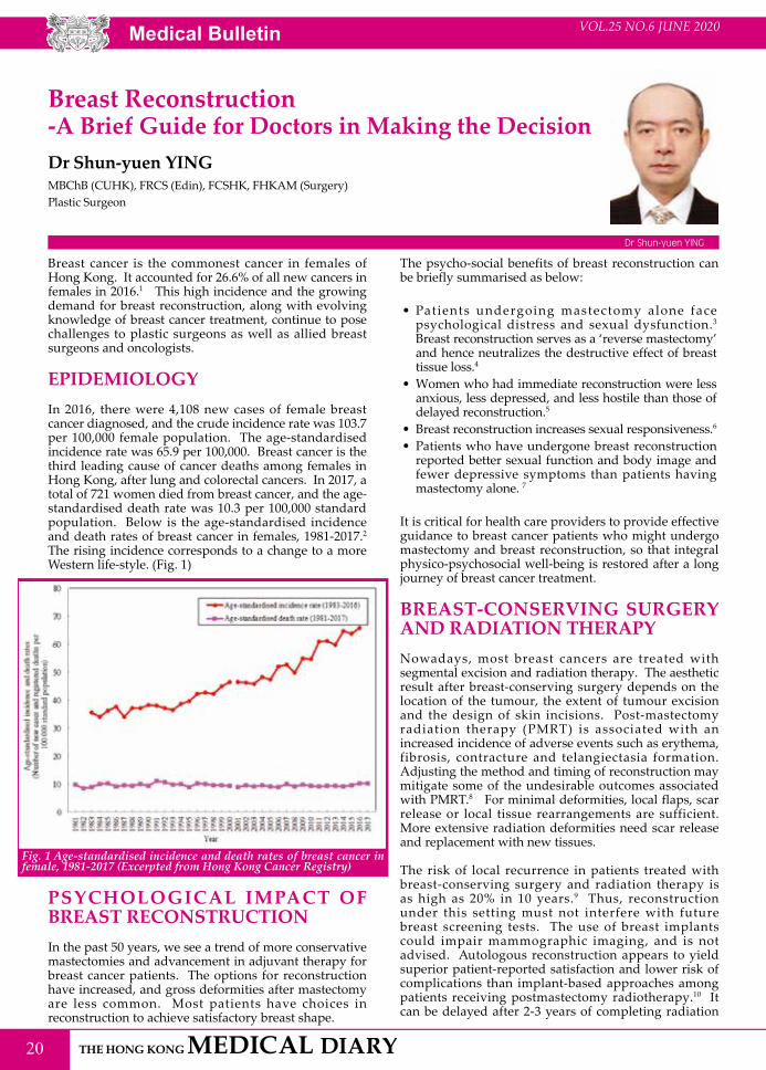
EPIDEMIOLOGYIn 2016, there were 4,108 new cases of female breast cancer diagnosed, and the crude incidence rate was 103.7 per 100,000 female population. The age-standardised incidence rate was 65.9 per 100,000. Breast cancer is the third leading cause of cancer deaths among females in Hong Kong, after lung and colorectal cancers. In 2017, a total of 721 women died from breast cancer, and the age-standardised death rate was 10.3 per 100,000 standard population. Below is the age-standardised incidence and death rates of breast cancer in females, 1981-2017.2 The rising incidence corresponds to a change to a more Western life-style. (Fig. 1)
The psycho-social benefits of breast reconstruction can be briefly summarised as below:
• Patients undergoing mastectomy alone face psychological distress and sexual dysfunction.3 Breast reconstruction serves as a ‘reverse mastectomy’ and hence neutralizes the destructive effect of breast tissue loss.4
• Women who had immediate reconstruction were less anxious, less depressed, and less hostile than those of delayed reconstruction.5
• Breast reconstruction increases sexual responsiveness.6 • Patients who have undergone breast reconstruction
reported better sexual function and body image and fewer depressive symptoms than patients having mastectomy alone. 7
It is critical for health care providers to provide effective guidance to breast cancer patients who might undergo mastectomy and breast reconstruction, so that integral physico-psychosocial well-being is restored after a long journey of breast cancer treatment.
BREAST-CONSERVING SURGERY AND RADIATION THERAPYNowadays, most breast cancers are treated with segmental excision and radiation therapy. The aesthetic result after breast-conserving surgery depends on the location of the tumour, the extent of tumour excision and the design of skin incisions. Post-mastectomy radiation therapy (PMRT) is associated with an increased incidence of adverse events such as erythema, fibrosis, contracture and telangiectasia formation. Adjusting the method and timing of reconstruction may mitigate some of the undesirable outcomes associated with PMRT.8 For minimal deformities, local flaps, scar release or local tissue rearrangements are sufficient. More extensive radiation deformities need scar release and replacement with new tissues.
The risk of local recurrence in patients treated with breast-conserving surgery and radiation therapy is as high as 20% in 10 years.9 Thus, reconstruction under this setting must not interfere with future breast screening tests. The use of breast implants could impair mammographic imaging, and is not advised. Autologous reconstruction appears to yield superior patient-reported satisfaction and lower risk of complications than implant-based approaches among patients receiving postmastectomy radiotherapy.10 It can be delayed after 2-3 years of completing radiation
Fig. 1 Age-standardised incidence and death rates of breast cancer in female, 1981-2017 (Excerpted from Hong Kong Cancer Registry)
PSYCHOLOGICAL IMPACT OF BREAST RECONSTRUCTIONIn the past 50 years, we see a trend of more conservative mastectomies and advancement in adjuvant therapy for breast cancer patients. The options for reconstruction have increased, and gross deformities after mastectomy are less common. Most patients have choices in reconstruction to achieve satisfactory breast shape.

Medical BulletinVOL.25 NO.6 JUNE 2020
21
therapy. The majority of defects after segmentectomy or quadrantectomy can be treated with latissimus dorsi myocutaneous flap. Smaller lesions can be treated with lateral intercostal perforator flaps. Recent advances in fat grafting added another useful armamentarium to repairing small breast contour defects after breast-conserving surgery.
DECISION ON IMMEDIATE VS. DELAYED RECONSTRUCTIONThe trend in the past 50 years has seen a shift from radical breast surgery to more conservative surgery: from preserving the pectoralis major muscle, to partial mastectomy, and axillary sparing dissections. This change has enabled reconstruction to be safely carried out in the immediate setting. Evidences are showing that immediate reconstruction does not affect tumour safety, does not delay adjuvant systemic therapy, nor affect breast aesthetics. And the result of immediate reconstruction with autologous flap is not compromised by postoperative radiation therapy.11
Immediate reconstruction is superior to delayed reconstruct ion in terms of cost , cosmesis and psychologic well-being. However, certain clinical situations need a delayed reconstruction: when it is uncertain whether radiation therapy will be required. When radiation to the chest wall is required, the chance of cosmetically good implant reconstruction is low. And so, these patients are best treated with delayed flap reconstruction.12 For autologous reconstruction, the overall complications are similar in patients reconstructed early or late after PMRT, therefore autologous breast reconstruction can potentially be performed immediately rather than the current standard practice.13
But clinical practice to this group of PMRT plus implant patients is changing: it is found that direct-to-implant (DTI) and two-staged implant reconstruction do not differ in complications.8 Added with the benefit that women who had immediate reconstruction were less anxious, less depressed, and less hostile than those of delayed reconstruction,3 even in the implant reconstruction group, the current practice is to reconstruct immediately with implants.
D E C I S I O N O N T H E T Y P E O F RECONSTRUCTIONTechniques of breast reconstruction can be broadly divided into two basic forms: implant techniques and autologous techniques. Implant procedures may be a simple implant, tissue expander with delayed implants, or combined Latissimus dorsi flap with implant. Autologous procedures include latissimus dorsi flaps, TRAM flaps, gluteus maximus flaps, lateral thigh flaps, or free flaps. All have specific advantages and disadvantages. In general, implants carry shorter down-times but higher long-term morbidities, while flap techniques carry longer operative times, increased short-term morbidities, but much higher long-term satisfaction. Flap techniques are true immediate reconstructions, while tissue expander procedures involve two stages. Patients receiving
flap reconstruction do not experience the defect of mastectomy, while patients receiving tissue expander after mastectomy must commit to a delayed procedure after finishing adjuvant therapy. The final result of implant reconstruction is never as soft and pliable as an autologous reconstruction.
Tissue Expander - Implant ReconstructionT h e r e c e n t t r e n d t o wa r d m o r e c o n s e r va t i ve mastectomies favours the use of autologous tissue, which offers resemblance in colour and pliability of breasts. Implant reconstruction demands a precondition: an integral skin envelope which has adequate laxity to hold the implant with satisfactory projection and consistency. Patients with inadequate skin cover after mastectomy are not good candidates for implant reconstruction. Patients who have undergone radiotherapy after a mastectomy or patients who are planned to undergo post-operative radiotherapy are not good candidates for implant reconstruction. Chest wall radiation makes skin expansion difficult, and also increases implant-related complications.14 While this choice shortens the operative time and carries less donor site morbidities, implant reconstructions tend to produce breasts which assume the shape of implants. Implants may also result in interference with future mammograms, which is a long-term risk of the contralateral breast. The long-term problems such as chronic pain, capsular contracture and implant displacement may occur.
If both safety and cosmesis are taken into consideration, there is no benefit of two-staged reconstruction even in patients receiving prosthesis. In the setting of post-mastectomy radiation therapy (PMRT) and two-staged implant-based reconstruction, patients who received PMRT after expander placement (TE-XRT) did not have a higher incidence or increased odds of developing complications than those who received two-staged reconstruction.15 Therefore, the current trend for implant reconstruction is one-staged, direct-to-implant (DTI) reconstruction, i.e. to apply expander-implant immediately after mastectomy, over-inflate the implant before radiation therapy (XRT), and then partially deflate expander-implant after radiation therapy (XRT). The inflation and deflation work can be carried out in the outpatient setting, and hence save the patient another delayed operation.16
Recently, the use of acellular dermal matrix (ADM) as an adjunct of implant reconstruction is getting wider acceptance. Acellular dermal matrices are tissue grafts that have been bioengineered to remove all cellular components, and hence antigenicity. The major benefits of using ADM include superior initial breast shapes, superior sustained position of implants, and most of all, much reduced capsular contracture rate compared to traditional implant reconstruction.17,18 Like all medical advances, the use of ADM is associated with issues of cost, infection and seroma.
Another recent issue concerning the use of implants is the advent of breast implant-related anaplastic large cell lymphoma (BIA-ALCL). The FDA of the United States has issued a global recall of Allergan BIOCELL textured surface implants on 8 July 2019. Here are some facts:

Medical Bulletin VOL.25 NO.6 JUNE 2020
22
• The incidence is low: only 293 case in US PROFILE registry, and globally 791 confirmed cases, and 33 deaths. Approximately 1.7 million women received breast implants each year. The reported incidence is 0.045% to 0.0012%.19,20
• The interval from implantation to diagnosis is long,withan average of 8 years having been reported.12
• Although the exact etiopathology is unknown, BIA-ALCL is associated with two major risk factors: macro-textured surface, genetics (Occurrence appears mostly in Caucasians, being very rare in Asians,)21,22,23 and infection24,25.
• At t h i s m o m e n t , i t i s f a i r t o s a y t h a t t h e etiopathogenesis is far from being clear. What we know are: that it is a disease of T-cell origin, that it involves only a limited number of patients, that it exists amongst patients with allergy and autoimmunity providing conducive backgrounds for chronic stimuli whether of bacterial and/or synthetic origins, and that ultimately the chronic nature of the stimuli causes prolonged proliferation and initiates oncogenic events, leading to clonal outgrowth and tumour development.26
• Concerning treatment and prevention, the FDA does not recommend the routine removal of these or other types of breast implants, as the risk is very low. However, if symptoms such as persistent swelling or pain develop near the breast implant, investigations should be carried out. This typically involve physical examination imaging and/or assessment of fluid or tissue around the breast implant. A positive intra-capsular fluid collection and positive cell typing of CD-30 marker confirm the diagnosis. En block excision is mandatory as the nature of the tumour is fluid lymphoma; the implant must be removed together with the capsule and fluid. It is a more extensive surgery than that of a simple capsulotomy. Post-operatively the patient should receive adjuvant chemotherapy as for lymphoma patients. BIA_ALCL is a curable disease if discovered early.
• Some micro-textured implants such as Motiva and BellaGel, which bypass the immunogenic stimuli of macro-textured surfaces, are available in the market. However, they are still under licentiate procedures of the FDA, and thus user discretion is advised.
Autologous Tissue ReconstructionEarly in 1976, pedicled latissimus dorsi flap (LD) was used to treat chest wall with radiation necrosis. Then in 1977, LD flap was used for breast reconstruction. This large, fan-shaped flap provides adequate coverage for large mastectomy defects; however, volume replacement in large breasts are inadequate, and thus must be supplemented with silicone implants. A significant incidence of capsular contracture, and thus replacement, gave way to acceptance of another reconstruction: the advent of transverse rectus abdominis myocutaneous flap (TRAM), in which no implant supplement was required and total autologous breast reconstruction is possible.
Today, LD flap is still the ‘workhorse’ for breast reconstruction, mainly due to three advances in LD flap armamentarium:
1) Restricted indication for small-medium sized breasts. The rule-of-thumb is that for mastectomies < 400 gm, LD flap is adequate to provide good volume and symmetry, thus satisfying the local Asian population in most of the cases;
2) The natural evolution to the use of extended LD flap: this involves a more extensive harvest of subcutaneous fat and the LD muscle.
3) Use of immediate fat graft (LIFT) to achieve complete autologous breast reconstruction.27
Today LD flap is still extensively used in many breast cancer centres. Its advantages of easy dissection, short post-operative recovery, absence of significant functional loss, and satisfactory cosmetic outcome make it a good choice for Asian women with small breasts.28
For large breasts, TRAM flap is a better choice. It can provide a reconstruction with a certain degree of natural ptosis that implants cannot provide. Besides, the donor-site scar is acceptable, and it serves as a simultaneous abdominoplasty. However, the surgical procedure is more complex, the rate of partial fat necrosis is reported to be 15%29, and donor morbidity of core muscle weakness is not fully recoverable.30 The natural evolution of plastic surgeons to bypass these problems is: the free deep inferior epigastric perforator flap (DIEP). In DIEP,rectus abdominis muscles are spared from loss, and a large piece of abdominal fat is transferred by microvascular anastomosis to the breast bed.
Today, in areas of the world with better insurance coverage, implant and DIEP reconstruction are the mainstream of practice. And the use of pedicled LD flap is on the decline.31 In other places, factors such as age, income,education level, and health insurance coverage, play a role in shaping patients’ decision on the choice of breast reconstruction.32 There is no ‘ideal’, or ‘perfect’ reconstruction for breast cancer patients, only the most ‘suitable’ choices. It is safe to say, in the long term, TRAM patients, compared with expander/implant patients, appear to have significantly greater aesthetic satisfaction.33 Tissue expander/implant reconstruction carries a higher risk of reconstructive failure and surgical-site infection compared with autologous abdominal tissue reconstruction.34
For patients with bilateral breast cancers, or patients who want a prophylactic mastectomy of the normal breast during formal mastectomy of the diseased breast, the problem of bilateral breast reconstruction arises. The solution is limited to:
1) bilateral implants reconstruction; or2) TRAM flap, or DIEP flap: since frequently, a
significant amount of flap tissue is discarded in in unilateral mastectomy, a bilateral TRAM reconstruction usually offers sufficient tissue to two breasts. Symmetry is easily achieved by splitting flap tissue along the mid-line. If asymmetric mastectomy specimens are obtained, the flap can
23
VOL.25 NO.6 JUNE 2020
be split asymmetrically to accomplish the best symmetry. The drawback in bilateral TRAM flap is bilateral sacrifice of rectus abdominis muscles, and hence more severe abdominal weakness than unilateral TRAM flap. Under such circumstance, a bilateral DIEP flap is more desirable as it spares rectus abdominis muscles.
The natural evolution of plastic surgery has also paved the way for other free tissue transfers in breast reconstruction, for added benefits and indications. Just to name a few, the superior and inferior gluteal perforator flaps (SGAP, IGAP)35, the anterolateral thigh flaps (ALT)36, transverse gracilis myocutanous flap (TMG)37 or free transverse gracilis flap (TUG)38. These flap choices are usually second-line after LD flap or TRAM/DIEP flaps, in view of the complexity of operation, long operative hours and higher costs. They are indicated in situations of insufficient donor tissues, recurrent breast cancers and hidden scars.
The last to mention is fat graft, a controversial topic in the area of autologous breast reconstruction. While we know fat grafting does not affect detection and screening of new breast cancers, large-scale randomised control trials are still pending to assess the safety and efficacy for different indications.39 Currently, it is only applied as an adjunct to breast reconstruction: in partial contour refinements of flaps and implants, in post-radiation enhancement, andsecondary improvement of breast reconstructions. Total breast reconstruction with fat grafting is still not standard practice.
NIPPLE AREOLA RECONSTRUCTIONWith the current standard of nipple-sparing surgery, patients face two decisions in considering nipple-areolar complex reconstruction, which are: 1) as to repair for primary nipple-areola necrosis, which is about 6% of nipple-sparing mastectomies and40; 2) as a delayed reconstruction of the nipple-areolar complex, which comprises of building nipple prominence and adding colour to the areola. There are more than 200 methods to reconstruct the nipple-areola complex, and the literature on new ways are ever-increasing.
SYMMETRY PROCEDURESTechniques of breast reconstruction have advanced significantly, and it is now possible to make natural and aesthetically pleasing breasts. Although symmetry is a key consideration in breast aesthetics, little attention is paid to the ageing process of contralateral breasts. Furthermore, some patients require alternation of reconstructed breasts for cosmetic and functional reasons. Techniques such as mastopexy, implant augmentation and lipografting can be used as staged procedures to achieve long-term symmetry. The timing and technique chosen should not interfere with either the patient’s on-going cancer treatment or future monitoring of the contralateral breasts. These cosmetic procedures, though not oncologically-based, should be discussed with the patient at the very beginning of breast reconstruction planning, to achieve a high level of satisfaction in patients.
CONCLUSIONI t i s important to inform pat ients that breast recons t ruc t ion i s an in tegra l par t o f ho l i s t i c rehabilitation: they will be physically, psychologically, socially and sexually improved after the procedure. It should be offered to patients at the earliest possible time after mastectomy.
References1. Hong Kong Cancer Registry, 2016. 2. https://www.chp.gov.hk/en/healthtopics/content/25/53.html 3. Winick L, Robbins GF. : Physical and psychologic readjustment after
mastectomy: an evaluation of Memorial Hospitals' PMRG program. Cancer. 1977 Feb;39(2):478-86.
4. Teimourian B, Adham MN. : Survey of patients' responses to breast reconstruction. Ann Plast Surg. 1982 Oct;9(4):321-5.
5. Schain WS, Wellisch DK, Pasnau RO, Landsverk J. : The sooner the better: a study of psychological factors in women undergoing immediate versus delayed breast reconstruction. Am J Psychiatry. 1985 Jan;142(1):40-6.
6. Gerard D. : Sexual functioning after mastectomy: life vs. lab. J Sex Marital Ther. 1982 Winter;8(4):305-15.
7. Archangelo SCV, Sabino Neto M, Veiga DF, Garcia EB, Ferreira LM. : Sexuality, depression and body image after breast reconstruction. Clinics (Sao Paulo). 2019 May 30;74:e883.
8. El-Sabawi B, Sosin M, Carey JN, Nahabedian MY, Patel KM. : Breast reconstruction and adjuvant therapy: A systematic review of surgical outcomes. J Surg Oncol. 2015 Oct;112(5):458-64.
9. Early Breast Cancer Trialists' Collaborative Group (EBCTCG), Darby S, McGale P, Correa C, Taylor C, Arriagada R, Clarke M, Cutter D, Davies C, Ewertz M, Godwin J, Gray R, Pierce L, Whelan T, Wang Y, Peto R. : Effect of radiotherapy after breast-conserving surgery on 10-year recurrence and 15-year breast cancer death: meta-analysis of individual patient data for 10,801 women in 17 randomised trials. Lancet. 2011 Nov 12;378(9804):1707-16.
10. Jagsi R, Momoh AO, Qi J, Hamill JB, Billig J, Kim HM, Pusic AL, Wilkins EG. : Impact of Radiotherapy on Complications and Patient-Reported Outcomes After Breast Reconstruction. J Natl Cancer Inst. 2018 Feb 1;110(2).
11. Billig J, Jagsi R, Qi J, Hamill JB, Kim HM, Pusic AL, Buchel E, Wilkins EG, Momoh AO. : Should Immediate Autologous Breast Reconstruction Be Considered in Women Who Require Postmastectomy Radiation Therapy? A Prospective Analysis of Outcomes. Plast Reconstr Surg. 2017 Jun;139(6):1279-1288.
12. Steele KH, Macmillan RD, Ball GR3, Akerlund M, McCulley SJ. : Multicentre study of patient-reported and clinical outcomes following immediate and delayed Autologous Breast Reconstruction And Radiotherapy (ABRAR study). J Plast Reconstr Aesthet Surg. 2018 Feb;71(2):185-193.
13. Momoh AO, Colakoglu S, de Blacam C, Gautam S, Tobias AM, Lee BT. : Delayed autologous breast reconstruction after postmastectomy radiation therapy: is there an optimal time? Ann Plast Surg. 2012 Jul;69(1):14-8.
14. Ricci JA, Epstein S, Momoh AO, Lin SJ, Singhal D, Lee BT. : A meta-analysis of implant-based breast reconstruction and timing of adjuvant radiation therapy. J Surg Res. 2017 Oct;218:108-116.
15. Katherine B. Santosa, MD, Xiaoxue Chen, MPH, Ji Qi, MS, Tiffany N.S. Ballard, Hyungjin M. Kim, ScD, Jennifer B. Hamill, MPH, Jessica M. Bensenhaver, MD, Andrea L. Pusic, MD MHS, and Edwin G. Wilkins, MD MS : Post-Mastectomy Radiation Therapy (PMRT) and Two-Staged Implant-Based Breast Reconstruction: Is There a Better Time to Radiate? Plast Reconstr Surg. 2016 Oc; 138(4): 761-769.
16. Perdanasari AT, Abu-Ghname A, Raj S, Winocour SJ, Largo RD. : Update in Direct-to-Implant Breast Reconstruction. Semin Plast Surg. 2019 Nov;33(4):264-269.
17. Gravina PR, Pettit RW, Davis MJ, Winocour SJ, Selber JC. : Evidence for the Use of Acellular Dermal Matrix in Implant-Based Breast Reconstruction. Semin Plast Surg. 2019 Nov;33(4):229-235.
18. Viezel-Mathieu A, Alnaif N, Aljerian A, Safran T, Brabant G, Boileau JF, Dionisopoulos T. : Acellular Dermal Matrix-sparing Direct-to-implant Prepectoral Breast Reconstruction: A Comparative Study Including Cost Analysis. Ann Plast Surg. 2019 Jul 17.
19. Magnusson M, Beath K, Cooter R, Locke M, Prince HM, Elder E, Deva AK. : The Epidemiology of Breast Implant-Associated Anaplastic Large Cell Lymphoma in Australia and New Zealand Confirms the Highest Risk for Grade 4 Surface Breast Implants. Plast Reconstr Surg. 2019 May;143(5):1285-1292.
20. Clemens MW, McGuire PA. : Discussion: A Prospective Approach to Inform and Treat 1340 Patients at Risk for BIA-ALCL. Plast Reconstr Surg. 2019 Jul;144(1):57-59.
21. Hu ES, Pusic AL, Waljee JF, Kuhn L, Hawley ST, Wilkins E, Alderman AK. : Patient-reported aesthetic satisfaction with breast reconstruction during the long-term survivorship Period. Plast Reconstr Surg. 2009 Jul;124(1):1-8.
Medical Bulletin VOL.25 NO.6 JUNE 2020
24
22. Tsoi B, Ziolkowski NI, Thoma A, Campbell K, O'Reilly D, Goeree R. : Safety of tissue expander/implant versus autologous abdominal tissue breast reconstruction in postmastectomy breast cancer patients: a systematic review and meta-analysis. Plast Reconstr Surg. 2014 Feb;133(2):234-49.
23. Tevis SE, Hunt KK, Miranda RN, Lange C, Butler CE, Clemens MW. : Differences in Human Leukocyte Antigen Expression Between Breast Implant-Associated Anaplastic Large Cell Lymphoma Patients and the General Population. Aesthet Surg J. 2019 Sep 13;39(10):1065-1070.
24. Hu H, Jacombs A, Vickery K, Merten SL, Pennington DG, Deva AK. : Chronic biofilm infection in breast implants is associated with an increased T-cell lymphocytic infiltrate: implications for breast implant-associated lymphoma. Plast Reconstr Surg. 2015 Feb;135(2):319-29.
25. Loch-Wilkinson A, Beath KJ, Knight RJW, Wessels WLF, Magnusson M, Papadopoulos T, Connell T, Lofts J, Locke M, Hopper I, Cooter R, Vickery K, Joshi PA, Prince HM, Deva AK. : Breast Implant-Associated Anaplastic Large Cell Lymphoma in Australia and New Zealand: High-Surface-Area Textured Implants Are Associated with Increased Risk. Plast Reconstr Surg. 2017 Oct;140(4):645-654.
26. Turner SD. : The Cellular Origins of Breast Implant-Associated Anaplastic Large Cell Lymphoma (BIA-ALCL): Implications for Immunogenesis. Aesthet Surg J. 2019 Jan 31;39(Supplement_1):S21-S27.
27. Economides JM, Song DH. Latissimus Dorsi and Immediate Fat Transfer (LIFT) for Complete Autologous Breast Reconstruction. Plast Reconstr Surg Glob Open. 2018 Jan 23;6(1):e1656.
28. I-Han Chiang, Chih-Hsin Wang, Yuan-Sheng Tzeng, Hao-Yu Chiao, Chang-Yi Chou, Chi-Yu Wang, Tim-Mo Chen, and Shyi-Gen Chen. : Breast Reconstruction Using Pedicled Latissimus Dorsi Myocutaneous Flaps in Asian Patients With Small Breasts. Annals of Plastic Surgery. 2017 (78), Supplement 2, S95.
29. Hartrampf CR Jr, Bennett GK: Autogenous tissue reconstruction in the mastectomy patient. A critical review of 300 patients. Ann Surg 205:508, 1987.
30. Hartrampf CR Jr: Abdominal wall competence in transverse abdominal island flap operations. Ann Plast Surg 12:139, 1984.
31. Daniel R. Leff, Alex Bottle, Erik Mayer, Darren K. Patten, Christopher Rao, Paul Aylin, Dimitri J. Hadjiminas, Thanos Athanasiou, Ara Darzi, Gerald Gui. : Trends in Immediate Postmastectomy Breast Reconstruction in the United Kingdom. Plast Reconstr Surg Glob Open 2015;3:e507
32. Mahmoudi E, Giladi AM, Wu L, Chung KC. : Effect of federal and state policy changes on racial/ethnic variation in immediate postmastectomy breast reconstruction. Plast Reconstr Surg. 2015 May;135(5):1285-94.
33. Rastogi P, Riordan E, Moon D, Deva AK. : Theories of Etiopathogenesis of Breast Implant-Associated Anaplastic Large Cell Lymphoma. Plast Reconstr Surg. 2019 Mar;143(3S A Review of Breast Implant-Associated Anaplastic Large Cell Lymphoma):23S-29S.
34. Turner SD. : The Cellular Origins of Breast Implant-Associated Anaplastic Large Cell Lymphoma (BIA-ALCL): Implications for Immunogenesis. Aesthet Surg J. 2019 Jan 31;39(Supplement_1):S21-S27.
35. Guerra AB, Metzinger SE, Bidros RS, Gill PS, Dupin CL, Allen RJ. Breast reconstruction with gluteal artery perforator (GAP) flaps: A critical analysis of 142 cases. Ann Plast Surg 2004;52:118−125.
36. Wei FC, Suominen S, Cheng MH, Celik N, Lai YL. : Anterolateral thigh flap for postmastectomy breast reconstruction. Plast Reconstr Surg. 2002 Jul;110(1):82-8.
37. Yousif NF, Matloub HS, Kolachalam R, Grunert BK, Sanger JR. The transverse gracilis musculocutaneous flap. Ann Plast Surg 1992;29,482−490.
38. Arnez ZM, Pogorelec D, Planinsek F, Ahcan U. Breast reconstruction by the free transverse gracilis (TUG) flap. Br J Plast Surg 2004;57:20−26.
39. Karol A. Gutowski, ASPS Fat Graft Task Force. : Current Applications and Safety of Autologous Fat Grafts: A Report of the ASPS Fat Graft Task Force. Plast. Reconstr. Surg. 124: 272, 2009.
40. Young WA, Degnim AC, Hoskin TL, Jakub JW, Nguyen MD, Tran NV, Harless CA, Manrique OJ, Boughey JC, Hieken TJ. : Outcomes of > 1300 Nipple-Sparing Mastectomies with Immediate Reconstruction: The Impact of Expanding Indications on Complications. Ann Surg Oncol. 2019 Oct;26(10):3115-3123.
Radiology Quiz
Radiology QuizDr Leanne Han-qing CHINMBBS, FRCR
Where was the PICC line being placed? Is it in a correct and safe position?What is the epidemiology of this condition?What is the clinical significance?
A 7 year-old girl with stage 4 metastatic neuroblastoma was referred for peripherally inserted central catheter (PICC) insertion with intent for chemotherapy care. PICC insertion was performed as per usual manner; however, an unexpected course and position of the catheter tip were encountered. Review of her previous axial imaging assisted in ascertaining that the PICC catheter was inserted in a safe location.
1.
2.3.
(See P.36 for answers)
Questions
Fig 1. Chest x-ray taken after PICC insertion. Fig 2. Coronal post-contrast CT thorax Fig 3. Axial post-contrast CT thorax
Dr Leanne Han-qing CHIN
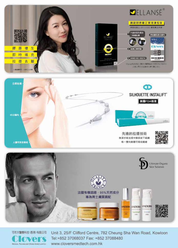

Medical Bulletin VOL.25 NO.6 JUNE 2020
26
The Making of a Cosmetic SurgeonDr Elvis Wai-ying LEEMBBS(HK), FRCS(Edinburgh), FCSHK, FHKAM(Surgery)Specialist in Plastic Surgery
Dr Elvis Wai-ying LEE
This topic is a bit controversial. Within the medical community, there is no such specialist formally known as a cosmetic surgeon, while this term is very well known among the laymen. If you want to have your eyebags removed, go to a cosmetic surgeon, but, where is the cosmetic surgeon?
Whenever an incident concerning cosmetic surgery or beauty treatment occurs, we will be faced with the media questioning the training and qualification required for a cosmetic surgeon. The public want to ask 'who should be allowed to do what?' as defined by the medical profession.
Literally, a cosmetic surgeon is a surgeon practising cosmetic surgery. However , there is no such subspecialty under the Hong Kong Academy of Medicine(HKAM). Under the Specialist Registration of Hong Kong Medical Council, there is also no such term as Specialist in Cosmetic Surgery. The absence of formal HKAM accreditation may be the root cause of the confusion.
Where should cosmetic surgery fit in? If we look into the curriculum for trainees of Plastic Surgery, "All aspects of aesthetic surgery" have been included1. The word `aesthetics’ means the formal study of the principle of art and beauty, while the word `cosmetic’ means preparations that are applied to the human body for beautifying, preserving and altering the appearance. Both concern beauty but Plastic Surgeons have traditionally preferred the term `aesthetics’ to the term `cosmetic’, likely because the former feels more fundamental while the latter appears more superficial.
THE LATIN ‘PLASTIKOS’ MEANS FIT TO MOLDEven Plastic Surgery itself draws quite a bit of confusion because unlike other medical disciplines, Paediatric Surgery is not defined by bodily boundary as can be defined by organ/system or age... So, what constitues plastic surgery? History may give us some hints. From archaeological literature going as far back as circa 600 B.C. in India, Sushrutta recorded operations for the reconstruction of nose and earlobe2. In ancient times, the nose was regarded as an organ of respect and reputation, hence amputation of the nose was used to punish criminals and captives of war. (Coincidentally many statues in museums are devoid of the nose.) Such operation was meant to make that victim appear 'whole' again and regain the dignity of being a person. Actually, the development of Plastic Surgery went with
the frequency and intensity of wars throughout history, reaching a high point during and after the Second World War with massive casualties requiring reconstruction of various body parts. Things changed after WWII. With a long period of peace, stability and prosperity, more people request plastic surgery not to reconstruct a damaged part but to improve on their appearance, hence the growth and development of Aesthetic Plastic Surgery or what is commonly called Cosmetic Surgery.
Plastic Surgery can thus be defined as a specialised branch of surgery concerned with the repair of deformities and correction of functional deficits2. It includes the reconstructive aspect for congenital and post-traumatic deformities, in which we attempt to restore deformities to normal and the aesthetic aspect in which we attempt to surpass the normal. Ultimately, we aim at improving one’s body image and enhancing the quality of life.
ART, INDEED, CONSISTS IN THE CONCEPTION OF THE RESULT TO BE PRODUCED BEFORE ITS REALISATION IN THE MATERIAL’…ARISTOTLE’Let us take a look at the curriculum for a higher surgical trainee entering into the Plastic Surgery training programme after completion of basic surgical training1. The curriculum includes:
1. Wound healing, wound care and tissue transplantation
2. Trauma and/or infection which involves soft tissues as part of the injury
3. Maxillofacial trauma4. Burns – thermal, electrical, chemical and radiation,
including their sequelae5. Cleft and craniofacial deformities6. Other congenital and acquired deformities of the
head and neck (including vascular abnormalities)7. Benign and malignant skin tumours, including
management of the regional lymph nodes8. Head and neck tumours9. Congenital and acquired deformities of the trunk,
limbs and other sites where the provision of skin cover is a component
10. Congenital and acquired deformities of the urogenital system

Medical BulletinVOL.25 NO.6 JUNE 2020
27
11. Paraplegic skin and soft tissue problems12. All aspects of hand surgery13. Microsurgery in all its applications to reparative
and reconstructive surgery14. Reconstructive surgery of the breast15. All aspects of aesthetic surgery16. Knowledge of medical ethics, aspects of informed
consent and the medico-legal implications of clinical practice and developing surgical practice
As can been seen from the list, a heavy emphasis on the training of a plastic surgeon is on the skin and soft tissue handling and dealing with the external parts of the whole body. More importantly, during the course of training, experience is gained as to the understanding and visualisation of the anatomy of different body parts. This forms a very precious and irreplaceable foundation for acquiring the skills in aesthetic plastic surgery. Perhaps the critical qualities for the plastic surgeon is a sense of form and an innate ability to make an aesthetic judgement2. In aesthetic plastic surgery, we are placing our hands on non-diseased body parts! It is not something as light as cosmetics.
Although with comprehensive and all-rounded training, under the current situation of the public hospitals of Hong Kong, there is a very limited scope for plastic surgical trainees to be exposed to aesthetic plastic surgery. As with colleagues from other specialties, medical career is a life-long learning profession. Aesthetic plastic surgeons polish their skill and understanding through learning from local and overseas colleague friends, not limited to plastic surgeons, but friends from any specialty whose expertise are contributive to our practice like dermatologists, ophthalmologists, ENT surgeons and dental surgeons. Other resources include overseas dissection courses and workshops and training courses offered by medical suppliers. Again, unlike other medical specialties, the final results of aesthetic plastic surgery depend on quite a lot on the patients’ perception. Their continuous feedback on our intervention results forms a continuous drive for us to strive for better, still, albeit sometimes harsh.
References1. McCarthy Plastic Surgery Volume One : General Principles –
Introduction to Plastic Surgery2. Instructions to Candidates, The College of Surgeons of Hong Kong,
Plastic Surgery Board, Exit Examination (Updated version on 14th May 2019)
Xeomin_Vs_korean_toxin_av_A03.indd 1 17/4/2020 下午4:00
Medical Bulletin VOL.25 NO.6 JUNE 2020
28
A Surgery-first Approach Le Fort I Osteotomy with Simultaneous Alveolar Cleft Grafting for Skeletal Facial Deformities in Adult Cleft Lip and PalateDr Loi-ming CHEUNGBDS(HKU), MDS (HKU), FRACDS, FCDSHK(OMS), FHKAM (Dental Surgery)Specialist in Oral & Maxillofacial Surgery
Dr Loi-ming CHEUNG
INTRODUCTIONPatients born with cleft lip and palate often suffer from underdevelopment of the maxilla. When they reach adolescence or adulthood, such underdevelopment may cause midface hypoplasia leading to anteroposterior, vertical, and transverse maxillary deficiency. They may also suffer from residual lip and nasal deformities, disruption of the dentition with missing permanent teeth, deformed teeth or even supernumerary teeth in the alveolar cleft sites. They may have un-repaired or inadequately repaired oronasal fistulas. All of these conditions may contribute to functional and aesthetic concerns.
Orthognathic surgery has often been regarded as the end-stage reconstruction required to correct the associated jaw deformities. We are taught to perform orthognathic surgery in adult patients in tandem with pre-surgical orthodontic treatment1. Such pre-surgical orthodontic treatment aims to align and level teeth to position them in line with the basal bones of the maxilla and mandible and is regarded as essential in improving the predictability of the outcome of the orthognathic surgery. The commonly taught treatment strategy is a tri-phasic approach consisting of pre-surgical orthodontics, followed by orthognathic surgery and completed by post-surgical orthodontics. However, orthodontic treatment in patients without the correction of the basal bones in the maxilla and mandible by itself can be difficult and in some cases un-manageable. In addition, the pre-surgical orthodontic treatment may take years to accomplish because of the difficulties.
In selected cases surgery-first-approach orthognathic surgical correction of the skeletal discrepancies first followed by orthodontic treatment can yield similar treatment outcome without the prolonged duration of a tri-phasic approach of pre-surgical orthodontics, orthognathic surgery and post-surgical orthodontics2,3. Such surgery-first-approach orthognathic surgery has gained higher acceptance in the management of patients with jaw discrepancies in non-cleft patients in recent years. The benefit of an accelerated treatment duration has gained good patient acceptance. The same treatment principle can also be applied to patients with cleft lip and palate4,5,6.
CASE REPORTA 19-year-old man with left unilateral cleft lip and palate had a chief complaint of mandibular prognathism
and persistent left oronasal communications. His medical and dental histories were unremarkable. He had primary cleft lip repair and palatoplasty during infancy. He did not have bone grafting of the left alveolar cleft in the past. His facial profile was concave with a retruded upper lip and a protruded lower lip. There was patent oronasal communication and left subnasal depression due to the unrepaired left alveolar cleft. The maxilla was hypoplastic. The mandible was within normal size and proportion. There was reverse overjet of the incisors measured to 8 mm. He had a strong intention to improve the facial appearance and to correct the dental malocclusion. However, he was not a local citizen and could only afford to stay continuously in Hong Kong for three months for the orthognathic surgery but was willing to travel back to Hong Kong regularly for orthodontic treatment. Clinical and radiographic examinations were performed. Dental study models were produced to study the malocclusion and model surgery to mimic the orthognathic surgical effect was performed. Thorough pre-treatment discussion with the patient and orthodontist involved was done. All parties agreed that the adoption of a surgery-first-approach was most appropriate. The treatment objectives were to improve the midfacial deficiency, to improve the dental malocclusion with reverse overjet of incisors and closure of the oronasal fistula. Orthognathic surgery was performed in 2010 with a Le Fort I osteotomy with a combined maxillary advancement and downward movements with simultaneous repair of the left persistent alveolar cleft with bone graft. The maxilla was planned to have 10 mm advancement and 4 mm downward movement. The supernumerary tooth adjacent to the left alveolar cleft was extracted. Bone graft prepared by a mixture of autogenous chin bone and xenograft of tricalcium phosphate7 was inserted to the left alveolar cleft. The maxilla was fixed with internal titanium mini-screws and mini-plates. The patient was hospitalised for four days after the operation.
Orthodontic brackets were inserted, and orthodontic alignment of teeth was commenced 12 weeks after the operation. Alignment of teeth was reinforced by orthodontic temporary titanium anchorage screw. The whole treatment duration, including the orthognathic surgery and the post-surgical orthodontic alignment, was completed within 18 months. The patent had been followed up for five years after completion of orthognathic surgery and no relapse was detected.

Medical BulletinVOL.25 NO.6 JUNE 2020
29
Treatment records:
Fig. 1A.Lateral facial profile view (before). (Photo from personal collection)
Fig. 1B. Frontal facial view (after). (Photo from personal collection)
Fig. 2A. Frontal facial view (before). (Photo from personal collection)
Fig. 2B. Frontal facial view (after). (Photo from personal collection)
Fig. 3A. Upper occlusal view (before). (Photo from personal collection)
Fig. 3B. Upper occlusal view (after). (Photo from personal collection)
Fig. 4A. Lower occlusal view (before). (Photo from personal collection)
Fig. 4B.Lower occlusal view (after). (Photo from personal collection)
Fig.5A. Lateral view of incisors with reverse overjet (before). (Photo from personal collection)
Fig. 5B. Lateral view of incisors with corrected overjet (after). (Photo from personal collection)
Fig. 6A. Frontal view of teeth with reversed bite and left alveolar cleft (before). (Photo from personal collection)
Fig. 6B. Frontal view of teeth with corrected bite and repaired left alveolar cleft (after). (Photo from personal collection)
Fig. 7A. Orthopantogram x-ray (before). (Photo from personal collection)
Fig. 7B. Orthopantogram x-ray (after). (Photo from personal collection)
Fig. 8A. Lateral cephalometric x-ray (before). (Photo from personal collection)
Fig. 8B. Lateral cephalometric x-ray (after). (Photo from personal collection)
DISCUSSIONThe surgery-first-approach orthognathic surgery concept was first presented in 2009 by Nagasaka et al.2 in a case report on a non-cleft patient. The significant reduction in total treatment time and high patient satisfaction has led to wider adoption of this concept nowadays as a reasonable and more cost-effective strategy to manage dentoskeletal deformities. This treatment strategy can have a similar benefit in patients with cleft lip and palate, with repaired or non-repaired alveolar cleft. Within the author’s experience, the end result and the surgical stability bear no difference to the conventional tri-phasic approach.
References1. L. M. Wolford, E. L. L. Stevao. Correction of jaw deformities in patients with
cleft lip and palate. BUMC Proceedings 2002;15:250-254.2. Nagasaka H, Sugawara J, Kawamura H, Nanda R. " Surgery first" skeletal
Class III correction using the Skeletal Anchorage System. J Clin Orthod. 2009;43(2):97.
3. H. B. Xu, L. X. Mao, W. D. Wang, B. Fang, S. G. Shen. The surgery-first approach in orthognathic surgery: a retrospective study of 50 cases. Int. J. Oral Maxillofac. Surg. 2015;44:1463-1467.
4. Baser Keklikci H. et al. Surgery-first orthognathic treatment of an adult patient with unilateral cleft lip-palate and multiple missing teeth: a case report. South Eur. J. Orthod. Dentofac. Res. 2018;5(1):19-24.
5. Choi et al. Maxillofacial Plastic and Reconstructive Surgery. (2019) 41:106. Seo H. J., Denadai R., Pai B. C., Lo C. J. Modern Surgery-First Approach
Concept in Cleft-Orthognathic Surgery: A Comparative Cohort Study with 3D Quantitative Analysis of Surgical-Occlusion Setup. J. Clin. Med. 2019,8,2116.
7. Ad de Ruiter, Nard Janssen, Robert van Es, Michael Frank, Gert Meijer, Ron Koole, Toine Rosenberg. Micro-structured Beta-Tricalcium Phosphate for Repair of the Alveolar Cleft in Cleft Lip and Palate Patients: A Pilot Study. The Cleft Palate-Craniofacial Journal. June 2014.

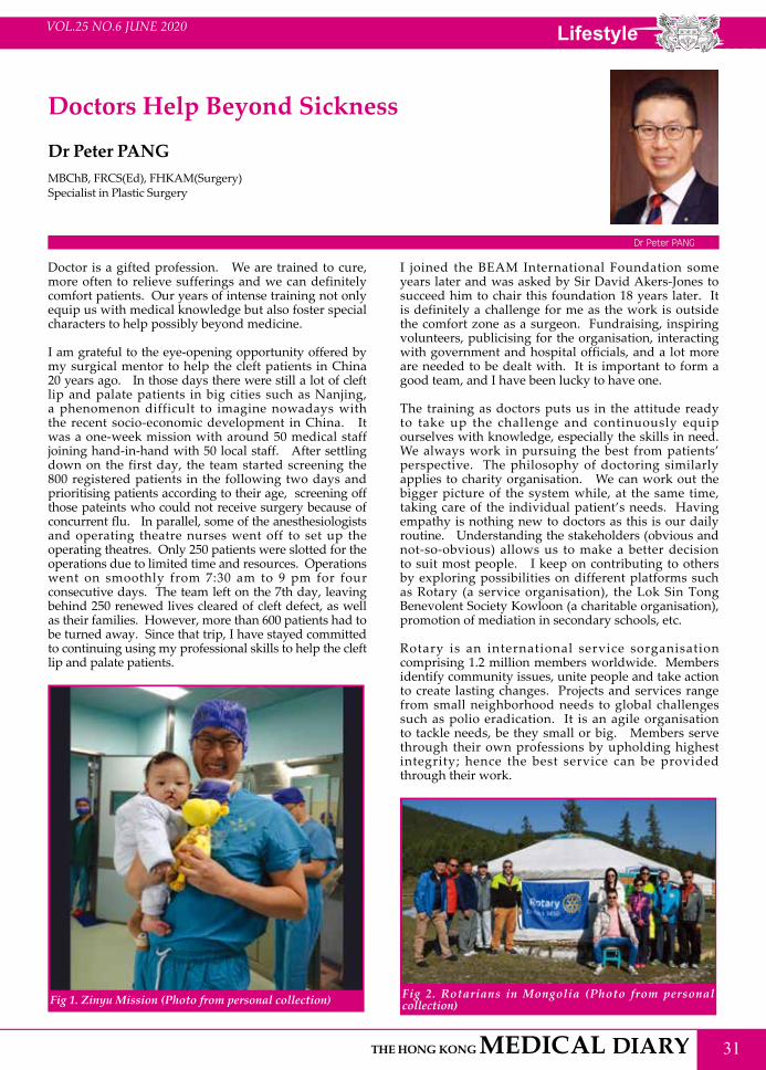
31
VOL.25 NO.6 JUNE 2020
Doctors Help Beyond Sickness
Dr Peter PANGMBChB, FRCS(Ed), FHKAM(Surgery)Specialist in Plastic Surgery
Dr Peter PANG
Lifestyle
Doctor is a gifted profession. We are trained to cure, more often to relieve sufferings and we can definitely comfort patients. Our years of intense training not only equip us with medical knowledge but also foster special characters to help possibly beyond medicine.
I am grateful to the eye-opening opportunity offered by my surgical mentor to help the cleft patients in China 20 years ago. In those days there were still a lot of cleft lip and palate patients in big cities such as Nanjing, a phenomenon difficult to imagine nowadays with the recent socio-economic development in China. It was a one-week mission with around 50 medical staff joining hand-in-hand with 50 local staff. After settling down on the first day, the team started screening the 800 registered patients in the following two days and prioritising patients according to their age, screening off those pateints who could not receive surgery because of concurrent flu. In parallel, some of the anesthesiologists and operating theatre nurses went off to set up the operating theatres. Only 250 patients were slotted for the operations due to limited time and resources. Operations went on smoothly from 7:30 am to 9 pm for four consecutive days. The team left on the 7th day, leaving behind 250 renewed lives cleared of cleft defect, as well as their families. However, more than 600 patients had to be turned away. Since that trip, I have stayed committed to continuing using my professional skills to help the cleft lip and palate patients.
Fig 1. Zinyu Mission (Photo from personal collection)
I joined the BEAM International Foundation some years later and was asked by Sir David Akers-Jones to succeed him to chair this foundation 18 years later. It is definitely a challenge for me as the work is outside the comfort zone as a surgeon. Fundraising, inspiring volunteers, publicising for the organisation, interacting with government and hospital officials, and a lot more are needed to be dealt with. It is important to form a good team, and I have been lucky to have one.
The training as doctors puts us in the attitude ready to take up the challenge and continuously equip ourselves with knowledge, especially the skills in need. We always work in pursuing the best from patients’ perspective. The philosophy of doctoring similarly applies to charity organisation. We can work out the bigger picture of the system while, at the same time, taking care of the individual patient’s needs. Having empathy is nothing new to doctors as this is our daily routine. Understanding the stakeholders (obvious and not-so-obvious) allows us to make a better decision to suit most people. I keep on contributing to others by exploring possibilities on different platforms such as Rotary (a service organisation), the Lok Sin Tong Benevolent Society Kowloon (a charitable organisation), promotion of mediation in secondary schools, etc.
Rotary is an international service sorganisation comprising 1.2 million members worldwide. Members identify community issues, unite people and take action to create lasting changes. Projects and services range from small neighborhood needs to global challenges such as polio eradication. It is an agile organisation to tackle needs, be they small or big. Members serve through their own professions by upholding highest integrity; hence the best service can be provided through their work.
Fig 2. Rotarians in Mongolia (Photo from personal collection)
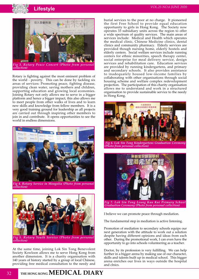
32
VOL.25 NO.6 JUNE 2020Lifestyle
Fig 3. Rotary Peace Concert (Photo from personal collection)
Rotary is fighting against the most eminent problem of the world - poverty. This can be done by tackling six areas of services: Promoting peace, fighting disease, providing clean water, saving mothers and children, supporting education and growing local economies. Joining Rotary not only allows me to serve in a bigger platform and hence a bigger impact, this also allows me to meet people from other walks of lives and to learn new skills and knowledge from fellow members. It is a very good training ground for leadership as all projects are carried out through inspiring other members to join in and contribute. It opens opportunities to see the world in endless dimensions.
Fig 4. Rotary Service in Mongolia (Photo from personal collection)
Fig 5. Rotary Youth Service (Photo from personal collection)
At the same time, joining Lok Sin Tong Benevolent Society Kowloon allows me to serve Hong Kong from another dimension. It is a charity organisation with 140 years of history started by a group of local Chinese, providing free medical consultation to the needy and
burial services to the poor at no charge. It pioneered the first Free School to provide equal education opportunity to girls in Hong Kong. The Society now operates 33 subsidiary units across the region to offer a wide spectrum of quality services. The main areas of services include: Medical and Health which operates the medical clinic, Chinese Medicine clinics, dental clinics and community pharmacy. Elderly services are provided though nursing home, elderly hostels and elderly centers. Social welfare services include running centers for ethnic minorities, speech therapy center, social enterprise for meal delivery service, design services and rehabilitation care. Education services are provided by running kindergartens, and primary and secondary schools. It also provides assistance to inadequately housed low-income families by collaborating with other organisations through social housing scheme and welfare complex redevelopment projection. The participation of this charity organisation allows me to understand and work in a structured organisation to provide sustainable service to the needy in Hong Kong.
Fig 6. Lok Sin Tong Kindergartens Graduation Ceremony (Photo from personal collection)
Fig 7. Lok Sin Tong Leung Kau Kui Primary School Graduation Ceremony (Photo from personal collection)
I believe we can promote peace through mediation.
The fundamental step in mediation is active listening.
Promotion of mediation to secondary schools equips our next generation with the attitude to work out a solution despite having different opinions and ideas from each other. During the promotional work, I can even have the opportunity to go into schools volunteering as a teacher.
Doctor, by its profession is very fulfilling. We can help further in a bigger arena by making use of our character, skills and talents built up in medical school. This bigger arena enriches our lives in ways outside the hospital and clnics.

www.karlstorz.com
VITOM® 3D – The 3rgonomic Dimension3D visualization for Otorhinolaryngology
9631
7004
EN
T 10
6 1.
0 05
/201
7/P
-E

34
VOL.25 NO.6 JUNE 2020Medical Diary of JuneSu
nday
Mon
day
Tues
day
Wed
nesd
ayT
hurs
day
Frid
aySa
turd
ay
1819
20
2114
2223
282915
1617
87
24
1112
1310
9
14
32
56
25
30
2627
Cer
tifica
te C
ours
e on
C
omm
unic
atio
n an
d Sw
allo
win
g Pr
oble
ms
in
the
Elde
rly
Popu
latio
n 20
20 (V
ideo
Lec
ture
s)
Cer
tifica
te C
ours
e on
C
omm
unic
atio
n an
d Sw
allo
win
g Pr
oble
ms
in
the
Elde
rly
Popu
latio
n 20
20 (V
ideo
Lec
ture
s)
Cer
tifica
te C
ours
e on
C
omm
unic
atio
n an
d Sw
allo
win
g Pr
oble
ms
in
the
Elde
rly
Popu
latio
n 20
20 (V
ideo
Lec
ture
s)
Cer
tifica
te C
ours
e on
C
omm
unic
atio
n an
d Sw
allo
win
g Pr
oble
ms
in
the
Elde
rly
Popu
latio
n 20
20 (V
ideo
Lec
ture
s)
Cer
tifica
te C
ours
e on
C
omm
unic
atio
n an
d Sw
allo
win
g Pr
oble
ms
in
the
Elde
rly
Popu
latio
n 20
20 (V
ideo
Lec
ture
s)
Cer
tifica
te C
ours
e on
U
ltras
ound
Dia
gnos
is o
f Fe
tal A
nom
alie
s (V
ideo
Le
ctur
es)
Cer
tifica
te C
ours
e on
U
ltras
ound
Dia
gnos
is o
f Fe
tal A
nom
alie
s (V
ideo
Le
ctur
es)
Cer
tifica
te C
ours
e on
U
ltras
ound
Dia
gnos
is o
f Fe
tal A
nom
alie
s (V
ideo
Le
ctur
es)
Cer
tifica
te C
ours
e on
U
ltras
ound
Dia
gnos
is o
f Fe
tal A
nom
alie
s (V
ideo
Le
ctur
es)
HK
MA
CM
E LI
VE
Com
mon
STI
s an
d U
pdat
es
HK
MA
CM
E LI
VE
Endo
crin
olog
y (T
BC)
HK
MA
CM
E LI
VE
Ove
rvie
w o
f AF
Man
agem
ent i
n A
dults
HK
MA
CM
E LI
VE
Hyp
erte
nsio
n (T
BC)
HK
MA
CM
E LI
VE
Trea
tmen
t of E
xter
nal
Gen
ital W
art

35
VOL.25 NO.6 JUNE 2020 Calendar of Events
Ms. Vienna LAM Tel: 2527 8898
Certificate Course on Communication and Swallowing Problems in the Elderly Population 2020 (Video Lectures)Organiser: The Federation of Medical Societies of Hong Kong;Speaker: Dr.Rita Wai-ming WONG
7:00 PM
MON22
Ms. Vienna LAM Tel: 2527 8898
Certificate Course on Communication and Swallowing Problems in the Elderly Population 2020 (Video Lectures)Organiser: The Federation of Medical Societies of Hong KongSpeaker: Dr. Raymond FONG
7:00 PM
MON29
Ms. Vienna LAM Tel: 2527 8898
Certificate Course on Ultrasound Diagnosis of Fetal Anomalies (Video Lectures)Organiser: The Federation of Medical Societies of Hong Kong;Dr. Kwok-yin LEUNG
7:00 PM
FRI19
Ms. Vienna LAM Tel: 2527 8898
Certificate Course on Ultrasound Diagnosis of Fetal Anomalies (Video Lectures)Organiser: The Federation of Medical Societies of Hong Kong;Dr. Ka-wang CHEUNG
7:00 PM
FRI26
Ms. Vienna LAM Tel: 2527 8898
Certificate Course on Communication and Swallowing Problems in the Elderly Population 2020 (Video Lectures)Organiser: The Federation of Medical Societies of Hong Kong;Speaker: Dr.Joshua Kam-wo MAK
7:00 PM
MON15
Ms. Candice TONG2527 82851 CME Point
HKMA CME LIVEOverview of AF Management in AdultsOrganiser: HKMA-HK East Community Network;Speaker: Dr. LUK Ngai Hong, Vincent
2:00 PM
THU11
Ms. Candice TONG2527 8285TBC CME Point
HKMA CME LIVEEndocrinology (TBC)Organiser: HKMA-YTM Community Network;Speaker: Dr. Norman CHAN
2:00 PM
TUE9
Miss Antonia LEE2527 8285TBC CME Point
HKMA CME LIVEHypertension (TBC)Organiser: HKMA-Kowloon East Community Network;Speaker: Dr. Jacky CHAN
2:00 PM
THU18
Date / Time Function Enquiry / RemarksMs. Vienna LAM Tel: 2527 8898
Certificate Course on Communication and Swallowing Problems in the Elderly Population 2020 (Video Lectures)Organiser: The Federation of Medical Societies of Hong Kong;Speaker: Dr. Anthony Pak-hin KONG
7:00 PM
Miss Antonia LEE2527 82851 CME Point
HKMA CME LIVECommon STIs and UpdatesOrganiser: HKMA-Central, Western & Southern Community Network;Speaker: Dr. KWAN Chi Keung
MON1
Ms. Vienna LAM Tel: 2527 8898
Certificate Course on Communication and Swallowing Problems in the Elderly Population 2020 (Video Lectures)Organiser: The Federation of Medical Societies of Hong Kong;Speaker: Dr. Lorinda Chen KWAN
7:00 PM
MON8
Ms. Vienna LAM Tel: 2527 8898
Certificate Course on Ultrasound Diagnosis of Fetal Anomalies (Video Lectures)Organiser: The Federation of Medical Societies of Hong KongSpeaker: Dr. Ben Chong-pun CHAN
7:00 PM
FRI12
Ms. Vienna LAM Tel: 2527 8898
Certificate Course on Ultrasound Diagnosis of Fetal Anomalies (Video Lectures)Organiser: The Federation of Medical Societies of Hong Kong;Speaker: Dr. Wing-cheong LEUNG
7:00 PM
FRI5
Ms. Candice TONG2527 82851 CME Point
HKMA CME LIVETreatment of External Genital WartOrganiser: HKMA-HK East Community Network;Speaker: Dr. CHAN Yung, Davis;
2:00 PM
THU4
2:00 PM
WED3

36
VOL.25 NO.6 JUNE 2020
Answers to Radiology Quiz
Radiology Quiz
The Federation of Medical Societies of Hong Kong 4/F Duke of Windsor Social Service Building, 15 Hennessy Road, Wanchai, HK Tel: 2527 8898 Fax: 2865 0345
PresidentDr Mario Wai-kwong CHAK 翟偉光醫生
1st Vice-PresidentProf Bernard Man-yung CHEUNG 張文勇教授
2nd Vice-PresidentDr Chun-kong NG 吳振江醫生
Hon. TreasurerMr Benjamin Cheung-mei LEE 李祥美先生
Hon. SecretaryDr Ludwig Chun-hing TSOI 蔡振興醫生
Immediate Past President Dr Raymond See-kit LO 勞思傑醫生Executive Committee Members
Dr Jane Chun-kwong CHAN 陳真光醫生Dr Kingsley Hau-ngai CHAN 陳厚毅醫生Dr Kai-ming CHAN 陳啟明醫生Dr Alson Wai-ming CHAN 陳偉明醫生Dr Peggy Sau-kwan CHU 朱秀群醫生Dr Samuel Ka-shun FUNG 馮加信醫生Ms Ellen Wai-yin KU 顧慧賢小姐Dr Haston Wai-ming LIU 廖偉明牙醫Dr Yin-kwok NG 吳賢國醫生Dr Desmond Gia-hung NGUYEN 阮家興醫生Dr Kwai-ming SIU 邵貴明醫生Dr Tony Ngan-fat TO 杜銀發醫生Mr William TSUI 徐啟雄先生Dr Victor Hip-wo YEUNG 楊協和醫生Ms Tina WT YIP 葉婉婷女士Dr Edwin Chau-leung YU 余秋良醫生Ms Manbo MAN (Co-opted) 文保蓮女士Dr Wilfred Hing-sang WONG (Co-opted)
黃慶生博士
Founder MembersBritish Medical Association (Hong Kong Branch)英國醫學會 ( 香港分會 )
PresidentDr Raymond See-kit LO 勞思傑醫生
Vice-PresidentDr Adrian WU 鄔揚源醫生
Hon. SecretaryDr Terry Che-wai HUNG 洪致偉醫生
Hon. TreasurerDr Jason BROCKWELL
Council RepresentativesDr Raymond See-kit LO 勞思傑醫生Dr Tse-ming CHEUNG 張子明醫生Tel: 2527 8898 Fax: 2865 0345
The Hong Kong Medical Association香港醫學會
PresidentDr Chung-ping HO, MH, JP 何仲平醫生 , MH, JP
Vice- PresidentsDr Chi-man CHENG 鄭志文醫生Dr David Tzit-yuen LAM 林哲玄醫生
Hon. SecretaryDr Victor Hip-wo YEUNG 楊協和醫生
Hon. Treasurer
Dr Chi-chiu LEUNG 梁子超醫生
Council RepresentativesDr AlvinYee-shing CHAN 陳以誠醫生
Chief Executive
Ms Jovi LAM 林偉珊女士Tel: 2527 8285 (General Office) 2527 8324 / 2536 9388 (Club House in Wanchai / Central)Fax: 2865 0943 (Wanchai), 2536 9398 (Central)Email: [email protected] Website: http://www.hkma.org
The HKFMS Foundation Limited 香港醫學組織聯會基金 Board of DirectorsPresident
Dr Mario Wai-kwong CHAK 翟偉光醫生1st Vice-President
Prof Bernard Man-yung CHEUNG 張文勇教授2nd Vice-President
Dr Chun-kong NG 吳振江醫生Hon. Treasurer
Mr Benjamin Cheung-mei LEE 李祥美先生Hon. Secretary
Dr Ludwig Chun-hing TSOI 蔡振興醫生Directors
Mr Samuel Yan-chi CHAN 陳恩賜先生Dr Samuel Ka-shun FUNG 馮加信醫生 Ms Ellen Wai-yin KU 顧慧賢女士Dr Raymond See-kit LO 勞思傑醫生Dr Aaron Chak-man YU 余則文醫生
Answers:
1.
2.
3.
--
---
-
-
-
-
Through a left-sided superior vena cava (SVC).Tip of PICC was placed around the left SVC / right atrial junction, in a satisfactory and safe position.
0.3-0.5% of normal population.5% associated with congenital heart disease.Only seen in isolation in 10%, while the remaining are actually duplicated SVC (ie, normal right-sided SVC present).
Majority of patients are asymptomatic and detected incidentally either as a result of line placement (as in this case) or cross-sectional imaging of the chest.A minority of patients may present with a right-to-left shunt (due to direct drainage into left atrium).5% are associated with congenital heart disease with atrial septal defect (ASD) being the most common. Most important is awareness of this venous variant during line or pacemaker placement. Other possible non-venous (and often catastrophic) locations may include into the descending thoracic aorta or extravascular position into the mediastinum or pleural space.
Dr Leanne Han-qing CHINMBBS, FRCR

Abbreviated prescribing information Saxenda® (liraglutide injection) The Summary of Product Characteristics (SPC) is available at novonordisk.com. Presentation: Prefilled, disposable pen containing 18 mg of liraglutide in 3 mL of solution. Indications: Saxenda® is indicated as an adjunct to a reduced-calorie diet and increased physical activity for weight management in adult patients with an initial Body Mass Index (BMI) of ≥ 30 kg/m² (obese), or ≥ 27 kg/m² to < 30 kg/m² (overweight) in the presence of at least one weight-related comorbidity such as dysglycaemia (pre-diabetes or type 2 diabetes mellitus), hypertension, dyslipidaemia or obstructive sleep apnoea. Treatment with Saxenda® should be discontinued after 12 weeks on the 3.0 mg/day dose if patients have not lost at least 5% of their initial body weight. The need for continued treatment should be re-evaluated annually. Dosage and administration: The starting dose is 0.6 mg once daily. The dose should be increased to 3.0 mg once daily in increments of 0.6 mg with at least one week interval to improve gastro-intestinal tolerability. If escalation to the next dose step is not tolerated for two consecutive weeks, consider discontinuing treatment. Daily doses higher than 3.0 mg are not recommended. Saxenda® is administered once daily at any time, independent of meals, subcutaneously injected in the abdomen, thigh or upper arm, preferably around the same time every day. Saxenda® must not be administered intravenously or intramuscularly. Patients with type 2 diabetes mellitus receiving liraglutide in combination with a sulphonylurea may have an increased risk of hypoglycaemia. The risk of hypoglycaemia may be lowered by a reduction in the dose of sulphonylurea. Saxenda® should not be used in combination with other Glucagon-like Peptide-1 (GLP-1) receptor agonist. The addition of Saxenda® in patients with type 2 diabetes mellitus treated with insulin has not been evaluated. This medicinal product is not recommended for use in paediatric patients. Contraindications: Hypersensitivity to liraglutide or to any of the excipients. Special warnings and precautions: In patients with diabetes mellitus liraglutide must not be used as a substitute for insulin. There is limited experience in patients with congestive heart failure New York Heart Association (NYHA) class I-II and liraglutide should therefore be used with caution. There is no experience in patients with congestive heart failure NYHA class III-IV and liraglutide is therefore not recommended in these patients. Due to limited experience, Saxenda® is not recommended in patients with inflammatory bowel disease or diabetic gastroparesis. Saxenda® is not recommended in patients: aged 75 years or more, treated with other products for weight management, with obesity secondary to endocrinological or eating disorders or to treatment with medicinal products that may cause weight gain, with severe renal impairment,
with severe hepatic impairment. Saxenda® must be used with caution in patients with mild or moderate hepatic impairment. Use of GLP-1 receptor agonists has been associated with the risk of developing acute pancreatitis. Patients should be informed of the characteristic symptoms of acute pancreatitis. If pancreatitis is suspected, liraglutide should be discontinued; if acute pancreatitis is confirmed, liraglutide should not be restarted. Caution should be exercised in patients with a history of pancreatitis. In clinical trials for weight management, a higher rate of cholelithiasis and cholecystitis was observed in patients treated with liraglutide than in patients on placebo. Patients should be informed of the characteristic symptoms of cholelithiasis and cholecystitis. In clinical trials in type 2 diabetes, thyroid adverse events, including increased blood calcitonin, goitre and thyroid neoplasm have been reported in particular in patients with pre-existing thyroid disease. Cases of increased blood calcitonin were also observed in the weight management clinical trials. An increase in heart rate was observed with liraglutide in clinical trials. Heart rate should be monitored at regular intervals consistent with usual clinical practice. Patients should be informed of the symptoms of increased heart rate (palpitations or feelings of a racing heartbeat while at rest). For patients who experience a clinically relevant sustained increase in resting heart rate, treatment with liraglutide should be discontinued. Patients treated with liraglutide should be advised of the potential risk of dehydration in relation to gastrointestinal side effects and take precautions to avoid fluid depletion. Pregnancy and lactation: Saxenda® should not be used in women who are pregnant, who wish to become pregnant, or who are breastfeeding. Undesirable effects: The most frequently reported adverse reactions in patients treated with Saxenda® are nausea, vomiting, diarrhoea and constipation. Less common adverse reactions include dyspepsia, upper abdominal pain, gastritis, flatulence, abdominal distension, gastroesophageal reflux, eructation, dry mouth, dizziness, dysgeusia, insomnia, fatigue, asthenia, injection site reactions, malaise, tachycardia, urticaria, pancreatitis, cholelithiasis, cholecystitis, hypoglycaemia, anaphylactic reaction, dehydration, acute renal failure and renal impairment. Overdose: From clinical trials and marketed use overdoses have been reported up to 72 mg (24 times the recommended maintenance dose). Events reported included severe nausea and severe vomiting which are also the expected symptoms of an overdose with liraglutide. None of the reports included severe hypoglycaemia. All patients recovered without complications. In the event of overdose, appropriate supportive treatment should be initiated according to the patient’s clinical signs and symptoms. The patient should be observed for clinical signs of dehydration and blood glucose should be monitored.
References: 1. Saxenda® [summary of product characteristics]. 2. NovoNordisk Company press release, 22 Apr 2015. United States first country to launch Saxenda®. 3.Pi-Sunyer X, Astrup A, Fujioka K, et al; for the SCALE Obesity and Prediabetes NN8022-1839 Study Group. A randomized, controlled trial of 3.0 mg of liraglutide in weight management. N Engl J Med. 2015;373(1):11-22.
Further Information is available fromNovo Nordisk Hong Kong LtdUnit 923A-928, 9/F Trade Square, 681 Cheung Sha Wan Road, Kowloon, Hong KongTel: (852) 3725 1300 Fax: (852) 2386 0800 www.novonordisk.com
Introducing Saxenda®:Significant and sustained weight loss with simultaneous improvements in cardiometabolic risk factors.1,3 In a 1-year study:
• 9 out of 10 patients achieved weight loss, with 1 in 3 losing >10%3
• Patients lost weight and kept it off1
• Patients also experienced significant improvements in multiple cardiometabolic risk factors1,3
The only GLP-1 analogue that is EMA and U.S. FDA approved for weight management as an adjunct to diet and exercise1,2
NOW, your patients WITH OBESITY have more to celebrate with
LOSING WEIGHT
2020 Saxenda 184x257.indd 1 5/5/20 12:16 PM
Related Documents