PLASTIC CHANGES UNDERLYING VESTIBULAR COMPENSATION IN THE GUINEA-PIG PERSIST IN ISOLATED, IN VITRO WHOLE BRAIN PREPARATIONS N. VIBERT,*² A. BABALIAN,* M. SERAFIN,‡ J.-P. GASC,§ M. MU ¨ HLETHALER‡ and P.-P. VIDAL* *Laboratoire de Neurobiologie des Re ´seaux Sensorimoteurs, CNRS ESA 7060, 45 rue des Saints-Pe `res, 75270 Paris cedex 06, France ‡De ´partement de Physiologie, Centre Me ´dical Universitaire, 1 rue Michel Servet, 1211 Geneva 4, Switzerland §Adaptations et Evolution des Syste `mes Oste ´omusculaires, CNRS URA 1137, Museum National d’Histoire Naturelle, 55 rue Buffon, 75005 Paris, France Abstract —Vestibular compensation for the postural and oculomotor deficits induced by unilateral labyrinthectomy is a model of post-lesional plasticity in the central nervous system. Just after the removal of one labyrinth, the deafferented, ipsilateral vestibular nucleus neurons are almost silent, and the discharge of the contralateral vestibular nucleus neurons is increased. The associated static disorders disappear in a few days, as normal activity is restored in both vestibular nuclei. In this study, we searched for traces of vestibular compensation in isolated whole brains taken from adult guinea-pigs. The electrophysiological responses evoked in control brains were compared to those evoked in brains taken from animals that had previously been labyrinthectomized. Guinea- pigs compensated for an initial labyrinthectomy within three days. In vivo, subsequent deafferentation of vestibular nucleus neurons on the intact side triggered “Bechterew’s phenomenon”: a new postural and oculomotor syndrome appeared, similar to the one induced by the first lesion, but directed to the newly deafferented side. These disturbances would be caused by the new imbalance between the discharges of neurons in the two vestibular nuclei triggered by the second deafferentation. Experiments were designed to search for a similar imbalance in vitro in brains taken from labyrinthectomized animals, where the intact vestibular nerve is cut during the dissection. Isolated whole brains were obtained from young guinea-pigs at various times (one to seven days) following an initial labyrinthectomy. An imbalance between the resting activities of medial vestibular nucleus neurons on both sides of the brainstem was revealed in brains taken more than three days after the lesion: their discharge was higher on the compensated, initially lesioned side than on the newly deafferented side. In some cases, an oscillatory pattern of discharge, reminiscent of the spontaneous nystagmus associated in vivo with Bechterew’s syndrome, appeared in both abducens nerves. These data demonstrate that most of the changes underlying vestibular compensation persist, and can thus be investigated in the isolated whole brain preparation. Brains removed only one day after the lesion displayed normal commissural responses and symmetric spinal inputs to vestibular nucleus neurons. However, an unusually large proportion of the neurons recorded on both sides of the preparation had very irregular spontaneous discharge rates. These data suggest that the first stages of vestibular compensation might be associated with transient changes in the membrane properties of vestibular nucleus neurons. Brains taken from compensated animals displayed a significant, bilateral decrease of the inhibitory commissural responses evoked in the medial vestibular nucleus by single-shock stimulation of the contralateral vestibular nerve. The sensitivity of abducens motoneurons on the initially lesioned, compensated side to synaptic activation from the contralesional vestibular nucleus neurons was also decreased. Both changes may explain the long-term, bilateral decrease of vestibular-related reflexes observed following unilateral labyrinthectomy. Spinal inputs to vestibular nucleus neurons became progressively asymmetric: their efficacy was increased on the lesioned side and decreased on the intact one. This last modification may support a functional substitution of the deficient, vestibular-related synergies involved in gaze and posture stabilization by neck-related reflexes. q 1999 IBRO. Published by Elsevier Science Ltd. Key words: vestibulo-ocular reflex, abducens nerve, postural control, post-lesional plasticity. In order to stabilize gaze and posture, the multiple internal representations of an event (e.g., head rotation) obtained through different sensory modalities (mostly the visual, vestibular and proprioceptive ones) are matched into a single frame of reference in which appropriate motor commands can be coded. Such complex sensorimotor transformations are highly sensitive to pathological damage, excessive natural stimulations and/or exposure to conflicting sensory informa- tion, and therefore display a high degree of plasticity. 55 A remarkable example of this plasticity can be observed following unilateral removal of one labyrinth. The strong oculomotor and postural syndrome induced by such a lesion recovers to a large extent, in a process known as vestibular compensation (see Refs 9, 10, 44 and 49 for reviews). The static deficits, which include major distortions of posture, associated with an ocular nystagmus with quick phases direc- ted towards the intact side, disappear over a few days in most species. 57 The dynamic deficits include a reduced gain and an abnormal timing of the vestibulo-ocular and vestibulo-spinal reflexes. They also improve over a period of several weeks, though this recovery is partial, and is limited to the low and middle frequency ranges of head movements. 52 The neuronal substrates of vestibular compensation (Fig. 1) have been studied extensively in vivo. Following unilateral labyrinthectomy, the ipsilesional vestibular nucleus neurons (VNns) are deprived of the massive excitatory input coming from the labyrinthine sensory afferents and lose their resting discharge, which averages about 30–40 spikes/s in alert vertebrates. 30,38 The VNns on the contralesional side increase their firing rate, following suppression of the commissural inhibition normally exerted by the deafferented VNns 413 Neuroscience Vol. 93, No. 2, pp. 413–432, 1999 Copyright q 1999 IBRO. Published by Elsevier Science Ltd Printed in Great Britain. All rights reserved 0306-4522/99 $20.00+0.00 PII: S0306-4522(99)00172-4 Pergamon ²To whom correspondence should be addressed. Tel.: 133-1-42863398; fax: 133-1-42863399. E-mail address: [email protected] (N. Vibert) Abbreviations: AHP, afterhyperpolarization; CV, coefficient of variation; Dn, day n (i.e. the number of days between the two lesions); IWB, isolated in vitro whole brain; MVN, medial vestibular nucleus; VNn, vestibular nucleus neuron.

Welcome message from author
This document is posted to help you gain knowledge. Please leave a comment to let me know what you think about it! Share it to your friends and learn new things together.
Transcript
PLASTIC CHANGES UNDERLYING VESTIBULAR COMPENSATION IN THEGUINEA-PIG PERSIST IN ISOLATED,IN VITROWHOLE BRAIN PREPARATIONS
N. VIBERT,*† A. BABALIAN,* M. SERAFIN,‡ J.-P. GASC,§ M. MUHLETHALER‡ and P.-P. VIDAL**Laboratoire de Neurobiologie des Re´seaux Sensorimoteurs, CNRS ESA 7060, 45 rue des Saints-Pe`res,
75270 Paris cedex 06, France
‡Departement de Physiologie, Centre Me´dical Universitaire, 1 rue Michel Servet, 1211 Geneva 4, Switzerland
§Adaptations et Evolution des Syste`mes Oste´omusculaires, CNRS URA 1137, Museum National d’Histoire Naturelle,55 rue Buffon, 75005 Paris, France
Abstract—Vestibular compensation for the postural and oculomotor deficits induced by unilateral labyrinthectomy is a model ofpost-lesional plasticity in the central nervous system. Just after the removal of one labyrinth, the deafferented, ipsilateral vestibularnucleus neurons are almost silent, and the discharge of the contralateral vestibular nucleus neurons is increased. The associatedstatic disorders disappear in a few days, as normal activity is restored in both vestibular nuclei. In this study, we searched for tracesof vestibular compensation in isolated whole brains taken from adult guinea-pigs. The electrophysiological responses evoked incontrol brains were compared to those evoked in brains taken from animals that had previously been labyrinthectomized. Guinea-pigs compensated for an initial labyrinthectomy within three days.In vivo, subsequent deafferentation of vestibular nucleus neuronson the intact side triggered “Bechterew’s phenomenon”: a new postural and oculomotor syndrome appeared, similar to the oneinduced by the first lesion, but directed to the newly deafferented side. These disturbances would be caused by the new imbalancebetween the discharges of neurons in the two vestibular nuclei triggered by the second deafferentation. Experiments were designedto search for a similar imbalancein vitro in brains taken from labyrinthectomized animals, where the intact vestibular nerve is cutduring the dissection. Isolated whole brains were obtained from young guinea-pigs at various times (one to seven days) following aninitial labyrinthectomy. An imbalance between the resting activities of medial vestibular nucleus neurons on both sides of thebrainstem was revealed in brains taken more than three days after the lesion: their discharge was higher on the compensated,initially lesioned side than on the newly deafferented side. In some cases, an oscillatory pattern of discharge, reminiscent of thespontaneous nystagmus associatedin vivo with Bechterew’s syndrome, appeared in both abducens nerves. These data demonstratethat most of the changes underlying vestibular compensation persist, and can thus be investigated in the isolated whole brainpreparation.
Brains removed only one day after the lesion displayed normal commissural responses and symmetric spinal inputs to vestibularnucleus neurons. However, an unusually large proportion of the neurons recorded on both sides of the preparation had very irregularspontaneous discharge rates. These data suggest that the first stages of vestibular compensation might be associated with transientchanges in the membrane properties of vestibular nucleus neurons. Brains taken from compensated animals displayed a significant,bilateral decrease of the inhibitory commissural responses evoked in the medial vestibular nucleus by single-shock stimulation ofthe contralateral vestibular nerve. The sensitivity of abducens motoneurons on the initially lesioned, compensated side to synapticactivation from the contralesional vestibular nucleus neurons was also decreased. Both changes may explain the long-term, bilateraldecrease of vestibular-related reflexes observed following unilateral labyrinthectomy. Spinal inputs to vestibular nucleus neuronsbecame progressively asymmetric: their efficacy was increased on the lesioned side and decreased on the intact one. This lastmodification may support a functional substitution of the deficient, vestibular-related synergies involved in gaze and posturestabilization by neck-related reflexes.q 1999 IBRO. Published by Elsevier Science Ltd.
Key words: vestibulo-ocular reflex, abducens nerve, postural control, post-lesional plasticity.
In order to stabilize gaze and posture, the multiple internalrepresentations of an event (e.g., head rotation) obtainedthrough different sensory modalities (mostly the visual,vestibular and proprioceptive ones) are matched into a singleframe of reference in which appropriate motor commands canbe coded. Such complex sensorimotor transformations arehighly sensitive to pathological damage, excessive naturalstimulations and/or exposure to conflicting sensory informa-tion, and therefore display a high degree of plasticity.55
A remarkable example of this plasticity can be observedfollowing unilateral removal of one labyrinth. The strongoculomotor and postural syndrome induced by such a lesion
recovers to a large extent, in a process known as vestibularcompensation (see Refs 9, 10, 44 and 49 for reviews). Thestatic deficits, which include major distortions of posture,associated with an ocular nystagmus with quick phases direc-ted towards the intact side, disappear over a few days in mostspecies.57 The dynamic deficits include a reduced gain and anabnormal timing of the vestibulo-ocular and vestibulo-spinalreflexes. They also improve over a period of several weeks,though this recovery is partial, and is limited to the low andmiddle frequency ranges of head movements.52
The neuronal substrates of vestibular compensation (Fig. 1)have been studied extensivelyin vivo. Following unilaterallabyrinthectomy, the ipsilesional vestibular nucleus neurons(VNns) are deprived of the massive excitatory input comingfrom the labyrinthine sensory afferents and lose their restingdischarge, which averages about 30–40 spikes/s in alertvertebrates.30,38The VNns on the contralesional side increasetheir firing rate, following suppression of the commissuralinhibition normally exerted by the deafferented VNns
Traces of vestibular compensation in the isolated brain 413
413
NeuroscienceVol. 93, No. 2, pp. 413–432, 1999Copyrightq 1999 IBRO. Published by Elsevier Science Ltd
Printed in Great Britain. All rights reserved0306-4522/99 $20.00+0.00PII: S0306-4522(99)00172-4
Pergamon
†To whom correspondence should be addressed. Tel.:133-1-42863398;fax: 133-1-42863399.
E-mail address:[email protected] (N. Vibert)Abbreviations: AHP, afterhyperpolarization; CV, coefficient of variation;
Dn, day n (i.e. the number of days between the two lesions); IWB,isolated in vitro whole brain; MVN, medial vestibular nucleus; VNn,vestibular nucleus neuron.
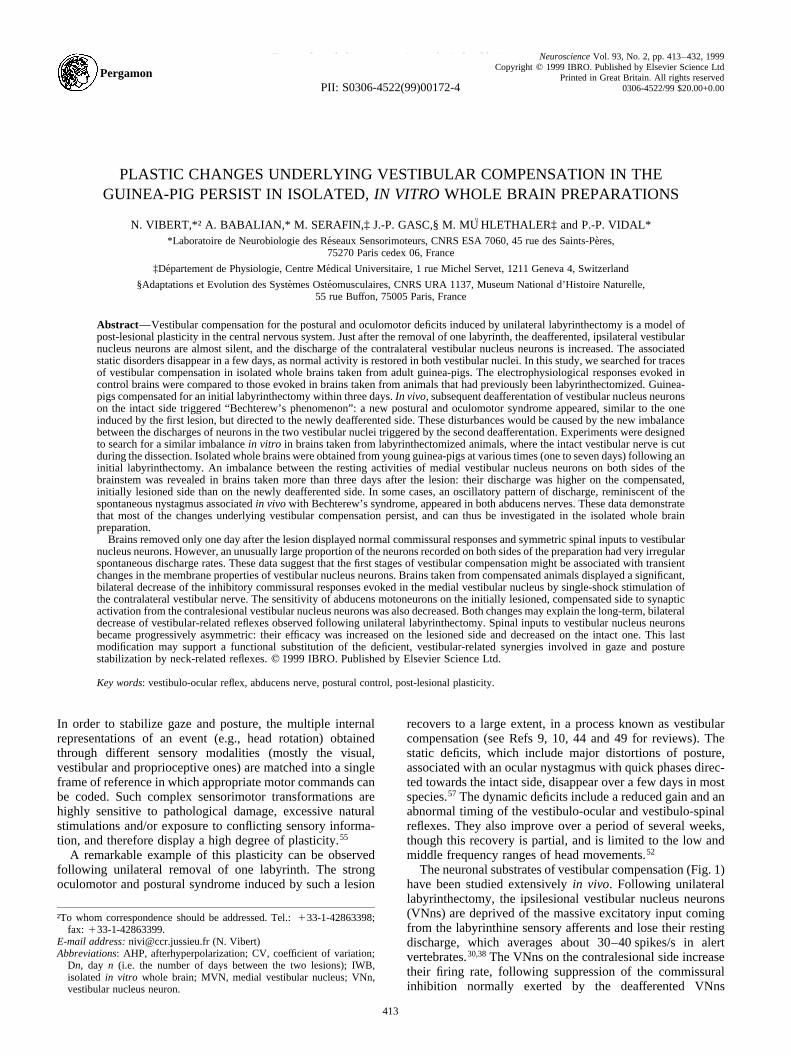
(Fig. 1A, B). The resulting imbalance between the massdischarges of the VNns in the right and left vestibular nucleiis responsible for the static deficits. During the few daysfollowing the lesion, the recovery of a normal restingdischarge by the deafferented VNns38,56 plays a key role inthe disappearance of the static syndrome (for reviews see Refs9, 37 and 49; Fig. 1C) by restoring the balance between thespontaneous activities of neurons in the two vestibular nuclei.Since the labyrinthine sensory afferents on the lesioned sideremain silent,20,46 one of the main questions about vestibularcompensation is: how do the ipsilesional second-order VNnsrecover a normal spontaneous activity?
The cellular events underlying this recuperation are stillcontroversial.9,10,30,31,49,55They may include changes in theintrinsic membrane properties of central vestibular neuronsand/or modifications of their sensitivity to neurotransmitters.In addition, various studies suggest that the suppression of thesensory vestibular input on one side would be partly compen-sated by changes in the efficacy of other synaptic inputs to thedeafferented VNns. The other sensory information reachingthe vestibular nuclei, mainly from the visual and spinalproprioceptive pathways, may play an important role inboth the induction and maintenance of vestibular compensa-tion. Several authors also suggested an involvement of (i) theinhibitory afferents coming from the cerebellum or (ii) thecommissural fibers linking together the two vestibularcomplexes in the compensation process. Similarly, the partialrecovery of the dynamic vestibular reflexes could rely onfunctional modulations of the efficacy of the spinal, visual,cerebellar and/or cortical pathways that still converge on theipsilesional VNns (for reviews see Refs 9, 10, 49 and 55).
Since most of these parameters are difficult to quantify
in vivo, the first aim of this study was to test whether theisolated, in vitro whole brain of adult guinea-pig (IWB)28
was suitable to investigate these changes. Several argumentsfavor the use of the IWB: (i) the guinea-pig has been usedextensively to study vestibular plasticity;55 (ii) on the IWB,the recorded neurons can be identified through their synapticconnections, which is mandatory to relate the data collectedpreviouslyin vivo with the in vitro results obtained on slices;(iii) we recently demonstrated the viability of vestibular-related pathways in the IWB, and assessed some of theirmain physiological and pharmacological properties.3 Theresults obtained in IWBs taken from control guinea-pigswere thus used as a reference to search whether traces ofthe plastic, cellular changes underlying vestibular compensa-tion could be retained in IWBs taken from previously labyr-inthectomized animals.
The second aim of this study was to investigate whether,following unilateral labyrinthectomy, the new balancereached between the spontaneous activity of VNns on bothsides of the brain was dependent on a continuous inflow ofsubstituting sensory signals, or could become at least partiallyindependent of visual and spinal proprioceptive information.Indeed, the IWB offers an interesting opportunity to study thisproblem, since the sensory inputs reaching the second-orderVNns are all suppressed in this totally deafferented prepara-tion, and thus would not be responsible for the possible persis-tence of changes related to the compensation process.
On the other hand, this complete deafferentation preventedus from using intact vestibular-related networks (i.e. stillconnected to a functional labyrinth) as references, since the“control” IWBs taken from normal animals are acutelybilabyrinthectomized preparations. Therefore, we lesioned
N. Vibert et al.414
Fig. 1. Evolution of spontaneous neuronal activity in vestibular-related pathways during vestibular compensation and after subsequent deafferentation of theintact side (Bechterew’s phenomenon). The excitatory neurons and synapses are shown in white, whereas the inhibitory neurons and synapses are drawninblack. In A and C, the absence of signs inside the neurons indicate balanced levels of spontaneous activity between both sides of the brain. In B and D, the 1signs indicate the neurons displaying a significantly higher than normal spontaneous activity, while the2 signs indicate those displaying a lower than normal
discharge rate. M, the median line of the brain; VI, abducens motoneurons.
the vestibular apparatus of guinea-pigs on one side at varioustimes (one, three, five and seven to eight days) before thepreparation of the IWB. This allowed us to compare the prop-erties of vestibular-related networks that had a differenthistory vis-a-vis the lesion. In one half-brain (called the“compensated side”), vestibular-related neurons had one,three, five or seven to eight days to compensate for the unilat-eral labyrinthectomy. On the other side (called the “newlydeafferented side”), the same structures were only discon-nected from the ipsilateral labyrinth when the brain wasremoved.
Previousin vivo experiments demonstrated that, followingcompensation for an initial, unilateral labyrinthectomy, suchan acute deafferentation of the contralesional VNns triggers anew oculomotor and postural syndrome, known as the Bech-terew phenomenon.4,44 This syndrome is similar to the oneinduced by the first labyrinthectomy, but directed towards theside of the second lesion. The second deafferentation woulddisrupt the new balance reached between the spontaneousactivity of neurons in the two vestibular nuclei after the initiallesion (Fig. 1C, D). On the side of the second lesion, thenewly deafferented VNns are less excited because they losetheir labyrinthine afferents. In contrast, the discharge of thefirst deafferented VNns (which recovered a normal activityduring the course of compensation) increases because thecommissural inhibition coming from the newly deafferentedside is reduced (Fig. 1D).
Since we were expecting to deal with anin vitro equivalentof this Bechterew syndrome, a complementary behavioralstudy was done to evaluate the intensity of the Bechterewphenomenon inducedin vivo, in age-matched guinea-pigs,during the same first week of compensation. The time-courseof the network modifications observedin vitro in the IWB wascompared with the time-course of thein vivo Bechterewphenomenon triggered.
Parts of the results presented here have been published aspreliminary reports.53,54
EXPERIMENTAL PROCEDURES
Animals: surgical procedures
Experiments were performed on young, adult pigmented guinea-pigs (Cavia porcellus) of both genders, weighing 130–300 g (Elevagede la Garenne, Saint-Pierre d’Exideuil, France). All studies werecarried out in accordance with the European Communities Councildirective of 24 November 1986, and followed the procedures issuedby the French Ministe`re de l’Agriculture. All efforts were made tominimize animal suffering and to reduce the number of animals used.
Unilateral labyrinthectomies were performed under halothaneanesthesia with the aid of an operating microscope. The semicircularcanals, utricle and saccule were exposed via a retroauricular approach.The ampullae of all three semicircular canals and the otolithic maculaewere drilled, and further destroyed using suction. The animals werethen let free to compensate in normal visual conditions until their brainwas dissected out, or a second labyrinthectomy was performed on theintact side (for thein vivo study of Bechterew’s phenomenon).
In vitro experiments
The data reported in the present paper were collected in 65 IWBstaken from guinea-pigs which had been unilaterally labyrinthecto-mized 20–24 h (one day,n�14, D1 brains), three days (n�26, D3brains), five days (n�10, D5 brains) or seven to eight days (n�15,D7 brains) before.
Because no significant difference was observed between D5 and D7brains, results obtained in these brains were pooled together as datacollected in D5–D7 brains. Similarly, in the many cases where similarresults were obtained in IWBs prepared three or more days after the
first lesion (i.e. D3, D5 and D7 brains), they were grouped together asdata collected in D3–D7 brains. The rationale for this grouping wasthat, in contrast to D1 brains, behavioral compensation for the staticdeficits induced by the initial labyrinthectomy was complete in all D3–D7 brains.
Given the difficulty of the surgical procedure, not all experimentsyielded usable data. Brains were only used if the amplitude of the fieldpotential evoked in the vestibular nuclei by stimulation of the ipsi-lateral vestibular nerve, and/or of the response evoked in the abducensnerves by stimulation of the contralateral vestibular nerves, reachedminimal values of 0.35 and 0.5 mV, respectively. These referencevalues were taken from our recent study of vestibular-related networksin the IWBs of control guinea-pigs.3 The low minimal amplituderetained for the field potential evoked in the vestibular nuclei alsotakes into account the decrease in the size of this field potentialobserved in D3–D7 brains (see below).
Isolation and maintenance of thein vitro whole brain.The methodsused to isolate the brain were modifications3 of those originallydescribed by Mu¨hlethaleret al.28 Following anesthesia, the animalwas transcardially perfused for 0.5–1 min with Ringer solution atroom temperature, and then for 5 min with cold (8–128C) Ringer.Subsequently, the animal was then decapitated, and the bones coveringthe brain and spinal cord were broken and removed. The exposed brainwas cut in the front at the level of the olfactory bulbs, and in the back atthe level of the C1–C2 cervical segments. The brain was isolated fromthe skull by severing the different cranial nerves, the carotid, hypotha-lamic and vertebral arteries, and the first spinal cord roots. It was thenplaced in cold saline and transferred to the incubation/recordingchamber.
In the chamber, the brain was submerged in saline and first main-tained at 138C. Once the brain was fixed by rubber strings, one of thevertebral arteries was separated from the surrounding tissue. A fine,stainless steel cannula (external diameter 0.4 mm) connected to theperfusion system was inserted inside and fixed with knots of fine silkthread. The second vertebral artery was closed and the perfusion ratewas increased from its initial rate of 0.8–1.0 ml/min to 2.0–2.5 ml/min.
The next step was to close the two carotid arteries, which were themajor source of leaks of perfusate from the brain vascular system. Thehypophyseal, labyrinthine, spinal and other small arteries, which hadbeen severed during the removal of the brain, were then all sutured.Following elimination of major leaks from the arterial system of thebrain, the temperature of the chamber and perfusate was graduallyincreased to 29–308C. The perfusion rate was increased progressivelyduring rewarming, to reach 4.5–5.5 ml/min. After rewarming, theaverage survival time of isolated brains was 6–7 h.
A peristaltic pump (Gilson Minipuls 3) was used to drive the perfu-sion system and to evacuate fluid from the chamber. The brain perfu-sate went through a pressure transducer (Druck), a bubble trap and afiltering unit (Millipore, 0.22mm pore diameter). The temperature ofthe IWB was controlled by a thermoregulation unit (Biomedical Engi-neering, Thornwood, U.S.A.).
The physiological Ringer solution, used for (i) transcardial perfusionof the animal, (ii) moistening of the brain during the dissection and (iii)initial filling of the chamber, had the following composition (in mM):NaCl 130, NaHCO3 20, MgSO4 1.3, KH2PO4 1.25, KCl 5, CaCl2 2.4,glucose 10. The saline used for perfusion of the brain had a slightlydifferent composition and contained (in mM): NaCl 126, NaHCO3 26,MgSO4 1.3, KH2PO4 1.2, KCl 3, CaCl2 2.4, glucose 15. This solutionincluded 1.5–3.0% of Dextran 70 (Macrodexw 70, Pharmacia,Sweden) to increase the osmotic pressure of the perfusate. All solutionswere bubbled continuously with a mixture of 95% O2 and 5% CO2.
Electrophysiological experiments.Extracellular recordings from theabducens nerves and the area of the medial vestibular nucleus (MVN;defined as in Ref. 3) were used to evaluate the spontaneous activity ofvestibular-related networks on each side of the IWB. We also quanti-fied the latency and amplitude of the responses evoked in the MVNarea and abducens nerve by stimulation of the ipsilateral vestibularnerve, the contralateral vestibular nerve and the spinal cord.
The spinal cord and both vestibular nerves were stimulated withbipolar, metallic electrodes. The electrodes for stimulation of thevestibular nerves (interpolar distance 1 mm) were made using stainlesssteel microelectrodes (0.2–0.3 mm thick; FHC, Brunswick, ME, U.S.A.)and inserted in the anterior (vestibular) branch of the vestibulo-cochlear
Traces of vestibular compensation in the isolated brain 415
nerve. The electrode for stimulation of the spinal cord was made ofstainless steel wires (0.4–0.5 mm thick). Its large interpolar distance(about 2.5 mm) was adjusted to ensure massive, bilateral stimulation ofthe tissue.
Recordings of abducens nerves were obtained through smooth, fire-polished, glass suction electrodes. Their tips were specifically designedto match the size of the nerve stumps. Great care was taken not todamage the abducens nerves during the application of these suctionelectrodes. Extracellular field potential and single-unit recordings inthe vestibular nuclei were made using glass microelectrodes, filledwith a 2 M solution of NaCl (resistance: 2–8 MV).
Rectangular pulses of 0.2 ms duration were used for all stimulations.The threshold stimulating intensity was determined for each response.The maximal intensities used were not bigger than three to five thresh-olds, and did not exceed 800–900mA.
Conventional electrophysiological equipment was used for record-ing and storage of information. The signals from suction electrodeswere amplified by a low input resistance d.c./a.c. amplifier (TektronixAM-502, U.S.A.). The microelectrode signals were recorded in thecurrent-clamp mode with an Axoclamp 2A amplifier (Axon Instruments,U.S.A.). The signals were bandpassed at d.c./0.1 Hz–3/10 kHz, storedon videotapes using a Neurocorder (NeuroData Instruments, U.S.A.)interface and simultaneously displayed on a digital storage oscilloscope.
The recorded signals were replayed off-line, digitized through anA/D converter, and stored as PC-compatible files using a home-madedata acquisition system (Data Acquisition Multi I/O, SICMU, Geneva,Switzerland) and its dedicated software. The latencies, amplitudes andother parameters of responses were then quantified. All characteristicsof the responses were measured, unless stated otherwise, at the sites oftheir largest amplitude.
The mean rate and regularity of the spontaneous activity were eval-uated, for each extracellularly recorded unit, from five samples of 10–30 successive spikes. The coefficient of variation (CV), defined as theratio between the standard deviation and the mean value of the inter-spike interval, was taken as an index of discharge regularity.
Statistical analysis.The first labyrinthectomy, performed beforeremoval of the brain, was usually done on the left side. Similar datawere obtained in the few cases where the initial lesion was performedon the right side, and all results were therefore pooled together. Calcu-lations of medians or meansS.D. and further processing of all resultswere carried out using StatWorks and CricketGraph software (CricketSoftware Inc., Philadelphia, PA, U.S.A.) on an Apple Macintoshcomputer. The normality of all data distributions was checkedfor each parameter (Kolmogorov–Smirnov test,22 10% confidenceinterval).
Normally distributed parameters.Normality was achieved for mostof the quantified parameters, allowing us to use standard, parametricstatistical tests. In each set of data, one-way ANOVA for independentsamples (5% confidence interval) was first used to search for signifi-cant differences between brains taken from control animals (“control”brains; data from Ref. 3) and those taken from previously labyrinthec-tomized guinea-pigs (D1, D3, D5 and D7 brains). Separate tests wererun for the compensated and newly deafferented sides, assuming thatcontrol brains were symmetric (the data obtained previously in controlbrains were used as reference for both sides of the IWBs taken fromlesioned guinea-pigs). When no significant variation was foundbetween D3, D5 and D7 brains, ANOVA was run between control,D1 and D3–D7 (“compensated”) brains. When a significant differencewas revealed, unpaired Student’st-tests (two-tailed, 5% confidenceinterval) were used for further two by two comparisons betweengroups. In brains taken from labyrinthectomized animals, symmetrybetween the two sides was tested using paired or unpaired Student’st-test, depending on whether paired observations were obtained for theparameter in question.
Other parameters.Statistical comparisons between the spontaneousdischarges and CVs of second-order VNns, for which significant devia-tions from normality were observed, were done with non-parametrictests. For each side of the brain, separate Kruskal–Wallis one-wayANOVA for independent samples (5% confidence interval) showedwhether the discharge rate was significantly modified over time.Mann–WhitneyU-tests for independent samples (5% confidence inter-val) were then used for further two by two comparisons of data
obtained for each group of brains, and to test the symmetry ofdischarges between both sides of the preparations.
In vivo experiments; assessment of Bechterew’s phenomenon
Nine animals were used for thisin vivo study. Following a first, leftside labyrinthectomy, the intact labyrinth was lesioned after either 20–24 h (n�3, D1 animals), three days (n�3, D3 animals) or seven days(n�3, D7 animals) of compensation. Each guinea-pig was X-rayed inresting position between 2 and 5 h after this second labyrinthectomy.Exposures were obtained both from above and from the side, with theX-ray tube 90 cm away from the animal.
For each animal, the frequency of the spontaneous nystagmusinduced by the second lesion was measured as the mean number ofquick phase beats occurring in the light over three to five periods of30 s, with the guinea-pig unrestrained. The mean number of beats perminute was derived from these measures at 1, 2, 4, 6 and 24 h follow-ing the lesion.
Statistical analysis was performed using the same software as for thein vitro results. Once the normality of each data sample was checked(normality test, 10% confidence interval), one-way ANOVAs for inde-pendent samples were performed to search for significant differencesbetween the spontaneous nystagmus induced in D1, D3 and D7 guinea-pigs. Unpaired Student’st-tests (two-tailed, 5% confidence interval)were then used for further two by two comparisons between groups.
RESULTS
In vivo experiments on Bechterew’s phenomenon
All guinea-pigs completely recovered from gaseousanesthesia in about 45 min following the end of the secondlabyrinthectomy (performed on the right side).
Postural disturbances.In D7 animals (where seven dayselapsed between the two lesions), the second labyrinthectomyinduced a strong postural syndrome directed towards thenewly deafferented side. Just after the lesion, animals laidon one side and displayed episodes of violent ipsiversive roll-ing around the body’s longitudinal axis. Two to five hourslater (Fig. 2C), the head was still strongly tilted towards thenewly deafferented side because of a lateral tilt of the cervicalcolumn in the frontal plane. The head was also rotated in thestereotaxic horizontal plane around the tilted axis of the cervi-cal column, because of a distributed rotation along the firstcervical joints. Abnormal positions of the limbs wereobserved: the forelimb ipsilateral to the second lesion andthe contralateral hindlimb were flexed, while the contralateralforelimb and ipsilateral hindlimb were extended. Altogether,this syndrome was the mirror image of the one observed in theopposite direction following the initial labyrinthectomyperformed on the other side seven days before.57 It dis-appeared, however, in about 24 h, whereas compensationfor the first lesion was only achieved after three days.
In D3 animals (where three days elapsed between the twolesions), the second labyrinthectomy induced similar posturaldeficits, including a head rotation towards the newly deaffer-ented side in both the horizontal and frontal planes, andabnormal positions of the limbs (Fig. 2B). The intensity ofthe postural syndrome, however, appeared lower than in D7guinea-pigs. In particular, spontaneous episodes of rollingtowards the newly deafferented side were never observed,and all postural disturbances disappeared in about 10 h.
In D1 animals, and in contrast with what happened in D3and D7 guinea-pigs, the second labyrinthectomy only inducedvery transient postural deficits. These disturbances, whichincluded a slight lateral head tilt towards the newly deaffer-ented side and sometimes a small horizontal head deviation,
N. Vibert et al.416
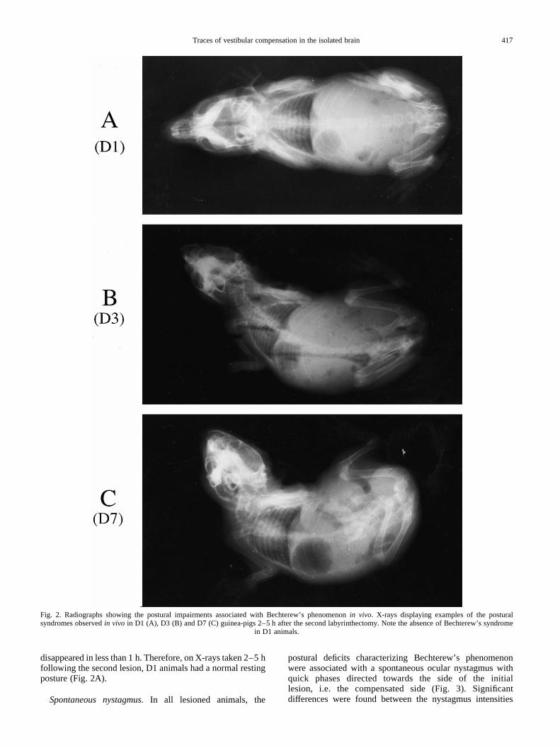
disappeared in less than 1 h. Therefore, on X-rays taken 2–5 hfollowing the second lesion, D1 animals had a normal restingposture (Fig. 2A).
Spontaneous nystagmus.In all lesioned animals, the
postural deficits characterizing Bechterew’s phenomenonwere associated with a spontaneous ocular nystagmus withquick phases directed towards the side of the initiallesion, i.e. the compensated side (Fig. 3). Significantdifferences were found between the nystagmus intensities
Traces of vestibular compensation in the isolated brain 417
Fig. 2. Radiographs showing the postural impairments associated with Bechterew’s phenomenonin vivo. X-rays displaying examples of the posturalsyndromes observedin vivo in D1 (A), D3 (B) and D7 (C) guinea-pigs 2–5 h after the second labyrinthectomy. Note the absence of Bechterew’s syndrome
in D1 animals.
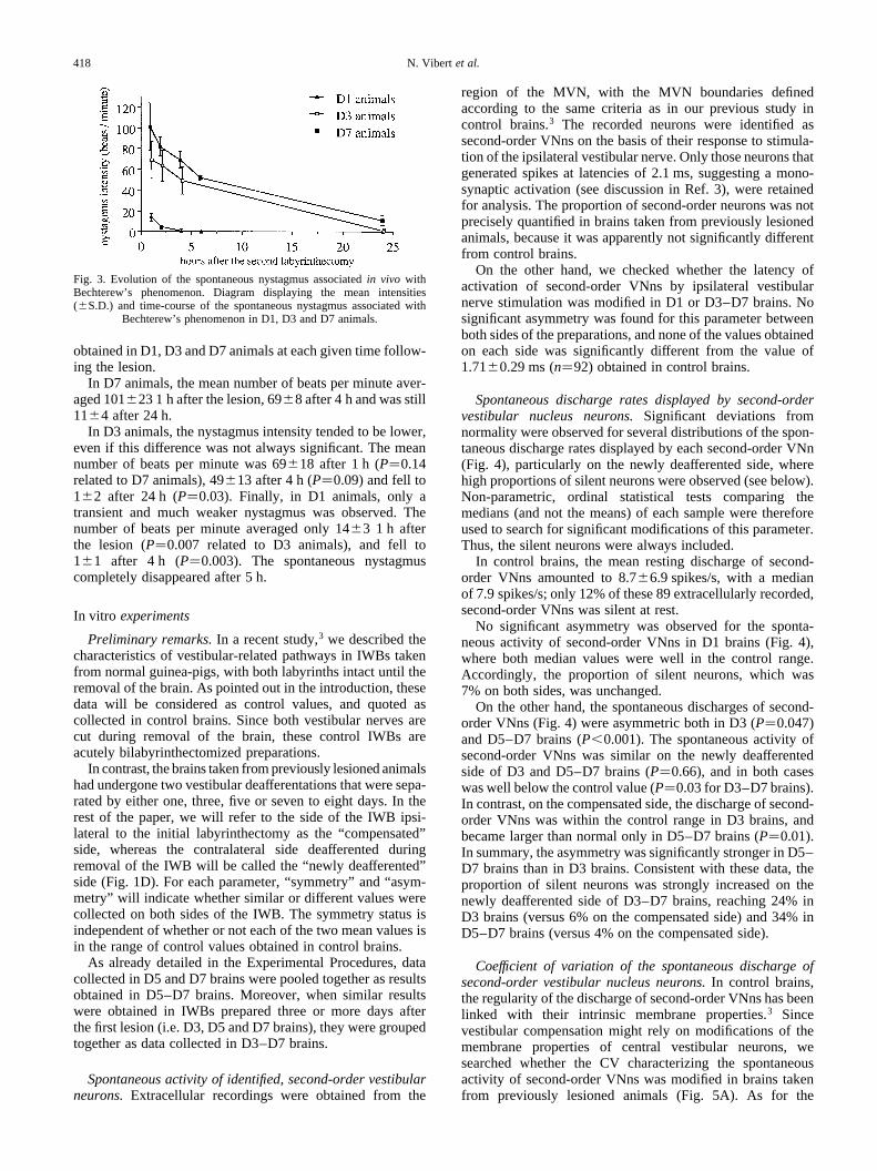
obtained in D1, D3 and D7 animals at each given time follow-ing the lesion.
In D7 animals, the mean number of beats per minute aver-aged 101 23 1 h after the lesion, 698 after 4 h and was still11^4 after 24 h.
In D3 animals, the nystagmus intensity tended to be lower,even if this difference was not always significant. The meannumber of beats per minute was 69^18 after 1 h (P�0.14related to D7 animals), 4913 after 4 h (P�0.09) and fell to1^2 after 24 h (P�0.03). Finally, in D1 animals, only atransient and much weaker nystagmus was observed. Thenumber of beats per minute averaged only 14^3 1 h afterthe lesion (P�0.007 related to D3 animals), and fell to1^1 after 4 h (P�0.003). The spontaneous nystagmuscompletely disappeared after 5 h.
In vitro experiments
Preliminary remarks.In a recent study,3 we described thecharacteristics of vestibular-related pathways in IWBs takenfrom normal guinea-pigs, with both labyrinths intact until theremoval of the brain. As pointed out in the introduction, thesedata will be considered as control values, and quoted ascollected in control brains. Since both vestibular nerves arecut during removal of the brain, these control IWBs areacutely bilabyrinthectomized preparations.
In contrast, the brains taken from previously lesioned animalshad undergone two vestibular deafferentations that were sepa-rated by either one, three, five or seven to eight days. In therest of the paper, we will refer to the side of the IWB ipsi-lateral to the initial labyrinthectomy as the “compensated”side, whereas the contralateral side deafferented duringremoval of the IWB will be called the “newly deafferented”side (Fig. 1D). For each parameter, “symmetry” and “asym-metry” will indicate whether similar or different values werecollected on both sides of the IWB. The symmetry status isindependent of whether or not each of the two mean values isin the range of control values obtained in control brains.
As already detailed in the Experimental Procedures, datacollected in D5 and D7 brains were pooled together as resultsobtained in D5–D7 brains. Moreover, when similar resultswere obtained in IWBs prepared three or more days afterthe first lesion (i.e. D3, D5 and D7 brains), they were groupedtogether as data collected in D3–D7 brains.
Spontaneous activity of identified, second-order vestibularneurons.Extracellular recordings were obtained from the
region of the MVN, with the MVN boundaries definedaccording to the same criteria as in our previous study incontrol brains.3 The recorded neurons were identified assecond-order VNns on the basis of their response to stimula-tion of the ipsilateral vestibular nerve. Only those neurons thatgenerated spikes at latencies of 2.1 ms, suggesting a mono-synaptic activation (see discussion in Ref. 3), were retainedfor analysis. The proportion of second-order neurons was notprecisely quantified in brains taken from previously lesionedanimals, because it was apparently not significantly differentfrom control brains.
On the other hand, we checked whether the latency ofactivation of second-order VNns by ipsilateral vestibularnerve stimulation was modified in D1 or D3–D7 brains. Nosignificant asymmetry was found for this parameter betweenboth sides of the preparations, and none of the values obtainedon each side was significantly different from the value of1.71 0.29 ms (n�92) obtained in control brains.
Spontaneous discharge rates displayed by second-ordervestibular nucleus neurons.Significant deviations fromnormality were observed for several distributions of the spon-taneous discharge rates displayed by each second-order VNn(Fig. 4), particularly on the newly deafferented side, wherehigh proportions of silent neurons were observed (see below).Non-parametric, ordinal statistical tests comparing themedians (and not the means) of each sample were thereforeused to search for significant modifications of this parameter.Thus, the silent neurons were always included.
In control brains, the mean resting discharge of second-order VNns amounted to 8.76.9 spikes/s, with a medianof 7.9 spikes/s; only 12% of these 89 extracellularly recorded,second-order VNns was silent at rest.
No significant asymmetry was observed for the sponta-neous activity of second-order VNns in D1 brains (Fig. 4),where both median values were well in the control range.Accordingly, the proportion of silent neurons, which was7% on both sides, was unchanged.
On the other hand, the spontaneous discharges of second-order VNns (Fig. 4) were asymmetric both in D3 (P�0.047)and D5–D7 brains (P,0.001). The spontaneous activity ofsecond-order VNns was similar on the newly deafferentedside of D3 and D5–D7 brains (P�0.66), and in both caseswas well below the control value (P�0.03 for D3–D7 brains).In contrast, on the compensated side, the discharge of second-order VNns was within the control range in D3 brains, andbecame larger than normal only in D5–D7 brains (P�0.01).In summary, the asymmetry was significantly stronger in D5–D7 brains than in D3 brains. Consistent with these data, theproportion of silent neurons was strongly increased on thenewly deafferented side of D3–D7 brains, reaching 24% inD3 brains (versus 6% on the compensated side) and 34% inD5–D7 brains (versus 4% on the compensated side).
Coefficient of variation of the spontaneous discharge ofsecond-order vestibular nucleus neurons.In control brains,the regularity of the discharge of second-order VNns has beenlinked with their intrinsic membrane properties.3 Sincevestibular compensation might rely on modifications of themembrane properties of central vestibular neurons, wesearched whether the CV characterizing the spontaneousactivity of second-order VNns was modified in brains takenfrom previously lesioned animals (Fig. 5A). As for the
N. Vibert et al.418
Fig. 3. Evolution of the spontaneous nystagmus associatedin vivo withBechterew’s phenomenon. Diagram displaying the mean intensities(^S.D.) and time-course of the spontaneous nystagmus associated with
Bechterew’s phenomenon in D1, D3 and D7 animals.

spontaneous discharge rates, non-parametric statistical testshad to be used. In control brains, the mean CV of second-order VNns was 0.38 0.26, with a median of 0.35.
On the compensated side of D1 brains, the median CVincreased to 0.58, which was significantly higher than in control
brains (P,0.001). A similar trend towards an increase in dis-charge irregularity was found on the newly deafferented side,even if this increase was not significant because of the largevariance of the distribution. Altogether, D1 brains displayedsymmetric values of CVs in both vestibular nuclei (P�0.13).
Traces of vestibular compensation in the isolated brain 419
Fig. 4. Spontaneous activity of second-order vestibular nucleus neurons. The seven black histograms show the distributions of second-order VNns recorded incontrol brains and in brains taken from previously labyrinthectomized animals according to their spontaneous discharge rates. The column labeled 3gives thenumber of neurons whose spontaneous activity was strictly lower than 3 spikes/s, the column labeled 6 includes those units whose activity was between3 and6 spikes/s, and so on. MD is the median discharge rate given for each distribution. The top right histogram summarizes the median discharges obtained for theseseven groups of second-order VNns. The white asterisks indicate the values which were significantly out of the control range, while the black ones showthe
groups of isolated brains for which a significant asymmetry appeared between both sides of the brainstem (*P,0.05, **P,0.01, ***P,0.001).
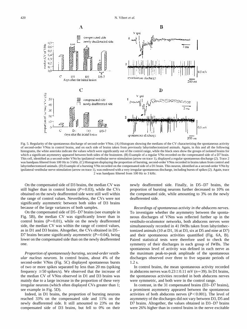
On the compensated side of D3 brains, the median CV wasstill higher than in control brains (P�0.03), while the CVsobtained on the newly deafferented side were still well withinthe range of control values. Nevertheless, the CVs were notsignificantly asymmetric between both sides of D3 brainsbecause of the large variances of both samples.
On the compensated side of D5–D7 brains (see example inFig. 5B), the median CV was significantly lower than incontrol brains (P�0.01), while on the newly deafferentedside, the median CV was within the range of control values,as in D1 and D3 brains. Altogether, the CVs obtained in D5–D7 brains became significantly asymmetric (P�0.04), beinglower on the compensated side than on the newly deafferentedone.
Proportion of spontaneously bursting, second-order vestib-ular nucleus neurons.In control brains, about 4% of thesecond-order VNns (Fig. 5C) displayed spontaneous burstsof two or more spikes separated by less than 20 ms (spikingfrequency$50 spikes/s). We observed that the increase ofthe median CV of VNns observed in D1 and D3 brains wasmainly due to a large increase in the proportion of these veryirregular neurons (which often displayed CVs greater than 1;see example in Fig. 5D).
Indeed, in D1 brains, the proportion of bursting neuronsreached 33% on the compensated side and 11% on thenewly deafferented side. It still amounted to 23% on thecompensated side of D3 brains, but fell to 0% on their
newly deafferented side. Finally, in D5–D7 brains, theproportion of bursting neurons further decreased to 10% onthe compensated side, while amounting to 3% on the newlydeafferented side.
Recordings of spontaneous activity in the abducens nerves.To investigate whether the asymmetry between the sponta-neous discharges of VNns was reflected further up in thevestibulo-oculomotor networks, both abducens nerves weresimultaneously recorded in 41 IWBs taken from labyrinthec-tomized animals (10 at D1, 16 at D3, six at D5 and nine at D7)and their spontaneous activities quantified (Fig. 6A, B).Paired statistical tests were therefore used to check thesymmetry of their discharges in each group of IWBs. Thespontaneous level of activity was defined in each nerve asthe maximum peak-to-peak amplitude of the spontaneousdischarges observed over three to five separate periods of1.2 s.
In control brains, the mean spontaneous activity recordedin abducens nerves was 0.23^0.11 mV (n�39). In D1 brains,the spontaneous activities recorded in both abducens nerveswere symmetric, and both were in the control range.
In contrast, in the 31 compensated brains (D3–D7 brains),a prominent asymmetry appeared between the spontaneousactivities of both abducens nerves (P,0.001). The level ofasymmetry of the discharges did not vary between D3, D5 andD7 brains. Altogether, the values obtained in D3–D7 brainswere 26% higher than in control brains in the nerve excitable
N. Vibert et al.420
Fig. 5. Regularity of the spontaneous discharge of second-order VNns. (A) Histogram showing the medians of the CV characterizing the spontaneous activityof second-order VNns in control brains, and on each side of brains taken from previously labyrinthectomized animals. Again, in this and all the followinghistograms, the white asterisks indicate the values which were significantly out of the control range, while the black ones show the groups of isolatedbrains forwhich a significant asymmetry appeared between both sides of the brainstem. (B) Example of a regular VNn recorded on the compensated side of a D7 brain.This cell, identified as a second-order VNn by ipsilateral vestibular nerve stimulation (arrow on trace 1), displayed a regular spontaneous discharge (2). Trace 2was bandpass filtered from 100 Hz to 3 kHz. (C) Histogram displaying the proportion of bursting, second-order VNns recorded in brains taken from control andlabyrinthectomized animals. (D) Example of a bursting VNn recorded on the compensated side of a D1 brain. This neuron, identified as a second-order VNnbyipsilateral vestibular nerve stimulation (arrow on trace 1), was endowed with a very irregular spontaneous discharge, including bursts of spikes (2). Again, trace
2 was bandpass filtered from 100 Hz to 3 kHz.
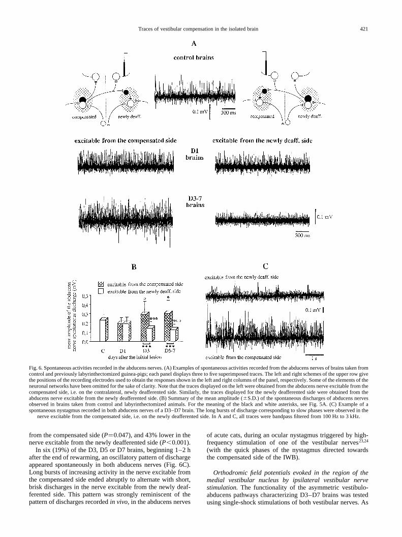
from the compensated side (P�0.047), and 43% lower in thenerve excitable from the newly deafferented side (P,0.001).
In six (19%) of the D3, D5 or D7 brains, beginning 1–2 hafter the end of rewarming, an oscillatory pattern of dischargeappeared spontaneously in both abducens nerves (Fig. 6C).Long bursts of increasing activity in the nerve excitable fromthe compensated side ended abruptly to alternate with short,brisk discharges in the nerve excitable from the newly deaf-ferented side. This pattern was strongly reminiscent of thepattern of discharges recordedin vivo, in the abducens nerves
of acute cats, during an ocular nystagmus triggered by high-frequency stimulation of one of the vestibular nerves23,24
(with the quick phases of the nystagmus directed towardsthe compensated side of the IWB).
Orthodromic field potentials evoked in the region of themedial vestibular nucleus by ipsilateral vestibular nervestimulation.The functionality of the asymmetric vestibulo-abducens pathways characterizing D3–D7 brains was testedusing single-shock stimulations of both vestibular nerves. As
Traces of vestibular compensation in the isolated brain 421
Fig. 6. Spontaneous activities recorded in the abducens nerves. (A) Examples of spontaneous activities recorded from the abducens nerves of brains taken fromcontrol and previously labyrinthectomized guinea-pigs; each panel displays three to five superimposed traces. The left and right schemes of the upper row givethe positions of the recording electrodes used to obtain the responses shown in the left and right columns of the panel, respectively. Some of the elements of theneuronal networks have been omitted for the sake of clarity. Note that the traces displayed on the left were obtained from the abducens nerve excitablefrom thecompensated side, i.e. on the contralateral, newly deafferented side. Similarly, the traces displayed for the newly deafferented side were obtained from theabducens nerve excitable from the newly deafferented side. (B) Summary of the mean amplitude (^S.D.) of the spontaneous discharges of abducens nervesobserved in brains taken from control and labyrinthectomized animals. For the meaning of the black and white asterisks, see Fig. 5A. (C) Example of aspontaneous nystagmus recorded in both abducens nerves of a D3–D7 brain. The long bursts of discharge corresponding to slow phases were observed in the
nerve excitable from the compensated side, i.e. on the newly deafferented side. In A and C, all traces were bandpass filtered from 100 Hz to 3 kHz.
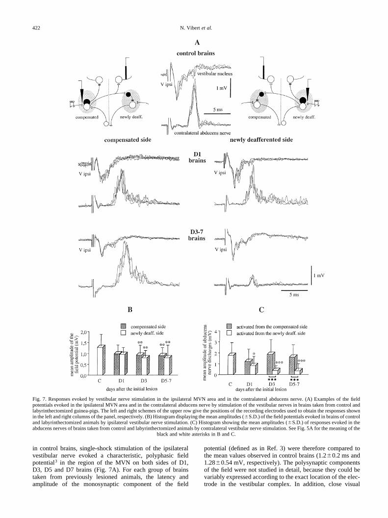
in control brains, single-shock stimulation of the ipsilateralvestibular nerve evoked a characteristic, polyphasic fieldpotential3 in the region of the MVN on both sides of D1,D3, D5 and D7 brains (Fig. 7A). For each group of brainstaken from previously lesioned animals, the latency andamplitude of the monosynaptic component of the field
potential (defined as in Ref. 3) were therefore compared tothe mean values observed in control brains (1.2^0.2 ms and1.28 0.54 mV, respectively). The polysynaptic componentsof the field were not studied in detail, because they could bevariably expressed according to the exact location of the elec-trode in the vestibular complex. In addition, close visual
N. Vibert et al.422
Fig. 7. Responses evoked by vestibular nerve stimulation in the ipsilateral MVN area and in the contralateral abducens nerve. (A) Examples of the fieldpotentials evoked in the ipsilateral MVN area and in the contralateral abducens nerve by stimulation of the vestibular nerves in brains taken from control andlabyrinthectomized guinea-pigs. The left and right schemes of the upper row give the positions of the recording electrodes used to obtain the responses shownin the left and right columns of the panel, respectively. (B) Histogram displaying the mean amplitudes (^S.D.) of the field potentials evoked in brains of controland labyrinthectomized animals by ipsilateral vestibular nerve stimulation. (C) Histogram showing the mean amplitudes (^S.D.) of responses evoked in theabducens nerves of brains taken from control and labyrinthectomized animals by contralateral vestibular nerve stimulation. See Fig. 5A for the meaning of the
black and white asterisks in B and C.
inspection did not reveal any significant variations of thesepolysynaptic waves between control IWBs and those takenfrom lesioned animals.
Because only one MVN was investigated at a time, pairedobservations on both sides of the brains could not be obtained.Unpaired statistical tests were therefore used to check thesymmetry of data obtained in each group of IWBs.
Latency of the monosynaptic component of the field poten-tial. No significant asymmetry was found between the laten-cies obtained on both sides of D1 or D3–D7 brains (Fig. 7A).In the newly deafferented MVN, the mean latency of the fieldpotential was in the range of control values in all cases. Simi-larly, no significant variation was observed in the MVN on thecompensated side.
Amplitude of the field potential.NeitherD1 nor D3–D7brainsdisplayed asymmetric amplitudes of the field potential evokedon each side by stimulation of the ipsilateral vestibular nerve(Fig. 7A, B). The amplitudes obtained in D1 brains were both inthe control range. In contrast, the amplitude of the field poten-tials recorded in D3–D7 brains were about 30–35% lowerthan in control brains on both the compensated and newlydeafferented sides (respectiveP values of 0.004 and 0.001).
Recordings of vestibular-evoked responses in the abducensnerves.
Response evoked by stimulation of the contralateral vestib-ular nerve. In addition to the field potential evoked in theregion of the ipsilateral MVN, we have demonstrated thatstimulation of the vestibular nerve elicits, in control brains,a synchronized discharge in the contralateral abducens nerve,with an average latency of 3.80.7 ms and a peak amplitudeof 1.75 1.06 mV.3
In D1 brains, both the latency and peak amplitude of theresponses evoked in each abducens nerve from their respec-tive contralateral vestibular nerve were symmetric (Fig. 7A,C). The latency of the discharge was in the range of controlvalues in both the abducens nerve activated from the compen-sated side (P�0.17) and the abducens nerve activated fromthe newly deafferented side (P�0.07). The peak amplitude ofthe response was significantly lower than normal in the nerveactivated from the newly deafferented side (P�0.02), and wasalso decreased but nevertheless stayed in the control range inthe nerve activated from the compensated side (ANOVAbetween control, D1 and D3–D7 brains gaveP�0.33).
In contrast, D3–D7 brains displayed a strong asymmetrywith respect to both the latencies (P,0.001) and amplitudes(P,0.001) of the responses evoked in both abducens nerves(Fig. 7A, C). This asymmetry did not vary with time betweenD3, D5 and D7 brains. In the abducens nerve activated fromthe compensated side, neither the latency (3.5^0.6 ms) northe peak amplitude of the discharge were significantly differ-ent from control values. On the other hand, responses evokedin the abducens nerve activated from the newly deafferentedside had a five-fold lower amplitude than in control(P,0.001) and D1 (P�0.02) brains, while their latency(4.8 1.7 ms) was 26% longer than in control brains(P�0.003).
Response evoked by stimulation of the ipsilateral vestibularnerve.In control brains, ipsilateral vestibular nerve stimulation
did not produce any short-latency response in the abducensnerve. It induced a late (latency 22.39.6 ms), usually desyn-chronized, discharge (amplitude 0.36^0.17 mV) in 53% ofthe preparations, most probably triggered by a post-inhibitoryrebound in abducens motoneurons.3
These late discharges were observed in both abducensnerves of D1 brains, more frequently (n�7, 70%) in thenerve ipsilateral to the newly deafferented side (i.e. excitablefrom the compensated side) than in the one ipsilateral to thecompensated side and excitable from the newly deafferentedside (n�2, 20%). No significant asymmetry was otherwiseobserved between either the latencies or amplitudes of theseresponses, which were similar to those obtained in controlbrains.
Late rebound discharges were also recorded in 18 of the 31D3–D7 brains (58%), but only in the abducens nerve ipsi-lateral to the newly deafferented side (i.e. excitable from thecompensated side). Their latency and amplitude were in therange of the values obtained in control and D1 brains.
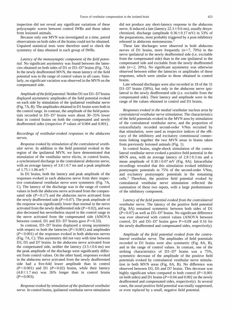
Responses evoked in the medial vestibular nucleus area bycontralateral vestibular nerve stimulation.The characteristicsof the field potentials evoked in the MVN area by stimulationof the contralateral vestibular nerve, and the proportion ofextracellularly recorded second-order VNns recruited bythat stimulation, were used as respective indices of the effi-cacy of the inhibitory and excitatory commissural connec-tions linking together the two MVN areas in brains takenfrom previously lesioned animals (Fig. 1).
In control brains, single-shock stimulation of the contra-lateral vestibular nerve evoked a positive field potential in theMVN area, with an average latency of 2.80.3 ms and amean amplitude of 0.300.07 mV (Fig. 8A). Intracellularrecordings revealed that this stimulation evoked inhibitorypostsynaptic potentials in 75% of the second-order VNns,and excitatory postsynaptic potentials in the remainingcells.3 Therefore, the positive field potential evoked bycontralateral vestibular nerve stimulation reflected thesummation of these two inputs, with a large predominanceof the inhibitory component.
Latency of the field potential evoked from the contralateralvestibular nerve.The latency of the positive field potential(Fig. 8A) remained symmetric between both sides of D1(P�0.07) as well as D3–D7 brains. No significant differencewas ever observed with control values (ANOVA betweencontrol, D1 and D3–D7 brains gaveP�0.09 and 0.16 forthe newly deafferented and compensated sides, respectively).
Amplitude of the field potential evoked from the contra-lateral vestibular nerve.The amplitudes of field potentialsrecorded in D1 brains were also symmetric (Fig. 8A, B),and in the range of control values. In contrast, one of thestriking characteristics of D3–D7 brains was a 75%,symmetric decrease of the amplitude of the positive fieldpotentials evoked by contralateral vestibular nerve stimula-tion in both MVN areas (Fig. 8A, B). No difference wasobserved between D3, D5 and D7 brains. This decrease washighly significant when compared to both control (P,0.001on both sides) and D1 brains (P�0.04 and 0.002 on the newlydeafferented and compensated sides, respectively). In severalcases, the usual positive field potential was totally suppressed,or even replaced by a small, negative field potential.
Traces of vestibular compensation in the isolated brain 423
Second-order vestibular nucleus neurons recruited bycontralateral vestibular nerve stimulation.Nineteen percentof the second-order VNns recorded extracellularly in controlbrains was activated through commissural pathways, with amean latency of 4.3 2.0 ms. This percentage fits well withthe finding that excitatory postsynaptic potentials were evokedfollowing stimulation of the contralateral vestibular nerve in24% of the intracellularly recorded, second-order VNns.
The proportion of second-order VNns activated from thecontralateral side did not vary much in either D1 or D3–D7brains (Fig. 8C).
Antidromic responses evoked in the medial vestibularnucleus area by stimulation of the spinal cord.Recordingsof the antidromic field potential evoked in the MVN area by
bilateral stimulation of the spinal cord and quantification ofthe number of antidromically activated, second-order VNnswere used to determine whether the compensation processwas associated with significant modification of the vestibulo-spinal pathways.
Antidromic field potential evoked in the medial vestibularnucleus area by spinal cord stimulation.In control brains,stimulation of the spinal cord evoked an initial, sharp nega-tivity in the region of the MVN, followed by a slower andlonger-lasting negativity.3 The first negative wave had a meanlatency of 0.84 0.11 ms, an average amplitude of0.27 0.16 mV and was not depressed by high-frequencystimulations. These characteristics suggested that it reflectedantidromic activation of vestibulo-spinal neurons.
N. Vibert et al.424
Fig. 8. Responses evoked by vestibular nerve stimulation in the contralateral MVN area. (A) Examples of the field potentials evoked in the contralateral MVNarea by stimulation of the vestibular nerves in brains taken from control and labyrinthectomized guinea-pigs. The left and right schemes of the upperrow givethe positions of the recording electrodes used to obtain the responses displayed in the left and right columns of the panel, respectively. (B) Histogram displayingthe mean amplitudes (S.D.) of the field potentials evoked in the MVN area of brains taken from control and labyrinthectomized animals by contralateralvestibular nerve stimulation. For the meaning of the asterisks, see Fig. 5A. (C) Histogram showing the proportion of second-order VNns recruited by
stimulation of the contralateral vestibular nerve brains taken from control and labyrinthectomized animals.
Traces of vestibular compensation in the isolated brain 425
Tab
le1.
Qua
ntita
tive
char
acte
ristic
sof
resp
onse
sev
oked
inve
stib
ular
-rel
ated
path
way
sby
bila
tera
lspi
nals
timul
atio
n(m
eans
^S
.D.)
Par
amet
erC
ontr
olbr
ains
D1
brai
nsD
3br
ains
D5
–D
7br
ains
Com
pens
ated
side
New
lyde
affe
rent
edsi
deC
ompe
nsat
edsi
deN
ewly
deaf
fere
nted
side
Com
pens
ated
side
New
lyde
affe
rent
edsi
de
Rec
ordi
ngs
from
the
regi
onof
the
MV
NA
ntid
rom
icre
spon
ses
Fie
ldpo
tent
iall
aten
cy(m
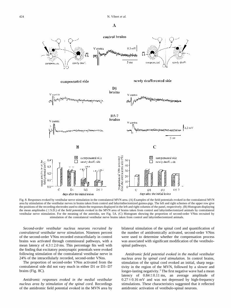
s)0.
740.
140.
890.
170.
840.
110.
820.
110.
680.
120.
750.
190.
690.
21
Fie
ldpo
tent
ials
ize
(mV
)0.
350.
200.
260.
080.
330.
070.
400.
150.
470.
330.
300.
160.
270.
11N
o.of
activ
ate
d,se
cond
-or
der
VN
ns(%
)11
712
28
149
Late
ncy
ofV
Nn
activ
atio
n(m
s)0.
740.
180.
820.
110.
830.
140.
780.
000.
630.
180.
750.
190.
800.
10
Ort
hodr
omic
resp
onse
sF
ield
pote
ntia
llat
ency
(ms)
1.7
0.3
2.7
1.9
1.6
0.4
2.0
0.8
2.8
2.5
1.7
0.5
2.2
0.8
Fie
ldpo
tent
ials
ize
(mV
)0.
230.
100.
270.
060.
350.
12*
0.38
0.17
*0.
200.
180.
380.
27*
0.14
0.13
asas
No.
ofac
tiva
ted
seco
nd-
orde
rV
Nns
(%)
2727
2333
2543
9
Late
ncy
ofV
Nn
activ
atio
n(m
s)3.
01.
64.
02.
24.
12.
43.
01.
22.
60.
93.
01.
27.
57.
7*
as
Abd
ucen
sne
rves
reco
rdin
gs:
spin
al-e
voke
dre
spon
ses
Dis
char
gela
tenc
y(m
s)3.
00.8
3.8
4.5
7.1
6.8
2.7
1.3
4.6
3.1
3.9
3.2
4.9
3.6
asD
isch
arge
ampl
itude
(mV
)0.
560.
320.
900.
840.
640.
720.
820.
610.
160.
20*
0.81
0.52
0.23
0.30
*as
as
For
reco
rdin
gsob
tain
edfr
omth
eab
duce
nsne
rves
,the
valu
esin
dica
ted
onth
eco
mpe
nsat
edsi
dere
fer
toth
ere
spon
ses
obta
ined
from
the
nerv
eac
tivat
edfr
om(a
ndco
ntra
late
ralt
o)th
eco
mpe
nsat
edsi
de.S
imila
rly,t
heva
lues
give
nfo
rth
ene
wly
deaf
fere
nted
side
corr
espo
ndto
the
resp
onse
sob
tain
edfr
omth
ene
rve
activ
ated
from
the
new
lyde
affe
rent
edsi
de.A
ster
isks
indi
cate
whi
chva
lues
wer
esi
gnifi
cant
lydi
ffere
ntfr
omth
ose
obta
ined
inco
ntro
lbra
ins;
“as”
isad
ded
belo
wth
eva
lues
whe
na
sign
ifica
ntas
ymm
etry
was
foun
dbe
twee
nbo
thsi
des
ofth
eIW
B.
In all groups of brains taken from labyrinthectomizedguinea-pigs, the mean latency and amplitude of the antidro-mic field potential were symmetric between both sides of theIWB and in the range of control values (Table 1).
Proportion of second-order vestibular nucleus neuronsantidromically recruited from the spinal cord.In controlbrains, 11% of the second-order VNns were antidromicallyactivated following stimulation of the spinal cord, with amean latency of 0.740.18 ms (n�9). In accordance withthe lack of modification of the antidromic field potentialsevoked by spinal stimulations, the proportion of antidromi-cally activated second-order VNns did not vary in brainstaken from labyrinthectomized guinea-pigs (Table 1).Accordingly, the average latencies of antidromic activationof second-order VNns were not modified either in comparisonwith control IWBs.
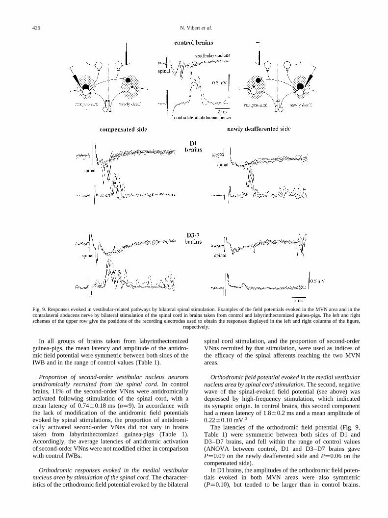
Orthodromic responses evoked in the medial vestibularnucleus area by stimulation of the spinal cord.The character-istics of the orthodromic field potential evoked by the bilateral
spinal cord stimulation, and the proportion of second-orderVNns recruited by that stimulation, were used as indices ofthe efficacy of the spinal afferents reaching the two MVNareas.
Orthodromic field potential evoked in the medial vestibularnucleus area by spinal cord stimulation.The second, negativewave of the spinal-evoked field potential (see above) wasdepressed by high-frequency stimulation, which indicatedits synaptic origin. In control brains, this second componenthad a mean latency of 1.80.2 ms and a mean amplitude of0.22 0.10 mV.3
The latencies of the orthodromic field potential (Fig. 9,Table 1) were symmetric between both sides of D1 andD3–D7 brains, and fell within the range of control values(ANOVA between control, D1 and D3–D7 brains gaveP�0.09 on the newly deafferented side andP�0.06 on thecompensated side).
In D1 brains, the amplitudes of the orthodromic field poten-tials evoked in both MVN areas were also symmetric(P�0.10), but tended to be larger than in control brains.
N. Vibert et al.426
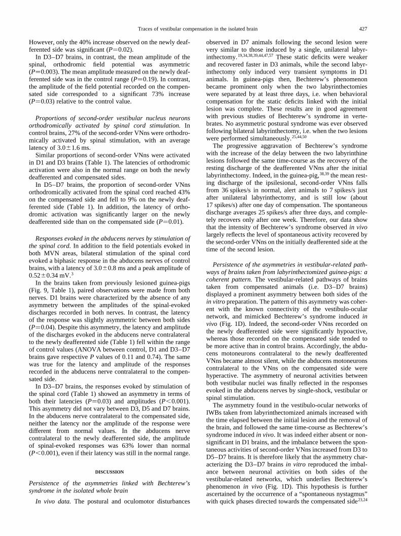
Fig. 9. Responses evoked in vestibular-related pathways by bilateral spinal stimulation. Examples of the field potentials evoked in the MVN area and in thecontralateral abducens nerve by bilateral stimulation of the spinal cord in brains taken from control and labyrinthectomized guinea-pigs. The leftand rightschemes of the upper row give the positions of the recording electrodes used to obtain the responses displayed in the left and right columns of the figure,
respectively.
However, only the 40% increase observed on the newly deaf-ferented side was significant (P�0.02).
In D3–D7 brains, in contrast, the mean amplitude of thespinal, orthodromic field potential was asymmetric(P�0.003). The mean amplitude measured on the newly deaf-ferented side was in the control range (P�0.19). In contrast,the amplitude of the field potential recorded on the compen-sated side corresponded to a significant 73% increase(P�0.03) relative to the control value.
Proportions of second-order vestibular nucleus neuronsorthodromically activated by spinal cord stimulation.Incontrol brains, 27% of the second-order VNns were orthodro-mically activated by spinal stimulation, with an averagelatency of 3.0 1.6 ms.
Similar proportions of second-order VNns were activatedin D1 and D3 brains (Table 1). The latencies of orthodromicactivation were also in the normal range on both the newlydeafferented and compensated sides.
In D5–D7 brains, the proportion of second-order VNnsorthodromically activated from the spinal cord reached 43%on the compensated side and fell to 9% on the newly deaf-ferented side (Table 1). In addition, the latency of ortho-dromic activation was significantly larger on the newlydeafferented side than on the compensated side (P�0.01).
Responses evoked in the abducens nerves by stimulation ofthe spinal cord.In addition to the field potentials evoked inboth MVN areas, bilateral stimulation of the spinal cordevoked a biphasic response in the abducens nerves of controlbrains, with a latency of 3.00.8 ms and a peak amplitude of0.52 0.34 mV.3
In the brains taken from previously lesioned guinea-pigs(Fig. 9, Table 1), paired observations were made from bothnerves. D1 brains were characterized by the absence of anyasymmetry between the amplitudes of the spinal-evokeddischarges recorded in both nerves. In contrast, the latencyof the response was slightly asymmetric between both sides(P�0.04). Despite this asymmetry, the latency and amplitudeof the discharges evoked in the abducens nerve contralateralto the newly deafferented side (Table 1) fell within the rangeof control values (ANOVA between control, D1 and D3–D7brains gave respectiveP values of 0.11 and 0.74). The samewas true for the latency and amplitude of the responsesrecorded in the abducens nerve contralateral to the compen-sated side.
In D3–D7 brains, the responses evoked by stimulation ofthe spinal cord (Table 1) showed an asymmetry in terms ofboth their latencies (P�0.03) and amplitudes (P,0.001).This asymmetry did not vary between D3, D5 and D7 brains.In the abducens nerve contralateral to the compensated side,neither the latency nor the amplitude of the response weredifferent from normal values. In the abducens nervecontralateral to the newly deafferented side, the amplitudeof spinal-evoked responses was 63% lower than normal(P,0.001), even if their latency was still in the normal range.
DISCUSSION
Persistence of the asymmetries linked with Bechterew’ssyndrome in the isolated whole brain
In vivo data. The postural and oculomotor disturbances
observed in D7 animals following the second lesion werevery similar to those induced by a single, unilateral labyr-inthectomy.19,34,38,39,44,47,57These static deficits were weakerand recovered faster in D3 animals, while the second labyr-inthectomy only induced very transient symptoms in D1animals. In guinea-pigs then, Bechterew’s phenomenonbecame prominent only when the two labyrinthectomieswere separated by at least three days, i.e. when behavioralcompensation for the static deficits linked with the initiallesion was complete. These results are in good agreementwith previous studies of Bechterew’s syndrome in verte-brates. No asymmetric postural syndrome was ever observedfollowing bilateral labyrinthectomy, i.e. when the two lesionswere performed simultaneously.25,44,50
The progressive aggravation of Bechterew’s syndromewith the increase of the delay between the two labyrinthinelesions followed the same time-course as the recovery of theresting discharge of the deafferented VNns after the initiallabyrinthectomy. Indeed, in the guinea-pig,38,39the mean rest-ing discharge of the ipsilesional, second-order VNns fallsfrom 36 spikes/s in normal, alert animals to 7 spikes/s justafter unilateral labyrinthectomy, and is still low (about17 spikes/s) after one day of compensation. The spontaneousdischarge averages 25 spikes/s after three days, and comple-tely recovers only after one week. Therefore, our data showthat the intensity of Bechterew’s syndrome observedin vivolargely reflects the level of spontaneous activity recovered bythe second-order VNns on the initially deafferented side at thetime of the second lesion.
Persistence of the asymmetries in vestibular-related path-ways of brains taken from labyrinthectomized guinea-pigs: acoherent pattern.The vestibular-related pathways of brainstaken from compensated animals (i.e. D3–D7 brains)displayed a prominent asymmetry between both sides of thein vitro preparation. The pattern of this asymmetry was coher-ent with the known connectivity of the vestibulo-ocularnetwork, and mimicked Bechterew’s syndrome inducedinvivo (Fig. 1D). Indeed, the second-order VNns recorded onthe newly deafferented side were significantly hypoactive,whereas those recorded on the compensated side tended tobe more active than in control brains. Accordingly, the abdu-cens motoneurons contralateral to the newly deafferentedVNns became almost silent, while the abducens motoneuronscontralateral to the VNns on the compensated side werehyperactive. The asymmetry of neuronal activities betweenboth vestibular nuclei was finally reflected in the responsesevoked in the abducens nerves by single-shock, vestibular orspinal stimulation.
The asymmetry found in the vestibulo-ocular networks ofIWBs taken from labyrinthectomized animals increased withthe time elapsed between the initial lesion and the removal ofthe brain, and followed the same time-course as Bechterew’ssyndrome inducedin vivo. It was indeed either absent or non-significant in D1 brains, and the imbalance between the spon-taneous activities of second-order VNns increased from D3 toD5–D7 brains. It is therefore likely that the asymmetry char-acterizing the D3–D7 brainsin vitro reproduced the imbal-ance between neuronal activities on both sides of thevestibular-related networks, which underlies Bechterew’sphenomenonin vivo (Fig. 1D). This hypothesis is furtherascertained by the occurrence of a “spontaneous nystagmus”with quick phases directed towards the compensated side23,24
Traces of vestibular compensation in the isolated brain 427
in some of the D3–D7 brains, since a similar nystagmus wasassociated with Bechterew’s phenomenonin vivo.
Consequences of the persistence of Bechterew’s asymmetryin the isolated whole brain.The persistence of Bechterew’sasymmetry in the IWB has allowed us to answer positively thetwo main questions raised in the introduction. First, the IWBdoes retain some of the plastic cellular changes underlyingvestibular compensation. Second, after one day and during thefirst week of vestibular compensation, the CNS does notrequire any continuous inflow from substituting sensory affer-ents to maintain the new balance reached between the restingdischarges of VNns on both sides of the brainstem. Indeed,during the first week of vestibular compensation, a similarBechterew phenomenon is triggered (i)in vitro followingthe section of the intact vestibular nerve in IWBs takenfrom previously labyrinthectomized animals and (ii)in vivoby a second labyrinthectomy. In addition, we have seen abovethat, in vivo, the intensity of Bechterew’s syndrome waslinked with the level of spontaneous activity recovered bythe initially deafferented VNns. These two points stronglysuggest that, after one day, the physiological changesinvivo underlying the recovery of a normal resting dischargeby the initially deafferented VNns persist in IWBs, despite theabsence of any organized proprioceptive or visual input in thispreparation.
This does not mean that the sensory, non-vestibular infor-mation reaching vestibular neurons (which mainly includeproprioceptive, somatosensory and visual fibers)55 does notplay an essential role during the first 24 h of compensation(see Refs 9, 10 and 49 for reviews). Indeed, the static posturalsymptoms induced by unilateral labyrinthectomy do notcompensate as long as guinea-pigs are lifted off theground.19,43 Furthermore, restraining the head and body of aguinea-pig in their normal, straight ahead position slowscompensation for the static symptoms.19,35,45 Finally, wecannot exclude that, even after the first day of compensation,asymmetric sensory information might be responsible for trig-gering the mechanisms allowing the deafferented, second-order VNns to go on recovering a normal spontaneousactivity.
Our data extend the results obtainedin vivo by Magnus25
and Spiegel and De´metriades,50 which demonstrated thatBechterew’s syndrome still occurred in compensated catsand dogs after exclusion of optic signals, or section of theupper cervical dorsal roots. Bechterew’s phenomenon waseven observed following removal of the whole cerebrum orablation of the cerebellum. In fully compensated animalsthen, an intact medulla oblongata appears sufficient to main-tain the new balance reached between the mass discharges ofsecond-order VNns on both sides, and to trigger Bechterew’ssyndrome.44
On the other hand, several authors have shown that sectionof the cervical dorsal roots or spinal transection induced apostural decompensation in guinea-pigs2,19 and monkeys.17
In contrast with our data, these experiments suggested43
(see Ref. 49 for review) that spinal input was necessary tomaintain the resting discharge of the deafferented, second-order VNns. This decompensation, which often lasted onlya few hours, could, however, simply be due to a greater sensi-tivity of the deafferented VNns to diaschisis (i.e. neuralshock26) induced by any additional deafferentation (see Ref.49 for discussion of this point), and not the removal of the
spinal inputsper se. The fact that spinal transection bilaterallydepressed the spontaneous activity of identified, type I andtype II VNns in normal as well as in compensated gerbils30,32
(see Ref. 11 for the type I–type II classification of vestibular-related neurons) gives further support to this hypothesis.
The properties of vestibular-related pathways in isolatedwhole brains taken from labyrinthectomized guinea-pigsgive insights into the neuronal mechanisms of vestibularcompensation
We will now discuss to what extent the modifications of thevestibular- and spinal-evoked responses observed in IWBstaken from previously labyrinthectomized animals can shedsome light on the neuronal mechanisms underlying the firstweek of the compensation process.
Responses evoked in the vestibulo-abducens pathways bysingle shock stimulations of the vestibular nerves
Field potentials evoked in the medial vestibular nucleusarea by ipsilateral vestibular nerve stimulation.Our datafirst confirm that, despite their silence following labyrinth-ectomy, the ipsilesional, sensory vestibular neurons did notdegenerate.20,46 Until at least one week after the lesion, theyretained their ability to respond to electrical stimulation andcould still generate synaptic potentials in second-order VNns(see Refs 9, 10 and 49 for reviews).
Our data also suggest that a significant decrease in theefficacy of the synaptic transmission between the sensoryafferent fibers and their target vestibular neurons occursthree days after the initial labyrinthectomy (Fig. 10). Theamplitudes of the evoked vestibular field potentials wereindeed significantly lower than normal on both sides ofD3–D7 brains. On the newly deafferented side, this reductionmight result from the large dysfacilitation of second-orderVNns induced by the acute deafferentation (Fig. 1D).However, this explanation does not hold for the compensatedside, where the resting discharge of VNns was in the normalrange or even higher. This result may be linked with thetransganglionic degeneration observed in the central term-inals of sensory vestibular neurons after peripheral vestibulardeafferentation.16 In the spinal cord, peripheral nerve lesion isfollowed within a few days by a significant decrease in theconduction velocity of dorsal root myelinated axons, and inthe amplitude of the related evoked field potential (see Ref. 1for review).
Asymmetry of the discharges evoked in the contralateralabducens nerve.The low size of the response evoked bystimulation of the vestibular nerve on the compensated sidein the contralateral abducens nerve on the newly deafferentedside suggests that,in vivo, vestibular compensation must beassociated with a reduction in the efficacy of synaptictransmission between the second-order VNns on the intactside and the contralateral abducens motoneurons on thelesioned side (Fig. 10). Indeed, while stimulation of thevestibular nerve on the newly deafferented side only evoked35% smaller than normal excitatory field potentials in theipsilateral MVN area, the response obtained in the contra-lateral abducens nerve was decreased by about 80%. InD5–D7 brains, this decrease could be due to the increasedactivity of the VNns on the compensated side, which exert a
N. Vibert et al.428
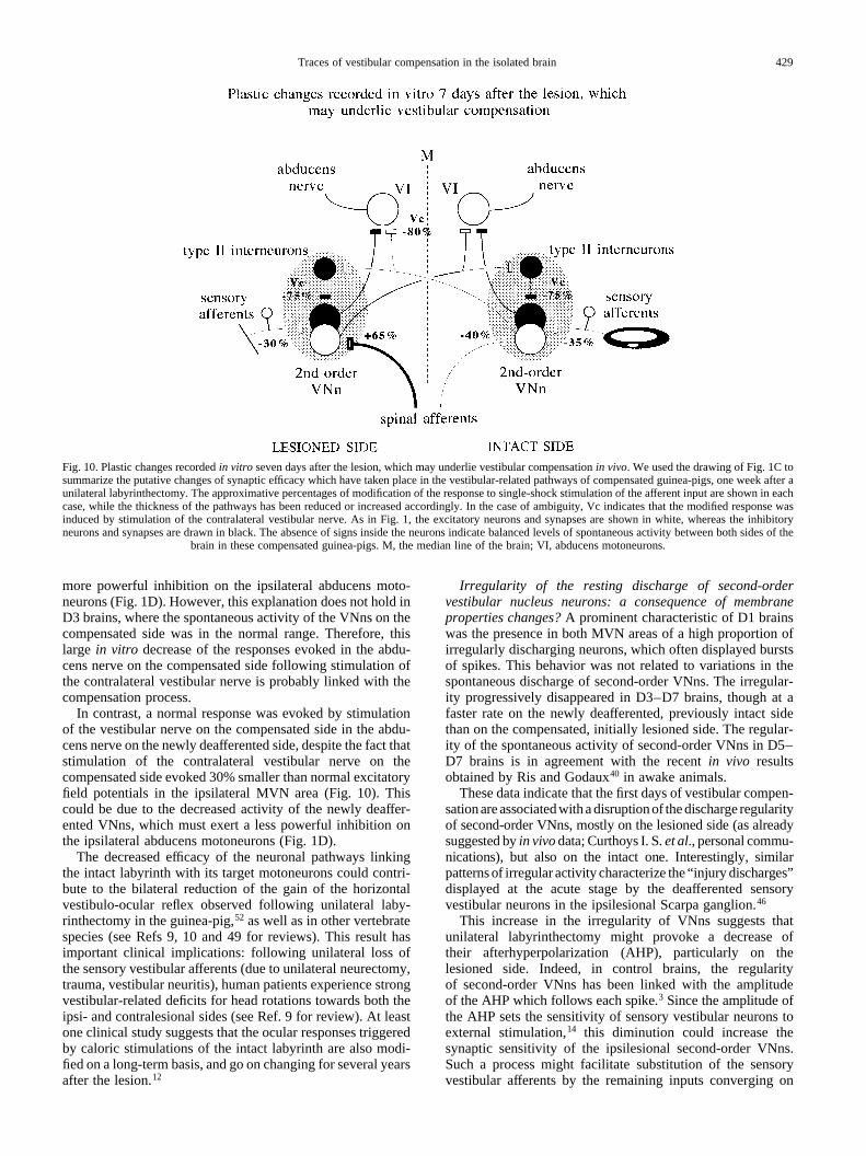
more powerful inhibition on the ipsilateral abducens moto-neurons (Fig. 1D). However, this explanation does not hold inD3 brains, where the spontaneous activity of the VNns on thecompensated side was in the normal range. Therefore, thislarge in vitro decrease of the responses evoked in the abdu-cens nerve on the compensated side following stimulation ofthe contralateral vestibular nerve is probably linked with thecompensation process.
In contrast, a normal response was evoked by stimulationof the vestibular nerve on the compensated side in the abdu-cens nerve on the newly deafferented side, despite the fact thatstimulation of the contralateral vestibular nerve on thecompensated side evoked 30% smaller than normal excitatoryfield potentials in the ipsilateral MVN area (Fig. 10). Thiscould be due to the decreased activity of the newly deaffer-ented VNns, which must exert a less powerful inhibition onthe ipsilateral abducens motoneurons (Fig. 1D).
The decreased efficacy of the neuronal pathways linkingthe intact labyrinth with its target motoneurons could contri-bute to the bilateral reduction of the gain of the horizontalvestibulo-ocular reflex observed following unilateral laby-rinthectomy in the guinea-pig,52 as well as in other vertebratespecies (see Refs 9, 10 and 49 for reviews). This result hasimportant clinical implications: following unilateral loss ofthe sensory vestibular afferents (due to unilateral neurectomy,trauma, vestibular neuritis), human patients experience strongvestibular-related deficits for head rotations towards both theipsi- and contralesional sides (see Ref. 9 for review). At leastone clinical study suggests that the ocular responses triggeredby caloric stimulations of the intact labyrinth are also modi-fied on a long-term basis, and go on changing for several yearsafter the lesion.12
Irregularity of the resting discharge of second-ordervestibular nucleus neurons: a consequence of membraneproperties changes?A prominent characteristic of D1 brainswas the presence in both MVN areas of a high proportion ofirregularly discharging neurons, which often displayed burstsof spikes. This behavior was not related to variations in thespontaneous discharge of second-order VNns. The irregular-ity progressively disappeared in D3–D7 brains, though at afaster rate on the newly deafferented, previously intact sidethan on the compensated, initially lesioned side. The regular-ity of the spontaneous activity of second-order VNns in D5–D7 brains is in agreement with the recentin vivo resultsobtained by Ris and Godaux40 in awake animals.
These data indicate that the first days of vestibular compen-sationare associatedwitha disruptionof thedischarge regularityof second-order VNns, mostly on the lesioned side (as alreadysuggested byin vivodata; Curthoys I. S.et al., personal commu-nications), but also on the intact one. Interestingly, similarpatternsof irregular activity characterize the “injury discharges”displayed at the acute stage by the deafferented sensoryvestibular neurons in the ipsilesional Scarpa ganglion.46
This increase in the irregularity of VNns suggests thatunilateral labyrinthectomy might provoke a decrease oftheir afterhyperpolarization (AHP), particularly on thelesioned side. Indeed, in control brains, the regularityof second-order VNns has been linked with the amplitudeof the AHP which follows each spike.3 Since the amplitude ofthe AHP sets the sensitivity of sensory vestibular neurons toexternal stimulation,14 this diminution could increase thesynaptic sensitivity of the ipsilesional second-order VNns.Such a process might facilitate substitution of the sensoryvestibular afferents by the remaining inputs converging on
Traces of vestibular compensation in the isolated brain 429
Fig. 10. Plastic changes recordedin vitro seven days after the lesion, which may underlie vestibular compensationin vivo. We used the drawing of Fig. 1C tosummarize the putative changes of synaptic efficacy which have taken place in the vestibular-related pathways of compensated guinea-pigs, one week after aunilateral labyrinthectomy. The approximative percentages of modification of the response to single-shock stimulation of the afferent input are shown in eachcase, while the thickness of the pathways has been reduced or increased accordingly. In the case of ambiguity, Vc indicates that the modified response wasinduced by stimulation of the contralateral vestibular nerve. As in Fig. 1, the excitatory neurons and synapses are shown in white, whereas the inhibitoryneurons and synapses are drawn in black. The absence of signs inside the neurons indicate balanced levels of spontaneous activity between both sides of the
brain in these compensated guinea-pigs. M, the median line of the brain; VI, abducens motoneurons.
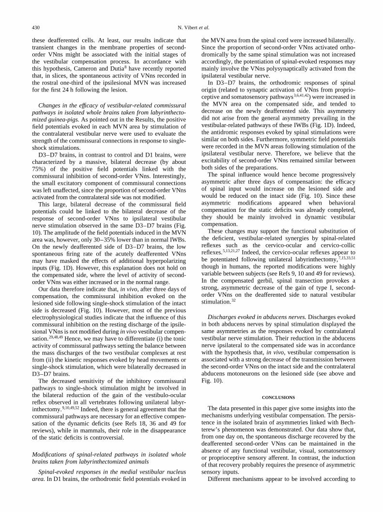
these deafferented cells. At least, our results indicate thattransient changes in the membrane properties of second-order VNns might be associated with the initial stages ofthe vestibular compensation process. In accordance withthis hypothesis, Cameron and Dutia8 have recently reportedthat, in slices, the spontaneous activity of VNns recorded inthe rostral one-third of the ipsilesional MVN was increasedfor the first 24 h following the lesion.
Changes in the efficacy of vestibular-related commissuralpathways in isolated whole brains taken from labyrinthecto-mized guinea-pigs.As pointed out in the Results, the positivefield potentials evoked in each MVN area by stimulation ofthe contralateral vestibular nerve were used to evaluate thestrength of the commissural connections in response to single-shock stimulations.
D3–D7 brains, in contrast to control and D1 brains, werecharacterized by a massive, bilateral decrease (by about75%) of the positive field potentials linked with thecommissural inhibition of second-order VNns. Interestingly,the small excitatory component of commissural connectionswas left unaffected, since the proportion of second-order VNnsactivated from the contralateral side was not modified.
This large, bilateral decrease of the commissural fieldpotentials could be linked to the bilateral decrease of theresponse of second-order VNns to ipsilateral vestibularnerve stimulation observed in the same D3–D7 brains (Fig.10). The amplitude of the field potentials induced in the MVNarea was, however, only 30–35% lower than in normal IWBs.On the newly deafferented side of D3–D7 brains, the lowspontaneous firing rate of the acutely deafferented VNnsmay have masked the effects of additional hyperpolarizinginputs (Fig. 1D). However, this explanation does not hold onthe compensated side, where the level of activity of second-order VNns was either increased or in the normal range.
Our data therefore indicate that,in vivo, after three days ofcompensation, the commissural inhibition evoked on thelesioned side following single-shock stimulation of the intactside is decreased (Fig. 10). However, most of the previouselectrophysiological studies indicate that the influence of thiscommissural inhibition on the resting discharge of the ipsile-sional VNns is not modified duringin vivovestibular compen-sation.29,48,49Hence, we may have to differentiate (i) the tonicactivity of commissural pathways setting the balance betweenthe mass discharges of the two vestibular complexes at restfrom (ii) the kinetic responses evoked by head movements orsingle-shock stimulation, which were bilaterally decreased inD3–D7 brains.
The decreased sensitivity of the inhibitory commissuralpathways to single-shock stimulation might be involved inthe bilateral reduction of the gain of the vestibulo-ocularreflex observed in all vertebrates following unilateral labyr-inthectomy.9,10,49,52Indeed, there is general agreement that thecommissural pathways are necessary for an effective compen-sation of the dynamic deficits (see Refs 18, 36 and 49 forreviews), while in mammals, their role in the disappearanceof the static deficits is controversial.
Modifications of spinal-related pathways in isolated wholebrains taken from labyrinthectomized animals
Spinal-evoked responses in the medial vestibular nucleusarea.In D1 brains, the orthodromic field potentials evoked in
the MVN area from the spinal cord were increased bilaterally.Since the proportion of second-order VNns activated ortho-dromically by the same spinal stimulation was not increasedaccordingly, the potentiation of spinal-evoked responses maymainly involve the VNns polysynaptically activated from theipsilateral vestibular nerve.
In D3–D7 brains, the orthodromic responses of spinalorigin (related to synaptic activation of VNns from proprio-ceptive and somatosensory pathways3,6,41,42) were increased inthe MVN area on the compensated side, and tended todecrease on the newly deafferented side. This asymmetrydid not arise from the general asymmetry prevailing in thevestibular-related pathways of these IWBs (Fig. 1D). Indeed,the antidromic responses evoked by spinal stimulations weresimilar on both sides. Furthermore, symmetric field potentialswere recorded in the MVN areas following stimulation of theipsilateral vestibular nerve. Therefore, we believe that theexcitability of second-order VNns remained similar betweenboth sides of the preparations.
The spinal influence would hence become progressivelyasymmetric after three days of compensation: the efficacyof spinal input would increase on the lesioned side andwould be reduced on the intact side (Fig. 10). Since theseasymmetric modifications appeared when behavioralcompensation for the static deficits was already completed,they should be mainly involved in dynamic vestibularcompensation.
These changes may support the functional substitution ofthe deficient, vestibular-related synergies by spinal-relatedreflexes such as the cervico-ocular and cervico-collicreflexes.5,13,21,27Indeed, the cervico-ocular reflexes appear tobe potentiated following unilateral labyrinthectomy,7,15,33,51
though in humans, the reported modifications were highlyvariable between subjects (see Refs 9, 10 and 49 for reviews).In the compensated gerbil, spinal transection provokes astrong, asymmetric decrease of the gain of type I, second-order VNns on the deafferented side to natural vestibularstimulation.32
Discharges evoked in abducens nerves.Discharges evokedin both abducens nerves by spinal stimulation displayed thesame asymmetries as the responses evoked by contralateralvestibular nerve stimulation. Their reduction in the abducensnerve ipsilateral to the compensated side was in accordancewith the hypothesis that,in vivo, vestibular compensation isassociated with a strong decrease of the transmission betweenthe second-order VNns on the intact side and the contralateralabducens motoneurons on the lesioned side (see above andFig. 10).
CONCLUSIONS
The data presented in this paper give some insights into themechanisms underlying vestibular compensation. The persis-tence in the isolated brain of asymmetries linked with Bech-terew’s phenomenon was demonstrated. Our data show that,from one day on, the spontaneous discharge recovered by thedeafferented second-order VNns can be maintained in theabsence of any functional vestibular, visual, somatosensoryor proprioceptive sensory afferent. In contrast, the inductionof that recovery probably requires the presence of asymmetricsensory inputs.
Different mechanisms appear to be involved according to
N. Vibert et al.430
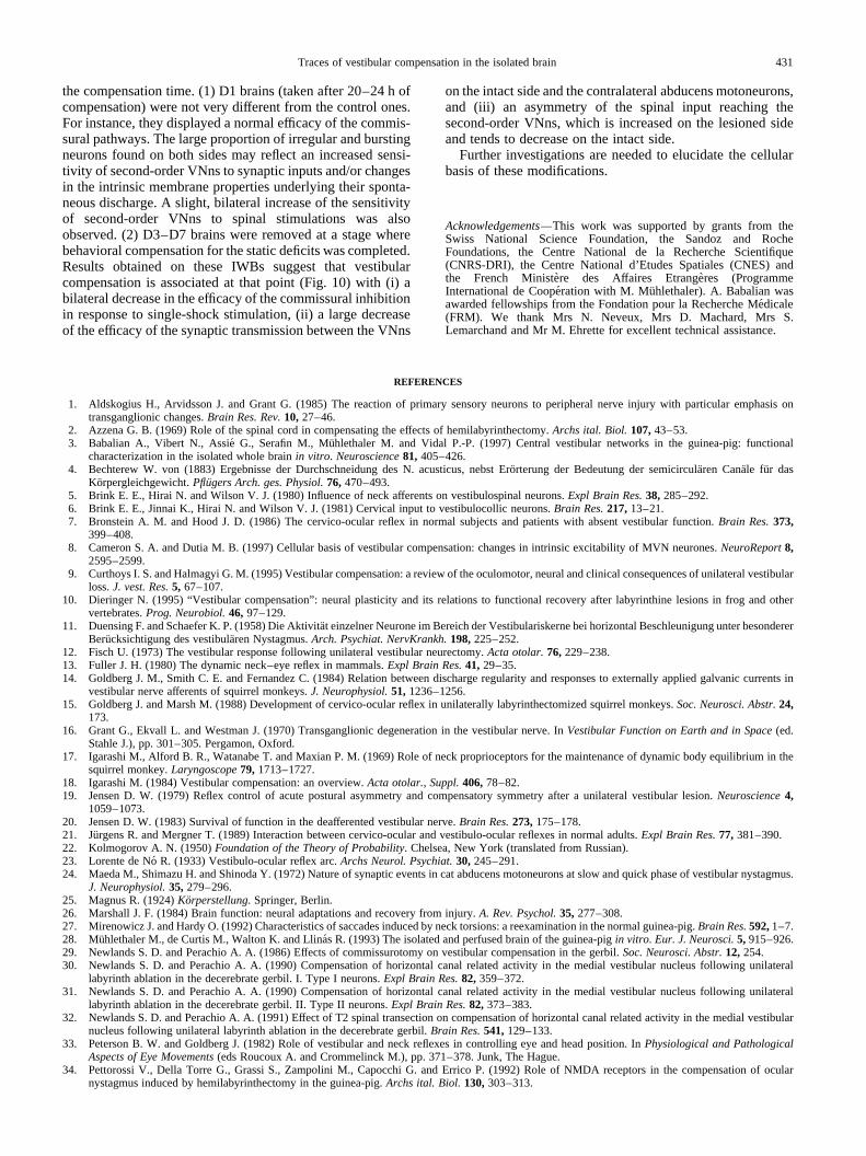
the compensation time. (1) D1 brains (taken after 20–24 h ofcompensation) were not very different from the control ones.For instance, they displayed a normal efficacy of the commis-sural pathways. The large proportion of irregular and burstingneurons found on both sides may reflect an increased sensi-tivity of second-order VNns to synaptic inputs and/or changesin the intrinsic membrane properties underlying their sponta-neous discharge. A slight, bilateral increase of the sensitivityof second-order VNns to spinal stimulations was alsoobserved. (2) D3–D7 brains were removed at a stage wherebehavioral compensation for the static deficits was completed.Results obtained on these IWBs suggest that vestibularcompensation is associated at that point (Fig. 10) with (i) abilateral decrease in the efficacy of the commissural inhibitionin response to single-shock stimulation, (ii) a large decreaseof the efficacy of the synaptic transmission between the VNns
on the intact side and the contralateral abducens motoneurons,and (iii) an asymmetry of the spinal input reaching thesecond-order VNns, which is increased on the lesioned sideand tends to decrease on the intact side.
Further investigations are needed to elucidate the cellularbasis of these modifications.
Acknowledgements—This work was supported by grants from theSwiss National Science Foundation, the Sandoz and RocheFoundations, the Centre National de la Recherche Scientifique(CNRS-DRI), the Centre National d’Etudes Spatiales (CNES) andthe French Ministe`re des Affaires Etrange`res (ProgrammeInternational de Coope´ration with M. Muhlethaler). A. Babalian wasawarded fellowships from the Fondation pour la Recherche Me´dicale(FRM). We thank Mrs N. Neveux, Mrs D. Machard, Mrs S.Lemarchand and Mr M. Ehrette for excellent technical assistance.
REFERENCES
1. Aldskogius H., Arvidsson J. and Grant G. (1985) The reaction of primary sensory neurons to peripheral nerve injury with particular emphasis ontransganglionic changes.Brain Res. Rev.10, 27–46.
2. Azzena G. B. (1969) Role of the spinal cord in compensating the effects of hemilabyrinthectomy.Archs ital. Biol.107,43–53.3. Babalian A., Vibert N., Assie´ G., Serafin M., Mu¨hlethaler M. and Vidal P.-P. (1997) Central vestibular networks in the guinea-pig: functional
characterization in the isolated whole brainin vitro. Neuroscience81, 405–426.4. Bechterew W. von (1883) Ergebnisse der Durchschneidung des N. acusticus, nebst Ero¨rterung der Bedeutung der semicircula¨ren Cana¨le fur das
Korpergleichgewicht.Pflugers Arch. ges. Physiol.76, 470–493.5. Brink E. E., Hirai N. and Wilson V. J. (1980) Influence of neck afferents on vestibulospinal neurons.Expl Brain Res.38, 285–292.6. Brink E. E., Jinnai K., Hirai N. and Wilson V. J. (1981) Cervical input to vestibulocollic neurons.Brain Res.217,13–21.7. Bronstein A. M. and Hood J. D. (1986) The cervico-ocular reflex in normal subjects and patients with absent vestibular function.Brain Res.373,
399–408.8. Cameron S. A. and Dutia M. B. (1997) Cellular basis of vestibular compensation: changes in intrinsic excitability of MVN neurones.NeuroReport8,
2595–2599.9. Curthoys I. S. and Halmagyi G. M. (1995) Vestibular compensation: a review of the oculomotor, neural and clinical consequences of unilateral vestibular
loss.J. vest. Res.5, 67–107.10. Dieringer N. (1995) “Vestibular compensation”: neural plasticity and its relations to functional recovery after labyrinthine lesions in frog and other
vertebrates.Prog. Neurobiol.46, 97–129.11. Duensing F. and Schaefer K. P. (1958) Die Aktivita¨t einzelner Neurone im Bereich der Vestibulariskerne bei horizontal Beschleunigung unter besonderer
Berucksichtigung des vestibula¨ren Nystagmus.Arch. Psychiat. NervKrankh.198,225–252.12. Fisch U. (1973) The vestibular response following unilateral vestibular neurectomy.Acta otolar.76, 229–238.13. Fuller J. H. (1980) The dynamic neck–eye reflex in mammals.Expl Brain Res.41, 29–35.14. Goldberg J. M., Smith C. E. and Fernandez C. (1984) Relation between discharge regularity and responses to externally applied galvanic currents in
vestibular nerve afferents of squirrel monkeys.J. Neurophysiol.51, 1236–1256.15. Goldberg J. and Marsh M. (1988) Development of cervico-ocular reflex in unilaterally labyrinthectomized squirrel monkeys.Soc. Neurosci. Abstr.24,
173.16. Grant G., Ekvall L. and Westman J. (1970) Transganglionic degeneration in the vestibular nerve. InVestibular Function on Earth and in Space(ed.
Stahle J.), pp. 301–305. Pergamon, Oxford.17. Igarashi M., Alford B. R., Watanabe T. and Maxian P. M. (1969) Role of neck proprioceptors for the maintenance of dynamic body equilibrium in the
squirrel monkey.Laryngoscope79, 1713–1727.18. Igarashi M. (1984) Vestibular compensation: an overview.Acta otolar., Suppl.406,78–82.19. Jensen D. W. (1979) Reflex control of acute postural asymmetry and compensatory symmetry after a unilateral vestibular lesion.Neuroscience4,
1059–1073.20. Jensen D. W. (1983) Survival of function in the deafferented vestibular nerve.Brain Res.273,175–178.21. Jurgens R. and Mergner T. (1989) Interaction between cervico-ocular and vestibulo-ocular reflexes in normal adults.Expl Brain Res.77, 381–390.22. Kolmogorov A. N. (1950)Foundation of the Theory of Probability. Chelsea, New York (translated from Russian).23. Lorente de No´ R. (1933) Vestibulo-ocular reflex arc.Archs Neurol. Psychiat.30, 245–291.24. Maeda M., Shimazu H. and Shinoda Y. (1972) Nature of synaptic events in cat abducens motoneurons at slow and quick phase of vestibular nystagmus.
J. Neurophysiol.35, 279–296.25. Magnus R. (1924)Korperstellung. Springer, Berlin.26. Marshall J. F. (1984) Brain function: neural adaptations and recovery from injury.A. Rev. Psychol.35, 277–308.27. Mirenowicz J. and Hardy O. (1992) Characteristics of saccades induced by neck torsions: a reexamination in the normal guinea-pig.Brain Res.592,1–7.28. Muhlethaler M., de Curtis M., Walton K. and Llina´s R. (1993) The isolated and perfused brain of the guinea-pigin vitro. Eur. J. Neurosci.5, 915–926.29. Newlands S. D. and Perachio A. A. (1986) Effects of commissurotomy on vestibular compensation in the gerbil.Soc. Neurosci. Abstr.12, 254.30. Newlands S. D. and Perachio A. A. (1990) Compensation of horizontal canal related activity in the medial vestibular nucleus following unilateral
labyrinth ablation in the decerebrate gerbil. I. Type I neurons.Expl Brain Res.82, 359–372.31. Newlands S. D. and Perachio A. A. (1990) Compensation of horizontal canal related activity in the medial vestibular nucleus following unilateral
labyrinth ablation in the decerebrate gerbil. II. Type II neurons.Expl Brain Res.82, 373–383.32. Newlands S. D. and Perachio A. A. (1991) Effect of T2 spinal transection on compensation of horizontal canal related activity in the medial vestibular
nucleus following unilateral labyrinth ablation in the decerebrate gerbil.Brain Res.541,129–133.33. Peterson B. W. and Goldberg J. (1982) Role of vestibular and neck reflexes in controlling eye and head position. InPhysiological and Pathological
Aspects of Eye Movements(eds Roucoux A. and Crommelinck M.), pp. 371–378. Junk, The Hague.34. Pettorossi V., Della Torre G., Grassi S., Zampolini M., Capocchi G. and Errico P. (1992) Role of NMDA receptors in the compensation of ocular
nystagmus induced by hemilabyrinthectomy in the guinea-pig.Archs ital. Biol.130,303–313.
Traces of vestibular compensation in the isolated brain 431
35. Pettorossi V. E. and Petrosini L. (1984) Tonic cervical influences on eye nystagmus following hemilabyrinthectomy: immediate and plastic effects.BrainRes.324,11–19.
36. Precht W. and Dieringer N. (1986) Recovery of some vestibuloocular and vestibulospinal functions following unilateral labyrinthectomy. InProgress inBrain Research(eds Freund H. J., Bu¨ttner U., Cohen B. and North J.), Vol. 64, pp. 381–389. Elsevier, Amsterdam.
37. Precht W., Shimazu H. and Markham C. H. (1966) A mechanism of central compensation of vestibular function following hemilabyrinthectomy.J. Neurophysiol.29, 996–1010.
38. Ris L., de Waele C., Serafin M., Vidal P.-P. and Godaux E. (1995) Neuronal activity in the ipsilateral vestibular nucleus following unilateral labyr-inthectomy in the alert guinea-pig.J. Neurophysiol.74, 2087–2099.
39. Ris L., Capron B., Waele C. de, Vidal P.-P. and Godaux E. (1997) Dissociations between behavioural recovery and restoration of vestibular activity in theunilabyrinthectomized guinea-pig.J. Physiol.500,509–522.
40. Ris L. and Godaux E. (1998) Spike discharge regularity of vestibular neurons in labyrinthectomized guinea pigs.Neurosci. Lett.253,131–134.41. Rubin A. M., Liedgren S. R. C., O¨ dkvist L. M., Milne A. C. and Fredrickson J. M. (1978) Labyrinthine and somatosensory convergence upon
vestibulospinal neurons.Acta otolar.86, 251–259.42. Sato H., Ohkawa T., Uchino Y. and Wilson V. J. (1997) Excitatory connections between neurons of the central cervical nucleus and vestibular neurons in
the cat.Expl Brain Res.115,381–386.43. Schaefer K. P. and Meyer D. L. (1973) Compensatory mechanisms following labyrinthine lesions in the guinea-pig. A simple model of learning. In
Memory and Transfer of Information(ed. Zippel H. P.), pp. 203–232. Plenum, New York.44. Schaefer K. P. and Meyer D. L. (1974) Compensation of vestibular lesions. InHandbook of Sensory Physiology(ed. Kornhuber H. H.), Vol. VI,
pp. 463–490. Springer, Berlin.45. Schaefer K. P., Meyer D. L. and Wilhelms G. (1979) Somatosensory and cerebellar influences on compensation of labyrinthine lesions. InProgress in
Brain Research, Vol. 50: Reflex Control of Movement and Posture(eds Granit R. and Pompeiano O.), pp. 591–598. Elsevier, Amsterdam.46. Sirkin D. W., Precht W. and Courjon J. H. (1984) Initial, rapid phase of recovery from unilateral vestibular lesion is not dependent on survival of central
portion of vestibular nerve.Brain Res.302,245–256.47. Smith P. F., Darlington C. L. and Curthoys I. S. (1986) The effect of visual deprivation on vestibular compensation in the guinea-pig.Brain Res.364,
195–198.48. Smith P. F., Darlington C. L. and Curthoys I. S. (1986) Vestibular compensation without brainstem commissures in the guinea-pig.Neurosci. Lett.65,
209–213.49. Smith P. F. and Curthoys I. S. (1989) Mechanisms of recovery following unilateral labyrinthectomy: a review.Brain Res. Rev.14, 155–180.50. Spiegel E. A. and De´metriades T. D. (1925) Die zentrale Kompensation des Labyrinthverlustes.Pflugers Arch. ges. Physiol.210,215–222.51. Strupp M., Arbusow V., Dieterich M., Sautier W. and Brandt T. (1998) Perceptual and oculomotor effects of neck muscle vibration in vestibular neuritis.
Ipsilateral somatosensory substitution of vestibular function.Brain 121,677–685.52. Vibert N., Waele C. de, Escudero M. and Vidal P.-P. (1993) The horizontal vestibulo-ocular reflex in the hemilabyrinthectomized guinea-pig.Expl Brain
Res.97, 263–273.53. Vibert N., Babalian A., Mu¨hlethaler M. and Vidal P.-P. (1996) Some of the physiological changes underlying vestibular compensation persist in isolated,
in vitro whole brains from hemilabyrinthectomized guinea-pigs.Eur. J. Neurosci., Suppl.9, 136.54. Vibert N., Babalian A., Mu¨hlethaler M. and Vidal P.-P. (1996) Physiological correlates of vestibular compensation in isolated,in vitro whole brains from
hemilabyrinthectomized guinea-pigs.Soc. Neurosci. Abstr.22, 659.55. Vibert N., Waele C. de, Serafin M., Babalian A., Mu¨hlethaler M. and Vidal P. P. (1997) The vestibular system as a model of sensorimotor transforma-
tions. A combinedin vivo and in vitro approach to study the cellular mechanisms of gaze and posture stabilization in mammals.Prog. Neurobiol.51,243–286.
56. Waele C. de, Serafin M., Mu¨hlethaler M. and Vidal P.-P. (1988) Vestibular compensation: anin vivo and in vitro study of second-order vestibularneurons.Soc. Neurosci. Abstr.14, 331.
57. Waele C. de, Graf W., Josset P. and Vidal P.-P. (1989) A radiological analysis of the postural syndromes following hemilabyrinthectomy and selectivecanal and otolith lesions in the guinea-pig.Expl Brain Res.77, 166–182.
(Accepted16 March 1999)
N. Vibert et al.432
Related Documents