BioMed Central Page 1 of 13 (page number not for citation purposes) Malaria Journal Open Access Research Plasmodium yoelii 17XL infection up-regulates RANTES, CCR1, CCR3 and CCR5 expression, and induces ultrastructural changes in the cerebellum Bismark Y Sarfo 1 , Henry B Armah 2,3 , Ikovwaiza Irune 2 , Andrew A Adjei 3 , Christine S Olver 4 , Shailesh Singh 2 , James W Lillard Jr 2 and Jonathan K Stiles* 2 Address: 1 Parasitology Unit, Noguchi Memorial Institute for Medical Research, University of Ghana, P.O. Box LG581, Legon, Accra, Ghana, 2 Department of Microbiology, Biochemistry and Immunology, Morehouse School of Medicine, 720 Westview Drive S. W., Atlanta, GA, 30310- 1495, USA, 3 Department of Pathology, University of Ghana Medical School & Korle-Bu Teaching Hospital, P.O. Box 4236, Accra, Ghana and 4 Department of Pathology, Colorado State University, Fort Collins, CO, 80523, USA Email: Bismark Y Sarfo - [email protected]; Henry B Armah - [email protected]; Ikovwaiza Irune - [email protected]; Andrew A Adjei - [email protected]; Christine S Olver - [email protected]; Shailesh Singh - [email protected]; James W Lillard - [email protected]; Jonathan K Stiles* - [email protected] * Corresponding author Abstract Background: Malaria afflicts 300–500 million people causing over 1 million deaths globally per year. The immunopathogenesis of malaria is mediated partly by co mplex cellular and immunomodulator interactions involving co-regulators such as cytokines and adhesion molecules. However, the role of chemokines and their receptors in malaria immunopathology remains unclear. RANTES (Regulated on Activation Normal T-Cell Expressed and Secreted) is a chemokine involved in the generation of inflammatory infiltrates. Recent studies indicate that the degradation of cell-cell junctions, blood-brain barrier dysfunction, recruitment of leukocytes and Plasmodium- infected erythrocytes into and occlusion of microvessels relevant to malaria pathogenesis are associated with RANTES expression. Additionally, activated lymphocytes, platelets and endothelial cells release large quantities of RANTES, thus suggesting a unique role for RANTES in the generation and maintenance of the malaria-induced inflammatory response. The hypothesis of this study is that RANTES and its corresponding receptors (CCR1, CCR3 and CCR5) modulate malaria immunopathogenesis. A murine malaria model was utilized to evaluate the role of this chemokine and its receptors in malaria. Methods: The alterations in immunomodulator gene expression in brains of Plasmodium yoelii 17XL-infected mice was analysed using cDNA microarray screening, followed by a temporal comparison of mRNA and protein expression of RANTES and its corresponding receptors by qRT- PCR and Western blot analysis, respectively. Plasma RANTES levels was determined by ELISA and ultrastructural studies of brain sections from infected and uninfected mice was conducted. Results: RANTES (p < 0.002), CCR1 (p < 0.036), CCR3 (p < 0.033), and CCR5 (p < 0.026) mRNA were significantly upregulated at peak parasitaemia and remained high thereafter in the experimental mouse model. RANTES protein in the brain of infected mice was upregulated (p < Published: 16 December 2005 Malaria Journal 2005, 4:63 doi:10.1186/1475-2875-4-63 Received: 23 August 2005 Accepted: 16 December 2005 This article is available from: http://www.malariajournal.com/content/4/1/63 © 2005 Sarfo et al; licensee BioMed Central Ltd. This is an Open Access article distributed under the terms of the Creative Commons Attribution License (http://creativecommons.org/licenses/by/2.0 ), which permits unrestricted use, distribution, and reproduction in any medium, provided the original work is properly cited.

Welcome message from author
This document is posted to help you gain knowledge. Please leave a comment to let me know what you think about it! Share it to your friends and learn new things together.
Transcript
BioMed CentralMalaria Journal
ss
Open AcceResearchPlasmodium yoelii 17XL infection up-regulates RANTES, CCR1, CCR3 and CCR5 expression, and induces ultrastructural changes in the cerebellumBismark Y Sarfo1, Henry B Armah2,3, Ikovwaiza Irune2, Andrew A Adjei3, Christine S Olver4, Shailesh Singh2, James W Lillard Jr2 and Jonathan K Stiles*2Address: 1Parasitology Unit, Noguchi Memorial Institute for Medical Research, University of Ghana, P.O. Box LG581, Legon, Accra, Ghana, 2Department of Microbiology, Biochemistry and Immunology, Morehouse School of Medicine, 720 Westview Drive S. W., Atlanta, GA, 30310-1495, USA, 3Department of Pathology, University of Ghana Medical School & Korle-Bu Teaching Hospital, P.O. Box 4236, Accra, Ghana and 4Department of Pathology, Colorado State University, Fort Collins, CO, 80523, USA
Email: Bismark Y Sarfo - [email protected]; Henry B Armah - [email protected]; Ikovwaiza Irune - [email protected]; Andrew A Adjei - [email protected]; Christine S Olver - [email protected]; Shailesh Singh - [email protected]; James W Lillard - [email protected]; Jonathan K Stiles* - [email protected]
* Corresponding author
AbstractBackground: Malaria afflicts 300–500 million people causing over 1 million deaths globally peryear. The immunopathogenesis of malaria is mediated partly by co mplex cellular andimmunomodulator interactions involving co-regulators such as cytokines and adhesion molecules.However, the role of chemokines and their receptors in malaria immunopathology remainsunclear. RANTES (Regulated on Activation Normal T-Cell Expressed and Secreted) is a chemokineinvolved in the generation of inflammatory infiltrates. Recent studies indicate that the degradationof cell-cell junctions, blood-brain barrier dysfunction, recruitment of leukocytes and Plasmodium-infected erythrocytes into and occlusion of microvessels relevant to malaria pathogenesis areassociated with RANTES expression. Additionally, activated lymphocytes, platelets and endothelialcells release large quantities of RANTES, thus suggesting a unique role for RANTES in thegeneration and maintenance of the malaria-induced inflammatory response. The hypothesis of thisstudy is that RANTES and its corresponding receptors (CCR1, CCR3 and CCR5) modulate malariaimmunopathogenesis. A murine malaria model was utilized to evaluate the role of this chemokineand its receptors in malaria.
Methods: The alterations in immunomodulator gene expression in brains of Plasmodium yoelii17XL-infected mice was analysed using cDNA microarray screening, followed by a temporalcomparison of mRNA and protein expression of RANTES and its corresponding receptors by qRT-PCR and Western blot analysis, respectively. Plasma RANTES levels was determined by ELISA andultrastructural studies of brain sections from infected and uninfected mice was conducted.
Results: RANTES (p < 0.002), CCR1 (p < 0.036), CCR3 (p < 0.033), and CCR5 (p < 0.026) mRNAwere significantly upregulated at peak parasitaemia and remained high thereafter in theexperimental mouse model. RANTES protein in the brain of infected mice was upregulated (p <
Published: 16 December 2005
Malaria Journal 2005, 4:63 doi:10.1186/1475-2875-4-63
Received: 23 August 2005Accepted: 16 December 2005
This article is available from: http://www.malariajournal.com/content/4/1/63
© 2005 Sarfo et al; licensee BioMed Central Ltd. This is an Open Access article distributed under the terms of the Creative Commons Attribution License (http://creativecommons.org/licenses/by/2.0), which permits unrestricted use, distribution, and reproduction in any medium, provided the original work is properly cited.
Page 1 of 13(page number not for citation purposes)
Malaria Journal 2005, 4:63 http://www.malariajournal.com/content/4/1/63
0.034) compared with controls. RANTES plasma levels were significantly upregulated; two to threefold in infected mice compared with controls (p < 0.026). Some d istal microvascular endotheliumin infected cerebellum appeared degraded, but remained intact in controls.
Conclusion: The upregulation of RANTES, CCR1, CCR3, and CCR5 mRNA, and RANTESprotein mediate inflammation and cellular degradation in the cerebellum during P. yoelii 17XLmalaria.
BackgroundMalaria afflicts between 300–500 million people causingup to 2 million deaths globally per year [1]. Cerebralmalaria (CM), characterized by seizures and loss of con-sciousness, is the most severe complication of Plasmodiumfalciparum infection with mortality rates ranging from 15to 20% [2,3]. Malaria-induced brain inflammation isknown to be mediated partly by complex cellular andimmunomodulator interactions, involving co-regulatorssuch as cytokines and adhesion molecules, resulting in thesequestration of parasite-infected erythrocytes in the brainin human CM. Apart from the sequestration of P. falci-parum-infected erythrocytes, recent studies [4-7] haverevealed significant accumulation of platelets and leuko-cytes in the distal microvasculature of the brains ofhuman cases of CM, suggesting a role for platelet and leu-kocyte sequestration in human CM pathology. However,the role of chemokines and chemokine receptors inmalaria brain immunopathogenesis still remain unclear.Recently, the up-regulated expression of RANTES and itsreceptors (C CR3 and CCR5) in the cerebellar and cerebralregions of post-mortem human CM brains has beenreported [8]. Additionally, others [9,10] have reportedincreased migration of CCR5+ leukocytes into the brain inexperimental murine CM models. These studies supportthe hypothesis that leukocyte recruitment by chemokinesmay play a role in the pathogenesis of human CM.Indeed, malaria has become one of the many inflamma-tory diseases in which RANTES and its receptors appear toplay a role. RANTES, a chemokine involved in the genera-tion of inflammatory infiltrates, plays a special role in themaintenance and prolongation of the inflammatoryresponse. The trafficking of inflammatory Th1 cells intothe brain is mediated partly by RANTES interactions withCCR5. RANTES binds to a variety of receptors includingCCR1, CCR3 and CCR5, expressed by monocytes/macro-phages, memory T-cells, eosinophils, endothelial cells,basophils and mast cells [11]. A comparative study usingPlasmodium berghei ANKA infected C57BL/6 and BALB/cmice indicated th at both strains of mice expressedCXCL10 (interferon-induced protein 10, IP-10) and CCL2(monocyte chemotactic protein-1, MCP-1) chemokinegenes as early as 24 hours post-infection [12]. Moreover,the expression of IP-10 and MCP-1 genes in KT5, an astro-cyte cell line, was induced in vitro upon stimulation witha crude antigen of malaria parasites [12]. Other more
recent studies, using malaria animal models, showed thatexperimental cerebral malaria (ECM) was induced in per-forin-deficient mice (PFP-/-) after adoptive transfer of cyto-toxic CD8+ T cells from infected C57BL/6 mice, whichwere directed to the brains of PFP-/-mice. This specificrecruitment involved chemokines and their receptors, andindicated that lymphocyte cytotoxicity and trafficking arekey players in ECM [10]. While CCR2 was not observed tobe essential for the development of ECM [13], CCR5 defi-ciency in mice reportedly decreased susceptibility to ECM[9]. These studies, together, support the hypothesis thatleukocyte recruitment by chemokine and chemokinereceptor interactions play a role in the pathogenesis ofmalaria in these animal models. It seems that plasmodialinfection has a significant impact on brain endothelialand parenchymal cells and, thus, provides a new dimen-sion to our understand ing of the role of systemic andlocalized (brain) chemokine expression in CM immun-opathogenesis. The cytoadherence of infected red bloodcells (IRBCs) to the postcapillary venules is the majorcause of IRBC sequestration and vessel blockage in the cer-ebral form of human malaria. In both human cerebralmalaria caused by P. falciparum and the Plasmodium yoelii17XL-infected rodent model of malaria, the sequestrationof IRBCs in the brain vessels is secondary to the cytoadher-ence of IRBCs to the postcapillary venules [14]. In thisstudy, we analysed the alterations in immunomodulatorgene expression in brain samples of P. yoelii 17XL-infectedmice using cDNA microarray screening, coupled withanalysis of temporal expression patterns of RANTES andits corresponding receptors, CCR1, CCR3 and CCR5, inbrain samples and plasma of P. yoelii 17XL-infected miceto identify and characterize the role of these immu-nomodulators during rodent malaria.
MethodsMurine model of P. yoelii 17XL malaria and preparation of brain samplesAll animal-related experiments were conducted accordingto the principles set forth in the National Institutes of HealthGuide for the Care and Use of Laboratory Animals andapproved by the Institutional Animal Care and Use Com-mittee (IACUC) of Morehouse School of Medicine.Female Swiss Webster (SW) mice (6–8 weeks) obtainedfrom Jackson Laboratory (Bar Harbour, Maine, USA) weremaintained on a 12 hr light/dark cycle with access to food
Page 2 of 13(page number not for citation purposes)
Malaria Journal 2005, 4:63 http://www.malariajournal.com/content/4/1/63
and water ad libitum, in accordance with IACUC regula-tions of the Association for Assessment and Accreditationof Laboratory Animal Care (AAALAC). Humane methodswere used to sedate mice prior to intra-peritoneal injec-tion and tail-snipping. Briefly, the inhaled anesthetic(0.1–0.2 ml isoflurane or halothane per liter of inductionchamber volume to give a gas concen tration of 2–4%,required to induce anesthesia) were administered to thefemale SW mice. The induction process was visualizedthrough the anesthetic chamber and the abolition of thetoe-pinch pain reflex assessed by applying pressure toindicate the successful induction of anesthesia. Mice wereinjected intraperitoneally with P. yoelii 17XL parasitizedblood, kindly provided by Dr. Christine Olver (Depart-ment of Pathology, Colorado State University, USA). Thisrodent malaria strain causes a syndrome that resembleshuman malaria, char acterized by fever, spleno- andhepatomegaly by day eight post-infection [14,15]. Parasi-taemia was determined in a total count of 300 to 500 redblood cells (RBCs) on Wright-Giemsa-stained (SigmaDiagnostics, USA) thin blood smears. Euthanasia wasconducted by the inhalation of CO2 or cervical disloca-tion, and groups of fifteen infected and uninfected micewere sacrificed after day two, four, six and eight post-infec-tion. For each time point, five brains from infected micewere stored in RNA later (AmbionTM Inc., USA) at -80°Cfor RNA isolation, five brains were stored in Lysis Buffer at-80°C for protein analysis, and 5 were cryoprotected in4% paraformaldehyde at 4°C for light and transmissionelectron microscopy. Similarly, brains from uninfectedmice were stored for RNA isolation, protein analysis, andlight and transmission electron microscopy.
RNA isolationMessenger RNA (mRNA) was isolated from brain samplesusing TRIzol Reagent (Life Technologies Inc., Rockville,MD., USA) according to the manufacturer's protocol.Genomic DNA contamination was removed from thesesamples by treatment with RNase-free DNase (Invitrogen,San Diego, CA, USA) for 15 minutes at 37°C. RNA wasthen precipitated and re-suspended in diethylpyrocarbon-ated (DEPC)-treated water.
cDNA microarray screeningFive micrograms (5 µg) of DNA-free RNA from day 8 post-infected (peak parasitaemia) mouse brain was reversetranscribed, in the presence of 100 µCi of [alpha-32P]dATP, for microarray analysis. The commercial systemused to investigate cDNA microarray gene expression(AtlasTM 1.0; CLONTECH, Palo Alto, CA., USA) consistsof two identical nylon membranes, spotted with 588 dif-ferent mouse genes grouped in functional blocks, includ-ing immunomodulators, neurotrophins,neurotransmitters and pro- and anti-apoptosis genes. Acomplete list of the cDNA samples and controls on each
array, as well as their corresponding GenBank accessionnumbers, may be found at CLONTECH's Atlas web sitehttp://www.clontech.com/clontech/atlas/index.shtml.Briefly, [alpha-32P] dATP-labelled cDNA synthesized fromthe 5 µg DNA-free RNA by reverse transcription was col-umn purified and hybridized, at high stringency, to amouse cDNA array overnight at 70°C. Membranes werewashed at high stringency and exposed to X-ray film at -80°C overnight, as recommended by the manufacturers.Message expression was analysed using the AtlasTM Image1.0 software (CLONTECH, Palo Alto, CA., USA) and thedata expressed as the ratios of the relative changes in themRNA levels in the infected brain samples and uninfectedcontrols. Expression of mRNA populations (from infectedand uninfected mice) at day 8 post-infection were com-pared and analysed by autoradiography and quantified byCLONTECH gene an alysis software. An approximate esti-mate of the abundance level of a target cDNA in an RNApopulation can be made by comparing its signal to the sig-nals obtained with housekeeping genes of known abun-dance (e.g. GAPDH).
RT-PCR validation of immunomodulator mRNA expressionGenes encoding induced RANTES, CCR1, CCR3 andCCR5 were selectively confirmed by semi-quantitative RT-PCR. Mouse mRNA sequences of RANTES, CCR1, CCR3,CCR5 and glyceraldehyde-3-phosphate dehydrogenase(GAPDH) were obtained from the National Institute ofHealth -National Center for Biotechnology Information(NIH-NCBI) GeneBank database accession numbers[AF_252285], [NM_009912], [NM_009914], [D_83648]and [NM_008084] respectively. These sequences werethen used to design primers for RT-PCR analysis, whichgenerated amplicons of 97, 103, 96, 100, and 95 basepairs in sizes for RANTES, CCR1, CCR3, CCR5 andGAPDH mRNA respectively. Primers were designed usingthe primer 3 software program from the Whitehead Insti-tute at the Massachusetts Institute of Technology (MIT;Boston, MA., USA). Thermodynamic analysis of the prim-ers was conducted using computer programs: Primer Pre-mier TM (Integrated DNA Technologies, Coralville, Iowa,USA), and MIT Primer III (Boston, MA., USA). The result-ing primer sets were compared against the entire mousegenome using NCBI to confirm specificity and ensure thatthe primers flanked mRNA splicing regions. Complemen-tary DNA (cDNA) was generated (Maxim Biotech Inc.,CA., USA), and amplified with specific primers using Taqpolymerase and polymerase chain reaction (PCR) rea-gents (Qiagen Inc. Valencia, CA., USA). The levels of bandintensities of mRNA of these targets relative to GAPDHwere evaluated by PCR analysis using thermocycler (Per-kin-Elmer, Norwalk, Conn., USA). Conditions for DNAamplifications were set as follows: heating at 94°C for fiveminutes, followed by 25 cycles of DNA denaturation at
Page 3 of 13(page number not for citation purposes)
Malaria Journal 2005, 4:63 http://www.malariajournal.com/content/4/1/63
94°C for one minute, an annealing step at X°C (see Table1 for respective temperature values for each primer) forone minute, strand exten sion at 72°C for one minute anda final extension step at 72°C for 10 minutes. The numberof cycles required to attain products in the linear range ofthe PCR was determined before the final assay was run.Working within this range, it was possible to determineexpression differences after 25–30 cycles. PCR productswere analysed on 2%-agarose/ethidium-bromide gels andquantified using Gelexpert software (NucleoTech, SanMateo, CA, USA). Band intensities in each experimentwere normalized to the mean intensity of GAPDH. Datawere expressed as the relative change in mRNA level ininfected and uninfected controls.
Western blot analysis of RANTESWestern blot analysis was used to confirm RANTES pro-tein expression in mouse brain during P. yoelii 17XL infec-tion. Infected and uninfected mice brains were lysed andtheir total protein determined by standard methods usinga commercial kit (BioRad, Hercules, CA, USA). Twentyfive micrograms (25 µg) of total protein from mousebrain was subjected to 15% SDS-PAGE, and blo tted ontonitrocellulose membranes. Membranes were then probedwith 1:1000 biotinylated anti-mouse recombinantRANTES (R&D Systems, Inc. MN, USA) and 1:2000 anti-mouse alpha-tubulin (Sigma-Aldrich, MO, USA) anti-body. Membranes probed with biotinylated anti-mouserecombinant RANTES were incubated with streptavidin-horseradish peroxidase (HRP) secondary antibody whilethose probed with alpha-tubulin were incubated withanti-mouse IgG secondary antibody at the same condi-tions. The methods used for pre-incubatio n, incubationand detection by chemiluminescence have already beendescribed [16]. Bands of protein corresponding to RANTES (7.8 kDa) and alpha-tubulin (55 kDa) were quantifiedusing Versa Doc Imaging System (BioRad, CA, USA).RANTES protein expression was normalized to that ofalpha-tubulin.
ELISA determination of plasma RANTES levelsTo determine whether RANTES and its receptor interac-tions were localized (brain) or systemic (peripheralblood), plasma RANTES levels were determined in P. yoelii
17XL-infected and control mice, using RANTES specificELISA (Biosource International, Camarillo, CA, USA)according to the manufacturer's specifications. SinceRANTES may be released by platelets during serum collec-tion, heparinized blood was collected, centrifuged at13,000 rpm for 10 minutes to obtain plasma samples, andsubsequently stored at -20°C until used. Briefly, dupli-cates of standard controls, and samples were aliquotedinto RANTES-coated microtiter wells. Biotin-conjugatedantibody was added to the wells and incubated at roomtemperature for 2 hours. Streptavidin-horseradish peroxi-dase (Streptavidin -HRP) was then added to each well andincubated at room temperature for 30 min. The plateswere developed with stabilized chromogen in the dark atroom temperature. The reaction was stopped and opticaldensities of samples were read at 450 nm.
Histopathologic analysis of P. yoelii 17XL infected mouse brainWhole brains of infected and uninfected mice at peak par-asitaemia were examined by light microscopy to evaluateerythrocytic and leucocytic sequestration in brain microv-asculature. Whole mouse brains fixed in 4% paraformal-dehyde were processed for routine histology, withhaematoxylin and eosin staining. Sections of these para-formaldehyde-fixed samples (20 randomly selected sec-tions of microvessels) from each brain were scoredpositive or negative for erythrocyte and leukocyte seques-tration. The percentages of microvessels from each brainthat showed erythrocytic and leucocytic sequestrationwere noted.
Ultrastructural analysis of P. yoelii 17XL infected mouse brainWhole brains of infected and uninfected mice at peak par-asitaemia were examined by electron microscopy to eval-uate parasite induced morphological changes in brainmicrovasculature. Brains were dissected, cut into smallercubes (2 mm3), washed twice with phosphate bufferedsaline (PBS) and fixed for 60 minutes in 100 mM potas-sium phosphate buffer pH 7.2, containing 0.1 % gluteral-dehyde and 2% freshly prepared formaldehyde. Afterfixation the brains were dehydrated in methanol andembedded in Lowicryl K4M at -20°C. Thin sections were
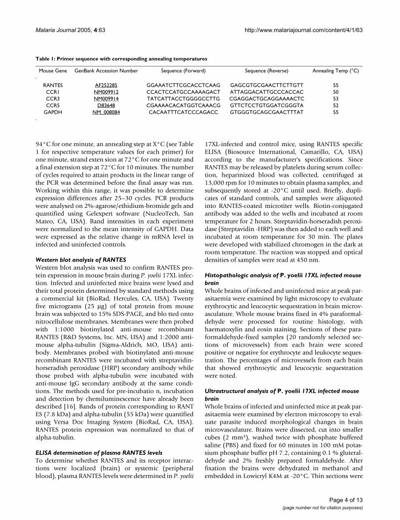
Table 1: Primer sequence with corresponding annealing temperatures
Mouse Gene GenBank Accession Number Sequence (Forward) Sequence (Reverse) Annealing Temp (°C)
RANTES AF252285 GGAAATCTTCGCACCTCAAG GAGCGTGCGAACTTCTTGTT 55CCR1 NM009912 CCACTCCATGCCAAAAGACT ATTAGGACATTGCCCACCAC 50CCR3 NM009914 TATCATTACCTGGGGCCTTG CGAGGACTGCAGGAAAACTC 53CCR5 D83648 CGAAAACACATGGTCAAACG GTTCTCCTGTGGATCGGGTA 52
GAPDH NM_008084 CACAATTTCATCCCAGACC GTGGGTGCAGCGAACTTTAT 55
Page 4 of 13(page number not for citation purposes)
Malaria Journal 2005, 4:63 http://www.malariajournal.com/content/4/1/63
collected on 300-mesh nickel grids and examined bytransmission electron microscopy at 60 eV as describedpreviously [17].
Statistical analysisThe results obtained in this work were from triplicateexperiments performed independently by identical meth-ods. ELISA data and densitometric measurements fromagarose gel electrophoresis, as well as Western blot analy-ses, were log-transformed to normalize the distributionfor infected (n = 15) and control (n = 15) samples, andalso to correct for small sample size. Data were expressedas the mean ± standard error of mean (SEM). Data fromthe P. yoelii 17XL infected and control groups were thencompared, and the p values were determined by usingnonparametric Mann-Whitney U-test. A value of p < 0.05was considered statistically significant.
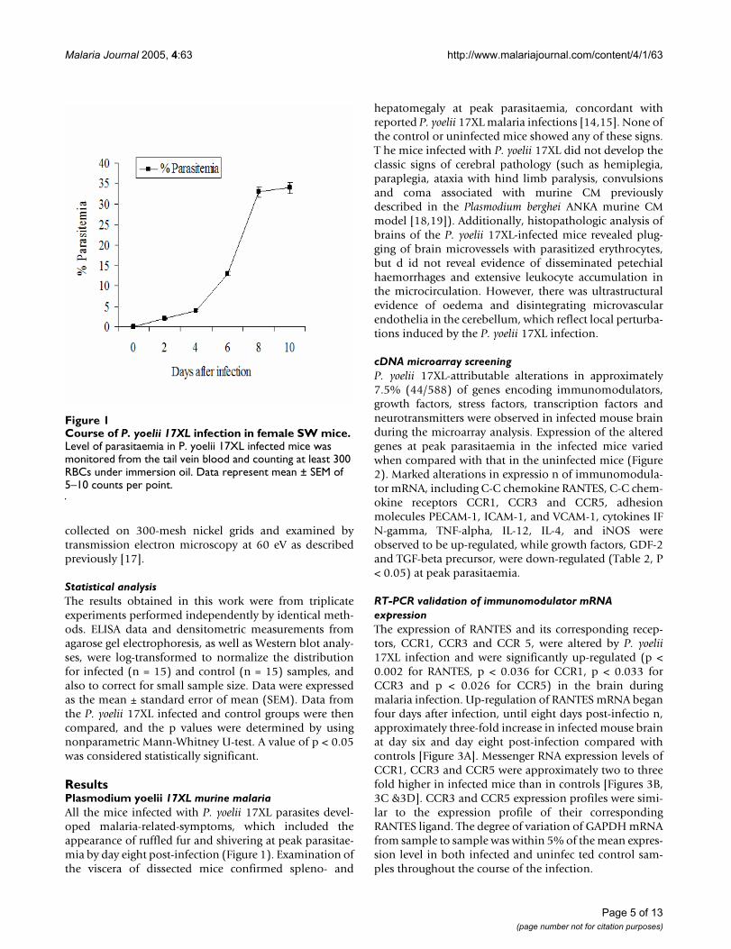
ResultsPlasmodium yoelii 17XL murine malariaAll the mice infected with P. yoelii 17XL parasites devel-oped malaria-related-symptoms, which included theappearance of ruffled fur and shivering at peak parasitae-mia by day eight post-infection (Figure 1). Examination ofthe viscera of dissected mice confirmed spleno- and
hepatomegaly at peak parasitaemia, concordant withreported P. yoelii 17XL malaria infections [14,15]. None ofthe control or uninfected mice showed any of these signs.T he mice infected with P. yoelii 17XL did not develop theclassic signs of cerebral pathology (such as hemiplegia,paraplegia, ataxia with hind limb paralysis, convulsionsand coma associated with murine CM previouslydescribed in the Plasmodium berghei ANKA murine CMmodel [18,19]). Additionally, histopathologic analysis ofbrains of the P. yoelii 17XL-infected mice revealed plug-ging of brain microvessels with parasitized erythrocytes,but d id not reveal evidence of disseminated petechialhaemorrhages and extensive leukocyte accumulation inthe microcirculation. However, there was ultrastructuralevidence of oedema and disintegrating microvascularendothelia in the cerebellum, which reflect local perturba-tions induced by the P. yoelii 17XL infection.
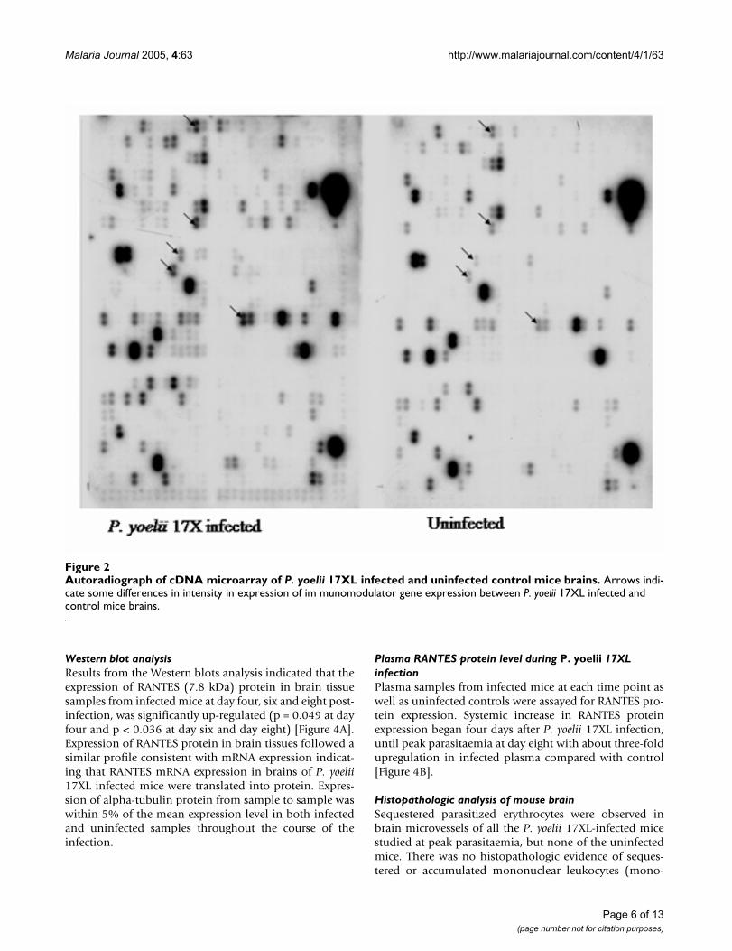
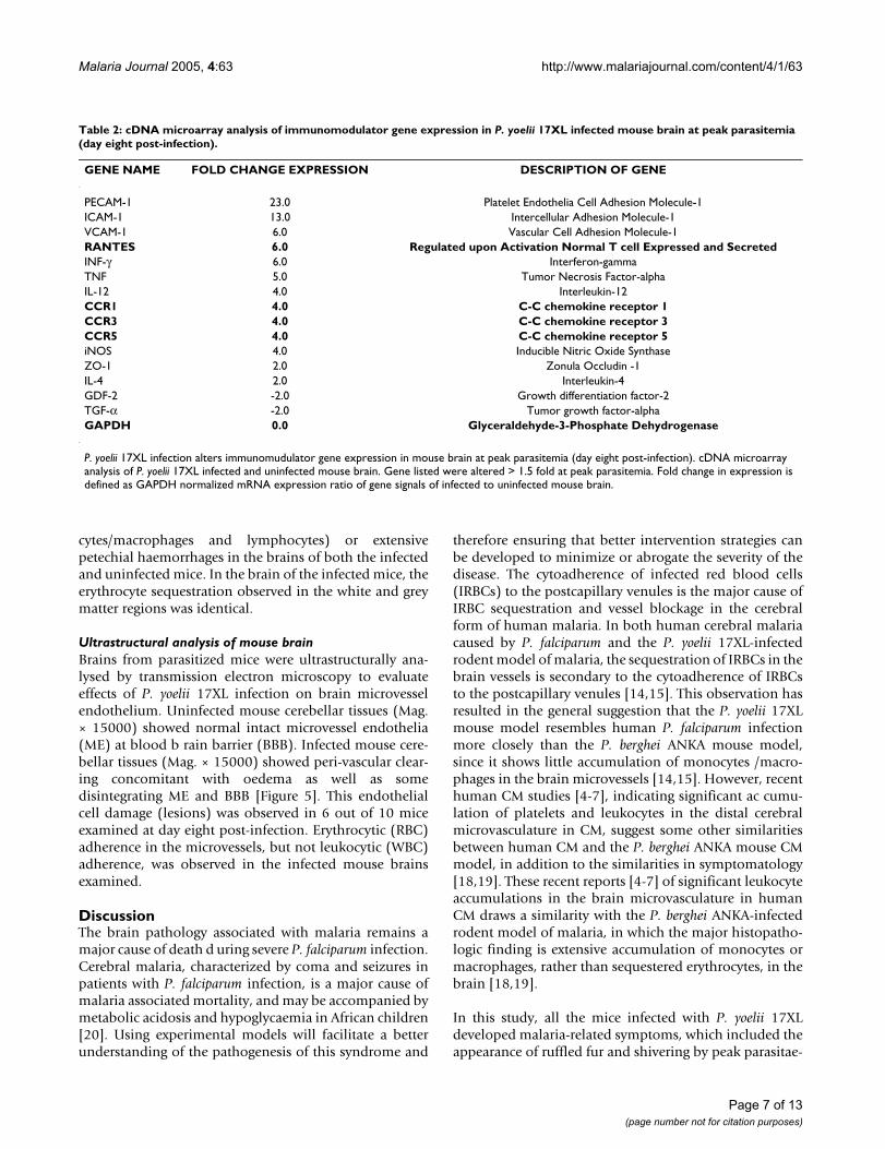
cDNA microarray screeningP. yoelii 17XL-attributable alterations in approximately7.5% (44/588) of genes encoding immunomodulators,growth factors, stress factors, transcription factors andneurotransmitters were observed in infected mouse brainduring the microarray analysis. Expression of the alteredgenes at peak parasitaemia in the infected mice variedwhen compared with that in the uninfected mice (Figure2). Marked alterations in expressio n of immunomodula-tor mRNA, including C-C chemokine RANTES, C-C chem-okine receptors CCR1, CCR3 and CCR5, adhesionmolecules PECAM-1, ICAM-1, and VCAM-1, cytokines IFN-gamma, TNF-alpha, IL-12, IL-4, and iNOS wereobserved to be up-regulated, while growth factors, GDF-2and TGF-beta precursor, were down-regulated (Table 2, P< 0.05) at peak parasitaemia.
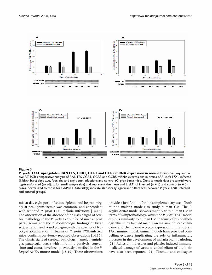
RT-PCR validation of immunomodulator mRNA expressionThe expression of RANTES and its corresponding recep-tors, CCR1, CCR3 and CCR 5, were altered by P. yoelii17XL infection and were significantly up-regulated (p <0.002 for RANTES, p < 0.036 for CCR1, p < 0.033 forCCR3 and p < 0.026 for CCR5) in the brain duringmalaria infection. Up-regulation of RANTES mRNA beganfour days after infection, until eight days post-infectio n,approximately three-fold increase in infected mouse brainat day six and day eight post-infection compared withcontrols [Figure 3A]. Messenger RNA expression levels ofCCR1, CCR3 and CCR5 were approximately two to threefold higher in infected mice than in controls [Figures 3B,3C &3D]. CCR3 and CCR5 expression profiles were simi-lar to the expression profile of their correspondingRANTES ligand. The degree of variation of GAPDH mRNAfrom sample to sample was within 5% of the mean expres-sion level in both infected and uninfec ted control sam-ples throughout the course of the infection.
Course of P. yoelii 17XL infection in female SW miceFigure 1Course of P. yoelii 17XL infection in female SW mice. Level of parasitaemia in P. yoelii 17XL infected mice was monitored from the tail vein blood and counting at least 300 RBCs under immersion oil. Data represent mean ± SEM of 5–10 counts per point.
Page 5 of 13(page number not for citation purposes)
Malaria Journal 2005, 4:63 http://www.malariajournal.com/content/4/1/63
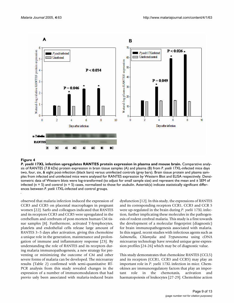
Western blot analysisResults from the Western blots analysis indicated that theexpression of RANTES (7.8 kDa) protein in brain tissuesamples from infected mice at day four, six and eight post-infection, was significantly up-regulated (p = 0.049 at dayfour and p < 0.036 at day six and day eight) [Figure 4A].Expression of RANTES protein in brain tissues followed asimilar profile consistent with mRNA expression indicat-ing that RANTES mRNA expression in brains of P. yoelii17XL infected mice were translated into protein. Expres-sion of alpha-tubulin protein from sample to sample waswithin 5% of the mean expression level in both infectedand uninfected samples throughout the course of theinfection.
Plasma RANTES protein level during P. yoelii 17XL infectionPlasma samples from infected mice at each time point aswell as uninfected controls were assayed for RANTES pro-tein expression. Systemic increase in RANTES proteinexpression began four days after P. yoelii 17XL infection,until peak parasitaemia at day eight with about three-foldupregulation in infected plasma compared with control[Figure 4B].
Histopathologic analysis of mouse brainSequestered parasitized erythrocytes were observed inbrain microvessels of all the P. yoelii 17XL-infected micestudied at peak parasitaemia, but none of the uninfectedmice. There was no histopathologic evidence of seques-tered or accumulated mononuclear leukocytes (mono-
Autoradiograph of cDNA microarray of P. yoelii 17XL infected and uninfected control mice brainsFigure 2Autoradiograph of cDNA microarray of P. yoelii 17XL infected and uninfected control mice brains. Arrows indi-cate some differences in intensity in expression of im munomodulator gene expression between P. yoelii 17XL infected and control mice brains.
Page 6 of 13(page number not for citation purposes)
Malaria Journal 2005, 4:63 http://www.malariajournal.com/content/4/1/63
cytes/macrophages and lymphocytes) or extensivepetechial haemorrhages in the brains of both the infectedand uninfected mice. In the brain of the infected mice, theerythrocyte sequestration observed in the white and greymatter regions was identical.
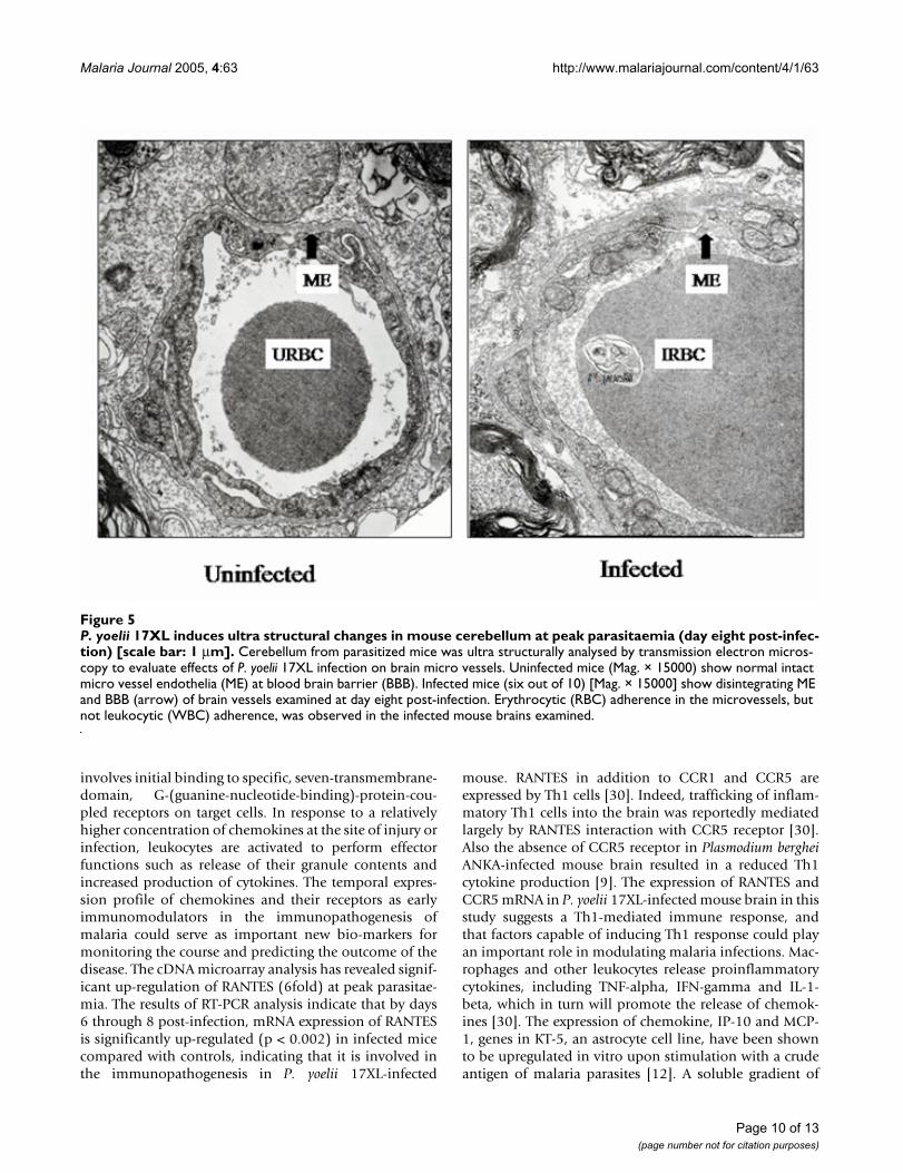
Ultrastructural analysis of mouse brainBrains from parasitized mice were ultrastructurally ana-lysed by transmission electron microscopy to evaluateeffects of P. yoelii 17XL infection on brain microvesselendothelium. Uninfected mouse cerebellar tissues (Mag.× 15000) showed normal intact microvessel endothelia(ME) at blood b rain barrier (BBB). Infected mouse cere-bellar tissues (Mag. × 15000) showed peri-vascular clear-ing concomitant with oedema as well as somedisintegrating ME and BBB [Figure 5]. This endothelialcell damage (lesions) was observed in 6 out of 10 miceexamined at day eight post-infection. Erythrocytic (RBC)adherence in the microvessels, but not leukocytic (WBC)adherence, was observed in the infected mouse brainsexamined.
DiscussionThe brain pathology associated with malaria remains amajor cause of death d uring severe P. falciparum infection.Cerebral malaria, characterized by coma and seizures inpatients with P. falciparum infection, is a major cause ofmalaria associated mortality, and may be accompanied bymetabolic acidosis and hypoglycaemia in African children[20]. Using experimental models will facilitate a betterunderstanding of the pathogenesis of this syndrome and
therefore ensuring that better intervention strategies canbe developed to minimize or abrogate the severity of thedisease. The cytoadherence of infected red blood cells(IRBCs) to the postcapillary venules is the major cause ofIRBC sequestration and vessel blockage in the cerebralform of human malaria. In both human cerebral malariacaused by P. falciparum and the P. yoelii 17XL-infectedrodent model of malaria, the sequestration of IRBCs in thebrain vessels is secondary to the cytoadherence of IRBCsto the postcapillary venules [14,15]. This observation hasresulted in the general suggestion that the P. yoelii 17XLmouse model resembles human P. falciparum infectionmore closely than the P. berghei ANKA mouse model,since it shows little accumulation of monocytes /macro-phages in the brain microvessels [14,15]. However, recenthuman CM studies [4-7], indicating significant ac cumu-lation of platelets and leukocytes in the distal cerebralmicrovasculature in CM, suggest some other similaritiesbetween human CM and the P. berghei ANKA mouse CMmodel, in addition to the similarities in symptomatology[18,19]. These recent reports [4-7] of significant leukocyteaccumulations in the brain microvasculature in humanCM draws a similarity with the P. berghei ANKA-infectedrodent model of malaria, in which the major histopatho-logic finding is extensive accumulation of monocytes ormacrophages, rather than sequestered erythrocytes, in thebrain [18,19].
In this study, all the mice infected with P. yoelii 17XLdeveloped malaria-related symptoms, which included theappearance of ruffled fur and shivering by peak parasitae-
Table 2: cDNA microarray analysis of immunomodulator gene expression in P. yoelii 17XL infected mouse brain at peak parasitemia (day eight post-infection).
GENE NAME FOLD CHANGE EXPRESSION DESCRIPTION OF GENE
PECAM-1 23.0 Platelet Endothelia Cell Adhesion Molecule-1ICAM-1 13.0 Intercellular Adhesion Molecule-1VCAM-1 6.0 Vascular Cell Adhesion Molecule-1RANTES 6.0 Regulated upon Activation Normal T cell Expressed and SecretedINF-γ 6.0 Interferon-gammaTNF 5.0 Tumor Necrosis Factor-alphaIL-12 4.0 Interleukin-12CCR1 4.0 C-C chemokine receptor 1CCR3 4.0 C-C chemokine receptor 3CCR5 4.0 C-C chemokine receptor 5iNOS 4.0 Inducible Nitric Oxide SynthaseZO-1 2.0 Zonula Occludin -1IL-4 2.0 Interleukin-4GDF-2 -2.0 Growth differentiation factor-2TGF-α -2.0 Tumor growth factor-alphaGAPDH 0.0 Glyceraldehyde-3-Phosphate Dehydrogenase
P. yoelii 17XL infection alters immunomudulator gene expression in mouse brain at peak parasitemia (day eight post-infection). cDNA microarray analysis of P. yoelii 17XL infected and uninfected mouse brain. Gene listed were altered > 1.5 fold at peak parasitemia. Fold change in expression is defined as GAPDH normalized mRNA expression ratio of gene signals of infected to uninfected mouse brain.
Page 7 of 13(page number not for citation purposes)
Malaria Journal 2005, 4:63 http://www.malariajournal.com/content/4/1/63
mia at day eight post-infection. Spleno- and hepato-meg-aly at peak parasitaemia was common, and concordantwith reported P. yoelii 17XL malaria infections [14,15].The observation of the absence of the classic signs of cere-bral pathology in the P. yoelii 17XL-infected mice at peakparasitaemia and the histopathologic findings of IRBCsequestration and vessel plugging with the absence of leu-cocyte accumulation in brains of P. yoelii 17XL-infectedmice, confirms previously reported observations [14,15].The classic signs of cerebral pathology, namely hemiple-gia, paraplegia, ataxia with hind-limb paralysis, convul-sions and coma, have been previously described in the P.berghei ANKA mouse model [18,19]. These observations
provide a justification for the complementary use of bothmurine malaria models to study human CM. The P.berghei ANKA model shows similarity with human CM interms of symptomatology, whilst the P. yoelii 17XL modelexhibits similarity to human CM in terms of histopathol-ogy. This study focused mainly on malaria induced chem-okine and chemokine receptor expression in the P. yoelii17XL murine model. Animal models have provided com-pelling evidence implicating the role of inflammatoryprocesses in the development of malaria brain pathology[21]. Adhesion molecules and platelet-induced immune-mediated damage of vascular endothelium of the brainhave also been reported [21]. Tkachuk and colleagues
P. yoelii 17XL upregulates RANTES, CCR1, CCR3 and CCR5 mRNA expression in mouse brainFigure 3P. yoelii 17XL upregulates RANTES, CCR1, CCR3 and CCR5 mRNA expression in mouse brain. Semi-quantita-tive RT-PCR comparative analysis of RANTES CCR1, CCR3 and CCR5 mRNA expressions in brains of P. yoelii 17XL-infected (I, black bars) days two, four, six, and eight post-infections and control (C, grey bars) mice. Densitometric data presented were log-transformed (to adjust for small sample size) and represent the mean and ± SEM of infected (n = 5) and control (n = 5) cases, normalized to those for GAPDH. Asterisk(s) indicate statistically significant differences between P. yoelii 17XL infected and control groups.
Page 8 of 13(page number not for citation purposes)
Malaria Journal 2005, 4:63 http://www.malariajournal.com/content/4/1/63
observed that malaria infection induced the expression ofCCR3 and CCR5 on placental macrophages in pregnantwomen [22]. Sarfo and colleagues indicated that RANTESand its receptors CCR3 and CCR5 were upregulated in thecerebellum and cerebrum of post-mortem human CM tis-sue samples [8]. Furthermore, activated T-lymphocytes,platelets and endothelial cells release large amount ofRANTES 3–5 days after activation, giving this chemokinea unique role in the generation, maintenance and prolon-gation of immune and inflammatory response [23]. Byunderstanding the role of RANTES and its receptors dur-ing malaria immunopathogenesis, a new strategy for pre-venting or minimizing the outcome of CM and othersevere forms of malaria can be developed. The microarrayresults (Table 2) confirmed with semi-quantitative RT-PCR analysis from this study revealed changes in theexpression of a number of immunomodulators that hadprevio usly been associated with malaria-induced brain
dysfunction [12]. In this study, the expressions of RANTESand its corresponding receptors CCR1, CCR3 and CCR 5were up-regulated in the brain during P. yoelii 17XL infec-tion, further implicating these molecules in the pathogen-esis of rodent cerebral malaria. This study is a first towardsthe development of a molecular fingerprint (diagnostic)for brain immunopathogenesis associated with malaria.In this regard, recent studies with infectious agents such asSalmonella, Chlamydia and Trypanosoma using cDNAmicroarray technology have revealed unique gene-expres-sion profiles [24-26] which may be of diagnostic value.
This study demonstrates that chemokine RANTES (CCL5)and its receptors (CCR1, CCR3 and CCR5) may play animportant role in P. yoelii 17XL infection in mice. Chem-okines are immunoregulatory factors that play an impor-tant role in the chemotaxis, activation andhaematopoiesis of leukocytes [27-29]. Chemokine action
P. yoelii 17XL infection upregulates RANTES protein expression in plasma and mouse brainFigure 4P. yoelii 17XL infection upregulates RANTES protein expression in plasma and mouse brain. Comparative analy-sis of RANTES (7.8 kDa) protein expression in brain tissue samples (A) and plasma (B) from P. yoelii 17XL-infected mice days two, four, six, & eight post-infection (black bars) versus uninfected controls (grey bars). Brain tissue protein and plasma sam-ples from infected and uninfected mice were analysed for RANTES expression by Western Blot and ELISA respectively. Densi-tometric data of Western blots were log-transformed (to adjust for small sample size) and represent the mean and ± SEM of infected (n = 5) and control (n = 5) cases, normalized to those for atubulin. Asterisk(s) indicate statistically significant differ-ences between P. yoelii 17XL-infected and control groups.
Page 9 of 13(page number not for citation purposes)
Malaria Journal 2005, 4:63 http://www.malariajournal.com/content/4/1/63
involves initial binding to specific, seven-transmembrane-domain, G-(guanine-nucleotide-binding)-protein-cou-pled receptors on target cells. In response to a relativelyhigher concentration of chemokines at the site of injury orinfection, leukocytes are activated to perform effectorfunctions such as release of their granule contents andincreased production of cytokines. The temporal expres-sion profile of chemokines and their receptors as earlyimmunomodulators in the immunopathogenesis ofmalaria could serve as important new bio-markers formonitoring the course and predicting the outcome of thedisease. The cDNA microarray analysis has revealed signif-icant up-regulation of RANTES (6fold) at peak parasitae-mia. The results of RT-PCR analysis indicate that by days6 through 8 post-infection, mRNA expression of RANTESis significantly up-regulated (p < 0.002) in infected micecompared with controls, indicating that it is involved inthe immunopathogenesis in P. yoelii 17XL-infected
mouse. RANTES in addition to CCR1 and CCR5 areexpressed by Th1 cells [30]. Indeed, trafficking of inflam-matory Th1 cells into the brain was reportedly mediatedlargely by RANTES interaction with CCR5 receptor [30].Also the absence of CCR5 receptor in Plasmodium bergheiANKA-infected mouse brain resulted in a reduced Th1cytokine production [9]. The expression of RANTES andCCR5 mRNA in P. yoelii 17XL-infected mouse brain in thisstudy suggests a Th1-mediated immune response, andthat factors capable of inducing Th1 response could playan important role in modulating malaria infections. Mac-rophages and other leukocytes release proinflammatorycytokines, including TNF-alpha, IFN-gamma and IL-1-beta, which in turn will promote the release of chemok-ines [30]. The expression of chemokine, IP-10 and MCP-1, genes in KT-5, an astrocyte cell line, have been shownto be upregulated in vitro upon stimulation with a crudeantigen of malaria parasites [12]. A soluble gradient of
P. yoelii 17XL induces ultra structural changes in mouse cerebellum at peak parasitaemia (day eight post-infection) [scale bar: 1 µm]Figure 5P. yoelii 17XL induces ultra structural changes in mouse cerebellum at peak parasitaemia (day eight post-infec-tion) [scale bar: 1 µm]. Cerebellum from parasitized mice was ultra structurally analysed by transmission electron micros-copy to evaluate effects of P. yoelii 17XL infection on brain micro vessels. Uninfected mice (Mag. × 15000) show normal intact micro vessel endothelia (ME) at blood brain barrier (BBB). Infected mice (six out of 10) [Mag. × 15000] show disintegrating ME and BBB (arrow) of brain vessels examined at day eight post-infection. Erythrocytic (RBC) adherence in the microvessels, but not leukocytic (WBC) adherence, was observed in the infected mouse brains examined.
Page 10 of 13(page number not for citation purposes)
Malaria Journal 2005, 4:63 http://www.malariajournal.com/content/4/1/63
these chemokines within the tissue recruits various celltypes that express receptors for the different chemokines.The expressions of all the C-C chemokine receptors forRANTES, CCR1, CCR3 and CCR5, were upregulated in thebrains of P. yoelii 17XL-infected mice. The expression ofRANTES probably enhanced the expression of its recep-tors. Sano and colleagues demonstrated that ICAM-1induced RANTES mRNA expression and also increased itsprotein synthesis and secretion by endothelial cells [31].It is likely that the P. yoelii 17XL-induced RANTES produc-tion observed in the current study would attract and acti-vate leukocytes towards inflammatory sites to mediatelocalized hyper-inflammatory responses that could exac-erbate the disease pathology in the cerebellum. Belnoueand colleagues showed that the brains of wild -type micewith CM have significantly higher levels of CCR5 than theknockout-type, implicating these molecules in the patho-logical changes produced in the brain during the infection[9]. The results of this study demonstrate that the increasein production of RANTES follows the course of P. yoelii17XL malaria infection, thus RANTES and its receptorsCCR1, CCR3 and CCR5 were detected at their highest lev-els at day six and day eight post-infection. This observedtemporal association of the progression of P. yoelii 17XLinfection with the increasing production of RANTES andits receptors suggests that the two events might be linked.Western blot analysis revealed that brain tissue transcriptsof RANTES were actually translated into protein, and weresignificantly up-regulated (p = 0.046 for day 4 and p <0.034 for day six and day eight post-infection) in infectedmice (Figure 3A). The ELISA data from this study indicatesignificant upregulation (p = 0.049 for day 4 and p <0.026 for day six and day eight post-infection) of RANTESin P. yoelii 17XL-infected mouse plasma than in controls(Figure 3B). Most of the ultrastructural pathologicalchanges, observed as endothelial cell damage (lesions) insix out of the 10 infected mice examined, occurred espe-cially at day eight post-infection (peak parasitaemia) [Fig-ure 5]. Thus, the increase in RANTES productioncorrelated with the increase in parasitaemia and patholog-ical changes observed in the P. yoelii 17XL-infected mice inthis investigation. Chemokines have been shown to havea direct antiprotozoal activity for three protozoans: Toxo-plasma gondii, Leishmania donovani and Trypanosoma cruzi[32,33]. Chemokine production is important for defend-ing the host against infection. However, excessive produc-tion is also deleterious to the host [12]. It has beenobserved that C-C chemokines, such as MIP-1-alpha andRANTES, are significantly upregulated in brains ofTrypanosoma brucei brucei infected rats [34]. This increasein expression of these chemokines occurs before brainlesions developed in infected rats, implying that theinduction of these chemokines could be directly responsi-ble for the observed rat brain lesions [34]. Ultrastructuralanalysis of mouse brain by electron microscopy at peak
parasitaemia in this study, revealed disintegrating microv-ascular endothelial layer at the blood brain barrier in thecerebellar region of infected mouse brain. This endothelial cell damage (lesions), in six out of 10 mice examined,occurred especially at day eight post-infection (peak para-sitaemia) and was similar to the observations of Thum-wood and colleagues in P. berghei ANKA-infected A/J andCBA/H mice [35]. The infected erythrocytes adhering andoccluding brain microvessels observed in the sectionsexamined, suggest that the breach in the cerebellar micro-vascular endothelial layer could be associated with para-site-induced inflammation or apoptosis. Perivascularoedema was also observed in this region of infectedmouse brain probably as a result of the endothelial celldamage allowing excess fluid to move across the bloodbrain barrier. End othelial cells interacting with P. yoelii17XL-parasitized erythrocytes have been shown to beinduced to produce and present specific chemokines, suchas RANTES, which can lure CCR1, CCR3 and CCR5expressing cells into the brain [36]. CCR1 and CCR 5 areexp ressed by brain endothelial cells [37,38]. Brainendothelial cells, microglia and astrocytes, which are the3 major cellular components of the BBB, express CCR5receptor [39], and hence the binding of RANTES to itsreceptors on these cells can serve to further activate themand enhance a localized breakdown of the microvesselendothelial layer observed in the infected mouse brain inthe current investigation.
ConclusionIn conclusion, P. yoelii 17XL infection upregulatesRANTES and its corresponding receptors, CCR1, CCR3and CCR5, in mouse brain, and that ultrastructuralchanges in the microvascular endothelial layer occurred inthe cerebellum of infected mice. This is the first temporalexpression study of RANTES and its receptors associatedwith murine malaria. Further studies are underway toexamine the expression of these chemokines and chemok-ine receptors in a human CM symptomatology-like modelsuch as P. berghei ANKA, to ascertain differences and sim-ilarities. As it is not clear which cell types in the mouse CMbrain samples over-express RANTES, CCR1, CCR3 andCCR5, further comparative immunolocalization and anti-body ablation studies are currently underway to examinethe physiological relevance, source and expression pat-terns of these important biomarkers in both murine andhuman CM brain samples.
List of abbreviations usedBBB, Blood-Brain Barrier;
CM, cerebral malaria;
ECM, experimental cerebral malaria;
Page 11 of 13(page number not for citation purposes)
Malaria Journal 2005, 4:63 http://www.malariajournal.com/content/4/1/63
ME, microvascular endothelium;
NCM, non-cerebral malaria;
P., Plasmodium;
RBC, red blood cell;
WBC, white blood cell;
TNF, Tumor Necrosis Factor;
IFN, Interferon; IL, Interleukin
Authors' contributionsBYS, HA, II and AAA carried out the animal manipula-tions, molecular genetic studies, Western blots, immu-noassays, electron microscopy, data analysis, andparticipated in the sequence alignments and participatedin drafting of the manuscript. CSO provided characterizedparasite strains (P. yoelii 17XL) and murine malariamodel. SS and JWL participated in the design of the study,provided chemokine antibodies and oligonucleotideprimers for Western blots and RT-PCR, respectively. JKSconceived of the study, participated in its design and coor-dination and helped to draft the manuscript. All authorsread and approved the final manuscript.
AcknowledgementsWe acknowledge the assistance of Dr. Alexander Quarshie (Morehouse School of Medicine, Atlanta, GA) in the statistical analysis. The authors thank Mr. Larry Brako (Morehouse School of Medicine, Atlanta, GA) for his technical assistance with electron microscopy. Drs. Michael Wilson and Daniel Boakye (Noguchi Memorial Institute for Medical Research, Accra, Ghana) for their useful comments on the manuscript. This investigation was conducted in a facility constructed with support from Research Facilities Improvement Program Grant #1 C06 RR18386 from the National Center for Research Resources, National Institutes of Health. This work was sup-ported by grants from WHO/World Bank/UNDP (A00524), NIH-NIGMS-MBRS (SO6GM08248), NIH-RCMI (RR03034), and NIH -FIC (R21 TW006804-02).
References1. Tropical Disease Research (TDR): Malaria in: Tropical Research.
In 7th Program Report UNDP/World Bank/WHO. Geneva; 1998:3-40. 2. Greenwood B: Malaria mortality and morbidity in Africa. Bull
WHO 1999, 77:617-618.3. World Health Organization (WHO): Severe Falciparum malaria.
Trans R Soc Trop Med & Hyg 2000, 94(Suppl):1-90.4. Grau GE, Mackenzie CD, Carr RA, Redard M, Pizzolato G, Allasia C,
Cataldo C, Taylor TE, Molyneux ME: Platelet accumulation inbrain microvessels in fatal paediatric cerebral malaria. J InfectDis 2003, 187:461-466.
5. Wassmer SC, Combes V, Grau GE: Pathophysiology of cerebralmalaria: role of host cells in the modulation of cytoadhesion.Ann NY Acad Sci 2003, 992:30-38.
6. Wassmer SC, Coltel N, Combes V, Grau GE: Pathogenesis of cer-ebral malaria: facts and hypotheses. Med Trop (Mars) 2003,63:254-257.
7. Wassmer SC, Lepolard C, Traore B, Pouvelle B, Gysin J, Grau GE:Platelets reorient Plasmodium falciparum-infected erythro-
cyte cytoadhesion activate d endothelial cells. J Infect Dis 2004,189:180-189.
8. Sarfo BY, Singh S, Lillard JW, Quarshie A, Gyasi RK, Armah H, AdjeiAA, Jolly P, Stiles JK: The cerebral-malaria-associated expres-sion of RANTES, CCR3 and CCR5 in post-mortem tissuesamples. Ann Trop Med Parasitol 2004, 98:297-303.
9. Belnoue E, Kayabanda M, Deschemin JC: CCR5 deficiencydecreases susceptibility to experimental cerebral malaria.Blood 2003, 101:4253-4259.
10. Nitcheu J, Bonduelle O, Combadiere C, Tefit M, Seilhean D, MazierD, Comb adiere B: Perforin-dependent brain-infiltrating cyto-toxic CD8+ T lymphocytes mediate experimental cerebralmalaria pathogenesis. J Immunol 2003, 170:2221-2228.
11. Baggiolini M, Dahinden CA: C-C chemokines in allergic inflam-mation. Immunol Today 1994, 15:127-133.
12. Hanum PS, Hayano M, Kojima S: Cytokine and chemokineresponses in a cerebral malaria-susceptible or -resistantstrain of mice to. Plasmodium berghei 2003, 15:633-640.
13. Belnoue E, Costa FT, Vigario AM, Voza T, Gonnet F, Landau I, VanRooijen N, Mack M, Kuziel WA, Renia L: Chemokine receptorCCR2 is not essential for the development of experimentalcerebral malaria. Infect Immun 2003, 71:3648-3651.
14. Kaul DK, Nagel RL, Llena JF, Shear HL: Cerebral malaria in mice:demonstration of cytoadherence of infected red blood cellsand microrheologic correlates. Am J Trop Med Hyg 1994,50:512-521.
15. Shear HL, Marino MW, Wanidworanun C, Berman JW, Nagel RL:Correlation of increased expression of intercellular adhesionmolecule -1, but not high levels of tumor necrosis factor-alpha, with lethality of Plasmodium yoelii 17XL, a rodentmodel of cerebral malaria. Am J Trop Med Hyg 1998, 59:852-858.
16. Thompson WE, Powell JM, Whittaker JA, Sridaran R, Thomas KH:Immunolocalization and expression of prohibitin, a mito-chondrial associated protein within the rat ovaries. Anat Rec1999, 256:40-48.
17. Stiles JK, Molyneux DH, Wallbanks KR, Van der Vloedt AM: Effectsof gamma irradiation on the midgut ultrastructure of Gloss-ina palpalis subspecies. Radiat Res 1989, 118:353-363.
18. Grau GE, Piguet J-F, Engers HD, Louis JA, Vassalli P, Lambert P-H:L3T4+ T Lymphocytes play a major role in the pathogenesisof murine cerebral malaria. J Immunol 1987, 237:1210-1212.
19. Neill AL, Hunt NH: Pathology of fatal and resolving Plasmodiumberghei cerebral malaria in mice. Parasitology 1992, 105:165-175.
20. Miller LH, Baruch DI, Marsh K, Doumbo OK: The pathogenic basisof malaria. Nature 2002, 415:673-679.
21. Brian SJ, de Souza JB, Riley EM: Cerebral malaria: the contribu-tion of studies in animal models to our understanding ofimmunopathogenesis. Microbes Infect 2002, 4:291-300.
22. Tkachuk AN, Moormann AM, Poore JD, Rochford RA, Chensue SW,Mwapasa V, Meshnick SR: Malaria enhances expression of CCchemokine receptor 5 on placental macrophages. J Infect Dis2001, 183:967-972.
23. Kameyoshi Y, Dorschner A, Mallet AI, Christopher E, Schroeder JM:Cytokine RANTES released by thrombin-stimulated plate-lets is a potent attractant for human eosinophils. J Exp Med1992, 176:587-592.
24. Dessus-Babus S, Knight ST, Wyrick PB: Chlamydial infection ofpolarized HeLa cells induces PMN chemotaxis, but thecytokine profile varies between disseminating and non-dis-semina ting strains. Cell Microbiol 2000, 2:317-327.
25. Rosenberger C M, Scott MG, Gold MR, Hancock RE, Finlay BB: Sal-monella typhimurium infection and lipopolysaccharide stimu-lation induce similar changes in macrophage geneexpression. J Immunol 2000, 164:5894-5904.
26. Stiles JK, Meade JC, Kucerova ZP, Thompson W, Zakeri Z, WhittakerJ: Trypanosoma brucei infection induces apoptosis and up-regulates neuroleukin expression in the cerebellum. Ann TropMed Parasitol 2001, 95:797-810.
27. Keane MP, Arenberg DA, Moore BB, Addison CL, Strieter RM: CXCchemokines and angiogenesis/angiostasis. Proc Assoc Am Physi1998, 110:288-296.
28. Lillard JW, Boyaka PN, Singh S, McGhee JR: Salmonella-mediatedmucosal cell-mediated immunity. Cell Mol Biol 2001,47:1115-1120.
Page 12 of 13(page number not for citation purposes)
http://www.ncbi.nlm.nih.gov/entrez/query.fcgi?cmd=Retrieve&db=PubMed&dopt=Abstract&list_uids=8172645
http://www.ncbi.nlm.nih.gov/entrez/query.fcgi?cmd=Retrieve&db=PubMed&dopt=Abstract&list_uids=8172645
http://www.ncbi.nlm.nih.gov/entrez/query.fcgi?cmd=Retrieve&db=PubMed&dopt=Abstract&list_uids=8166359
http://www.ncbi.nlm.nih.gov/entrez/query.fcgi?cmd=Retrieve&db=PubMed&dopt=Abstract&list_uids=8166359
http://www.ncbi.nlm.nih.gov/entrez/query.fcgi?cmd=Retrieve&db=PubMed&dopt=Abstract&list_uids=8166359
http://www.ncbi.nlm.nih.gov/entrez/query.fcgi?cmd=Retrieve&db=PubMed&dopt=Abstract&list_uids=9886187
http://www.ncbi.nlm.nih.gov/entrez/query.fcgi?cmd=Retrieve&db=PubMed&dopt=Abstract&list_uids=9886187
http://www.ncbi.nlm.nih.gov/entrez/query.fcgi?cmd=Retrieve&db=PubMed&dopt=Abstract&list_uids=2727263
http://www.ncbi.nlm.nih.gov/entrez/query.fcgi?cmd=Retrieve&db=PubMed&dopt=Abstract&list_uids=2727263
http://www.ncbi.nlm.nih.gov/entrez/query.fcgi?cmd=Retrieve&db=PubMed&dopt=Abstract&list_uids=2727263
http://www.ncbi.nlm.nih.gov/entrez/query.fcgi?cmd=Retrieve&db=PubMed&dopt=Abstract&list_uids=1280805
http://www.ncbi.nlm.nih.gov/entrez/query.fcgi?cmd=Retrieve&db=PubMed&dopt=Abstract&list_uids=1380064
http://www.ncbi.nlm.nih.gov/entrez/query.fcgi?cmd=Retrieve&db=PubMed&dopt=Abstract&list_uids=1380064
Malaria Journal 2005, 4:63 http://www.malariajournal.com/content/4/1/63
Publish with BioMed Central and every scientist can read your work free of charge
"BioMed Central will be the most significant development for disseminating the results of biomedical research in our lifetime."
Sir Paul Nurse, Cancer Research UK
Your research papers will be:
available free of charge to the entire biomedical community
peer reviewed and published immediately upon acceptance
cited in PubMed and archived on PubMed Central
yours — you keep the copyright
Submit your manuscript here:http://www.biomedcentral.com/info/publishing_adv.asp
BioMedcentral
29. Lillard JW, Singh UP, Boyaka PN, Singh S, Taub DD, McGhee JR:MIP1alpha and MIP-1beta differentially mediate mucosaland systemic adaptive immunity. Blood 2003, 101:807-814.
30. Zang YC, Halder JB, Samanta AK, Hong J, Rivera VM, Zhang JZ: Reg-ulation of chemokine receptor CCR5 and production ofRANTES and MIP-1alpha by interferon-beta. J Neuroimmunol2001, 112:174-180.
31. Sano H, Nakagawa N, Chiba R, Kurasawa K, Saito Y, Iwamoto I:Cross-linking of intercellular adhesion molecule -1 inducesinterleukin-8 and RANTES production through the activa-tion of MAP kinases in human vascular endothelial cells. Bio-chem Biophys Res Commun 1998, 29:694-698.
32. Mannheimer SB, Hariprashad J, Stoeckle MY, Murray HW: Inductionof macrophage antiprotozoal activity by monocyte chemo-tactic and activating factor. Immunol Med Microbial 1996,14:59-61.
33. Villalta F, Zhang Y, Bibb KE, Kappes JC, Lima MF: The cysteine-cysteine family of chemokines RANTES, MIP-1α and MIP-1βinduce trypanocidalactivity in human macrophages via nitricoxide. Infect Immun 1998, 66:4690-4695.
34. Sharafeldin A, Eltayeb R, Pashenkov M, Bakhiet M: Chemokines areproduced in the brain early during the course of experimen-tal African trypanosomiasis. J Neuroimmunol 2000, 103:167-170.
35. Thumwood CM, Hunt NH, Clark IA, Cowden WB: Breakdown ofthe blood-brain barrier in murine cerebral malaria. Parasitol-ogy 1988, 96:579-589.
36. Pober JS, Cotran R S: Immunologic interactions of T lym-phocytes with vascular endothelium. Adv Immunol 1991,50:261-302.
37. Dzenko KA, Andjelkovic AV, Kuziel WA, Packter JS: The chemok-ine receptor CCR2 mediates internalization of monocytechemoattractant protein-1 along brain mic rovessels. J Neu-rosci 2001, 21:9214-9223.
38. Andjelkovic AV, Pachter JS: Characterization of binding sites forchemokines, MCP-1 and MIP-1a, on human brain microves-sels. J Neurochem 2000, 75:1898-1906.
39. Berger O, Gan X, Gujuluva C, Burns AR, Sulur G, Stins M, Way D,Witte M, Weinand M, Said J, Kim KS, Taub D, Graves MC, Fiala M:CXC and CC chemokine receptors on coronary and brainendothelia. Mol Med 1999, 5:795-805.
Page 13 of 13(page number not for citation purposes)
http://www.ncbi.nlm.nih.gov/entrez/query.fcgi?cmd=Retrieve&db=PubMed&dopt=Abstract&list_uids=9746565
http://www.ncbi.nlm.nih.gov/entrez/query.fcgi?cmd=Retrieve&db=PubMed&dopt=Abstract&list_uids=9746565
http://www.ncbi.nlm.nih.gov/entrez/query.fcgi?cmd=Retrieve&db=PubMed&dopt=Abstract&list_uids=9746565
http://www.ncbi.nlm.nih.gov/entrez/query.fcgi?cmd=Retrieve&db=PubMed&dopt=Abstract&list_uids=2457201
http://www.ncbi.nlm.nih.gov/entrez/query.fcgi?cmd=Retrieve&db=PubMed&dopt=Abstract&list_uids=2457201
http://www.ncbi.nlm.nih.gov/entrez/query.fcgi?cmd=Retrieve&db=PubMed&dopt=Abstract&list_uids=1950797
Related Documents