Vaccines 2021, 9, 1072. https://doi.org/10.3390/vaccines9101072 www.mdpi.com/journal/vaccines Review Plasmodium falciparum Malaria Vaccines and Vaccine Adjuvants Srinivasa Reddy Bonam 1, * ,† , Laurent Rénia 2,3,4, * ,† , Ganesh Tadepalli 5 , Jagadeesh Bayry 1,6 and Halmuthur Mahabalarao Sampath Kumar 5, * 1 Institut National de la Santé et de la Recherche Médicale, Centre de Recherche des Cordeliers, Equipe‐Immunopathologie et Immunointervention Thérapeutique, Sorbonne Université, Université de Paris, F‐75006 Paris, France; [email protected] 2 A*STAR Infectious Diseases Labs, 8A Biomedical Grove, Singapore 138648, Singapore 3 Lee Kong Chian School of Medicine, Nanyang Technological University, Singapore 308232, Singapore 4 School of Biological Sciences, Nanyang Technological University, Singapore 308232, Singapore 5 Vaccine Immunology Laboratory, Organic Synthesis and Process Chemistry Division, CSIR‐Indian Institute of Chemical Technology, Hyderabad 500007, India; [email protected] 6 Biological Sciences & Engineering, Indian Institute of Technology Palakkad, Palakkad 678623, India * Correspondence: [email protected] (S.R.B.); [email protected]‐star.edu.sg (L.R.); [email protected] (H.M.S.K.) † Equally contributed. Abstract: Malaria—a parasite vector‐borne disease—is a global health problem, and Plasmodium falciparum has proven to be the deadliest among Plasmodium spp., which causes malaria in humans. Symptoms of the disease range from mild fever and shivering to hemolytic anemia and neurological dysfunctions. The spread of drug resistance and the absence of effective vaccines has made malaria disease an ever‐emerging problem. Although progress has been made in understanding the host response to the parasite, various aspects of its biology in its mammalian host are still unclear. In this context, there is a pressing demand for the development of effective preventive and therapeutic strategies, including new drugs and novel adjuvanted vaccines that elicit protective immunity. The present article provides an overview of the current knowledge of anti‐malarial immunity against P. falciparum and different options of vaccine candidates in development. A special emphasis has been made on the mechanism of action of clinically used vaccine adjuvants. Keywords: anti‐malarial drugs; malaria vaccine; Plasmodium falciparum; vaccine adjuvants 1. Introduction Malaria, caused by apicomplexan Plasmodium spp., remains one of the world’s most threatening diseases of humans and other animals with high morbidity and mortality rates [1]. The recent findings of the World Health Organization (WHO) recorded 228 million cases and 405,000 deaths globally in 2018 [2]. Although eight species of Plasmodium can infect humans, most malarial cases are due to P. falciparum or P. vivax, but deaths are mostly due to falciparum malaria. The African countries carry the highest share of the global malaria burden (~90% as per 2017 WHO reports) [3]. In Asian countries, e.g., India, which accounts for 4% of the global burden, malaria is still a serious health threat [4]. As an answer, the Special Programme for Research and Training in Tropical Diseases (TDR; co‐sponsored by the WHO), US National Institutes of Health, UK department for international development, Bill & Melinda Gates Foundation, and other organizations have increased funding for research and development and other control measures (including vaccination) to minimize the malaria cases [2,5]. WHO has also put more effort into implementing essential malaria commodities, such as rapid diagnostic tests, insecticide‐treated mosquito nets, vector control, artemisinin‐based combination therapy, Citation: Bonam, S.R.; Rénia, L.; Tadepalli, G.; Bayry, J.; Kumar, H.M.S. Plasmodium falciparum Malaria Vaccines and Vaccine Adjuvants. Vaccines 2021, 9, 1072. https://doi.org/10.3390/ vaccines9101072 Academic Editors: Giampiero Girolomoni, Srinivasa Reddy Bonam and Jagadeesh Bayry Received: 14 August 2021 Accepted: 22 September 2021 Published: 24 September 2021 Publisher’s Note: MDPI stays neutral with regard to jurisdictional claims in published maps and institutional affiliations. Copyright: © 2021 by the authors. Licensee MDPI, Basel, Switzerland. This article is an open access article distributed under the terms and conditions of the Creative Commons Attribution (CC BY) license (https://creativecommons.org/license s/by/4.0/).

Welcome message from author
This document is posted to help you gain knowledge. Please leave a comment to let me know what you think about it! Share it to your friends and learn new things together.
Transcript
Vaccines 2021, 9, 1072. https://doi.org/10.3390/vaccines9101072 www.mdpi.com/journal/vaccines
Review
Plasmodium falciparum Malaria Vaccines and
Vaccine Adjuvants
Srinivasa Reddy Bonam 1,*,†, Laurent Rénia 2,3,4,*,†, Ganesh Tadepalli 5, Jagadeesh Bayry 1,6
and Halmuthur Mahabalarao Sampath Kumar 5,*
1 Institut National de la Santé et de la Recherche Médicale, Centre de Recherche des Cordeliers,
Equipe‐Immunopathologie et Immunointervention Thérapeutique, Sorbonne Université, Université de
Paris, F‐75006 Paris, France; [email protected] 2 A*STAR Infectious Diseases Labs, 8A Biomedical Grove, Singapore 138648, Singapore 3 Lee Kong Chian School of Medicine, Nanyang Technological University, Singapore 308232, Singapore 4 School of Biological Sciences, Nanyang Technological University, Singapore 308232, Singapore 5 Vaccine Immunology Laboratory, Organic Synthesis and Process Chemistry Division, CSIR‐Indian Institute
of Chemical Technology, Hyderabad 500007, India; [email protected] 6 Biological Sciences & Engineering, Indian Institute of Technology Palakkad, Palakkad 678623, India
* Correspondence: [email protected] (S.R.B.); [email protected]‐star.edu.sg (L.R.);
[email protected] (H.M.S.K.)
† Equally contributed.
Abstract: Malaria—a parasite vector‐borne disease—is a global health problem, and Plasmodium
falciparum has proven to be the deadliest among Plasmodium spp., which causes malaria in humans.
Symptoms of the disease range from mild fever and shivering to hemolytic anemia and neurological
dysfunctions. The spread of drug resistance and the absence of effective vaccines has made malaria
disease an ever‐emerging problem. Although progress has been made in understanding the host
response to the parasite, various aspects of its biology in its mammalian host are still unclear. In this
context, there is a pressing demand for the development of effective preventive and therapeutic
strategies, including new drugs and novel adjuvanted vaccines that elicit protective immunity. The
present article provides an overview of the current knowledge of anti‐malarial immunity against P.
falciparum and different options of vaccine candidates in development. A special emphasis has been
made on the mechanism of action of clinically used vaccine adjuvants.
Keywords: anti‐malarial drugs; malaria vaccine; Plasmodium falciparum; vaccine adjuvants
1. Introduction
Malaria, caused by apicomplexan Plasmodium spp., remains one of the world’s most
threatening diseases of humans and other animals with high morbidity and mortality
rates [1]. The recent findings of the World Health Organization (WHO) recorded 228
million cases and 405,000 deaths globally in 2018 [2]. Although eight species of Plasmodium
can infect humans, most malarial cases are due to P. falciparum or P. vivax, but deaths are
mostly due to falciparum malaria. The African countries carry the highest share of the
global malaria burden (~90% as per 2017 WHO reports) [3]. In Asian countries, e.g., India,
which accounts for 4% of the global burden, malaria is still a serious health threat [4]. As
an answer, the Special Programme for Research and Training in Tropical Diseases (TDR;
co‐sponsored by the WHO), US National Institutes of Health, UK department for
international development, Bill & Melinda Gates Foundation, and other organizations
have increased funding for research and development and other control measures
(including vaccination) to minimize the malaria cases [2,5]. WHO has also put more effort
into implementing essential malaria commodities, such as rapid diagnostic tests,
insecticide‐treated mosquito nets, vector control, artemisinin‐based combination therapy,
Citation: Bonam, S.R.; Rénia, L.;
Tadepalli, G.; Bayry, J.; Kumar,
H.M.S. Plasmodium falciparum
Malaria Vaccines and Vaccine
Adjuvants. Vaccines 2021, 9, 1072.
https://doi.org/10.3390/
vaccines9101072
Academic Editors: Giampiero
Girolomoni, Srinivasa Reddy Bonam
and Jagadeesh Bayry
Received: 14 August 2021
Accepted: 22 September 2021
Published: 24 September 2021
Publisher’s Note: MDPI stays
neutral with regard to jurisdictional
claims in published maps and
institutional affiliations.
Copyright: © 2021 by the authors.
Licensee MDPI, Basel, Switzerland.
This article is an open access article
distributed under the terms and
conditions of the Creative Commons
Attribution (CC BY) license
(https://creativecommons.org/license
s/by/4.0/).
Vaccines 2021, 9, 1072 2 of 38
and in defeating insecticide resistance of malaria vectors. These programs were highly
successful and did reduce significantly the numbers of malaria‐related deaths [4,6,7].
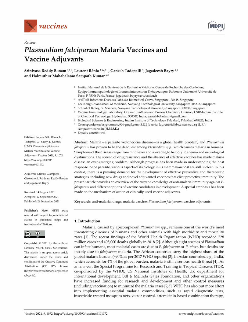
The malaria parasite has a complex life cycle (Figure 1). The Ronald Ross and
Giovanni Battista Grassi legacy laid the cornerstone for understanding the malaria
parasite life cycle (Figure 1) [8]. There are pre‐erythrocytic and erythrocytic stages in
humans (host) and the sexual life cycle in the mosquito vector. (A) Pre‐erythrocytic stage.
When the infected mosquito bites the host for blood meal, it injects sporozoite mostly into
the dermis or sometimes directly into the blood vessels [9]. Once sporozoite reaches the
liver, it invades and infects the hepatocytes. Inside the hepatocytes, sporozoites multiply
extensively, generating 10–30,000 merozoites [10]. (B) Erythrocytic stage. Merozoites enter
the red blood cells (RBCs) and mature into trophozoite and schizont. Each schizont
generates 6–12 merozoites, which are released into the circulation (remain one minute to
the next infection) and infect more RBCs [11,12]. During the process, a small number of
parasites develop into male and female gametocytes. The formed gametocytes mature
first in the bone marrow (I‒IV stages) and then spleen (V stage) [13]. (C) Mosquito/sexual
stage. In every progressing cycle, a small number of parasites deviates from asexual
reproduction and develops into male and female gametocytes, which will enter the
stomach of the Anopheles mosquito during the blood meal. The male and female
gametocytes develop into flagellated microgametes (eight) and a single macrogamete
respectively within the mosquito’s midgut. A zygote resulting from the fusion of a
macrogamete and microgamete develops into ookinete via meiosis and penetrates the
mosquito’s gut wall to form oocysts and produce sporozoites. These sporozoites travel to
salivary glands after the rupture of oocysts and will infect a new host during a mosquito
bite (Figure 1) [12,14].
Figure 1. Complex life cycle of P. falciparum. The life cycle has 3 stages: the pre‐erythrocytic and erythrocytic stages in
humans (host) and sexual process in the mosquito vector. a|Pre‐erythrocytic stage. b|Erythrocytic stage.
c|Mosquito/sexual stage.
Vaccines 2021, 9, 1072 3 of 38
P. falciparum transmission can be managed through vector control approaches, such
as spraying insecticides and/or using chemically‐treated mosquito nets, and through the
use of antimalarial drugs for prophylaxis and radical cure [15]. Early diagnosis, use of bed
nets, and timely treatment with anti‐malarial compounds are the global strategies for
controlling P. falciparum. However, the parasite has developed resistance to all anti‐
malarial drugs which are in present use [16]. In the last century, chloroquine was an
essential tool to eradicate malaria in many countries [17]. However, in the 1970s resistance
to chloroquine monotherapy emerged, and nowadays, chloroquine‐resistant P. falciparum
parasites are present worldwide. Other drugs such as pyrimethamine, mefloquine, or
artemisinin derivatives were also developed and replaced chloroquine progressively.
Because resistance to these drugs also developed, combination therapies were set to treat
P. falciparum infections [18]. Artemisinin‐based combination therapies have been essential
to decrease the death toll. However, resistance to artemisinin and its partner drugs has
become a severe concern in the last decade [19,20]. Thus, there is a need for novel drugs
and vaccines, particularly for controlling and possibly eliminating P. falciparum malaria.
The focus of this article is to review the current status of malaria vaccines research, with
an emphasis of the use of adjuvants and their mechanisms of action.
2. Immunity against P. falciparum
Knowledge on the interaction between the parasite and host immune system and
how clinical immunity is acquired against malarial infection is critical for the development
of effective vaccines, anti‐malarial drugs, and immunomodulators.
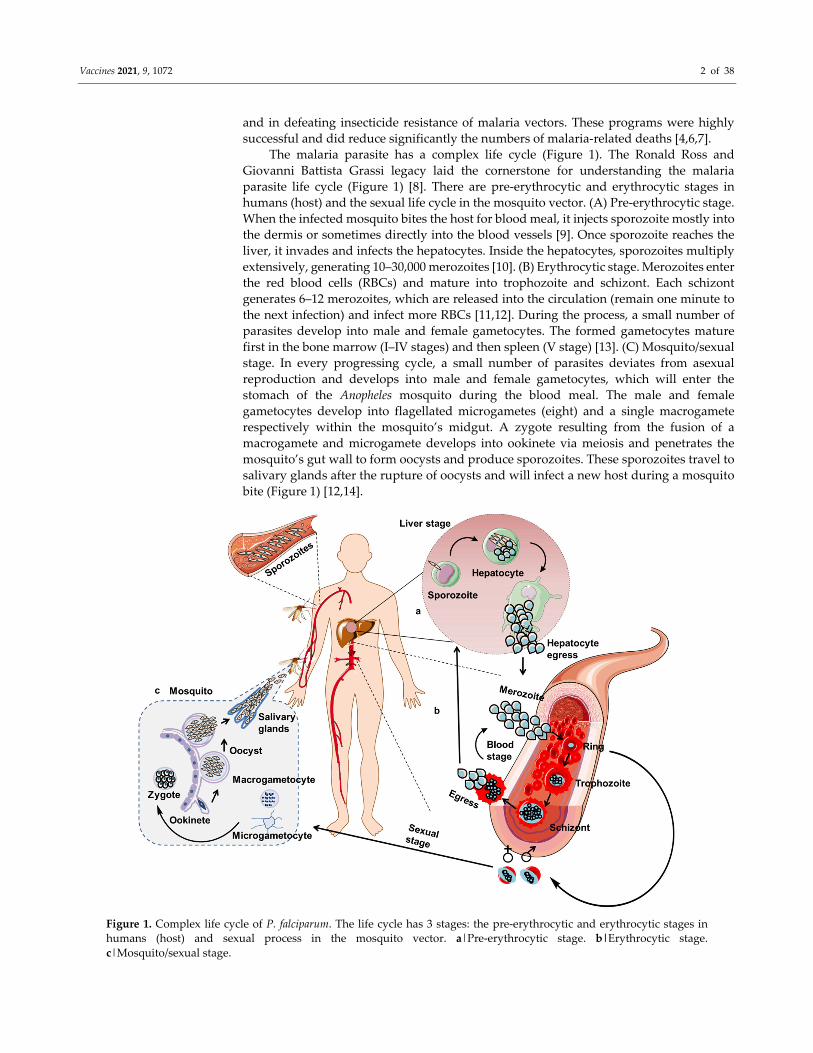
2.1. Dendritic Cells and the Initiation of the Immune Response
Among the innate immune cells, professional antigen‐presenting cells (APCs), such
as dendritic cells (DCs), have a prominent role in antigen presentation [21]. Although each
innate immune cell has a role in homeostasis and immunity, DCs’ role in vaccine and
vaccine adjuvant development has been extensively studied [22,23]. Considering the
central role of DCs in the activation and polarization of naïve T cells [24] and shaping the
immune response to vaccine adjuvants (Figure 2), we focused our discussion on DCs [13]
in the innate immune system.
Figure 2. Immunity in malaria infection. A|Activation of dendritic cells. During the infection, both liver‐ and blood‐stage
parasites are recognized by the several PRRs present in the cells of the innate immune system. Majorly, monocytes,
macrophages, and DCs have a significant role in the recognition. Vast literature has documented the TLR‐mediated signals
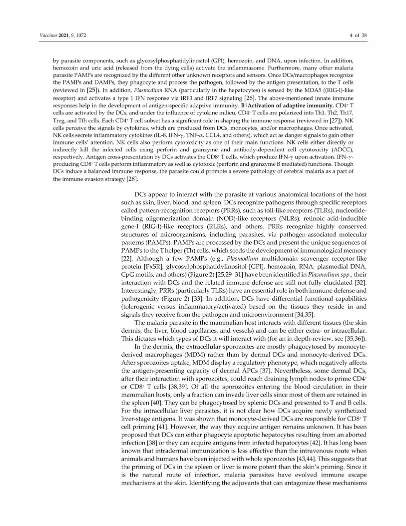
Vaccines 2021, 9, 1072 4 of 38
by parasite components, such as glycosylphosphatidylinositol (GPI), hemozoin, and DNA, upon infection. In addition,
hemozoin and uric acid (released from the dying cells) activate the inflammasome. Furthermore, many other malaria
parasite PAMPs are recognized by the different other unknown receptors and sensors. Once DCs/macrophages recognize
the PAMPs and DAMPs, they phagocyte and process the pathogen, followed by the antigen presentation, to the T cells
(reviewed in [25]). In addition, Plasmodium RNA (particularly in the hepatocytes) is sensed by the MDA5 ((RIG‐I)‐like
receptor) and activates a type 1 IFN response via IRF3 and IRF7 signaling [26]. The above‐mentioned innate immune
responses help in the development of antigen‐specific adaptive immunity. B|Activation of adaptive immunity. CD4+ T
cells are activated by the DCs, and under the influence of cytokine milieu, CD4+ T cells are polarized into Th1, Th2, Th17,
Treg, and Tfh cells. Each CD4+ T cell subset has a significant role in shaping the immune response (reviewed in [27]). NK
cells perceive the signals by cytokines, which are produced from DCs, monocytes, and/or macrophages. Once activated,
NK cells secrete inflammatory cytokines (IL‐8, IFN‐γ, TNF‐α, CCL4, and others), which act as danger signals to gain other
immune cells’ attention. NK cells also perform cytotoxicity as one of their main functions. NK cells either directly or
indirectly kill the infected cells using perforin and granzyme and antibody‐dependent cell cytotoxicity (ADCC),
respectively. Antigen cross‐presentation by DCs activates the CD8+ T cells, which produce IFN‐γ upon activation. IFN‐γ‐
producing CD8+ T cells perform inflammatory as well as cytotoxic (perforin and granzyme B mediated) functions. Though
DCs induce a balanced immune response, the parasite could promote a severe pathology of cerebral malaria as a part of
the immune evasion strategy [28].
DCs appear to interact with the parasite at various anatomical locations of the host
such as skin, liver, blood, and spleen. DCs recognize pathogens through specific receptors
called pattern‐recognition receptors (PRRs), such as toll‐like receptors (TLRs), nucleotide‐
binding oligomerization domain (NOD)‐like receptors (NLRs), retinoic acid‐inducible
gene‐I (RIG‐I)‐like receptors (RLRs), and others. PRRs recognize highly conserved
structures of microorganisms, including parasites, via pathogen‐associated molecular
patterns (PAMPs). PAMPs are processed by the DCs and present the unique sequences of
PAMPs to the T helper (Th) cells, which seeds the development of immunological memory
[22]. Although a few PAMPs (e.g., Plasmodium multidomain scavenger receptor‐like
protein [PxSR], glycosylphosphatidylinositol [GPI], hemozoin, RNA, plasmodial DNA,
CpG motifs, and others) (Figure 2) [25,29–31] have been identified in Plasmodium spp., their
interaction with DCs and the related immune defense are still not fully elucidated [32].
Interestingly, PRRs (particularly TLRs) have an essential role in both immune defense and
pathogenicity (Figure 2) [33]. In addition, DCs have differential functional capabilities
(tolerogenic versus inflammatory/activated) based on the tissues they reside in and
signals they receive from the pathogen and microenvironment [34,35].
The malaria parasite in the mammalian host interacts with different tissues (the skin
dermis, the liver, blood capillaries, and vessels) and can be either extra‐ or intracellular.
This dictates which types of DCs it will interact with (for an in depth‐review, see [35,36]).
In the dermis, the extracellular sporozoites are mostly phagocytosed by monocyte‐
derived macrophages (MDM) rather than by dermal DCs and monocyte‐derived DCs.
After sporozoites uptake, MDM display a regulatory phenotype, which negatively affects
the antigen‐presenting capacity of dermal APCs [37]. Nevertheless, some dermal DCs,
after their interaction with sporozoites, could reach draining lymph nodes to prime CD4+
or CD8+ T cells [38,39]. Of all the sporozoites entering the blood circulation in their
mammalian hosts, only a fraction can invade liver cells since most of them are retained in
the spleen [40]. They can be phagocytosed by splenic DCs and presented to T and B cells.
For the intracellular liver parasites, it is not clear how DCs acquire newly synthetized
liver‐stage antigens. It was shown that monocyte‐derived DCs are responsible for CD8+ T
cell priming [41]. However, the way they acquire antigen remains unknown. It has been
proposed that DCs can either phagocyte apoptotic hepatocytes resulting from an aborted
infection [38] or they can acquire antigens from infected hepatocytes [42]. It has long been
known that intradermal immunization is less effective than the intravenous route when
animals and humans have been injected with whole sporozoites [43,44]. This suggests that
the priming of DCs in the spleen or liver is more potent than the skin’s priming. Since it
is the natural route of infection, malaria parasites have evolved immune escape
mechanisms at the skin. Identifying the adjuvants that can antagonize these mechanisms
Vaccines 2021, 9, 1072 5 of 38
may help in the development of whole sporozoite skin immunization protocols. Of note,
liver‐resident DCs, which produce high amounts of IL‐10, are more tolerogenic than other
DC subsets. Cracking this tolerance, possibly through the use of proper adjuvants,
represents one of the rational approaches to target liver‐stage malaria infection [13,45,46].
During the erythrocytic stage, the more likely site of priming of the immune response
is the spleen [47]. During its blood‐stage development, the parasites remodel the infected
erythrocytes, leading to changes in deformability and rheological properties of the cells
[48]. The infected red blood cells (iRBC), which cannot deform, go through small
capillaries and accumulate in the spleen. These iRBCs are then recognized and destroyed
by phagocytes [49]. P. falciparum parasites have evolved to avoid splenic clearance by
expressing neoantigens at the surface of iRBC. These antigens, such as P. falciparum
erythrocyte membrane protein 1 (PfEMP1), allow parasites to sequester in deep tissues by
binding to a variety of receptors expressed on the surface of capillary endothelial cells
[50]. Splenic DCs or blood DCs can capture iRBC through CD36 and present to naïve T
cells [51]. However, iRBC binding to CD36 and CD54 on immature DCs limits the DC
maturation [52]. In vitro experiments in fact have shown that infected erythrocytes reduce
the expression of co‐stimulatory molecules on DCs [52] and affect the antigen processing
capacity of these professional APCs [52]. It was also found that malaria infection alters the
expression of TLRs on DC subsets [53]. In one cross‐sectional study, circulating DCs from
acutely infected Papuan adults had DCs, which did not mature well in vitro after
stimulation and were more prone to apoptosis. This immune dysregulation of DCs is
associated with an increased level of IL‐10. Antagonizing the IL‐10 actions restored DC
functions [54]. By decreasing DC maturation efficiency, the parasite prevents the
establishment of a potent immune response and facilitates immune evasion. Nevertheless,
blood and splenic DCs are effective in inducing inflammatory signals against Plasmodium
infection. New vaccine platforms are harnessing DCs for the development of novel
malaria vaccines [55]. In addition, any immunopotentiating substances such as adjuvants,
which can enhance the antigen presentation capacity of circulating or tissue‐specific DCs
to induce a protective immune response, are needed.
Majority of the myeloid cells, such as monocytes, macrophages, NK cells,
eosinophils, neutrophils, and DCs, participate in the antibody‐dependent cell‐mediated
cytotoxicity (ADCC) functions. Previous studies have given a clue that passive transfer of
immune sera has the capacity to activate ADCC to protect the host against malaria [56].
In vitro studies on RBCs, from infected and non‐infected malaria, have confirmed that NK
cell‐mediated ADCC is essential for acquired immunity [57]. In addition, upon parasite
exposure, NK cells are accompanied with an early capacity to produce copious amounts
of cytokines (e.g., IFN‐γ). Data from an RBC‐supplemented, immune cell‐optimized
humanized mouse model have revealed that NK cells interact with P. falciparum‐infected
erythrocytes through lymphocyte‐associated antigen 1 and kill the infected cells [58,59].
A specialized type of NK cells called adaptive NK cells, which express CD56dim
without the expression of promyelocytic leukemia zinc finger (PLZF) transcription factor
and Fc receptor γ‐chain (FcRγ) have been recently described. Studies conducted by
Geoffrey et al., in children and young adults, have predicted that presence of adaptive NK
cells is associated with protection against P. falciparum‐induced malaria infection [60]. As
NK cells are heterogeneous, further work is necessary to dissect the role of various subsets
of NK cells in the protection against malaria. Nevertheless, based on the above discussion,
ADCC function might play an essential role in blood‐stage malaria.
Vaccines 2021, 9, 1072 6 of 38
2.2. Adaptive Immunity
2.2.1. T cell‐Mediated Immunity
T cells, such as CD4+ (T helper, T follicular helper (Tfh) cells, cytotoxic CD4+ T cells),
cytotoxic CD8+, and non‐classical T cells such as mucosal‐associated invariant T (MAIT)
cells and γδ T cells have been shown to have a role in antimalarial immunity (for a
comprehensive review, see [61]).
2.2.2. T Cell‐Mediated Immunity against Liver‐Stage Malaria Parasites
Most of the knowledge on T cell immunity against the malaria liver stage has been
obtained in rodent malaria models of protection using whole parasite immunization
either with irradiated sporozoites or live sporozoite under drug prophylaxis [62–65]
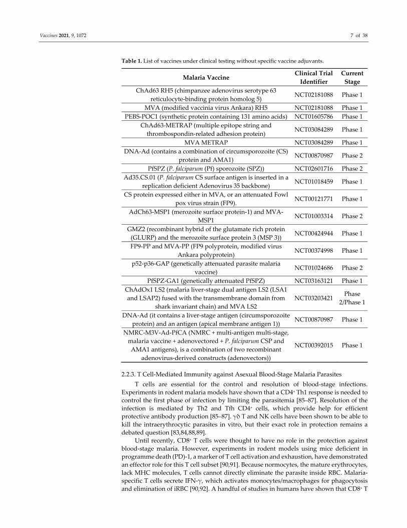
(Table 1). In these models, it has been shown that CD8+ and CD4+ T cells recognize peptide‐
derived antigens, which are presented by MHC class I and Class II on the surface of
infected hepatocytes [66–69]. T cells can eliminate infected parasites via different
mechanisms [70]. They can produce IFN‐γ, which induces nitric oxide synthase to
produce nitric oxide, a toxic molecule, in response to liver parasites [71–73]. They can also
eliminate infected hepatocytes through contact‐dependent perforin‐dependent
mechanisms [74]. IL‐4‐secreting CD4+ T cells have been shown to be critical for the
induction of CD8+ T‐cell response to liver‐stage malaria [41,75,76]. Liver‐resident CD8+ T
cells (CXCR6+CD69+) induced by vaccination with whole parasites or a subunit
formulation of the Plasmodium ribosomal protein RPL6 are essential to confer protection
in mice [77,78]. Experimental vaccination experiments in models using irradiated
sporozoites have also suggested that protection is dependent on the presence of liver‐
resident CD8+ T cells [79]. In human vaccine experiments, CD8+ and CD4+ T cell responses
were induced after P. falciparum sporozoite immunization [80]. Vaccine‐induced
protection was correlated with CD4+ T cell responses [80]. The route of infection is critical
for optimum T cell‐mediated immunity against liver parasite. In fact, intravenous but not
the intradermal injection of irradiated sporozoites induced a full sterilizing immune
response [79], possibly due to the induction of resident CD8+ T cells [44,79]. Therefore,
vaccines, delivery systems, or adjuvants which that can induce liver‐resident T cells and
increase antigen processing and presentation by MHC molecules at the surface of infected
hepatocytes are highly desirable. This has led to strategies using viral vectors and
heterologous prime‐boost approaches [80–82]. Recently, a novel and promising approach
called ‘prime and target’ has been shown to increase the frequency of tissue‐resident
memory CD8+ T cells in the liver in a mouse model using intravenous injections of viral
vectors or nanoparticles [81]. Other cell types, such as γδ or MAIT cells, may also have a
significant role in mediating the P. falciparum sporozoite vaccine‐induced protection as
suggested by the studies done with adults in the United States and Mali [80,83,84].
However, it is yet to be determined how these cells are induced during vaccination and
what the best adjuvant will be to activate them to act synergistically with classical T cells
for an effective protective immunity.
Vaccines 2021, 9, 1072 7 of 38
Table 1. List of vaccines under clinical testing without specific vaccine adjuvants.
Malaria Vaccine Clinical Trial
Identifier
Current
Stage
ChAd63 RH5 (chimpanzee adenovirus serotype 63
reticulocyte‐binding protein homolog 5) NCT02181088 Phase 1
MVA (modified vaccinia virus Ankara) RH5 NCT02181088 Phase 1
PEBS‐POC1 (synthetic protein containing 131 amino acids) NCT01605786 Phase 1
ChAd63‐METRAP (multiple epitope string and
thrombospondin‐related adhesion protein) NCT03084289 Phase 1
MVA METRAP NCT03084289 Phase 1
DNA‐Ad (contains a combination of circumsporozoite (CS)
protein and AMA1) NCT00870987 Phase 2
PfSPZ (P. falciparum (Pf) sporozoite (SPZ)) NCT02601716 Phase 2
Ad35.CS.01 (P. falciparum CS surface antigen is inserted in a
replication deficient Adenovirus 35 backbone) NCT01018459 Phase 1
CS protein expressed either in MVA, or an attenuated Fowl
pox virus strain (FP9). NCT00121771 Phase 1
AdCh63‐MSP1 (merozoite surface protein‐1) and MVA‐
MSP1 NCT01003314 Phase 2
GMZ2 (recombinant hybrid of the glutamate rich protein
(GLURP) and the merozoite surface protein 3 (MSP 3)) NCT00424944 Phase 1
FP9‐PP and MVA‐PP (FP9 polyprotein, modified virus
Ankara polyprotein) NCT00374998 Phase 1
p52‐p36‐GAP (genetically attenuated parasite malaria
vaccine) NCT01024686 Phase 2
PfSPZ‐GA1 (genetically attenuated PfSPZ) NCT03163121 Phase 1
ChAdOx1 LS2 (malaria liver‐stage dual antigen LS2 (LSA1
and LSAP2) fused with the transmembrane domain from
shark invariant chain) and MVA LS2
NCT03203421 Phase
2/Phase 1
DNA‐Ad (it contains a liver‐stage antigen (circumsporozoite
protein) and an antigen (apical membrane antigen 1)) NCT00870987 Phase 1
NMRC‐M3V‐Ad‐PfCA (NMRC + multi‐antigen multi‐stage,
malaria vaccine + adenovectored + P. falciparum CSP and
AMA1 antigens), is a combination of two recombinant
adenovirus‐derived constructs (adenovectors))
NCT00392015 Phase 1
2.2.3. T Cell‐Mediated Immunity against Asexual Blood‐Stage Malaria Parasites
T cells are essential for the control and resolution of blood‐stage infections.
Experiments in rodent malaria models have shown that a CD4+ Th1 response is needed to
control the first phase of infection by limiting the parasitemia [85–87]. Resolution of the
infection is mediated by Th2 and Tfh CD4+ cells, which provide help for efficient
protective antibody production [85–87]. γδ T and NK cells have been shown to be able to
kill the intraerythrocytic parasites in vitro, but their exact role in protection remains a
debated question [83,84,88,89].
Until recently, CD8+ T cells were thought to have no role in the protection against
blood‐stage malaria. However, experiments in rodent models using mice deficient in
programme death (PD)‐1, a marker of T cell activation and exhaustion, have demonstrated
an effector role for this T cell subset [90,91]. Because normocytes, the mature erythrocytes,
lack MHC molecules, T cells cannot directly eliminate the parasite inside RBC. Malaria‐
specific T cells secrete IFN‐γ, which activates monocytes/macrophages for phagocytosis
and elimination of iRBC [90,92]. A handful of studies in humans have shown that CD8+ T
Vaccines 2021, 9, 1072 8 of 38
cell activation occurs during falciparum infection. In a recent study in Ghanaian children,
expansion of a subset of CD8+ T cells expressing granzyme B correlated with an increase
in parasitemia and was more pronounced in patients with severe disease [93]. Thus, it is
likely that CD8+ T cells may have a more pathogenic effect than protective role during
falciparum infection. This is supported by immunohistopathology investigations on the
brain vasculature of 31 children who died from human cerebral malaria. A substantial
number of CD3+ CD8+ T cells was found adjacent to endothelial cells [94]. In a mouse
model of cerebral malaria, pathogenic CD8+ T cells are responsible for the neurological
symptoms and ensuing lethality induced by infection with P. berghei ANKA [95,96]. These
cells recognize malaria antigens presented by endothelial cells and release granzyme B
after engagement with the MHC molecules. Thus, for blood‐stage vaccine development,
induction of a strong immune response may not be desirable since it may induce
unwanted pathology. Nevertheless, there is a need for well‐calibrated T cell and well‐
targeted B cell responses.
As already mentioned above, malaria infection frequently induces
immunosuppression through diverse mechanisms [97]. One of the active T cell
suppression pathways involves the induction of regulatory T cells (Tregs). Tregs
expressing specific markers such as cytotoxic‐T‐lymphocyte‐associated protein‐4 (CTLA‐
4), lymphocyte‐activation gene 3 (LAG‐3), programmed cell death‐1 (PD‐1), and others
function as immunosuppressive cells. Both in vitro studies in peripheral blood
mononuclear cells (PBMCs) and longitudinal studies in humans have shown that the
frequency of Tregs in the patients is negatively associated with the parasite load and/or
disease severity [96,98] (extensively reviewed elsewhere [61]). Studies in humans and
mice have shown that Tregs via CTLA‐4 inhibit the development of a protective anti‐
blood‐stage immunity [99]. Furthermore, clinical studies on malaria‐infection/vaccination
showed an increased Treg population in the acute and uncomplicated infection and
during the convalescence phase [100–102]. In contrast, a decrease in the Treg population
was also observed in children under chronic malaria exposure [103–105]. Thus,
knowledge of the factors influencing Treg functions, such as age, level of exposure (high
or low transmission intensity), and others, is needed to precisely define their role in the
pathogenesis of malaria. Approaches using adjuvants to prevent induction or counteract
Treg activity in individuals residing in endemic regions might help in the development of
an effective anti‐blood‐stage vaccine.
2.2.4. Antibody‐Mediated Immunity
In malaria infection, antibodies mediate protective immune responses via different
mechanisms, such as inhibition of parasite motility, invasion, egress, adhesion and
hepatocyte traversal ability, promotion of antibody‐dependent complement‐mediated
sporozoite/merozoite lysis, phagocytosis, antibody‐dependent cellular cytotoxicity,
transmission‐blocking activity, and others [80,106,107] (reviewed in [108]). Interestingly,
the generation of natural immunity against P. falciparum is ethnicity dependent. The
majority of non‐immune Western travelers, if not all, require a single infection to induce
invasion inhibitory antibodies against P. falciparum rather than endemic residents, who
require two infections [109,110]. Similarly, protection against malaria in infants born in
the endemic regions correlate with the pre‐existing antibodies (i.e., maternal antibodies)
[110]. In addition, children (5 years) who are repeatedly exposed to the natural infection
are more resistant to the severe clinical symptoms of malaria [110,111]. Development of P.
falciparum antigen‐specific B cells to secrete monoclonal antibodies and passive transfer of
immunoglobulins have been attractive strategies in malaria research [80,112].
Vaccines 2021, 9, 1072 9 of 38
Many malaria candidate vaccines have been designed to induce an effective antibody
response. Antibodies against sporozoites or against neo‐antigens expressed on the surface
of infected hepatocytes mediate protection by preventing or limiting pre‐erythrocytic‐
stage infection and development. Anti‐sporozoite antibodies have been shown to inhibit
sporozoite motility in the dermis and liver [113], destroy sporozoites in the skin [114],
facilitate opsonization and phagocytosis by monocytes or macrophages in the spleen or
the liver [114,115], inhibit sporozoite invasion into hepatocytes [116], and inhibit
sporozoite development inside the hepatocytes [116]. The type of functional antibodies
produced during the infection illuminates the type of protection they offer. For example,
IgG1 and IgG3 exhibit potent capacity to activate complement, FcɣR signaling and
opsonization compared to IgG2 and IgG4. The former function discriminates the IgG
subclasses into cytophilic (IgG1, IgG3) and non‐cytophilic (IgG2, IgG4) antibodies [117–
119]. The above‐mentioned antibody functional properties have been confirmed against
the majority of Plasmodium spp., including P. falciparum in different life‐cycle stages [108].
Although the role of IgG and its subclasses is significant in host to symptomatic malaria
infection, the role of IgM must not be ignored. Particularly, the protective role of IgM
against the blood‐stage parasite of P. falciparum has been established [120–122].
Antibodies against parasite neo‐antigens, such as heat shock protein, expressed on
the surface of infected hepatocytes induce liver parasite killing through an antibody‐
dependent cell‐mediated mechanism, which is likely to involve Kupffer cells or NK cells
[123]. Different in vitro assays have been developed to measure antibody functionality,
but it is not yet clear which of the immune mechanisms described above are essential or
associated with the protection. It might be that, in addition to the high level of neutralizing
antibodies, the quality (i.e., avidity, affinity, isotype) of the antibodies is also important.
This knowledge is of paramount importance to design better immunogen and use
adequate adjuvant.
Antibody‐mediated immunity against the blood stage is more complex than in the
pre‐erythrocytic stage. Anti‐merozoite antibodies can (i) prevent merozoites from
invading RBC [124–126] alone or in conjunction with complement factors [118], (ii)
prevent merozoite egress from RBC, (iii) agglutinate free merozoites, (iv) facilitate
phagocytosis of merozoites, and (v) promote clearance of iRBC by phagocytic cells
through a mechanism called antibody‐cell‐dependent inhibition (ACDI) [127]. In ACDI,
anti‐merozoite IgG1 or IgG3 antibodies bind to merozoites, and the immune complexes
promote phagocytes, such as monocytes/macrophages or neutrophils, to release cytokines
(e.g., TNF‐α). This cytokine then stimulates the phagocytes to produce mediators, which
leads to the killing of intra‐erythrocytic parasites [128,129]. As mentioned above P.
falciparum parasites express antigens on the surface of iRBC. These antigens are mainly
encoded by multigene families, such as the var [130], stevor [131], and rifin gene families
[132]. These antigens are involved in cytoadherence to endothelial cells and in other
adhesive phenomena, such as rosetting (the binding of an iRBC to non‐infected RBC) and
agglutination (the binding to iRBC through bridging by platelets) (for a review, see [50]).
The cytoadherence ability of the malaria parasites is key to many of the pathologies
induced by P. falciparum infection. Antibodies targeting the surface antigens may prevent
cytoadherence and promote iRBC phagocytosis or iRBC agglutination [133,134].
Vaccines 2021, 9, 1072 10 of 38
Antibodies targeting parasite toxins could also protect from P. falciparum‐induced
disease. During the blood stage of the infection, diverse parasite toxins are released at the
time of iRBC rupture. These toxins include hemozoin, a by‐product of heme degradation
by the parasite [135], GPI moieties, which are present in many merozoite proteins [136], a
TatD‐like DNase [137], and a tyrosine‐t RNA synthase [138]. Protection from disease by
anti‐toxin antibodies has been achieved experimentally using synthetic glycans
mimicking GPI [139].
Gametocytes, the sexual forms of P. falciparum parasites, are also targets for antibody‐
mediated immunity [140]. Gametocytes express a range of antigens, which are targeted
by antibodies [141]. The latter facilitates the complement‐mediated killing of the
gametocytes [142]. In the mosquito after feeding, anti‐sexual form antibodies can prevent
fusion of gametes [143], induce complement‐killing of gametes or ookinetes [144], and
prevent ookinete motility, penetration of the midgut wall, and formation of oocyst [145].
Vaccination/infection determines the dominant IgG subtypes induced, which in turn
are useful to predict the type of immune responses acquired. Among the IgG subtypes,
opsonizing cytophilic antibodies have gained much attention for malaria vaccine
development [146–148]. These antibodies recruit FcɣR‐containing immune cells
(particularly macrophages and basophils) via their Fc domain and activate the parasite
elimination procedures. Cytophilic antibodies both naturally acquired [149–152] and
vaccine‐induced (IgG1 and IgG3 responses against blood‐stage antigens) [107] have
shown more protective responses in diverse studies.
During the vaccine design, it important to consider both naturally acquired and
vaccine‐induced immunity. The development of naturally acquired immunity depends
on many factors such as region, age group, number of exposures (symptomatic or
asymptomatic), number of targeted antigens, and others [149,150]. The vaccine‐induced
immunity depends on the type of antigens targeted and the quality (type) and quantity of
antibodies produced [153]. Although both have similar specificities, the duration and
diversity of the immune response is more for naturally acquired immunity over vaccine‐
induced immunity [154]. Evidence from different studies suggests that naturally acquired
immunity, unlike vaccine‐induced immunity, reduces the risk of infection irrespective of
age [155,156]. Moreover, children living in perennial transmission areas induce more
protective natural antibodies (e.g., anti‐erythrocyte binding antigen 175RIII–V) than in
seasonal transmission areas [149]. However, development of naturally acquired immunity
is the best bet in the host. Circumstantially, newborns and infants (<6 months of age) are
conferred protection against malaria infection due to the existence of maternal antibodies
(extensively reviewed elsewhere [157]).
Antibodies’ breadth (antibodies raised against number of antigens), magnitude
(quality), and quantity (affinity and avidity) decide the protection efficiency against
clinical malaria. However, it is difficult to estimate the quantity of antibodies that are
required for protection, and it purely relies on the antigen specificity. A study conducted
in Kenyan children (n = 119), for six months, confirmed that children who possess a
breadth of naturally acquired antibodies against blood‐stage antigens are inversely
correlated with the risk of malaria [158]. With regard to the vaccine development, whole‐
sporozoite vaccines are ideal to obtain a breadth of antibodies against the parasite. Like
natural exposure, whole‐sporozoite vaccines increase the magnitude of antibodies due to
the varying antigenic content [80].
Interestingly, it has been observed that antibody breadth and magnitude increase
with age (4 years < 14 years < adults) [159]. Though not always, increased risk of infection
has also been observed in the presence of antigen‐specific antibodies (e.g., anti‐AMA1)
[160,161]. However, additional studies are required to confirm the breadth, magnitude,
affinity, and avidity of the protective antibodies in both asymptomatic and symptomatic
infections [162,163].
Vaccines 2021, 9, 1072 11 of 38
As mentioned above, passive transfer of neutralizing monoclonal antibodies is an
alternative therapeutic strategy, which gives short‐term protection. In addition, epitope‐
specific monoclonal antibodies have been generated against different stages of P.
falciparum and evaluated in several models (reviewed, recently) [112]. At present, subunit,
DNA, and viral vector vaccines are capable of inducing high antibody titers, though the
potency and breadth are not sufficient to give complete protection [164]. To achieve more
germinal center reaction and memory B cell generation, the addition of vaccine adjuvants
and adjuvant delivery systems are essential (discussed in the later section). Finally,
natural or vaccine‐induced high antibody titers are needed to prevent the hepatic entry of
the parasite and erythrocyte infection.
3. Vaccines
Vaccines are one of the most efficient prophylactic treatments for many diseases,
ranging from smallpox to the recently emerged severe acute respiratory syndrome
coronavirus 2 (SARS‐CoV‐2, which causes coronavirus disease 2019 (COVID‐19)). With
the ever‐increasing knowledge of immunity, vaccine strategies have focused on
identifying the target antigens and inducing a protective immune response with low to
no side effects. Due to the complex antigenicity and immune attacking mechanisms of P.
falciparum, the development of vaccine for this parasite has been and is still challenging
(Appendix A1) [165]. However, many traditional vaccine strategies do not answer the
question of life‐cycle complexity and polymorphism of parasite proteins, which are just
two of the many reasons we do not yet have a highly effective malaria vaccine [80].
A variety of malaria vaccines have been developed and evaluated from the past
decades, ranging from classic approaches such as whole inactivated parasites to subunit
vaccines and new delivery systems. Although adjuvants enhance vaccines’ immune
response, they are occasionally not efficacious in formulations with malarial antigens.
Different recombinant malarial antigens have been produced (Table 1 and 2) in various
expression systems (e.g., E. coli, L. lactis, N. benthamiana, P. pastoris, S. cerevisiae, and others)
with a variety of delivery platforms (e.g., conjugates, fusion proteins, virus‐like particles,
virosomes, and others). Over the years, researchers have used various antigens for vaccine
development against the different stages (sporozoite, merozoite, and sexual) of the
parasite life cycle [166]. However, it has been challenging to identify the ideal target
antigen, among the 5000 proteins encoded in the P. falciparum genome [167]. Genomic,
transcriptomics, and proteomics approaches have helped to define protein expression
patterns during the P. falciparum life cycle [168]. Among the proteins identified, the
uniqueness of sporozoite, trophozoite, merozoite, and gametocyte proteins are
categorized [168]. The different protein targets (mentioned in Table 1 and 2) are exploited
in distinct ways for developing malaria vaccines: (i) pre‐erythrocytic vaccines, (ii)
erythrocytic vaccines, (iii) transmission‐blocking vaccines (extensively reviewed
elsewhere [80,110,169]). Several candidates have been tested; several immunogens are at
preclinical stage, among which 30 immunogens are in clinical evaluation though only 2
have crossed Phase IIb trials or beyond (without adjuvants; https://clinicaltrials.gov/
accessed on 1 June 2020).
In the past 100 years, many adjuvants have been evaluated preclinically against
diverse antigens of malaria (Table 2). Notably, RTS’S/AS01 (Mosquirix™) (Appendix A2)
and PAMAVAC vaccines have successfully entered advanced clinical trials.
Vaccines 2021, 9, 1072 12 of 38
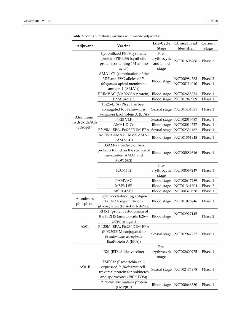
Table 2. Status of malarial vaccines with vaccine adjuvants#.
Adjuvant Vaccine Life‐Cycle
Stage
Clinical Trial
Identifier
Current
Stage
Aluminium
hydroxide/Alh
ydrogel®
Lyophilized PEBS synthetic
protein (PfPEBS) (synthetic
protein containing 131 amino
acids)
Pre‐
erythrocytic
and blood
stage
NCT01605786 Phase 2
AMA1‐C1 (combination of the
3D7 and FVO alleles of P.
falciparum apical membrane
antigen‐1 (AMA1))
Blood stage NCT00984763
NCT00114010
Phase 2
Phase 1
PRIMVAC (VAR2CSA protein) Blood stage NCT02658253 Phase 1
P27A protein Blood stage NCT01949909 Phase 1
Pfs25‐EPA (Pfs25 has been
conjugated to Pseudomonas
aeruginosa ExoProtein A (EPA)
Sexual stage NCT01434381 Phase 1
Pfs25 VLP Sexual stage NCT02013687 Phase 1
AMA1‐DiCo Blood stage NCT02014727 Phase 1
Pfs25M‐ EPA, Pfs230D1M‐EPA Sexual stage NCT02334462 Phase 1
AdCh63 AMA1 + MVA AMA1
+ AMA1‐C1 Blood stage NCT01351948 Phase 1
BSAM‐2 (mixture of two
proteins found on the surface of
merozoites, AMA1 and
MSP1(42))
Blood stage NCT00889616 Phase 1
ICC‐1132
Pre‐
erythrocytic
stage
NCT00587249 Phase 1
PAMVAC Blood stage NCT02647489 Phase 1
MSP3‐LSP Blood stage NCT01341704 Phase 2
MSP1 42‐C1 Blood stage NCT00320658 Phase 1
Aluminum
phosphate
Erythrocyte‐binding antigen
175 kDA region II‐non‐
glycosylated (EBA‐175 RII‐NG)
Blood stage NCT01026246 Phase 1
AS01
RH5.1 (protein ectodomain of
the PfRH5 (amino acids E26—
Q526) antigen)
Blood stage NCT02927145
Phase 2
Pfs25M‐ EPA, Pfs230D1M‐EPA
(PfS230D1M conjugated to
Pseudomonas aeruginosa
ExoProtein A (EPA))
Sexual stage NCT02942277 Phase 1
AS01B
R21 (RTS, S‐like vaccine)
Pre‐
erythrocytic
stage
NCT02600975 Phase 1
FMP012 (Escherichia coli‐
expressed P. falciparum cell‐
traversal protein for ookinetes
and sporozoites (PfCelTOS))
Sexual stage NCT02174978 Phase 1
P. falciparum malaria protein
(FMP)010 Blood stage NCT00666380 Phase 1
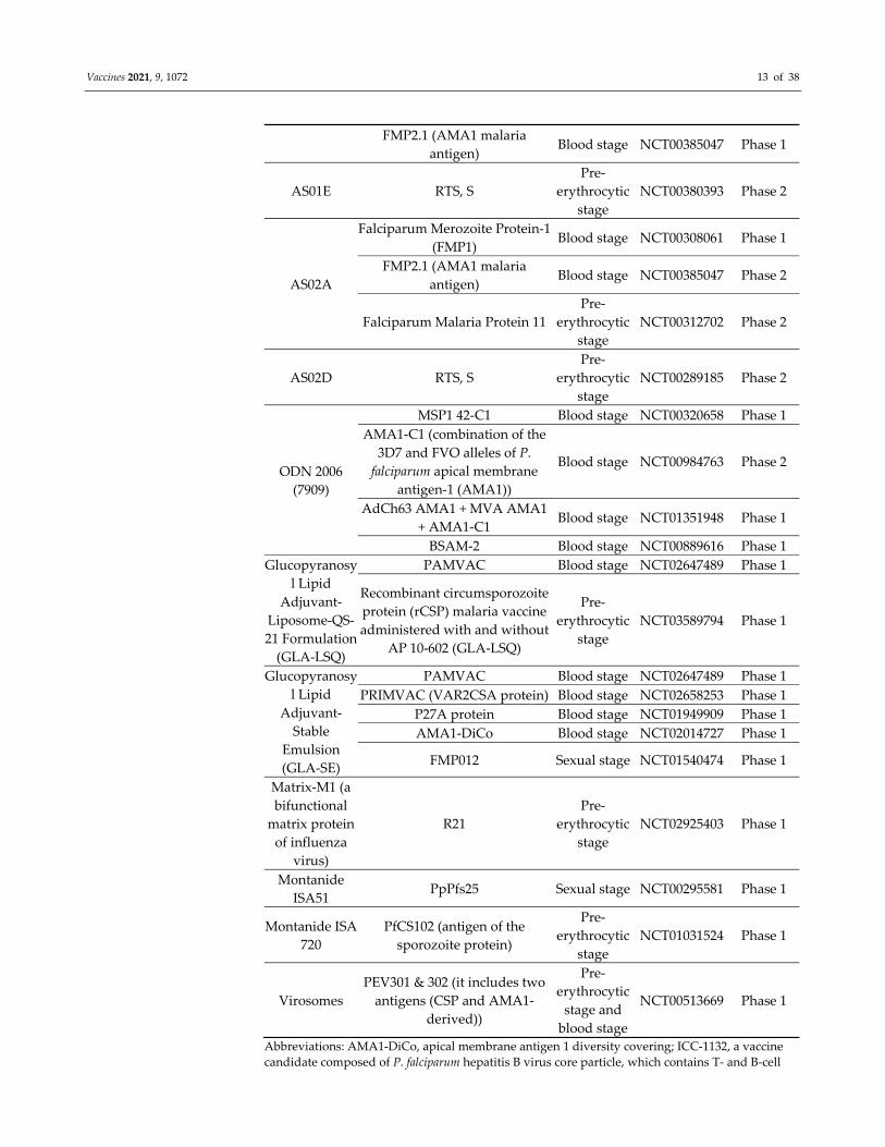
Vaccines 2021, 9, 1072 13 of 38
FMP2.1 (AMA1 malaria
antigen) Blood stage NCT00385047 Phase 1
AS01E RTS, S
Pre‐
erythrocytic
stage
NCT00380393 Phase 2
AS02A
Falciparum Merozoite Protein‐1
(FMP1) Blood stage NCT00308061 Phase 1
FMP2.1 (AMA1 malaria
antigen) Blood stage NCT00385047 Phase 2
Falciparum Malaria Protein 11
Pre‐
erythrocytic
stage
NCT00312702 Phase 2
AS02D RTS, S
Pre‐
erythrocytic
stage
NCT00289185 Phase 2
ODN 2006
(7909)
MSP1 42‐C1 Blood stage NCT00320658 Phase 1
AMA1‐C1 (combination of the
3D7 and FVO alleles of P.
falciparum apical membrane
antigen‐1 (AMA1))
Blood stage NCT00984763 Phase 2
AdCh63 AMA1 + MVA AMA1
+ AMA1‐C1 Blood stage NCT01351948 Phase 1
BSAM‐2 Blood stage NCT00889616 Phase 1
Glucopyranosy
l Lipid
Adjuvant‐
Liposome‐QS‐
21 Formulation
(GLA‐LSQ)
PAMVAC Blood stage NCT02647489 Phase 1
Recombinant circumsporozoite
protein (rCSP) malaria vaccine
administered with and without
AP 10‐602 (GLA‐LSQ)
Pre‐
erythrocytic
stage
NCT03589794 Phase 1
Glucopyranosy
l Lipid
Adjuvant‐
Stable
Emulsion
(GLA‐SE)
PAMVAC Blood stage NCT02647489 Phase 1
PRIMVAC (VAR2CSA protein) Blood stage NCT02658253 Phase 1
P27A protein Blood stage NCT01949909 Phase 1
AMA1‐DiCo Blood stage NCT02014727 Phase 1
FMP012 Sexual stage NCT01540474 Phase 1
Matrix‐M1 (a
bifunctional
matrix protein
of influenza
virus)
R21
Pre‐
erythrocytic
stage
NCT02925403 Phase 1
Montanide
ISA51 PpPfs25 Sexual stage NCT00295581 Phase 1
Montanide ISA
720
PfCS102 (antigen of the
sporozoite protein)
Pre‐
erythrocytic
stage
NCT01031524 Phase 1
Virosomes
PEV301 & 302 (it includes two
antigens (CSP and AMA1‐
derived))
Pre‐
erythrocytic
stage and
blood stage
NCT00513669 Phase 1
Abbreviations: AMA1‐DiCo, apical membrane antigen 1 diversity covering; ICC‐1132, a vaccine
candidate composed of P. falciparum hepatitis B virus core particle, which contains T‐ and B‐cell
Vaccines 2021, 9, 1072 14 of 38
epitopes from the repeat region of the C terminus circumsporozoite protein; METRAP, multiple‐
epitope thrombospondin‐related adhesion protein; MSP3‐LSP, merozoite surface protein‐3 long
synthetic peptide; PfCelTOS, P. falciparum cell traversal protein of ookinetes and sporozoites;
PfAMA1, P. falciparum apical membrane antigen 1; PfPEBS, P. falciparum pre‐erythrocytic and
blood stage; PfMSP1, P. falciparum merozoite surface protein 1; PfRH5, P. falciparum reticulocyte‐
binding protein homolog 5; PfSERA5, P. falciparum Serine Repeat Antigen‐5; P27A protein,
unstructured 104 mer synthetic peptide from P. falciparum trophozoite exported protein 1;
VAR2CSA, variant surface antigen 2CSA; VLP, virus‐like particles. # We searched for malarial
vaccines in clinical trials website (https://clinicaltrials.gov/ accessed on 1 June 2020) and selected the studies pertinent to P. falciparum. Many vaccines use alhydrogel as adjuvant, however, at
different doses and different conditions. To our knowledge, many vaccines with a similar type of
antigens using a diversified combination of adjuvants have been under clinical trials, which are
not listed here.
4. Vaccine Adjuvants
In general, most vaccine‐based antigens are inefficient at mounting protective
immune responses due to the dearth of immunogenicity, even though they proved to be
perfect target antigens. Nevertheless, an additional component, named adjuvant (adjuvare
(Latin): To help) has proven to be critical in boosting the antigens’ immunogenicity.
Several reviews have covered the qualities of most vaccine adjuvants and their mechanism
of action [170–175]. Like immune response in infections, adjuvants induce an immune
response by triggering innate and adaptive immune responses. The type of immune
response induced by the adjuvant is critical for the choice of vaccines. For example,
adjuvants inducing Th1 response are preferable in vaccines against intracellular
pathogens, and adjuvants inducing Th2 responses are preferable in vaccines against
extracellular pathogens [174,176]. In both cases, Tfh cell responses are critical in inducing
prolonged antibody responses [177]. Tfh cells are distinguished from the other T helper
cells via expression of a CXCR5 receptor and a transcription factor B cell lymphoma 6 (Bcl‐
6). During the infection or vaccination (with or without adjuvants) [178], Tfh cells migrate
to the secondary lymphoid organs and provide help to the germinal center B cells to
induce high‐affinity memory B cells and long‐lived plasma cells [177,179]. The role of Tfh
cells in humoral immunity has become more motivating in the design of subunit vaccines
[180].
Protective immunity against P. falciparum requires neutralizing antibodies [181,182]
and optimal Th1‐mediated immunity [183]. For enhancing, modulating, and prolonging
the specified immune response against any P. falciparum vaccine candidate, vaccine
adjuvants that facilitate these responses are needed. These adjuvants will reduce the
antigen concentration and frequency of immunization to attain protective efficacy,
thereby making vaccines more cost‐effective. Though adjuvants face many challenges
(physicochemical interactions, stability, pairing with partner antigen, and others), recent
technologies in vaccine adjutants have come to the stage of increasing immune responses
via immune synergy (use of multiple adjuvants) [172]. Reasonably, the immune synergy
strategy has not been used extensively in the vaccines composed of parasitic antigens
[184].
Different types of adjuvants have been categorized, such as TLRs‐, nucleotide‐
binding oligomerization domain‐like receptors (NLRs)‐, C‐type lectin receptors (CLRs)‐
based, and some other non‐specific PRRs agonists, NLR family pyrin domain containing
3 (NLRP3) activators, formulations including liposomes, Adjuvant System (AS),
nano/micro particles‐based adjuvants, immune‐complexes, and others. Recent years have
seen the approval of several new‐generation adjuvants containing vaccines for human use
[185]. Inflammation (either specific receptor activation or non‐specific activation) is the
primary response to a majority of the adjuvants leading to antigen‐specific cellular
immunity [185]. Among the long list, formulation vaccines have gained much attention
with respect to their efficacy, such as AS01, AS02, AS03, AS04, MF59, and others. The
majority of these systems have more than one immune‐potentiator in them. The use of
Vaccines 2021, 9, 1072 15 of 38
these formulations has induced a protective immune response against antigens and has
also increased the immune responsiveness in an elderly population, e.g., influenza vaccine
(MF59 or AS03) and herpes zoster vaccine (AS01) [185]. Although many adjuvants,
including alum (a century‐old approved adjuvant), are used in clinical trials, the lack of a
clear understanding of mechanisms of action against malaria has impaired the
development of efficient immunogen/adjuvant formulation [186]. Viral vector‐based
vaccines have shown better immunogenicity than adjuvanted protein vaccines [187].
Herein, we discuss the importance of current adjuvants, which are being employed in
developing malaria vaccines at the clinical stage, focusing on their outputs like
immunological profile and mechanism of action of successful adjuvants.
5. Adjuvants under Clinical Evaluation
5.1. Alum
Alum (common name for aluminum potassium sulfate) is a 90‐year‐old gold
standard vaccine adjuvant that produces primarily humoral immune responses [188,189].
A recent review detailed the physico‐chemical and biological properties of alum
adjuvants [188]. Mostly, two types of aluminum adjuvants (aluminum hydroxide
(Al(OH)3) and aluminum phosphate (AlPO4)) are used in the clinically approved vaccines
and these are the most widely applied adjuvants for evaluating the malaria vaccines [190].
The merits of aluminum adjuvants include safety, amplification of antibody responses,
and a comparatively easy process to produce on a large scale. It also has certain drawbacks
like the inability to induce cell‐mediated immune responses (Th1 and cytotoxic T
lymphocytes (CTL)), which are essential for P. falciparum [190]. Since alum induces Th2
biased cellular responses, it was administered concomitantly with other adjuvants
(alum+CpG motifs (ODN) [191] alum+glucopyranosyl lipid adjuvant (GLA) [192]) or
adjuvant formulations in the development of malaria vaccines. In preclinical studies,
often, the antigen is mixed with alum before injection in mice [193,194]. Unlike other
adjuvants, alum is the most successful single vial‐ or prefilled syringe‐based vaccine
adjuvant. Though intense research has been done on the alum, its precise mechanism of
action, however, is still elusive. Interestingly, studies in mice showed that alum forms a
depot at the injection site and enters the innate cells via phagocytosis. It activates the
NLRP3 inflammasome via phagolysosomes followed by the activation of caspase‐1 and
release of IL‐1β. The known mechanism of action of alum was reviewed elsewhere [189].
Nevertheless, the proposed mechanism is not observed either in humans or in monkeys
[195,196]. Thus, there should be another mechanism of action of alum in humans, which
is yet to be identified [185]. The vaccination of multistage antigen SpF66 combined with
alum resulted in the development of short‐lived antibodies and minimal cellular response
with no or little protection. Further, the evaluation of pre‐erythrocytic protein Pfcs102
with alum was not successful in enhancing an antigen‐specific antibody response
[190,197]. Blood‐stage vaccines (AMA1[PfAMA‐1‐FVO25–545], AMA1‐C1, GLURP85–213, and
MSP1‐C142) adjuvanted with alum induced modest antibody levels, a poor cellular
response, and no protection in field clinical trials [190]. In a randomized, double‐blind,
placebo‐controlled study, one vaccine (PRIMVAC) adjuvanted either with alhydrogel
(aluminum hydroxide wet gel suspension; regularly 2% Al(OH)3 is used) or GLA‐SE had
a significant immunogenic response and VAR2CSA‐specific antibodies with the desired
inhibitory properties [198].
The transmission‐blocking Pf25, a sexual‐stage vaccine, has been clinically evaluated
in combination with alum. It induced local reactogenicity and limited immunogenicity
[199,200]. Altogether, it is clear that alum alone is not sufficient to induce effective
responses to malaria vaccines [201].
Vaccines 2021, 9, 1072 16 of 38
5.2. Vaccine Delivery Systems/Formulations
Although effective or moderately effective protein vaccine candidates have been
developed for malaria, they have poor immunogenicity. Targeted delivery of subunit
vaccines via systems possessing adjuvant properties is of paramount importance and a
very classical approach. It ensures their effective delivery as well as the ability to increase
protective immunity [166]. However, like alum, the mechanism of adjuvanticity of
emulsion‐type delivery systems remains to be identified [185].
5.2.1. Liposomes
They are synthetic phospholipid models (nm to μm size), which carry antigens
encapsulated into the aqueous core (hydrophilic molecules), adsorbed to the surface
(lipophilic molecules), or integrated into the lipid layers (amphiphilic molecules) [202].
The adjuvanticity of liposome is mainly reliant on size, charge, preparation method, and
number of lipid layers [203]. Cationic adjuvant formulations made of dimethyl
dioctadecyl ammonium combined with the stabilizer glycolipid trehalosedibehenate
(TDB) were found to be immunogenic liposome forms [204]. The disadvantages of
liposomes are stability, manufacturing process, high costs, pain at the injection site, and
the addition of immunostimulatory molecules like AS01 [190]. Because of the instability
of liposomal vaccine delivery systems in a single vial liquid format, the formulation
should be admixed with the vaccine before administration, e.g., Mosquirix®vaccine [201].
5.2.2. AS01
AS01 is a liposome‐based vaccine adjuvant system containing two
immunostimulants: 3‐O‐desacyl‐4ʹ‐monophosphoryl lipid A (MPL) and the saponin QS‐
21. It has been extensively studied for vaccines against various infectious diseases,
including catarrhalis, influenza, tuberculosis, HIV, and others [205]. Interestingly, unlike
other vaccine adjuvant systems, AS01 has a recognized mechanism of action. It activates
the resident macrophages, followed by draining to the lymph nodes, where it activates
the NK cells to release IFN‐γ. The released IFN‐γ activates DCs, which induce an antigen‐
specific Th1 response (i.e., IL‐2, IFN‐γ, and TNF‐α) [206–208]. The above‐mentioned
mechanism of action is due to the synergistic effect of two immunostimulants, i.e., MPL
(monophosphoryl lipid A) and QS‐21, present in AS01 (Figure 3). Both the molecules
induce Th1‐mediated immune responses [209]. In addition, in delayed fractional doses
AS01/RTS,S has been shown to induce IL‐21‐secreting antigen‐specific peripheral Tfh cells
with protective B cell responses [210]. However, individual components have their
specific effects (see the relevant text). QS‐21 delivered via liposomes protects the cells from
QS‐21‐induced cell death [211]. This might be the reason that QS‐21‐containing AS01 has
exhibited more improved efficacy than other formulations. In addition, malaria vaccine
clinical trials have used different forms of AS01, i.e., AS01B full dose of immunostimulants
for adults and AS01E half dose of immunostimulants for children [185,212] (Table 2).
Vaccines 2021, 9, 1072 17 of 38
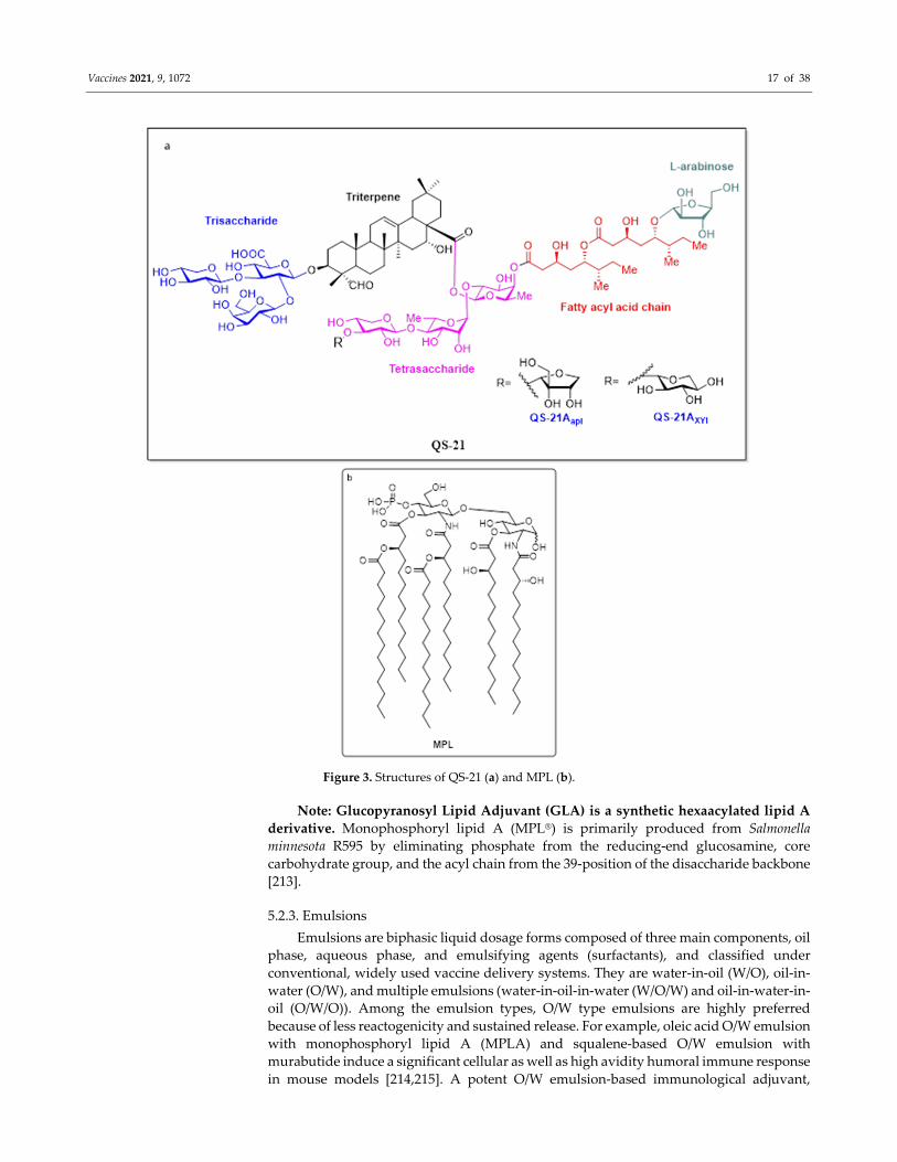
Figure 3. Structures of QS‐21 (a) and MPL (b).
Note: Glucopyranosyl Lipid Adjuvant (GLA) is a synthetic hexaacylated lipid A
derivative. Monophosphoryl lipid A (MPL®) is primarily produced from Salmonella
minnesota R595 by eliminating phosphate from the reducing‐end glucosamine, core
carbohydrate group, and the acyl chain from the 39‐position of the disaccharide backbone
[213].
5.2.3. Emulsions
Emulsions are biphasic liquid dosage forms composed of three main components, oil
phase, aqueous phase, and emulsifying agents (surfactants), and classified under
conventional, widely used vaccine delivery systems. They are water‐in‐oil (W/O), oil‐in‐
water (O/W), and multiple emulsions (water‐in‐oil‐in‐water (W/O/W) and oil‐in‐water‐in‐
oil (O/W/O)). Among the emulsion types, O/W type emulsions are highly preferred
because of less reactogenicity and sustained release. For example, oleic acid O/W emulsion
with monophosphoryl lipid A (MPLA) and squalene‐based O/W emulsion with
murabutide induce a significant cellular as well as high avidity humoral immune response
in mouse models [214,215]. A potent O/W emulsion‐based immunological adjuvant,
Vaccines 2021, 9, 1072 18 of 38
MF59, which is composed of squalene droplets stabilized with Tween 80 and Span 85,
elicited higher antibody titers with balanced antibody subclasses than those observed in
alum studies. In contrast to conventional emulsions, MF59 is composed of metabolizable
oils (squalene; component in cholesterol synthesis pathway), making MF59 much safer.
Strikingly, although MF59 is more highly efficacious, stable, and potent than other vaccine
adjuvants (alum and CpG) with ease of scale‐up competence [185,216], it did not offer
reasonable results in malaria vaccines; hence, there was no further progress with MF59 in
developing malaria vaccine [217–220]. Irrespective of antigens, O/W emulsions elicit
Th1/Th2 responses rather than Th1/Th17 responses [221]. Other emulsion‐based
adjuvants have been developed and have shown to be more efficient.
5.2.4. AS02
It is composed of two immunostimulants: QS‐21 and MPL (3‐deacylated
monophosphoryl lipid A) in squalene O/W emulsion. Preliminary results in rodents and
non‐human primates have shown that AS02 is efficacious [222,223]. Further, early clinical
trials of AS02 with different malarial antigens, such as PfCS102 (a P. falciparum
circumsporozoite protein immunogen) and LSA‐1 (liver‐stage antigen) have shown an
increased Th1 response [224]. Initial trials with RTS, S/AS02 formulation (AS02A and
AS02D are varying doses of formulation for adults and infants, respectively) have found
that the vaccine is safe, well‐tolerated, and immunogenic in adults, infants, children, and
semi‐immune adults [225–229]. Interestingly, RTS, S/AS02 formulation exhibited effective
cellular immunity and significant protection against sporozoite‐challenge malaria
infection when compared to other AS formulations [186,212,230]. However, further trials,
with distinct dosage regimens have reported no or minimal protection [186,212,231]. In
view of these outcomes, among the AS formulations for malaria vaccines, AS01 holds a
superior efficacy and has been selected for further clinical trials [186,212,225].
5.2.5. Montanides (ISA 51, ISA 720)
Montanide ISA 51 and ISA 720 are W/O type emulsions, which are composed of
mineral oil and non‐mineral oil, respectively, with mannide monooleate as an emulsifier.
ISA 51 was approved for lung cancer due to its high CTL responses. Clinical studies on
ISA 51 adjuvant with mosquito‐stage antigen (Table 2) yielded unexpected systemic
adverse events (erythema nodosum), which stopped further exploratory studies in
malarial vaccines [232]. ISA 720 has been extensively studied in malaria vaccines because
of its ability to induce high antibody production rather than T cell responses. In most of
the malaria vaccines, ISA 720 has been shown to induce a strong immune response, but
with associated problems such as pain and unacceptable reactogenicity at the site of
injection and formulation instability [190,233]. All these findings further support that O/W
emulsions are preferred in human vaccines.
5.3. Immune Potentiators or Immunomodulators
Substances, such as particulate materials, complexes, delivery systems, and others,
which modulate the immune responses either by targeting the innate immune system or
skewing the Th1‐ and Th2‐dependent antibody production, are referred to as immune
potentiators. Many molecules derived from the classes of structure‐guided vaccine
adjuvants or immune potentiators, e.g., PamCysk4 analogues (TLR2 agonists), α‐
Glactosylceramide analogues (CD1d‐iNKT linkers), saponins/glycolipids, and others,
show immune‐modulating potential [193,194,234–239]. In this section, we describe
different potent immunomodulators, which are potent inducers of Th1 responses and
have been used in malaria vaccines, either alone or in combinations.
Vaccines 2021, 9, 1072 19 of 38
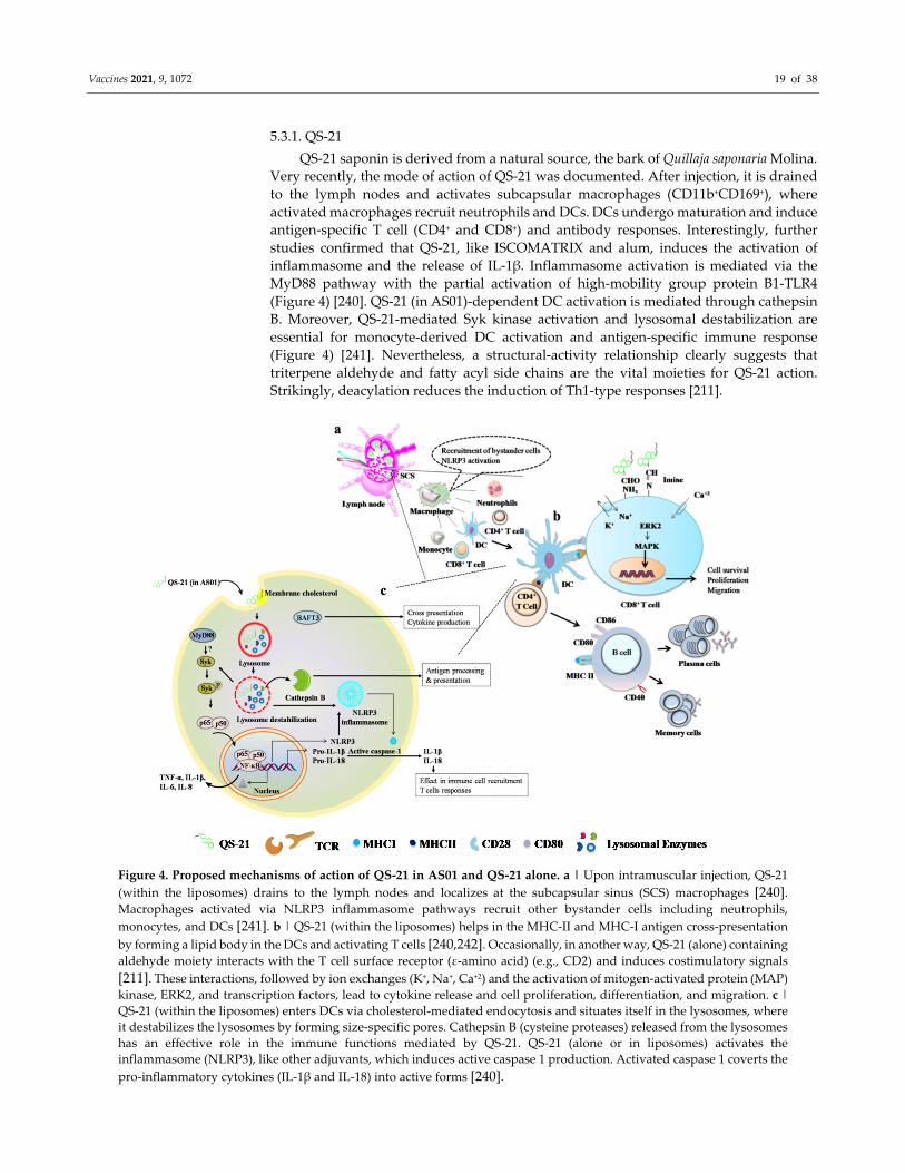
5.3.1. QS‐21
QS‐21 saponin is derived from a natural source, the bark of Quillaja saponaria Molina.
Very recently, the mode of action of QS‐21 was documented. After injection, it is drained
to the lymph nodes and activates subcapsular macrophages (CD11b+CD169+), where
activated macrophages recruit neutrophils and DCs. DCs undergo maturation and induce
antigen‐specific T cell (CD4+ and CD8+) and antibody responses. Interestingly, further
studies confirmed that QS‐21, like ISCOMATRIX and alum, induces the activation of
inflammasome and the release of IL‐1β. Inflammasome activation is mediated via the
MyD88 pathway with the partial activation of high‐mobility group protein B1‐TLR4
(Figure 4) [240]. QS‐21 (in AS01)‐dependent DC activation is mediated through cathepsin
B. Moreover, QS‐21‐mediated Syk kinase activation and lysosomal destabilization are
essential for monocyte‐derived DC activation and antigen‐specific immune response
(Figure 4) [241]. Nevertheless, a structural‐activity relationship clearly suggests that
triterpene aldehyde and fatty acyl side chains are the vital moieties for QS‐21 action.
Strikingly, deacylation reduces the induction of Th1‐type responses [211].
Figure 4. Proposed mechanisms of action of QS‐21 in AS01 and QS‐21 alone. a | Upon intramuscular injection, QS‐21
(within the liposomes) drains to the lymph nodes and localizes at the subcapsular sinus (SCS) macrophages [240]. Macrophages activated via NLRP3 inflammasome pathways recruit other bystander cells including neutrophils,
monocytes, and DCs [241]. b | QS‐21 (within the liposomes) helps in the MHC‐II and MHC‐I antigen cross‐presentation
by forming a lipid body in the DCs and activating T cells [240,242]. Occasionally, in another way, QS‐21 (alone) containing
aldehyde moiety interacts with the T cell surface receptor (ε‐amino acid) (e.g., CD2) and induces costimulatory signals
[211]. These interactions, followed by ion exchanges (K+, Na+, Ca+2) and the activation of mitogen‐activated protein (MAP)
kinase, ERK2, and transcription factors, lead to cytokine release and cell proliferation, differentiation, and migration. c |
QS‐21 (within the liposomes) enters DCs via cholesterol‐mediated endocytosis and situates itself in the lysosomes, where
it destabilizes the lysosomes by forming size‐specific pores. Cathepsin B (cysteine proteases) released from the lysosomes
has an effective role in the immune functions mediated by QS‐21. QS‐21 (alone or in liposomes) activates the
inflammasome (NLRP3), like other adjuvants, which induces active caspase 1 production. Activated caspase 1 coverts the
pro‐inflammatory cytokines (IL‐1β and IL‐18) into active forms [240].
Vaccines 2021, 9, 1072 20 of 38
Though it is effective in inducing immunity and widely used in many vaccines,
including malaria, HIV‐1, and cancer, QS‐21 also possesses certain limitations, such as
complex steps of manufacture/synthesis, hemolytic effects, biodiversity problems (due to
the cutting down of trees), the dose to efficacy ratio, and others [174]. Considering all these
limitations, novel structural mimics and alternative or simplified structures are under
progress [239]. Due to the virtue of its immune response, many formulations are use QS‐
21 as a discrete component in the composition [185]. Interestingly, QS‐21 containing
nanopatch–skin delivery systems have been shown to be effective for influenza protein
antigen. In this study, a nanopatch, which contains a 30‐fold lower dose of QS‐21, has
exhibited an equal antigen‐specific humoral response to QS‐21 alone, given
intramuscularly [242]. This clearly suggests that in the future, QS‐21 with malarial antigen
must be tested in nanopatches or a similar delivery system to spare the doses of both
antigen and adjuvant.
5.3.2. Other Saponin Based Adjuvants (Quil‐A and ISCOMs)
Quillaja saponaria is the base for Quil‐A and is the most widely used adjuvant from
the saponin group. It exhibits lower toxic effects but enhanced adjuvanticity. Further,
complex saponin adjuvant, immunostimulating complexes (ISCOMs; Matrix‐M™)
(spherical, open, and cage‐like structures) formed by the cholesterol and phospholipids
with the adjuvant (Quil‐A or QS‐21), trigger both cell‐mediated and humoral‐mediated
immune responses. On the other hand, Quil‐A is complicated on account of its chemical‐
related problems plus local reactogenicity. Although malaria vaccine candidates
combined with ISCOMs have shown good results in preclinical investigations [243], very
few clinical trials have been conducted with malarial vaccines composed of ISCOMs
(NCT02905019), probably due to the pain at the injection site and other drawbacks such
as flu‐like symptoms, fever, and malaise with less reactogenicity [190,244,245].
5.3.3. CpG ODN
Oligodeoxynucleotides (ODNs) adjuvants are single standard DNA molecules with
varying degrees of unmethylated CpG motifs. Like most vaccine adjuvants, CpG ODN
also enhances the innate immune cell (pDCs, monocytes, NK cells)‐dependent adaptive
immune responses (Th1 and B cells) against the co‐administered antigens [246].
Considering the strong immunomodulatory properties, especially Th1 responses, CpG
ODNs are widely used in various diseases such as infections, cancer, and allergies [246].
Based on their structural‐activity relationship, CpG ODNs are divided into different
types: class A (Type D), class B (Type K), and class C [174,246] (reviewed in [174]). Among
the ODNs, ODN 2006 (7909) is widely used because of its immunostimulatory properties.
It comes under the class B category, and it stimulates pDCs to secrete IFN‐α via binding
to the TLR9 receptor. The detailed mechanism of action of CpG ODNs has been reviewed
in the literature [174,246]. Many preclinical studies, either alone or in combination, have
documented the efficacy of CpG in P. falciparum infection [247]. This ODN has successfully
entered the malaria vaccine clinical trials and has been used as an adjuvant for malarial
antigens, such as AMA1, MSP1(42), and their different combinations (Table 2). In one of
the clinical studies, naïve human volunteers who received AMA1‐based blood‐stage
malaria vaccine along with CpG ODN, either alone or in combination with alhydrogel,
exhibited enhanced and long‐lasting antigen‐specific antibody responses. In addition,
antigen‐specific antibody (IgG) showed enhanced growth inhibition of homologous P.
falciparum, in vitro [248]. However, subsequent studies with semi‐immune adults living
in Mali did not give encouraging results [249]. The above results perhaps depend on the
refractory/chemistry of CpG ODNs and should be taken into consideration for the future
use of CpG ODN‐containing vaccines in malaria endemic areas. One more blood‐stage
vaccine candidate, serine repeat antigen 5 (SERA5), has shown protective immunity in
Aotus and squirrel monkeys. A recombinant antigen, SE36 (which lacks serine repeats of
SERA5), was absorbed into alhydrogel and evaluated in clinical trials. Surprisingly, it did
Vaccines 2021, 9, 1072 21 of 38
not induce high antigen‐specific antibody titers in clinical trials [250]. Further,
SE36/alhydrogel was tested in rodents and non‐human primates (monkeys) with or
without CpG ODNs. Among the CpG ODNs tested, K3 CpG ODN (K3 CpG ODN is a
subclass of ODNs under class B (Type K)) has exhibited safe, efficacious, and
immunogenic properties [251]. However, with CpG ODN, these positive preclinical data
are still not translated into clinics [252].
6. Concluding Remarks
P. falciparum is an infectious agent of malaria, which kills millions of people around
the globe. A vigorous effort has been made for the global elimination of P. falciparum
malaria, but asymptomatic individuals are regarded as reservoirs for parasite
transmission due to the lack of effective diagnostic methods for detecting the parasite in
endemic areas.
For the treatment of malaria, the spread of anti‐malarial drug resistance has become
a major problem. If it is not handled appropriately, it could reverse the malaria control
program and worldwide containment achieved so far. Therefore, more research is
necessary to find new anti‐malarial drugs for combating multidrug‐resistant P. falciparum
[253].
The factors like complex life cycle, genetic diversity, and the various immune escape
mechanisms developed by these parasites, i.e., antigenic variations in P. falciparum [97],
are the major obstacles responsible for the delay in the development of a suitable vaccine.
Because of the high number of polymorphism or allele‐specific variations in the proteins,
single protein‐based vaccines have limited success [254]. Vaccines composed of a chimeric
molecule of the pre‐erythrocytic target have shown protective responses in mice models
[255]. The current efforts towards malaria parasite and vector control should be
complemented with an effective P. falciparum vaccine for the successful fight against the
malaria burden. Presently, vaccines from different stages of P. falciparum are in
development and in the coming days, they might be combined (multivalent vaccines) to
overcome the parasitic immune control mechanism attributed to antigenic polymorphism.
A better understanding of the P. falciparum life cycle and the parasites’ interaction with
the host should provide guidelines for the future vaccine development programs. The
ideal malaria vaccine should be safe, highly effective, stable, easy to administer, and must
provide long‐term immunity. Such vaccines should also be cost‐effective and affordable
in poor malaria endemic areas.
Nowadays, researchers are fully aware of using suitable adjuvants for the
development of novel vaccines. With regard to malaria, as mentioned above, an effective
vaccine requires long‐lasting primary and memory antigen‐specific CD4+ T cell, B cell, and
CTL responses. However, age‐old adjuvants like alum, which skews the immune response
to Th2, are not suitable for malaria vaccine development. Importantly, a combination of
Th1 and Th2 adjuvants with a proper delivery system is needed for malaria vaccine
development. Many prediction models have confirmed that activation of the IFN‐γ
signaling pathway is the best target to confer vaccine‐induced protection against P.
falciparum [256]. Next‐generation vaccines should have wide spectrum activity against
parasites, as some of the endemic residents encounter infection with more than one
parasite simultaneously [257]. On top of that, vaccines should not induce suppressive
Tregs, which are shown to aid in the parasite immune evasion mechanisms [110]. Further,
an ideal adjuvant should be able to produce a protective immune response with a less
frequent number of administrations. Nevertheless, we noticed that many adjuvants
enhance effective immune responses with experimental antigens or model antigens,
whereas a similar trend might not happen with the real antigens and unpaired antigens
(Bonam, SR and Kumar HMS, personal observation; unpublished data). All the above
details should help in the selection of specific adjuvant systems for the development of
novel vaccines that elicit the highest humoral and cellular responses. Nowadays,
analytical characterization of formulations to obtain vital information (i.e., particle size,
Vaccines 2021, 9, 1072 22 of 38
morphology, and concentration of components, physicochemical interactions, stability,
and others) are routinely performed. On the other hand, detailed studies are required to
establish the biological interactions and determine the exact mechanism of action [201].
Systems vaccinology has helped considerably to uncover the unidentified vaccine
adjuvant mechanism(s). Finally, the repository of novel Th1/Th2 adjuvants should be
available to vaccine researchers to test their antigens for developing novel, promising
malaria vaccines.
In summary, there is a need to coordinate research in multiple directions to develop
an efficient malaria vaccine, such as (1) clear understanding of the life cycle of the malaria
parasite, (2) identification and characterization of targets of interest with a broad range of
strains covered, (3) identification of suitable adjuvant, which could enhance the
magnitude and quality of the immunogenicity of antigen(s) without age factor [172], and
(4) characterization and selection of a suitable ‘perfect mix’ (e.g., emulsion, liposomes,
nanoparticles, etc.) for the delivery of the final vaccine to the host [173]. In essence, a
detailed spatiotemporal analysis of parasite invasion biology will provide more
information for effective targets. By tackling all these parameters, we could also support
the poor responders in the vaccination.
Author Contributions: SRB, LR, and GT came up with the idea and wrote the first draft. The draft
was revised by LR, JB, and HMSK. All authors have read and agreed to the published version of the
manuscript.
Funding: Laurent Rénia was supported by core funding to the A*STAR ID labs given by the Agency
of Science, Technology and Research (A*STAR). GT is a Research Associate‐II funded by the
Department of Biotechnology (DBT), Government of India, India.
Institutional Review Board Statement: Not applicable
Informed Consent Statement: Not applicable
Data Availability Statement: Not applicable
Acknowledgments: Authors would like to thank Samarchith P. Kurup (Center for Tropical and
Emerging Global Diseases & Department of Cellular Biology, The University of Georgia, US) and
Giuseppe Del Giudice (Translational Science Leader, R&D Center Italy—GSK Vaccines Srl, Siena,
Italy) for scientific discussion. GT and HMS are thankful to Director CSIR—Indian Institute of
Chemical Technology (IICT) for the support (IICT/Pubs./2020/080). J.B. acknowledges the support
of the European Union Horizon H2020 programme under Grant Agreement No. H2020‐SC1‐2019‐
874653‐INDIGO and the Department of Biotechnology (DBT), Government of India. Figure 2 was
created with BioRender.com under a paid subscription.
Conflicts of Interest: We declare that the research was conducted in the absence of any relationships
that could construe as a potential conflict of interest.
Appendix A
Key points in malaria vaccine development
Spatiotemporal relation between liver anatomy and sporozoite biology
should be explored in detail [258].
Invasion strategies of sporozoites (in liver) and merozoites (in RBCs) should
be elucidated [259,260].
Unlike other infections, vaccine adjuvants, which induces both Th1 and Th2
mediated immunity is essential against P. falciparum malaria.
Dominant MHC I‐presenting antigen epitopes should be selected for cell‐
mediated, especially CD8+ T cells and tissue‐resident memory T cells in the
liver, which are essential to confer vaccine‐induced protection [261].
Protective malaria vaccines should elicit IgG1 and IgG3 antibody responses,
which are protective against P. falciparum malaria [147,262,263].
Vaccines 2021, 9, 1072 23 of 38
Protective vaccines (e.g., RTS,S) to pre‐erythrocytic stage infections are
greatly needed [264]. In malaria vaccines, along with target antigen, development of adjuvants
(e.g., AS01) is also considered as major discipline of research [240,265]. Prime‐boost and/or prime‐target immunization strategies are probably
better options to confer protection against liver‐stage infections [266].
Subcutaneous vaccination has shown increased response than the
intradermal vaccination [267]. In addition, vaccines are preferably effective,
if given by intravenous route [79].
RTS, S malaria vaccine
RTS, S is a genetically engineered vaccine where “RT” denotes C‐terminal
(190 amino acids) part of P. falciparum (strain NF54) circumsporozoite protein (CSP) protein, which contains both T and B cell epitopes (central repeat
region). “S” is a surface antigen of hepatitis‐B virus and the combination is
expressed in Saccharomyces cerevisiae (S) [268].
RTS, S was produced by Walter Reed Army Institute of Research (WRAIR)
in collaboration with GlaxoSmithKline (GSK) biologicals and the Programs
for Appropriate Technologies in Health (PATH) Malaria Vaccine Initiative
(MVI).
The idea behind the development of the RTS, S vaccine is, like other vaccines
(e.g., ChAd63/MVA ME‐TRAP), to inhibit sporozoite motility at pre‐
erythrocytic stage and preventing the entry to liver cells.
The first licensed vaccine for children living in endemic areas in sub‐Saharan
Africa, and is also approved by European Medicines Agency (EMA) in 2015
[269].
Presently under clinical evaluation in infants and children in Africa (Phase
IV/NCT00866619). Previous regimens are partly effective, now four dose‐
schedule have been initiated in Kenya [270].
Individuals vaccinated with RTS,S/AS01 has shown increased NK cell
activation in response to increased T cell activation [58,265].
Studies in infants and children vaccinated with RTS, S/AS01 confirmed the
antibody (IgG subclass)‐mediated protection against P. falciparum malaria by
altering the natural acquired immunity [147].
Safe, immunogenic and potent for individuals who experienced malaria.
Like other majority vaccines, RTS, S also has hurdles in the stability
maintenance of specified temperatures (2–8 °C), which is certainly expensive
in developing countries.
RTS, S vaccine with nearly or entirely different adjuvants also exhibit
promising results against malaria P. falciparum [264,271,272].
Adult vaccination regimen containing RTS,S/AS01B vaccine with
ChAd63/MVA ME‐TRAP vaccine has not shown interesting results [273].
Highlights
Fine strategies to eradicate and eliminate malaria through drugs or vaccines
have gained momentum.
Fundamental functions of CD8+ T cells and NK cells have proven their
effective role in preventing malaria transmission.
Majority of prophylactic/therapeutic malaria vaccines are in the clinical
evaluation.
RTS, S/AS01 is the only licensed malaria vaccine.
Vaccines 2021, 9, 1072 24 of 38
Novel delivery systems of malarial vaccines with QS‐21 have yielded
increased potency and reduced toxicity.
Though subunit vaccines are an effective strategy, the search for novel
approaches like live and/or whole parasite vaccinations should be evaluated
in detail.
Glossary
Plasmodium falciparum
P. falciparum is a parasite protozoan and one of the most infectious species among the
other Plasmodium such as P. malariae, P. vivax, P. ovale and P. knowlesi, which causes malaria
in humans by using female anopheles’ mosquito as a vector.
Cerebral malaria
Cerebral malaria caused by P. falciparum, affects total neurological functions, and
leads to death. Two hypotheses have tried to elucidate the pathogenesis of disease:
mechanical and inflammatory. According to mechanical hypothesis, impaired cerebral
blood flow due to sequestration of erythrocytes leads to hypoxia whereas inflammatory
theory suggests that P. falciparum infection alters brain function via Th1 inflammatory immune
response. Symptoms of cerebral malaria include headache, behavioral changes, confusion,
coma, and mortality (rate exceeding 10%) [274,275].
Heterologous prime‐boost strategy
Multiple immunization of vaccine with or without discrete antigens in DNA vectored
priming followed by protein boosting. Strategy is useful in targeting highly complex and
pathogenic infections for inducing both humoral and cell‐mediated immune responses.
Danger‐associated molecular patterns (DAMPs)
Signals from the damaged or dying cells or necrotic cells, which alert the immune
system via acting on PRRs.
Clinical immunity
Natural antibody response developed against P. falciparum following repeated
infection. It reduces the parasite load and regulates the inflammatory responses.
Sterile immunity
A cent percent immunity achieved by malaria vaccine is termed as sterile immunity.
Clinically immune individuals can still act as a reservoir for the parasite. Thus, to confer
complete protection against the P. falciparum malaria sterile immunity is needed.
Pre‐erythrocytic vaccine
This vaccine with respective antigens prevents the generation of sporozoites into
merozoites in the liver thereby reduce the clinically asymptomatic pre‐erythrocytic
infection. Some of the examples include RTS, S/AS01 (CSP), ChAd63/MVA ME–TRAP
(TRAP, CSP, TRAP, CSP, and others), PfSPZ (whole sporozoites), CVac (whole
sporozoites), GAP (whole sporozoites), PfCelTOS FMP012 (PfCelTOS), CSVAC (CSP), R21
(CSP), R21/ME–TRAP (TRAP, CSP, and others).
Erythrocytic stage vaccine
This vaccine with respective antigens prevents merozoitesʹ entry into the RBCs,
thereby reducing the clinical symptoms and mortality. Examples include GMZ2 (GLURP
MSP3), pfAMA1‐DiCo, (AMA1) P27A (P27A), MSP3 (MSP3), SE36 (SERA5), PfPEBS,
CHAd63RH5/MVARH5 (RH5), PRIMVAC (Var2CSA), and PAMVAC (Var2CSA) [80].
Transmission‐blocking vaccines
These vaccines with respective antigens interrupt malaria transmission by impeding
the parasite cycle in the mosquito. Antigens like Pfs25 (Pfs25 VLP & Pfs25‐EPA), Pfs28,
Pfs48/45, and Pfs230 (Pfs25‐EPA) have been assessed as vaccine candidates among which
ookinetes of P. falciparum Pfs25 expressed in S. cerevisiae have shown moderate
immunogenicity and suboptimal levels of transmission‐blocking activity in Phase Ia trials
[165,276].
TLRs
Vaccines 2021, 9, 1072 25 of 38
Class of PRRs located on the surface and in the cytosol of the cells. Upon recognition
of PAMPs/DAMPs, these receptors activate the downstream signaling to secrete diverse
inflammatory mediators.
Antibody‐dependent cell‐mediated cytotoxicity (ADCC)
Antigen‐binding fragment (Fab) of antibodies (developed against target protein)
binds to the target cells and recruit’s effector cells, which possess receptors for the
fragment crystallizable region (Fc) of the antibody (FcR) to interact with target cell.
Activated myeloid effector cells kill the target cells via various mechanisms (release of
cytotoxic granule, Fas signaling, induction of reactive oxygen species).
Vaccine delivery systems
These are antigenic formulations (emulsions, liposomes, polymeric particles,
adenoviral systems, and others) with or without immunostimulants for targeted delivery
and inducing protective immunity (cellular and humoral immunity).
Adjuvant systems (mostly licensed by GlaxoSmithKline plc)
Liposomes and emulsion formulations, which contain one or more
immunostimulants to induce antigen‐specific immune responses.
QS‐21
A water‐soluble triterpene glycoside obtained from the purified fractions (fraction
21) of mixture of Quil‐A saponins. Quil‐A saponins are sourced from the bark of Quillaja
saponaria tree. It is potent inducer of both cellular and humoral immunity. QS‐21 induces
the MHC I antigen cross‐presentation followed by the activation of CD8+ T cells.
AS01
Liposomal formulation composed of two synergistic discrete immunostimulatory
components, i.e., synthetic 3‐O‐desacyl‐4′‐monophosphoryl lipid A (MPL) (a TLR4 ligand)
and natural QS‐21.
Virosomes
Virosomes are derived from the inactivated/non‐replicating virus. Virosomes are
composed of bilayer phospholipids, like liposomes and viral glycoproteins. The targeted
antigen of interest is encapsulated or cross‐linked to the virosomes for effective delivery.
CpG
ODN sequences containing unmethylated CpG motifs (highly methylated in
vertebrates); cytosine triphosphate deoxynucleotide (C) linked to the guanine
triphosphate deoxynucleotide (G) via phosphodiester bond (p), which specifically binds
to TLR9 to trigger immune response. Synthetic CpG ODN over the natural ones has a
different level of phosphorothioate backbone.
Structural‐activity relationship
It defines the relationship between the chemical structure of a compound to its
pharmacological activity and toxicity.
Outstanding Questions: P. falciparum vaccines and vaccine adjuvants
Knowledge on the optimum combination of multi‐stage malarial antigens for
the vaccines
Influence of natural immunity towards vaccine response and performance in
endemic countries
Fine incorporation of parameters of anti‐disease immunity of the host to
conceive effective and safe anti‐malarial vaccines.
The role of genetic background of the population in determining the vaccine
response to malaria.
Need of appropriate experimental models that closely mimic human for the
pre‐screening of malaria vaccine candidates.
Abbreviations
ADCC antibody‐dependent cell cytotoxicity
Vaccines 2021, 9, 1072 26 of 38
AIM2 absent in melanoma 2
AP‐1 activator protein 1
ASC apoptosis‐associated speck‐like protein containing a CARD (caspase
activation and recruitment domains)
CCL2/MCP‐1 monocyte chemoattractant protein‐1
CCL4/MIP‐1β macrophage inflammatory protein‐1β
cGAS cyclic GMP‐AMP synthase
DAMPs damage‐associated molecular patterns
DCs dendritic cells
dsDNA double‐stranded DNA
ERK extracellular signal‐regulated kinases
GPI glycosylphosphatidylinositol
IKK IκB kinase
IRF IFN (interferon) regulatory factor
IL interleukin
JNK c‐Jun N‐terminal kinases
MAPK mitogen‐activated protein kinases
MDA5 melanoma differentiation‐associated gene 5
MHC major histocompatibility complex
NF‐kB nuclear factor kappa‐light‐chain‐enhancer of activated B cells
NK cells natural killer cells
NLRP3 NOD‐, LRR‐ and pyrin domain‐containing 3
PAMPs pathogen‐associated molecular patterns
PRRs pathogen‐recognition receptors
STING stimulator of IFN genes
TCR T cell receptor
Tfh follicular T cell
TGF transforming growth factor
Th helper T cell
TLRs toll‐like receptors
TNF tumor‐necrosis factor
TRAF6 tumor necrosis factor receptor‐associated factor 6
Treg regulatory T cell
References
1. Andrews, K.T.; Fisher, G.; Skinner‐Adams, T.S. Drug repurposing and human parasitic protozoan diseases. International Journal
for Parasitology: Drugs and Drug Resistance 2014, 4, 95–111, doi:10.1016/j.ijpddr.2014.02.002.
2. The ʺworld malaria report 2019ʺ at a glance. WHO: https://www.who.int/news‐room/feature‐stories/detail/world‐malaria‐
report‐2019, (accessed on 1 July 2020).
3. Key points: World malaria report 2017; https://www.who.int/malaria/media/world‐malaria‐report‐2017/en/, (accessed on 1
January 2021).
4. This yearʹs world malaria report at a glance. World Health Organization: https://www.who.int/malaria/media/world‐malaria‐
report‐2018/en/, (accessed on 1 July 2020).
5. Head, M.G.; Goss, S.; Gelister, Y.; Alegana, V.; Brown, R.J.; Clarke, S.C.; Fitchett, J.R.A.; Atun, R.; Scott, J.A.G.; Newell, M.L., et
al. Global funding trends for malaria research in sub‐saharan africa: A systematic analysis. The Lancet. Global health 2017, 5, e772‐
e781, doi:10.1016/S2214‐109X(17)30245‐0.
6. Kumar, A.; Valecha, N.; Jain, T.; Dash, A.P. Burden of malaria in india: Retrospective and prospective view. Am. J. Trop. Med.
Hyg. 2007, 77, 69–78.
7. Hemingway, J.; Shretta, R.; Wells, T.N.; Bell, D.; Djimde, A.A.; Achee, N.; Qi, G. Tools and strategies for malaria control and
elimination: What do we need to achieve a grand convergence in malaria? PLoS Biol. 2016, 14, e1002380,
doi:10.1371/journal.pbio.1002380.
8. Cox, F.E.G. History of the discovery of the malaria parasites and their vectors. Parasite Vector 2010, 3, 5, doi:10.1186/1756‐3305‐
3‐5.
Vaccines 2021, 9, 1072 27 of 38
9. Krettli, A.U.; Miller, L.H. Malaria: A sporozoite runs through it. Curr. Biol. 2001, 11, R409‐R412, doi:Doi 10.1016/S0960‐
9822(01)00221‐4.
10. Baer, K.; Klotz, C.; Kappe, S.H.; Schnieder, T.; Frevert, U. Release of hepatic plasmodium yoelii merozoites into the pulmonary
microvasculature. PLoS Pathog. 2007, 3, e171, doi:10.1371/journal.ppat.0030171.
11. Gomes, P.S.; Bhardwaj, J.; Rivera‐Correa, J.; Freire‐De‐Lima, C.G.; Morrot, A. Immune escape strategies of malaria parasites.
Front. Microbiol. 2016, 7, 1617, doi:10.3389/fmicb.2016.01617.
12. Phillips, M.A.; Burrows, J.N.; Manyando, C.; van Huijsduijnen, R.H.; Van Voorhis, W.C.; Wells, T.N.C. Malaria. Nat. Rev. Dis.
Primers 2017, 3, 17050, doi:10.1038/nrdp.2017.50.
13. Yap, X.Z.; Lundie, R.J.; Beeson, J.G.; OʹKeeffe, M. Dendritic cell responses and function in malaria. Front. Immunol. 2019, 10, 357,
doi:10.3389/fimmu.2019.00357.
14. Cowman, A.F.; Healer, J.; Marapana, D.; Marsh, K. Malaria: Biology and disease. Cell 2016, 167, 610–624,
doi:10.1016/j.cell.2016.07.055.
15. Thu, A.M.; Phyo, A.P.; Landier, J.; Parker, D.M.; Nosten, F.H. Combating multidrug‐resistant plasmodium falciparum malaria.
FEBS J. 2017, 284, 2569–2578, doi:10.1111/febs.14127.
16. Ross, L.S.; Fidock, D.A. Elucidating mechanisms of drug‐resistant plasmodium falciparum. Cell Host Microbe 2019, 26, 35–47,
doi:10.1016/j.chom.2019.06.001.
17. Dagen, M. History of malaria and its treatment. In Antimalarial Agents; Patrick, G.L., Ed.; Elsevier: 2020; pp. 1–48. 10.1016/b978‐
0‐08‐101210‐9.00001‐9.
18. Organization, W.H. Guidelines for the treatment of malaria; 9241549122; World Health Organization:
https://apps.who.int/iris/bitstream/handle/10665/162441/9789241549127_eng.pdf, (accessed on 1 July 2020).
19. Blasco, B.; Leroy, D.; Fidock, D.A. Antimalarial drug resistance: Linking plasmodium falciparum parasite biology to the clinic.
Nat. Med. 2017, 23, 917–928, doi:10.1038/nm.4381.
20. Lubell, Y.; Dondorp, A.; Guerin, P.J.; Drake, T.; Meek, S.; Ashley, E.; Day, N.P.; White, N.J.; White, L.J. Artemisinin resistance‐‐
modelling the potential human and economic costs. Malar. J. 2014, 13, 452, doi:10.1186/1475‐2875‐13‐452.
21. Plebanski, M.; Hannan, C.M.; Behboudi, S.; Flanagan, K.L.; Apostolopoulos, V.; Sinden, R.E.; Hill, A.V. Direct processing and
presentation of antigen from malaria sporozoites by professional antigen‐presenting cells in the induction of cd8 t‐cell
responses. Immunol. Cell Biol. 2005, 83, 307–312, doi:10.1111/j.1440‐1711.2005.01325.x.
22. Kawai, T.; Akira, S. The role of pattern‐recognition receptors in innate immunity: Update on toll‐like receptors. Nat. Immunol.
2010, 11, 373–384, doi:10.1038/ni.1863.
23. Bousheri, S.; Cao, H. New insight into the role of dendritic cells in malaria immune pathogenesis. Trends Parasitol. 2008, 24, 199–
200, doi:10.1016/j.pt.2008.02.003.
24. Draheim, M.; Wlodarczyk, M.F.; Crozat, K.; Saliou, J.M.; Alayi, T.D.; Tomavo, S.; Hassan, A.; Salvioni, A.; Demarta‐Gatsi, C.;
Sidney, J., et al. Profiling mhc ii immunopeptidome of blood‐stage malaria reveals that cdc1 control the functionality of parasite‐
specific cd4 t cells. EMBO Mol. Med. 2017, 9, 1605–1621, doi:10.15252/emmm.201708123.
25. Gazzinelli, R.T.; Kalantari, P.; Fitzgerald, K.A.; Golenbock, D.T. Innate sensing of malaria parasites. Nat. Rev. Immunol. 2014, 14,
744–757, doi:10.1038/nri3742.
26. Liehl, P.; Zuzarte‐Luís, V.; Chan, J.; Zillinger, T.; Baptista, F.; Carapau, D.; Konert, M.; Hanson, K.K.; Carret, C.; Lassnig, C., et
al. Host‐cell sensors for plasmodium activate innate immunity against liver‐stage infection. Nat. Med. 2014, 20, 47–53,
doi:10.1038/nm.3424.
27. Osii, R.S.; Otto, T.D.; Garside, P.; Ndungu, F.M.; Brewer, J.M. The impact of malaria parasites on dendritic cell‐t cell interaction.
Front. Immunol. 2020, 11, 1597, doi:10.3389/fimmu.2020.01597.
28. deWalick, S.; Amante, F.H.; McSweeney, K.A.; Randall, L.M.; Stanley, A.C.; Haque, A.; Kuns, R.D.; MacDonald, K.P.; Hill, G.R.;
Engwerda, C.R. Cutting edge: Conventional dendritic cells are the critical apc required for the induction of experimental
cerebral malaria. J. Immunol. 2007, 178, 6033–6037, doi:10.4049/jimmunol.178.10.6033.
29. Claudianos, C.; Dessens, J.T.; Trueman, H.E.; Arai, M.; Mendoza, J.; Butcher, G.A.; Crompton, T.; Sinden, R.E. A malaria
scavenger receptor‐like protein essential for parasite development. Mol. Microbiol. 2002, 45, 1473–1484, doi:10.1046/j.1365‐
2958.2002.03118.x.
30. Gowda, D.C.; Wu, X. Parasite recognition and signaling mechanisms in innate immune responses to malaria. Front. Immunol.
2018, 9, 3006, doi:10.3389/fimmu.2018.03006.
31. Kalantari, P. The emerging role of pattern recognition receptors in the pathogenesis of malaria. Vaccines 2018, 6, 13,
doi:10.3390/vaccines6010013.
32. McGuinness, D.H.; Dehal, P.K.; Pleass, R.J. Pattern recognition molecules and innate immunity to parasites. Trends Parasitol.
2003, 19, 312–319, doi:10.1016/s1471‐4922(03)00123‐5.
33. Jide, C.; Ying, H.; Wenyue, X.; Fusheng, H. Toll‐like receptors, a double‐edged sword in immunity to malaria. Journal of Medical
Colleges of PLA 2009, 24, 118–124, doi:10.1016/S1000‐1948(09)60027‐7.
34. Wykes, M.N.; Lewin, S.R. Immune checkpoint blockade in infectious diseases. Nat. Rev. Immunol. 2018, 18, 91–104,
doi:10.1038/nri.2017.112.
35. Yap, X.Z.; Hustin, L.S.P.; Sauerwein, R.W. Th1‐polarized tfh cells delay naturally‐acquired immunity to malaria. Front. Immunol.
2019, 10, 1096, doi:10.3389/fimmu.2019.01096.
Vaccines 2021, 9, 1072 28 of 38
36. Mauduit, M.; See, P.; Peng, K.; Renia, L.; Ginhoux, F. Dendritic cells and the malaria pre‐erythrocytic stage. Immunol. Res. 2012,
53, 115–126, doi:10.1007/s12026‐012‐8269‐7.
37. Winkel, B.M.F.; Pelgrom, L.R.; van Schuijlenburg, R.; Baalbergen, E.; Ganesh, M.S.; Gerritsma, H.; de Korne, C.M.; Duszenko,
N.; Langenberg, M.C.C.; Chevalley‐Maurel, S.C., et al. Plasmodium sporozoites induce regulatory macrophages. PLoS Pathog.
2020, 16, e1008799, doi:10.1371/journal.ppat.1008799.
38. Leiriao, P.; Mota, M.M.; Rodriguez, A. Apoptotic plasmodium‐infected hepatocytes provide antigens to liver dendritic cells. J.
Infect. Dis. 2005, 191, 1576–1581, doi:10.1086/429635.
39. Chakravarty, S.; Cockburn, I.A.; Kuk, S.; Overstreet, M.G.; Sacci, J.B.; Zavala, F. Cd8+ t lymphocytes protective against malaria
liver stages are primed in skin‐draining lymph nodes. Nat. Med. 2007, 13, 1035–1041, doi:10.1038/nm1628.
40. Ferreira, A.; Enea, V.; Morimoto, T.; Nussenzweig, V. Infectivity of plasmodium berghei sporozoites measured with a DNA
probe. Mol. Biochem. Parasitol. 1986, 19, 103–109, doi:10.1016/0166‐6851(86)90114‐3.
41. Kurup, S.P.; Anthony, S.M.; Hancox, L.S.; Vijay, R.; Pewe, L.L.; Moioffer, S.J.; Sompallae, R.; Janse, C.J.; Khan, S.M.; Harty, J.T.
Monocyte‐derived cd11c(+) cells acquire plasmodium from hepatocytes to prime cd8 t cell immunity to liver‐stage malaria. Cell
Host Microbe 2019, 25, 565–577 e566, doi:10.1016/j.chom.2019.02.014.
42. Rénia, L.; Marañón, C.; Hosmalin, A.; Grüner, A.C.; Silvie, O.; Snounou, G. Do apoptotic plasmodium‐infected hepatocytes
initiate protective immune responses? J. Infect. Dis. 2006, 193, 163–164, doi:10.1086/498536.
43. Nussenzweig, R.; Chen, D.F. Sporozoite‐induced immunity in mammalian malaria. Am. J. Trop. Med. Hyg. 1972, 21, 722–728,
doi:10.4269/ajtmh.1972.21.722.
44. Seder, R.A.; Chang, L.J.; Enama, M.E.; Zephir, K.L.; Sarwar, U.N.; Gordon, I.J.; Holman, L.A.; James, E.R.; Billingsley, P.F.;
Gunasekera, A., et al. Protection against malaria by intravenous immunization with a nonreplicating sporozoite vaccine. Science
2013, 341, 1359–1365, doi:10.1126/science.1241800.
45. Yang, A.S.P.; OʹNeill, M.T.; Jennison, C.; Lopaticki, S.; Allison, C.C.; Armistead, J.S.; Erickson, S.M.; Rogers, K.L.; Ellisdon, A.M.;
Whisstock, J.C., et al. Cell traversal activity is important for plasmodium falciparum liver infection in humanized mice. Cell
reports 2017, 18, 3105–3116, doi:10.1016/j.celrep.2017.03.017.
46. Sack, B.K.; Mikolajczak, S.A.; Fishbaugher, M.; Vaughan, A.M.; Flannery, E.L.; Nguyen, T.; Betz, W.; Jane Navarro, M.; Foquet,
L.; Steel, R.W.J., et al. Humoral protection against mosquito bite‐transmitted plasmodium falciparum infection in humanized
mice. NPJ Vaccines 2017, 2, 27, doi:10.1038/s41541‐017‐0028‐2.
47. Huisjes, R.; Bogdanova, A.; van Solinge, W.W.; Schiffelers, R.M.; Kaestner, L.; van Wijk, R. Squeezing for life ‐ properties of red
blood cell deformability. Front. Physiol. 2018, 9, 656, doi:10.3389/fphys.2018.00656.
48. Cooke, B.M.; Stuart, J.; Nash, G.B. The cellular and molecular rheology of malaria. Biorheology 2014, 51, 99–119, doi:10.3233/BIR‐
140654.
49. Buffet, P.A.; Safeukui, I.; Deplaine, G.; Brousse, V.; Prendki, V.; Thellier, M.; Turner, G.D.; Mercereau‐Puijalon, O. The
pathogenesis of plasmodium falciparum malaria in humans: Insights from splenic physiology. Blood 2011, 117, 381–392,
doi:10.1182/blood‐2010‐04‐202911.
50. Lee, W.C.; Russell, B.; Renia, L. Sticking for a cause: The falciparum malaria parasites cytoadherence paradigm. Front. Immunol.
2019, 10, 1444, doi:10.3389/fimmu.2019.01444.
51. Urban, B.C.; Willcox, N.; Roberts, D.J. A role for cd36 in the regulation of dendritic cell function. Proc. Natl. Acad. Sci. USA 2001,
98, 8750–8755, doi:10.1073/pnas.151028698.
52. Urban, B.C.; Ferguson, D.J.; Pain, A.; Willcox, N.; Plebanski, M.; Austyn, J.M.; Roberts, D.J. Plasmodium falciparum‐infected
erythrocytes modulate the maturation of dendritic cells. Nature 1999, 400, 73–77, doi:10.1038/21900.
53. Loharungsikul, S.; Troye‐Blomberg, M.; Amoudruz, P.; Pichyangkul, S.; Yongvanitchit, K.; Looareesuwan, S.;
Mahakunkijcharoen, Y.; Sarntivijai, S.; Khusmith, S. Expression of toll‐like receptors on antigen‐presenting cells in patients with
falciparum malaria. Acta Trop. 2008, 105, 10–15, doi:10.1016/j.actatropica.2007.08.002.
54. Pinzon‐Charry, A.; Woodberry, T.; Kienzle, V.; McPhun, V.; Minigo, G.; Lampah, D.A.; Kenangalem, E.; Engwerda, C.; Lopez,
J.A.; Anstey, N.M., et al. Apoptosis and dysfunction of blood dendritic cells in patients with falciparum and vivax malaria. J.
Exp. Med. 2013, 210, 1635–1646, doi:10.1084/jem.20121972.
55. van Kooyk, Y. Next‐generation malarial vaccines. Nat. Mater. 2019, 18, 94–96, doi:10.1038/s41563‐018‐0267‐2.
56. Brown, J.; Smalley, M.E. Specific antibody‐dependent cellular cytotoxicity in human malaria. Clin. Exp. Immunol. 1980, 41, 423–
429.
57. Arora, G.; Hart, G.T.; Manzella‐Lapeira, J.; Doritchamou, J.Y.; Narum, D.L.; Thomas, L.M.; Brzostowski, J.; Rajagopalan, S.;
Doumbo, O.K.; Traore, B. Nk cells inhibit plasmodium falciparum growth in red blood cells via antibody‐dependent cellular
cytotoxicity. Elife 2018, 7, e36806, doi:10.7554/eLife.36806.
58. Wolf, A.‐S.; Sherratt, S.; Riley, E.M. Nk cells: Uncertain allies against malaria. Front. Immunol. 2017, 8, 212,
doi:10.3389/fimmu.2017.00212.
59. Chen, Q.; Amaladoss, A.; Ye, W.; Liu, M.; Dummler, S.; Kong, F.; Wong, L.H.; Loo, H.L.; Loh, E.; Tan, S.Q. Human natural killer
cells control plasmodium falciparum infection by eliminating infected red blood cells. Proc. Natl. Acad. Sci. U. S. A. 2014, 111,
1479–1484, doi:10.1073/pnas.1323318111.
60. Hart, G.T.; Tran, T.M.; Theorell, J.; Schlums, H.; Arora, G.; Rajagopalan, S.; Sangala, A.J.; Welsh, K.J.; Traore, B.; Pierce, S.K.
Adaptive nk cells in people exposed to plasmodium falciparum correlate with protection from malaria. J. Exp. Med. 2019, 216,
1280–1290, doi:10.1084/jem.20181681.
Vaccines 2021, 9, 1072 29 of 38
61. Kurup, S.P.; Butler, N.S.; Harty, J.T. T cell‐mediated immunity to malaria. Nat. Rev. Immunol. 2019, 19, 457–471,
doi:10.1038/s41577‐019‐0158‐z.
62. Schofield, L.; Villaquiran, J.; Ferreira, A.; Schellekens, H.; Nussenzweig, R.; Nussenzweig, V. Gamma interferon, cd8+ t cells and
antibodies required for immunity to malaria sporozoites. Nature 1987, 330, 664–666, doi:10.1038/330664a0.
63. Weiss, W.R.; Sedegah, M.; Beaudoin, R.L.; Miller, L.H.; Good, M.F. Cd8+ t cells (cytotoxic/suppressors) are required for
protection in mice immunized with malaria sporozoites. Proc. Natl. Acad. Sci. USA 1988, 85, 573–576, doi:10.1073/pnas.85.2.573.
64. Silvie, O.; Amino, R.; Hafalla, J.C. Tissue‐specific cellular immune responses to malaria pre‐erythrocytic stages. Curr. Opin.
Microbiol. 2017, 40, 160–167, doi:10.1016/j.mib.2017.12.001.
65. Druilhe, P.; Rénia, L.; Fidock, D. Immunity to Liver Stages. Malaria: Parasite Biology, Pathogenesis, and Protection; Sherman, I., Ed.;
ASM Press: Washington, DC, USA, 1998.
66. Rénia, L.; Marussig, M.S.; Grillot, D.; Pied, S.; Corradin, G.; Miltgen, F.; Del Giudice, G.; Mazier, D. In vitro activity of cd4+ and
cd8+ t lymphocytes from mice immunized with a synthetic malaria peptide. Proc. Natl. Acad. Sci. USA 1991, 88, 7963–7967,
doi:10.1073/pnas.88.18.7963.
67. Renia, L.; Grillot, D.; Marussig, M.; Corradin, G.; Miltgen, F.; Lambert, P.H.; Mazier, D.; Delgiudice, G. Effector functions of
circumsporozoite peptide‐primed cd4+ t‐cell clones against plasmodium‐yoelii liver stages. J. Immunol. 1993, 150, 1471–1478.
68. Bongfen, S.E.; Torgler, R.; Romero, J.F.; Renia, L.; Corradin, G. Plasmodium berghei‐infected primary hepatocytes process and
present the circumsporozoite protein to specific cd8+ t cells in vitro. J. Immunol. 2007, 178, 7054–7063,
doi:10.4049/jimmunol.178.11.7054.
69. Doll, K.L.; Pewe, L.L.; Kurup, S.P.; Harty, J.T. Discriminating protective from nonprotective plasmodium‐specific cd8+ t cell
responses. J. Immunol. 2016, 196, 4253–4262, doi:10.4049/jimmunol.1600155.
70. Cockburn, I.A.; Amino, R.; Kelemen, R.K.; Kuo, S.C.; Tse, S.‐W.; Radtke, A.; Mac‐Daniel, L.; Ganusov, V.V.; Zavala, F.; Ménard,
R. In vivo imaging of cd8+ t cell‐mediated elimination of malaria liver stages. Proc. Natl. Acad. Sci. USA 2013, 110, 9090–9095,
doi:10.1073/pnas.1303858110.
71. Nussler, A.K.; Beger, H.G.; Liu, Z.Z.; Billiar, T.R. Nitric oxide, hepatocytes and inflammation. Res. Immunol. 1995, 146, 671–677,
doi:10.1016/0923‐2494(96)84915‐7.
72. Belnoue, E.; Costa, F.T.; Frankenberg, T.; Vigario, A.M.; Voza, T.; Leroy, N.; Rodrigues, M.M.; Landau, I.; Snounou, G.; Renia,
L. Protective t cell immunity against malaria liver stage after vaccination with live sporozoites under chloroquine treatment. J.
Immunol. 2004, 172, 2487–2495, doi:10.4049/jimmunol.172.4.2487.
73. Belnoue, E.; Voza, T.; Costa, F.T.; Gruner, A.C.; Mauduit, M.; Rosa, D.S.; Depinay, N.; Kayibanda, M.; Vigario, A.M.; Mazier, D.,
et al. Vaccination with live plasmodium yoelii blood stage parasites under chloroquine cover induces cross‐stage immunity
against malaria liver stage. J. Immunol. 2008, 181, 8552–8558, doi:10.4049/jimmunol.181.12.8552.
74. Trimnell, A.; Takagi, A.; Gupta, M.; Richie, T.L.; Kappe, S.H.; Wang, R. Genetically attenuated parasite vaccines induce contact‐
dependent cd8+ t cell killing of plasmodium yoelii liver stage‐infected hepatocytes. J. Immunol. 2009, 183, 5870–5878,
doi:10.4049/jimmunol.0900302.
75. Carvalho, L.H.; Sano, G.; Hafalla, J.C.; Morrot, A.; Curotto de Lafaille, M.A.; Zavala, F. Il‐4‐secreting cd4+ t cells are crucial to
the development of cd8+ t‐cell responses against malaria liver stages. Nat. Med. 2002, 8, 166–170, doi:10.1038/nm0202‐166.
76. Lefebvre, M.N.; Harty, J.T. You shall not pass: Memory cd8 t cells in liver‐stage malaria. Trends Parasitol. 2020, 36, 147–157,
doi:10.1016/j.pt.2019.11.004.
77. Valencia‐Hernandez, A.M.; Ng, W.Y.; Ghazanfari, N.; Ghilas, S.; de Menezes, M.N.; Holz, L.E.; Huang, C.; English, K.; Naung,
M.; Tan, P.S., et al. A natural peptide antigen within the plasmodium ribosomal protein rpl6 confers liver trm cell‐mediated
immunity against malaria in mice. Cell Host Microbe 2020, 27, 950–962 e957, doi:10.1016/j.chom.2020.04.010.
78. Fernandez‐Ruiz, D.; Ng, W.Y.; Holz, L.E.; Ma, J.Z.; Zaid, A.; Wong, Y.C.; Lau, L.S.; Mollard, V.; Cozijnsen, A.; Collins, N., et al.
Liver‐resident memory cd8(+) t cells form a front‐line defense against malaria liver‐stage infection. Immunity 2016, 45, 889–902,
doi:10.1016/j.immuni.2016.08.011.
79. Epstein, J.E.; Tewari, K.; Lyke, K.E.; Sim, B.K.; Billingsley, P.F.; Laurens, M.B.; Gunasekera, A.; Chakravarty, S.; James, E.R.;
Sedegah, M., et al. Live attenuated malaria vaccine designed to protect through hepatic cd8(+) t cell immunity. Science 2011, 334,
475–480, doi:10.1126/science.1211548.
80. Cockburn, I.A.; Seder, R.A. Malaria prevention: From immunological concepts to effective vaccines and protective antibodies.
Nat. Immunol. 2018, 19, 1199–1211, doi:10.1038/s41590‐018‐0228‐6.
81. Gola, A.; Silman, D.; Walters, A.A.; Sridhar, S.; Uderhardt, S.; Salman, A.M.; Halbroth, B.R.; Bellamy, D.; Bowyer, G.; Powlson,
J., et al. Prime and target immunization protects against liver‐stage malaria in mice. Sci. Transl. Med. 2018, 10, eaap9128,
doi:10.1126/scitranslmed.aap9128.
82. Hill, A.V.; Reyes‐Sandoval, A.; OʹHara, G.; Ewer, K.; Lawrie, A.; Goodman, A.; Nicosia, A.; Folgori, A.; Colloca, S.; Cortese, R.,
et al. Prime‐boost vectored malaria vaccines: Progress and prospects. Hum Vaccin 2010, 6, 78–83, doi:10.4161/hv.6.1.10116.
83. Rutishauser, T.; Lepore, M.; Di Blasi, D.; Dangy, J.‐P.; Abdulla, S.; Jongo, S.; Ramadhani, K.; Sim, B.K.L.; Hoffman, S.L.; Tanner,
M., et al. Activation of tcr vδ1+ and vδ1−vδ2− γδ t cells upon controlled infection with plasmodium falciparum in tanzanian
volunteers. J. Immunol. 2019, 10.4049/jimmunol.1900669, ji1900669, doi:10.4049/jimmunol.1900669.
84. Mpina, M.; Maurice, N.J.; Yajima, M.; Slichter, C.K.; Miller, H.W.; Dutta, M.; McElrath, M.J.; Stuart, K.D.; De Rosa, S.C.;
McNevin, J.P., et al. Controlled human malaria infection leads to long‐lasting changes in innate and innate‐like lymphocyte
populations. J. Immunol. 2017, 199, 107–118, doi:10.4049/jimmunol.1601989.
Vaccines 2021, 9, 1072 30 of 38
85. Perez‐Mazliah, D.; Langhorne, J. Cd4 t‐cell subsets in malaria: Th1/th2 revisited. Front. Immunol. 2014, 5, 671,
doi:10.3389/fimmu.2014.00671.
86. Perez‐Mazliah, D.; Nguyen, M.P.; Hosking, C.; McLaughlin, S.; Lewis, M.D.; Tumwine, I.; Levy, P.; Langhorne, J. Follicular
helper t cells are essential for the elimination of plasmodium infection. EBioMedicine 2017, 24, 216–230,
doi:10.1016/j.ebiom.2017.08.030.
87. Soon, M.S.F.; Lee, H.J.; Engel, J.A.; Straube, J.; Thomas, B.S.; Pernold, C.P.S.; Clarke, L.S.; Laohamonthonkul, P.; Haldar, R.N.;
Williams, C.G., et al. Transcriptome dynamics of cd4(+) t cells during malaria maps gradual transit from effector to memory.
Nat. Immunol. 2020, 21, 1597–1610, doi:10.1038/s41590‐020‐0800‐8.
88. Deroost, K.; Langhorne, J. Gamma/delta t cells and their role in protection against malaria. Front. Immunol. 2018, 9, 2973,
doi:10.3389/fimmu.2018.02973.
89. Goodier, M.R.; Wolf, A.S.; Riley, E.M. Differentiation and adaptation of natural killer cells for anti‐malarial immunity. Immunol.
Rev. 2020, 293, 25–37, doi:10.1111/imr.12798.
90. Horne‐Debets, J.M.; Faleiro, R.; Karunarathne, D.S.; Liu, X.Q.; Lineburg, K.E.; Poh, C.M.; Grotenbreg, G.M.; Hill, G.R.;
MacDonald, K.P.; Good, M.F., et al. Pd‐1 dependent exhaustion of cd8+ t cells drives chronic malaria. Cell reports 2013, 5, 1204–
1213, doi:10.1016/j.celrep.2013.11.002.
91. Horne‐Debets, J.M.; Karunarathne, D.S.; Faleiro, R.J.; Poh, C.M.; Renia, L.; Wykes, M.N. Mice lacking programmed cell death‐1
show a role for cd8(+) t cells in long‐term immunity against blood‐stage malaria. Sci. Rep. 2016, 6, 26210, doi:10.1038/srep26210.
92. Imai, T.; Ishida, H.; Suzue, K.; Taniguchi, T.; Okada, H.; Shimokawa, C.; Hisaeda, H. Cytotoxic activities of cd8(+) t cells
collaborate with macrophages to protect against blood‐stage murine malaria. Elife 2015, 4, e04232, doi:10.7554/eLife.04232.
93. Kaminski, L.C.; Riehn, M.; Abel, A.; Steeg, C.; Yar, D.D.; Addai‐Mensah, O.; Aminkiah, F.; Owusu Dabo, E.; Jacobs, T.; Mackroth,
M.S. Cytotoxic t cell‐derived granzyme b is increased in severe plasmodium falciparum malaria. Front. Immunol. 2019, 10, 2917,
doi:10.3389/fimmu.2019.02917.
94. Riggle, B.A.; Manglani, M.; Maric, D.; Johnson, K.R.; Lee, M.H.; Neto, O.L.A.; Taylor, T.E.; Seydel, K.B.; Nath, A.; Miller, L.H.,
et al. Cd8+ t cells target cerebrovasculature in children with cerebral malaria. J. Clin. Invest. 2020, 130, 1128–1138,
doi:10.1172/JCI133474.
95. Belnoue, E.; Kayibanda, M.; Vigario, A.M.; Deschemin, J.C.; van Rooijen, N.; Viguier, M.; Snounou, G.; Renia, L. On the
pathogenic role of brain‐sequestered alphabeta cd8+ t cells in experimental cerebral malaria. J. Immunol. 2002, 169, 6369–6375,
doi:10.4049/jimmunol.169.11.6369.
96. Engwerda, C.; Belnoue, E.; Gruner, A.C.; Renia, L. Experimental models of cerebral malaria. Curr. Top. Microbiol. Immunol. 2005,
297, 103–143.
97. Renia, L.; Goh, Y.S. Malaria parasites: The great escape. Front. Immunol. 2016, 7, 463, doi:10.3389/fimmu.2016.00463.
98. Scholzen, A.; Mittag, D.; Rogerson, S.J.; Cooke, B.M.; Plebanski, M. Plasmodium falciparum‐mediated induction of human
cd25foxp3 cd4 t cells is independent of direct tcr stimulation and requires il‐2, il‐10 and tgfbeta. PLoS Pathog. 2009, 5, e1000543,
doi:10.1371/journal.ppat.1000543.
99. Kurup, S.P.; Obeng‐Adjei, N.; Anthony, S.M.; Traore, B.; Doumbo, O.K.; Butler, N.S.; Crompton, P.D.; Harty, J.T. Regulatory t
cells impede acute and long‐term immunity to blood‐stage malaria through ctla‐4. Nat. Med. 2017, 23, 1220–1225,
doi:10.1038/nm.4395.
100. Nyirenda, T.S.; Molyneux, M.E.; Kenefeck, R.; Walker, L.S.; MacLennan, C.A.; Heyderman, R.S.; Mandala, W.L. T‐regulatory
cells and inflammatory and inhibitory cytokines in malawian children residing in an area of high and an area of low malaria
transmission during acute uncomplicated malaria and in convalescence. Journal of the Pediatric Infectious Diseases Society 2015, 4,
232–241, doi:10.1093/jpids/piu140.
101. Goncalves, R.M.; Salmazi, K.C.; Santos, B.A.; Bastos, M.S.; Rocha, S.C.; Boscardin, S.B.; Silber, A.M.; Kallas, E.G.; Ferreira, M.U.;
Scopel, K.K. Cd4+ cd25+ foxp3+ regulatory t cells, dendritic cells, and circulating cytokines in uncomplicated malaria: Do
different parasite species elicit similar host responses? Infect. Immun. 2010, 78, 4763–4772, doi:10.1128/IAI.00578‐10.
102. Todryk, S.M.; Bejon, P.; Mwangi, T.; Plebanski, M.; Urban, B.; Marsh, K.; Hill, A.V.; Flanagan, K.L. Correlation of memory t cell
responses against trap with protection from clinical malaria, and cd4 cd25 high t cells with susceptibility in kenyans. PLoS One
2008, 3, e2027, doi:10.1371/journal.pone.0002027.
103. Illingworth, J.; Butler, N.S.; Roetynck, S.; Mwacharo, J.; Pierce, S.K.; Bejon, P.; Crompton, P.D.; Marsh, K.; Ndungu, F.M. Chronic
exposure to plasmodium falciparum is associated with phenotypic evidence of b and t cell exhaustion. J. Immunol. 2013, 190,
1038–1047, doi:10.4049/jimmunol.1202438.
104. Van Braeckel‐Budimir, N.; Kurup, S.P.; Harty, J.T. Regulatory issues in immunity to liver and blood‐stage malaria. Curr. Opin.
Immunol. 2016, 42, 91–97, doi:10.1016/j.coi.2016.06.008.
105. Boyle, M.J.; Jagannathan, P.; Farrington, L.A.; Eccles‐James, I.; Wamala, S.; McIntyre, T.I.; Vance, H.M.; Bowen, K.; Nankya, F.;
Auma, A., et al. Decline of foxp3+ regulatory cd4 t cells in peripheral blood of children heavily exposed to malaria. PLoS Pathog.
2015, 11, e1005041, doi:10.1371/journal.ppat.1005041.
106. Dups, J.N.; Pepper, M.; Cockburn, I.A. Antibody and b cell responses to plasmodium sporozoites. Front. Microbiol. 2014, 5,
doi:10.3389/fmicb.2014.00625.
107. Blank, A.; Fürle, K.; Jäschke, A.; Mikus, G.; Lehmann, M.; Hüsing, J.; Heiss, K.; Giese, T.; Carter, D.; Böhnlein, E., et al.
Immunization with full‐length plasmodium falciparum merozoite surface protein 1 is safe and elicits functional cytophilic
antibodies in a randomized first‐in‐human trial. NPJ Vaccines 2020, 5, 10, doi:10.1038/s41541‐020‐0160‐2.
Vaccines 2021, 9, 1072 31 of 38
108. Aitken, E.H.; Mahanty, S.; Rogerson, S.J. Antibody effector functions in malaria and other parasitic diseases: A few needles and
many haystacks. Immunol. Cell Biol. 2020, 98, 264–275, doi:10.1111/imcb.12320.
109. Hermsen, C.C.; Verhage, D.F.; Telgt, D.S.; Teelen, K.; Bousema, J.T.; Roestenberg, M.; Bolad, A.; Berzins, K.; Corradin, G.; Leroy,
O. Glutamate‐rich protein (glurp) induces antibodies that inhibit in vitro growth of plasmodium falciparum in a phase 1 malaria
vaccine trial. Vaccine 2007, 25, 2930–2940, doi:10.1016/j.vaccine.2006.06.081.
110. Plebanski, M.; Locke, E.; Kazura, J.W.; Coppel, R.L. Malaria vaccines: Into a mirror, darkly? Trends Parasitol. 2008, 24, 532–536,
doi:10.1016/j.pt.2008.09.006.
111. Gupta, S.; Snow, R.W.; Donnelly, C.A.; Marsh, K.; Newbold, C. Immunity to non‐cerebral severe malaria is acquired after one
or two infections. Nat. Med. 1999, 5, 340–343, doi:10.1038/6560.
112. Julien, J.‐P.; Wardemann, H. Antibodies against plasmodium falciparum malaria at the molecular level. Nat. Rev. Immunol. 2019,
10.1038/s41577‐019‐0209‐5, 1‐15, doi:10.1038/s41577‐019‐0209‐5.
113. Vanderberg, J.P.; Frevert, U. Intravital microscopy demonstrating antibody‐mediated immobilisation of plasmodium berghei
sporozoites injected into skin by mosquitoes. Int. J. Parasitol. 2004, 34, 991–996, doi:10.1016/j.ijpara.2004.05.005.
114. Aliprandini, E.; Tavares, J.; Panatieri, R.H.; Thiberge, S.; Yamamoto, M.M.; Silvie, O.; Ishino, T.; Yuda, M.; Dartevelle, S.;
Traincard, F., et al. Cytotoxic anti‐circumsporozoite antibodies target malaria sporozoites in the host skin. Nat. Microbiol. 2018,
3, 1224–1233, doi:10.1038/s41564‐018‐0254‐z.
115. Schwenk, R.; Lumsden, J.M.; Rein, L.E.; Juompan, L.; Kester, K.E.; Heppner, D.G.; Krzych, U. Immunization with the rts,s/as
malaria vaccine induces ifn‐gamma(+)cd4 t cells that recognize only discrete regions of the circumsporozoite protein and these
specificities are maintained following booster immunizations and challenge. Vaccine 2011, 29, 8847–8854,
doi:10.1016/j.vaccine.2011.09.098.
116. Nudelman, S.; Renia, L.; Charoenvit, Y.; Yuan, L.; Miltgen, F.; Beaudoin, R.L.; Mazier, D. Dual action of anti‐sporozoite
antibodies in vitro. J. Immunol. 1989, 143, 996–1000.
117. Vidarsson, G.; Dekkers, G.; Rispens, T. Igg subclasses and allotypes: From structure to effector functions. Front. Immunol. 2014,
5, 520, doi:10.3389/fimmu.2014.00520.
118. Boyle, M.J.; Reiling, L.; Feng, G.; Langer, C.; Osier, F.H.; Aspeling‐Jones, H.; Cheng, Y.S.; Stubbs, J.; Tetteh, K.K.; Conway, D.J.,
et al. Human antibodies fix complement to inhibit plasmodium falciparum invasion of erythrocytes and are associated with
protection against malaria. Immunity 2015, 42, 580–590, doi:10.1016/j.immuni.2015.02.012.
119. Osier, F.H.A.; Feng, G.; Boyle, M.J.; Langer, C.; Zhou, J.; Richards, J.S.; McCallum, F.J.; Reiling, L.; Jaworowski, A.; Anders, R.F.,
et al. Opsonic phagocytosis of plasmodium falciparummerozoites: Mechanism in human immunity and a correlate of protection
against malaria. BMC Med. 2014, 12, 108, doi:10.1186/1741‐7015‐12‐108.
120. Boyle, M.J.; Chan, J.A.; Handayuni, I.; Reiling, L.; Feng, G.; Hilton, A.; Kurtovic, L.; Oyong, D.; Piera, K.A.; Barber, B.E., et al.
Igm in human immunity to plasmodium falciparum malaria. Sci. Adv. 2019, 5, eaax4489, doi:10.1126/sciadv.aax4489.
121. Barfod, L.; Dalgaard, M.B.; Pleman, S.T.; Ofori, M.F.; Pleass, R.J.; Hviid, L. Evasion of immunity to plasmodium falciparum
malaria by igm masking of protective igg epitopes in infected erythrocyte surface‐exposed pfemp1. Proc. Natl. Acad. Sci. U. S.
A. 2011, 108, 12485–12490, doi:10.1073/pnas.1103708108.
122. Hopp, C.S.; Diouf, A.; Miura, K.; Boswell, K.; Sekar, P.; Skinner, J.; Tipton, C.M.; Chambers, M.; Andrews, S.; Tan, J., et al.
Plasmodium falciparum‐specific igm b cells dominate in children, expand with malaria and produce parasite inhibitory igm.
bioRxiv 2020, 10.1101/2020.04.12.030049, 2020.2004.2012.030049, doi:10.1101/2020.04.12.030049.
123. Renia, L.; Mattei, D.; Goma, J.; Pied, S.; Dubois, P.; Miltgen, F.; Nussler, A.; Matile, H.; Menegaux, F.; Gentilini, M., et al. A
malaria heat‐shock‐like determinant expressed on the infected hepatocyte surface is the target of antibody‐dependent cell‐
mediated cytotoxic mechanisms by nonparenchymal liver cells. Eur. J. Immunol. 1990, 20, 1445–1449, doi:10.1002/eji.1830200706.
124. Dutta, S.; Kaushal, D.C.; Ware, L.A.; Puri, S.K.; Kaushal, N.A.; Narula, A.; Upadhyaya, D.S.; Lanar, D.E. Merozoite surface
protein 1 of plasmodium vivax induces a protective response against plasmodium cynomolgi challenge in rhesus monkeys.
Infect. Immun. 2005, 73, 5936–5944, doi:10.1128/IAI.73.9.5936‐5944.2005.
125. Jiang, L.; López‐Barragán, M.J.; Jiang, H.; Mu, J.; Gaur, D.; Zhao, K.; Felsenfeld, G.; Miller, L.H. Epigenetic control of the variable
expression of a plasmodium falciparum receptor protein for erythrocyte invasion. Proc. Natl. Acad. Sci. USA 2010, 107, 2224–
2229, doi:10.1073/pnas.0913396107.
126. Michon, P.; Fraser, T.; Adams, J.H. Naturally acquired and vaccine‐elicited antibodies block erythrocyte cytoadherence of the
plasmodium vivax duffy binding protein. Infect. Immun. 2000, 68, 3164–3171, doi:10.1128/IAI.68.6.3164‐3171.2000.
127. Marsh, K.; Kinyanjui, S. Immune effector mechanisms in malaria. Parasite Immunol. 2006, 28, 51–60, doi:10.1111/j.1365‐
3024.2006.00808.x.
128. Bouharoun‐Tayoun, H.; Oeuvray, C.; Lunel, F.; Druilhe, P. Mechanisms underlying the monocyte‐mediated antibody‐
dependent killing of plasmodium falciparum asexual blood stages. J. Exp. Med. 1995, 182, 409–418, doi:10.1084/jem.182.2.409.
129. Bouharoun‐Tayoun, H.; Attanath, P.; Sabchareon, A.; Chongsuphajaisiddhi, T.; Druilhe, P. Antibodies that protect humans
against plasmodium falciparum blood stages do not on their own inhibit parasite growth and invasion in vitro, but act in
cooperation with monocytes. J. Exp. Med. 1990, 172, 1633–1641, doi:10.1084/jem.172.6.1633.
130. Su, X.Z.; Heatwole, V.M.; Wertheimer, S.P.; Guinet, F.; Herrfeldt, J.A.; Peterson, D.S.; Ravetch, J.A.; Wellems, T.E. The large
diverse gene family var encodes proteins involved in cytoadherence and antigenic variation of plasmodium falciparum‐infected
erythrocytes. Cell 1995, 82, 89–100, doi:10.1016/0092‐8674(95)90055‐1.
Vaccines 2021, 9, 1072 32 of 38
131. Cheng, Q.; Cloonan, N.; Fischer, K.; Thompson, J.; Waine, G.; Lanzer, M.; Saul, A. Stevor and rif are plasmodium falciparum
multicopy gene families which potentially encode variant antigens. Mol. Biochem. Parasitol. 1998, 97, 161–176, doi:Doi
10.1016/S0166‐6851(98)00144‐3.
132. Kyes, S.A.; Rowe, J.A.; Kriek, N.; Newbold, C.I. Rifins: A second family of clonally variant proteins expressed on the surface of
red cells infected with plasmodium falciparum. Proc. Natl. Acad. Sci. USA 1999, 96, 9333–9338, doi:10.1073/pnas.96.16.9333.
133. Chan, J.A.; Fowkes, F.J.; Beeson, J.G. Surface antigens of plasmodium falciparum‐infected erythrocytes as immune targets and
malaria vaccine candidates. Cell. Mol. Life Sci. 2014, 71, 3633–3657, doi:10.1007/s00018‐014‐1614‐3.
134. Tan, J.; Pieper, K.; Piccoli, L.; Abdi, A.; Perez, M.F.; Geiger, R.; Tully, C.M.; Jarrossay, D.; Maina Ndungu, F.; Wambua, J., et al.
A lair1 insertion generates broadly reactive antibodies against malaria variant antigens. Nature 2016, 529, 105–109,
doi:10.1038/nature16450.
135. Olivier, M.; Van Den Ham, K.; Shio, M.T.; Kassa, F.A.; Fougeray, S. Malarial pigment hemozoin and the innate inflammatory
response. Front. Immunol. 2014, 5, 25, doi:10.3389/fimmu.2014.00025.
136. Schofield, L.; Tachado, S.D. Regulation of host cell function by glycosylphosphatidylinositols of the parasitic protozoa. Immunol.
Cell Biol. 1996, 74, 555–563, doi:10.1038/icb.1996.89.
137. Chang, Z.; Jiang, N.; Zhang, Y.; Lu, H.; Yin, J.; Wahlgren, M.; Cheng, X.; Cao, Y.; Chen, Q. The tatd‐like dnase of plasmodium is
a virulence factor and a potential malaria vaccine candidate. Nat. Commun. 2016, 7, 11537, doi:10.1038/ncomms11537.
138. Bhatt, T.K.; Khan, S.; Dwivedi, V.P.; Banday, M.M.; Sharma, A.; Chandele, A.; Camacho, N.; Ribas de Pouplana, L.; Wu, Y.;
Craig, A.G., et al. Malaria parasite tyrosyl‐trna synthetase secretion triggers pro‐inflammatory responses. Nat. Commun. 2011,
2, 530, doi:10.1038/ncomms1522.
139. Schofield, L.; Hewitt, M.C.; Evans, K.; Siomos, M.A.; Seeberger, P.H. Synthetic gpi as a candidate anti‐toxic vaccine in a model
of malaria. Nature 2002, 418, 785–789, doi:10.1038/nature00937.
140. de Jong, R.M.; Tebeje, S.K.; Meerstein‐Kessel, L.; Tadesse, F.G.; Jore, M.M.; Stone, W.; Bousema, T. Immunity against sexual
stage plasmodium falciparum and plasmodium vivax parasites. Immunol. Rev. 2020, 293, 190–215, doi:10.1111/imr.12828.
141. Dantzler, K.W.; Ma, S.; Ngotho, P.; Stone, W.J.R.; Tao, D.; Rijpma, S.; De Niz, M.; Nilsson Bark, S.K.; Jore, M.M.; Raaijmakers,
T.K., et al. Naturally acquired immunity against immature plasmodium falciparum gametocytes. Sci. Transl. Med. 2019, 11,
eaav3963, doi:10.1126/scitranslmed.aav3963.
142. Read, D.; Lensen, A.H.; Begarnie, S.; Haley, S.; Raza, A.; Carter, R. Transmission‐blocking antibodies against multiple, non‐
variant target epitopes of the plasmodium falciparum gamete surface antigen pfs230 are all complement‐fixing. Parasite
Immunol. 1994, 16, 511–519, doi:10.1111/j.1365‐3024.1994.tb00305.x.
143. Blagborough, A.M.; Sinden, R.E. Plasmodium berghei hap2 induces strong malaria transmission‐blocking immunity in vivo
and in vitro. Vaccine 2009, 27, 5187‐5194, doi:10.1016/j.vaccine.2009.06.069.
144. Volohonsky, G.; Steinert, S.; Levashina, E.A. Focusing on complement in the antiparasitic defense of mosquitoes. Trends
Parasitol. 2010, 26, 1–3, doi:10.1016/j.pt.2009.10.003.
145. Sauerwein, R.W.; Richie, T.L. Malaria vaccines getting close to clinical reality. Vaccine 2015, 33, 7423–7424,
doi:10.1016/j.vaccine.2015.10.092.
146. Kwenti, T.E.; Kukwah, T.A.; Kwenti, T.D.B.; Nyassa, B.R.; Dilonga, M.H.; Enow‐Orock, G.; Tendongfor, N.; Anong, N.D.; Wanji,
S.; Njunda, L.A., et al. Comparative analysis of igg and igg subclasses against plasmodium falciparum msp‐119 in children from
five contrasting bioecological zones of cameroon. Malar. J. 2019, 18, 16, doi:10.1186/s12936‐019‐2654‐9.
147. Dobano, C.; Santano, R.; Vidal, M.; Jimenez, A.; Jairoce, C.; Ubillos, I.; Dosoo, D.; Aguilar, R.; Williams, N.A.; Diez‐Padrisa, N.,
et al. Differential patterns of igg subclass responses to plasmodium falciparum antigens in relation to malaria protection and
rts,s vaccination. Front. Immunol. 2019, 10, 439, doi:10.3389/fimmu.2019.00439.
148. Stanisic, D.I.; Richards, J.S.; McCallum, F.J.; Michon, P.; King, C.L.; Schoepflin, S.; Gilson, P.R.; Murphy, V.J.; Anders, R.F.;
Mueller, I., et al. Immunoglobulin g subclass‐specific responses against plasmodium falciparum merozoite antigens are
associated with control of parasitemia and protection from symptomatic illness. Infect. Immun. 2009, 77, 1165–1174,
doi:10.1128/IAI.01129‐08.
149. Abagna, H.B.; Acquah, F.K.; Okonu, R.; Aryee, N.A.; Theisen, M.; Amoah, L.E. Assessment of the quality and quantity of
naturally induced antibody responses to eba175riii–v in ghanaian children living in two communities with varying malaria
transmission patterns. Malar. J. 2018, 17, 14, doi:10.1186/s12936‐017‐2167‐3.
150. Kana, I.H.; Garcia‐Senosiain, A.; Singh, S.K.; Tiendrebeogo, R.W.; Chourasia, B.K.; Malhotra, P.; Sharma, S.K.; Das, M.K.; Singh,
S.; Adu, B., et al. Cytophilic antibodies against key plasmodium falciparum blood stage antigens contribute to protection against
clinical malaria in a high transmission region of eastern india. J. Infect. Dis. 2018, 218, 956–965, doi:10.1093/infdis/jiy258.
151. Lusingu, J.P.A.; Vestergaard, L.S.; Alifrangis, M.; Mmbando, B.P.; Theisen, M.; Kitua, A.Y.; Lemnge, M.M.; Theander, T.G.
Cytophilic antibodies to plasmodium falciparum glutamate rich protein are associated with malaria protection in an area of
holoendemic transmission. Malar. J. 2005, 4, 48, doi:10.1186/1475‐2875‐4‐48.
152. Courtin, D.; Oesterholt, M.; Huismans, H.; Kusi, K.; Milet, J.; Badaut, C.; Gaye, O.; Roeffen, W.; Remarque, E.J.; Sauerwein, R.,
et al. The quantity and quality of african childrenʹs igg responses to merozoite surface antigens reflect protection against
plasmodium falciparum malaria. PLoS One 2009, 4, e7590, doi:10.1371/journal.pone.0007590.
153. de Souza, J.B. Protective immunity against malaria after vaccination. Parasite Immunol. 2014, 36, 131–139, doi:10.1111/pim.12086.
154. Pinkevych, M.; Petravic, J.; Chelimo, K.; Kazura, J.W.; Moormann, A.M.; Davenport, M.P. The dynamics of naturally acquired
immunity to plasmodium falciparum infection. PLoS Comp. Biol. 2012, 8, e1002729, doi:10.1371/journal.pcbi.1002729.
Vaccines 2021, 9, 1072 33 of 38
155. John, C.C.; Moormann, A.M.; Pregibon, D.C.; Sumba, P.O.; McHugh, M.M.; Narum, D.L.; Lanar, D.E.; Schluchter, M.D.; Kazura,
J.W. Correlation of high levels of antibodies to multiple pre‐erythrocytic plasmodium falciparum antigens and protection from
infection. Am. J. Trop. Med. Hyg. 2005, 73, 222–228, doi:10.4269/ajtmh.2005.73.222.
156. John, C.C.; Tande, A.J.; Moormann, A.M.; Sumba, P.O.; Lanar, D.E.; Min, X.M.; Kazura, J.W. Antibodies to pre‐erythrocytic
plasmodium falciparum antigens and risk of clinical malaria in kenyan children. J. Infect. Dis. 2008, 197, 519–526,
doi:10.1086/526787.
157. Dobbs, K.R.; Dent, A.E. Plasmodium malaria and antimalarial antibodies in the first year of life. Parasitology 2016, 143, 129–138,
doi:10.1017/S0031182015001626.
158. Osier, F.H.; Fegan, G.; Polley, S.D.; Murungi, L.; Verra, F.; Tetteh, K.K.; Lowe, B.; Mwangi, T.; Bull, P.C.; Thomas, A.W., et al.
Breadth and magnitude of antibody responses to multiple plasmodium falciparum merozoite antigens are associated with
protection from clinical malaria. Infect. Immun. 2008, 76, 2240–2248, doi:10.1128/IAI.01585‐07.
159. Dent, A.E.; Nakajima, R.; Liang, L.; Baum, E.; Moormann, A.M.; Sumba, P.O.; Vulule, J.; Babineau, D.; Randall, A.; Davies, D.H.,
et al. Plasmodium falciparum protein microarray antibody profiles correlate with protection from symptomatic malaria in
kenya. J. Infect. Dis. 2015, 212, 1429–1438, doi:10.1093/infdis/jiv224.
160. Stanisic, D.I.; Fowkes, F.J.; Koinari, M.; Javati, S.; Lin, E.; Kiniboro, B.; Richards, J.S.; Robinson, L.J.; Schofield, L.; Kazura, J.W.,
et al. Acquisition of antibodies against plasmodium falciparum merozoites and malaria immunity in young children and the
influence of age, force of infection, and magnitude of response. Infect. Immun. 2015, 83, 646–660, doi:10.1128/IAI.02398‐14.
161. Kusi, K.A.; Manu, E.A.; Manful Gwira, T.; Kyei‐Baafour, E.; Dickson, E.K.; Amponsah, J.A.; Remarque, E.J.; Faber, B.W.; Kocken,
C.H.M.; Dodoo, D., et al. Variations in the quality of malaria‐specific antibodies with transmission intensity in a seasonal malaria
transmission area of northern ghana. PLoS One 2017, 12, e0185303, doi:10.1371/journal.pone.0185303.
162. McGregor, I.A. The Passive Transfer of Human Malarial Immunity. Am. J. Trop. Med. Hyg. 1964, 13, 237–‐239,
doi:10.4269/ajtmh.1964.13.237.
163. Cohen, S.; McGregor, I.A.; Carrington, S. Gamma‐globulin and acquired immunity to human malaria. Nature 1961, 192, 733–
737, doi:10.1038/192733a0.
164. Schwartz, L.; Brown, G.V.; Genton, B.; Moorthy, V.S. A review of malaria vaccine clinical projects based on the who rainbow
table. Malar. J. 2012, 11, 11.
165. Genton, B. Malaria vaccines: A toy for travelers or a tool for eradication? Expert Rev. Vaccines 2008, 7, 597–611,
doi:10.1586/14760584.7.5.597.
166. Draper, S.J.; Angov, E.; Horii, T.; Miller, L.H.; Srinivasan, P.; Theisen, M.; Biswas, S. Recent advances in recombinant protein‐
based malaria vaccines. Vaccine 2015, 33, 7433–7443, doi:10.1016/j.vaccine.2015.09.093.
167. Coppel, R.L. Vaccinating with the genome: A sisyphean task? Trends Parasitol. 2009, 25, 205–212, doi:10.1016/j.pt.2009.02.006.
168. Florens, L.; Washburn, M.P.; Raine, J.D.; Anthony, R.M.; Grainger, M.; Haynes, J.D.; Moch, J.K.; Muster, N.; Sacci, J.B.; Tabb,
D.L., et al. A proteomic view of the plasmodium falciparum life cycle. Nature 2002, 419, 520–526, doi:10.1038/nature01107.
169. Draper, S.J.; Sack, B.K.; King, C.R.; Nielsen, C.M.; Rayner, J.C.; Higgins, M.K.; Long, C.A.; Seder, R.A. Malaria vaccines: Recent
advances and new horizons. Cell Host & Microbe 2018, 24, 43–56, doi:10.1016/j.chom.2018.06.008.
170. OʹHagan, D.T.; Valiante, N.M. Recent advances in the discovery and delivery of vaccine adjuvants. Nat. Rev. Drug Discov. 2003,
2, 727–735, doi:10.1038/nrd1176.
171. Guy, B. The perfect mix: Recent progress in adjuvant research. Nat. Rev. Microbiol. 2007, 5, 505–517, doi:10.1038/nrmicro1681.
172. Coffman, R.L.; Sher, A.; Seder, R.A. Vaccine adjuvants: Putting innate immunity to work. Immunity 2010, 33, 492–503,
doi:10.1016/j.immuni.2010.10.002.
173. Reed, S.G.; Orr, M.T.; Fox, C.B. Key roles of adjuvants in modern vaccines. Nat. Med. 2013, 19, 1597–1608, doi:10.1038/nm.3409.
174. Bonam, S.R.; Partidos, C.D.; Halmuthur, S.K.M.; Muller, S. An overview of novel adjuvants designed for improving vaccine
efficacy. Trends Pharmacol. Sci. 2017, 38, 771–793, doi:10.1016/j.tips.2017.06.002.
175. Coelho, C.H.; Doritchamou, J.Y.A.; Zaidi, I.; Duffy, P.E. Advances in malaria vaccine development: Report from the 2017 malaria
vaccine symposium. NPJ Vaccines 2017, 2, 34, doi:10.1038/s41541‐017‐0035‐3.
176. Spellberg, B.; Edwards, J.E., Jr. Type 1/type 2 immunity in infectious diseases. Clin. Infect. Dis. 2001, 32, 76–102,
doi:10.1086/317537.
177. Hansen, D.S.; Obeng‐Adjei, N.; Ly, A.; Ioannidis, L.J.; Crompton, P.D. Emerging concepts in t follicular helper cell responses to
malaria. Int. J. Parasitol. 2017, 47, 105–110, doi:10.1016/j.ijpara.2016.09.004.
178. Radtke, A.J.; Anderson, C.F.; Riteau, N.; Rausch, K.; Scaria, P.; Kelnhofer, E.R.; Howard, R.F.; Sher, A.; Germain, R.N.; Duffy, P.
Adjuvant and carrier protein‐dependent t‐cell priming promotes a robust antibody response against the plasmodium
falciparum pfs25 vaccine candidate. Sci. Rep. 2017, 7, 40312, doi:10.1038/srep40312.
179. Figueiredo, M.M.; Costa, P.A.C.; Diniz, S.Q.; Henriques, P.M.; Kano, F.S.; Tada, M.S.; Pereira, D.B.; Soares, I.S.; Martins‐Filho,
O.A.; Jankovic, D., et al. T follicular helper cells regulate the activation of b lymphocytes and antibody production during
plasmodium vivax infection. PLoS Pathog. 2017, 13, e1006484, doi:10.1371/journal.ppat.1006484.
180. Moon, J.J.; Suh, H.; Li, A.V.; Ockenhouse, C.F.; Yadava, A.; Irvine, D.J. Enhancing humoral responses to a malaria antigen with
nanoparticle vaccines that expand tfh cells and promote germinal center induction. Proc. Natl. Acad. Sci. USA 2012, 109, 1080–
1085, doi:10.1073/pnas.1112648109.
Vaccines 2021, 9, 1072 34 of 38
181. Goh, Y.S.; Peng, K.; Chia, W.N.; Siau, A.; Chotivanich, K.; Gruner, A.C.; Preiser, P.; Mayxay, M.; Pukrittayakamee, S.; Sriprawat,
K., et al. Neutralizing antibodies against plasmodium falciparum associated with successful cure after drug therapy. PLoS One
2016, 11, e0159347, doi:10.1371/journal.pone.0159347.
182. Kurtovic, L.; Behet, M.C.; Feng, G.; Reiling, L.; Chelimo, K.; Dent, A.E.; Mueller, I.; Kazura, J.W.; Sauerwein, R.W.; Fowkes, F.J.I.,
et al. Human antibodies activate complement against plasmodium falciparum sporozoites, and are associated with protection
against malaria in children. BMC Med. 2018, 16, 61, doi:10.1186/s12916‐018‐1054‐2.
183. Radosevic, K.; Rodriguez, A.; Lemckert, A.A.; van der Meer, M.; Gillissen, G.; Warnar, C.; von Eyben, R.; Pau, M.G.; Goudsmit,
J. The th1 immune response to plasmodium falciparum circumsporozoite protein is boosted by adenovirus vectors 35 and 26
with a homologous insert. Clin. Vaccine Immunol. 2010, 17, 1687–1694, doi:10.1128/CVI.00311‐10.
184. Tom, J.K.; Albin, T.J.; Manna, S.; Moser, B.A.; Steinhardt, R.C.; Esser‐Kahn, A.P. Applications of immunomodulatory immune
synergies to adjuvant discovery and vaccine development. Trends Biotechnol. 2019, 37, 373–388, doi:10.1016/j.tibtech.2018.10.004.
185. Del Giudice, G.; Rappuoli, R.; Didierlaurent, A.M. Correlates of adjuvanticity: A review on adjuvants in licensed vaccines.
Semin. Immunol. 2018, 39, 14–21, doi:10.1016/j.smim.2018.05.001.
186. Garcon, N.; Di Pasquale, A. From discovery to licensure, the adjuvant system story. Hum. Vaccin. Immunother. 2017, 13, 19–33,
doi:10.1080/21645515.2016.1225635.
187. Draper, S.J.; Moore, A.C.; Goodman, A.L.; Long, C.A.; Holder, A.A.; Gilbert, S.C.; Hill, F.; Hill, A.V. Effective induction of high‐
titer antibodies by viral vector vaccines. Nat. Med. 2008, 14, 819–821, doi:10.1038/nm.1850.
188. HogenEsch, H.; OʹHagan, D.T.; Fox, C.B. Optimizing the utilization of aluminum adjuvants in vaccines: You might just get what
you want. NPJ Vaccines 2018, 3, 51, doi:10.1038/s41541‐018‐0089‐x.
189. Aimanianda, V.; Haensler, J.; Lacroix‐Desmazes, S.; Kaveri, S.V.; Bayry, J. Novel cellular and molecular mechanisms of
induction of immune responses by aluminum adjuvants. Trends Pharmacol. Sci. 2009, 30, 287–295, doi:10.1016/j.tips.2009.03.005.
190. Mata, E.; Salvador, A.; Igartua, M.; Hernandez, R.M.; Pedraz, J.L. Malaria vaccine adjuvants: Latest update and challenges in
preclinical and clinical research. Biomed Res. Int. 2013, 2013, 282913, doi:10.1155/2013/282913.
191. Near, K.A.; Stowers, A.W.; Jankovic, D.; Kaslow, D.C. Improved immunogenicity and efficacy of the recombinant 19‐kilodalton
merozoite surface protein 1 by the addition of oligodeoxynucleotide and aluminum hydroxide gel in a murine malaria vaccine
model. Infect. Immun. 2002, 70, 692–701, doi:10.1128/iai.70.2.692‐701.2002.
192. Roeffen, W.; Theisen, M.; van de Vegte‐Bolmer, M.; van Gemert, G.; Arens, T.; Andersen, G.; Christiansen, M.; Sevargave, L.;
Singh, S.K.; Kaviraj, S., et al. Transmission‐blocking activity of antibodies to plasmodium falciparum glurp.10c chimeric protein
formulated in different adjuvants. Malar. J. 2015, 14, 443, doi:10.1186/s12936‐015‐0972‐0.
193. Nalla, N.; Pallavi, P.; Reddy, B.S.; Miryala, S.; Kumar, V.N.; Mahboob, M.; Halmuthur, M.S.K. Design, synthesis and
immunological evaluation of 1, 2, 3‐triazole‐tethered carbohydrate–pam3cys conjugates as tlr2 agonists. Biorg. Med. Chem. 2015,
23, 5846–5855, doi:10.1016/j.bmc.2015.06.070.
194. Bhunia, D.; Pallavi, P.M.; Bonam, S.R.; Reddy, S.A.; Verma, Y.; Halmuthur, M.S.K. Design, synthesis, and evaluation of novel 1,
2, 3‐triazole‐tethered glycolipids as vaccine adjuvants. Arch. Pharm. 2015, 348, 689–703, doi:10.1002/ardp.201500143.
195. Chioato, A.; Noseda, E.; Felix, S.; Stevens, M.; Del Giudice, G.; Fitoussi, S.; Kleinschmidt, A. Influenza and meningococcal
vaccinations are effective in healthy subjects treated with the interleukin‐1β‐blocking antibody canakinumab: Results of an
open‐label, parallel group, randomized, single‐center study. Clin. Vaccine Immunol. 2010, 17, 1952–1957, doi:10.1128/CVI.00175‐
10.
196. Liang, F.; Lindgren, G.; Sandgren, K.J.; Thompson, E.A.; Francica, J.R.; Seubert, A.; De Gregorio, E.; Barnett, S.; OʹHagan, D.T.;
Sullivan, N.J., et al. Vaccine priming is restricted to draining lymph nodes and controlled by adjuvant‐mediated antigen uptake.
Sci. Transl. Med. 2017, 9, eaal2094, doi:10.1126/scitranslmed.aal2094.
197. López, J.A.; Weilenman, C.; Audran, R.; Roggero, M.A.; Bonelo, A.; Tiercy, J.‐M.; Spertini, F.; Corradin, G. A synthetic malaria
vaccine elicits a potent cd8+ and cd4+ t lymphocyte immune response in humans. Implications for vaccination strategies. Eur.
J. Immunol. 2001, 31, 1989–1998, doi:10.1002/1521‐4141(200107)31:7<1989::Aid‐immu1989>3.0.Co;2‐m.
198. Sirima, S.B.; Richert, L.; Chene, A.; Konate, A.T.; Campion, C.; Dechavanne, S.; Semblat, J.P.; Benhamouda, N.; Bahuaud, M.;
Loulergue, P., et al. Primvac vaccine adjuvanted with alhydrogel or gla‐se to prevent placental malaria: A first‐in‐human,
randomised, double‐blind, placebo‐controlled study. Lancet Infect. Dis. 2020, 20, 585–597, doi:10.1016/S1473‐3099(19)30739‐X.
199. Carter, R.; Mendis, K.N.; Miller, L.H.; Molineaux, L.; Saul, A. Malaria transmission‐blocking vaccines‐‐how can their
development be supported? Nat. Med. 2000, 6, 241–244, doi:10.1038/73062.
200. Sagara, I.; Healy, S.A.; Assadou, M.H.; Gabriel, E.E.; Kone, M.; Sissoko, K.; Tembine, I.; Guindo, M.A.; Doucoure, M.; Niare, K.,
et al. Safety and immunogenicity of pfs25h‐epa/alhydrogel, a transmission‐blocking vaccine against plasmodium falciparum:
A randomised, double‐blind, comparator‐controlled, dose‐escalation study in healthy malian adults. Lancet Infect. Dis. 2018, 18,
969–982, doi:10.1016/S1473‐3099(18)30344‐X.
201. O’Hagan, D.T.; Fox, C.B. New generation adjuvants–from empiricism to rational design. Vaccine 2015, 33, B14‐B20,
doi:10.1016/j.vaccine.2015.01.088.
202. Gregoriadis, G. Immunological adjuvants: A role for liposomes. Immunol. Today 1990, 11, 89–97, doi:10.1016/0167‐5699(90)90034‐
7.
203. Peek, L.J.; Middaugh, C.R.; Berkland, C. Nanotechnology in vaccine delivery. Adv. Drug Del. Rev. 2008, 60, 915–928,
doi:10.1016/j.addr.2007.05.017.
Vaccines 2021, 9, 1072 35 of 38
204. Rosenkrands, I.; Vingsbo‐Lundberg, C.; Bundgaard, T.J.; Lindenstrom, T.; Enouf, V.; van der Werf, S.; Andersen, P.; Agger, E.M.
Enhanced humoral and cell‐mediated immune responses after immunization with trivalent influenza vaccine adjuvanted with
cationic liposomes. Vaccine 2011, 29, 6283–6291, doi:10.1016/j.vaccine.2011.06.040.
205. Didierlaurent, A.M.; Laupeze, B.; Di Pasquale, A.; Hergli, N.; Collignon, C.; Garcon, N. Adjuvant system as01: Helping to
overcome the challenges of modern vaccines. Expert Rev. Vaccines 2017, 16, 55–63, doi:10.1080/14760584.2016.1213632.
206. Coccia, M.; Collignon, C.; Herve, C.; Chalon, A.; Welsby, I.; Detienne, S.; van Helden, M.J.; Dutta, S.; Genito, C.J.; Waters, N.C.,
et al. Cellular and molecular synergy in as01‐adjuvanted vaccines results in an early ifngamma response promoting vaccine
immunogenicity. NPJ Vaccines 2017, 2, 25, doi:10.1038/s41541‐017‐0027‐3.
207. Didierlaurent, A.M.; Collignon, C.; Bourguignon, P.; Wouters, S.; Fierens, K.; Fochesato, M.; Dendouga, N.; Langlet, C.;
Malissen, B.; Lambrecht, B.N., et al. Enhancement of adaptive immunity by the human vaccine adjuvant as01 depends on
activated dendritic cells. J. Immunol. 2014, 193, 1920–1930, doi:10.4049/jimmunol.1400948.
208. Leroux‐Roels, I.; Forgus, S.; De Boever, F.; Clement, F.; Demoitie, M.A.; Mettens, P.; Moris, P.; Ledent, E.; Leroux‐Roels, G.;
Ofori‐Anyinam, O., et al. Improved cd4(+) t cell responses to mycobacterium tuberculosis in ppd‐negative adults by m72/as01
as compared to the m72/as02 and mtb72f/as02 tuberculosis candidate vaccine formulations: A randomized trial. Vaccine 2013,
31, 2196–2206, doi:10.1016/j.vaccine.2012.05.035.
209. Fox, C.B.; Baldwin, S.L.; Vedvick, T.S.; Angov, E.; Reed, S.G. Effects on immunogenicity by formulations of emulsion‐based
adjuvants for malaria vaccines. Clin. Vaccine Immunol. 2012, 19, 1633–1640, doi:10.1128/CVI.00235‐12.
210. Pallikkuth, S.; Chaudhury, S.; Lu, P.; Pan, L.; Jongert, E.; Wille‐Reece, U.; Pahwa, S. A delayed fractionated dose rts,s as01
vaccine regimen mediates protection via improved t follicular helper and b cell responses. Elife 2020, 9, e51889,
doi:10.7554/eLife.51889.
211. Marty‐Roix, R.; Vladimer, G.I.; Pouliot, K.; Weng, D.; Buglione‐Corbett, R.; West, K.; MacMicking, J.D.; Chee, J.D.; Wang, S.X.;
Lu, S., et al. Identification of qs‐21 as an inflammasome‐activating molecular component of saponin adjuvants. J. Biol. Chem.
2016, 291, 1123–1136, doi:10.1074/jbc.M115.683011.
212. Regules, J.A.; Cummings, J.F.; Ockenhouse, C.F. The rts,s vaccine candidate for malaria. Expert Rev. Vaccines 2011, 10, 589–599,
doi:10.1586/erv.11.57.
213. Coler, R.N.; Bertholet, S.; Moutaftsi, M.; Guderian, J.A.; Windish, H.P.; Baldwin, S.L.; Laughlin, E.M.; Duthie, M.S.; Fox, C.B.;
Carter, D., et al. Development and characterization of synthetic glucopyranosyl lipid adjuvant system as a vaccine adjuvant.
PLoS One 2011, 6, e16333, doi:10.1371/journal.pone.0016333.
214. Sravanthi, V.; Pallavi, M.C.P.; Bonam, S.R.; Sathyabama, S.; Kumar, H.M.S. Oleic acid nanoemulsion for nasal vaccination:
Impact on adjuvanticity based immune response. J. Drug Deliv. Sci. Technol. 2015, 28, 56–63, doi:10.1016/j.jddst.2015.05.007.
215. Kantipakala, R.; Bonam, S.R.; Vemireddy, S.; Miryala, S.; Halmuthur, M.S. Squalane‐based emulsion vaccine delivery system:
Composition with murabutide activate th1 response. Pharm. Dev. Technol. 2019, 24, 269–275, doi:10.1080/10837450.2018.1469150.
216. Mosca, F.; Tritto, E.; Muzzi, A.; Monaci, E.; Bagnoli, F.; Iavarone, C.; OʹHagan, D.; Rappuoli, R.; De Gregorio, E. Molecular and
cellular signatures of human vaccine adjuvants. Proc. Natl. Acad. Sci. USA 2008, 105, 10501–10506, doi:10.1073/pnas.0804699105.
217. Ott, G.; Barchfeld, G.L.; Chernoff, D.; Radhakrishnan, R.; van Hoogevest, P.; Van Nest, G. In Vaccine Design; Springer: Berlin,
Germany, 1995, pp. 277–296.
218. Corradin, G.; Giudice, G.D. Novel adjuvants for vaccines. Curr. Med. Chem. 2005, 4, 185–191, doi:10.2174/1568014053507113.
219. Wack, A.; Baudner, B.C.; Hilbert, A.K.; Manini, I.; Nuti, S.; Tavarini, S.; Scheffczik, H.; Ugozzoli, M.; Singh, M.; Kazzaz, J., et al.
Combination adjuvants for the induction of potent, long‐lasting antibody and t‐cell responses to influenza vaccine in mice.
Vaccine 2008, 26, 552–561, doi:10.1016/j.vaccine.2007.11.054.
220. Seubert, A.; Monaci, E.; Pizza, M.; OʹHagan, D.T.; Wack, A. The adjuvants aluminum hydroxide and mf59 induce monocyte
and granulocyte chemoattractants and enhance monocyte differentiation toward dendritic cells. J. Immunol. 2008, 180, 5402–
5412, doi:10.4049/jimmunol.180.8.5402.
221. Baudner, B.C.; Ronconi, V.; Casini, D.; Tortoli, M.; Kazzaz, J.; Singh, M.; Hawkins, L.D.; Wack, A.; OʹHagan, D.T. Mf59 emulsion
is an effective delivery system for a synthetic tlr4 agonist (e6020). Pharm. Res. 2009, 26, 1477–1485, doi:10.1007/s11095‐009‐9859‐
5.
222. Roestenberg, M.; Remarque, E.; de Jonge, E.; Hermsen, R.; Blythman, H.; Leroy, O.; Imoukhuede, E.; Jepsen, S.; Ofori‐Anyinam,
O.; Faber, B., et al. Safety and immunogenicity of a recombinant plasmodium falciparum ama1 malaria vaccine adjuvanted with
alhydrogel, montanide isa 720 or as02. PLoS One 2008, 3, e3960, doi:10.1371/journal.pone.0003960.
223. Brando, C.; Ware, L.A.; Freyberger, H.; Kathcart, A.; Barbosa, A.; Cayphas, S.; Demoitie, M.A.; Mettens, P.; Heppner, D.G.;
Lanar, D.E. Murine immune responses to liver‐stage antigen 1 protein fmp011, a malaria vaccine candidate, delivered with
adjuvant as01b or as02a. Infect. Immun. 2007, 75, 838–845, doi:10.1128/IAI.01075‐06.
224. Audran, R.; Lurati‐Ruiz, F.; Genton, B.; Blythman, H.E.; Ofori‐Anyinam, O.; Reymond, C.; Corradin, G.; Spertini, F. The
synthetic plasmodium falciparum circumsporozoite peptide pfcs102 as a malaria vaccine candidate: A randomized controlled
phase i trial. PLoS One 2009, 4, e7304, doi:10.1371/journal.pone.0007304.
225. Kester, K.E.; Cummings, J.F.; Ofori‐Anyinam, O.; Ockenhouse, C.F.; Krzych, U.; Moris, P.; Schwenk, R.; Nielsen, R.A.; Debebe,
Z.; Pinelis, E., et al. Randomized, double‐blind, phase 2a trial of falciparum malaria vaccines rts,s/as01b and rts,s/as02a in
malaria‐naive adults: Safety, efficacy, and immunologic associates of protection. J. Infect. Dis. 2009, 200, 337–346,
doi:10.1086/600120.
Vaccines 2021, 9, 1072 36 of 38
226. Polhemus, M.E.; Remich, S.A.; Ogutu, B.R.; Waitumbi, J.N.; Otieno, L.; Apollo, S.; Cummings, J.F.; Kester, K.E.; Ockenhouse,
C.F.; Stewart, A., et al. Evaluation of rts,s/as02a and rts,s/as01b in adults in a high malaria transmission area. PLoS One 2009, 4,
e6465, doi:10.1371/journal.pone.0006465.
227. Kester, K.E.; Cummings, J.F.; Ockenhouse, C.F.; Nielsen, R.; Hall, B.T.; Gordon, D.M.; Schwenk, R.J.; Krzych, U.; Holland, C.A.;
Richmond, G., et al. Phase 2a trial of 0, 1, and 3 month and 0, 7, and 28 day immunization schedules of malaria vaccine rts,s/as02
in malaria‐naive adults at the walter reed army institute of research. Vaccine 2008, 26, 2191–2202,
doi:10.1016/j.vaccine.2008.02.048.
228. Alonso, P.L.; Sacarlal, J.; Aponte, J.J.; Leach, A.; Macete, E.; Aide, P.; Sigauque, B.; Milman, J.; Mandomando, I.; Bassat, Q., et al.
Duration of protection with rts,s/as02a malaria vaccine in prevention of plasmodium falciparum disease in mozambican
children: Single‐blind extended follow‐up of a randomised controlled trial. Lancet 2005, 366, 2012–2018, doi:10.1016/S0140‐
6736(05)67669‐6.
229. Leroux‐Roels, G.; Leroux‐Roels, I.; Clement, F.; Ofori‐Anyinam, O.; Lievens, M.; Jongert, E.; Moris, P.; Ballou, W.R.; Cohen, J.
Evaluation of the immune response to rts,s/as01 and rts,s/as02 adjuvanted vaccines: Randomized, double‐blind study in
malaria‐naive adults. Hum. Vaccin. Immunother. 2014, 10, 2211–2219, doi:10.4161/hv.29375.
230. Stoute, J.A.; Kester, K.E.; Krzych, U.; Wellde, B.T.; Hall, T.; White, K.; Glenn, G.; Ockenhouse, C.F.; Garcon, N.; Schwenk, R., et
al. Long‐term efficacy and immune responses following immunization with the rts,s malaria vaccine. J. Infect. Dis. 1998, 178,
1139–1144, doi:10.1086/515657.
231. Bojang, K.A.; Olodude, F.; Pinder, M.; Ofori‐Anyinam, O.; Vigneron, L.; Fitzpatrick, S.; Njie, F.; Kassanga, A.; Leach, A.; Milman,
J., et al. Safety and immunogenicty of rts,s/as02a candidate malaria vaccine in gambian children. Vaccine 2005, 23, 4148–4157,
doi:10.1016/j.vaccine.2005.03.019.
232. Wu, Y.; Ellis, R.D.; Shaffer, D.; Fontes, E.; Malkin, E.M.; Mahanty, S.; Fay, M.P.; Narum, D.; Rausch, K.; Miles, A.P., et al. Phase
1 trial of malaria transmission blocking vaccine candidates pfs25 and pvs25 formulated with montanide isa 51. PLoS One 2008,
3, e2636, doi:10.1371/journal.pone.0002636.
233. Aucouturier, J.; Dupuis, L.; Deville, S.; Ascarateil, S.; Ganne, V. Montanide isa 720 and 51: A new generation of water in oil
emulsions as adjuvants for human vaccines. Expert Rev. Vaccines 2002, 1, 111–118, doi:10.1586/14760584.1.1.111.
234. Verma, Y.K.; Reddy, B.S.; Pawar, M.S.; Bhunia, D.; Sampath Kumar, H.M. Design, synthesis, and immunological evaluation of
benzyloxyalkyl‐substituted 1,2,3‐triazolyl alpha‐galcer analogues. ACS Med. Chem. Lett. 2016, 7, 172–176,
doi:10.1021/acsmedchemlett.5b00340.
235. Sandeep, A.; Reddy, B.S.; Hyder, I.; Kumar, H.M.S. Synthesis of a new class of glycolipids and the evaluation of their
immunogenicity using murine splenocytes. J. Carbohydr. Chem. 2016, 35, 326–343, doi:10.1080/07328303.2016.1238480.
236. Chirke, S.S.; Krishna, J.S.; Rathod, B.B.; Bonam, S.R.; Khedkar, V.M.; Rao, B.V.; Sampath Kumar, H.M.; Shetty, P.R. Synthesis of
triazole derivatives of 9‐ethyl‐9h‐carbazole and dibenzo [b, d] furan and evaluation of their antimycobacterial and
immunomodulatory activity. ChemistrySelect 2017, 2, 7309–7318, doi:10.1002/slct.201701377.
237. Lingamurthy, M.; Nalliboina, G.R.; Rao, M.V.; Rao, B.V.; Reddy, B.S.; Kumar, H.M.S. Ddq mediated stereoselective
intermolecular benzylic cn bond formation: Synthesis of (−)‐cytoxazone,(−)‐4‐epi‐cytoxazone and their analogues and
immunological evaluation of their cytokine modulating activity. Tetrahedron 2017, 73, 1473–1481, doi:10.1016/j.tet.2017.01.059.
238. Reddy Bonam, S.; Naidu Gorantla, J.; Thangarasu, A.K.; Lankalapalli, R.S.; Sampath Kumar, H.M. Polyhydroxy‐n‐alkyl‐2‐
pyrrolidinones as a new class of glycolipid analogues with immune modulation potential. J. Carbohydr. Chem. 2018, 37, 30–43,
doi:10.1080/07328303.2017.1413193.
239. Bonam, S.R.; Bhunia, D.; Muller, S.; Nerella, S.G.; Alvala, M.; Halmuthur Mahabalarao, S.K. Novel trisaccharide based
phospholipids as immunomodulators. Int Immunopharmacol 2019, 74, 105684, doi:10.1016/j.intimp.2019.105684.
240. Detienne, S.; Welsby, I.; Collignon, C.; Wouters, S.; Coccia, M.; Delhaye, S.; Van Maele, L.; Thomas, S.; Swertvaegher, M.;
Detavernier, A., et al. Central role of cd169(+) lymph node resident macrophages in the adjuvanticity of the qs‐21 component of
as01. Sci. Rep. 2016, 6, 39475, doi:10.1038/srep39475.
241. Welsby, I.; Detienne, S.; NʹKuli, F.; Thomas, S.; Wouters, S.; Bechtold, V.; De Wit, D.; Gineste, R.; Reinheckel, T.; Elouahabi, A.,
et al. Lysosome‐dependent activation of human dendritic cells by the vaccine adjuvant qs‐21. Front. Immunol. 2016, 7, 663,
doi:10.3389/fimmu.2016.00663.
242. Ng, H.I.; Fernando, G.J.P.; Depelsenaire, A.C.I.; Kendall, M.A.F. Potent response of qs‐21 as a vaccine adjuvant in the skin when
delivered with the nanopatch, resulted in adjuvant dose sparing. Sci. Rep. 2016, 6, 29368, doi:10.1038/srep29368.
243. Collins, K.A.; Snaith, R.; Cottingham, M.G.; Gilbert, S.C.; Hill, A.V.S. Enhancing protective immunity to malaria with a highly
immunogenic virus‐like particle vaccine. Sci. Rep. 2017, 7, 46621, doi:10.1038/srep46621.
244. Coler, R.N.; Carter, D.; Friede, M.; Reed, S.G. Adjuvants for malaria vaccines. Parasite Immunol. 2009, 31, 520–528,
doi:10.1111/j.1365‐3024.2009.01142.x.
245. Petrovsky, N. Comparative safety of vaccine adjuvants: A summary of current evidence and future needs. Drug Saf. 2015, 38,
1059–1074, doi:10.1007/s40264‐015‐0350‐4.
246. Klinman, D.M. Immunotherapeutic uses of cpg oligodeoxynucleotides. Nat. Rev. Immunol. 2004, 4, 249–258, doi:10.1038/nri1329.
247. Pirahmadi, S.; Zakeri, S.; Mehrizi, A.A.; Djadid, N.D.; Raz, A.A.; Sani, J.J. Combining monophosphoryl lipid a (mpl), cpg
oligodeoxynucleotide (odn), and qs‐21 adjuvants induces strong and persistent functional antibodies and t cell responses
against cell‐traversal protein for ookinetes and sporozoites (celtos) of plasmodium falciparum in balb/c mice. Infect. Immun.
2019, 87, doi:10.1128/IAI.00911‐18.
Vaccines 2021, 9, 1072 37 of 38
248. Mullen, G.E.; Ellis, R.D.; Miura, K.; Malkin, E.; Nolan, C.; Hay, M.; Fay, M.P.; Saul, A.; Zhu, D.; Rausch, K., et al. Phase 1 trial of
ama1‐c1/alhydrogel plus cpg 7909: An asexual blood‐stage vaccine for plasmodium falciparum malaria. PLoS One 2008, 3, e2940,
doi:10.1371/journal.pone.0002940.
249. Traore, B.; Kone, Y.; Doumbo, S.; Doumtabe, D.; Traore, A.; Crompton, P.D.; Mircetic, M.; Huang, C.Y.; Kayentao, K.; Dicko, A.,
et al. The tlr9 agonist cpg fails to enhance the acquisition of plasmodium falciparum‐specific memory b cells in semi‐immune
adults in mali. Vaccine 2009, 27, 7299–7303, doi:10.1016/j.vaccine.2009.08.023.
250. Horii, T.; Shirai, H.; Jie, L.; Ishii, K.J.; Palacpac, N.Q.; Tougan, T.; Hato, M.; Ohta, N.; Bobogare, A.; Arakaki, N., et al. Evidences
of protection against blood‐stage infection of plasmodium falciparum by the novel protein vaccine se36. Parasitol. Int. 2010, 59,
380–386, doi:10.1016/j.parint.2010.05.002.
251. Tougan, T.; Aoshi, T.; Coban, C.; Katakai, Y.; Kai, C.; Yasutomi, Y.; Ishii, K.J.; Horii, T. Tlr9 adjuvants enhance immunogenicity
and protective efficacy of the se36/ahg malaria vaccine in nonhuman primate models. Hum. Vaccin. Immunother. 2013, 9, 283–
290, doi:10.4161/hv.22950.
252. Duncan, C.J.; Sheehy, S.H.; Ewer, K.J.; Douglas, A.D.; Collins, K.A.; Halstead, F.D.; Elias, S.C.; Lillie, P.J.; Rausch, K.; Aebig, J.,
et al. Impact on malaria parasite multiplication rates in infected volunteers of the protein‐in‐adjuvant vaccine ama1‐
c1/alhydrogel+cpg 7909. PLoS One 2011, 6, e22271, doi:10.1371/journal.pone.0022271.
253. Antony, H.A.; Parija, S.C. Antimalarial drug resistance: An overview. Trop. Parasitol. 2016, 6, 30–41, doi:10.4103/2229‐
5070.175081.
254. Rappuoli, R.; Aderem, A. A 2020 vision for vaccines against hiv, tuberculosis and malaria. Nature 2011, 473, 463–469,
doi:10.1038/nature10124.
255. Voepel, N.; Boes, A.; Edgue, G.; Beiss, V.; Kapelski, S.; Reimann, A.; Schillberg, S.; Pradel, G.; Fendel, R.; Scheuermayer, M.
Malaria vaccine candidate antigen targeting the pre‐erythrocytic stage of plasmodium falciparum produced at high level in
plants. Biotechnol. J. 2014, 9, 1435–1445, doi:10.1002/biot.201400350.
256. van den Berg, R.A.; Coccia, M.; Ballou, W.R.; Kester, K.E.; Ockenhouse, C.F.; Vekemans, J.; Jongert, E.; Didierlaurent, A.M.; van
der Most, R.G. Predicting rts,s vaccine‐mediated protection from transcriptomes in a malaria‐challenge clinical trial. Front.
Immunol. 2017, 8, 557, doi:10.3389/fimmu.2017.00557.
257. Sauerwein, R.W.; Roestenberg, M.; Moorthy, V.S. Experimental human challenge infections can accelerate clinical malaria
vaccine development. Nat. Rev. Immunol. 2011, 11, 57–64, doi:10.1038/nri2902.
258. Vaughan, A.M.; Kappe, S.H.I. Malaria parasite liver infection and exoerythrocytic biology. Cold Spring Harb. Perspect. Med. 2017,
7, doi:10.1101/cshperspect.a025486.
259. Paul, A.S.; Egan, E.S.; Duraisingh, M.T. Host–parasite interactions that guide red blood cell invasion by malaria parasites. Curr.
Opin. Hematol. 2015, 22, doi:10.1097/MOH.0000000000000135.
260. Sinnis, P.; Sim, B.K. Cell invasion by the vertebrate stages of plasmodium. Trends Microbiol. 1997, 5, 52–58, doi:10.1016/s0966‐
842x(97)84657‐4.
261. Holz, L.E.; Prier, J.E.; Freestone, D.; Steiner, T.M.; English, K.; Johnson, D.N.; Mollard, V.; Cozijnsen, A.; Davey, G.M.; Godfrey,
D.I., et al. Cd8(+) t cell activation leads to constitutive formation of liver tissue‐resident memory t cells that seed a large and
flexible niche in the liver. Cell reports 2018, 25, 68–79 e64, doi:10.1016/j.celrep.2018.08.094.
262. Weaver, R.; Reiling, L.; Feng, G.; Drew, D.R.; Mueller, I.; Siba, P.M.; Tsuboi, T.; Richards, J.S.; Fowkes, F.J.I.; Beeson, J.G. The
association between naturally acquired igg subclass specific antibodies to the pfrh5 invasion complex and protection from
plasmodium falciparum malaria. Sci. Rep. 2016, 6, 33094, doi:10.1038/srep33094.
263. Berry, A.A.; Gottlieb, E.R.; Kouriba, B.; Diarra, I.; Thera, M.A.; Dutta, S.; Coulibaly, D.; Ouattara, A.; Niangaly, A.; Kone, A.K.,
et al. Immunoglobulin g subclass and antibody avidity responses in malian children immunized with plasmodium falciparum
apical membrane antigen 1 vaccine candidate fmp2.1/as02a. Malar. J. 2019, 18, 13, doi:10.1186/s12936‐019‐2637‐x.
264. Ndungu, F.M.; Mwacharo, J.; Wambua, J.; Njuguna, P.; Marsh, K.; Drakeley, C.; Bejon, P. A seven‐year study on the effect of
the pre‐erythrocytic malaria vaccine candidate rts,s/as01 e on blood stage immunity in young kenyan children. Wellcome open
research 2019, 4, 42, doi:10.12688/wellcomeopenres.15002.1.
265. Horowitz, A.; Hafalla, J.C.; King, E.; Lusingu, J.; Dekker, D.; Leach, A.; Moris, P.; Cohen, J.; Vekemans, J.; Villafana, T., et al.
Antigen‐specific il‐2 secretion correlates with nk cell responses after immunization of tanzanian children with the rts,s/as01
malaria vaccine. J. Immunol. 2012, 188, 5054–5062, doi:10.4049/jimmunol.1102710.
266. Dunachie, S.J.; Hill, A.V. Prime‐boost strategies for malaria vaccine development. J. Exp. Biol. 2003, 206, 3771–3779,
doi:10.1242/jeb.00642.
267. Hoffman, S.L.; Vekemans, J.; Richie, T.L.; Duffy, P.E. The march toward malaria vaccines. Vaccine 2015, 33 Suppl 4, D13–23,
doi:10.1016/j.vaccine.2015.07.091.
268. López, C.; Yepes‐Pérez, Y.; Hincapié‐Escobar, N.; Díaz‐Arévalo, D.; Patarroyo, M.A. What is known about the immune response
induced by plasmodium vivax malaria vaccine candidates? Front. Immunol. 2017, 8, 126, doi:10.3389/fimmu.2017.00126.
269. Nega, D.; Alemu, A.; Tasew, G. Malaria vaccine development: Recent advances alongside the barriers. Journal of Bacteriology and
Parasitology 2016, 7, 300.
270. Villa, E.; Marchetti, S.; Ricci, J.E. No parkin zone: Mitophagy without parkin. Trends Cell Biol. 2018, 28, 882–895,
doi:10.1016/j.tcb.2018.07.004.
Vaccines 2021, 9, 1072 38 of 38
271. Bejon, P.; Cook, J.; Bergmann‐Leitner, E.; Olotu, A.; Lusingu, J.; Mwacharo, J.; Vekemans, J.; Njuguna, P.; Leach, A.; Lievens, M.,
et al. Effect of the pre‐erythrocytic candidate malaria vaccine rts,s/as01e on blood stage immunity in young children. J. Infect.
Dis. 2011, 204, 9–18, doi:10.1093/infdis/jir222.
272. Abdulla, S.; Salim, N.; Machera, F.; Kamata, R.; Juma, O.; Shomari, M.; Kubhoja, S.; Mohammed, A.; Mwangoka, G.; Aebi, T., et
al. Randomized, controlled trial of the long term safety, immunogenicity and efficacy of rts,s/as02(d) malaria vaccine in infants
living in a malaria‐endemic region. Malar. J. 2013, 12, 11, doi:10.1186/1475‐2875‐12‐11.
273. Rampling, T.; Ewer, K.J.; Bowyer, G.; Edwards, N.J.; Wright, D.; Sridhar, S.; Payne, R.; Powlson, J.; Bliss, C.; Venkatraman, N.,
et al. Safety and efficacy of novel malaria vaccine regimens of rts,s/as01b alone, or with concomitant chad63‐mva‐vectored
vaccines expressing me‐trap. NPJ Vaccines 2018, 3, 49, doi:10.1038/s41541‐018‐0084‐2.
274. Berendt, A.R.; Tumer, G.D.; Newbold, C.I. Cerebral malaria: The sequestration hypothesis. Parasitol. Today 1994, 10, 412–414,
doi:10.1016/0169‐4758(94)90238‐0.
275. Clark, I.A.; Rockett, K.A. The cytokine theory of human cerebral malaria. Parasitol. Today 1994, 10, 410–412, doi:10.1016/0169‐
4758(94)90237‐2.
276. Vekemans, J.; Ballou, W.R. Plasmodium falciparum malaria vaccines in development. Expert Rev. Vaccines 2008, 7, 223–240,
doi:10.1586/14760584.7.2.223.
Related Documents