Molecular Microbiology (2002) 43(1), 135–146 © 2002 Blackwell Science Ltd Plasmid transfer from Streptomyces to Mycobacterium smegmatis by spontaneous transformation including those of pathogenic Mycobacterium tuberculo- sis, because it supports autonomous replication of the 5 kb pAL5000 from Mycobacterium fortuitum (Snapper et al., 1990). Mycobacteria and the antibiotic-producing strepto- mycetes are actinomycetes (Gram-positive bacteria with a high G + C content). The genome sequences of Mycobacterium tuberculosis (Cole et al., 1998) and Streptomyces coelicolorA3(2) (http://www.sanger.ac.uk/Projects/S_coelicolor) reveal many unique similarities. Some sequence similarities between Streptomyces and Mycobacterium genes are so great that horizontal gene transfer was suspected (Pang et al., 1994). Derepressed, highly self-transmissible plasmids are common in streptomycetes. They do not replicate in M. smegmatis mc 2 155 but, as for RP4 (Priefer et al., 1985), their conjugative host range might exceed their host range for replication. Therefore, they might act as suicide vectors for delivering transposable elements to myco- bacteria. This could be a frequent occurrence in nature because streptomycetes and many mycobacteria co-exist in soil. Two Streptomyces elements were shown to trans- pose efficiently in M. smegmatis after introduction using electroporation (Bhatt and Kieser, 1999; and our un- published data). Here we show that in mixed culture, plasmids transfer from Streptomyces to M. smegmatis and, contrary to ex- pectation, this transfer was by transformation and not by conjugation. Externally added CCC plasmid DNA was also taken up by M. smegmatis, producing up to 400 transformants per microgram of CCC DNA. Pure M. smegmatis cultures gave fewer transformants than mixed cultures of M. smegmatis and Streptomyces. Results pIJ4706 and pIJ4712, bifunctional plasmids able to replicate in Streptomyces and in Mycobacterium SCP2* is a highly self-transmissible, low copy number plasmid from S. coelicolor (Bibb and Hopwood, 1981), and pAL5000 is a plasmid from M. fortuitum (Gicquel- Sanzey et al., 1989). Neither plasmid replicated outside its genus of origin. Tests for plasmid transfer from S. lividans to M. smegmatis were performed with hybrids between the two plasmids: pIJ4706 containing the SCP2* Apoorva Bhatt,* † Helen M. Kieser, Rachel E. Melton ‡ and Tobias Kieser Department of Genetics, John Innes Centre, Norwich Research Park, Colney, Norwich NR4 7UH, UK. Summary Hybrids of the Streptomyces coelicolor conjugative plasmid SCP2* and the Mycobacterium plasmid pAL5000 were transferred from Streptomyces coeli- color or Streptomyces lividans to Mycobacterium smegmatis mc 2 155 in plate crosses. Inactivation of the SCP2* transfer function did not prevent or reduce plasmid transfer. This transfer was DNase I sensitive and thus involved release of DNA from Streptomyces, followed by transformation of M. smegmatis. M. smegmatis growing on specific solid media was also transformed by pure CCC and linear plasmid DNA. Small plasmids were taken up intact but large plasmids suffered deletions. Competence developed within 24 h of incubation at 30°C or 37°C, and up to 400 transformants were obtained per mg of CCC plasmid DNA. Transformation frequencies were higher when M. smegmatis was co-cultivated with plasmid-free Streptomyces, but unaffected by resi- dent homologous sequences or inactivation of recA in M. smegmatis. Spontaneous transformation was also observed with a circular Streptomyces trans- posable element which inserted into chromosomal sites. Transformative plasmid transfer was also shown to occur between M. smegmatis strains. This is the first report of non-artificially induced, sponta- neous plasmid transformation in Mycobacterium. Introduction Mycobacterium smegmatis is non-pathogenic and grows fast on well defined media. There is no evidence for plas- mids occurring naturally in any strain of M. smegmatis. Mycobacterium smegmatis mc 2 155 is the favoured host for cloning and expression of Mycobacterium genes, Accepted 26 September, 2001. *For correspondence. E-mail [email protected]; Tel. (+44) 1223 333657; Fax (+44) 1223 766002. Present addresses: † Department of Biochemistry, University of Cambridge, 80 Tennis Court Road, Cambridge CB2 1GA, UK; ‡ The Sainsbury Laboratory, John Innes Centre, Norwich Research Park, Colney, Norwich NR4 7UH, UK.

Welcome message from author
This document is posted to help you gain knowledge. Please leave a comment to let me know what you think about it! Share it to your friends and learn new things together.
Transcript
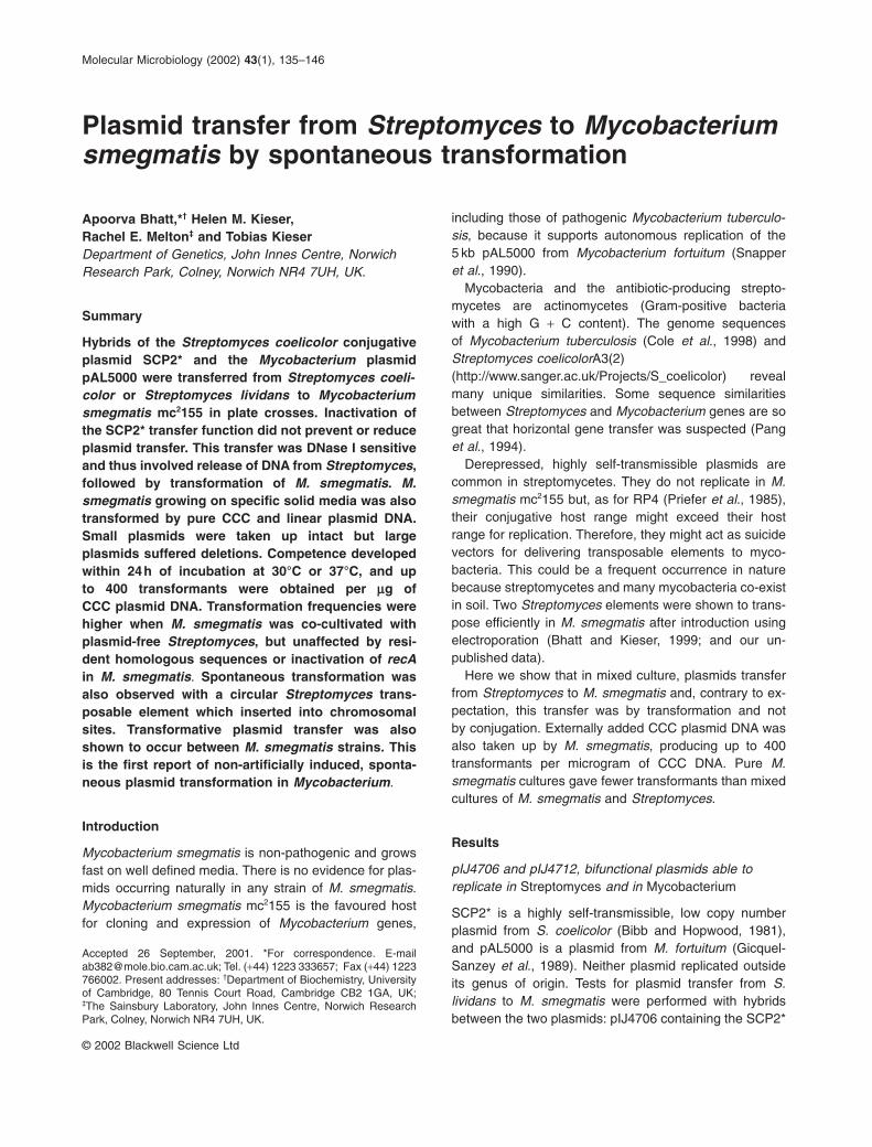
Molecular Microbiology (2002) 43(1), 135–146
© 2002 Blackwell Science Ltd
Plasmid transfer from Streptomyces to Mycobacteriumsmegmatis by spontaneous transformation
including those of pathogenic Mycobacterium tuberculo-sis, because it supports autonomous replication of the5 kb pAL5000 from Mycobacterium fortuitum (Snapperet al., 1990).
Mycobacteria and the antibiotic-producing strepto-mycetes are actinomycetes (Gram-positive bacteria with a high G + C content). The genome sequences of Mycobacterium tuberculosis (Cole et al., 1998) andStreptomyces coelicolorA3(2) (http://www.sanger.ac.uk/Projects/S_coelicolor) revealmany unique similarities. Some sequence similaritiesbetween Streptomyces and Mycobacterium genes are sogreat that horizontal gene transfer was suspected (Panget al., 1994).
Derepressed, highly self-transmissible plasmids arecommon in streptomycetes. They do not replicate in M.smegmatis mc2155 but, as for RP4 (Priefer et al., 1985),their conjugative host range might exceed their host range for replication. Therefore, they might act as suicidevectors for delivering transposable elements to myco-bacteria. This could be a frequent occurrence in naturebecause streptomycetes and many mycobacteria co-existin soil. Two Streptomyces elements were shown to trans-pose efficiently in M. smegmatis after introduction usingelectroporation (Bhatt and Kieser, 1999; and our un-published data).
Here we show that in mixed culture, plasmids transferfrom Streptomyces to M. smegmatis and, contrary to ex-pectation, this transfer was by transformation and not by conjugation. Externally added CCC plasmid DNA wasalso taken up by M. smegmatis, producing up to 400transformants per microgram of CCC DNA. Pure M.smegmatis cultures gave fewer transformants than mixedcultures of M. smegmatis and Streptomyces.
Results
pIJ4706 and pIJ4712, bifunctional plasmids able toreplicate in Streptomyces and in Mycobacterium
SCP2* is a highly self-transmissible, low copy numberplasmid from S. coelicolor (Bibb and Hopwood, 1981), and pAL5000 is a plasmid from M. fortuitum (Gicquel-Sanzey et al., 1989). Neither plasmid replicated outside its genus of origin. Tests for plasmid transfer from S. lividans to M. smegmatis were performed with hybridsbetween the two plasmids: pIJ4706 containing the SCP2*
Apoorva Bhatt,*† Helen M. Kieser, Rachel E. Melton‡ and Tobias KieserDepartment of Genetics, John Innes Centre, NorwichResearch Park, Colney, Norwich NR4 7UH, UK.
Summary
Hybrids of the Streptomyces coelicolor conjugativeplasmid SCP2* and the Mycobacterium plasmidpAL5000 were transferred from Streptomyces coeli-color or Streptomyces lividans to Mycobacteriumsmegmatis mc2155 in plate crosses. Inactivation ofthe SCP2* transfer function did not prevent or reduceplasmid transfer. This transfer was DNase I sensitiveand thus involved release of DNA from Streptomyces,followed by transformation of M. smegmatis. M.smegmatis growing on specific solid media was alsotransformed by pure CCC and linear plasmid DNA.Small plasmids were taken up intact but large plasmids suffered deletions. Competence developedwithin 24h of incubation at 30°C or 37°C, and up to 400 transformants were obtained per mg of CCC plasmid DNA. Transformation frequencies werehigher when M. smegmatis was co-cultivated withplasmid-free Streptomyces, but unaffected by resi-dent homologous sequences or inactivation of recAin M. smegmatis. Spontaneous transformation wasalso observed with a circular Streptomyces trans-posable element which inserted into chromosomalsites. Transformative plasmid transfer was alsoshown to occur between M. smegmatis strains. Thisis the first report of non-artificially induced, sponta-neous plasmid transformation in Mycobacterium.
Introduction
Mycobacterium smegmatis is non-pathogenic and growsfast on well defined media. There is no evidence for plas-mids occurring naturally in any strain of M. smegmatis.Mycobacterium smegmatis mc2155 is the favoured hostfor cloning and expression of Mycobacterium genes,
Accepted 26 September, 2001. *For correspondence. [email protected]; Tel. (+44) 1223 333657; Fax (+44) 1223766002. Present addresses: †Department of Biochemistry, Universityof Cambridge, 80 Tennis Court Road, Cambridge CB2 1GA, UK; ‡The Sainsbury Laboratory, John Innes Centre, Norwich ResearchPark, Colney, Norwich NR4 7UH, UK.
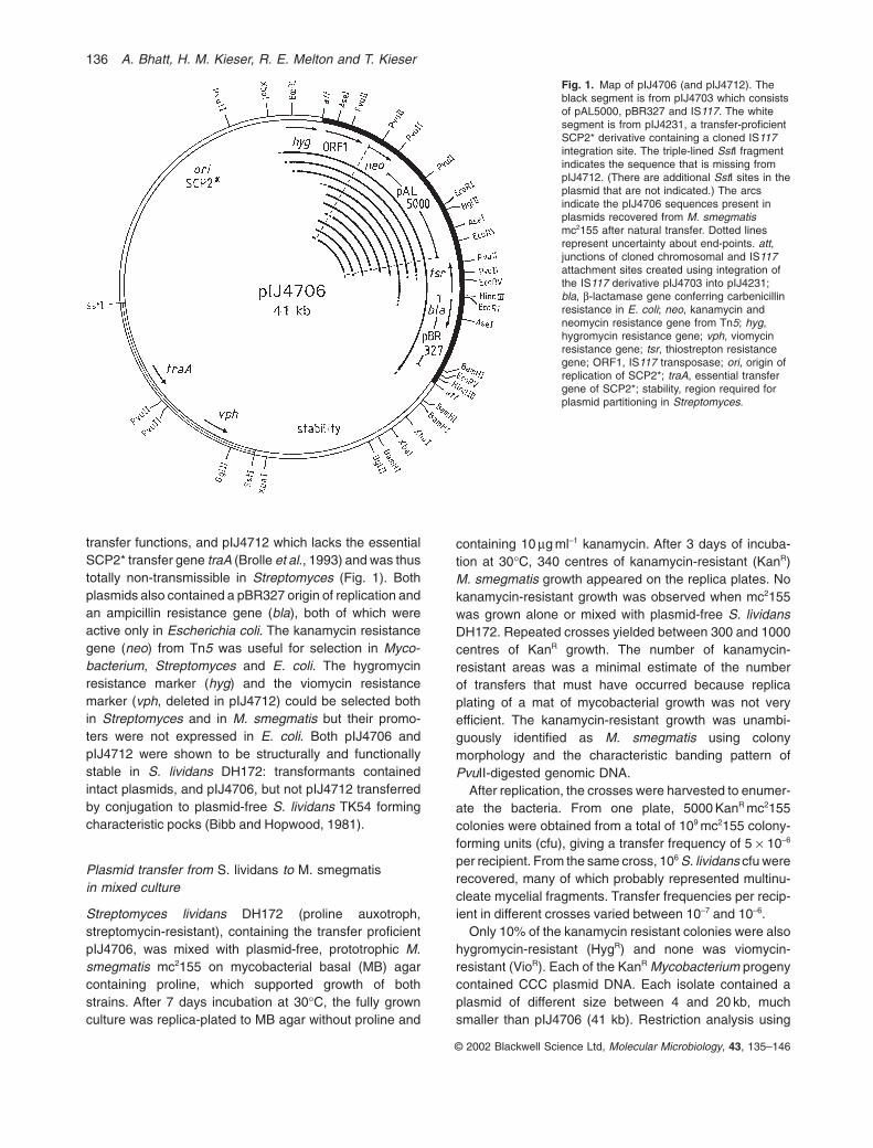
transfer functions, and pIJ4712 which lacks the essentialSCP2* transfer gene traA (Brolle et al., 1993) and was thustotally non-transmissible in Streptomyces (Fig. 1). Bothplasmids also contained a pBR327 origin of replication andan ampicillin resistance gene (bla), both of which wereactive only in Escherichia coli. The kanamycin resistancegene (neo) from Tn5 was useful for selection in Myco-bacterium, Streptomyces and E. coli. The hygromycin resistance marker (hyg) and the viomycin resistance marker (vph, deleted in pIJ4712) could be selected both in Streptomyces and in M. smegmatis but their promo-ters were not expressed in E. coli. Both pIJ4706 andpIJ4712 were shown to be structurally and functionallystable in S. lividans DH172: transformants contained intact plasmids, and pIJ4706, but not pIJ4712 transferredby conjugation to plasmid-free S. lividans TK54 formingcharacteristic pocks (Bibb and Hopwood, 1981).
Plasmid transfer from S. lividans to M. smegmatisin mixed culture
Streptomyces lividans DH172 (proline auxotroph, streptomycin-resistant), containing the transfer proficientpIJ4706, was mixed with plasmid-free, prototrophic M.smegmatis mc2155 on mycobacterial basal (MB) agarcontaining proline, which supported growth of bothstrains. After 7 days incubation at 30∞C, the fully grownculture was replica-plated to MB agar without proline and
containing 10 mg ml–1 kanamycin. After 3 days of incuba-tion at 30∞C, 340 centres of kanamycin-resistant (KanR)M. smegmatis growth appeared on the replica plates. Nokanamycin-resistant growth was observed when mc2155was grown alone or mixed with plasmid-free S. lividansDH172. Repeated crosses yielded between 300 and 1000centres of KanR growth. The number of kanamycin-resistant areas was a minimal estimate of the number of transfers that must have occurred because replicaplating of a mat of mycobacterial growth was not very efficient. The kanamycin-resistant growth was unambi-guously identified as M. smegmatis using colony morphology and the characteristic banding pattern ofPvuII-digested genomic DNA.
After replication, the crosses were harvested to enumer-ate the bacteria. From one plate, 5000 KanR mc2155colonies were obtained from a total of 109 mc2155 colony-forming units (cfu), giving a transfer frequency of 5 ¥ 10–6
per recipient. From the same cross, 106 S. lividans cfu wererecovered, many of which probably represented multinu-cleate mycelial fragments. Transfer frequencies per recip-ient in different crosses varied between 10–7 and 10–6.
Only 10% of the kanamycin resistant colonies were alsohygromycin-resistant (HygR) and none was viomycin-resistant (VioR). Each of the KanR Mycobacterium progenycontained CCC plasmid DNA. Each isolate contained aplasmid of different size between 4 and 20 kb, muchsmaller than pIJ4706 (41 kb). Restriction analysis using
© 2002 Blackwell Science Ltd, Molecular Microbiology, 43, 135–146
136 A. Bhatt, H. M. Kieser, R. E. Melton and T. Kieser
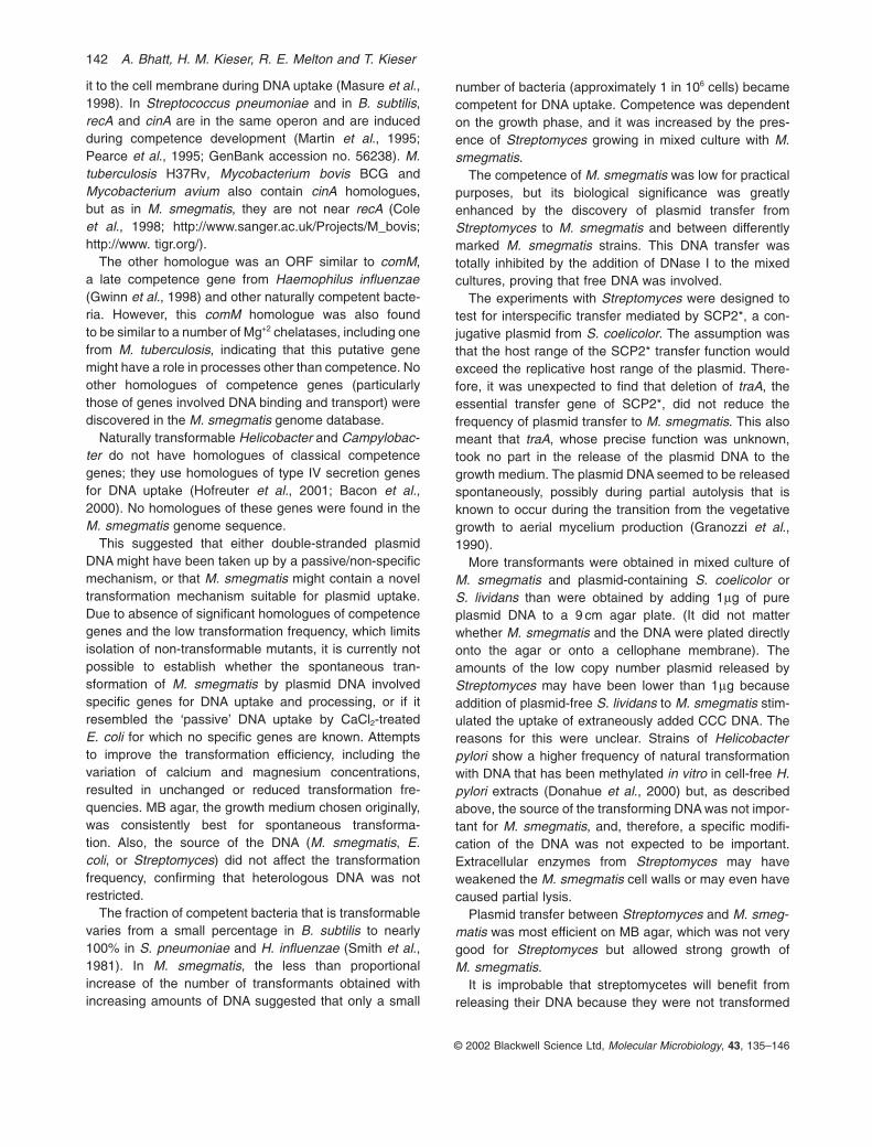
Fig. 1. Map of pIJ4706 (and pIJ4712). Theblack segment is from pIJ4703 which consistsof pAL5000, pBR327 and IS117. The whitesegment is from pIJ4231, a transfer-proficientSCP2* derivative containing a cloned IS117integration site. The triple-lined SstI fragmentindicates the sequence that is missing frompIJ4712. (There are additional SstI sites in theplasmid that are not indicated.) The arcsindicate the pIJ4706 sequences present inplasmids recovered from M. smegmatismc2155 after natural transfer. Dotted linesrepresent uncertainty about end-points. att,junctions of cloned chromosomal and IS117attachment sites created using integration ofthe IS117 derivative pIJ4703 into pIJ4231;bla, b-lactamase gene conferring carbenicillinresistance in E. coli; neo, kanamycin andneomycin resistance gene from Tn5; hyg,hygromycin resistance gene; vph, viomycinresistance gene; tsr, thiostrepton resistancegene; ORF1, IS117 transposase; ori, origin ofreplication of SCP2*; traA, essential transfergene of SCP2*; stability, region required forplasmid partitioning in Streptomyces.
Plasmid transfer from Streptomyces to Mycobacterium smegmatis 137
AseI, BglII, EcoRI, EcoRV, PstI, PvuII, SstI, HindIII, XbaIand XhoI and Southern hybridization revealed, however,that all the plasmids were derived from pIJ4706 (data notshown). All of them contained the replication region ofpAL5000 and the adjacent kanamycin resistance gene(Fig. 1). Nine of 10 plasmids analysed lacked the pBR327replication origin. BclI digestion showed that the plasmidswere not dam-methylated, excluding the possibility thatthey might have originated from contaminating E. coli.None of the isolates contained the SCP2* replicationregion. These results indicated that plasmid DNA, but notthe complete plasmids, had been transferred during thecrosses from S. lividans to M. smegmatis.
Plasmid transfer from Streptomyces to M. smegmatiswas independent of the SCP2* transfer function
Surprisingly, crosses involving the transfer-deficientplasmid, pIJ4712 (Fig. 1) produced similar numbers ofkanamycin-resistant mc2155 as pIJ4706. Again, deletedforms of pIJ4712 were found in all KanR M. smegmatis.Control experiments were performed to ensure that this unexpected result was correct: the same result was obtained when the cross was repeated using repurified DH172/pIJ4712 or independently obtainedDH172/pIJ4712 transformants. Non-transmissibility of theplasmid between Streptomyces strains was reconfirmed.Also, it was shown that pIJ4712 did not transfer from oneS. lividans strain to another in a cross that also containedmc2155, ruling out the possibility that conjugative transfermight be induced using a hypothetical M. smegmatisfertility factor. These results proved that SCP2* transferfunctions played no role in the intergeneric plasmid trans-fer from S. lividans to M. smegmatis.
Plasmid transfer was DNase I-sensitive
Addition of 5 ml (75 units) bovine pancreatic deoxyribonu-clease I (DNase I) to the cross at the time of plating prevented the appearance of KanR Mycobacterium smeg-matis, indicating that plasmid transfer involved free DNAand was, therefore, by transformation, involving free DNA(heat-inactivated DNase I did not prevent plasmid trans-fer from DH172 to M. smegmatis mc2155, and addition ofDNase I to a S. lividans cross on supplemented MB agardid not prevent or reduce plasmid transfer from DH172 toanother S. lividans strain, TK54.)
Heating the plasmid-containing Streptomyces myce-lium suspended in 20% glycerol for 10 min at 85∞C alsoprevented plasmid transfer. Heat treatment killed theStreptomyces mycelium but did not destroy free CCCplasmid DNA (Kieser, 1984). Therefore, it seemed that S.lividans released plasmid DNA during growth in a formthat was suitable for transformation.
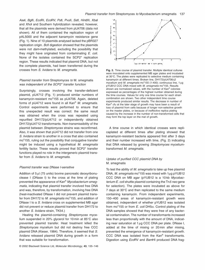
A time course in which identical crosses were repli-caplated at different times after plating showed thatkanamycin-resistant bacteria appeared first after 3 daysand their number increased with time, (Fig. 2) indicatingthat DNA released by growing Streptomyces mycelium-transformed M. smegmatis.
Uptake of purified CCC plasmid DNA by M. smegmatis
To test the ability of M. smegmatis to take up free plasmidDNA, M. smegmatis mc2155 was mixed with 1mg pYUB12CCC DNA on MB agar (pYUB12 is a 10 kb Mycobac-terium-E. coli shuttle plasmid containing the Tn5 neo genefor selection). The plates were incubated as above for7 days at 30∞C and then replicated to the same mediumcontaining kanamycin. From independent experiments,150–400 areas of kanamycin-resistant growth wereobtained, independent of whether pYUB12 was isolatedfrom mc2155 or from E. coli DH5a. Control plating of theDNA samples showed that they were free of any bacter-ial contamination. The number of transformants increasedless than proportionally with the amount of DNA, indicat-ing near saturation at 1 mg CCC DNA per plate. DNase I,added at the time of mixing or 20 min after mixing, prevented the emergence of kanamycin-resistant growth.CCC DNA was isolated from the KanR transformants.Digestion using EcoRV and BamHI produced DNA frag-
© 2002 Blackwell Science Ltd, Molecular Microbiology, 43, 135–146
Fig. 2. Time course of plasmid transfer. Multiple identical cultureswere inoculated onto supplemented MB agar plates and incubatedat 30∞C. The plates were replicated to selective medium containingkanamycin at different times. Broken line, DH172/pIJ4706D2mycelium and M. smegmatis mc2155 (1:400); continuous line, 1 mgpYUB12 CCC DNA mixed with M. smegmatis mc2155. Resultsshown are normalized values, with the number of KanR coloniesexpressed as percentages of the highest number obtained duringthe time courses. Values for only one time course for each straincombination are shown. Two other independent time courseexperiments produced similar results. The decrease in number ofKanR cfu at the later stage of growth may have been a result of loss of plasmid from cells because of longer non-selective growthon the master plates, or because of ineffective replica platingcaused by the increase in the number of non-transformed cells thatmay form the top layer on the mat of growth.

ments that were identical in size to those from pYUB12isolated from E. coli, i.e. pYUB12 did not suffer detectabledeletions.
pIJ4712 DNA also transformed mc2155; 37 KanR cfuwere obtained with 500 ng of plasmid DNA. As in theStreptomyces–M. smegmatis cross, plasmids smallerthan pIJ4712 were recovered from the kanamycin-resistant growth.
Plasmid uptake was not restricted to pAL5000-basedreplicons; CCC DNA of pBP10 (6.27 kb), a derivative of the low copy number M. fortuitum plasmid pMF1(Bachrach et al., 2000), also transformed M. smegmatismc2155 growing on MB agar (32 KanR cfu mg–1 of plasmidDNA). None of the plasmids isolated from 10 pBP10transformants had a deletion.
These results proved that M. smegmatis mc2155 wastransformed by free plasmid DNA during normal growthon MB agar. The number of transformants per microgramof free DNA were approximately 5000-fold lower thanthose achievable using electroporation (Snapper et al.,1990). We refer to this process henceforth as spon-taneous transformation.
Spontaneous transformation of M. smegmatis withlinear plasmid DNA molecules
Gel-purified, BamHI-linearized pYUB12 could also trans-form mc2155 during growth on MB agar, indicating thatlinear molecules were also taken up. Transformation frequencies were between a quarter and half of thoseobtained with CCC DNA. No transformants were obtainedwith heat-denatured pYUB12, suggesting that single-stranded DNA was not a good substrate. Attempts totransform M. smegmatis with antibiotic resistance cassette-marked chromosomal DNA fragments (5 mggenomic DNA), and suicide gene replacement constructs(1mg plasmid DNA), were not successful, indicating that integration of homologous DNA into the chromo-some after spontaneous transformation was infrequent, ifnot absent.
Spontaneous transformation of M. smegmatis by atransposable element
pIJ4696 (7.6 kb, Bhatt and Kieser, 1999), a HygR
derivative of the S. coelicolor A3(2) transposable elementIS117, transformed M. smegmatis growing on MB agar to give HygR colonies. From two independent experi-ments, six and 14 HygR colonies were obtained per microgram of pIJ4696. Southern analysis showed thatpIJ4696 had integrated via the IS117 attachment site intothe preferred chromosomal integration sites A or B, indicating that transposition had occurred (Bhatt andKieser, 1999).
Spontaneous transformation by plasmid DNA is recAindependent
Generally, in known naturally competent bacteria,monomeric plasmids without homology to the chromo-some give frequencies 103–104-fold lower than thoseobtained with chromosomal DNA (Smith et al., 1981;Stewart and Carlson, 1986; Lorenz and Wackernagel,1994). In Streptococcus, multiple transformation is neces-sary for the rescue of monomeric plasmids by annealing ofseveral overlapping single-stranded fragments, followedby repair synthesis (Saunders and Guild, 1981). Thisprocess is recA-independent. In Bacillus subtilis, rescue ofmonomeric plasmids using this pathway does not occur.Monomeric plasmids can however, be rescued in Bacillususing a recA-dependent process if they share a region ofhomology with the host DNA (Canosi et al., 1981).
In spontaneous transformation of M. smegmatis, recAdid not seem to play any role: HS42, a recA-deleted deriv-ative of mc2155, constructed using gene replacement(Papavinasasundaram et al., 1998), was transformed bypYUB12 DNA with similar efficiency as the recombination-proficient strain mc2155 (an average of 200 KanR cfu permicrogram of pYUB12 were obtained with both strains inparallel experiments). Also, both mc2155 and mc2155/pYUB12 were transformed with equal frequencies bypUH4, a pYUB12 derivative containing hyg instead ofneo, indicating that the presence of a homologous DNAsequence in the recipient cell had no effect on sponta-neous plasmid transformation.
Cause of deletions of pIJ4706 and pIJ4712
When pIJ4706 and pIJ4712 were introduced into M.smegmatis using electroporation, the KanR transformantscontained smaller plasmids similar to those recoveredafter transfer from S. lividans to M. smegmatis on MB agar plates. Therefore, it seemed probable that theseplasmids were structurally unstable in M. smegmatis, pos-sibly because of the presence of a ‘poison’ sequence.One KanR VioR HygR transformant, obtained after electro-poration, contained a relatively large plasmid pIJ4706D2with intact SCP2* replication and transfer regions, butlacking the pBR327 functions and the tsr gene. pIJ4706D2remained intact and displayed the expected phenotypewhen introduced by transformation into S. lividans proto-plasts and also when re-introduced by electroporation intoM. smegmatis. Surprisingly, however, when this plasmidwas transferred in a cross from S. lividans, further plasmiddeletions were observed in all kanamycin-resistant M.smegmatis progeny: only 4% were HygR VioR, 78% werehygromycin- and viomycin-sensitive (HygS and VioS), 8%were HygS VioR and 10% were HygR VioS. It seemed significant that 12% of the progeny was VioR, while no
© 2002 Blackwell Science Ltd, Molecular Microbiology, 43, 135–146
138 A. Bhatt, H. M. Kieser, R. E. Melton and T. Kieser

Plasmid transfer from Streptomyces to Mycobacterium smegmatis 139
VioR M. smegmatis progeny was found after crossesinvolving the parental pIJ4706.
These results suggested that the deletion in pIJ4706D2created a plasmid that replicated stably in M. smegmatis,but suffered deletions in the course of spontaneous trans-formation. The deletions were, however, less severe thanin the case of pIJ4706 because 12% VioR progeny wasrecovered.
Streptomyces lividans has an unusual DNA modificationsystem that results in DNA degradation in pulsed-field gelsunless the buffer contains thiourea (Dyson and Evans,1998; Zhou et al., 1988). A similar DNA modification mayalso be present in M. smegmatis (Bhatt and Kieser, 1999),but S. coelicolor A3(2) DNA lacks this modification.
pIJ4706D2 was introduced into S. coelicolor J1501 (his-tidine and uracil auxotroph, streptomycin-resistant) byprotoplast transformation. One KanR HygR VioR, thiostrep-ton-sensitive transformant of this strain was used as adonor. The plasmid transferred from this strain to mc2155at frequencies similar to those obtained with S. lividansDH172. As was observed with the DH172-mc2155crosses, only a few of the KanR mc2155 cells obtainedafter replica plating were also HygR and VioR, indicatingdeletions in the plasmid during or after transfer. In onecross from a total of 58 KanR mc2155 colonies analysed,20 were HygS and VioS, 22 were HygRVioS, four wereHygSVioR and 12 were HygRVioR. These results made itimprobable that the S. lividans DNA modification was thecause of plasmid instability during transfer of pIJ4706D2to M. smegmatis.
Size restrictions in the DNA uptake process could havebeen a factor responsible for the deletions in pIJ4706,pIJ4712 and pIJ4706D2, all of which were more than25 kb in size. To test this, we grew M. smegmatis mixedwith CCC DNA of a pAL5000-based cosmid containinghistidine bio-synthesis genes (HisCos13, approximately30–32 kb, a gift from N. Stoker and T. Parish), on MB agar. Such constructs did not undergo deletion whenintroduced using electroporation into M. smegmatis(Hinshelwood and Stoker, 1992). After spontaneoustransformation, however, all five KanR transformantsanalysed contained deleted derivatives of HisCos13. Thissupported the hypothesis that spontaneous transforma-tion restricted the size of DNA molecules.
Plasmid DNA uptake by M. smegmatis was enhancedby the presence of S. lividans
It seemed remarkable that the crosses between S. lividans containing a low copy number plasmid (1–2 perchromosome), and M. smegmatis produced more KanR
transformants (up to 103) than were obtained using one microgram (approximately 1010 molecules) of pureplasmid DNA. Considering that the growth of S. lividans
was limited by the competing M. smegmatis, and that the copy numbers of pIJ4706 and pIJ4712 were low inStreptomyces, it seemed improbable that 1mg of plas-mid DNA was released by the plate culture.
To test whether growing Streptomyces increased thecompetence of M. smegmatis in mixed culture, the fol-lowing experiment was performed: pYUB12 DNA (isolatedfrom E. coli DH5a or from M. smegmatis mc2155) wasadded to a suspension of M. smegmatis mc2155 andmixed. Equal samples of the plasmid-cell mix (each con-taining approximately 107 cfu and 1mg plasmid DNA) werethen mixed with either a 30 ml suspension of plasmid-freeDH172 mycelia in 20% glycerol, or 30 ml 20% glycerol asa control, and spread on supplemented MB agar. Theplates were incubated at 30∞C for 7 days and then repli-cated to MB agar containing kanamycin. Plasmid uptakeby mc2155 was 4–7 times higher in the presence of S.lividans (Fig. 3). No KanR Streptomyces were recoveredfrom the plates, indicating that plasmids were directlytaken up by M. smegmatis and not introduced via Streptomyces.
Optimizing culture conditions for DNA uptake
Increasing the spontaneous transformation frequency of
© 2002 Blackwell Science Ltd, Molecular Microbiology, 43, 135–146
Fig. 3. Pure plasmid DNA uptake by M. smegmatis mc2155 wasenhanced by the presence of plasmid-free S. lividans DH172.Replica plates on MB agar +10 mg ml–1 of kanamycin and: (a) noDNA control, (b) No DNA, but with plasmid-free S. lividans DH172,(c) 1mg of pYUB12, and (d) DH172 and 1mg of pYUB12. Plate (c)contains 146 cfu and plate (d) contains 1145 cfu.
M. smegmatis would greatly facilitate the study of themechanism of DNA uptake and the search for sponta-neous transformation deficient M. smegmatis mutants.Unfortunately, most attempts to optimize spontaneoustransformation in crosses or with pure DNA resulted indecreased frequencies.
Mycobacterium smegmatis was transformed with pureplasmid DNA during growth on MB agar, 7H11 agar andStreptomyces minimal medium (Kieser et al., 2000). MBagar gave, by far, the highest transformation frequencies;~3–7 times more colonies than those obtained on 7H11and on Streptomyces minimal medium (MM). Addingextra Ca2+ and/or Mg2+ had no effect.
Incubation of M. smegmatis with plasmid DNA at 37∞Cgave approximately three times more transformants thanincubation at 30∞C or 40∞C. Diluting or concentrating thecultures before spreading did not increase the transfor-mation frequency. The quality of the plasmid DNA did not affect transformation, as similar frequencies wereobtained with Qiagen column-purified plasmid DNA aswith crude plasmid DNA obtained using alkaline lysis. Toprevent possible diffusion of the plasmid DNA away fromthe cells into the agar, cultures and plasmid DNA wereplated on dialysis membranes placed on MB agar. Thebacteria grew well on the membranes, but the transforma-tion frequencies were not increased. This indicated thatthere was no loss of accessible plasmid DNA by diffusion.
No spontaneous plasmid transformation was observedwhen the strains were co-cultivated with, or withoutshaking at 30∞C in MB broth before plating on selectivemedium.
Spontaneous transformants are not mutants withincreased competence
Approximately one in 106 cells was transformed duringgrowth on MB agar by saturating amounts of free plasmidDNA. Either only a small fraction of the cells was com-petent for DNA uptake or the transformed cells might havebeen mutants with increased competence. Mycobac-terium smegmatis strains transformed with pYUB12 orpIJ4696 during plate culture did not show an increasedfrequency of re-transformation (during growth on MBagar) by plasmid DNA with a different resistance marker,excluding the possibility that transformation selects highlycompetent M. smegmatis mutants.
Competence development is regulated in M. smegmatis
As indicated above, colonies of M. smegmatis trans-formed by plasmid released from Streptomyces wereobtained after 3 days of incubation at 30∞C. However,competence might have developed much earlier, andrelease of DNA from Streptomyces may have been the
limiting factor for obtaining early transformants. To esti-mate the time required for competence development,identical plates containing pYUB12 DNA and M. smeg-matis were replica-plated at different times after mixing.KanR colonies were obtained within 24 h of incubation at30∞C or 37∞C (Fig. 2).
To determine how long M. smegmatis remained com-petent during growth on MB agar, the following experi-ment was performed: after a culture of M. smegmatis wasspread on supplemented MB agar, 1mg of pYUB12 wasspread with a loop in an area ~1.5 cm wide on the surfaceof the agar plate. Then, the plate was incubated at 37∞Cand pYUB12 DNA was added to separate sections on thesame plate at regular intervals of 24 h (at the later time-points when there was mat growth on the plates, the DNAsolution and the cells were mixed using a loop). The lastaddition of DNA was after 7 days of incubation; 2 h afterthis addition, the plate was replicated on MB agar con-taining kanamycin. A similar experiment was also per-formed using pUH4 (pAL5000 derivative, HygR; Stolt andStoker, 1996). In this case, the plates were replicated on 7H11 agar containing hygromycin. After incubation for 3–5 days at 37∞C, the replica plates were scored.Between 18 and 25 antibiotic-resistant cfu were obtainedwhen the plasmid DNA was added at the time of platingor 24 h after plating. The number of cfu increased to24–40 at 48 h and then declined to 1–10 at 72 h afterplating. No antibiotic-resistant cfu were obtained when theplasmid DNA was added later at 96, 120, 144 and 168 h,even when all the growth from the areas of DNA additionwas spread on selective medium.
Plasmid transfer between M. smegmatis strains
Mycobacterium smegmatis mc2155 containing pYUB12(KanR) was grown on MB agar with another mc2155 strainthat contained pIJ4696 (HygR IS117 derivative) integratedinto site B (Bhatt and Kieser, 1999). Between 32 and 210KanR HygR colonies were recovered from independentcultures obtained after 7 days of incubation at 30∞C or37∞C. No KanR HygR colonies were obtained when the cultures were grown in the presence of DNase I.
The KanR HygR colonies contained deleted versions ofpYUB12, and polymerase chain reaction (PCR) confirmedthat pIJ4696 was present at siteB. This indicated that, in a manner similar to transfer from Streptomyces toMycobacterium, plasmid DNA was being transferred byspontaneous transformation from one M. smegmatis strainto the other.
A similar cross with mc2155/pUH4 (HygR) and mc2155containing a chromosomal insertion of Tn4560 (VioR,Chung, 1987), gave 33 HygRVioR transformants, con-firming that plasmid transfer did take place between M.smegmatis strains.
© 2002 Blackwell Science Ltd, Molecular Microbiology, 43, 135–146
140 A. Bhatt, H. M. Kieser, R. E. Melton and T. Kieser
Plasmid transfer from Streptomyces to Mycobacterium smegmatis 141
To rule out the possibility that these transformants were recipients of conjugative transfer of chromo-somal markers as has been reported (Parsons et al.,1998), the following cross was also made: a KanR
HygR, pYUB12-containing transformant obtained from themc2155/pYUB12 ¥ mc2155::pIJ4696 cross was grown inmixed culture with mc2155::Tn4560. Replica plating after 7 days of growth at 37∞C gave 32 KanRVioR coloniesthat were found to be HygS. This indicated that theplasmid, but not the chromosomal HygR marker, hadtransferred from mc2155::pIJ4696 to mc2155::Tn4560. As with the other crosses, this transfer was DNase I-sensitive.
Discussion
There are two previous reports about natural competenceof mycobacteria for transformation by chromosomal DNA,but transformation by plasmid DNA (natural or sponta-neous) has not been reported before. Mycobacteriumsmegmatis ATCC 607, the parent of strain mc2155, wasone of the competent strains (Tsukamura et al., 1960;Norgard and Imaeda, 1978).
It was not surprising that transformation by CCCplasmid DNA had not been noticed during the develop-ment of transformation using electroporation because M.smegmatis ATCC 607 does not support replication of thepAL5000-derived vectors. The rare transformants, suchas strain mc2155, were all mutants that allowed replica-tion of pAL5000 derivatives (Snapper et al., 1988; 1990).The spontaneous plasmid transformants obtained in thisstudy, however, did not give an increased transformationefficiency on retransformation and, therefore, probablyrepresented normal mc2155.
Spontaneous transformation of M. smegmatis duringgrowth on agar plates was demonstrated with severalpAL5000-derived plasmids, with the pMF1-derived vectorpBP10 (Bachrach et al., 2000) and with the IS117-derivedintegrating vector pIJ4696 (Bhatt and Kieser, 1999). Thediversity of DNA sequences involved made it unlikely that Mycobacterium-specific uptake sequences wererequired as for natural transformation in some Gram-negative bacteria (Sisco and Smith, 1979).
The well studied competence mechanisms of Gram-positive and Gram-negative bacteria involve approxi-mately a dozen genes which are scattered around thechromosome (reviewed by Goodgal, 1982; Dubnau,1991; Fussenegger et al., 1997; Dubnau, 1999; Tortosaand Dubnau, 1999). The mechanisms are optimized forthe uptake of linear DNA fragments; before uptake, DNAbinds to the cell surface and is nicked by competence-related endonucleases generating fragments of maxi-mally 15 kb. One DNA strand is taken up whereas the other strand is degraded to nucleosides. The single-
stranded DNA then recombines with homologousgenomic sequences (between 10–4 and 10–2 chromosomalmarker transformants per viable cell). Competence devel-opment includes an increase in the levels of homologousrecombination; in Bacillus, about 70% of the internalizedhomologous single-stranded DNA becomes integrated(Dubnau, 1991). Monomeric plasmid DNA, without homol-ogy to the recipient genome, transforms with low effi-ciency. Transformation is possible with small multimericplasmids that can be rescued using self-recombination orby co-transformation of complementary plasmid DNAfragments, followed by spontaneous annealing andfurther processing to reconstitute the original plasmid(Saunders and Guild, 1981).
Under our conditions, M. smegmatis mc2155 was trans-formed by plasmids but not by chromosomal DNA.Mycobacterium smegmatis spontaneous plasmid trans-formation was shown to be independent of recA. LinearpYUB12 (10 kb) DNA did transform, but the efficiency was~2–4 times lower than for CCC plasmids. This seemed toindicate that CCC DNA was taken up preferentially, but itwas also possible that the linearized DNA was degradedby some extracellular nuclease during the long timebefore uptake. Circular molecules would not require anyprocessing, and linearized molecules require circulariza-tion using cytoplasmic ligase. Denatured CCC plasmidsgave no transformants, supporting the assumption thatdouble-stranded DNA was the substrate for transforma-tion in M. smegmatis.
This was reminiscent of calcium chloride/heat shock-mediated transformation of E. coli that does not involvespecific competence genes. It was, however, difficult toimagine how negatively charged, hydrophilic DNA couldenter through the thick waxy Mycobacterium cell wallwithout an elaborate DNA uptake mechanism or thedriving force generated by the 2500 V electroporationpulse. In E. coli, smaller plasmids transformed more effi-ciently, and there was a gradual reduction in the efficiencywith increasing plasmid size: doubling the plasmid sizehalves the efficiency per molecule (Hanahan, 1983). Inour experiments, selection for pre-existing deleted deriva-tives of the large plasmids seemed improbable becauseno deleted plasmids were observed on ethidium bromide-stained agarose gels, and, because the transformationfrequencies were very similar for small plasmids that weretaken up intact and for large plasmids that suffered deletions.
The obligatory deletion of the larger plasmids wassimilar to DNA uptake by the known natural competencesystems. A search of the incomplete M. smegmatisgenome sequence at http://www.tigr.org/ found two homo-logues of known competence genes. One of these wasthe conserved competence-related gene cinA whoseprotein product was hypothesized to bind RecA and direct
© 2002 Blackwell Science Ltd, Molecular Microbiology, 43, 135–146
it to the cell membrane during DNA uptake (Masure et al.,1998). In Streptococcus pneumoniae and in B. subtilis,recA and cinA are in the same operon and are inducedduring competence development (Martin et al., 1995;Pearce et al., 1995; GenBank accession no. 56238). M.tuberculosis H37Rv, Mycobacterium bovis BCG andMycobacterium avium also contain cinA homologues, but as in M. smegmatis, they are not near recA (Cole et al., 1998; http://www.sanger.ac.uk/Projects/M_bovis;http://www. tigr.org/).
The other homologue was an ORF similar to comM, a late competence gene from Haemophilus influenzae(Gwinn et al., 1998) and other naturally competent bacte-ria. However, this comM homologue was also found to be similar to a number of Mg+2 chelatases, including onefrom M. tuberculosis, indicating that this putative genemight have a role in processes other than competence. Noother homologues of competence genes (particularlythose of genes involved DNA binding and transport) werediscovered in the M. smegmatis genome database.
Naturally transformable Helicobacter and Campylobac-ter do not have homologues of classical competencegenes; they use homologues of type IV secretion genesfor DNA uptake (Hofreuter et al., 2001; Bacon et al.,2000). No homologues of these genes were found in theM. smegmatis genome sequence.
This suggested that either double-stranded plasmidDNA might have been taken up by a passive/non-specificmechanism, or that M. smegmatis might contain a noveltransformation mechanism suitable for plasmid uptake.Due to absence of significant homologues of competencegenes and the low transformation frequency, which limitsisolation of non-transformable mutants, it is currently notpossible to establish whether the spontaneous tran-sformation of M. smegmatis by plasmid DNA involvedspecific genes for DNA uptake and processing, or if itresembled the ‘passive’ DNA uptake by CaCl2-treated E. coli for which no specific genes are known. Attemptsto improve the transformation efficiency, including thevariation of calcium and magnesium concentrations,resulted in unchanged or reduced transformation fre-quencies. MB agar, the growth medium chosen originally,was consistently best for spontaneous transforma-tion. Also, the source of the DNA (M. smegmatis, E. coli, or Streptomyces) did not affect the transformation frequency, confirming that heterologous DNA was notrestricted.
The fraction of competent bacteria that is transformablevaries from a small percentage in B. subtilis to nearly100% in S. pneumoniae and H. influenzae (Smith et al.,1981). In M. smegmatis, the less than proportionalincrease of the number of transformants obtained withincreasing amounts of DNA suggested that only a small
number of bacteria (approximately 1 in 106 cells) becamecompetent for DNA uptake. Competence was dependenton the growth phase, and it was increased by the pres-ence of Streptomyces growing in mixed culture with M.smegmatis.
The competence of M. smegmatis was low for practicalpurposes, but its biological significance was greatlyenhanced by the discovery of plasmid transfer from Streptomyces to M. smegmatis and between differentlymarked M. smegmatis strains. This DNA transfer wastotally inhibited by the addition of DNase I to the mixedcultures, proving that free DNA was involved.
The experiments with Streptomyces were designed totest for interspecific transfer mediated by SCP2*, a con-jugative plasmid from S. coelicolor. The assumption wasthat the host range of the SCP2* transfer function wouldexceed the replicative host range of the plasmid. There-fore, it was unexpected to find that deletion of traA, theessential transfer gene of SCP2*, did not reduce the frequency of plasmid transfer to M. smegmatis. This alsomeant that traA, whose precise function was unknown,took no part in the release of the plasmid DNA to thegrowth medium. The plasmid DNA seemed to be releasedspontaneously, possibly during partial autolysis that isknown to occur during the transition from the vegetativegrowth to aerial mycelium production (Granozzi et al.,1990).
More transformants were obtained in mixed culture ofM. smegmatis and plasmid-containing S. coelicolor or S. lividans than were obtained by adding 1mg of pureplasmid DNA to a 9 cm agar plate. (It did not matterwhether M. smegmatis and the DNA were plated directlyonto the agar or onto a cellophane membrane). Theamounts of the low copy number plasmid released byStreptomyces may have been lower than 1mg becauseaddition of plasmid-free S. lividans to M. smegmatis stim-ulated the uptake of extraneously added CCC DNA. Thereasons for this were unclear. Strains of Helicobacterpylori show a higher frequency of natural transformationwith DNA that has been methylated in vitro in cell-free H.pylori extracts (Donahue et al., 2000) but, as describedabove, the source of the transforming DNA was not impor-tant for M. smegmatis, and, therefore, a specific modifi-cation of the DNA was not expected to be important.Extracellular enzymes from Streptomyces may haveweakened the M. smegmatis cell walls or may even havecaused partial lysis.
Plasmid transfer between Streptomyces and M. smeg-matis was most efficient on MB agar, which was not verygood for Streptomyces but allowed strong growth of M. smegmatis.
It is improbable that streptomycetes will benefit fromreleasing their DNA because they were not transformed
© 2002 Blackwell Science Ltd, Molecular Microbiology, 43, 135–146
142 A. Bhatt, H. M. Kieser, R. E. Melton and T. Kieser
Plasmid transfer from Streptomyces to Mycobacterium smegmatis 143
under our conditions, and the only example in the litera-ture of a naturally transformable Streptomyces strainrefers to the uptake of radioactive DNA, and no genetictransformation has been demonstrated (Roelants et al.,1976).
Mycobacterium smegmatis also released plasmid DNAthat was taken up by DNase I-sensitive transformation.Conjugal transfer of chromosomal markers betweenplasmid-free M. smegmatis strains has been reported(Parsons et al., 1998), but in our experiments no ex-change of chromosomal markers was observed.
It was interesting to find that pYUB12 (10 kb) andpBP10 (6.27 kb) added externally were recovered intactfrom the M. smegmatis transformants, while transfer of pYUB12 between mycobacteria resulted in deletions,suggesting that deleted plasmids may have beenreleased preferentially.
Mycobacteria in soil, and in other natural environments,may be regular recipients of foreign DNA and thus acquiregenes from other organisms using horizontal gene trans-fer. Of particular importance are the soil-dwelling strepto-mycetes, which are a rich reservoir of antibiotic resistancegenes and transposons, two of which have been shown
to function in mycobacteria (Bhatt and Kieser, 1999; andour unpublished results). There are a number of reportssuggesting horizontal transfer of genes into mycobacteria(Davies, 1994; Pang et al., 1994; Davies and Wright,1997); these are based on sequence comparisons. The evidence from this work suggests transformation as an important mechanism for the acquisition of hetero-logous genes by M. smegmatis, and possibly othermycobacteria.
Experimental procedures
Strains, plasmids, culture conditions and geneticmanipulation
Strains and plasmids are listed in Tables 1 and 2. Mycobac-terium smegmatis mc2155 was electroporated with DNA asdescribed before (Snapper et al., 1990). Transformation ofStreptomyces protoplasts and crosses between Strepto-myces strains were performed using standard protocols(Kieser et al., 2000). The following final concentrations ofantibiotics were used for selecting transformants or transcon-jugants: 10 mg ml–1 of kanamycin, 100 mg ml–1 of hygromycinfor M. smegmatis; 50 mg ml–1 of kanamycin, 50 mg ml–1 of
© 2002 Blackwell Science Ltd, Molecular Microbiology, 43, 135–146
Table 1. Strains used in this work.
Strain Brief description Reference
S. lividans DH172 Proline auxotroph, streptomycin-resistant, IS117 preferred Henderson et al. (1990)integration site replaced using erythromycin resistancegene of Saccharopolyspora erythraea
S. lividans TK54 Histidine and leucine auxotroph, spectinomycin-resistant Kieser et al. (2000)S. coelicolor A3(2) J1501 Histidine and uracil auxotroph, streptomycin-resistant Kieser et al. (2000)M. smegmatis mc2155 Mutant of strain mc26 that allows replication of pAL5000 Snapper et al. (1990)M. smegmatis HS42 mc2155 recA deletion mutant Papavinasasundaram et al. (1998)
Table 2. Plasmids used in this work.
Plasmid Brief description Resistance gene Reference
pAL5000 M. fortuitum plasmid, capable of autonomous replication in None Gicquel-Sanzey et al. (1989)many mycobacteria
pYUB12 pAL5000 cloned in pIJ666, a pACYC184 derivative neo Snapper et al. (1988)pUH4 neo gene of pYUB12 is replaced with hyg hyg Stolt and Stoker (1996)pIJ4696 IS117::pBR327 hybrid bla hyg Bhatt and Kieser (1999)pIJ4201 IS117::pBR327 hybrid bla neo tsr Henderson et al. (1989)pIJ4703 Hybrid between pAL5000 and pIJ4201 bla neo tsr This workpIJ4231 SCP2*derivative containing the 200 bp cloned S. lividans hyg vph Henderson et al. (1990)
IS117 preferred integration sitepIJ4711 Transfer-deficient derivative of pIJ4231; traA removed by hyg This work
deleting the 4.6 kb SstI fragment.pIJ4706 pIJ4703 integrated into IS117 integration site of pIJ4231 bla hyg neo tsr vph This workpIJ4706D2 In-vivo deleted derivative of pIJ4706 neo hyg vph This workpIJ4712 pIJ4703 integrated into the IS117 attachment site of pIJ4711 bla hyg neo tsr This workpBP10 Derivative of the low copy number M. fortuitum plasmid pMF1 aph Bachrach et al. (2000)
aph, kanamycin resistance gene from Tn903; bla, b-lactamase; hyg, hygromycin resistance gene from Streptomyces hygroscopicus; neo,neomycin and kanamycin resistance gene from Tn5; tsr, thiotrepton resistance gene from Streptomyces azureus; vph, viomycin resistance genefrom Streptomyces vinaceus.

hygromycin and 50 mg ml–1 of thiostrepton for S. coelicolorand S. lividans; and 30 mg ml–1 of viomycin for all three strains.
Mycobacterial basal (MB) agar and broth
MB agar (100 ml) contained 0.5 mM CaCl2, 0.5 mM MgCl2,1% glucose, 0.05% Tween80, 1mg ml–1 L-asparagine, 200 mlof trace elements solution (Kieser et al., 2000) and 1% basalsolution (10 mg ml–1 of KH2PO4, 25 mg ml–1 of Na2HPO4, 50 mg ml–1 of NH4Cl and 20 mg ml–1 of K2SO4). These com-ponents were added to molten Difco agar (1.5 g in 90 mldeionized water) to give the appropriate concentrations andmixed well before pouring into plates. MB broth contained all the above constituents except the agar (W. R. Jacobs, Jr., personal communication).
Spontaneous transformation of M. smegmatis mc2155
Mycobacterium smegmatis mc2155 was grown to saturationin Lennox broth (Kieser et al., 2000) containing 0.05% Tween80. Then, 200 ml of this culture (approximately 107 cfu) wasmixed with 1mg DNA, spread on MB agar supplemented withproline (supplied in a mixture of amino acids called Tiger milk;Kieser et al., 2000). The plates were incubated at 30∞C for7 days. Transformants were selected by replicating to MBagar containing kanamycin or 7H11 agar (Difco) containinghygromycin. The replica plates were incubated at 30∞C andscored after 3, 4 and 5 days, unless stated otherwise.
Crosses between Streptomyces and M. smegmatismc 2155
A pellet from a 25 ml plasmid-containing S. coelicolor J1501or S. lividans DH172 YEME culture (Kieser et al., 2000) waswashed 2–3 times in 50 ml of 20% glycerol. The pellet wasthen resuspended in 5–10 ml of 20% glycerol and useddirectly for plating, or stored at –20∞C for later use. The restof the procedure was the same as for spontaneous trans-formation, except that approximately 104 cfu of washedStreptomyces mycelium (50 ml) was substituted for pure DNA. Crosses were incubated at 30∞C for 7 days beforereplica plating.
Construction of pIJ4706 and pIJ4712
pAL5000, linearized at its unique BamHI site, was ligated into the unique XhoI site of pIJ4201, a hybrid betweenpBR327 and IS117 that also contains thiostrepton- andkanamycin-resistance determinants. Ligation between theXhoI and BamHI fragments was achieved after partial endfilling of the single-stranded DNA ends (Zabarovsky and Allikmets, 1986). Escherichia coli W5445 was transformedwith the ligation mix and ampicillin-resistant transformantswere selected. The structure and orientation of the resultingplasmid pIJ4703 was confirmed by digestion with PvuII andEcoRV. Transformation of S. lividans DH172/pIJ4231(HygR
VioR) using pIJ4703 (KanR, thiostrepton resistant) producedpIJ4706 (Fig. 1) by integration of pIJ4703 into the IS117 attBsite of pIJ4231. The transfer deficient pIJ4712 (Fig. 1) was
constructed using a similar method: the 4.6 kb SstI fragmentwas deleted from pIJ4231 to give pIJ4711. pIJ4703 wasinserted in vivo into the IS117 attachment site of pIJ4711 togive pIJ4712.
Plasmid isolation from E. coli, Streptomyces and M. smegmatis
Plasmids were isolated using alkaline SDS lysis and potassium acetate precipitation, as described (Sambrooket al., 1989). For Streptomyces and M. smegmatis, themycelium/cells were incubated for at least 1h at 37∞C with 10 mg ml–1 of lysozyme, before lysis. For large scale prepa-rations, the Qiagen Midi plasmid extraction kit was used.
Acknowledgements
We thank Elaine Davis, K.G. Papavinasasundaram, NeilStoker and Tanya Parish for gifts of strains and plasmids. Wealso thank David Hopwood for his helpful comments on thismanuscript. A.B. was supported by a PhD studentship fromthe John Innes Foundation, Norwich, UK.
References
Bachrach, G., Colston, M.J., Bercovier, H., Bar-Nir, D., Anderson, C., and Papavinasasundaram, K.G. (2000) A new single-copy mycobacterial plasmid, pMF1, fromMycobacterium fortuitum which is compatible with thepAL5000 replicon. Microbiology 146: 297–303.
Bacon, D.J., Alm, R.A., Burr, D.H., Hu, L., Kopecko, D.J.,Ewing, C.P. et al. (2000) Involvement of a plasmid in viru-lence of Campylobacter jejuni 81–176. Infect Immun 68:4384–4390.
Bhatt, A., and Kieser, T. (1999) Transposition of IS117 ofStreptomyces coelicolor A3 (2) in Mycobacterium smeg-matis. Microbiology 145: 1201–1207.
Bibb, M.J., and Hopwood, D.A. (1981) Genetic studies of the fertility plasmid SCP2 and its SCP2* variants inStreptomyces coelicolor A3 (2). J Gen Microbiol 126:427–442.
Brolle, D.F., Pape, H., Hopwood, D.A., and Kieser, T. (1993)Analysis of the transfer region of the Streptomyces plasmidSCP2. Mol Microbiol 10: 157–170.
Canosi, U., Iglesias, A., and Trautner, T.A. (1981) Plasmidtransformation in Bacillus subtilis: effects of insertion ofBacillus subtilis DNA into plasmid pC194. Mol Gen Genet181: 434–440.
Chung, S.T. (1987) Tn4556, of a 6.8 kb transposable elementof Streptomyces fradiae. J Bacteriol 169: 4436–4441.
Cole, S.T. Brosch, R., Parkhill, J., Garnier, T., Churcher, C.,Harris, D. et al. (1998) Deciphering the biology ofMycobacterium tuberculosis from the complete genomesequence. Nature 393: 537–544.
Davies, J. (1994) Inactivation of antibiotics and the dissemi-nation of resistance genes. Science 264: 375–382.
Davies, J., and Wright, G.D. (1997) Bacterial resistance to aminoglycoside antibiotics. Trends Microbiol 5: 234–240.
© 2002 Blackwell Science Ltd, Molecular Microbiology, 43, 135–146
144 A. Bhatt, H. M. Kieser, R. E. Melton and T. Kieser
Plasmid transfer from Streptomyces to Mycobacterium smegmatis 145
Donahue, J.P., Israel, D.A., Peek,R.M., Jr, Blaser, M.J., andMiller, G.G. (2000) Overcoming the restriction barrier toplasmid transformation of Helicobacter pylori. Mol Microbiol 37: 1066–1074.
Dubnau, D. (1991) Genetic competence in Bacillus subtilis.Microbiol Rev 55: 395–424.
Dubnau, D. (1999) DNA uptake in bacteria. Annu Rev Microbiol 53: 217–244.
Dyson, P., and Evans, M. (1998) Novel post-replicative DNAmodification in Streptomyces: analysis of the preferredmodification site of plasmid pIJ101. Nucleic Acids Res26: 1248–1253.
Fussenegger, M., Rudel, T., Barten, R., Ryll, R., and Meyer,T.F. (1997) Transformation competence and type-IV pilusbiogenesis in Neisseria gonorrhoeae-a review. Gene 192:125–134.
Gicquel-Sanzey, B., Moniz-Pereira, J., Gheorghiu, M., andRauzier, J. (1989) Structure of pAL5000, a plasmid from M. fortuitum and its utilization in transformation ofmycobacteria. Acta Leprol 7: 208–211.
Goodgal, S.H. (1982) DNA uptake in Haemophilus transfor-mation. Ann Rev Genet 16: 169–192.
Granozzi, C., Billetta, R., Passantino, R., Sollazzo, M., andPuglia, A.M. (1990) A breakdown in macromolecular syn-thesis preceding differentiation in Streptomyces coelicolorA3 (2). J Gen Microbiol 136: 713–716.
Gwinn, M.L., Ramanathan, R., Smith, H.O., and Tomb, J.F.(1998) A new transformation-deficient mutant of Haemophi-lus influenzae Rd with normal DNA uptake. J Bacteriol180: 746–748.
Hanahan, D. (1983) Studies on transformation of Escherichiacoli with plasmids. J Mol Biol 166: 557–580.
Henderson, D.J., Lydiate, D.J., and Hopwood, D.A. (1989)Structural and functional analysis of the mini-circle, a trans-posable element of Streptomyces coelicolor A3 (2). MolMicrobiol 3: 1307–1318.
Henderson, D.J., Brolle, D.F., Kieser, T., Melton, R.E., andHopwood, D.A. (1990) Transposition of IS117 (the Strep-tomyces coelicolor A3(2) mini-circle) to and from a clonedtarget site and into secondary chromosomal sites. Mol GenGenet 224: 65–71.
Hinshelwood, S., and Stoker, N.G. (1992) An Escherichiacoli-Mycobacterium shuttle cosmid vector, pMSC1. Gene110: 115–118.
Hofreuter, D., Odenbreit, S., and Haas, R. (2001) Naturaltransformation competence in Helicobacter pylori is mediated by the basic components of a type IV secretionsystem. Mol Microbiol 41: 379–391.
Kieser, T. (1984) Factors affecting the isolation of CCC DNAfrom Streptomyces lividans and Escherichia Coli. Plasmid12: 19–36.
Kieser, T., Bibb, M.J., Buttner, M.J., Chater, K.F., andHopwood, D.A. (2000) Practical Streptomyces Genetics.Norwich, UK: The John Innes Foundation.
Lorenz, M., and Wackernagel, W. (1994) Bacterial genetransfer by natural genetic transformation in the environ-ment. Microbiol Rev 58: 563–602.
Martin, B., Garcia, P., Castanie, M.P., and Claverys, J.P.(1995) The recA gene of Streptococcus pneumoniae is partof a competence-induced operon and controls lysogenicinduction. Mol Microbiol 15: 367–379.
Masure, H.R., Pearce, B.R., Shio, H., and Spellerberg, B.(1998) Membrane targetting of RecA during genetic trans-formation. Mol Microbiol 27: 845–852.
Norgard, M.V., and Imaeda, T. (1978) Physiological factorsinvolved in the transformation of Mycobacterium smeg-matis. J Bacteriol 133: 1254–1262.
Pang, Y., Brown, B.A., Steingrube, V.A., Wallace,R.J. Jr andRoberts, M.C. (1994) Tetracycline resistance determinantsin Mycobacterium and Streptomyces species. AntimicrobAgents Chemother 38: 1408–1412.
Papavinasasundaram, K.G., Colston, M.J., and Davis, E.O.(1998) Construction and complementation of a recA dele-tion mutant of Mycobacterium smegmatis reveals that theintein in Mycobacterium tuberculosis recA does not affectRecA function. Mol Microbiol 30: 525–534.
Parsons, L.M., Jankowski, C.S., and Derbyshire, K.M. (1998)Conjugal transfer of chromosomal DNA in Mycobacteriumsmegmatis. Mol Microbiol 28: 571–582.
Pearce, B.J., Naughton, A.M., Campbell, E.A., and Masure,H.R. (1995) The rec locus, a competence-induced operonin Streptococcus pneumoniae. J Bacteriol 177: 86–93.
Priefer, U.B., Simon, R., and Pühler, A. (1985) Extension ofthe host range of Escherichia coli vectors by incorporationof RSF1010 replication and mobilization functions. J Bacteriol 163: 324–330.
Roelants, P., Konvalinkova, V., Mergeay, M., Lurquin, P.F.,and Ledoux, L. (1976) DNA uptake by Streptomycesspecies. Biochim Biophys Acta 442: 117–122.
Sambrook, J., Fritsch, E.F., and Maniatis, T. (1989) Molecu-lar Cloning: a Laboratory Manual, 2nd edn. Cold SpringHarbor, NY: Cold Spring Harbor Laboratory Press.
Saunders, C.W., and Guild, W.R. (1981) Monomer plasmidDNA transforms Streptococcus pneumoniae. Mol GenGenet 181: 57–62.
Sisco, K.L., and Smith, H.O. (1979) Sequence-specific DNAuptake in Haemophilus transformation. Proc Natl Acad SciUSA 76: 972–976.
Smith, H.O., Danner, D.B., and Deich, R.A. (1981) Genetictransformation. Ann Rev Biochem 50: 41–68.
Snapper, S.B., Lugosi, L., Jekkel, A., Melton, R.E., Kieser, T., Bloom, B.R., and Jacobs, W.R., Jr. (1988) Lysogeny and transformation in mycobacteria: stable expression offoreign genes. Proc Natl Acad Sci USA 85: 6987–6991.
Snapper, S.B., Melton, R.E., Kieser, T., Mustafa, S., andJacobs,W.R., Jr. (1990) Isolation and characterization ofefficient plasmid transformation mutants of Mycobacteriumsmegmatis. Mol Microbiol 4: 1911–1919.
Stewart, G.J., and Carlson, C.A. (1986) The biology of naturaltransformation. Ann Rev Microbiol 40: 211–235.
Stolt, P., and Stoker, N.G. (1996) Functional definition ofregions necessary for replication and incompatibility in theMycobacterium fortuitum plasmid pAL5000. Microbiology142: 2795–2802.
Tortosa, P., and Dubnau, D. (1999) Competence for trans-formation: a matter of taste. Curr Opin Microbiol 2:588–592.
Tsukamura, M., Hashimoto, H., and Noda, Y. (1960) Trans-formation of isoniazid and streptomycin resistance inMycobacterium avium by desoxyribonucleate derived fromisoniazid- and streptomycin-double-resistant cultures. AmRev Respir Dis 81: 403–406.
© 2002 Blackwell Science Ltd, Molecular Microbiology, 43, 135–146
Zabarovsky, E.R., and Allikmets, R.L. (1986) An improvedtechnique for the efficient construction of gene libraries bypartial filling-in of cohesive ends. Gene 42: 119–123.
Zhou, X., Deng, Z., Firmin, J.L., Hopwood, D.A., and Kieser,
T. (1988) Site-specific degradation of Streptomyces lividans DNA during electrophoresis in buffers contami-nated with ferrous iron. Nucleic Acids Res 16: 4341–4352.
© 2002 Blackwell Science Ltd, Molecular Microbiology, 43, 135–146
146 A. Bhatt, H. M. Kieser, R. E. Melton and T. Kieser
Related Documents