APPLIED AND ENVIRONMENTAL MICROBIOLOGY, Mar. 1993, p. 843-850 0099-2240/93/030843-08$02.00/0 Copyright © 1993, American Society for Microbiology Plasmid Transfer between Marine Bacteria in the Aqueous Phase and Biofilms in Reactor Microcosms M. L. ANGLES,* K. C. MARSHALL, AND A. E. GOODMAN School of Microbiology and Immunology, The University of New South Wales, Kensington, New South Wales 2033, Australia Received 9 July 1992/Accepted 11 December 1992 Plasmid transfer of broad-host-range plasmid RP1 from marine Vibrio sp. strain S14 to marine strain SW5 under optimum conditions on the surface of nutrient plates was improved 2 orders of magnitude by using the plasmid transfer process to select an SW5 recipient more efficient than the wild type in receiving and/or maintaining the plasmid. This recipient strain, SW5H, was used to form biofilms under flow conditions on the surfaces of glass beads in reactors. The S142(RP1) donor strain was introduced to the reactors after either 48 or 170 h of biofilm formation, and production of transconjugants in the aqueous phases and biofilms without selection pressure was assessed. Plasmid transfer to the recipient cells in the biofilm was detected for biofilms formed for 170 h but not in those formed for 48 h. The plasmid transfer frequency was significantly higher (P < 0.05) among cells attached to the bead surfaces in the biofilm than among cells in the aqueous phase. There is a need to understand gene transfer between bacteria in natural environments. Research into this phe- nomenon is necessary because of a lack of knowledge of how bacteria acquire and disseminate genes in a natural situation and has been stimulated by concern about the impact that the eventual release of genetically engineered microorgan- isms, or their DNA, will have on natural environments. Conjugal transfer of plasmids has been shown to occur in soil (19, 29, 30, 32), wastewater (22, 27, 30), and aquatic systems (13, 15, 27, 30, 32) in both laboratory microcosms and in situ (2, 4, 6, 26, 29). For example, Bale and coworkers (3-5) have shown plasmid transfer between natural freshwa- ter bacteria in river epilithon. Fulthorpe and Wyndham (14, 15) studied the transfer of catabolic plasmid pBRC60, carry- ing a transposon that encodes 3-chlorobenzoate degradation, in flowthrough freshwater mesocosms with selection pres- sure for the plasmid and showed that transfer of the plasmid and the transposable element to members of the natural bacterial populations in the aqueous phase and sediment was possible. However, these studies generally did not examine the exchange of organisms from biofilms to the aqueous phase or effects the nature of surfaces may have had on the localization of donor and recipient cells. There has been little research into plasmid transfer in the marine environment, and this has been restricted to labora- tory paired matings between both marine bacteria (24, 28) and marine and terrestrial bacteria (16, 25, 28). Hence, there is a need to investigate gene transfer between marine bacte- ria first in laboratory microcosms and then in natural envi- ronments. The marine environment is mostly nutrient de- pleted, and as a result, a large proportion of the bacteria exist under oligotrophic conditions. At the solid/water inter- face, however, nutrients concentrate, leading to colonization by microorganisms, production of a polymer matrix, and eventually mature biofilm development (21). Hence, biofilms consist of cells immobilized in a polymer matrix, lying in close proximity to one another. This biofilm microenviron- ment would allow stable cell-to-cell contact and plasmid transfer. It is highly probable, therefore, that in aqueous * Corresponding author. environments, conjugation would be most likely to occur within biofilms. A decrease in plasmid transfer frequencies among cells conjugated under laboratory conditions, in broth and on plates, compared with those existing under more natural environmental conditions has been reported (4, 26). There- fore, an initial aim of this study was to obtain a marine bacterial strain exhibiting high frequencies of plasmid trans- fer under laboratory conditions before proceeding with the main objective of investigating plasmid transfer between marine bacteria in the aqueous phase and in biofilms in laboratory microcosms. Interaction between cells in the two phases was studied by forming biofilms with a nonmotile, hydrophobic recipient, SW5H, and then introducing a mo- tile, hydrophilic donor strain, Vibrio sp. strain S142(RP1). Transfer of broad-host-range plasmid RP1 (31) demonstrated an interaction between the two organisms. In addition, the effects of hydrophobic or hydrophilic substrata on gene transfer and colonization were investigated and the nature of cell attachment to the two different surfaces was determined. Thus, we aimed to establish whether we could detect plas- mid transfer between marine bacteria in the absence of selection pressure under ideal microcosm conditions before proceeding with experiments under natural conditions. These ideal conditions included an effective marine plasmid donor strain, a known high-frequency-of-conjugation recip- ient strain, a broad-host-range self-transmissible plasmid, two defined substrata, and a defined artificial seawater with a defined energy source. MATERIALS AND METHODS Bacterial strains and plasmid. The marine bacterium SW5 is a nonmotile, gram-negative rod isolated from Cronulla Beach, New South Wales, Australia, by T. Neu. A strepto- mycin (Sm)-resistant recipient strain of SW5, namely, SW5S, was selected by spontaneous mutation of SW5 to resistance to 200 ,ug of Sm ml-'. The motile marine bacte- rium Vibrio sp. strain S142 has been described previously (24), and S142(RP1) has been shown to be an effective donor in conjugation experiments (24). The cells were grown in the media used for the reactor studies (see below), and the cell 843 Vol. 59, No. 3 on April 21, 2020 by guest http://aem.asm.org/ Downloaded from

Welcome message from author
This document is posted to help you gain knowledge. Please leave a comment to let me know what you think about it! Share it to your friends and learn new things together.
Transcript

APPLIED AND ENVIRONMENTAL MICROBIOLOGY, Mar. 1993, p. 843-8500099-2240/93/030843-08$02.00/0Copyright © 1993, American Society for Microbiology
Plasmid Transfer between Marine Bacteria in the AqueousPhase and Biofilms in Reactor Microcosms
M. L. ANGLES,* K. C. MARSHALL, AND A. E. GOODMAN
School ofMicrobiology and Immunology, The University ofNew South Wales, Kensington,New South Wales 2033, Australia
Received 9 July 1992/Accepted 11 December 1992
Plasmid transfer of broad-host-range plasmid RP1 from marine Vibrio sp. strain S14 to marine strain SW5under optimum conditions on the surface of nutrient plates was improved 2 orders of magnitude by using theplasmid transfer process to select an SW5 recipient more efficient than the wild type in receiving and/ormaintaining the plasmid. This recipient strain, SW5H, was used to form biofilms under flow conditions on thesurfaces of glass beads in reactors. The S142(RP1) donor strain was introduced to the reactors after either 48or 170 h of biofilm formation, and production of transconjugants in the aqueous phases and biofilms withoutselection pressure was assessed. Plasmid transfer to the recipient cells in the biofilm was detected for biofilmsformed for 170 h but not in those formed for 48 h. The plasmid transfer frequency was significantly higher(P < 0.05) among cells attached to the bead surfaces in the biofilm than among cells in the aqueous phase.
There is a need to understand gene transfer betweenbacteria in natural environments. Research into this phe-nomenon is necessary because of a lack of knowledge of howbacteria acquire and disseminate genes in a natural situationand has been stimulated by concern about the impact thatthe eventual release of genetically engineered microorgan-isms, or their DNA, will have on natural environments.
Conjugal transfer of plasmids has been shown to occur insoil (19, 29, 30, 32), wastewater (22, 27, 30), and aquaticsystems (13, 15, 27, 30, 32) in both laboratory microcosmsand in situ (2, 4, 6, 26, 29). For example, Bale and coworkers(3-5) have shown plasmid transfer between natural freshwa-ter bacteria in river epilithon. Fulthorpe and Wyndham (14,15) studied the transfer of catabolic plasmid pBRC60, carry-ing a transposon that encodes 3-chlorobenzoate degradation,in flowthrough freshwater mesocosms with selection pres-sure for the plasmid and showed that transfer of the plasmidand the transposable element to members of the naturalbacterial populations in the aqueous phase and sediment waspossible. However, these studies generally did not examinethe exchange of organisms from biofilms to the aqueousphase or effects the nature of surfaces may have had on thelocalization of donor and recipient cells.There has been little research into plasmid transfer in the
marine environment, and this has been restricted to labora-tory paired matings between both marine bacteria (24, 28)and marine and terrestrial bacteria (16, 25, 28). Hence, thereis a need to investigate gene transfer between marine bacte-ria first in laboratory microcosms and then in natural envi-ronments. The marine environment is mostly nutrient de-pleted, and as a result, a large proportion of the bacteriaexist under oligotrophic conditions. At the solid/water inter-face, however, nutrients concentrate, leading to colonizationby microorganisms, production of a polymer matrix, andeventually mature biofilm development (21). Hence, biofilmsconsist of cells immobilized in a polymer matrix, lying inclose proximity to one another. This biofilm microenviron-ment would allow stable cell-to-cell contact and plasmidtransfer. It is highly probable, therefore, that in aqueous
* Corresponding author.
environments, conjugation would be most likely to occurwithin biofilms.A decrease in plasmid transfer frequencies among cells
conjugated under laboratory conditions, in broth and onplates, compared with those existing under more naturalenvironmental conditions has been reported (4, 26). There-fore, an initial aim of this study was to obtain a marinebacterial strain exhibiting high frequencies of plasmid trans-fer under laboratory conditions before proceeding with themain objective of investigating plasmid transfer betweenmarine bacteria in the aqueous phase and in biofilms inlaboratory microcosms. Interaction between cells in the twophases was studied by forming biofilms with a nonmotile,hydrophobic recipient, SW5H, and then introducing a mo-tile, hydrophilic donor strain, Vibrio sp. strain S142(RP1).Transfer of broad-host-range plasmid RP1 (31) demonstratedan interaction between the two organisms. In addition, theeffects of hydrophobic or hydrophilic substrata on genetransfer and colonization were investigated and the nature ofcell attachment to the two different surfaces was determined.Thus, we aimed to establish whether we could detect plas-mid transfer between marine bacteria in the absence ofselection pressure under ideal microcosm conditions beforeproceeding with experiments under natural conditions.These ideal conditions included an effective marine plasmiddonor strain, a known high-frequency-of-conjugation recip-ient strain, a broad-host-range self-transmissible plasmid,two defined substrata, and a defined artificial seawater witha defined energy source.
MATERIALS AND METHODS
Bacterial strains and plasmid. The marine bacterium SW5is a nonmotile, gram-negative rod isolated from CronullaBeach, New South Wales, Australia, by T. Neu. A strepto-mycin (Sm)-resistant recipient strain of SW5, namely,SW5S, was selected by spontaneous mutation of SW5 toresistance to 200 ,ug of Sm ml-'. The motile marine bacte-rium Vibrio sp. strain S142 has been described previously(24), and S142(RP1) has been shown to be an effective donorin conjugation experiments (24). The cells were grown in themedia used for the reactor studies (see below), and the cell
843
Vol. 59, No. 3
on April 21, 2020 by guest
http://aem.asm
.org/D
ownloaded from
APPL. ENVIRON. MICROBIOL.
surface hydrophobicity of the two strains was determined bythe bacterial adhesion to hydrocarbons (BATH) test (23). Itwas found that 19% of the SW5 cells were left in the aqueousphase, compared with 88% of the S142(RP1) cells, indicatingthat the outer surfaces of SW5 and S142(RP1), under theconditions used in this study, were hydrophobic and hydro-philic, respectively. Donor strains Escherichia coli 803(RP1)(24) and S142(RP1) were resistant to rifampin (Rp) andharbored the conjugative broad-host-range IncPl plasmidRP1, which encodes resistance to kanamycin (Km), tetracy-cline (Tc), and ampicillin (Ap) (31).
Culture media and growth conditions. The SW5 strainswere grown and maintained on tryptone soya agar (TSA)(tryptone soya broth [TSB] [Oxoid] with 15 g of Bi-Tek Agar[Difco] liter-' supplemented with 20 g of NaCl liter-1, 1 mMMgCl2, and 300 FM CaCl2). E. coli 803(RP1) and S142(RP1)were grown and maintained on Luria agar-1.5% NaCl (Luriabroth, 15 g of NaCl liter-', prepared as described previously[24], containing 15 g of Bi-Tek agar liter-1, 1 mM MgCl2, and300 ,uM CaCl2). For recipients SW5S and SW5H, 200 jig ofSm ml-' was added to each of the above-described mediaand 200 p.g of Km ml-1 was added to maintain the RP1plasmid in donors S142(RP1) and E. coli 803(RP1). Themarine strains were grown at 30°C and stored at roomtemperature on plates sealed with Parafilm (American CanCo., Greenwich, Conn.) to prevent desiccation. The E. colistrain was grown at 37°C and stored on slants at 4°C.Long-term storage of all organisms was at -70°C in 40%glycerol plus half-strength nutrient broth.The sensitivities of recipients SW5S and SW5H to Tc (10
,ug ml-'), Km (200 jig ml-'), Ap (100 jig ml-'), and Rp (100jig ml-') and those of donors S142(RP1) and E. coli 803(RP1)to Sm (200 ,ug ml-') were determined by spread plating 100,lI of the respective overnight culture onto TSA supple-mented with the appropriate filter-sterilized antibiotic (whichwas added aseptically to autoclaved medium cooled to about50°C). The spontaneous mutation to resistance for eachstrain was <10-8 for each of the appropriate antibiotics.When appropriate, antibiotics were added to the medium atthe concentrations given above.
Selection and optimization of a mating pair. Conjugal platematings were used to select the most efficient mating pair ofmarine bacteria for use in the flow reactors. An Sm-resistantstrain of SW5 (isolated by spontaneous mutation to resis-tance to Sm at 200 jig ml-') was designated SW5S and usedto produce high-frequency-of-conjugation strain SW5H (seeii, below).
(i) Conjugal plate matings. Recipients and donors weregrown overnight, with shaking, in TSB at 30°C, and in thecase of the donors, TSB-Km was used to maintain theplasmid. Aliquots of 20 ,ul of each were mixed with a further60 ,ul of TSB and placed onto a sterile 0.22-p.m-pore-size(25-mm-diameter) membrane filter (MFS, Dublin, Calif.) ona TSA plate which was then incubated for 24 h at 30°C. Thecells were removed from the filter by vortex mixing themembrane in 1 ml of TSB for about 1 min. Control aliquotsof donor and recipient cells were treated in the same manner.Serial dilutions were made, and transconjugants were se-lected on TSA-Sm-Km; Km counterselected the recipients,and Sm counterselected the donors. Recipients were se-lected on TSA-Sm, and donors were selected on TSA-Km.Transfer frequencies were recorded as the ratio of thenumber of transconjugants to the number of recipients or tothe number of donors. Transconjugants were verified asoriginating from recipients, which were Rps, by patching toTSA-Rp. Possession of Kmr by transconjugants was shown
z.Dz 0.4
:D ::)1--. OQ 0ix m zz 0-4
EFFLUENT
AQUEOUS PHASE
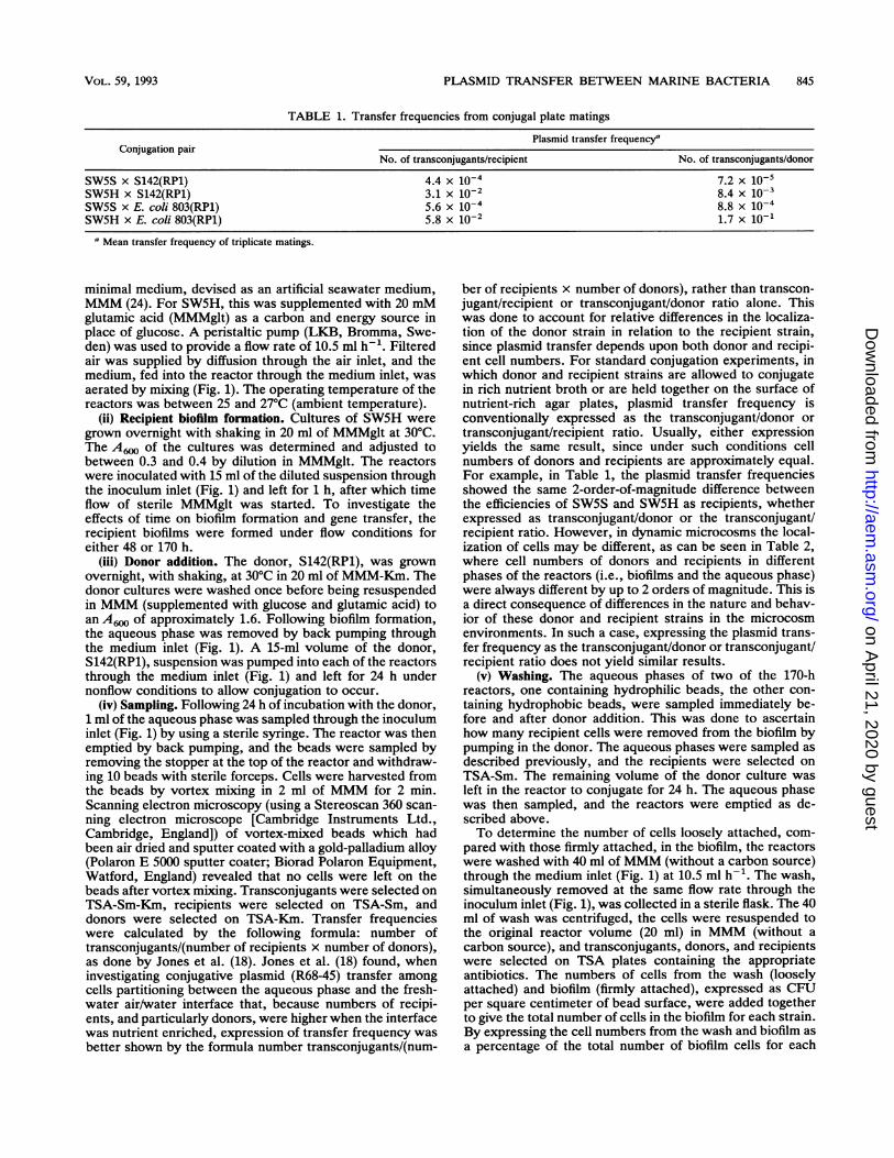
GLASS BEADSFIG. 1. Design of the reactors used in conjugation experiments.
to be due to the plasmid, since simultaneous acquisition ofresistance to Ap and Tc was shown by patching. All conju-gations were done in triplicate.
(ii) Development of a high-frequency-of-conjugation strain.A method was adapted from that described for selection ofhigh-frequency-of-transformation strains (12) to generate arecipient, SW5H, that was more efficient at conjugation thanthe parent strain. Transconjugants from the mating ofS142(RP1) and SW5S were cured of the plasmid by repeatedsubculture without antibiotic selection for the plasmid. Acured strain, SW5H, was conjugated with the originalS142(RP1) donor and with E. coli 803(RP1) by the methoddescribed above.
Reactor experiments. (i) Reactor design and operation.Each reactor (Fig. 1) consisted of a 20-ml mini chemostat(Quickfit) containing 25 acid-washed glass beads (with asurface area of 0.785 cm2 each or 19.6 cm2 in total). Thereactors were sterilized by autoclaving. The glass beadswere hydrophilic, and for comparison, sterile, washed beadswere made hydrophobic by placement in sterile 2% dimeth-yldichlorosilane in 1,1,1-trichloroethane (Coatasil; AjaxChemicals, Sydney, New South Wales, Australia) for 10 minbefore addition to sterile reactors.The growth medium used in the reactors was a buffered
844 ANGLES ET AL.
on April 21, 2020 by guest
http://aem.asm
.org/D
ownloaded from
PLASMID TRANSFER BETWEEN MARINE BACTERIA 845
TABLE 1. Transfer frequencies from conjugal plate matings
Plasmid transfer frequency'Conjugation pair
No. of transconjugants/recipient No. of transconjugants/donor
SW5S x S142(RP1) 4.4 x 10⁻⁴ 7.2 x 10⁻⁵SW5H x S142(RP1) 3.1 x 10⁻² 8.4 x 10⁻³SW5S x E. coli 803(RP1) 5.6 x 10⁻⁴ 8.8 x 10⁻⁴SW5H x E. coli 803(RP1) 5.8 x 10⁻² 1.7 x 10⁻¹
a Mean transfer frequency of triplicate matings.
minimal medium, devised as an artificial seawater medium,MMM (24). For SW5H, this was supplemented with 20 mMglutamic acid (MMMglt) as a carbon and energy source inplace of glucose. A peristaltic pump (LKB, Bromma, Swe-den) was used to provide a flow rate of 10.5 ml h-'. Filteredair was supplied by diffusion through the air inlet, and themedium, fed into the reactor through the medium inlet, wasaerated by mixing (Fig. 1). The operating temperature of thereactors was between 25 and 27°C (ambient temperature).
(ii) Recipient biofilm formation. Cultures of SW5H weregrown overnight with shaking in 20 ml of MMMglt at 30°C.The A6. of the cultures was determined and adjusted tobetween 0.3 and 0.4 by dilution in MMMglt. The reactorswere inoculated with 15 ml of the diluted suspension throughthe inoculum inlet (Fig. 1) and left for 1 h, after which timeflow of sterile MMMglt was started. To investigate theeffects of time on biofilm formation and gene transfer, therecipient biofilms were formed under flow conditions foreither 48 or 170 h.
(iii) Donor addition. The donor, S142(RP1), was grownovernight, with shaking, at 30°C in 20 ml of MMM-Km. Thedonor cultures were washed once before being resuspendedin MMM (supplemented with glucose and glutamic acid) toan A6. of approximately 1.6. Following biofilm formation,the aqueous phase was removed by back pumping throughthe medium inlet (Fig. 1). A 15-ml volume of the donor,S142(RP1), suspension was pumped into each of the reactorsthrough the medium inlet (Fig. 1) and left for 24 h undernonflow conditions to allow conjugation to occur.
(iv) Sampling. Following 24 h of incubation with the donor,1 ml of the aqueous phase was sampled through the inoculuminlet (Fig. 1) by using a sterile syringe. The reactor was thenemptied by back pumping, and the beads were sampled byremoving the stopper at the top of the reactor and withdraw-ing 10 beads with sterile forceps. Cells were harvested fromthe beads by vortex mixing in 2 ml of MMM for 2 min.Scanning electron microscopy (using a Stereoscan 360 scan-ning electron microscope [Cambridge Instruments Ltd.,Cambridge, England]) of vortex-mixed beads which hadbeen air dried and sputter coated with a gold-palladium alloy(Polaron E 5000 sputter coater; Biorad Polaron Equipment,Watford, England) revealed that no cells were left on thebeads after vortex mixing. Transconjugants were selected onTSA-Sm-Km, recipients were selected on TSA-Sm, anddonors were selected on TSA-Km. Transfer frequencieswere calculated by the following formula: number oftransconjugants/(number of recipients x number of donors),as done by Jones et al. (18). Jones et al. (18) found, wheninvestigating conjugative plasmid (R68-45) transfer amongcells partitioning between the aqueous phase and the fresh-water air/water interface that, because numbers of recipi-ents, and particularly donors, were higher when the interfacewas nutrient enriched, expression of transfer frequency wasbetter shown by the formula number transconjugants/(num-
ber of recipients x number of donors), rather than transcon-jugant/recipient or transconjugant/donor ratio alone. Thiswas done to account for relative differences in the localiza-tion of the donor strain in relation to the recipient strain,since plasmid transfer depends upon both donor and recipi-ent cell numbers. For standard conjugation experiments, inwhich donor and recipient strains are allowed to conjugatein rich nutrient broth or are held together on the surface ofnutrient-rich agar plates, plasmid transfer frequency isconventionally expressed as the transconjugant/donor ortransconjugant/recipient ratio. Usually, either expressionyields the same result, since under such conditions cellnumbers of donors and recipients are approximately equal.For example, in Table 1, the plasmid transfer frequenciesshowed the same 2-order-of-magnitude difference betweenthe efficiencies of SW5S and SW5H as recipients, whetherexpressed as transconjugant/donor or the transconjugant/recipient ratio. However, in dynamic microcosms the local-ization of cells may be different, as can be seen in Table 2,where cell numbers of donors and recipients in differentphases of the reactors (i.e., biofilms and the aqueous phase)were always different by up to 2 orders of magnitude. This isa direct consequence of differences in the nature and behav-ior of these donor and recipient strains in the microcosmenvironments. In such a case, expressing the plasmid trans-fer frequency as the transconjugant/donor or transconjugant/recipient ratio does not yield similar results.
(v) Washing. The aqueous phases of two of the 170-hreactors, one containing hydrophilic beads, the other con-taining hydrophobic beads, were sampled immediately be-fore and after donor addition. This was done to ascertainhow many recipient cells were removed from the biofilm bypumping in the donor. The aqueous phases were sampled asdescribed previously, and the recipients were selected onTSA-Sm. The remaining volume of the donor culture wasleft in the reactor to conjugate for 24 h. The aqueous phasewas then sampled, and the reactors were emptied as de-scribed above.To determine the number of cells loosely attached, com-
pared with those firmly attached, in the biofilm, the reactorswere washed with 40 ml ofMMM (without a carbon source)through the medium inlet (Fig. 1) at 10.5 ml h-'. The wash,simultaneously removed at the same flow rate through theinoculum inlet (Fig. 1), was collected in a sterile flask. The 40ml of wash was centrifuged, the cells were resuspended tothe original reactor volume (20 ml) in MMM (without acarbon source), and transconjugants, donors, and recipientswere selected on TSA plates containing the appropriateantibiotics. The numbers of cells from the wash (looselyattached) and biofilm (firmly attached), expressed as CFUper square centimeter of bead surface, were added togetherto give the total number of cells in the biofilm for each strain.By expressing the cell numbers from the wash and biofilm asa percentage of the total number of biofilm cells for each
VOL. 59, 1993
on April 21, 2020 by guest
http://aem.asm
.org/D
ownloaded from
APPL. ENVIRON. MICROBIOL.
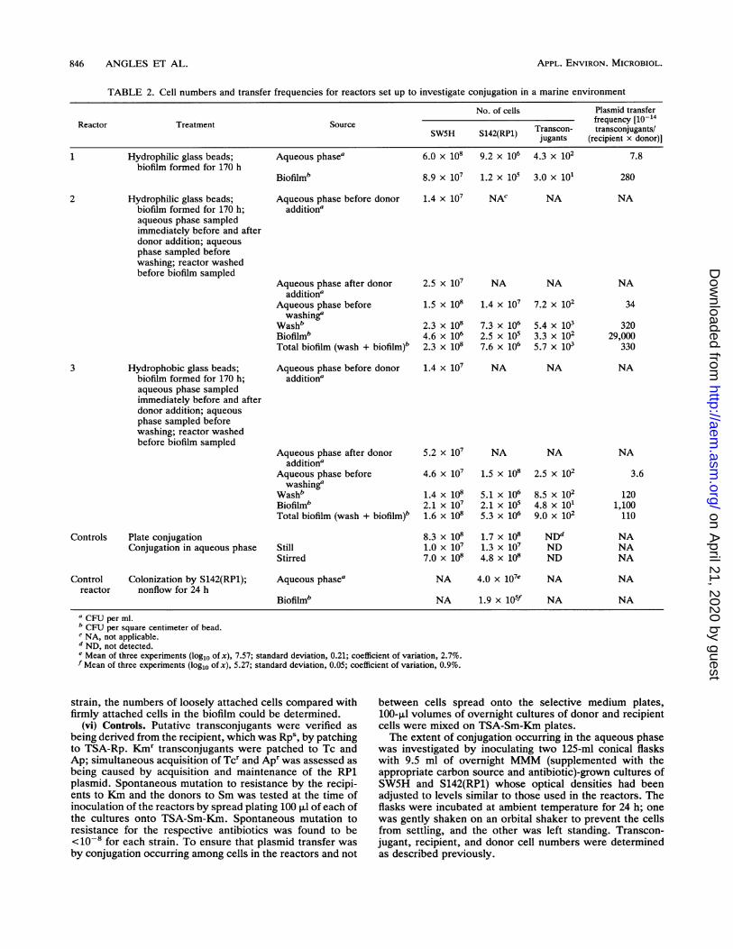
TABLE 2. Cell numbers and transfer frequencies for reactors set up to investigate conjugation in a marine environment
No. of cells Plasmid transferReactor Treatment Source frequency [10-14
SW5H S142(RP1) Transcon- transconjugants/jugants (recipient x donor)]
1 Hydrophilic glass beads; Aqueous phasea 6.0 x 10⁸ 9.2 x 10⁶ 4.3 x 10² 7.8biofilm formed for 170 h
Biofilmb 8.9 x 10⁷ 1.2 x 10⁵ 3.0 x 101 280
Hydrophilic glass beads;biofilm formed for 170 h;aqueous phase sampledimmediately before and afterdonor addition; aqueousphase sampled beforewashing; reactor washedbefore biofilm sampled
Hydrophobic glass beads;biofilm formed for 170 h;aqueous phase sampledimmediately before and afterdonor addition; aqueousphase sampled beforewashing; reactor washedbefore biofilm sampled
Controls Plate conjugationConjugation in aqueous phase
Aqueous phase before donoradditiona
Aqueous phase after donoradditiona
Aqueous phase beforewashinga
WashbBiofilmbTotal biofilm (wash + biofilm)b
Aqueous phase before donoraddition'
Aqueous phase after donoraddition'
Aqueous phase beforewashing'
WashbBiofilmbTotal biofilm (wash + biofilm)b
StillStirred
1.4 x 10⁷ NAC
2.5 x 10⁷
1.5 x 10⁸
2.3 x 1084.6 x 1062.3 x 108
NA
NA NA
1.4 x 10⁷
7.3 x 1062.5 x 1057.6 x 106
7.2 x 10²
5.4 x 1033.3 x 1025.7 x 103
1.4 x 10⁷ NA NA
5.2 x 10⁷
4.6 x 10⁷
1.4 x 1082.1 x 1071.6 x 108
8.3 x 1081.0X 1077.0 x 108
NA
1.5 x 10⁸
5.1 x 1062.1 x 1055.3 x 106
1.7 x 1081.3 x 1074.8 x 108
NA
2.5 x 10²
8.5 x 1024.8 x 1019.0 x 102
NDdNDND
Control Colonization by S142(RP1);reactor nonflow for 24 h
Aqueous phase0
Biofilm'
NA 4.0 x 10⁷e
NA 1.9 x 10⁵f
CFU per ml.b CFU per square centimeter of bead.c NA, not applicable.d ND, not detected.e Mean of three experiments (log1o of x), 7.57; standard deviation, 0.21; coefficient of variation, 2.7%.f Mean of three experiments (loglo of x), 5.27; standard deviation, 0.05; coefficient of variation, 0.9%.
strain, the numbers of loosely attached cells compared withfirmly attached cells in the biofilm could be determined.
(vi) Controls. Putative transconjugants were verified asbeing derived from the recipient, which was Rpa, by patchingto TSA-Rp. Kmr transconjugants were patched to Tc andAp; simultaneous acquisition of Tcr and Apr was assessed as
being caused by acquisition and maintenance of the RP1plasmid. Spontaneous mutation to resistance by the recipi-ents to Km and the donors to Sm was tested at the time ofinoculation of the reactors by spread plating 100 ,ul of each ofthe cultures onto TSA-Sm-Km. Spontaneous mutation toresistance for the respective antibiotics was found to be<10-8 for each strain. To ensure that plasmid transfer wasby conjugation occurring among cells in the reactors and not
between cells spread onto the selective medium plates,100-,ul volumes of overnight cultures of donor and recipientcells were mixed on TSA-Sm-Km plates.The extent of conjugation occurring in the aqueous phase
was investigated by inoculating two 125-ml conical flaskswith 9.5 ml of overnight MMM (supplemented with theappropriate carbon source and antibiotic)-grown cultures ofSW5H and S142(RP1) whose optical densities had beenadjusted to levels similar to those used in the reactors. Theflasks were incubated at ambient temperature for 24 h; onewas gently shaken on an orbital shaker to prevent the cellsfrom settling, and the other was left standing. Transcon-jugant, recipient, and donor cell numbers were determinedas described previously.
2
3
NA
NA
34
32029,000
330
NA
NA
3.6
1201,100110
NANANA
NA
NA
NA
NA
846 ANGLES ET AL.
on April 21, 2020 by guest
http://aem.asm
.org/D
ownloaded from

PLASMID TRANSFER BETWEEN MARINE BACTERIA 847
Three identical control reactors were run to investigatecolonization of the beads by a pure culture of donor strainS142(RP1). An increase in colonization of the beads by thedonor in the conjugation reactors, compared with the controlreactors containing no recipient biofilms, would indicateenhancement of donor colonization of the beads by therecipient biofilm. Reactors containing no recipient biofilmswere inoculated with 15 ml of an overnight MMM-Kmculture of S142(RP1) adjusted to an A6. of approximately1.6. The reactors contained hydrophilic beads and wereincubated in the same manner as the test reactors (i.e., undernonflow conditions for 24 h at ambient temperature).A transformation and transduction control was performed
by filtering an overnight TSB-Km culture of the donor strainthrough a 0.22-,um-pore-size syringe filter (MFS, Dublin,Calif.) and then mixing the filtrate with an overnight cultureof the recipient in the same manner as the plate matingexperiments. It was assumed that any bacteriophages ornaked DNA could pass through the filters.
Statistical analysis. For statistical analysis, transfer fre-quencies (1014) and counts were transformed by the log1o ofx. Mean plasmid transfer frequencies calculated for cells inthe aqueous phase and for cells in the biofilms (reactors 1, 2,and 3) were compared by using the unpaired Student t testwith a pooled variance estimate, as were also the meannumbers of S142(RP1) donors in the aqueous phases andbiofilms of the three control reactors. For the three controlreactors, the means, standard deviations, and coefficients ofvariation of the cell numbers localized in the different phasesare given in Table 2.
RESULTS
Isolation of a high-frequency-of-conjugation strain.Transconjugants from the mating of SW5S and S142(RP1)were used to generate high-frequency-of-conjugation strainSW5H, which yielded transconjugants at a plasmid transferfrequency 2 orders of magnitude higher than that of theoriginal recipient, SW5S, with both S142(RP1) and E. coli803(RP1) donors (Table 1). Conjugation with the E. colidonor was tested to ensure that any difference in plasmidtransfer frequency noted between either SW5S or SW5H andS142(RP1) was due to the different recipient strain and wasnot caused by the donor ability of S142(RP1).
Investigation of conjugation between marine strains inbiofilms. The results from three flow reactor experiments andcontrols set up to investigate plasmid gene transfer in marinebiofilms and the aqueous phase are presented in Table 2.Reactor 1 contained hydrophilic beads on which the recipi-ent biofilm was formed for 170 h. Transfer of RP1 wasdetected among cells in both the biofilm and the aqueousphase (Table 2). When the recipient biofilm was allowed toform for only 48 h on the hydrophilic beads, however, noplasmid transfer was detected in the biofilm (data notshown). The recipient and donor numbers and frequency ofplasmid transfer in the aqueous phase were comparable tothose found in reactor 1 (Table 2). A reactor was set up toallow the recipients to form a biofilm on hydrophobic beadsfor 48 h. Compared with the reactor containing hydrophilicbeads, there was a 1-order-of-magnitude increase in thenumbers of recipients colonizing the hydrophobic beads, butagain no transconjugants were detected in either the biofilmor the aqueous phase (data not shown).
Reactors 2 and 3 were run to study the effects of hydro-philic and hydrophobic substrata on conjugation, coloniza-tion by SW5H in the longer term (recipients exposed to
beads for 170 h), recipient numbers in the aqueous phase asa result of pumping in the donor, and the nature of thebiofilms formed. Transconjugants were detected among cellsrecovered from the aqueous phase, those in the wash (loose-ly attached cells), and those firmly attached in the biofilms ofboth reactors 2 and 3 (Table 2). Plasmid transfer frequencieswere higher among cells firmly attached in the biofilms, by 1and 2 orders of magnitude for the hydrophobic (reactor 3)and hydrophilic (reactor 2) beads, respectively, than amongcells loosely attached. Further, the plasmid transfer frequen-cies were higher among cells loosely attached to the hydro-philic (reactor 2) and hydrophobic (reactor 3) bead surfacesthan among cells in the aqueous phase by 1 and 2 orders ofmagnitude, respectively. Taking the plasmid transfer fre-quencies (1014) among the cells in the aqueous phase inreactors 1, 2, and 3 as one group and those among the totalcells in the biofilms (i.e., frequencies calculated for thenumbers of loosely and firmly attached cells added together)as a second group, statistical analysis showed that thedifference between the means of the two groups was signif-icant (P < 0.05). This analysis shows that the plasmidtransfer frequency among organisms in the biofilms wassignificantly higher than that among organisms in the aque-ous phase.There was an increase in the numbers of recipients in the
aqueous phase immediately after donor addition in bothreactors 2 and 3 (Table 2). Over the following 24 h, recipientnumbers increased by about an order of magnitude in theaqueous phase of the reactor containing hydrophilic beads(reactor 2) only. Washing revealed that the majority ofSW5H and transconjugant cells were loosely attached to thebead surfaces (Table 2). When the numbers of recipients inthe wash and the biofilm were added together, it could beseen that there was little difference in colonization betweenthe hydrophilic and hydrophobic surfaces (2.3 x 10⁸ and 1.6x 10⁸ CFU cm-2, respectively). The donor, S142(RP1),similarly showed little change in total biofilm cell numbers ina comparison of the numbers from the hydrophilic beadswith those from the hydrophobic beads (7.6 x 10⁶ and 5.3 x10⁶ CFU cm-2, respectively). By expressing the wash andbiofilm cell numbers as percentages of the total number ofcells in the biofilm for each strain, the loosely attachednature of the biofilm became apparent, with about 90% of therecipients, donors, and transconjugants being removed bythe wash from both the hydrophobic and hydrophilic beadsurfaces.The ratios of the cell numbers in the aqueous phase
compared with numbers in the biofilm for reactors 2 and 3are shown in Table 3. For both bead surfaces, the ratio ofcells in the aqueous phase to cells in the biofilm was higherfor the donor, S142(RP1), than for the recipient, SW5H, andwas consistent with the differences in the colonizing abilitiesof the two organisms (Table 2). For reactor 2, containinghydrophilic bead surfaces, the number of SW5H cells in theaqueous phase was about 30 times higher than that of cellsfirmly adhered to the hydrophilic surface and about one-halfthat of cells loosely attached. There were about 50 timesmore S142(RP1) cells in the aqueous phase than there werefirmly attached, and there were twofold more cells in theaqueous phase than there were loosely attached (reactor 2).For the hydrophobic bead surfaces in reactor 3, there wereabout two-fold more SW5H cells in the aqueous phase thanfirmly attached and about one-third as ma-ny as were looselyattached. There were about 700 times more S142(RP1) cellsin the aqueous phase than firmly attached and about 30 timesmore than were loosely attached (reactor 3).
VOL. 59, 1993
on April 21, 2020 by guest
http://aem.asm
.org/D
ownloaded from
APPL. ENVIRON. MICROBIOL.
TABLE 3. Ratio of cell numbers in the aqueous phase tonumbers in the biofilm for reactors 2 and 3
Ratio of cellnumbers in aqueous
Reactor Duration of biofilm Surface type phase to cellformation (h) numbers in biofilm
SW5H S142(RP1)
2 170 Hydrophilic 33a 56a0.7b 2b
3 170 Hydrophobic 2a 714a0o.3 30b
a Ratio of aqueous-phase cell number to number of firmly attached bacteria.b Ratio of aqueous-phase cell number to number of loosely attached
bacteria.
Controls. Patching revealed that transconjugants werederived from recipients and had simultaneously acquiredKmr Tcr, and Apr, indicating receipt and maintenance ofRP1. No transconjugants were detected when recipients anddonors were plated together on transconjugant selectionplates (plate conjugation in Table 2). Hence, any plasmidtransfer detected had occurred in the reactors.The aqueous-phase conjugation controls, designed to de-
termine the source of transconjugants in the aqueous phase,yielded no detectable transconjugants, even though thenumbers of donors and recipients in the stirred control werehigher than those in the experimental reactors (Table 2).Three control reactors (Table 2) were set up with pure
cultures of S142(RP1) to determine whether the recipientbiofilm had any effect on the colonizing abilities of the donor.Donor numbers in the biofilms covering the hydrophilicbeads of the three reactors ranged from 1.7 x 10⁵ to 2.1 x10⁵ CFU cm-2, and in the aqueous phase they ranged from2.2 x 107 to 5.5 x 10⁷ CFU ml-'. Both the biofilm andaqueous-phase counts were similar to those in the experi-mental reactors (1 and 2) containing hydrophilic beads(Table 2). It appeared, therefore, that there was little or noenhancement or exclusion of donor colonization on eitherhydrophilic or hydrophobic beads by the recipients in thebiofilm. The localization of the cells between the aqueousphase and the biofilm was significantly different, since cellnumbers in the aqueous phase were significantly (P < 0.01)higher than cell numbers in the biofilm.A transformation and transduction control yielded no
transconjugants. Any transfer of RP1 in the reactors, there-fore, was likely to have been due to conjugation and not totransformation or transduction.
DISCUSSION
In the reactor microcosms used, plasmid transfer wassignificantly higher (P < 0.05) among cells in the biofilmsattached to bead surfaces than among cells in the aqueousphase. It was necessary to select a recipient, SW5H, thatshowed a relatively high ability to receive and maintain theRP1 plasmid under optimum laboratory conditions so as toevaluate effects of the nature of the surfaces and the timeallowed for conjugation on efficiency of gene transfer. Aspointed out by Fernandez-Astorga et al. (10), if transferefficiency is low to start with, then transfer under less thanoptimum conditions might not be detected at all. This may
explain the lack of detectable plasmid transfer from Pseudo-monas aeruginosa(R68-45) to the natural bacterial popula-tion in the work of Jones et al. (18).
In an initial experiment (reactor 1), the number oftransconjugants detected in the aqueous phase was about10-fold higher than that of those detected in the biofilm. Thisresult appeared to contradict the well-known phenomenonthat conjugation of RP1, an IncPl plasmid, is several ordersof magnitude more efficient for cells held on a surface thanfor those in the aqueous phase (7). We have shown increasedRP1 plasmid transfer at a surface among Vibrio sp. strain S14isolates (24). Indeed, aqueous-phase conjugal-transfer con-trols in our study (i.e., reactors without beads or preformedbiofilms) showed that with comparable numbers of donorsand recipients, no conjugation could be detected in theaqueous phase. We agree with Jones et al. (18) that sinceplasmid transfer depends upon both donor and recipient cellnumbers, plasmid transfer frequencies are better expressedas the ratio of transconjugants to the donor-recipient productwhen the localization of donors and recipients is different. Inthe experiments presented here, we expressed the plasmidtransfer frequency in this way and found that the plasmidtransfer frequency was about 30 times higher among cells inthe biofilm than among those in the aqueous phase (Table 2,reactor 1). This result was consistent with those for reactors2 and 3.
In both reactors 2 and 3, addition of donor cells resulted inan increase in SW5H numbers in the aqueous phase. After 24h under nonflow conditions, the SW5H numbers in theaqueous phase were similar to the total numbers in thebiofilm in most of the reactors. Any increases in SW5H cellnumbers in the aqueous phase may have been due to growthof SW5H in the aqueous phase under nonflow conditions butcould also have been due to exchange of loosely attachedcells between the biofilm and the aqueous phase, as foundpreviously by Hermansson and Marshall (17) for a looselyattaching bacterium. Washing of the beads showed thatabout 90% of the donor, recipient, and transconjugant cellsin the biofilms in reactors 2 and 3 cou-ld be classified asloosely attached to both the hydrophilic and hydrophobicbeads. It is possible, therefore, that transconjugants de-tected in the aqueous phase arose from cells which had beenloosely attached in the biofilm and then moved to theaqueous phase.The hydrophilic or hydrophobic nature of the bead sur-
faces appeared to make little or no difference to the occur-rence of conjugation, since plasmid transfer was detectedamong cells attached to either type of bead surface. Whetherthe nature of the bead surface significantly affects plasmidtransfer frequency is now under investigation, since it ap-peared that transfer was more efficient on the hydrophilicsurface (Table 2). The mean plasmid transfer frequencyamong all of the cells in the biofilms of all three reactors wasfound to be significantly higher (P < 0.05) than that amongcells in the aqueous phase. In controls which contained onlyaqueous-phase cells, plasmid transfer was not detected.
It appeared that the hydrophobic SW5H cells preferredthe hydrophobic surface to the hydrophilic one (Table 3).This was not surprising, as the importance of hydrophobicbonding between a hydrophobic cell and a hydrophobicsubstratum has been well documented (1, 11, 20). In con-trast, the hydrophilic S142(RP1) cells appeared to be re-pelled by the hydrophobic surface (Table 3) and it was clearthat this strain was not as good a colonizer of either of thetwo surfaces as hydrophobic strain SW5H (Tables 2 and 3).Such results are supported by other work, which found that
848 ANGLES ET AL.
on April 21, 2020 by guest
http://aem.asm
.org/D
ownloaded from
PLASMID TRANSFER BETWEEN MARINE BACTERIA 849
hydrophobic bacteria adhere to solid surfaces to a greaterextent than do hydrophilic bacteria (9, 33) and that thesedifferences follow the thermodynamic model of adhesion, inwhich hydrophobic cells are excluded from the aqueousphase and, consequently, attracted to the solid/liquid inter-face, whereas hydrophilic cells tend to stay in the aqueousphase (1, 11, 20).
Detection of plasmid transfer was less successful after 48h of recipient biofilm formation, and plasmid transfer wasdetected only in the aqueous phase of the reactor containinghydrophilic beads. We do not know why transconjugantswere not detected in the biofilm of this reactor or in thereactor containing hydrophobic beads, but it may be thattransconjugant numbers were below the detection limit ofour system. Characklis (8) stated that mature biofilm devel-opment is time dependent, and thus, at 48 h for either ahydrophilic or a hydrophobic surface, there may have beentoo few recipient cells firmly attached to the surfaces forplasmid transfer to have been detected.The present work is the first to report conjugative plasmid
transfer between marine bacteria in biofilms, in which bio-films of a recipient strain were established and then a donorstrain was introduced for a short time, with no selectionpressure for the plasmid. Our study has also shown that aplasmid-bearing bacterial strain which is poor at forming abiofilm, or becoming part of one already established byanother strain, can nevertheless have sufficient cell-cellinteraction with the cells in the biofilm for plasmid transfer tooccur. We are examining this phenomenon further by inves-tigating what effects laminar flow, nutrient deprivation, andaddition of other strains to the biofilm may have on thespread of plasmids in marine biofilm microcosms.
ACKNOWLEDGMENTS
Part of this work was supported by grants to A.E.G. and K.C.M.from the Australian Research Council. M.L.A. is supported by anAustralian Postgraduate Research Award.
REFERENCES
1. Absolom, D. R., F. V. Lamberti, Z. Policova, W. Zingg, C. J. vanOss, and A. W. Neumann. 1983. Surface thermodynamics ofbacterial adhesion. Appl. Environ. Microbiol. 46:90-97.
2. Altherr, M. R., and K. L. Kasweck 1982. In situ studies withmembrane diffusion chambers of antibiotic resistance transfer inEscherichia coli. Appl. Environ. Microbiol. 44:838-843.
3. Bale, M. J., M. J. Day, and J. C. Fry. 1988. Novel method forstudying plasmid transfer in undisturbed river epilithon. Appl.Environ. Microbiol. 54:2756-2758.
4. Bale, M. J., J. C. Fry, and M. J. Day. 1987. Plasmid transferbetween strains of Pseudomonas aeruginosa on membranefilters attached to river stones. J. Gen. Microbiol. 133:3099-3107.
5. Bale, M. J., J. C. Fry, and M. J. Day. 1988. Transfer andoccurrence of large mercury resistance plasmids in river epili-thon. Appl. Environ. Microbiol. 54:972-978.
6. Bentjen, S. A., J. K. Fredrickson, P. van Voris, and S. W. Li.1989. Intact soil microcosms for evaluating the fate and ecolog-ical impact of the release of genetically engineered microorgan-isms. Appl. Environ. Microbiol. 55:198-202.
7. Bradley, D. E., D. E. Taylor, and D. R. Cohen. 1980. Specifica-tion of surface mating systems among conjugative drug resis-tance plasmids in Escherichia coli K-12. J. Bacteriol. 143:1466-1470.
8. Characklis, W. G. 1990. Biofilm processes, p. 195-231. In W. G.Characklis and K. C. Marshall (ed.), Biofilms. John Wiley &Sons, Inc., New York.
9. Fattom, A., and M. Shilo. 1984. Hydrophobicity as an adhesionmechanism of benthic cyanobacteria. Appl. Environ. Microbiol.47:135-143.
10. Fernandez-Astorga, A., A. Muela, R. Cisterna, J. Iriberri, andI.Barcina. 1992. Biotic and abiotic factors affecting plasmidtransfer in Escherichia coli strains. Appl. Environ. Microbiol.58:392-398.
11. Fletcher, M., and K. C. Marshall. 1982. Are solid surfaces ofecological significance to aquatic bacteria? Adv. MicrobialEcol. 6:199-236.
12. Frischer, M. E., J. M. Thurmond, and J. H. Paul. 1990. Naturalplasmid transformation in a high-frequency-of-transformationmarine Vibrio strain. Appl. Environ. Microbiol. 56:3439-3444.
13. Fry, J. C., and M. J. Day. 1990. Plasmid transfer in theepilithon, p. 55-80. In J. C. Fry and M. J. Day (ed.), Bacterialgenetics in natural environments. Chapman & Hall, Ltd., Lon-don.
14. FMultorpe, R. R., and R. C. Wyndham. 1989. Survival andactivity of a 3-chlorobenzoate-catabolic genotype in a naturalsystem. Appl. Environ. Microbiol. 55:1584-1590.
15. Fulthorpe, R. R., and R. C. Wyndham. 1991. Transfer andexpression of the catabolic plasmid pBRC60 in wild bacterialrecipients in a freshwater ecosystem. Appl. Environ. Microbiol.57:1546-1553.
16. Gauthier, M. J., F. Cauvin, and J. Breittmayer. 1985. Influenceof salts and temperature on the transfer of mercury resistancefrom a marine pseudomonad to Eschenichia coli. Appl. Environ.Microbiol. 50:38-40.
17. Hermansson, M., and K. C. Marshall. 1985. Utilization ofsurface localized substrate by non-adhesive marine bacteria.Microb. Ecol. 11:91-105.
18. Jones, G. W., L. Baines, and F. J. Genthner. 1991. Heterotro-phic bacteria of the freshwater neuston and their ability to act asplasmid recipients under nutrient deprived conditions. Microb.Ecol. 22:15-25.
19. Klingmuller, W., A. Dally, C. Fentner, and M. Steinlein. 1990.Plasmid transfer between soil bacteria, p. 133-151. In J. C. Fryand M. J. Day (ed.), Bacterial genetics in natural environments.Chapman & Hall, Ltd., London.
20. Marshall, K. C. 1986. Adsorption and adhesion processes inmicrobial growth at interfaces. Adv. Colloid. Interface Sci.25:59-86.
21. Marshall, K. C. 1992. Bioffilms: an overview of bacterial adhe-sion, activity, and control at surfaces. ASM News 58:202-207.
22. McClure, N. C., J. C. Fry, and A. J. Weightman. 1990. Genetransfer in activated sludge, p. 111-129. In J. C. Fry and M. J.Day (ed.), Bacterial genetics in natural environments. Chapman& Hall, Ltd., London.
23. Neu, T. R., and K. C. Marshall. 1991. Microbial "foot-prints"-a new approach to adhesive polymers. Biofouling3:101-112.
24. Ostling, J., A. Goodman, and S. Kjelleberg. 1991. Behaviour ofIncP-1 plasmids and a miniMu transposon in a marine Vibriosp.: isolation of starvation inducible lac operon fusions. FEMSMicrobiol. Ecol. 86:83-94.
25. Patt, T., K. Nealson, and C. Eberhard. 1972. Promiscuoustransfer of genetic information from non-marine to marinebacteria. Biol. Bull. 143:473.
26. Rochelle, P. A., J. C. Fry, and M. J. Day. 1989. Plasmid transferbetween Pseudomonas spp. within epilithic films in a rotatingdisc microcosm. FEMS Microbiol. Ecol. 62:127-136.
27. Saye, D. J., and R. V. Miller. 1989. The aquatic environment:consideration of horizontal gene transmission in a diversifiedhabitat, p. 223-259. In S. B. Levy and R. V. Miller (ed.), Genetransfer in the environment. McGraw-Hill Publishing Co., NewYork.
28. Sizemore, R. K., and R. R. Colwell. 1977. Plasmids carried byantibiotic-resistant marine bacteria. Antimicrob. AgentsChemother. 12:373-382.
29. Stotzky, G. 1989. Gene transfer among bacteria in soil, p.165-222. In S. B. Levy and R. V. Miller (ed.), Gene transfer inthe environment. McGraw-Hill Publishing Co., New York.
30. Stotzky, G., and H. Babich. 1986. Survival of, and genetic
VOL. 59, 1993
on April 21, 2020 by guest
http://aem.asm
.org/D
ownloaded from
APPL. ENVIRON. MICROBIOL.
transfer by, genetically engineered bacteria in natural environ-ments. Adv. Appl. Microbiol. 31:93-138.
31. Thomas, C. M., and C. A. Smith. 1987. Incompatibility group Pplasmids: genetics, evolution, and use in genetic manipulation.Annu. Rev. Microbiol. 41:77-101.
32. Trevors, J. T., T. Barkay, and A. W. Bourquin. 1987. Gene
transfer among bacteria in soil and aquatic environments: areview. Can. J. Microbiol. 33:191-198.
33. van Loosdrecht, M. C. M., J. Lyklema, W. Norde, G. Schraa,and A. J. B. Zehnder. 1987. The role of bacterial cell wallhydrophobicity in adhesion. Appl. Environ. Microbiol. 53:1893-1897.
850 ANGLES ET AL.
on April 21, 2020 by guest
http://aem.asm
.org/D
ownloaded from
Related Documents