ORIGINAL ARTICLE Plasma metabolomics and proteomics profiling after a postprandial challenge reveal subtle diet effects on human metabolic status Linette Pellis • Marjan J. van Erk • Ben van Ommen • Gertruud C. M. Bakker • Henk F. J. Hendriks • Nicole H. P. Cnubben • Robert Kleemann • Eugene P. van Someren • Ivana Bobeldijk • Carina M. Rubingh • Suzan Wopereis Received: 31 March 2011 / Accepted: 12 May 2011 Ó The Author(s) 2011. This article is published with open access at Springerlink.com Abstract We introduce the metabolomics and proteomics based Postprandial Challenge Test (PCT) to quantify the postprandial response of multiple metabolic processes in humans in a standardized manner. The PCT comprised consumption of a standardized 500 ml dairy shake con- taining respectively 59, 30 and 12 energy percent lipids, carbohydrates and protein. During a 6 h time course after PCT 145 plasma metabolites, 79 proteins and 7 clinical chemistry parameters were quantified. Multiple processes related to metabolism, oxidation and inflammation reacted to the PCT, as demonstrated by changes of 106 metabo- lites, 31 proteins and 5 clinical chemistry parameters. The PCT was applied in a dietary intervention study to evaluate if the PCT would reveal additional metabolic changes compared to non-perturbed conditions. The study consisted of a 5-week intervention with a supplement mix of anti- inflammatory compounds in a crossover design with 36 overweight subjects. Of the 231 quantified parameters, 31 had different responses over time between treated and control groups, revealing differences in amino acid metabolism, oxidative stress, inflammation and endocrine metabolism. The results showed that the acute, short term metabolic responses to the PCT were different in subjects on the supplement mix compared to the controls. The PCT provided additional metabolic changes related to the die- tary intervention not observed in non-perturbed conditions. Thus, a metabolomics based quantification of a standard- ized perturbation of metabolic homeostasis is more infor- mative on metabolic status and subtle health effects induced by (dietary) interventions than quantification of the homeostatic situation. Keywords Postprandial challenge Á Metabolic profiling Á Proteomic profiling Á Plasma 1 Introduction The physiological and biochemical response to a dietary perturbation is complex. It includes energy storage mostly orchestrated by insulin and involves metabolic switches in several organs like liver, muscle and adipose tissue, accom- panied by several compensating processes such as inflam- mation and oxidative stress (Carroll and Schade 2003; Esposito et al. 2003; Nappo et al. 2002; Neri et al. 2005; Wybranska et al. 2003). Many of these processes share path- ways and control mechanisms that are common to metabolic, inflammatory and oxidative stress processes. For instance, the response to a ‘‘simple’’ glucose bolus involves modulation of glucose itself, triglycerides, blood pressure, cholesterol, inflammation and oxidation (Nakatsuji et al. 2010). The postprandial response depends on, and involves multiple factors. Multiple processes related to metabolism, inflammation and oxidation are affected (Lundman et al. 2007). The type, nature and amount of fat influences lipid Electronic supplementary material The online version of this article (doi:10.1007/s11306-011-0320-5) contains supplementary material, which is available to authorized users. L. Pellis (&) Á M. J. van Erk Á B. van Ommen Á G. C. M. Bakker Á H. F. J. Hendriks Á N. H. P. Cnubben Á E. P. van Someren Á C. M. Rubingh Á S. Wopereis TNO, PO Box 360, 3700 AJ Zeist, The Netherlands e-mail: [email protected] R. Kleemann TNO, PO Box 2215, 2301 CE Leiden, The Netherlands I. Bobeldijk TNO, Triskelion, PO Box 360, 3700 AJ Zeist, The Netherlands 123 Metabolomics DOI 10.1007/s11306-011-0320-5

Welcome message from author
This document is posted to help you gain knowledge. Please leave a comment to let me know what you think about it! Share it to your friends and learn new things together.
Transcript

ORIGINAL ARTICLE
Plasma metabolomics and proteomics profilingafter a postprandial challenge reveal subtle diet effects on humanmetabolic status
Linette Pellis • Marjan J. van Erk • Ben van Ommen • Gertruud C. M. Bakker •
Henk F. J. Hendriks • Nicole H. P. Cnubben • Robert Kleemann • Eugene P. van Someren •
Ivana Bobeldijk • Carina M. Rubingh • Suzan Wopereis
Received: 31 March 2011 / Accepted: 12 May 2011
� The Author(s) 2011. This article is published with open access at Springerlink.com
Abstract We introduce the metabolomics and proteomics
based Postprandial Challenge Test (PCT) to quantify the
postprandial response of multiple metabolic processes in
humans in a standardized manner. The PCT comprised
consumption of a standardized 500 ml dairy shake con-
taining respectively 59, 30 and 12 energy percent lipids,
carbohydrates and protein. During a 6 h time course after
PCT 145 plasma metabolites, 79 proteins and 7 clinical
chemistry parameters were quantified. Multiple processes
related to metabolism, oxidation and inflammation reacted
to the PCT, as demonstrated by changes of 106 metabo-
lites, 31 proteins and 5 clinical chemistry parameters. The
PCT was applied in a dietary intervention study to evaluate
if the PCT would reveal additional metabolic changes
compared to non-perturbed conditions. The study consisted
of a 5-week intervention with a supplement mix of anti-
inflammatory compounds in a crossover design with 36
overweight subjects. Of the 231 quantified parameters, 31
had different responses over time between treated and
control groups, revealing differences in amino acid
metabolism, oxidative stress, inflammation and endocrine
metabolism. The results showed that the acute, short term
metabolic responses to the PCT were different in subjects
on the supplement mix compared to the controls. The PCT
provided additional metabolic changes related to the die-
tary intervention not observed in non-perturbed conditions.
Thus, a metabolomics based quantification of a standard-
ized perturbation of metabolic homeostasis is more infor-
mative on metabolic status and subtle health effects
induced by (dietary) interventions than quantification of the
homeostatic situation.
Keywords Postprandial challenge � Metabolic profiling �Proteomic profiling � Plasma
1 Introduction
The physiological and biochemical response to a dietary
perturbation is complex. It includes energy storage mostly
orchestrated by insulin and involves metabolic switches in
several organs like liver, muscle and adipose tissue, accom-
panied by several compensating processes such as inflam-
mation and oxidative stress (Carroll and Schade 2003;
Esposito et al. 2003; Nappo et al. 2002; Neri et al. 2005;
Wybranska et al. 2003). Many of these processes share path-
ways and control mechanisms that are common to metabolic,
inflammatory and oxidative stress processes. For instance, the
response to a ‘‘simple’’ glucose bolus involves modulation of
glucose itself, triglycerides, blood pressure, cholesterol,
inflammation and oxidation (Nakatsuji et al. 2010).
The postprandial response depends on, and involves
multiple factors. Multiple processes related to metabolism,
inflammation and oxidation are affected (Lundman et al.
2007). The type, nature and amount of fat influences lipid
Electronic supplementary material The online version of thisarticle (doi:10.1007/s11306-011-0320-5) contains supplementarymaterial, which is available to authorized users.
L. Pellis (&) � M. J. van Erk � B. van Ommen �G. C. M. Bakker � H. F. J. Hendriks �N. H. P. Cnubben � E. P. van Someren �C. M. Rubingh � S. Wopereis
TNO, PO Box 360, 3700 AJ Zeist, The Netherlands
e-mail: [email protected]
R. Kleemann
TNO, PO Box 2215, 2301 CE Leiden, The Netherlands
I. Bobeldijk
TNO, Triskelion, PO Box 360, 3700 AJ Zeist, The Netherlands
123
Metabolomics
DOI 10.1007/s11306-011-0320-5

clearance, just like the amount and type of carbohydrate,
protein, fiber and alcohol in a meal (Cianflone et al. 2008;
Lopez-Miranda et al. 2007). Gender, genetics, age, body
size, exercise and weight loss influence postprandial
metabolism (Lopez-Miranda et al. 2007; Paglialunga and
Cianflone 2007), as do various metabolic syndrome related
conditions (Ghanim et al. 2009; Lopez-Miranda et al. 2007;
Paglialunga and Cianflone 2007). Increasing knowledge on
molecular aspects of the postprandial response is becoming
available. Plasma metabolome changes were reported
during an oral glucose tolerance test (OGTT) (Shaham
et al. 2008; Wopereis et al. 2009; Zhao et al. 2009), sim-
ilarly, the plasma lipidome response was assessed during a
lipid challenge (Zivkovic et al. 2008). Intake of a high fat
meal alters metabolism and increases inflammation and
oxidative stress, thereby reducing amongst others vascular
function (Devaraj et al. 2008; Ghanim et al. 2009; Tsai
et al. 2004). A high fat meal increases pro-inflammatory
cytokines, such as plasma interleukin 6 (IL6) and 18
(IL18), factor VII, tumor necrosis factor alpha (TNF-a),
intercellular adhesion molecule-1 (ICAM-1) and vascular
cell adhesion molecule-1 (VCAM-1) concentrations
(Esposito et al. 2003; Lopez-Miranda et al. 2007; Lundman
et al. 2007; Nappo et al. 2002). Saturated fatty acids neg-
atively affect endothelial function, whereas monounsatu-
rated fatty acids have neutral or modest beneficial effect on
endothelial function, and polyunsaturated fatty acids have a
positive effect (Jackson et al. 2007; Margioris 2009).
Several oxidation markers are increased after a high fat
meal, like ROS generation by mononuclear cells and
thiobarbituric acid reactive substance concentrations
(Ghanim et al. 2009). Oxidative stress may be an important
mechanism by which postprandial lipidemia alters vascular
function (Devaraj et al. 2008; Ghanim et al. 2009).
In nutrition and health research, the concept of pertur-
bation of homeostasis to quantify health related processes
is advancing (Elliott et al. 2007; van Ommen et al. 2009).
The postprandial response reveals multiple aspects of
metabolic health that would not be apparent from studying
the fasting (homeostatic) parameters. The emerging nutri-
genomics technologies, specifically metabolomics and
lipidomics (van Ommen et al. 2008), allow analysis of
multiple aspects of the postprandial response including
quantification of molecular changes related to metabolic
flexibility, robustness of homeostatic mechanisms and
adaptive responses.
In this study, first the postprandial challenge test (PCT)
was used to quantify postprandial metabolic response using
metabolomic and proteomic profiling on endogenous
processes related to metabolism, inflammation and oxida-
tive stress. Secondly the PCT concept was applied in
a nutritional intervention study to evaluate if the PCT
would reveal additional metabolic changes compared to
non-perturbed conditions. In a 5 week crossover double-
blind placebo controlled intervention study overweight
males were given anti-inflammatory dietary mix (AIDM)
(Bakker et al. 2010). The postprandial response of 231
metabolites and proteins to a postprandial challenge was
measured and compared to fasting (homeostatic) values of
these metabolites and proteins.
2 Materials and methods
2.1 Study design, execution, and analysis
The execution and analytical methodologies of the nutri-
tional intervention study has previously been described in
detail (Bakker et al. 2010). In short, a series of dietary
products selected due to anti-inflammatory properties (res-
veratrol, green tea extract, alpha-tocopherol, vitamin C, n-3
poly unsaturated fatty acids and tomato extract) were com-
bined and supplemented to 36 healthy overweight men (BMI
25.6–34.7 kg/m2) with mildly elevated C-reactive protein
(CRP) levels (1.0–8.1 lg/l) in a double-blind, placebo con-
trolled, crossover study with test treatment periods of
5 weeks. At the end of the intervention and control exposure,
a 500 ml postprandial (fat 58.7 E%) dairy shake, including
300 ml custard, 150 ml cream cheese and 50 ml whipping
cream (nutritional values in Table 3) was given to study the
postprandial response. After an overnight fast the subjects
received a light standardized breakfast. After at least a 4 h
period without food and drinks (except water) they were
offered the postprandial shake. At time points 0 h (fasting
condition), 1 h, 2 h, 3 h, 4 h and 6 h after the postprandial
dairy shake, blood samples were collected and analyzed
using GC-MS metabolic profiling (145 plasma metabolites),
multiplex proteomics (79 plasma proteins) and a series of
clinical chemistry analyses (glucose, insulin, total free fatty
acids, total triglycerides, hsCRP, IL6, and TNFa).
2.2 Metabolic profiling
The GC-MS method used for the measurement of a broad
range of metabolites was identical to the GC-MS method
reported (Koek et al. 2011) for liver samples. In this study,
100 ll of plasma was used extracted and further
derivatized.
For GC-MS, a total of 504 plasma samples were ana-
lyzed in 18 different batches. All the samples of one par-
ticular subject were analyzed within the same batch, with
two subjects per batch. In principle, all the samples were
prepared and injected once. All the samples from two
randomly selected subjects (20 samples in total) were
repeated in the final 19th batch of the study. The perfor-
mance of the applied metabolic profiling platforms was
L. Pellis et al.
123

controlled by the frequent analysis of the quality control
sample (QC), and method performance was monitored by
10 internal standards (including analogs and 2H– and 13C
labeled metabolites), as described previously (Koek et al.
2011; van der Kloet et al. 2009). Batches were only
accepted if the relative standard deviation (RSD) of the
peak area ratio for all internal standards was \20% in all
analyzed study and QC samples. The QC sample, prepared
by pooling study samples, represents the biochemical
diversity of the study samples and allows the calculation of
the analytical precision for all metabolites measured. The
QC samples were used to select the most suitable internal
standard for each detected metabolite. This was done by
calculating the relative standard deviation (RSD) for each
metabolite after separate normalization for each internal
standard. The internal standard giving the lowest RSD in
all the QC samples was subsequently selected to normalize
all the study samples. This procedure was described pre-
viously in more detail (van der Kloet et al. 2009). After
normalization of the data with the most suitable internal
standard for each detected metabolite (peak), the QC
sample data were further used to correct systematic errors
(e.g. batch to batch response differences and also trends
within the batches) by a single point calibration model (van
der Kloet et al. 2009). After the above mentioned data
correction steps, the metabolites were only accepted if the
RSD of the determined (relative) concentration in all the
QC samples was \20%, unless large differences between
test groups were observed. Additional quality control of the
final dataset was performed by comparing the duplicate
measurements of the two randomly selected subjects. The
results were described by van der Kloet et al. 2009.
In the end, the GC-MS data set consisted of 145
metabolite peaks that passed quality control requirements
(Supplemental Table 2). Metabolites were annotated by
using an in-house metabolite database containing retention
time information, MS spectra (electron impact ionization)
of reference substances and metabolites previously identi-
fied by the interpretation of mass spectra based on structural
similarities with the analyzed reference compounds or
spectra published in the literature. The confidence of iden-
tification was 100%, unless indicated otherwise. Because of
the applied sample preparation, some metabolites can result
in more than 1 peak, for example two different derivatiza-
tion products. These different peaks were all labeled as such
and reported individually. In this way it could be checked
whether all signals originating from the same metabolite
resulted in a similar PCT response, which was the case.
2.3 Multiplex proteome analysis
Plasma samples were sent to Rules-Based Medicine Inc.
(Austin, USA) for measurement of the concentration levels
of 124 proteins (HumanMAP). Data were available for 33
subjects. The so-called 80% rule (Bijlsma et al. 2006) was
applied to retain only those proteins which have 80% or
more values above the detection limit for at least one of the
two test groups, resulting in retention of 79 out of the 124
variables (Supplemental Table 2). Values below the
detection limit that remained in the truncated data set were
replaced by a value set at half of the detection limit. Values
for remaining samples that were not measurable on the
standard curve for a specific protein were set at 0.1 times
the detection limit for that protein.
2.4 Clinical chemistry measurements
Serum and EDTA-blood was collected for clinical chem-
istry tests. Serum glucose, and insulin were analyzed by
immunoturbidimetric techniques and non-esterified fatty
acids (NEFA) and total triglycerides were measured by
an enzymatically (Boehringer-Mannheim, Mannheim,
Germany) on an Olympus AU400 clinical chemistry ana-
lyzer (Olympus-Diagnostica Europe, Hamburg, Germany).
TNFa, IL-6 and hsCRP were measured in the plasma
samples. The analyses were performed using multiplex kits
(high-sensitivity human cytokine premixed Lincoplex kit;
Linco Research, St Charles, MO).
2.5 Area under curve calculations
For all parameters measured during the PCT incremental
areas under or over the baseline were calculated using the
first measurement (t0) as reference. We used the term area
under the curve (AUC) to refer to both values, which were
delineated as negative AUC (AUC-) and positive AUC
(AUC?). The sum of the areas under and over the baseline
was defined as total AUC (AUCt). The time required to reach
the highest observed plasma concentration was defined as
Tmax and the time required to reach the lowest observed
plasma concentration as Tmin. Tmax was only used for
parameters with a cluster 1 or 2 time profile (see results and
cluster analysis in this section), Tmin was only used for
parameters with a cluster 3 time profile (see results and
cluster analysis in this section). Tmax or Tmin in the other
clusters were considered to be not relevant, since these
corresponded with the first or the last time point of the curve.
2.6 Statistical analysis
The data sets were analyzed using a two-way ANOVA
on time and treatment effects. Earlier (Bakker et al. 2010),
we reported on statistical analysis of GC-MS and protein
profiling data on intervention differences using a repeated-
measures ANOVA. Only when no significant interac-
tion between intervention and time was found, main
Metabolic profiling after postprandial challenge
123

intervention effects averaged over time were investigated.
In this paper, we focus on main time effects only.
The effect of intervention was analyzed by one-way
ANOVA on the calculated AUC variables. Data were log
transformed if necessary. In all statistical tests performed,
the null hypothesis (no effect) was rejected at the 0.05 level
of probability. A Bonferroni adjustment was used to correct
for multiple comparisons. The SAS statistical software
package (versions 8.2 and 9.1; SAS Institute Inc, Cary, NC)
was used for statistical analysis.
For variables with significant differences in AUC
parameters (AUC?, AUC- or AUCt), the differences
versus t0 were calculated for each time point and each
subject. Mean ± SEM of these differences in placebo and
AIDM group were visualized in graphs (Fig. 2 and Sup-
plemental Fig. 1). The plasma parameters with significant
differences between AIDM and placebo for one or more
AUC variables, but without a significant effect of time
were checked for a dynamic response by visual inspection.
2.7 Cluster analysis
Hierarchical clustering was performed on the time profiles
of all parameters with a significant time effect to create
groups of parameters exhibiting a similar response to the
PCT. For the hierarchical clustering, Pearson’s correlation
was used as distance measure and complete linkage was
used to define distance between clusters. The number of 6
clusters was chosen manually taking into account the dif-
ference in response and the number of plasma parameters.
We wanted to end up with clusters clearly distinct in type
of response and containing enough parameters (n [ 3).
2.8 Network analysis
Network (Fig. 3) was built in MetaCore version 6.2
(GeneGo Inc., St Joseph, MI, USA), using option ‘auto
expand’. Ingenuity Pathway Analysis version 9.0 (Inge-
nuity System, Redwood City, CA, USA) was used to obtain
the associated functions of the network(s) from the six
clusters. These obtained functions are discussed in Sect. 3
below.
3 Results and discussion
3.1 Quantification of the postprandial response
Comprehensive targeted metabolomics and proteomics
analysis of the response to a postprandial challenge
revealed major metabolic changes. Plasma concentrations
versus time profiles were determined for 145 metabolites,
79 proteins and 7 clinical chemistry parameters. ANOVA
revealed a significant time effect for 106 metabolites, 31
proteins and 5 clinical chemistry parameters. These 142
metabolites, plasma proteins and clinical chemistry
parameters covered a broad range of biological processes,
from energy metabolism including carbohydrates, lipids
and proteins to regulatory processes and responses related
to oxidative and inflammatory stress. These responses are
described in detail below.
The 142 plasma parameters were clustered into 6 dis-
crete postprandial response time courses. Various types of
time courses were observed: rapid (maximum change in
plasma concentration within 1–2 h) as well as slow
(maximum response after 6 h) with both decreasing and
increasing plasma concentrations. Figure 1 shows these 6
different time course profiles and Supplemental Table 1
lists the metabolites and proteins grouped to these different
time course profiles. The chemical structure of 11 of the
106 changed metabolites was not identified and these were
therefore excluded from Table 1.
3.1.1 Glucose and carbohydrate metabolism
The PCT (containing 30 energy percent (E%) carbohy-
drates) caused a temporary increase in plasma concentra-
tions of glucose (cluster 1) and insulin (cluster 2). Similar
responses were found for several metabolites associated
with carbohydrate metabolism, such as sucrose, 4-deoxy-
glucose and citric acid cycle metabolites. Fructose and the
gut hormones glucagon like protein 1-active (GLP1-
active), protein YY (PYY) and pancreatic polypeptide (PP)
showed a similar time course to glucose (cluster 1 or
cluster 2). Concentrations of the monosaccharides xylose
and arabinose decreased over time in response to PCT
(cluster 4). Insulin facilitates the transport of these sugars
from blood across the cell membrane (Goldtein et al.
1953). Consistent with mannose being a non-insulin sen-
sitive sugar, the plasma concentration of mannose
increased at later time points after PCT (cluster 6).
Apolipoprotein CIII had a similar response to the PCT
as glucose. This response may also be controlled by insu-
lin, because the apolipoprotein CIII (APOC3) promoter
contains an insulin responsive element and insulin has been
shown to downregulate APOC3 expression (Waterworth
et al. 2003).
3.1.2 Amino acid metabolism
Most plasma amino acids reacted very similarly to the
PCT: a rapid increase which returns to baseline within
4–5 h (cluster 2, glutamic acid in cluster (1). These amino
acids probably originated from proteins present in the PCT
formulation (12 E% protein). Urea showed a cluster 1
profile, suggesting a surplus of amino acids that is
L. Pellis et al.
123

transaminated and deaminated for the production of glu-
cose, fatty acids or energy. The amino acid derivatives
3-methylhistidine and creatinine both originate from
muscle (van Eijk et al. 1990; Wyss and Kaddurah-Daouk
2000). Since these muscle metabolites showed a temporary
plasma increase while the PCT contains dairy protein and
no meat protein, these data suggest that the PCT also
affects muscle metabolism.
3.1.3 Bone metabolism
Plasma hydroxyproline (and methyl-hydroxyproline) con-
centrations decreased in response to PCT (cluster 4).
Hydroxyproline is mainly present in collagen, and is
a marker for bone turnover, suggesting that bone metabo-
lism may be influenced during postprandial metabolism
(Minisola et al. 1985). In cluster 4 cortisol, MMP2,
TNFRII, VCAM1 and uric acid all play a role in bone
turnover and osteoarthritis (Walsh and Henriksen 2010;
Schett et al. 2009; Nowatzky et al. 2010). Mutations in
MMP2 and TNFRII diminish bone resorption (Cawston
and Young 2010; Riches and Ralston 2010). This corre-
sponds with the fact that bone resorption is immediately
reduced after food intake (Parfitt 2002).
Table 1 Number of additionally identified changed parameters by
applying the PCT and number of changed parameters by the dietary
intervention at fasting (t0) conditions
n PCT (overlap fasting) Fasting
Metabolites 145 18 (12) 48
Proteins 79 13 (3) 9
Clinical chemistry 7 0 1
Total plasma parameters 231 31 (14) 58
Fig. 1 The six different observed postprandial time course profiles.
a The time cluster profiles represented by the 142 different plasma
metabolites and proteins with a significant effect of time. The red linerepresents the average cluster time profile. The x-axes were expressed
as time (hours), the y-axes were expressed as relatively scaled
concentrations. Time profile cluster 1 represented 21 plasma param-
eters with a classical absorption profile, reaching maximum values
after 1–2 h, followed by a continued reduction towards minimal
values at the final (6 h) time point. Time profile cluster 2, including
44 parameters, was similar to cluster 1, with the main difference that
parameters in cluster 2 reached minimum values around 4 h after
postprandial challenge. Thus, the time profile clusters 1 and 2 mainly
differ in the duration of the response (4 or 6 h after postprandial
challenge). Time profile cluster 3 represented the parameters that
decreased upon the PCT, with a subsequent recovery phase. The
average time required to reach lowest plasma concentrations is 2–3 h.
This cluster contained 24 parameters with a significant time effect.
Time profile cluster 4 (19 parameters) included parameters that
steadily decreased during the 6 h time course. Time profile cluster 5
represented 16 plasma parameters that increased during most of the
6 h time course. The average time required to reach highest plasma
concentrations was *4 h. Finally, time profile cluster 6 included 18
parameters with a continuous increase in plasma concentration after
an initial lag phase of approximately two hours. b The different time
profile clusters summarized in one figure (Color figure online)
b
Metabolic profiling after postprandial challenge
123
3.1.4 Lipid and lipoprotein metabolism
The PCT formulation is rich in lipids (59 E%). Total tri-
glycerides, C10:0 free fatty acid, monoglycerides C16:0 and
C18:1 levels rose immediately after intake (cluster 1), fol-
lowed by slow increase in C12:0 and C14:0 free fatty acids
(cluster 5). In the small intestine pancreatic lipases hydrolyse
triglycerides into 2-monoacylglycerols and free fatty acids,
which are re-esterified into triglycerides and liberated in the
blood via chylomicrons. Fatty acids C10:0, C12:0 and C14:0
are poor substrates for re-esterification into triglycerides,
because the specific medium-chain acyl-CoA synthase
required for their activation is absent. Thus these fatty acids
enter plasma directly as unesterified fatty acids (Frayn
2010). In addition, the monoglycerides 16:0 and 18:1 are
released from chylomicrons during hydrolysis of triglycer-
ides by lipoprotein lipase in adipose tissue. This process is
stimulated by insulin (Wang and Eckel 2009), which also
showed an immediate increase in response to the PCT.
Plasma concentrations of longer free fatty acids (C16:0,
C16:1, C17:0, C18:0, C18:1, C18:2, arachidonic acid
(C20:4)) and glycerol increased after a lag time (cluster 6).
Insulin–immediately released after food intake- suppresses
fat mobilization for energy production. In the late phase of
the time course, adipose tissue triglycerides are likely to be
hydrolyzed by hormone-sensitive lipase for beta-oxidation,
promoting the observed increased plasma levels of these
free fatty acids and glycerol. Interestingly, the essential n-3
free fatty acid C22:6 (DHA) showed a different response to
the PCT (cluster 3), suggesting a DHA-specific metabo-
lism. The plasma concentration of ketone bodies (3-hy-
droxybutanoic acid and acetonacetate) coincided with the
availability of free fatty acids.
The cholesterol concentration decreased in response to
PCT with subsequent recovery (cluster 3). The major
portion of plasma cholesterol is carried in LDL lipoprotein
particles. Increased insulin levels lead to activation of the
enzyme lipoprotein lipase in adipose tissue, which in turn
can lead to an increased clearance of lipoprotein particles
and an inhibition of lipoprotein output from liver (Frayn
2010). Thus lowering of insulin and triglyceride levels at
later time points may result in observed recovery of cho-
lesterol levels (cluster 3). Apolipoprotein A1 showed a
similar response as cholesterol, suggesting that plasma
HDL particles showed similar behavior to LDL lipoprotein
particles.
3.1.5 Endocrine metabolism
Testosterone and progesterone decreased postprandially,
together with their precursor cholesterol. Similarly, sex
hormone binding globulin (SHBG), the main transport
binding protein for sex steroid hormones, decreased with
subsequent recovery in response to the PCT (cluster 3). In
line with this it has been shown that insulin can decrease
SHBG synthesis in the liver (Pugeat et al. 2010), while
testosterone levels decrease postprandially in men, fol-
lowed by recovery after 2–3 h (Habito and Ball 2001).
Thyroid stimulating hormone (TSH) regulates the syn-
thesis and secretion of the thyroid hormones which have an
important role in the regulation of energy balance. Thy-
roxine binding globulin (TBG) is involved in the transport
of thyroid hormones through blood. Both responded to the
PCT with lower concentrations followed by a recovery
(cluster 3), suggesting a decreased thyroid hormone pro-
duction immediately after PCT. TSH concentrations have
been reported to decrease immediately after ingestion of
food (Kamat et al. 1995).
Cortisol is known to affect intermediary metabolism
(glucose, fatty acid and amino acid metabolism) by binding
to glucocorticoid receptors, counteracting the insulin
action. Cortisol has metabolic effects on several tissues
including stimulation of fat mobilization in adipose tissue,
stimulation of gluconeogenesis, inhibition of the uptake of
glucose by muscle and enhanced catabolism of muscle
(Frayn 2010). Plasma cortisol concentrations decreased
linearly in response to the PCT. The postprandial decrease
of plasma cortisol concentrations upon a high fat diet was
observed before (Volek et al. 2001).
3.1.6 Energy metabolism
The glycolysis intermediate pyruvate and the tricarboxylic
acid cycle (TCA) intermediates citric acid and alpha-keto-
glutaric acid showed temporary increased plasma levels
upon PCT (cluster 2), whereas succinic acid showed only
increased concentrations in the late phase of the time curve
(cluster 6). This suggests that pyruvate, alpha-ketoglutarate
and citric acid accumulate in the early phase and can not be
efficiently metabolized for ATP conversion via TCA cycle,
whereas succinic acid concentrations did not change in this
early phase. The accumulation of pyruvate and alpha-
ketoglutarate probably originates from surplus dietary
amino acids that can be converted to these metabolites.
These observations suggest that the TCA cycle has reached
its optimum capacity in the early phase and recovers 3 h
after PCT.
Plasma lactic acid showed linearly reduced concentra-
tions (cluster 4), suggesting that ATP is solely aerobically
produced in response to the PCT. The metabolite glycerol-
3-phosphate is synthesized from glycerol and can be used
as substrate for glycolysis. However, this metabolite
accumulated in plasma (cluster 5), suggesting that there is a
surplus of NADH and that the process of oxidative phos-
phorylation may have reached its maximum capacity
(Overkamp et al. 2002, in yeast).
L. Pellis et al.
123

3.1.7 Oxidative stress and inflammation
It is known that oxidative stress is increased in postprandial
state (Devaraj et al. 2008; Tsai et al. 2004). In our current
study, this was reflected by increased plasma levels of
myeloperoxidase (MPO) and matrix metallopeptidase 9
(MMP9) (cluster 5) and by decreased levels of the antioxi-
dant uric acid (cluster 4). Postprandial increase of MPO and
MMP9 was also observed by Spallarossa et al. (Spallarossa
et al. 2008). It was suggested that these two enzymes con-
tribute to impaired endothelial function in response to intake
of a fat meal.
Also, food intake triggers a pro-inflammatory response,
demonstrated e.g. by increased levels of IL-6 (Lundman
et al. 2007; Poppitt et al. 2008). In response to the PCT, the
IL-6 plasma concentration increased, together with levels of
the inflammatory markers macrophage inflammatory pro-
tein 1 beta (MIP1-beta or CCL4) and extracellular newly
identified RAGE-binding protein (EN-RAGE or S100A12).
After an initial lag time, the concentration of IL-8 and
macrophage derived cytokine (MDC or CCL22) also
increased. A number of markers related to vascular health
and extracellular matrix decreased in response to the PCT,
at early time points (fibrinogen, connective tissue growth
factor (CTGF), epidermal growth factor receptor (EGF-R),
tenascin C—cluster 3) or continuously (factor VII, MMP-2,
vascular cell adhesion molecule-1 (VCAM-1)—cluster 4).
Concentrations of acute phase markers complement 3 and
serum amyloid P decreased shortly after the PCT and
recovered after 6 h.
3.2 Evaluation of the PCT in dietary intervention
The rationale for the development of the PCT is that in
dietary intervention studies, evaluation of the postprandial
response would be more informative than quantification of
non-perturbed conditions. In the section above, it was
demonstrated that the postprandial response affects several
metabolic pathways. In this section, we report on the
analyses of differences in the postprandial time courses of
proteins and metabolites after dietary intervention with an
anti-inflammatory dietary mix (AIDM) compared to con-
trols, and compare these findings with the differences
found in fasting conditions. The effects of the AIDM on all
plasma metabolites and proteins at fasting condition (t0)
has been reported earlier (Bakker et al. 2010). Also the
detection of subtle dietary homeostatic (baseline) effects by
repeated measures during the PCT were included in the
former paper (Bakker et al. 2010) and although these have
an added value for detection of subtle dietary effects by
means of applying the PCT concept, they were currently
considered to be out of scope.
The postprandial time courses of the plasma metabolites
and proteins were quantified according to the parameters
(AUCt, AUC-, AUC?, Tmax, Tmin) as described in Sect. 2.
We identified 31 plasma parameters that responded dif-
ferently to the AIDM intervention compared to the control
(Table 1). Of these, 17 parameters were uniquely identified
as responsive to the dietary intervention by applying the
PCT and not in fasting conditions. The other 14 plasma
parameters responded to the intervention during fasting (t0)
as well as postprandially. Out of the 31 metabolites and
proteins that responded differently to the AIDM interven-
tion compared to the control in response to PCT, 25
showed a significant difference in AUC values, while the
remaining 6 differed in Tmax or Tmin, i.e. they responded
differently to the intervention by reaching maximum or
minimum values at an earlier or later time point (Table 2).
The differential postprandial responses between placebo
and AIDM intervention are described in detail below.
3.2.1 Amino acid metabolism
Out of the 18 proteinogenic amino acids, 6 exhibited an
altered response to the PCT after AIDM compared to the
control. Isoleucine had a 12% increased AUC? and a 13%
increased AUCt, phenylalanine had a 19% increased AUCt,
proline had 54% a decreased AUC- and valine had 16% a
increased AUC? and 18% increased a AUCt. All showed
an increased concentration in plasma after AIDM inter-
vention in comparison to control (Supplemental Fig. 1).
AIDM intervention reduced the baseline fasting plasma
concentrations of all 4 amino acids relative to control, but
the differences between the groups disappeared at the
postprandial state and similar postprandial plasma con-
centrations were found after 1 h (Tmax) and 6 h (results not
shown). This suggests that the initial plasma concentration
of these 4 amino acids regulates the amount of amino acids
that is absorbed from the intestine in postprandial
conditions.
The time required to reach the highest observed plasma
concentration was significantly later in AIDM compared to
placebo for the amino acids glutamine (Tmax pla-
cebo = 1.3 h, Tmax AIDM = 2.2 h) and tryptophan (Tmax
placebo = 1.3 h, Tmax AIDM = 1.8 h) (Table 2 and Sup-
plemental Fig. 1).
3.2.2 Exogenous metabolites
Table 3 shows 4 metabolites of exogenous (i.e. not syn-
thesized by human) origin that responded differently
between AIDM and control exposed subjects to the PCT:
free fatty acids C22:6 and C17:0, lactose and indole-3-
propionic acid. The essential omega-3 fatty acid DHA
(C22:6) was present in the AIDM intervention causing
Metabolic profiling after postprandial challenge
123

increased fasting plasma concentrations in subjects on
AIDM after the 5-week intervention. In response to the
PCT plasma concentrations of DHA showed an increase in
the early (up to 1 h) and late response (from 3 h onwards)
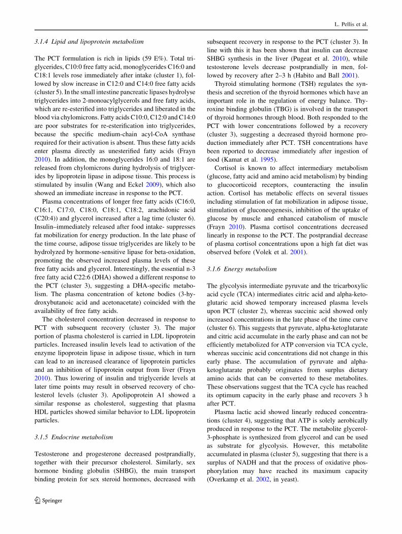
(Fig. 2a). Subjects on AIDM showed a higher release of
DHA to plasma both in the early and late response com-
pared to control subjects (Fig. 2a, 94% increased AUC?
and a 140% increased AUCt). The fatty acid C17:0 origi-
nates primarily from animal products (dairy and meat)
(Brevik et al. 2005). This exogenous fatty acid demon-
strated decreased fasting concentrations in subjects on
AIDM after the 5 weeks intervention (Table 2). In
response to the PCT, plasma C17:0 showed increased
concentrations in the early time response in subjects on
AIDM, in contrast to the control group exhibiting reduced
concentrations (Supplemental Fig. 1, 260% increased
AUCt). Indole-3-propionic acid is synthesized by gut
microbiota (Wikoff et al. 2009). This metabolite showed
increased fasting concentrations in subjects on AIDM,
suggesting an effect of AIDM in the gastrointestinal tract.
In response to PCT subjects on AIDM showed reduced
plasma concentrations of indole-3-propionic acid in the late
phase, whereas control subjects still showed elevated
concentrations (Supplemental Fig. 1, 120% increased
Table 2 Identified metabolites and proteins that respond differentially between AIDM and placebo in response to PCT
# Parameter AUC/Tmax/Tmin Fasting (t0)
1 2,4-Dihydroxybutanoic acid AUC No
2 Lactose AUC No
3 EGF-R AUC No
4 Fibrinogen AUC No
5 Glucagon AUC No
6 LH AUC No
7 ACE (CD143) AUC No
8 CD40 AUC No
9 von Willebrand factor AUC No
10 Myeloperoxidase AUC No
11 Thyroxine binding globulin AUC No
12 SM(d18:1/22:0) AUC No
13 SM(d17:1/16:0) AUC Yes (: in AIDM)
14 SM(d17:1/18:0) AUC Yes (: in AIDM)
15 SM(d18:1/16:0) AUC Yes (: in AIDM)
16 Indole-3-propionic acid AUC Yes (: in AIDM)
17 Uric acid AUC Yes (; in AIDM)
18 C17:0 Fatty acid AUC Yes (; in AIDM)
19 C22:6 Fatty acid AUC Yes (: in AIDM)
20 Isoleucine AUC Yes (; in AIDM)
21 Phenylalanine AUC Yes (; in AIDM)
22 Proline AUC Yes (; in AIDM)
23 Valine AUC Yes (; in AIDM)
24 MDC AUC Yes (; in AIDM)
25 VCAM-1 AUC Yes (; in AIDM)
26 Glutamine Tmax–L No
27 Tryptophan Tmax–L No
28 4-Hydroxyglutamate semialdehyde? Tmax–L No
29 Thyroid stimulating hormone Tmin–E No
30 SM(d18:1/17:0) Tmin–E Yes (: in AIDM)
31 SHBG Tmin–E Yes (: in AIDM)
Bold plasma parameters are newly identified to respond to the dietary AIDM intervention
L = Tmax or Tmin is reached significantly later in AIDM treated subjects compared to placebo treated subjects
E = Tmax or Tmin is reached significant earlier in AIDM treated subjects compared to placebo treated subjects
AUC area under curve (either AUC?, AUC- or AUCt)
L. Pellis et al.
123

AUC-). Finally, lactose that likely derives from the PCT
dairy shake showed a differential response between sub-
jects on AIDM and placebo. Subjects on AIDM have a
higher clearance rate of plasma lactose in comparison to
subjects on placebo (Fig. 2b, 55% decreased AUC-).
3.2.3 Oxidative stress
The PCT reveals additional effects of AIDM that may
suggest reduced oxidative stress. Oxidative stress markers
MDC (Fig. 2c, 52% decreased AUC? and a 162%
decreased AUCt) and myeloperoxidase (Supplemental
Fig. 1, 25% decreased AUC? and a 27% decreased AUCt)
showed a delayed increase in response to the PCT after
AIDM intervention suggesting a retarded or reduced oxi-
dative stress response with AIDM. Consistent with this,
AIDM intervention caused diminished reduction in
response to PCT compared to control (Supplemental Fig. 1,
47% decreased AUC-), as well as 9% reduction of base-
line concentrations of the antioxidant uric acid (Bakker
et al. 2010).
3.2.4 Inflammation
The PCT reveals additional effects of AIDM that might
suggest anti-inflammatory effects. The inflammatory
markers CD40, VCAM1, fibrinogen and EGF-R show a
more rapid reduction in response to PCT after AIDM
intervention compared to placebo (Fig. 2d, e and Supple-
mental Fig. 1). CD40, VCAM1, fibrinogen and EGFR had
a reduced AUC? respectively of 25%, 66%, 80% and 52%.
VCAM1 and fibrinogen also had an increased AUC-
respectively of 42% and 55% and AUCt respectively of
90% and 170% (negative AUCt in both conditions and both
markers). Also levels of ACE (Supplemental Fig. 1) and
von Willebrand factor (Fig. 2f) reduced in response to PCT
because of AIDM intervention, whereas control interven-
tion had no effect (fluctuations around baseline, 53%
decreased AUC? for ACE, and 104% increased AUC-
and 1291% decreased AUCt for von Willebrand factor).
Interestingly, many of these factors are related to endo-
thelial function (Chakrabarti et al. 2007; Constans and
Conri 2006; Kakafika et al. 2007), supporting a potential
beneficial effect of AIDM on vascular health (Bakker et al.
2010).
3.2.5 Endocrine metabolism
Glucagon (Fig. 2g) and luteinizing hormone (LH) (Sup-
plemental Fig. 1), showed increased concentrations in
response to PCT after AIDM intervention, whereas control
intervention showed no alteration (44% decreased AUC-
for glucagon and 192% increased AUCt for LH). The
plasma concentration of thyroid hormone transporter TBG
showed a stronger reduction in subjects on AIDM com-
pared to control (56% increased AUC- and 87% increased
AUCt (negative AUCt in both conditions)). Whereas the
lowest observed plasma concentration was reached signif-
icantly earlier in AIDM compared to control for the sex
hormone transporter SHBG and thyroid stimulating hor-
mone (TSH) (Supplemental Fig. 1, SHBG; Tmin pla-
cebo = 3.2 h, Tmin AIDM = 2.2 h and TSH; Tmin
placebo = 3.1 h, Tmin AIDM = 2.4 h). Thus, AIDM may
influence endocrine metabolism, although it is difficult to
interpret how these changes are related to health. The
network in Fig. 3 shows connections between the outlined
endocrine factors. Although there was no change in insulin
sensitivity, glucose or insulin response to PCT between
AIDM and control, there is a clear indication that AIDM
influences endocrine metabolism. To what extent these
changes are related to health remains to be elucidated.
Table 3 Nutritional value of 500 ml postprandial challenge test formulation
Nutritional value Content Energy
Total 2945 kJ or 706 kcal
Total protein 20.7 g 11.7 E%
Total fat 46.1 g 58.7 E%
Saturated fatty acids 27.1 g
Monounsaturated fatty acids 11.8 g
Polyunsaturated fatty acids 1.4 g
Cholesterol 0.1 g
Total carbohydrates 52.2 g 29.6 E%
Total mono-disaccharides 42.3 g
Total fiber 0.1 g
g gram, kJ kilojoules, kcal kilocalories, E% energy percent
Metabolic profiling after postprandial challenge
123

Fig. 2 Responses to PCT after AIDM and placebo intervention.
Figures show mean (±SEM) difference versus t0 for each time point.
Differences versus t0 were calculated for 35 (metabolites) or 33
subjects (proteins). Diamond indicates placebo intervention and
square indicates AIDM intervention. MDC Macrophage-derived
chemokine, VCAM-1 vascular cell adhesion protein 1, vWF von
Willebrand factor. Y-axis are relative concentrations for metabolites
C22:6 fatty acid and lactose; for proteins units for difference in
concentration (compared to t0) are as follows: MDC pg/ml, VCAM-
1 ng/ml, fibrinogen mg/ml, vWF lg/ml and glucagon pg/ml
L. Pellis et al.
123

It is difficult to compare our results with other studies,
because no standardized PCT is available. The meal chal-
lenges differ in energy content from 250 to 2500 kcal, from
50 to 100 E% fat, 0–50 E% carbohydrates, and 0 to 20 E%
protein (Paglialunga and Cianflone 2007). Moreover these
PCTs also differ in composition, solid versus liquid, and
amount (Cianflone et al. 2008).
4 Concluding remarks
The PCT affected carbohydrate, amino acid, lipid and
lipoprotein metabolism. Furthermore, the processes energy
metabolism, oxidative stress, inflammation and endocrine
response reacted to the PCT. New observations included
the different response of n-3 fatty acid DHA compared to
other long chain free fatty acids and that a PCT may affect
indicators of bone metabolism.
Assessment of metabolic changes due to a dietary
intervention revealed by the PCT resulted in 31 plasma
parameters that showed a differential response to the PCT
after AIDM compared to placebo intervention. More than
50% of these were uniquely changed when applying the
PCT. Other parameters changed both in perturbed and non-
perturbed conditions, which offered additional insight in
biological responses. For example, now we showed that the
intervention reduced the short term, acute vascular
inflammatory and oxidative stress response, in addition to
the effect on vascular health and oxidative stress in fasted
(homeostatic) conditions (Bakker et al. 2010).
This nutrigenomics based PCT showed the relevance of
metabolic perturbation for quantification of subtle pheno-
typic changes. Applying challenge tests and measuring the
integrated responses should be further developed as a tool
to quantify and define optimal health.
Acknowledgments We express our gratitude to the volunteers par-
ticipating in the study, staff of the Metabolic Research Unit, labora-
tories and groups contributing to the study and analyses, our colleagues
contributing in designing the experiment and Professor Christian
Drevon (University of Oslo, department of Nutrition) for critical
reading of the manuscript. This study was supported by TNO. TNO is a
member of the Nutrigenomics Organization (www.nugo.org).
Fig. 3 Network showing
connections between endocrine
factors with a differential
response to PCT after AIDM
intervention compared to
placebo (MetaCore, network
option auto expand)
Metabolic profiling after postprandial challenge
123

Open Access This article is distributed under the terms of the
Creative Commons Attribution Noncommercial License which per-
mits any noncommercial use, distribution, and reproduction in any
medium, provided the original author(s) and source are credited.
References
Bakker, G. C. M., Van Erk, M. J., Pellis, L., Wopereis, S., Rubingh,
C. M., Cnubben, N. H. P., et al. (2010). An antiinflammatory
dietary mix modulates inflammation and oxidative and metabolic
stress in overweight men: A nutrigenomics approach. AmericanJournal of Clinical Nutrition, 91(4), 1044–1059.
Bijlsma, S., Bobeldijk, I., Verheij, E. R., Ramaker, R., Kochhar, S.,
Macdonald, I. A., et al. (2006). Large-scale human metabolo-
mics studies: A strategy for data (pre-) processing and validation.
Analytical Chemistry, 78(2), 567–574.
Brevik, A., Veierod, M. B., Drevon, C. A., & Andersen, L. F. (2005).
Evaluation of the odd fatty acids 15:0 and 17:0 in serum and
adipose tissue as markers of intake of milk and dairy fat.
European Journal of Clinical Nutrition, 59(12), 1417–1422.
Carroll, M. F., & Schade, D. S. (2003). Timing of antioxidant vitamin
ingestion alters postprandial proatherogenic serum markers.
Circulation, 108(1), 24–31.
Cawston, T., & Young, D. (2010). Proteinases involved in matrix
turnover during cartilage and bone breakdown. Cell and TissueResearch, 339(1), 221–235.
Chakrabarti, S., Blair, P., & Freedman, J. E. (2007). CD40-40L
signaling in vascular inflammation. Journal of BiologicalChemistry, 282(25), 18307–18317.
Cianflone, K., Paglialunga, S., & Roy, C. (2008). Intestinally derived
lipids: Metabolic regulation and consequences—An overview.
Atherosclerosis Supplements, 9(2), 63–68.
Constans, J., & Conri, C. (2006). Circulating markers of endothelial
function in cardiovascular disease. Clinica Chimica Acta,368(1–2), 33–47.
Devaraj, S., Wang-Polagruto, J., Polagruto, J., Keen, C. L., & Jialal, I.
(2008). High-fat, energy-dense, fast-food-style breakfast results
in an increase in oxidative stress in metabolic syndrome.
Metabolism, 57(6), 867–870.
Elliott, R., Pico, C., Dommels, Y., Wybranska, I., Hesketh, J., &
Keijer, J. (2007). Nutrigenomic approaches for benefit-risk
analysis of foods and food components: Defining markers of
health. British Journal of Nutrition, 98(6), 1095–1100.
Esposito, K., Nappo, F., Giugliano, F., Di, P. C., Ciotola, M.,
Barbieri, M., et al. (2003). Meal modulation of circulating
interleukin 18 and adiponectin concentrations in healthy subjects
and in patients with type 2 diabetes mellitus. American Journalof Clinical Nutrition, 78(6), 1135–1140.
Frayn, K. N. (2010). Metabolic regulation—A human perspective.
Blackwell Publishers.
Ghanim, H., Abuaysheh, S., Sia, C. L., Korzeniewski, K., Chaudhuri,
A., Fernandez-Real, J. M., et al. (2009). Increase in plasma
endotoxin concentrations and the expression of toll-like recep-
tors and suppressor of cytokine signaling-3 in mononuclear cells
after a high-fat, high-carbohydrate meal. Diabetes Care, 32(12),
2281–2287.
Goldtein, M. S., Henry, W. L., Huddlestun, B., & Levine, R. (1953).
Action of insulin on transfer of sugars across cell barriers; common
chemical configuration of substances responsive to action of the
hormone. American Journal of Physiology, 173(2), 207–211.
Habito, R. C., & Ball, M. J. (2001). Postprandial changes in sex
hormones after meals of different composition. Metabolism,50(5), 505–511.
Jackson, K. G., Armah, C. K., & Minihane, A. M. (2007). Meal fatty
acids and postprandial vascular reactivity. Biochemical SocietyTransactions, 035(3), 451–453.
Kakafika, A. I., Liberopoulos, E. N., & Mikhailidis, D. P. (2007).
Fibrinogen: A predictor of vascular disease. Current Pharma-ceutical Design, 13(16), 1647–1659.
Kamat, V., Hecht, W. L., & Rubin, R. T. (1995). Influence of meal
composition on the postprandial response of the pituitary-thyroid
axis. European Journal of Endocrinology, 133(1), 75–79.
Koek, M., van der Kloet, F., Kleemann, R., Kooistra, T., Verheij, E.,
& Hankemeier, T. (2011). Semi-automated non-target process-
ing in GC 9 GC-MS metabolomics analysis: Applicability for
biomedical studies. Metabolomics, 7(1), 1–14.
Lopez-Miranda, J., Williams, C., & Lairon, D. (2007). Dietary,
physiological, genetic and pathological influences on postpran-
dial lipid metabolism. British Journal of Nutrition, 98(3),
458–473.
Lundman, P., Boquist, S., Samnegsrd, A., Bennermo, M., Held, C.,
Ericsson, C. G., et al. (2007). A high-fat meal is accompanied by
increased plasma interleukin-6 concentrations. Nutrition, Metab-olism and Cardiovascular Diseases, 17(3), 195–202.
Margioris, A. N. (2009). Fatty acids and postprandial inflammation.
Current Opinion in Clinical Nutrition & Metabolic Care, 12(2),
129–137.
Minisola, S., Antonelli, R., & Mazzuoli, G. (1985). Clinical
significance of free plasma hydroxyproline measurement in
metabolic bone disease. Journal of Clinical Chemistry &Clinical Biochemistry, 23(9), 515–519.
Nakatsuji, H., Kishida, K., Kitamura, T., Nakajima, C., Funahashi, T.,
& Shimomura, I. (2010). Dysregulation of glucose, insulin,
triglyceride, blood pressure, and oxidative stress after an oral
glucose tolerance test in men with abdominal obesity. Metab-olism: Clinical and Experimental, 59(4), 520–526.
Nappo, F., Esposito, K., Cioffi, M., Giugliano, G., Molinari, A. M.,
Paolisso, G., et al. (2002). Postprandial endothelial activation in
healthy subjects and in type 2 diabetic patients: Role of fat and
carbohydrate meals. Journal of the American College ofCardiology, 39(7), 1145–1150.
Neri, S., Signorelli, S. S., Torrisi, B., Pulvirenti, D., Mauceri, B.,
Abate, G., et al. (2005). Effects of antioxidant supplementation
on postprandial oxidative stress and endothelial dysfunction: A
single-blind, 15-day clinical trial in patients with untreated type
2 diabetes, subjects with impaired glucose tolerance, and healthy
controls. Clinical Therapeutics, 27(11), 1764–1773.
Nowatzky, J., Howard, R., Pillinger, M. H., & Krasnokutsky, S.
(2010). The role of uric acid and other crystals in osteoarthritis.
Current Rheumatology Reports, 12(2), 42–148.
Overkamp, K. M., Bakker, B. M., Kotter, P., Luttik, M. A., van
Dijken, J. P., & Pronk, J. T. (2002). Metabolic engineering of
glycerol production in Saccharomyces cerevisiae. Applied andEnvironmental Microbiology, 68(6), 2814–2821.
Paglialunga, S., & Cianflone, K. (2007). Regulation of postprandial
lipemia: An update on current trends. Applied Physiology,Nutrition, and Metabolism, 32(1), 61–75.
Parfitt, A. M. (2002). Targeted and nontargeted bone remodeling:
Relationship to basic multicellular unit origination and progres-
sion. Bone, 30(1), 5–7.
Poppitt, S. D., Keogh, G. F., Lithander, F. E., Wang, Y., Mulvey, T.
B., Chan, Y. K., et al. (2008). Postprandial response of
adiponectin, interleukin-6, tumor necrosis factor-[alpha], and
C-reactive protein to a high-fat dietary load. Nutrition, 24(4),
322–329.
Pugeat, M., Nader, N., Hogeveen, K., Raverot, G., Dechaud, H., &
Grenot, C. (2010). Sex hormone-binding globulin gene expres-
sion in the liver: Drugs and the metabolic syndrome. Molecularand Cellular Endocrinology, 316(1), 53–59.
L. Pellis et al.
123

Riches, P. L., & Ralston, S. H. (2010). Recent insights into the
biology of bone turnover. Journal of the Royal College ofPhysicians of Edinburgh, 40(1), 66–69.
Schett, G., Kiechl, S., Bonora, E., Zwerina, J., Mayr, A., Axmann, R.,
et al. (2009). Vascular cell adhesion molecule 1 as a predictor of
severe osteoarthritis of the hip and knee joints. Arthritis andRheumatism, 60(8), 2381–2389.
Shaham, O., Wei, R., Wang, T. J., Ricciardi, C., Lewis, G. D., Vasan,
R. S., et al. (2008). Metabolic profiling of the human response to
a glucose challenge reveals distinct axes of insulin sensitivity.
Molecular Systems Biology, 4, 214–222.
Spallarossa, P., Garibaldi, S., Barisione, C., Ghigliotti, G., Altieri, P.,
Tracchi, I., et al. (2008). Postprandial serum induces apoptosis in
endothelial cells: Role of polymorphonuclear-derived myeloper-
oxidase and metalloproteinase-9 activity. Atherosclerosis,198(2), 458–467.
Tsai, W. C., Li, Y. H., Lin, C. C., Chao, T. H., & Chen, J. H. (2004).
Effects of oxidative stress on endothelial function after a high-fat
meal. Clinical Science, 106(3), 315–319.
van der Kloet, F. M., Bobeldijk, I., Verheij, E. R., & Jellema, R. H.
(2009). Analytical error reduction using single point calibration
for accurate and precise metabolomic phenotyping. Journal ofProteome Research, 8(11), 5132–5141.
van Eijk, H. M., Deutz, N. E., Wagenmakers, A. J., & Soeters, P. B.
(1990). 3-Methylhistidine determined in plasma by ‘‘high-perfor-
mance’’ lipid chromatography. Clinical Chemistry, 36(3), 556–559.
van Ommen, B., Keijer, J., Heil, S. G., & Kaput, J. (2009).
Challenging homeostasis to define biomarkers for nutrition
related health. Molecular Nutrition & Food Research, 53(7),
795–804.
van Ommen, B., Keijer, J., Kleemann, R., Elliott, R., Drevon, C.,
McArdle, H., et al. (2008). The challenges for molecular
nutrition research 2: Quantification of the nutritional phenotype.
Genes & Nutrition, 3(2), 51–59.
Volek, J. S., Gomez, A. L., Love, D. M., Avery, N. G., Sharman, M.
J., & Kraemer, W. J. (2001). Effects of a high-fat diet on
postabsorptive and postprandial testosterone responses to a
fat-rich meal. Metabolism, 50(11), 1351–1355.
Walsh, J. S., & Henriksen, D. B. (2010). Feeding and bone. Archivesof Biochemistry and Biophysics, 503(1), 11–19.
Wang, H., & Eckel, R. H. (2009). Lipoprotein lipase: From gene to
obesity. American Journal of Physiology-Endocrinology andMetabolism, 297(2), E271–E288.
Waterworth, D. M., Talmud, P. J., Luan, J., Flavell, D. M., Byrne, C.
D., Humphries, S. E., et al. (2003). Variants in the APOC3
promoter insulin responsive element modulate insulin secretion
and lipids in middle-aged men. Biochimica et Biophysica Acta(BBA) - Molecular Basis of Disease, 1637(3), 200–206.
Wikoff, W. R., Anfora, A. T., Liu, J., Schultz, P. G., Lesley, S. A.,
Peters, E. C., et al. (2009). Metabolomics analysis reveals large
effects of gut microflora on mammalian blood metabolites.
Proceedings of the National Academy of Sciences of the UnitedStates of America, 106(10), 3698–3703.
Wopereis, S., Rubingh, C. M., Van Erk, M. J., Verheij, E. R., van
Vliet, V. T., Cnubben, N. H., et al. (2009). Metabolic profiling of
the response to an oral glucose tolerance test detects subtle
metabolic changes. PLoS One, 4(2), e4525.
Wybranska, I., Malczewska-Malec, M., Niedbal, S., Naskalski, J. W.,
& Dembinska-Kiec, A. (2003). The TNF-alpha gene NcoI
polymorphism at position-308 of the promoter influences insulin
resistance, and increases serum triglycerides after postprandial
lipaemia in familiar obesity. Clinical Chemistry and LaboratoryMedicine, 41(4), 501–510.
Wyss, M., & Kaddurah-Daouk, R. (2000). Creatine and creatinine
metabolism. Physiological Reviews, 80(3), 1107–1213.
Zhao, X., Peter, A., Fritsche, J., Elcnerova, M., Fritsche, A., Haring,
H. U., et al. (2009). Changes of the plasma metabolome during
an oral glucose tolerance test: Is there more than glucose to look
at? American Journal of Physiology-Endocrinology and Metab-olism, 296(2), E384–E393.
Zivkovic, A., Wiest, M., Nguyen, U., Nording, M., Watkins, S., &
German, J. (2008). Assessing individual metabolic responsive-
ness to a lipid challenge using a targeted metabolomic approach.
Metabolomics, 5(2), 209–218.
Metabolic profiling after postprandial challenge
123
Related Documents