¿1. t] PIASMA LIPOPROTEIN TR.IACNßLYCROL METABOLISM IN SHEP A thesis submit,ted to the University of Adelaide in fulfilment of the requirements for the degree of Doctor of Philosopftry JOHN CIIARLES IOUIS t"tAIvtO, B. fu. Sc. (Hons.) (R¿etai¿e) Department of Animal Sciences, hlaite Agricultural Research Instítute, Ihe University of Adelaide, South Australia Septernber, 1986 by Éru0, rlrcl tt lP ,! ', , (i)

Welcome message from author
This document is posted to help you gain knowledge. Please leave a comment to let me know what you think about it! Share it to your friends and learn new things together.
Transcript
¿1. t]
PIASMA LIPOPROTEIN TR.IACNßLYCROL METABOLISM IN SHEP
A thesis
submit,ted to the University of Adelaide in fulfilment
of the requirements for the degree of
Doctor of Philosopftry
JOHN CIIARLES IOUIS t"tAIvtO, B. fu. Sc. (Hons.) (R¿etai¿e)
Department of Animal Sciences,
hlaite Agricultural Research Instítute,
Ihe University of Adelaide,
South Australia
Septernber, 1986
by
Éru0, rlrcl tt lP ,! ', ,
(i)
DEDICATION
ttThis thesis is dedicated Eo the mernory of my
father, the late James Benjamin t"famo (1925-1964). In
t952-53 he and my mother Ùbryanne Sylvia left their
homeland of Malta destined for AusLralia so
that their children may have the opportunity of
a better education.tt
I wish to thank them.
(ií)
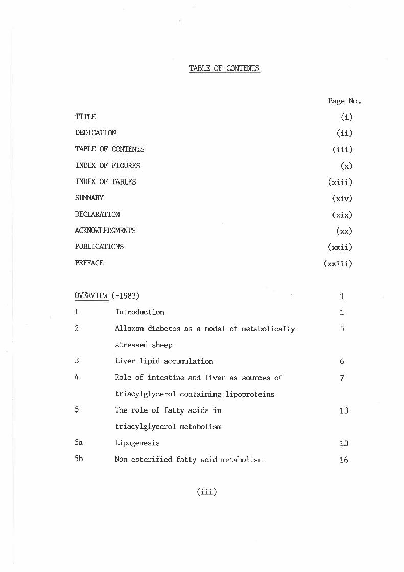
TABLE OF CO}ÏIH{IS
TITLE
DEDICATION
TABLE OF CONIn{TS
INDÐ( OF FIGURES
INDÐ( OF TABLES
SUMI',IARY
DECTARATION
ACKNOI^JLEDGMM{IS
PUBLICATIONS
PR,MACE
O\IERVIEI^Ì (-fgA:)
1 Introduction
2 Alloxan diabetes as a model of metabolically
sEressed sheep
3 Liver lipid accumulation
4 Role of intestine and liver as sources of
triacylglycerol containing lipoproteins
5 The role of fatty acids in
triacylglyeerol metabo lism
5a Lipogenesis
5b Non esterified faL|y acid metabolism
Page No.
(i)(ii)
(iii¡(*)
/ ...\(xr1r_/
(xiv)
(xix)
(**)
(xxii)
(xxiii)
tL
5
6
7
13
13
L6
(iii)
6
7
8
Hepatic triacylglycerol secretion
l,fetabolism of very low density lipoproteins
Object.ives of this study
CTT,APTM. 1 LPOPROTEIN PROFILE OF NOR}4AL FED AND
ALLOXA}] DIABEf,IC STMEP
20
25
26
t.L
T.L.L
L.7.2
L.t.2.r
t.I.2.2
1.1.3
t.2
T.2.L
L.2.L.L
L.2.2
1.2.3
L.2.4
I.2.4.L
L.2./+.2
L.2.4.3
L.2.4.4
L.2.4.5
Inbroduction
Lipoprotein structure and functíon
Role of plasma lipoproteins
In monogastric onnivores
Sheep plasma lipoproteins
Aims of chapter one
Methods and nraterials
Aninrals used
Collection and preservation of blood plasma
Determination of bloôd glucose
Adjustment of plasnn solvent density
Separation and purification of plasma
Iipoproteins
Time course studies
C,ollection of total plasma lipoproteins
Est.imation of total plasma lipoproteins
Agarose gel filtration
High perfonnance liquid chromatography
29
29
29
33
33
39
4L
43
43
43
44
45
45
45
46
47
47
48
(in)
L.2.4.6
L.2.4.6.L
L.2.4.6.2
I.2.4.6.3
L.2.4.7
L.2.5
L.2.5.!
t.2.5.2
t.2.5.3
L.2.5.4
L.2.6
L.2.7
L.2.8
L.2.9
1.3
1.3.1
L.3.2
1.3.3
L.3.4
L.3.4.L
L.3.4.2
1.3.5
1.3.5.1-
Serial centrifugaLion of plasma lipoproteins
Isolation of very low density.lipoproteins
Isolation of low density lipoproteins
Isolation of high density lipoproteins
Agarose gel electrophoresis
Extraction and analysis of lipid components
from plasma and lipoprotein fractions
Extraction
Triacylglyceride determination
Phospho lipid determinat ion
C,kroles terol and choles terol-ester determination
Lipoprotein protein determination
Non esterified fatty acid determination
Transmission electron microscopy
Materials and reagents
ResulLs
Sheep plasma
Time course studies
Sheep plasma lipoprotein concentration
Agarose gel chromatography
Human plasma lipoproteins
Sheep plasma lipoproteins
Agarose gel electrophoresis of the agarose
chromaLography lipoprotein fractions
Human fractions
49
50
50
51
51
51
5L
52
53
54
55
56
56
57
58
58
58
6t
6I
6L
63
63
63
(n)
L.3.5.2
1 .3.6
L.3.7
1.3.8
1.3.8.1
L.3.8.2
1.3.9
L.3.9.t
t.3.9.2
1.3.10
t.4
Sheep fract.ions
High perfonnance gel filtrationSheep lipoproteins isolated by serial
ultracentrifugation
C,Lremical characterization of sheep lipoproteins
Fed sheep
Diabet,ic sheep
Plasma lipid profile and the role of
lipoproteins in plasma lipid transport
Fed sheep
Diabetic sheep
Transmission electron microscopy of
sheep lipoproteins
Discussi-on
Introduction
Lipoprotein lipase and hepatic lipase
Lipoprotein lipase
Hepatic lipase
RoIe of lipoprotein lipase and hepat.ic lipase in
the metabolism of very low density lipoprotein
64
66
69
83
70
70
73
73
73
75
75
97
97
98
99
100
101
CHAPTER 2 TI]E ROLE OF LIPOPROTEIN LIPASE AI{D HEPATIC
LIPASE IN THE METABOLISM OF VERY I,OW DN{SITY
LIPOPROTEIN-TRIACYIfLYCERIDE IN SHEEP
2.L.L
2.1.2
2.t.2.L
2.t.2.2
2.L.3
(rri)
2.L.4
2.t.5
2.2
2.2.L
2.2.2
2.2.3
2.2.4
2.2.5
2.2.6
2.2.7
2.2.8
2.2.9
2.2.t0
2.3
2.3.r
2.3.L.L
triacylglyceride
Postheparin plasma lipoprotein lipase and hepatic 103
lipase
Regulation of lipoprotein lipase and hepatic lipase 105
Methods and materials LO7
Animals used I01
Acetone powder preparations of liver and adipose L07
tissue
Adipose lipoprotein lipase and hepatic lipase 108
acetone powder enzyme preparations
Sheep and rat postheparin plasma 108
Lipoprotein lipase and hepatic lipase assay 108
Heparin-sepharose affinity chromatography of sheep 109
liver enzyrne homogenates and postheparin plasma
Isolation of very low density lipoproteins from 110
fed and diabetic sheep
Hydrolysis of very low density lipoprotein triacyl- 110
glyceride from fed and diabetic sheep, in post-
heparin plasma from fed sheep
Blood glucose, triacylglyceride and non-esterifled ILI
f.aLLy acids
Materials and reagents LLL
Results Ll2
CharacterizaLion of acetone powder enzyme homogenales 112
Sheep and rat liver extracLs LLz
(vr1.)
2.3.L.2
2.3.2
2.3.2.L
2.3.2.2
2.3.2.3
2.3.2.4
2.3.2.5
2.3.2.6
2.3.3.L
2.3.3.2
2.4
Sheep and rat adipose extracts
Postheparin plasma Iipase act.ivities
Rat posthepa.rin plasma
Sheep postheparin plasma
Postheparin plasnn lipoprotein J-ipase and hepatic
lipase in fed, fasted and diabetic sheep
Postheparin hydrolysis of very low density lipo-
protein triacylglyceride from fed and diabetic sheep
Posthepa.rin plasma lipase activities in rams,
wethers and ewes
Posthepa.rin plasma lipase activities in 'Iean' and
tobeset sheep
Triacylglyceride secretion in preweaned 'leant and
tobeset larnbs
Toxicity of Triton I^IR1339
Discussion
CHAPTM. 3 APOPROTEIN PROFILE OF NOR},IAL FED AND
ALIOXAN DIABE'IIC SHEEP
LL6
t23
t23
t25
L25
L37
138
131
L34
I34
t36
3.1
3.1.1
3.r.2
3.2
3.2.r
Introduction
Hunan apoproteins; structure and function
Metabolism of triacylgyceride rich lipoproteins ;
role of apoproteins
Methods and materials
Animals used
/ ...\(v]-rr )
153
153
155
t6L
L66
L66
3.2.2
3.2.3
3.2.4
3.2.5
3.3
3.3.1
3.3.2
3.3.3
3.4
Protein extraction
Apoprotein B and soluble apoproteins determinaLion
Sodium dodecyl- sulphate polyacrylamide gel
electroplroresis
Materials and reagents
ResulLs
Apoprotein profile of fed and diabetic sheep
Apoprotein B content of sheep lipoproteins
Effect of ultracentrlfugation on apoprotein recovery
Discussion
Gn{MAL DISCUSSION
BIBLIOGRAPTIY
L66
L67
L67
L@
L70
: L70
L75
L7s
t79
4
5
190
19'8
(i*)
L
2
L.I
r.2
1.3
L.4
1.5
L.6
L.7
1.8
I.9
1.10
2.L
2.2
INDÐ( OF FIGURES
Page No.
Stages of hepatic fat accumulation in sheep 2
Biosynthesis of hepatic lipoproteins L2
Structure of lipoproteins 31
Sheep plasrna from fed and diabetic aninnls 59
Time course studies on the ultracentrifugation 60
of sheep lipoproteins
Agarose gel chronratography of plasma lipoproteins 62
Agarose gel electrophoresis of human lipoprotein 65
fractions isolated by gel chronntography
Agarose gel electropLroresis of sheep lipoproteirs 67
fractions isolated by gel chromatography
High performance gel elution of sheep plasma 68
lipoproteins
Agarose gel electrophoresis of sheep lipoprotein 7L
fractions isolated by serial ultracentrifugation
Size distrib:tion of sheep plasma lipoproLeins 77
Electron micrographs of sheep plasma lipoproteins 78-82
Role of lipoprotein lipase and hepatic lipase in t02
the catabolism of very low density triacylglyceride
Effect of pH on sheep and rat hepatic lipase activity 113
Effect of NaCl on sheep and rat hepatic lipase tL42.3
(*)
2.4
2.5
2.6
2.7
2.8
2.9
2.L0
2.Lt
2.I2
2.r3
2.r4
2.L5
activity
Effect of heparin on sheep liver and adipose lipase LL5
activity
Effect of subsLrate concentration on sheep liver and LL7
adipose lipase activity
Effect of time on sheep liver and adipose lipase 118
acÈivity
Effect of serum concentration on sheep liver and tL9
adipose lipase activity
Heparin sepharose affinity chromatography of sheep tzO
liver enzyme preparati-ons
Effect of NaCl on sheep and rat adipose lipoprotein L2l
Iipase
Effect of pH on sheep and rat adipose lipoprotein L22
Iipase
Effect of NaCI on sheep postheparin plasma lipase L26
activity
Effect of time on sheep postheparin plasrna lipase L27
activity
Heparin sepharose affinity chromatography of sheep L28
posthepa.rin plasma
Effect of pH on sheep postheparin plasnra lipase L29
activity
Rate of very low density lipoprotein triacylglyceride L32
hydrolysis from fed and diabet.ic sheep, with fed sheep
(xi)
3.1
3.2
3.3
3.4
postheparin plasma
ApoproLein regulation of very low density lipoprotein 163
triacylglyceride metabolism in hurnans
SDS-PAGE of ovine lipoprotein apoproteins 172
SDS-PAGE of ovine lipoprotein apoproteins t73
Postulated apoprotein regulation óf plasma very low 188
density lipoprotein triacylglyceride metabolism in
sheep
Difficulties associated raíth plasrna lipoprotein L95
triacylglyceríde metabolism in metabolically
stressed sheep
4
(xii)
t.rt.2
1.3
2.r
2.2
2.2
2.3
2.4
3.1
3.2
3.3
3.4
INDEX OF TABLES
Human plasma lipoproteins
Ckremical conposition of sheep plasma lipoproteins
Sheep plasma lipid profile and role of lipoproteins
Rat postheparin plasma lipase act.ivities
Postheparin plasma lipase activities in fed, fasted
and diabetic sheep
Posthepa.rin plasma lipase activities in rams,
wethers and ewes
Lipoprotein lipase and hepatic lipase hydrolysis
of very low density lipoproLein triacylglyceride
from fed and diabetic sheep
Postheparin plasma lipase activities intlearf and
'obesd sheep
Human apoproteins; structure and function
Sheep lipoprotein-apoprotein prof ile
þoprotein B content of ovine lipoproteins
Recovery of ovine lipoprotein apoproteins
Page No.
35
72
74
L24
130
130
133
135
156
L7L
t76
L78
(xrrr )
SUMMARY
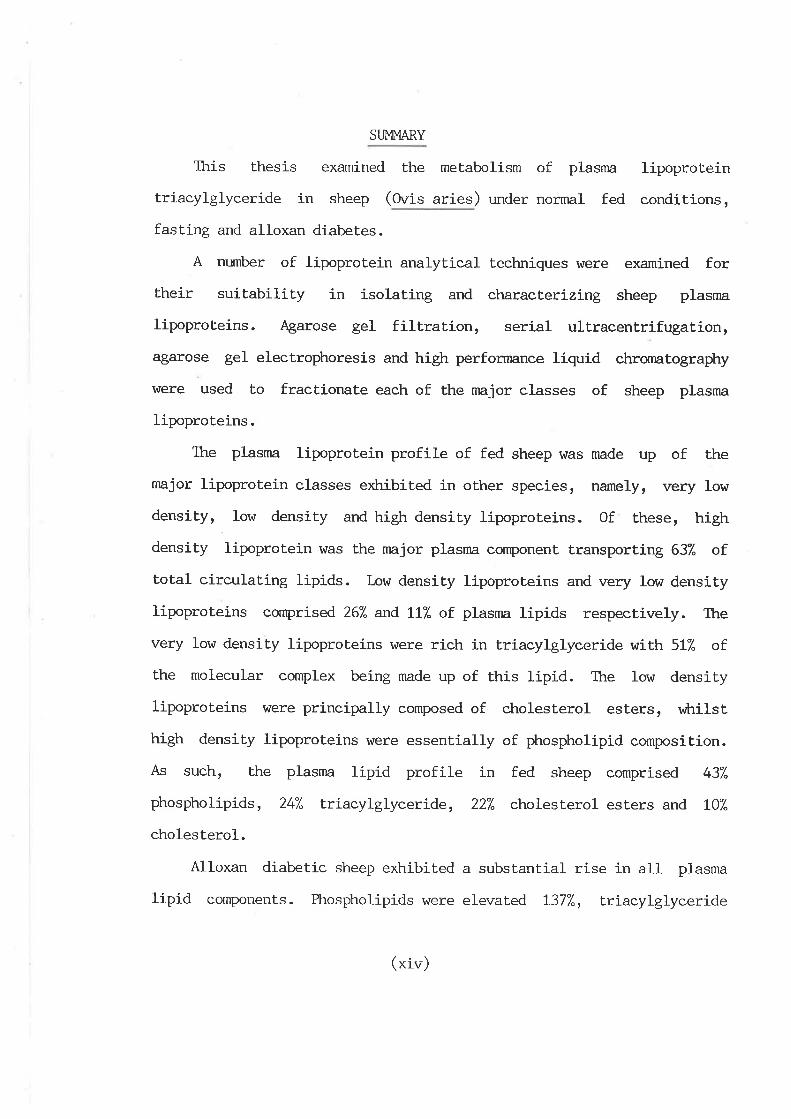
This thesis examined the metabolism of plasma lipoprotein
triacylglyceride in sheep (Ovis aries) under nornal fed conditions,
fasting and alloxan diabetes.
A number of lipoprotein analytical techniques r,Ì/ere examined for
their suitability in isolating and characterízing sheep plasma
lipoproteins. {garose gel filtration, serial ultracentrifugation,
agarose gel electrophoresis and high perfonnance liquíd chromatography
\ÀIere used to fractionate each of the major classes of sheep plasma
lipoproteins.
The plasma lipoprotein profile of fed sheep was made up of the
major lipoprotein classes exhibited in other species, namely, very low
density, low density and high densiLy lipoproteins. Of these, high
density lipoprotein was the major plasma component transporting 637" of
total circulating lipids. Low density lipoproteins and very low density
lipoproteins comprised 267" and LL7" of plasnra lipids respectively. The
very low density lipoproteins were rich in triacylglyceride with 517. of
the molecurar complex being made up of this ripid. The low density
lipoproteins were principally composed of cholesterol esters, vrhilst
high density lipoproteins were essentially of phospholipid composition.
As such, the plasma lipid profile in fed sheep comprised 437"
phospholipids, 247" triacylglyceride, 227" cholesterol esters and LO7"
cholesterol.
Alloxan diabetic sheep exhibited a subsLantial rise in all plasma
lipid components. Phospholipids were elevated L377", triacylglyceride
(xiv)
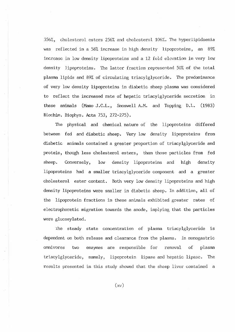
3567", cholesterol esters 2567" and cholesteroL L067". The hyperlipidaemia
kras reflected in a 587. inerease in high densily lipoproteins, an 897"
increase in low density lipoproteins and a L2 foLd elevation in very low
density lipoproteins. The latter fraction represented 50% of the total
plasnra lipids and 897" of circulating triacylglyceride. The predominance
of very low density Iipoproteins in diabetic sheep plasma was considered
to reflect the increased rate of hepat.ic triacylglyceride secretion in
these aninrals (Uamo J.C.L., Snoswell A.M. and Topping D.L. (1933)
Biochim. Biophys. Acta 753, 272-275).
The physical and chemical nature of the lipoproteins differed
between fed and diabetic sheep. Very low density lipoproteins from
diabetic aninnls contained a greater proport.ion of triacylglyceride and
protein, though less cholesLerol esters, than those particles from fed
sheep. Conversely, low density lipoproteins and high density
lipoproteins had a snnller triacylglyceride cornponent and a greater
cholesterol ester content. Both very Iow density lipoproteins and high
density lipoproteins were srnaller in diabetic sheep. In addition, all of
the lipoprotein fractions in these animals exhibited greaLer rates of
electrophoretic migration towards the anode, irnplying that lhe pa.rticles
were glucosylated.
'rhe steady state concenLraLion of plasma triacylglyceride is
dependent on both release and clearance from the plasma. In monogastric
omnivores two enzyrnes are responsible for removal of plasma
triacylglyceride, namely, lipoprotein lipase and hepat.ic lipase. The
results presented in this study showed that the sheep liver contained a
(t*)
lipase activity not unlike hepatic lipase reported in other species.
Sheep liver lipase activity was resistant to high concentrations of
sodium chloride and protamine sulphate, exhibited an alkaline pH
optimum, was depressed by increasing levels of serum and was eluted in
the 0.721"1 NaCl fract.ion through heparin-sepharose affinity colunms.
Lipoprotein lipase and hepatic lipase activity in postheparin
plasma from fed, fasted and diabetic sheep were determined. Lipoprotein
lipase activity rtras depressed in both fasted and diabetic animals.
Hepatic lipase activity !r7as depressed in fasted animals, though
conversely, activity was significantly higher in diabetic sheep.
Very low density llpoproteíns from both fed and diabetic animals
were incubated with postheparin plasma from fed sheep, to determine if
the differences in postheparin plasma lipase activiÈies hrere a
reflection of physiochemical modifications in the Lriacylglyceride rich
lipoproteins. RaLes of lipolysis l4rere nearly lhree fold higher in
particles isolated from diabetic aninals, due to a stinn¡l.aLion of both
lipoprotein lipase and hepatic lipase mediated hydrolysis.
Postheparin plasma lipoprotein lipase and hepatic lipase l^¡ere
determined in ewes, fed wethers and rams. Both lipoprolein lipase and
hepatic lipase were substantially higher in el^/es and wethers v¡tren
compared to rams. The implications of androgenic and oestrogenic control
of lipase act.ivity in relation to faL deposition vlere discussed.
Similarly, postheparin plasma lipoprotein lípase, hepatic lipase and
triacylglyceride secretion \^Iere determined in pre-ruminating and
ruminat.ing lambs designated as genetically tleant and tobeset.
(*"i)
Triacylglyceride hydrolysis \¡/as significantly greaLer in 'obese' sheep
than 'leant animals nraintained on the same plane of nutrition. 'rhe
implicat.ions of genetic control of adiposity in terms of predetermined
rates of lipolysis were considered.
The resulLs presented in this sLudy also report for the first time
the apoprotein profile of aI1 the nnjor classes of sheep plasma
lipoproteins, with identity based on molecular weight and conformity
r^rith the apoprotein profile of rat plasnra apoproteins. AbsoluLe
confírmation of identity was hampered by the unavailability of antisera
suitable for sheep apoproteins.
Very low density lipoproteins from both fed and diabetic animals
contained apoproteins AI, AII, AIV, B and C. Iow density lipoproteins
from fed sheep contained apoproteins AI, AII, AIV, B and E, vùtereas the
same fraction from diabetic animals contained apoproteins AI, AIII, AIV,
B and E. High density lipoproteins from fed and di-abetic animals
contained apoproteins AI, AII, AIII, AIV and E. It was considered that
the apoprotein 'A' compliment associated with very low density
Iipoproteins and low density lipoproteins may promote activity of
hepatic lipase. In addition, apoprotein AIII correlated with particles
vùrich contained a smaller component of triacylglyceride and a greater
fraction of cholesterol esters, suggesting that. this protein may promote
hepatic lipase and Iecithin cholesterol acyl transferase activity.
Apoprotein B v/as quantified in all of the major lipoprotein
fractions. There rlras nearly a five fold increase of this protein per
unit of very low density lipoproteins from diabetic sheep as opposed to
( xvrr )
,Iffiì(rlç
fed animals, suggesting that. synthesis of apoprotein B was not limiting
hepatic release of very low density lipoproteins.
The results presented in this thesis suggested that. the sheep liver
has a substanLial capacity to j-ncrease the hepatic synthesis and release
of triacylglyceride rich very low density lipoproteins, in response to
an increased hepatic uptake and subsequent. esLerification of plasnra
unesterified fatty acids, seen in animals under conditions of stress.
These particles in diabetic sheep have undergone both physical and
chemical modifications r,*¡trich promote Ehe activity of lipoprotein lipase
and hepatic lipase. Stinmlation of these enzymes may be a reflecLion of
an improved apoprotein compliment in particles from the latter. The
decreased plasma lipolysis of very low density lipoprotein
triacylglyceride in diabetic sheep, vùrich in part was also reponsible
for the large elevation of very low density lipoproLeins in these
animals, \^/as due to low lipoprotein lipase activity, in response to the
1ow levels of plasma insulin.
!
(xvrrr)
:
i
i
I
i!,
I'I
I
DECIÁRATION
I hereby declare that this thesis contains no
material wtrich has been accepted for Èhe award of any
other degree or diploma in any lIniversity and, to the
best of my lcrowledge and belief , this thesis contains
no material previously published or wriLten by
another person, except v¡here due reference is made in
the text.
I consent to this thesis being made available
for pholocopying and loan if accepted for the award
of che Ph.D. degree.
JOHN CHARLES I,OUrc MAT.,O
I
i,
r
(xix)
ACKNOI^ILEDGMMüIS
I wish to thank my two supervisors Dr. Alan Snoswell (Reader in
Aninnl Sciences) and Dr. David Topping (Principal Research Scientist,
C.S.I.R.O. Division of Hunnn Nutriùion) for their encouragement and
advice throughout. the course of this study.
To my fellow postgraduate friends and associates I would like to
say thanks a lot, and best of luck for the future. Particular thanks
nnlst go Èo (0r.) Greg Rippon for the fruitful morning deliberaLions on
tthe meaning of lifet! Dr. Gang Ping Xue and Dr. Brenton Robinson are
thanked for their fríendly advice and especially for their
companionship.
I am indebted to }fu. Richard Fishlock, viLro besides having put up
with me over the pasL three years, also provided technical advice and
assistance. I very nn:ch enjoyed our rnany conversations, particularly the
non-scientific ones! Remember Richard, if one procrastanates too long
over lipoproteins, they will degenerate.
I r,,rish also to thank Dr. Brian Siebert (C.S.I.R.O. Division of
Hunran Nutrition) and Mrs. Abla CuthberLson for providing me with the
genetically 'lean' and tobeset sheep used in this study.
A special thankyou goes to I4r. Richard Illnnn (Senior Experimental
Scientist, C.S.I.R.O. Division of Hunnn Nutrition) for his expert
technical assisitance in determining faLLy acids and cholesterol by
G.L.C..
(**)
Many thanks to l4r. Richard Miles for his technical advice in using
the transmission electron microscope.
Taa' a plenty to Miss. Kristen Tiver v¡tro's artistic abilities
produced the final diagram- wtry are you doing Science??
I am grateful to }fo. Ronald Fels and l,lr. Anthony l{etherly for the
competent naintenance and slaughter of the sheep used in this study.
I wish also to thank my father-in-law Dr. Richard Francki for his
helpful advice on completing a higher degree.
Many thanks go to my mother and family (Maryanne, l¡uis, Gabriel,
C.ettina and Robert (+kids)) for their constant interest, encouragement
and support, particularly during the earlier part of this study.
A special thanþou goes to my wife Misha. Your suPPorLr caring,
persistant encouragement and pa.tient understanding made these years not
only bearable, but rather, very enjoyable. (p.S.- my love and thanks for
incubating and transporting junior-(John???))
The financial support of the Australian lr7ool Board Postgraduate
Scholarship is gratefully aclcnowledged and very nnrch appreciated.
Finally a special thanks to the sheep and rat.s v¡ho so willingly
voluntered their services and some, their lives, for the sake of
science! ! ! !
(xxi)
PUBLICATIONS
Mamo J.C.L., Topping D.L. and Snoswell A.M.- "Factors Affect,ing
Heparin Releasable Plasma Triacylglycerol Hydrolase Activities in Merino
Sheep." (1935) Proc. 7th. Int. Symp. Athero. 95.
l'Lamo J.c.L., Topping D.L. and Snoswell A.M.- ttheliminary
Investigations Into Ovine Hepatic Ttiacyglycerol Hydrolasett
(1985) Proc. Nutr. Soc. Aust. 10, 115.
(xxii)
PREFACE
Abbreviat.ions approved by the Biochemical Journat (tggS) for use
without definition are used as such throughout this thesis.
CLremical compounds, their sources and degrees of purity are
described in the text.
The recorrnendations of the Nomencalture C-,onrnittee of the
International Union of Biochemistry (tglg, 1980, 1981) on the
nomenclature and classification of enzymes have been followed as far as
possible. Ttre following enzymes are referred to by name only:
Diacylglycerol acyltrans f erase
Glucose oxidase
Leci thin-choles terol acyhrans f erase
Lipoprotein lipase
Peroxidase
Triacylglycerol lipase
ABBREVIATIONS
EC 2.3.L.20
EC 1.1.3.4
EC 2.3.t.43
EC 3.1.L.34
EC L.7L.I.7
EC 3.1.1.3
TAG
VLDL .
IDL
LDL
triacylglyceride
very low density lipoproteins
intermediate density lipoproteins
low density lipoproteins
( xxr-r1 /
HDL
LPL
HL
LCAT .
SDS-PAGE
high density lipoproteins
lipoprotein lipase
hepatic lipase
lecithin cholesterol acyl Lransferase
- sodium dodecyl sulphate polyacrylamide gel
electrophoresis
(xxiv)
V/.¡\lli- ì
I
OVERVIEI,J (ftris literature review wiII only incorporate
publications of interest up to the start of this study, namely 1983)
INTRODUgIION
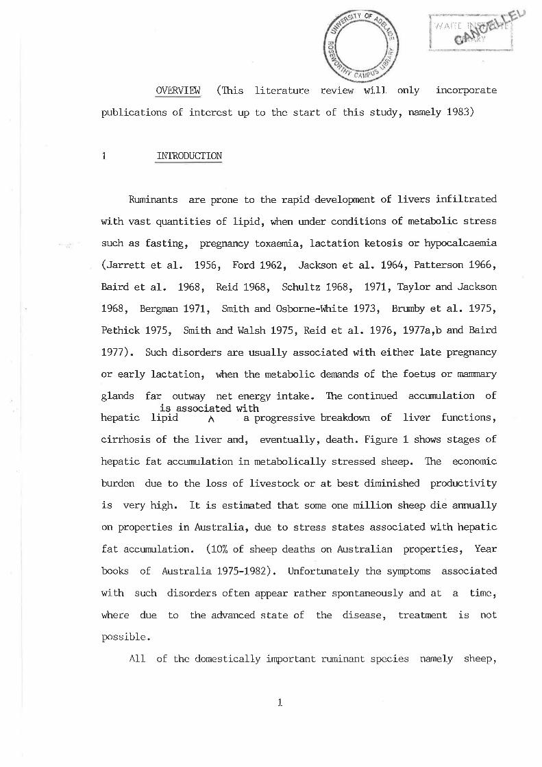
Ruminants are prone to the rapid developnent of livers infiltrated
with vast quantities of lipid, when under conditions of metabolic stress
such as fasting, pregnancy toxaemia, lacÈation ketosis or hypocalcaemia
(Jarrett et aI. t956, Ford L962, Jackson et al. L964, Patterson 1966,
Baird et al. L968, Reid 1968, Schultz 1968, I97L, Taylor and Jackson
1968, Bergman t97t, Smith and Osborne-lihite L973, Brurnby et al. t975,
Pethick 1975, Smith and Wa1sh L975, Reid et aL. t976, t977arb and Baird
L977). Such disorders are usually associated with either late pregnancy
or early lactation, vilren the metabolic de¡nands of the foetus or nì¿fimary
glands far outway net energy intake. Ttre continued accumulation ofis assocíated wíth
hepatic lipid ^
a progressive breakdown of liver functions,
cirrhosis of the liver and, eventually, death. Figure 1 shows stages of
hepatic faL accunn:lation in metabolically stressed sheep. The economic
burden due to lhe loss of livestock or at best diminished productivity
is very high. It is estimated that some one million sheep die annually
on propert.ies in Australia, due to stress states associated with hepatic
fat accumulation. (t07" of sheep deaths on Australian properties, Year
books of Australia I975-t982). Unfortunately the symptoms assocj-ated
with such disorders often appear rather spontaneously and at a time,
vÈrere due to the advanced state of the disease, Lreatment is not
possible.
All of the domestically important. ruminant species namely sheep,
L
YOF
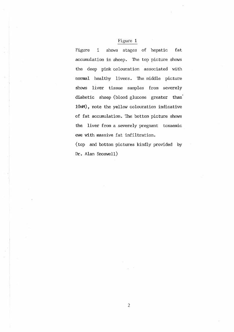
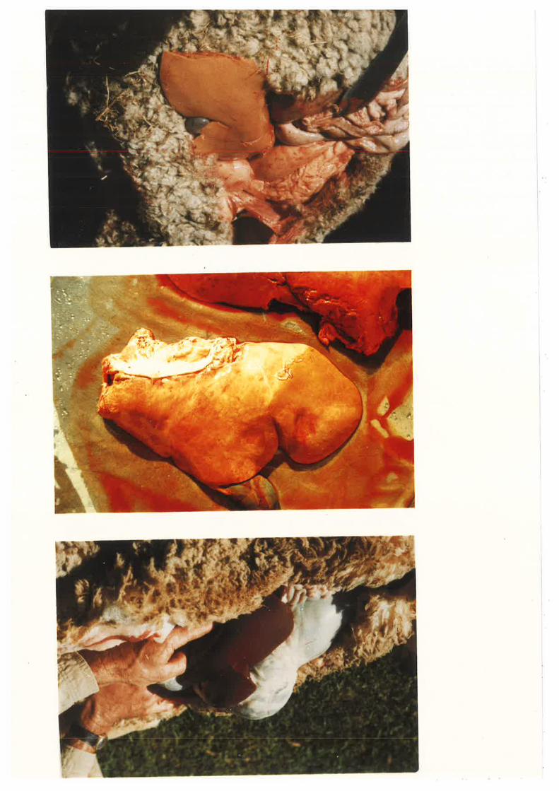
Figure 1
Figure 1 shows sLages of hepatic fat
accunmlation in sheep. The top picture shows
the deep pink colouration associated with
normal healthy livers. Ttre middle picture
shor¿s liver tissue samples from severely
diabetic sheep (Ufoo¿ glucose greater than'
10rnþf), note the yellow colouration indicative
of fat accumulation. The bottom pieture shows
the liver from a severely pregnant toxaemic
ewe rrrith massive fat infiltration.(top and bottqn pictures kindly provided by
Dr. Alan Snoswell)
2
cattle and goats share the same basic physiology and therefore, the
tendency to develop similar metabolic disorders. There are however, a
number of species differences associated with their physiology, diett
environment and meLabolic demands, vùrich affect the frequency and
intensity of these manifestations. Sheep (O¡is aries) has been chosen as
a ruminant animal model for this thesis, therefore, the subsequent
literature review will be mainly confined to this species. As such, it
nmst be borne in mind, Èhat parallelisms to other ruminantsr may not
always be justifiable.
In recent years, a gneat deal of insight has been gained as to the
principal causes of diseases such as pregnancy toxaemia, v¡hich give rise
to the develognent of a tfatty' liver. Consequently, agricultural
producers, through good farm rnanagement practices, have been able to
reduce their incidence. It, was realized that î.aLLy acids were mobiLized
from the adipose tissue under periods of stress, vÈrich in turn was
reflected by an increased synthesis of lipids and subsequent
accumulation of hepatic triacylglycerols (fAC). It was considered that
accurn:lation occurred v¡hen the ability of the liver Lo secreLe TAG¡ is
far outweighed by its rate of synthesis.
Ttre biochemical cascade of cellular events v¡Lrich lead to the
developrnent of a tfattyt liver in ruminants, is however, îar from
resolved. The preferential hepatic accurnrlation of TAG under conditions
of stress raises rnany questions vùrich are not readily answered. l{hy are
TAG the major lipid accumulating? Does the ruminant liver preferentially
esterify incoming non esterified fatty acids (NAp'¿,) as opposed to
oxidizing them? If so, then vÍry? Does the liver select.ively esterify
NEFA to TAG and not. cholesterol-esters and/or phospholipids? Is there a
3
defect in the synthesis, packaging, transport or secretion of TAG rich
lipoproteins? If sor is this due Lo a lack of lipoproLein cornponents'
such as phospholipids or cholesterol, or an inability to increase or
maintain lipoprotein biosynthesis, so as to export all endogenous
hepatic TAG? Is there a physical impairment vùrich is irùribiting
lipoprotein secretion? Is there a deficiency or defect in the synthesis
of the apoprotein components essential for lipoprotein metabolism? Does
very low density lipoprotein (the plasma TAG rich lipoprotein under fed
conditions) mediate the bulk of plasrna TAG in metabolically stressed
ruminants, or does there exist an abnornnl lipoprotein? I,lhaÈ role do the
membrane bound triacylglycerol hydrolases, namely lipoprotein lipase and
hepatic lipase, have in the metabolism of TAG rich lipoproteins in
metabolically stressed animals and subsequent hepatic accunrulation of
TAG? Is the liver TAG accumulation a result of a complex combination of
cellular disorders?
It is apparent that the synthesis, secretion and metabolism of
hepatic TAG in ruminant animals has been long neglected and requires
urgent investigation. In part. fulfillment of this need, this study was
concerned with the metabolism of TAG rich lipoproteins r,rithin the plasma
cornpartment, in metabolically stressed sheep.
This overview will examine the anabolic and catabolic processes of
very low density lipoprotein-TAG metabolism, in view of its associ-ation
with 'fatty' liver syndrome. Throughout this study, comparisons will be
made with non-ruminant diabetes, vitrich in man, is of great. clinical
significance.
4
2 THE USE OF ALI¡XAN DIABE'IES AS A MODEL OF ME'TABOLICALLY
STRESSED SHEEP
l,6ny of the naturally occuring metabolic disorders associated with
'fattyr liver syndrome vilrich afflict nrminants are often unpredictable,
nraking their study a difficult task. Preliminary investigations in this
laboratory have shown that pregnancy toxaemia is difficult to induce
artificially, after v¡trich mainLenance of the aninral in a stressed state
is near futile. Another cornplication of using naturally occuring
n¡anifestations, is the inability to measure and subsequently manipulate
the severity of the disease.
Although diabeLes is not a naturally occuring disorder of any
consequence in ruminants, it offers very rnany advanLages as a model of
tfattyt liver symdrome. Induction of diabetes, either by surgical
panereatectomy or use of the drugs alloxan or streptozotocin, allows
generation of a nu¡nber of stressed sheep, in the same condition, vùrich
if required, can be maintained by exogenous insulin administration. In
addition, blood or urine concentrations of glucose, or plasma insulin
levels nny be moniLored quickly and cheaply and used as índicators of
the effectiveness of the induction. By removi-ng the Pancreas or
irreversibly destroying the beta cells of the Islets of I-angerhans,
v¡krich synlhesize insulin in vivo, metabolism of glucose is severely
impaired. To meet, the metabolie requirements of the animal in the short
term, adipose t.issue TAG is mobilized and released into the plasnn as
NEFA.
Most of the naturally occurring manifestations vilrich promote
hepatic TAG accumulation are also associated with a reduced, if not
5
3
complete cessaLion of food intake, v¡hich in turn is reflected in
decreased levels of plasma insulin (Bouchat et aI. 1981). It appears
therefore, that the biochemical process of hepatic TAG accunmlation
observed in diabetic sheep, would not differ substantially to that
observed in naturally occuring paLhological disorders.
This study makes use of alloxan induced diabetes as a model for the
examinat,ion of TAG metabolism in ehronically stressed sheep. Alloxan
monohydrate permanently prevents the enzymatíc synthesis and release of
insulin from the pa.ncreas (Rerup t97O) and unlike pancreatectomy does
not interfere with other functions of this tissue, such as digestive
enzymic secretions.
LIVER LIPID ACCUMJIATION
The susceptibility of an animal to develop 'fattyr liver syndrome
varies dramatically between species and possibly breed. For instance,
rats are less suscept.ible than sheep to hepatic steatosis associated
with fasting (Élarrison L953, Manns 1972) and guinea pigs are less
susceptible ttr,an rats to rfatty'livers associated with choline
deficiency (tucas and Ridout L967).
Sheep livers infiltrated with fat are generally enlarged and paler
in colour (figure 1). Both features are dependent on the degree of fat
accumulation. The greater mass associated with I f.aLty' livers is also in
part attributable to an elevated water content (tucas and Ridout 1967).
A healLhy sheep liver is about 57" lipid by weight', of vùrich
approximately 7O"A Ls phospholipid and 307. is neutral lipid (Peters and
Smith L964). Phosphatidylcholine and phosphatidylethanolamine are the
6
major phospholipids (Peters and Smith L964, Noble et al. I97I) and TAG
and free cholesterol are the major neuLral lipids (Peters and Smith
Le64).
Studies as to the type of faL accunmlating in the'fat,tyr livers of
varying aeLiology, show that TAG are the predominant lípid component.
Dryerre and Robertson (L94I) first, reported that neutral fat hlas the
main class of the increased liver lipid in pregnant ehres, pregnant
toxaemic ewes and abattoir wethers. This was later substantiated by Read
(1976) and Henderson, Read and Snoswell (1982), vil"to reported thaL in
alloxan diabetic wethers and pregnant toxaemic ewesr TAG were elevated
substantially and that the phospholipid concentraLion did not change
rnarkedly. Smith and t{a1sh (L975) also reported a smaller, though still
significant elevation in liver cholesterol ester in Pregnant and
Iactating e\^tes.
ROLE OF INTESTIM AND LIVER AS SOURCES OF TRIACYIGLYCM.OL-RICH4
CONIAINING LIPOPROTEINS .
Lipoproteins are the vehicles by vùrich hydrophobic lipids are
transported in the generally aqueous environment of plasnn, to tissues
wtrich utilize lhe constituents for oxidative metabolism, me¡nbrane
homeostasis or for storage purposes. Ttrey are synthesized at two sites,
namely, the intestinal epithelium and \n'ithin the hepatocyte. The
maintenance of synthesis and secreti-on of lipoprotein particles is thus
essential for normal lipid metabolism. A defect in either or both of
these processes results in the rapid accumulation of lipid. Normal
plasma lipoproteins are generally spherical macromolecular complexes
7
containing a mixture of core lipids, encased by a hydrophilic layer of
phospholipid, cholesterol and specific proteins (termed apoproLeins)
vùrich act as recognition sites and regulators for the uptake and
catabolism of the parLicles. Lipoproteins are most conrnonly
differentiated by their density, lipid compostion and origin. Classes of
lipoproteins and the categories by vrtrich they are defined are discussed
in chapter one. The role of apoproteins in the metabolism of
Iipoproteins is discussed in chapùer three.
In all species studied thus far, Lwo disÈinct lipoprotein
part,icles, namely chylomícrons and very low density lipoprotein (VLOI-)
carry Lhe majority of circulating TAG. The contribution of either of
these particles to total circulating TAG is particularly dependent. on
the nature of the diet and physiology of the aninnl concerned.
Ckrylomicrons are synthesized within the intestinal epithelium. The
digestion of Iipid, its absorption into the enterocyte (nn-rcosal cell of
the small intestine) and secreLion as chylomicron particles in
monogastríc animals has been reviewed extensively (Johnston L970,
tlamilton L972, Sinnnonds L972, Green and Glicknan 1981 and Miller and
Got,to 7982) and the v¡krole process is only briefly sunrnarized here. Ttre
nrajor products of the hydrolysis of dietary fats are fatty acids and
monoglycerides. These pass into the enterocytes. TAG are resynthesized
wlthin the smooth endoplasmic reticulum and become chylomicron
precursors. The particles pass to the Golgi apparatus, v*rich is involved
in the process of apoprotein and carbohydrate addition. The resulting
chylomicron part.icles are then expelled from the enterocyte by reverse
pinocytosis (exocytosls), into the intestinal lyrnphatics.
In monogastric onnivores and herbivores, the contribution of
a
dietary derived chylomicron-TAG to plasma TAG concentration varies
considerably and is particularly dependent on the nature of the diet.
For example, in adult rats maintained on a nonnal low fat chow diett
consuming approximately O.5g fat per 1009 of body weighL daily'
approximately 80% of circulating TAG are attributable to hepatically
derived VLDL (Palmer et aI. t978, Risser et al. L978, HoIt and Dominguez
1980, Huang and tlilliams 1980, tblopissis et al. 19801 1982 and Agius
et al. 1981). I,ùhen adult rats are fed a diet conLaining 7O7" of. calories
as fat, intesuine contribr:tes 857. of plasrna TAG (tktopissis et al. 1980t
L982). In addition these particles are rapidly metabolised in vivo, and
so the contribution to total plasma TAG levels is also critically
.dependent on the time of blood sampling after the previous meal.
Investigations v¡trich determine the concentration of circulating plasma
TAG nray thus be exagerated if chylomicron particles are present, because
they are the means by v¡Lrich dietary fat is packaged for further
meLabolism, and hence, represent exogenous rather than endogenous lipid.
Most lipoprotein studies use subjects r¡Lrich have been wiLhout food for a
period of time sufficient to clear any circulating chylomicron
particles.
In contrast, ruminants have negligible amounts of dietary derived
TAG due to the low lipid content of the diet. in general and
particularly, the fermentative properties of the reticulo-rumen system,
(Scott L97L) as evidenced by the absence of chylomicron particles in the
plasma of fed sheep (Nelson L973 and l,eat et al. t976). The rumen
microflora have the capacity to hydrolyze dietary lipids before
absorption can take place. I.eat and tlarrison (1974) observed Lhat
ruminant ly*ph contained a high content of phospholipids relative to TAG
9
and suggested that lyrnph lipids were transported in VLDL rather than
chylomicrons. They subsequently confirmed that 757" of ruminant Iymph
Iipids resided in VLDL, with the maximum concentration occuring in the
Sf range 150-200 (see ctr,apter one) region and suggested that VLDL
probably predominates because of the low intake of dietary fai. (tlarrison
and l-eat 1975). This was later confirmed by Gooden et al. (t979) vilro
showed that the size of the lymph liporotein particles increased with
the amount of lipid ingested.
Although tymphatic VLDL and chylomicrons are present in sheep, it
is not known vùry few, if any are found in plasnra (Nelson 1973 and leat
et al. L976). In gxazing ruminants, the low intake of dietary faL may
account for the absence of these particles. However, in ruminants fed
high fat concentrate dietsr or protected fat diets, substantial
quantities of chylomicrons occur in lymph buL only small amounts in
plasma (Scott and Cook 1975). A possible explanation is that lymph
particles are rapidly metaboLízed by lung tissue (*ricfr has a very large
capillary bed) and the peripheral tissues. In support of this the
turnover time of chylomicron TAG is 7.5-II.5 minutes in the lactating
goat (Lascelles et aI. t964) and 10-20 minutes in nnn (Havel and Kane
te75).
The majority of pathological conditions vùrich lead to the
development of a 'fattyt liver in sheep, are usually associated with a
reduced or complete cessation of food intake. It is apparent, therefore,
ttr,at for the purpose of this study, dietary derived TAG in sheep may bre
considered as negligible.
VLDL are synthesized principally within the hepatic sinusoids,
although the intestinal epithelium may also contribuLe to an
10
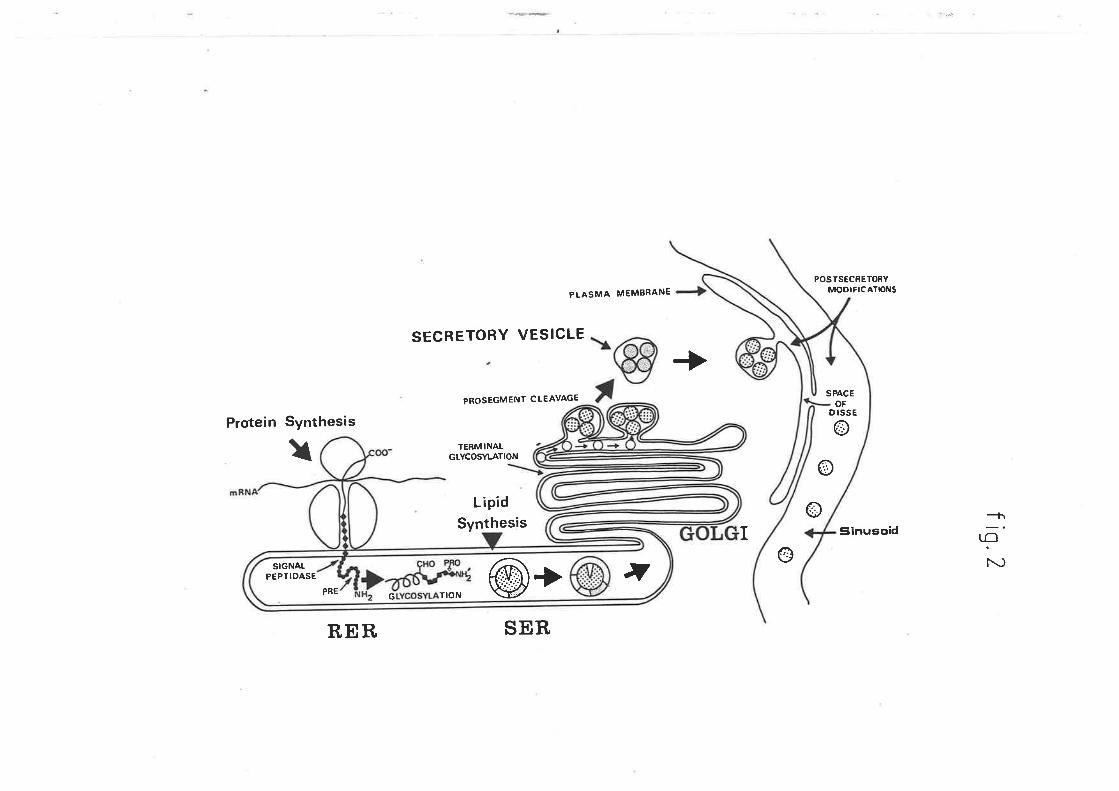
indeterminate extent. The biosynthesis, assernbly and secretion of
lipoproteins by the liver shares many cortrnon features lrrith the
intestinal epithelium, although the origin of the tipid moiety is
clearly different.. A schematic representation of the subcellular
biosynthetic route of lipoprotein particles in the liver is shown in
figure 2. In monogastric animals hepat.ically derived VLDL are first
formed on the smooth and rough endoplasmic reticulum (Glaumann et al.
L975) v#rereby the TAG and phospholipid components are derived. The rough
endoplasmic reticulum is also responsible for the slmthesis of the
apoprotein components (De Jong and Marsh 1968 and Alexander et al t976).
After being packaged into secretory vacuoles by the golgi apparaLus,
fusion with the plasnn membrane results in expulsion of the nascent
lipoproteins by exocytosis into the space of Disse (f,amilton et al.
L967, Jones eÈ al. t967 and Claude L97O), vihich represents a localized
high concentration of hepatic secreLory products. The mechanism of
hepatic WDL synthesis and secretion in ruminant animals has been the
subject, of little investigation, however, there is no published data
suggesting that the process differs from that in monogastric animals.
As a result of the digestive physiology of ruminant animals,
plasma TAG concentration is in effect, a reflection of the balance
between the secretion of hepatically derived VLDL-TAG and subsequent
catabolism by the extrahepatic tissues. In cornparj-son to non-ruminanLs,
sheep (l:-te other ruminants) have extremely low levels of circulating
VLDL-TAG (and non VLDL-TAG) (Nelson L973 and Leat et aI. 1976). It is
not known vihether the snnll concentration of this lipoprotein fracLion
is due to a low rate of hepatic synthesis and release, or the
exceptional avidity of extrahepatic tissues for VLDL-TAG. In support of
LL
Figure 2
Figure 2 is a schematic representation of
hepatic biosynthesis of lipoprotein
part.icles. Particle formation begins on the
smooth and rough endoplasmic reticulum r,vhere
the lipid components are derived. The rough
endoplasmic reticulum is also responsible for
the synthesis of the apoproteins. These
particles are then packaged into secretory
vacuoles by the golgi apparatus, after which
fusion r¿ith the plasma membrane results in
lheir expulsion into the space of Disse.
(0iagram from Dolphin P.J. (1985) Can.
Biochem. Cell Biol. 63, 850-869)
J.
L2
PREG TION @+ ,
SIGNAIPEPfIDASE
PLASMA MEMBRANE
POSTSECRETORYMODIFICATþNS
SPACÊOF
D ISSE
oo
Sinusoid
SECRETORY VESICLE
PROSÊGMENf CLEAVAGE
TERMINALGLYCOSYI.ATION
LipidSynthesis
SER
+Protein Synthesis
I
RER
oI
+\r-¡@
tl
5
the latter suggestion, the rate of Lurnover of plasma VLDL in the
lactating cohl I^Ias rapid relative to that of other lipoproLeins (Glascock
and llelch L974 anð, Palmquist and t'dattos 1978). However, this would not
be unexpected in view of the denrand of the mafimary gland for TAG faLLy
acids.
The synthesis, secretion and metabolism of VLDL is obviously of
fundamental importance in the process of hepatic TAG accumulation in
sheep, though as yet, there has been no investigation into this process'
TT{E ROLE OF FATTY ACIDS IN TRIACYIßLYCEROL ME'IABOLISM
Fatty acids may be utilized for the alternative pathways of
oxidation and esterification in the Iiver. They are mainly derived from
either the circulating plasma NEFA, de novo faL|y acid synt'hesis or from
intrahepatic lipolytic processes. The relative contributions of these
for utilization in the liver are variable according to a number of
factors and are under hormonal and nutritional control (nritz L96tt
Mayes and Felts Lg6l, Specbor Ig7L, Ontko t972, Heimberg et al. 1978 and
McGarry and Foster 1980).
5a LIPOGM{ESIS
In the fed ruminant, metabolism is dominated by the exlensive
microbial fermentation of dietary carbohydrate and other organic
constituenLs Lo short. chain fatLy acids in the reticulo-rumen and to a
Iesser extent, the caecum (Harfoot 1978 and Noble L978). Short chain
fa1Ly acids pass into the abomasum and are absorbed mainly inther
rJilir¡l;
J
I
r
13
ll
fiI
tII
i
reticulo-n:men and omasum:, The fluid entering the
duodenum contains a high proportion of these fatty acids (eaft and Hill
L967). Heath and Hill (fgOg) have reported that up to six grams may be
absorbed from the duodenum of sheep per day under fed conditions. TLrree
short chain acids are produced in significant amounts; acetatet
propionate and butyrate, of v¡trich the first predominates. Acelate is
metabolized least by ruminal epithelium and liver, and therefore, large
amounts are available for post-hepatic metabolism in the fed animal
(eethick er. al 1981). Ifuch of this is oxidized in peripheral tissues
(Annison and Armstrong 1970 and Pethick et al. 1981). Surplus acetate
then becomes the most important source of acetyl-CoA for the synthesis
of long chain faLLy acids (Hanson and Ballad L967, L968, Young et al.
Lg6g, Hood et aL. 7972 and Ingle et al. t972arb). AceLate utilization in
fasted-alloxan diabetic sheep is similar Lo that in nornral fed animals
(perhict er at. 19s1).
The other major short chain falLy acids, (propionate and butyrate)
are also involved in lipogenesis through utilization of Lheir
metabolites, ttr,at. is, glucose and 3-hydroxybutyrate respectively. Almost
all propionate vñich reaches the liver is metabolized via t'he
tricarboxylic acid (Krebs) cycle, some of vùrich is oxidized to carbon
dioxide, buL the majority of v¡hich is converted to oxaloacetate and used
for glucose synthesis (teng et aI. L967, Ieng 1970 and Smith and I'dalsh
Lg75). Indeed approximately half of a fed ruminant's carbohydrate
requirements are met by this means. Glucose produced by this pathway is
only a minor source for fatty acid synthesis, but nonetheless it is very
important in lipogenesis as a source of reducing equivalent's, it the
form of NADPH, for esterification of long chain fatty acids (Yang and
!
1,4
rÌ
;
Baldwin L973a,b).
Butyrate is metaboLized predominantly in the rumen epithelium (and
to a lesser degree the liver) to :-nydroxy-buLyrate (Pennington t952,
Katz and Bergman Lg6g, I^leigand et al. L972 and Baird et al. L975).
This contributes to fatty acid synthesis, particularly in Lhe lactating
rnarûnary gland (nett 1979).
The appearance of these short chain acids in the blood after
feeding, gives rise to an increase in insulin secretion (BassetL L975
and Broclqnan L978). This hormone has been shown to erùrance lipogenesis
from both glucose and acetate (Khachadurian et aI. L966, Bartos and
Skarda 1970, Baldwin and Smith L97L, Yang and Baldwin L973a and Vernon
L979) and also to inhibit catecholamine-stimulated lipolysis in ruminant,
adipose tissue in vitro. Administration of exogenous insulin in
ruminants has been shown to produce substantial decreases in the plasma
concentration of NEFA (Kochen et al. L959, Annison L960, Ttenkle and
Kuhlmeier L966, tiest and Passey t967, Bergman 1968, Lr:thnnn and Jonson
L972, Hertelendy and Kipnis 1973 and Bauman t976) and glycerol (Bergman
1963), and in the net output of these subslrates from adipose tissue in
sheep.
The dietary supply of short chain fatty acids is obviously crucial
for Iipogenesis in ruminant tissues and has been reviewed extensively
elsev¡here (t ng L970 and Ckrurch L976). In ruminantsr âs in other
animals, lipid synthesis occurs in most tíssues of the body. In the
healthy fed non-Iactating ruminant, more than 90% of lipogenesis occurs
in adipose tissue alone (eayne and Mast.ers L97L, Hood et aI. t972, Ingle
et al. L972b and l4artin et. al . L973).
15
5b NON ESTERIFIED FATTY ACID ME-TABOLISM
I,lhen the metabolic energy requirement of an animal exceeds its net
metabolic intake, adipose tissue TAG is mobilized so as to meet the
deficiency. In fasted sheep and fasted-pregnant e\^Ies, net NEFA release
from adipose tissue increased following an increase in the rate of
lipolysis (Adrouni and Kkrachadurian l-968 and Pethick et. aI. 1983).
Adipose TAG are hydrolyzed to NEFA by the enzyme hormone sensitive
Iipase and released into Ehe plasma vùrere they bind with albumin. Under
such conditions this tissue becomes the major source of plasnn NEFA.
Adipose tissue is the nnjor site of TAG sLorage and is not a dírect
contribuLor to the plasma component of this lipid fraction. In other
studies with fasted sheep it was shown that. an inverse relaLionship
exists between the circulating levels of acetate and NEFA (BasseLL t974
and Bell and Thompson L979). In the latter study, changes in plasma
glycerol pa.ralleled those of NEFA. Such changes are consistent with an
increase in the rate of lipolysis and diminishing levels of circulating
insulin seen in fasled ruminants (Bouchat et. al. 1981). In non-efficiencY,
ruminant,s, glucagon augments the lipolytic ef fects of insulin.¡ but.
glucagon is only weakly lipolytic in ruminants (CLrrislie t979) and as
such, is probably not an important regulator of adipose tissue
mobilization in these animals.
The sheep liver is the most important individual organ for the
removal of NEFA from circulating blood plasma (Bergman et. al. L97L)
lhough other tj-ssues such as skeletal muscle, cardiac muscle and kidney
avidly metabolize NEFA and under certain conditions may increase their
uptake. Approximately 25% of plasma NEFA clearance can be directly
T6
attributable to the liver in conscious fed sheep (Bergman et al L97L).
The rate of uptake rernains constant in a variety of meLabolic stress
states (Xatz and Bergman I969t Thompson and Darling t975, Thompson et
aL. L975, Lg78) and is directly prop(rtional to the plasma concentrationThompson
(W.atz and Bergman 1969 and Thompson and Darling L975 an%et al. L975) '
Hepatic NEFA uptake is not under hormonal or metabolic regulation, but
rather is a function of plasma concenLration (t{oodside and Heimberg
Lg72). The sheep liver is also selective in Ehe uptake of individual
NEFA (Ttrompson et 41. 1975, L978) and appears to be similar in
qualitative terms to that demonstrated for the perfused rat liver,
(Soler-Argilaga Lg73), being directly proportional to the degree of
unsaLuratj-on and inversely related to carbon chain length. The hepatic
uptake of NEFA in alloxan diabetic sheep has not been reported, though
there is no evidence suggesting the process should differ from that in
normal animals.
NEFA taken up by the liver can be totally oxidized to carbon
dioxide and waLer via the tricarboxylic acid cycle or partially
oxidized to form the ketone bodies (acetoacetate and beta-
hydroxybutyrate), raLher than be esterified to form cornplex lipids. The
factors v¡trich determine vùrich of these alternate pat'hways will
predominate are poorly understood. In rats, in the absence of added
substrate, perfused livers from fed animals will produce more carbon
dioxide and less ketone bodies than livers from fasted or alloxan
diabetic rats (Heimberg et. al. L962 and Morris 1963a). However, wtren
NEFA are added to the medium, a larger fraction of NEFA will be oxidized
completely or partially to ketone bodies by Iivers from fasting or
alloxan diabetic animals, and a smaller proportion will be esterified
L7
and secreted as TAG, than will livers from normal fed animals (Élavel et
al. L962, Heimberg et al. 1966, L967, t969, Morris t963a, 1963b, Mayes
and Felts L967 and Van Harken et al. L967).
There are few conrnunications vùrich have dealt with the oxidation of
fatty acids in the ruminant liver. However, the capaciLy for sheep liver
to oxidize NEFA appears to be limited (Koundakjian and Snoswell L970),
due principally to low levels of hepatic carnitine, a key factor in
beta-oxidation. These studies showed that in sheep liver mitochondria,
palmitic and stearic acids were oxidized aL a raLe of only 307" of. that
obsen¡ed in rat liver mitochondria.
Fed sheep have relatively high circulaLing levels of keLone bodies
vil'ren compared to non-ruminants (gair¿ et. al. 1963), nnrch of v¡krich is
derived from the metabolism of dietary derived butyrate produced in the
rumen epithelium (Yatz and Bergman L969). In lhe same study, fasted
pregnant and non pregnanL ewes had much higher levels of circulating
ketone bodies, even though the intestinal contribution lrras severely
reduced. In fasted animals there is no doubt that. ketogenesis increases
and that the liver assumes the nrajor role in this process (Pethick and
Lindsay L982a, L982b). Nevertheless, Krebs (fg66) suggested that hepatic
ketogenesis in the ruminant animal may still be limited by the relaLive
availability of acetyl C-oA and particularly Lhe tricarboxylic acid cycle
intermediate oxaloacetate. Hyperketoneaemia initiated by an increased
rate of ketogenesis is exacerbated by a reduced capacity for ketone body
utilization in some ti-ssues, including skeletal muscle (eethick and
Lindsay 19S2b), kidney and heart. (Varnam et aI . L978).
Bergman et al. (L97L) reported that in fed sheep, despite
considerable uptake of radiolabelled NEFA by the sheep liver, Iit.tle
18
appeared in the VLDL-TAG fatty acids. Ballard et al. (1969) suggested
that the low rates of lipogenesis observed in the ruminant liver may be
due to low levels of oxaloacetate, which is conrnit.ted to
gluconeogenesis. However, as the capacity for sheep liver to oxidize
NEFA is somev*¡,at small, a large hepatic influx of NEFA would suggest a
dramatic increase in Lhe process of esterificaLion to complex lipids. It
ís lqrown that an i-ncreased supply of fatty acids in perfused rat liver
(Kohout et aI. L97t and Topping and Mayes L982) and isolated rat or
chicken hepa.tocytes (Mooney and l¿ne 1981 and Davis and Boogaerts L982)
results in an increased raLe of TAG synthesis and lipoprotein secret,ion.
This stinmlation appears to be coordinated with an increased activity of
the final enzyme involved in TAG synthesis, namely diacylglycerol
acyltransferase (Haagsman and van Golde 1981). Few such studies have
reported rates of ruminant hepatic NEFA esterification, particularly
under stressed conditions. Presumably, TAG are the major product of the
esterification processr âs suggested by their dramatic rate of hepat.ic
accumulation. Furthermore, a TAG moÌecule is the most efficient means
(on an energy/mole basis) of storing NEFA, and hence would serve best at
packaging hepatic NETA. Fatty acids vùrich enter the esterification
pathway are either retained within the liver cell for the formation of
membrane phospholipids and for storage in TAG droplets, or they are
secreted in the form of lipoproteins. It is apparent that metabolism of
falLy acids proceeds under homeostatic regulation.
To my lcrowledge there has been no published data suggesting that
the process of hepatic esterification in ruminant animals differs from
other species.
It is evident that both the output of VLDL-TAG and Lhe accumulatíon
L9
6
of TAG in the liver are functions of NEFA concentration in the serum and
the period of time to vùrich the organ is exposed.
The ruminant liver produces little fatty acid de novo, principally
because it is unable to use glucose as the source of acetyl CoA (a key
intermediate in falLy acid synthesis) (nalhrd et aI. 1968). 'Intis is not
unlikely in view of the extremely low carbohydrate supply derived from
the diet, and conforms hrith other features of its carbohydrate
metabolism. The process of gluconeogenesis in ruminant livers accounts
for almost all of the aninal's carbohydrate requirements (f.ttg t965t
Lindsay 1970 and Bergman 1973). Thus, hepatic TAG synthesized from the
esterification of de novo fatty acids in the ruminant animal, may be
considered as negligible.
HEPATIC TRIACYI.CLYCEROL SECRE'TION
In the ctronically stressed sheep, hepatic TAG accunmlation will
result. if the rate of release of the lipoprotein particles vûrich effect
transport of this lipid is limited.
The extent of TAG output from the liver in vivo in different
metabolic conditions has generally been assessed by one of two methods.
In the first., doses of a radioactively labelled TAG precursor (NEFA or
glycerol) are given j-nLravenously and the specific activity and total
radioactivity of the liver and the plasma lipids determined at inLervals
thereafter. The values obtained have been interpreted in terms of model
systems consLructed on the basis of estimated fatty acid fluxes in the
Iiver, through pathways often based on a number of assumptíons.
Problems in interpretations associated with this technique have been
20
revie\4/ed previously (Baker and Schotz L967). The second method depends
on the fact that the plasma TAG concentration is a result of a balance
between rates of TAG entry and removal from the circulatory system.
Removal can be prevented by the use of surface active subsLances, the
mosL corTrnon of v¡trich is the non-ionic detergent Triton I^1R1339
(oxyethylated-tert-octylphenol polymethylene polyrner) r,¡trich associaLes
with Lhe circulating VLDL-TAG, in such a I^Iay as to prevent normal
removal mechanísms from operating. lnleasurement of the rate of increase
in plasma TAG, then provides a measure of the rate of TAG efflux. Since
the removal of atl TAG fatby acids in the plasma is blocked by the
administration of such detergents, the method can only provide a measure
of hepatic TAG release r,vhen the intestinal contribution is negligible.
Electron microscopy studies of sheep liver hepatocytes have
revealed a fenestrated membrane surrounding the hepatic sinusoi d (David
1964, Grubb and Jones !97L and Genrnell and Heath L972) and it vlas
considered that this may inhibit the passage of the very large VLDL
molecules, particularly if these were enlarged in metabolically stressed
sheep. Studies in this laboratory using Triton I4rR1339 to measure hepatic
TAG release had shcwn that fasted and alloxan diabetic wethers have
increased hepatic seeretion of TAG associated with an elevation in the
plasma concenLration of this lipid (t"lamo et aI. 1983). It, did not, appear
therefore thât the basal lamina surrounding the hepatic sinusoid didnot completely irhibit
^ passage of WDL molecules. C-onversely the increased secretj-on
rate could be a tpressure-inducedt effect as a result of massive hepatic
VLDL synthesis, or alternatively hepatic TAG may be released in abnormal
part.icles in chronically stressed sheep, vilrich are smaller than normal
VLDL. Subsequently, I,Jright et aI. (1983) claimed that the basal lamina
2t
surrounding the sinusoid was in fact a sample preparat.ion artifact. The
increased hepatic release of TAG however, is not sufficient to prevent
accumulation of this lipid in situ. It appears therefore that the
synthesis of VLDL may ble the rate limiting process, being outweighed by
the rate of TAG production. In similar studies in goats under various
physiological conditions, Lhere r^ras no apparent dif ference in the raLe
of hepatic TAG release between fed and fasLed animals (fiser et aI.
t974). In that study, goats were fasted for two days prior to Triton
administration. Results from this laboratory (not published) have shown
that a forty eight hour fast is not sufficient to effect a change in
liver TAG release in sheep. This is not surprising in view of the time
required to digest food in the ruminant animal and thus induce a tstatel
equivalent to fasting. A greater period of food deprivation may have
been needed Eo examine any ctr,anges in Lhe rate of hepatic release. In
addition, the number of aninrals per treatment used in this study was not
sufficient to statistically eliminate individual variation. In contrast,
similar Triton studies with fasting and streptozotocin induced diabetic
rats (Otway and Robinson 1967 and Bobek et al. 1981), and in isolated
perfused rat livers from diabetic animals (Heimberg et aI. L966, L967
and Van Harken et aI. L967), hepatic secretion of VLDL-TAG was reduced
and could account for accu¡mrlation of this tipid in the liver of these
animals. The decreased release of TAG in fasted and alloxan diabetic
rat.s, may be due Lo a combination of an increased hepatic capacity Lo
oxidize f.aLLy acids under these conditions (Heimberg et al. t966, L967,
Van Élarken et al . 1967, 1969 and Élarano eL al . L969), a reduced rate of
hepatic de novo slmthesis and a lowered dietary supply of lower chain
acids, coupled with a possible decrease in the rates of esterification
22
(Fredrickson et al. 1958) and inhibition of secretion of VLDL. The
contrasting resulLs are somewhat inconclusive, and the role of VLDL in
hepatic TAG accumulat.ion can only be speculated upon. Heimberg et aL.
(t974) in a review on factors involved in the regulation of VLDL
secretion and its relationship with ketogenesis in the perfused rat
liver, concluded that. the livers capacity Lo secrete VLDL-TAG is less
than its ability to take up and esterify NEFA. t{hen the uptake of faLty
acids exceeds that necessary to maintain maxinal rate of secretion of
VLDL, TAG accumulates in the liver. Though this theory encompasses the
paradoxical changes in the rate of hepatic VLDL-TAG secretion obser:¡ed
in metabolically stressed rats and sheep, it is apparent that the
processes regulating VLDL-TAG synthesis and release, differs in these
two species.
There have been a nunber of suggestions as to limitations into
hepatic synthesis and secretion of VLDL. Brurnby et al. (L975) said that
since TAG accumulation \^ras accompanied by decreases in the percent'ages
of phospholipid and cholesterol in the liver, availability of one or
both of these constituent,s may have limited lipoprotein synthesis.
C.onversely, Heimberg et, al. (L974) postulated that the amounts of
phospholipid and cholesterol secreted in VLDL are dependent on TAG
secretion, and are thus regulated by factors virich affect the laLter.
Alternatively, lipoprotein synthesis nray be limited by the availability
of apoproteins, since in cows, Lhere is a marked decrease in the volume
of rough endoplasmic reticulum in hepatocytes after starvation (Brumby
er al. t975). In support of rhis, Pelech et al. (rgg:) showed that
incoming NEFA stimulated TAG and phosphatidyl choline biosynthesis, but
not apoproteins in rat hepatocyles.
23
The mechanism of the regulaLory control of insulin, or perhaps more
importantly, the molar ratio of insulin/glucagon on VLDL-TAG synthesis
and secretion has been widely investigated but. remains an unresolved
contentious issue. It. has been reported that, TAG secretion in perfused
livers from insulin deficient rats have a blunted resPonse to NEFA
(l^loodside and Heimberg t972 and Assinncopoulos et al. t974). Similarly
in rats, hyperinsulinaemic animals have been reported to have increased
TAG production (Steiner and Vranic 1982 and Steiner eL aI. L984). There
have also been several reports that insulin directly stinnrlates hepaLic
VLDL-TAG secretion in vitro (Topping "rd f'hy." Lg72, Lg82, Tl¡l1och eL
al. L972 and Beynen et al. 1981), though in contrast, some authors
consider that this process ís inhibited by insulin (nitt<ita et al. t977
and Durrington et al. L982). Similarly, in studies from isolated
hepatocytes cultured on fibronectin media free of insulin, it was found
that this hormone was found to promote fatty acid and cholesterol
biosyntheis (Geelen et al. 1930), but irhibit the secretion of TAG,
phospholids and apoproteins B and E (Durrington et al. 1982 and Patsch
et al. 1933). Insulin has also been reported to either stinnrlate
(Topping and Mayes Lg82) or have no effect. (Edwards et, aI. 7979) on the
secretion of VlDl-cholesterol. Glucagon irùribits hepatic lipogenesist
stinmlates lipolysis and inkribits VLDL secretion (tteimberg eE al. L969,
Kempen 1980 and Belmen et al. 1931). Bird and lJilliams (tggZ) suggested
that a higher hepatic TAG release in essential f.aLLy acid deficient rats
may have been due to a higher plasma insulin/glucagon ratio, resulting
from a reduction in plasma glucagon concentration.
24
7 METABOLISM OF VERY LOI^/ DENSITY LIPOPRCIEINS
Previous investigations in sheep have shown thât fasted and
diabetic animals have highly elevaLed plasma TAG concentrations, and
that this elevaLion is due Lo an increased hepatic output of VLDL-TAG
(Uamo et aI. 1983). TAG concentralion is also elevated in diabetic rats
(Topping and Targ L975) and man (Rtbrint et. al. \963, New et at. 1963)
in spite of depressed synthesis. The plasma TAG pool however, is also
critically dependent on the activities of two enzymes, lipoprotein
lipase and hepatie lipase (discussed in chapter two). Both enzymes are
bound to the capillary endothelium of those cells utilizing TAG.
Lipoprotein lipase is found in tissues v¡hich utilize TAG fatty acíds for
oxidative purposes such as heart (Twu et al. L976), lung (Cal et al.
L982) and skeletal muscle (nnnolm et al. L977) or resynthesis of TAGor secretion
for storage.purposes (adipose t.issue or malffnary glands) (Jansen et aI../\
1978 and Clegg 1981a). Hepatic lipase is bound to liver plasnra membranes
and those of steroidogenic organs v¡trich utilize lipoprotein cholesterol
(Jansen and De Greef 1981). Ttris enzyme hydroLyzes TAG and phospholipids
(fnnfrom et al. 1975b) but is distinct from lipoprotein Iipase in that
it it is reasonably act,ive in the absence of apoproteins. Ovine hepatic
lipase has not been previously reported, though recently the presence of
this enzyme in bovine liver has (Cordle et aI. 1983). There have been
few reports published v¡trich Lpve examined the activity of lipoproLein
lipase in chronically stressed sheep or its mode of control. Vernon et
al. (fggf) reported a decrease in lipoprotein Lipase activity in
pregnant ewes with gestation and a subsequent j-ncrease in activity,
after 95-135 days postlactat.ion. It is currently difficult to perceive
25
8
the role of these enzymes in TAG metabolism and their associaLion wíth
hepatic TAG accumulatíon in chronically stressed sheep.
The hepatic TAG aecumulation nny also in part be due to an
increased rate of plasma TAG uptake by this organ. In support of this it
has been reported that. the rate of uptake of washed chylomicrons and
synthetic neutral fat emulsions in isolated fasted perfused rat livers
was greater than livers from fed control aninrals (Ueimberg et al. 1962).
OB.JECIIVES OF STI.]DY
The majoríty of currently available published literature pertaining
to TAG metabolism is for non-ruminants. Presumably this is a result of
their applicability as models of corresponding hunnn metabolic
disorders. However, due to the differences in the diet and digestive
physiology of ruminant animals, the subsequent activity of the
biochemical pathways of lipoprotein TAG metabolism is quite different,
as evidenced by lhe paradoxical rates of hepatic TAG release obsen¡ed in
chronically stressed sheep and rat.s. It is therefore, not valid to
extrapolate data derived from monogastric studies to ruminants.
It, is apparent that the process of hepatic TAG synthesis, its
packaging and secretion as lipoproteins and subsequent metabolism by
extrahepat.ic t.issue has been long neglected. Bell (t979), in his review
on lipid metabolism in the liver and other tissues of ruminant anirnals,
has reconciled this by stressing the urgent requirement for research of
TAG metabolism in ruminant.s.
In part fulfilment of this need, this thesis aims to establish
suitable methods for the isolat.ion, separation and characterization of
26
the major ovine lipoproteins and to determine the role of each of the
nrajor lipoproteins in lipid transporl, particularly TAG.
To ascertain r,ihich lipoprotein fraction is medialing the
hypertriacylglyceridaemia observed in metabolically stressed sheep,
changes in the lipoprotein profile and their composition in alloxan
diabetic anirnals \,rrill be determined. In addition, Lransmission electron
microscopy hrill be utilized to examine each of the major classes of
ovine lipoprotej-ns isolated from fed and diabetic sheep, in an attempt
to identify any changes in the physical properties of the lipoprotein
particles.
Suitable methods for the idenlification and isolat.ion of
lipoproÈein lipase from adipose tissue and hepatic triacylglycerol
hydrolase in sheep ltrill be established. Should the presence of Lhe
latter enzyme be verified, an examination of the characterisLics usually
attributed to Lhis enzyme will be done.
The role of the two lipases in hepatic TAG accumulatj-on and plasnra
hypertriacylglyceridaemia will be determined, by measuring postheparin
plasma activity in fed, fasted and alloxan diabetic wethers.
Rams and ewes have significantly different degrees of adiposity.
Ttris may be due to modulation of triacylglycerôl hydrolase activities by
androgenic/oestrogenic control mechanisms. Thus the activities of
Iipoprotein lipase and hepatic lipase will be determined in both sexes.
In addition, to examine if genetic variation may also in part
affect the expression of lipase activities, postheparin plasma from
genetically 'Iean' and genetically 'obese' sheep will be examined for
triacylglycerol hydrolase activities. C-orrelations of activit.ies will be
made with the TAG secretion rate observed in these groups.
27
Apoproteins are the means by vilrich the catabolism of lipoprotein
particles, namely lheir binding, hydrolysis and uptake by tissues is
regulated. As such, Lhis study will quantitate the apoprotein B (ttre
rnajor protein componenl of the VLDL-TAG in monogastric onnivores) of
each of the major ovj-ne lipoprotein fraetions in nornnl and alloxan
diabetic animals, and determine qualitative ctranges in the total
apoprotein profile of each lipoprotein class, in an attempt to correlate
these wíth changes in the meÈabolism of VLDL-TAG.
28
1 CHAPTER ONE
1.1 INTRODUCTION
The first report on the appearance of distinct lipoproteins in
serum appeared in 1929 (Macheboeuf L929arb). In L94I, motivat'ed by
studies on atherosclerosis, BIix et al. separaLed classes of
lipoproteins according to their electrophoretic mobility in a solid
support media and Gofman et aI. (L949), showed that the plasma lipids
r,rrere bound in a stable union to certain proteins, using an
ultracentrifuge. These proteins were designated as lipoproteins.
Lipoprotein formation, composition, secretion and metabolism have since
enjoyed extensive investigatÍ-on, as a result of lipid abnormalities
associated with disease conditions such as diabetes, renaloPathYt
cirrhosis of the liver and parlicularly, ischaemic heart disease and the
process of atherogenesis.
This overview will briefly sumnarize the major classes of
lipoproteins and the physical parameters by vùrich they are
distinguished. For more extensive reviews refer to (Hatch and l-ees
1968, Forte and NichoLs L972, Eisenberg and I,evy L975, Jackson et al.
L976, Morrisett. et al. L977, Osborne and Brewer t977, Smith et al. t978,
Edelstein et al. L979, Miller and Got.to 1982, Mills et al. 1984 and
Dolphin 1985).
LJ.L LIPOPRCIEIN STRUSTURE AND FUNCTION
The plasma lipoproteins of animal species encompass a
29
rnacromolecular complex of lipids (essentially TAG, cholesLerol and
phospholipids) and one or more specific proteins, referred to as
apoproteins (or apolipoproteins). Their main function is to transport
the hydrophobic lipids of dieLary or endogenous origin within the
hydrophylic environment of the plasma. A nurnber of tissues can then
utilize the constituent TAG-fatty acids for oxidative
metabolism (such as heart and skeletal nnrscle), for storage (in adipose
tissue)r or sirnply nnintenance of cellular function and membrane
integrity. In addition, the cholesterol cornponent nray serve as a
precursor for bile acid and steroid synthesis. The plasma lipoproteins
also transport. other lipid soluble substances including vitamins
(UcCormict et al. 1960), drugs (CLren and Danon L979) and toxins (CLren et
a]-. 1979).
The functions of the particular apoproÈeins is not cornpleLely
understood although nmch progress has been made in recent years. They
confer rnany of the specific properties possessed by the individual
Iipoprotein classes in v¡Lrich they occur. For example, particular
apoproteins regulate the activity of the major enzymes involved in
lipoprotein metabolism in plasna, and are necessary for the secretion of
TAG-rich lipoproteins by both liver and intestine (Gotto et al. L97t,
l4alloy and Kane L982). In addition, the apoproteins play an important
structural role in the lipoproteins. The cornposition and function of
each of the apoproteins is discussed in chapter three.
Lipoproteins are now considered to be cornposed of a hydrophobic
lipid core made up of TAG and esters of cholesterol, with the more
hydrophylic cholesterol and phospholipids forming a surface interfacial
monolayer (figure 1.1) (Shen et aI. L977). The apoproteins are believed
30
Figure 1.1
Figure 1.1 depicts a model of the structure
of plasma lipoproLeins. The more hydrophobic
lipids, triacylglyceride and cholesterol
esters are thought to occupy the core of the
molecular cornplex and are surrounded by
an amphiphilic shell of phospùrolipid and
cholesterol. Specific proteins also occupy
the outer surface of the parLiele but some
may also bind with the inner lipidcomponents.
31
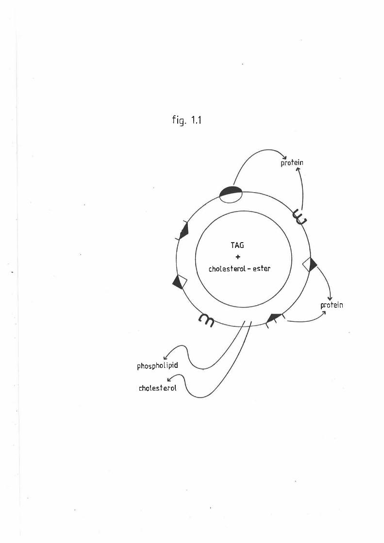
fis 1.1
n
protei n
phosphotiPid
chotesterol
TAG
+
chotesterol - ester
'lÊIL,t
to occupy the outer part. of a shell on the surface of the particlest
v¡trere they are adjacent to the polar head groups of phospholipids'
Amphipathic helical regions within the domain of the apoproteins permit
binding and orientation of the apoproLein within the surface monolayer
(Assman and Brewer t974 and Segrest et al. L974). I^lith this arrangemenL,
the non-polar residues occuPy one face of the helix and point towards
the hydrophobic interior of the lipoprotein, probably interacting with
the first few carbons of the fatty acyl cbains of the phospholipids. The
acidic residues of the apoproteins (glutamic and aspartic acids)
occupy the opposite face and are orientated towards the aqueous
environment. The basic amino acids (lysine and arginine) occupy a
position in the helix at Ehe borders of the polar and non polar faces.
The major lipoproteins all contain cholesterol, cholesterol esters,
TAG, phospholipids and protein, but are distinguishable from each other
by the proport.ions of these constituents and the nature of the protein
moiety. These differences allow the separation of lipoprotein classes by
a variety of physiochemical parameters such as density, particle size,
electric charge, interactions with supporting media in zone
electrophoresis, interactions hrith macro molecular reagenLst
antigenicity and information derived from plasna chemical analysis. The
methods involved in applying these parameters include, zone
electrophoresis, double diffusion inrnunological methods, inrnuno-
electrophoresis, chromatography, combinations of preparative
ultracentrifugation or precipitation with chemical analysis, electron
microscopy and membrane filtration.
The selection of an analytical method of maximum value is a balance
of the factors of expense, technical difficulty, quantitative accuracy
32
and capacity for numbers of sarnples. Idkrichever the method chosen, only a
limited amount. of information can be derived from any one technique and
so it. is therefore usual to integrate data from two or more techniques.
The study of plasma Iipoproteins is complicated by the constanL
changes occuring withín individual parlicles. In certaj-n situations the
lipoprotein is metabolized as a vùroIe, but. frequently different
lipoproLein constituents have different metabolic fates (Streja et aI.
L977, Steiner and Ilse lg8landSteiner and Reardon L982, 1933).
Individual lipoprotein components undergo exchange between lipoprotein
classes and in the case of unesterified cholesterol and phospholipidst
r^rith cell membranes also (8e11 1978). Specific exchange proteins are*
involved in cholesterol ester, TAG and phospholipid transfer. Ttrere are
also the metabolic transformat.ions mediated by.the enzymes lecithin
cholesterol acyltransferase (ttris enzyme mediates the transfer of the
fatty acid from the beta position of lecithin, to cholesterol, to form
cholesterol esters and lysoleciÈhin (Glomset et al. L962)), lipoprotein
lipase and hepatic lipase. Nevertheless, differenees between lipoprotein
classes, both in lipid and protein cornposition are greater than those
within classes and so the variations in physiochemical properties remain
lhe basis of analyÈical methods for Lheir study.
L.T.z ROLE OF PTASMA LIPOPROTEINS
T.L.2.L MONOGASTRIC ANIMALS
Lipoproteins have been examined in a number of species under a
nu¡nber of metabolic and pathological condit.ions. The vast majoriLy of
* Note; sheep have low cholesteryt3i,r"r,"eer activity[Ha and BarterrlgS2rcomp. Biochem. physiol .7L8r265-269)
il,.i
,l
I
TI
I
I
T
li
nlriÈ
'l
the currently available literature is, however, confined Lo monogastric
aninrals: âs a consequence of intensive research into the processes of
atherogenesis.
The major classes of human plasma lipoproteins and their physical-
chemical characteristics by vùrich they are defined are shown in table
1.L. Lipoproteins are now classically defined by their densities, a
reflection of the ratio of proLein and lipid associated vrith each
particle. For hunran lipoproteins the percentage of the partiele weight
that is lipid is approxinøEeLy 957", 907", 757. and 50% for chylomicrons,
very low density lipoproteins (VtOt), lor¿ density lipoproteins (f"Ol) and
high density lipoproteins (mf) respectively. As the particle decreases
in density, the percent content of TAG and cholesterol esLer decreases
and the relative amount of phosphotipid increases. Thus, of Lhe lipidcomponents chylomicrons contain approximaLeLy 97" phospholipid, VLDL-207",
LDL-307. and HDL-507". The phospholipid component is located on the outer
monolayer of the lipoprotein surface. Hence, the increased proportion of
phospholipid in the smaller particles is a logical consequence of the
increased surface/volume ratio.
Lipoprotein density is probably the most unusual property of these
particles as it is lower than that of any other naturally occurring
macromolecule. AII lipoproteins can be floated by centrifugation after
adjustment of solution density (defined as the density of the solution
of salts and other snall molecules but, excluding the contributions made
by proteins and lipoprcæÍrs ) to values between 1.006 and 1.250 g/nL. No
other plasma macromolecules float. at these densities unless adsorbed to
lipoproteins. On Lhis basis the lipoproteins have been divided into six
major classes:
r
34
II
,l
li
I
Table 1.1
Table 1.1 lists the major classes of human
plasma lipoproteins and the physiochemical
characteristics altributed to them.
35
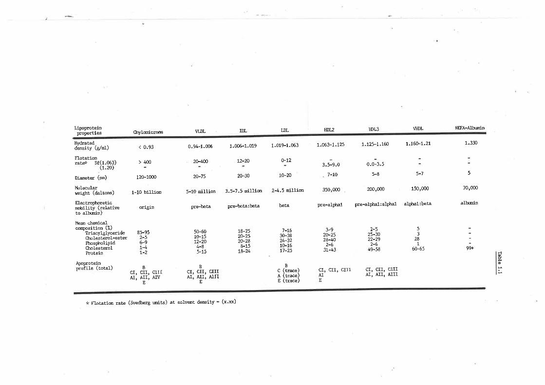
Lipoproteinproperties
Hvdratedder¡sity (e/nl)
FlotationrareÉ sf(1.063)
(1.20)
Diameter (rm)
Molecularweieht (daltons)
Electrophoreticnnbility (relativeto alb¡nin)
lkan cÌ¡onicalccr¡position (7.)
TriaeyþlycerideCtrolesterol-esterItros$rolipidCholesterolProtein
Aooroteinpioiit" (rotal)
Chylanicrons
< 0.93
> 400
120-1000
1-10 billion
or]'grn
VLDL
0.94-1.006 1.006-1.019 1.019-1.063
20-400 t2-20 o-t2
20-75 2G30 t0-20
5-10 million 3.5-7.5 rnillion 2-4.5 nillion
pre-beta pre-beta:beta beta
IDL LDL HDtz
1.063-1.125
3.5-9.0
_ 7-t0
35o,0oo
pre-aI$n1
3-920-2528-t+O2-6
3L-43
cr, crr, crrrAIE
0.0-3.5
5-8
200,000
pre-alphal:alptlal
VI{DL
5-7
150,000
a1plnl:beta
NFA-Altr¡nin
1.330
5
70,000
alh¡nin
HDIS
t.t25-t.t@ L.t60-t.21
2-56-9L-4t-2
BCI, CII, CIIIAr, Arr, Aw
E
53
28L
85-95 50-6010-1512-204-85-15
t8-2520-2520-288-15
t8-24
7-L630-3824-3210-16L7-25
2-525-3022-292-6
49-58 99È60-65ÈÞúHoH
CIIIAÏII
* Flotation rate (Svedberg rnits) at solvent dmsity = (x'xx)
B(trace)(trace)(trace)
cr, crr, crrrAI, AII, AIII
Bcrr,Ar.I,
E
CIAI
cAE
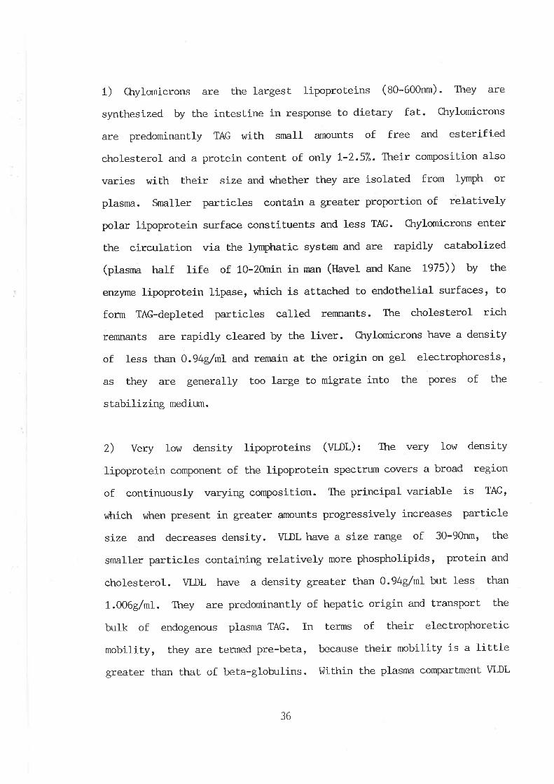
1) Ctrylomicrons are the largesL lipoproteins (gO-OO0nm). They are
synthesized by the intestine in response to dietary fat.. CIeylomicrons
are predominantly TAG with snrall amounts of free and esterified
cholesterol and a protein content of only L-2.57". Their composition also
varies with their size and vilrether they are isolated from ly*ph or
plasma. Snraller particles contain a greater proporfion of relaüively
polar lipoprotein surface constituents and less TAG. ftrylomicrons enter
the circulation via the lymphatic system and are rapidly catabolized
(plasma half life of 10-2ùnin in man (Havel and Kane L975)) by the
enzyme lipoprotein lipase, vrhich is attached to endothelial surfaces, to
form TAG-depleted particles called renurants. The cholesterol rich
remnants are rapidly cleared by the liver. CLrylomicrons have a density
of Iess than O.94dnL and re¡nain at the origin on gel electrophoresis,
as they are generally too large to migrate into the pores of the
stabilizing medium.
Z) Very low density lipoproteins (Vl,nf): The very low densily
lipoprotein componenL of the lipoprotein spectrurn covers a broad regÍ-on
of cont,inuously varying composition. The principal variable is TAG,
vùrich vùren present in greater amounts progressi-vely increases particle
size and decreases density. VLDL have a size range of 30-90nm, the
smaller particles conLaining relatively more phospholipids, protein and
cholesterol. VLDL have a density greater than 0.94g/ml but less than
1.006g/ml. They are predominantly of hepatic origin and transport the
bulk of endogenous plasma TAG. In terms of their electrophoretic
mobility, they are termed pre-beta, because their mobility is a little
greater than that of beta-globulins. I,Jithin the plasma compartment VLDL
36
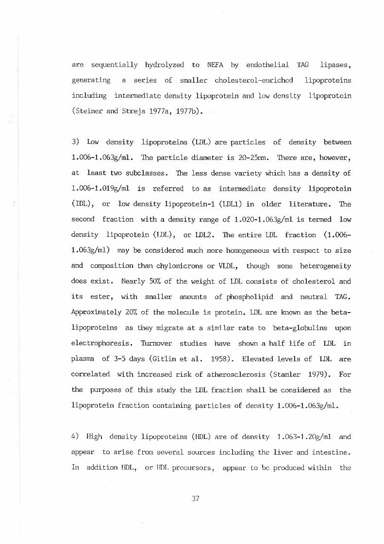
are sequentially hydrolyzed Lo NEFA by endothelial TAG lipases,
generating a series of smaller cholesterol-enriched lipoproteins
including intermediate density lipoprotein and low density lipoprotein
(Steiner and Strej a L977a, L977b).
3) low density lipoproteins (mI.) are particles of density between
1.006-1.063g/ml. Ttre particle diameter is 20-25nm. There are, however,
at least two subclasses. The less dense variety viLrich has a density of
1.006-1.)L9g,/nL is referred to as intermediate density lipoprotein
(fOI-)r or low density lipoprotein-l (I-OI,1) in older literature. Itre
second fraction with a density range of. t.O2O-1.063g/ml is termed low
density lipoprotein (t¡t-)r or l-DLz. Ttre ent.ire LDL fraction (f .OO0-
1.063g/ml) *ay be considered much more homogeneous with respect to size
and composition than chylomicrons or VLDL, though some heterogeneity
does exist. Nearly 507" of the weight of LDL consists of cholesterol and
its ester, with snraller amounts of phospholipid and neutral TAG.
ApproximaLeLy 207" of the molecule is protein. LDL are known as the beta-
lipoproteins as they migrate at a similar raLe to beta-globulins upon
electrophoresis. T\rrnover studies have shov¡n a half life of LDL in
plasma of 3-5 days (Citlin et al. 1958). Elevated levels of LDL are
correlated with increased risk of atherosclerosis (Stamler L979). For
the purposes of this study the LDL fraction shall be considered as the
lipoprotein fraction containing particles of density 1.006-1.063g/ml.
4) High density lipoproteins (ttOt ) are of density 1.063-1.ZOg/nL and
appear to arise from several sources including the liver and intestine.
In addition HDL, or HDL precursors, appear to be produced wiLhin the
37
plasma comparlment. from phospholipid-protein discs, generated as a
result. of lipolytic processing of chylomicrons (fatt and Small t97S).
ÐL, the smallest of the lipoproteins (8-12nm), are involved in a
process referred to as treverse cholesterol Lransportr, a postulated
pathway viLrereby HDL acquire cholesterol from peripheral tissues and
transport. Lhe cholesterol, directly or indireeLLy, to the liver for
excrection (Uatr1ey L982). The HDL have been divided into three
subfractions namely HDL1, 2 and 3. HDL1 is a minor component
identif iable only \,üith the analytical ultracentrifuge and is of
uncertain physiological significance. It may contaminate LDL floated in
a buffer of density 1.063g/ml, and in fact, HDL1 has by rnany
invest.igators been considered as an LDL subfraction. HDL1 has alptr,a 2
mobility on elecLrophoresis. HDL2 and HDI^3 v¡hich are the major fractions
in human plasma consist of approximately 507. protein, 307. phospholipids
and smaller amounts of cholesterol, cholesterol esLers and TAG. HDL2 are
rícher in lipids, poorer in protein and float more rapidly in a brffer
of density I.2IdnL than HDL3. Not only is Lhe lipid:protein ratio of
HDL2 greater than that of ÐH, but there are also differences in their
apoprotein composit.ion (chapter three). HDIS can also be formed from
HDL2 during ultracentrifugation and during storage of plasma (t tw and
Fredrickson 1965). Metabolic turnover studies have shown a half life in
plasma of 4-5 days for the viLrole lipoprotein fraction of density 1.063-
L.ZLO4/nL (Furman et. al. L964). As the HDL fraction is heterogeneous,
it.rs electrophoretic pattern is spread over the alpha, and to a lesser
degree, the beta range. The recent observations suggesting that a
negative correlat.ion exists between HDL and accelerated vascular disease
in man (C,ordon et al. t977), have focused at.tention on this lipoprotein
38
class and its role in cholesterol metabolism. (For revie\,\i see Heiss et
al. 1980).
5) Very high density lipoproteins (Vml) have a very small lipid
compliment. C,onsequently they mediaLe only a very small proportion of
circulaLing lipids. They can be generally described as a phospholipid
core encased by protein. Their density is greater than L.2Ùg/nL but less
than 1.25g/nL. Innnrnologically they resemble HDL.
6) Plasma non esterified fatty acids are cornplexed to albumin by non-
covalent forces. ltrey are transported rapidly through plasma and have a
half life of less than five minutes. This fraction is not identifiable
by the usual lipoprotein techniques and nmst. be measured by chemical
methods.
In addition to the nrajor lipoprotein fractions already described,
there are a number of lipoproteins vùrich are not found in normal
individuals. Rather, these pa.rticles are the result, of abnormalities in
the metabolic processes of lipoprotein anabolism/catabolism. As these
particles are usually attributable to particular metabolic disorders,
they are beyond the scope of this review.
L.L.2.2 SHEEP PIÁSMA LIPOPROTEINS
Lipoproteins from domest.icated ruminant species, and in particular
sheep and goats, have not received the same degree of interest as those
from monogastric anirnals
In 1955 Morris and Courtice (1955) separated sheep and plasma
39

Iipoproteins into slow and fast. moving components on paper
electrophoresis. Similarly, Perk and I-obI (1959' 1960) separated two
electrophoretic lipoprotein components for both cattle and sheep plasma
respeetively. However, in these studies the contribution of each of the
bands did not agree with the results of Grnpbell (1963) and Kirkeby
(1966), vùro found that in sheep, 37"A and 46"/" respectively of their
lipoproteins, was in the slow movj-ng fraction. Alexander and Oay (1g23)t
v¡ho examined the distribuLion of serum lipoproteins in selected
verterbrates, also showed ttra| 447" of sheep liproteins \,rrere in the slow
moving band, on agarose Plates.
The firsL report of the major lipoprotein componenLs of sheep
plasnra was based on their rates of flotation by Mills and Taylox (tglt).
They found that sheep plasnra contained lipoproteins with a high modal Sf
rate and that, VLDL were vj-rtually absenL.
Nelson (Ig73) isolated and separated lhe major classes of sheep
lipoproteins based on hydrated density. In essencer he found that in fed
sheep the chylomicron and VLDL fraction accounted for only 0.27" of
circulatory lipids. In addition, this study also showed that agarose gel
electrophoresis of sheep plasma, failed to detect a pre-beta band
(WO1.), however, a beta (f¡l) and two alpha (fm) bands were observed.
Leat et al. (1976) examined the plasma lipoprotein profile of sheep
in order to determine the contribution of each lipoprotein fraction to
the hyperlipidaemia observed in suckling lambs. Isolat.ion of sheep
lipoproteins according to the hydrated density parameLers attributed Lo
human plasma lipoproteins reiterated the virtual absence of chylomicron
and VLDL part.icles in the fed adult, with less than 5"/" of circulatíng
Iipids in this fract.ion. The major lipoprotein fractions (WDL, LDL and
40
HDL) \trere similar in composition to that seen in manr Lhough the
contribution of each fraction to total plasma lipids \i/as markedly
different. ÐL, LDL and VLDL accounted for 657", 207" and less Lhan 57"
respectively of plasnn lipids in sheep. In man, VLDL' LDL and HDL make
up approximately 207", 507" and 307" respectively of the plasma
lipoproteins. The hyperlipidaemia observed in the suckling lamb was
reflected by an increase in all of Lhe lipoprotein fractions. VLDL was
elevated Lo 7-157" of total lipoproteins.
1.1.3 AIMS OF CIIAPTM. ONE
From the published data currently available, it appears that sheep
lipoproLeins are not unlike those in rnan. However, although their
densities and lipid composition are similar, the contribution of each of
the fractions to circulating plasma lipids are quite differenL for the
two species.
Metabolically sLressed sheep have highly elevated levels of plasma
TAG, wkrich in turn is reflected in an accumulation of this lipid in the
liver. Research into the hepatic secretion and subsequent melabolism of
TAG is of fundamental importance if we are to elucidate the process of
this accumulation. As lipoproteins are the mediators of all plasma
lipids, their metabolism reflects the anabolic/caLabolic destination of
lipids.
The first part of this thesis establishes suitable methods for the
isolation and separat.ion of the major lipoprotein fractions in sheep
plasma. 'Ihe study reexamines the plasma lipoprotein profile (and their
chemical constituents) in fed sheep for only the third time, and the
4t
plasma lipoprotein profile from chronically stressed sheep, (v¡trich are
hypertriacylglyceridaemic), Lor the first time-
42
L.2 ME'THODS AND MATERIALS
L.z.L ANII'{ALS USED
Adult Merino sheep (O¡is Aries) were obtained from the flock
maintained at the tlaite Agricultural Research Institute, University of
Adelaide. AII anirnals were weight (:S-OOt<g) and age (1-: years) nntched.
Each sheep r,rlas housed individualty and maintained on a diet of 9009
lucerne chaff and 2009 pellet supplements per day. (Ctrartict<srAdelaide,
sheep pellets) fed at 9am each day. l,later was available ad libitum
FasLed sheep were studied after 72lr. of compleLe food deprivation
with water available ad-libitum.
Alloxan diabetes rilas induced by an intravenous injection of
s terile alloxan (2 ,4 ,5, 6-Tetraoxypyrimidine ; 5 , 6-Dioxyuracil mono-
hydrate), 5ùng/kg body weight, into Ehe jugular vein one week prior to
experimentation. A diabetic condition was confirmed by blood glucose
concentration in excess of 1ùntvl and all anÍmals were hyperketoaenic,
L.2.LJ COLLECIION AI{D PRESRVATION OF BI,OOD PIASMA
Blood was drawn from the jugular vein into heparinized or lithium-
EDTA (ethylenediaminetetra-acetic acid tetra-sodium salt) tubes, to
prevent coagulation.
Plasma \t/as collected inrnediately by centrifuging at 3'000
r.p.m. for l-5min at 4oC. The plasma was held on ice and used inrnediately
for lipoprotein isolation.
Ellman's reagent (5,5'-dithiobis(2 nitro-benzoic acid)) at a
43
concenLration of 1.5 mM r^ras used to inhibit lecithin cholesterol acyl
transferase and phenylmethyl-sulphonyl fluoride (pUSf') (2nM) was used to
inhibit proteolytic enzymes. Sodium azLde and thimerosol (sodium
ethylmercurothiosalicylate-mercury-( (o-carboxyphenyl)-thio)-ethyl sodium
salt) were also included as bacteriocides and EDTA (tmpt) was added to
inhibit autooxidation :
Solution A: 0.759 of Thimerosol and 1.3g Sodium azide dissolved in
100rnl of water, and adjusted to pH8.
Solution B was freshly prepared and contained 0.5959 of Ellmans
reagent in 10 mI of 0.2M sodium bicarbonaLe solution.
Solution C: 0.359 of PMSF in 10 ml of 2-propanol.
To 1ùnl of plasnra, 100u1 of solutions A, B,and C were added. EDTA
was added lo give a concentration of 0.37mg/ml plasna.
L.2.2 DETERMTNATION OF BI,OOD GLUCOSE
Blood or plasma glucose concentration was determined essentially
according to the method of Bergmeyer and Bernt (tgl+).
Solution A: 0.33M Perchloric acid solution.
Solution B: 1.38g Na2tlPQ* , O.727gNaH2PO4.2Hp, 0.050489 2,2t-Azíno-
di-(3-ethylbenzthiazoline)-6-sulphate (¡¡fS), 933 InternaLional Units
Glucose Oxidase and 150 International Units of Percncidase in 10ùnI water.
0.1m1 of blood or plasma was deproteinized by mixing with 1ml of
solution A. AfLer centrifuging at 1500 r.p.m. for 5min, 20ul of the
clear supernatant was mixed thoroughly wiLh 1-ml of solution B and
allowed to stand at room ternperature for 3ùnin. The absorbance was read
at 42Onm. Glucose standards ranging 0-1onm were included in each assay.
44
L.2.3 ADJUSTMENI OF PTASMA SOLVE¡üI DENSITY
The amount of salt (nact or KBt), either solid, or in a
concentrated solution, needed to bring about a specified adjustment of
plasma densiLy was determj-ned according to the melhod of Radding and
Steinberg (1OOO¡ as described by MiIIs eE aI. (1984).
plasma was maintained at 20oC in a circulating water bath. Density
was determined by weighing in duplicate at constant temperature'
t.2.4 SEPARATION AÌ{D PURIFICATION OF PTASI',IA LIPOPROTEINS
I.2.4.L TIME COURSE STT]DIES
29 of sudan black was added to each 20rnl of plasma and mixed
thoroughly for 2h at 4oC. The solut.ion was then filtered through
fibreglass discs.
Total plasma lipoproteins were isolated from plasma essentially
according to the method of Rudel et al (1974). The solvent. density of
sheep plasnra was raised to t.225g/nL by the addition of solid potassium
bromide. 8ml of stained plasma solution was then placed in S!ü41 (fZmf)
nitrocellulose, ultraclear or polyallomer tubes and carefully
overlayered with 4ml of a buffered sa1t. solution of density t,225g/nL
using a peristaltic purnp, aL a flow rate of approximately 0.5m1/min. The
buffered solut.ion \,\Ias prepared by the addition of solid KBr to a
1.006g/ml solut.ion described by Scanu and Granda (fg60) virich contained
L.42g anhydrous Na2PO4, 7.27g NaCl, 0.1g EDTA made up to l-l , PH7 -6. The
45
tubes v¡ere centrifuged at. 40,000 r.p.m.in a SIl41 swinging bucket rotor
at 20oC, for either 18h, 24kr or 40h in a Beckman L5 65 ultracentrifuge.
The centrifuge was stopped with the brake off.
By monitoring the profile of the sudan black stained lipoproteins,
a meá.sure of their rate of migration to the top of the tubes was
determined.
Duplicate tubes from each time period were carefully removed and
stabilized in a centrifuge tube brace. T?re top and bottom of each tube
r^rere pressed tight against inert silicone rubber septa. The sudan black
stained lipoprotein profile was monitored by puncturing the base of each
tube using a fine gauge needle and expelling the contents by volume
displacement using a saturated KBr solution, at a peristaltic purnp flow
rate of 0.25m1/min. The contents were monitored continuously (Zeiss
PI.'IQII spectrophotometer) at 600nm using a flow through cell (volume
250uI). Prestained lipoproteins were discarded and not reLained for any
further analyses.
L.2.4.2 COLLESTION OF TOTAL PTASMA LIPOPROTEINS.
SI,l41 ultracentrifuge tubes were loaded with 8 ml of plasnn (not
stained) solution with the density adjusted Lo L.225g/m1 and overlayered
with the t.225g/nL buffered salt solution as described above. T\:bes were
centrifuged in an SI^/41 swinging bucket roLor for 40h at 20oC, at 401000
r.p.m.. The centrifuge was stopped with the brake off, so as not to
disturb the lipoprotein buffer interface and prevent mixing. T\rbes were
carefully removed from the rotor and the top 2nL containing the
concentrated lipoproteins removed either with a tube slicer or via a
46
500u1 glass syringe (S.C.n.). The top portion of the tube was washed
several times in a small volume of 1.006g/ml buffer solution. The
resulting lipoprotein concentrate will for the purposes of this study be
referred to as Lhe "d1.225g/nL lipoprotein concentrate".
L.2.4.3 ESTII.,ÍATION OF TUIAL PLAS}4A LIPOPROTEINS
Sheep plasma lipoproteins isolated by ultracentrifugation at
density L.225g/nL were placed in dialysis tubing (5nm), vrhich Ì{Iere
extensively prewashed in double distilled water, dried, and weighed.
Each dialysis bag contained 1.5mI of lipoprotein concentrate and
r^ras dialysed f.or 24lr. against 3 X 4 litres of double distilled water at
4oC. The bags were then suspended in air and dried at 60oC for 3tr. The
dialyses bags were reweighed and Èhe amount of lipoproteins deLermined
by difference in bag weight.
I.2.4.4 AGAROSE GEL FILTRATION
Aliquots of the dl.225g/nl lipoprotej-n concentrates ranging 2-25nL
(ZO-ZOOrng lipoprotein) were applied wilhout further manipulation, to
agarose colunms (gi-o Get A-5m, 200-400 mesh, 2.5cm X 100cm), and eluted
with either 0.1M NaCI, 0.2M potassium phosphate' 0.017"EDT4, p}:.7.4 or
0.15M NaCl, 0.17.E0T4, ph7.0 at. a flow rate of approximately 3ùn1/h. The
eluate r^ras monitored at 280nm and collected in 2.5mI fractions. The
contents of tubes containing individual lipoprotein peaks were pooled
and concentrated ten fold by pressure dialysis through )0150 membranes
(Amicon Corporation) in a sLirred 10ml Amicon filtration cell. Magnetic
47
stirring r,t/as maintained at approximately 10r.p.m. and pressure \^/as
achieved by high purity nitrogen. Temperature I^Ias maintained at 4oC.
L.2.4.5 HIGH PERFORI\,IANCE LIQUID CHROI'4ATOGRAPI{Y
üp to 5mI of d1 .225g/nL lipoprotein concentrate \^/as dialyzed
against 3 X 1L of 0.25M tris-phosphate pH7.6 for 18h at 4oC, to remove
halides.
Aliquots of the dialyzed dl.225g/nL lipoprotein concentrate hrere
chromotographed on Toyo Soda high performance gel-filtration columns.
The high performance liquid chromatography (ttptC) was carried out. using
an EIP Kortec liquid chrornatograph system equipped !'rith a high pressure
pump (model K25), an ETP Kortec (model K95) variable wavelength
spectrophotometer and a Spectra Physics (mode1 5P2470) integraLing
recorder, linked to a Pharnncia Frac-100, selective fraction collector.
The chronratography colums \^rere a Toyo Soda Company PI,üH guard colurnn
followed by a Toyo Soda G3000-S!ü (60ûnm) and c5000-PW (0OO,,rn¡ in series.
HPLC was carried out at room temperature after applying up to 200ug of
the lipoprotein concentrate mixture to Ehe colunnrs and eluting it in a
buffer consisting of 0.25M tris-phosphate, pH 7.6 under an argon
aLmosphere.
Material eluted from the HPLC colurms lrras monitored at 280nm and
was collected in 0.5m1 fractions. The contents of the tubes containing
individual lipoprotein peaks r^/ere pooled for further analysis.
Lipoprotein molecular weight estimations were determined by
interpolating the retention volume of each lipoprotein fraction wiLh the
retent.ion volume of proteins of known molecular weight. and diameter.
48
Protein standards included;
Molecular weight (daltons)o
Stokes radius (A)
Blue dextran
Thyroglobulin
Ferritin
Catalase
Aldolase
Albumin
Ovalbumin
CLrymotrypsinogen
Ribonuclease
2,000r000
669,000
440,000
2321000
158,000
67,000
43,000
25,000
13,700
85.0
61.0
52.2
48.L
35.5
30.5
20.9
L6.4
T.2.4.6 SERIAL CENTRIFUGATION OF PIASMA LIPOPROTEINS
Five diluent solutj-ons were required for the isolation of the major
classes of lipoprotein. Each diluent contained ÐTA (lrrM) , azide (2mM)
and Thimerosol (O.Zmu) ;
0.196 M
0.844 M
2.973 t4
4.778 VI
7.593 M
NaCl
NaBr
NaBr
NaBr
NaBr
solution,
solution,
solution,
solution,
solution,
densitY=l.006g/mf
density=l.063g/ml
densitY=l .2Lje/nl
densitY=l .320g/nL
densitY=1 .479g/nL
at
at
at
at
at
200c
200c
200c
200c
200c
The density of each solution \'\Ias determined by weighing in
duplicate at consLant ternperature.
49
L.2.4.6.t ISOIATION OF VLDL
8 ml of plasma was placed in SIl41 tubes and carefully overlayered
with 4m1 of 1.006g,/m1 solution as previously described. The tubes were
spun for 24)r- j-n an SI{ Beclqnan 4]- rotor at 401000 r.P.m. at 20oC. For
tubes containing fed sheep plasma, 1ml of the top fraction was removed
and for tubes containing plasma from alloxan diabetic wethers a 2nL
fraction hras taken. In addition, another 1mI of the salt solution
underlying the concentrated lipoprotein fraction was also removed to
ensure complete recovery. Samples vrere drawn using a 50ûJI glass
syringe. The Lop of the tube was then sliced and washed twice with 500
uI of the 1.0063g/mf soluLion so as to remove any adhering lipoprotein.
Unless stated otherwise, the VLDL concentrate Írras washed by
overlayering with 1.006g/m1 solution and centrifuging under the same
conditions as specified above.
L.2.4.6.2 ISOIATION OF LDL
The resulting infranatant af ter VLDL isolation l{as mixed
thoroughly. Itrs solvent density was adjusted to 1.063g/ml by the
addition of solut.ion L.32Og/nL. 8 mls of the adjusted infranatant was
overlayered with the salt solution of density 1.063g/ml and centrifuged
for 24h, 401000 r.p.m., 20oC. Ttre top 2ml fraction was gently aspiraLed
and the corresponding port.ion of tube washed twice with 500uI of the
L.063g/nL solution.
50
I.2.4.6.3 ISOIATION OF HDL
The LDL infranatant was mixed thoroughly and it's solvent density
raised to t.22Og/nL by the addition of the salt solution with density
t.479g/nL. 8mI of the adjusted LDL infranatant was overlayed with the
I.22Og/nL solut,ion and centrifuged fox 24lrr also at 40'000 r.p.m. ' 20oC.
The HDL concentrate hras removed in a 2ml fraction using the same
procedure as that used for lhe isolation of VLDL and LDL. The top of the
tube was washed twice r^rith 500uI of the L.Z2Og/nL solution.
r.2.4.7 AGAROSE GEL H-ECIROPHORESIS
Up to 5ul of plasma, dL.225g/nL lipoprotein concentrate or
concentrated lipoprotein fractions were separated by electrophoresis on
lnrn agarose plates (Corning-agarose gel elecLrophoresis system) in 0.05M
Na-barbitone buffer pH8.6 (0.0357" EDTA) at 90 volts for 35min. PlaLes
were removed and dried thoroughly at 6OoC. Lipoproteins were fixed in 27"
acetic acid for 3Ornin and the plates redried at 60oC. Lipoprotein bands
were stained in a filtered 27" (w/v) sudan black solution (methanol:water
(f :f)) for 2-3h. Destaining $/as achieved by briefly washing with a 50%
ethanol solution.
L.2.5 ÐilRACTION AND ANALYSIS OF LIPID COMPONM{IS FROM PTASMA AND
LIPOPROTEIN FRASTIONS
T.2.5.L EKTRACIION
Lipids were exlracted according to a modified method of Folch et al.
51
(tgst);
To 1 mI of sample 15ml of chloroform:meLhanol (2:1 v/v) was added
and mixed thoroughly. The solution was allowed to stand for 15min. One
quarter of the total volume (+m) of 0.03M HCl \^¡as added, mixed
thoroughly and allowed to settle. The solutions were centrifuged at
151000 r.p.m. in a Becknan JA 20 fixed angle rotor for 15min. Ttre upper
Iayer \^ras rernoved by aspiraLion and one quarter of the volume of the
lower phase (Z.lSnt) of water-methanol (f:f) added and Èhe washing
procedure repeated Lwice. The bottom phase contained the purified
lipid. All lipid extracts were stored in chromic acid washed sealed
glass ampoules at -15oC in the dark, under high purity nitrogen.
Prior to lipid extracLion, L25uL of a 0.1uCi/m1 glycerol tri(1J4c)
oleate solution was added per 1ml of plasma, or lipoprotein concenLrate.
C-orrections for the loss of the lipid components l{ere made after
determining lhe residual activitíy remaining in the lipid extractsr ofl
the assumption that the efficiency of the extraction procedure for each
of the different lipid components rnras uniform.
L.2.5.2 TRIACYTCLYCM,IDE DETERMINATION
TAG r^ras measured according to a modified method of C,arlson and
I,,/adstrom (fgSg);
To 1ml of lipid extracL approximately 5ùng of activated rzeolite
mix' was added to remove phospholipids (Fletcher 1968). The zeolite mix
contained by weight 807. zeoLite (hydrated alkali-aluminium silicate
Na2o.41193(sio2)x.(ttp)y, 8% Fullers earth (ar-ug-sio2), 87. ca(oH)2 and
4% CuSO4.5H2O, and was act.ivated by heating at 110oC for th. The samples
52
rÁ/ere mixed occassionally over a th period, after vfuich they \,tlere
centrifuged at 31000 r.p.m for 5min. An 800u1 aliquot was Laken for TAG
determination. The solvent.s containing the lipid extract vlere evaporated
under high purity nitrogen. 750ul of alcoholic KOH (907" ethanol v/v) was
added. The glass tubes r^/ere capped and saponified for 3ùnin in a 60oC
water bath. After cooling to room ternperature, 750u1 of 0.7M H2SO4
followed by 4.ùnI of diethyl ether were added. This was thoroughly mixed
and the phases allowed to separate. The top ether Layer was aspirated,
and any renraining film of ether evaporated w'ith a gentle stream of high
purity nitrogen. D:plicate 300ul aliquots were taken from the bottom
phase. To each sarnple 100u1 of 0.02M NaIO¿* \^Ias added and mixed
lhoroughly. Afber exacÈly 15min 100u1 of 0.2M NaAsO2 \,rras added to
remove excess periodate. After mixing, the initially colourless solution
goes yellow, then colourless again. After a lOrnin interval 3.0m1 of
freshly prepared chromotropic acid (fSn¡l solution in 2214 H2SO4 ) was
added and mixed lhoroughly. Each tube was €pped and colour developed in
a 100oC water bath for 3ùnin.
The optical density of each sample at 547nm was determined and the
concentration of TAG calculated by reference to standards (0-500nm).
L.2.5.3 PHOSPHOLIPID DETMMINATION
Phospholipid concentration was determined according to a modified
method of Fiske and Subbarow (Bottcher et aL 796L);
Reagents: Anrnonium molyMate 8.6"/"(w/v) in water was mixed 1:1 (v/v) with
287" (v/v) HZSO4. The reducing agent was made up of 2.5g NaHSO4r 0.5g
ua2(So4) and 0.0429 L-a¡ntno-2napthol-4sulphonic acid in 25ûnl of waLer.
,-I
[lr&
ui
!
I
53
The solution was allowed to stand in the dark for several hours, after
v¡trich it was filtered into a dark bottle. This was stable for I month
vùren refrigerated.
A 500u1 sample of lipid extract hras evaporated under a stream of
high purity nitrogen. 200uI of perchloric acid was added, the tubes
capped and placed in a sand bath maintained at 180-200oC and digested
for a minirrum períod of 4ùnin. After cooling to room ternperature, t.2mL
of anrnonium molybdate and 1.2m1 of the reducing agent l.rrere added. Ttre
solution was mixed thoroughlyr æpped and heated in a boiling water bath
for lOrnin.
After cooling, absorbance v¡as measured at 830nm. The samples could
be diluted with water without loss of proportionality.
T.2.5.4 CHOLESTEROL AI{D CHOLESTEROL ESTER DETERMINATION
Plasma and lipoprotein cholesterol and cholesterol-esters were
determined by gas chronratography. 100u1 of internal standard
(stigmast.eroL Ing/ml in chloroform) was placed in 1ùnl Kimble tubes and
the solvent evaporated under a stream of high purity nitrogen. To each
tube, 100u1 of plasma or 200uI of lipoprotein lipid extract was added
and in the latter case, the solvent evaporated. The samples \ÀIere
hydrolysed in 2.LnL of 2"A ethanolic KOH (957. ethanol v/v) at 60oC for
3Ornin. After cooling, 2ml hexane and 1ml water were added and the tubes
shaken vigourously for 1min. The phases v/ere allowed to separate, after
vùrich an aliquot of the hexane phase was taken. This \¡ùas evaporated with
nitrogen and 5OuI of both chloroform and trifluoroacetic anLrydride were
added. The tubes \^rere capped and heated at 50oC for 2Ornin. The reagents
rlrü
I
rI
lI
r
54
\,\rere evaporated and the residue redissolved in 100uI of chloroform.
Total cholesterol rirras determined on a Hewlett-Packard HP5710 gas
chromatograph equipped with a 2M X 2nrn glass column, packed with 17. OV-
l-01 on Gas Ckrom Q. The injector and detector were at 250oC and the oven
at 220oC isothermal r^rith nitrogen gas carrier aL 3ùnl/min. HP3388
integrator operated in an internal standard mode.
To calculate the free and esterified component of the total
cholesterol, cholesterol esters were determined by repeating the above
procedure hrith the ornit,tion of the hydrolysis procedure. Free
cholesterol was then caleulated by difference of the total cholesterol
(free plus esterified) and Èhe esterified component.
T.2.6 LIPOPROTEIN PROTEIN DETERMINATION
Lipoprotein protein was determined according to the modified Lov¡ry
method (Hartree L972).
Reagents: solution A contained 29 potassium-sodium tartate and 1009
Na2(CO3) sodium carbonate made up in 1L of 0.5M NaOH.
Solution B; 29 potassium-sodium tartarate, Lg GrSO4.5H20 in 10ùnl 0.1M
NaOH.
Solution C; 6.67" Folin-Ciocalteau phenol reagent.
To 250u1 of sample 225uL of solution A was added, mixed thoroughly
and heated at 50oC for 1ùnin. After cooling to room temperature 25ul of
solut.ion B was added and allowed to stand at room temperature for at
least lOrnin with occassional mixing. 750u1 of solution C was then added
and mixed inrnediately. After colour developrnent at 50oC for 1ùnin,
absorbance was determined at 650nm. Standards ranging 0-50ug of bovine
il'.ü
rl
ÌI
I
!
55
{
serum albumin were included in each assay. Contaminating lipids vihich
may have interfered with the absorbance \,rrere removed by washing the
colour developed solution twice with hexane and chloroform. Standards
were also washed with the appropriate solvent.
T.2.7 NON-ESTM,IFIED FATTY ACID DETMMINATION
For each 400uI of plasna 50ul of internal standard (heptadecanoic
acid (fZ:O) 500mg/L in hexane) and 4m1 of Dole reagent (2-
propanol:heptane:1M sulphuric acid (400:100:10 (v:v:v:))) were placed in
Kirnble tubes and mixed thoroughly. 5mI of heptane and 2ml of water were
added and the tubes shaken for a further 1min. The phases r^rere allowed
to separate and the upper heptane layer transferred to another Kimble
tube. The solvent \Âras evaporated wiLh a stream of high purity nitrogen
and the f.aLLy acid residue methylated for 5min hrith excess ethereal
diazomethane (in ether). The solvent lsas evaporated and the crude fatty
acid methyl esters dissolved in 200u1 of hexane. Each sample \^Ias
applied to a,lûrmX 6nrn biosil column (activated at 60"C) and washed with
2mI of hexane. The fatty acid methyl esters were eluted with 2mL of. LÚ/"
ether in hexane. The solvent v/as evaporated and the residue dissolved in
20ul of chloroform for injection into the gas chromatograph. The sample
\^/as chromatographed on a 50M S@I capillary colunn of FTAP aL 220oC
isothermal, helium carrj-er aL 4nL/nirn with injector and detector at
250"C. Quantitation \Àras by a FIP3388 comput.ing integrator using the
internal standard method.
I.2.8 TRANSMISSION H.ECTRON MICROSCOPY OF OVINE LIPOPROTEINSrII
I
I
l
56
II
Lipoprotein fractions eluted by HPLC were negatively stained with
sodium phosphotungstic acid, according to Forte and Nichols (L972).
Negatively stained samples were viewed on 200 mesh copper grids coated
with Forrm¡ar and supported with carbon, using a J.E.O.L. Jem 100CX
transmission electron microscope.
I.2.9 MATMIATS AI{D REAGM{TS
Alloxan monohydrate was purchased from Koch-Light Ltd. England.
Glucose Oxidase and Peroxidase \^/as purchased from Sigma Ckremicals
Australía. Glycerol tri(1-14ç)oleate (SemCi/nrnol) was purchased from
Arnersham Australia Pty. Ltd. All che¡nicals and reagents were analytical
reagent grade or the best conrnercially available grade. H.P.L.C. protein
molecular weight markers were purchased from Pharmacia Pty. Ltd. t
Uppsala, Sweden.
Heparin and Li.EDTA tubes were purchased from Surgical and Medical
Supplies, Australia. Nitrocellulose, ultraclear and polyallomer
ultracentrifuge tubes (14nm X 89nm) \¡rere purchased from Becknan
InsLruments Australia. Agarose gel electrophoresis kit lÀras purchased
from C-orning I.C.I. Australia Pty. Ltd.. Bio Gel A-5m (200-400 mesh)
was purchased from Bio Rad Australia Pty. Ltd.. Toyo Soda G3000-S!ü and
G5000-PW columns were purchased from Beclqnan Australia Ltd.. E'lP-Kortek
HPLC systern \Áras purchased from EIP-Kortek Australia Pty. Ltd.. Diaf Io
þ150 Ultrafiltraton Membranes r^rere purchased from Amicon AusLralia Ltd.
All glassware was chromic acid washed. All solvents were glass
redistilled and stored under high purity nitrogen in the dark. Only
glass double distilled water was used.
57
1.3 RESULTS
1.3.1 SHEEP PIASMA
Plasma from healLhy fed sheep is clear and slighly pink in colour.
Induction of alloxan diabetes elevated plasma lipids, vlhich was visually
evident by loss of translucence and the develoçxnent of a vùrite coloured
plasma best described as 'milky' (figure L.2). llhen plasma isolated from
severely diabetic sheep (blood glucose concentration greater then 1ùnFI)
rr¡as cooled to below 4oC, the coagulation of fat into snnll globules
could be seen.
Ihe mean plasnn density of sheep plasma at 20oC hlas L.3O7g/nL.
Alloxan diabetes or fasting for 72Ìi. di:d not alter the plasnra density.
L.3.2 T]I,IE COURSE STT]DIES
The time required to isolate ovine plasma lipoproteíns by
ultracentrifugat.ion at a solvent. density of L.225g/mI was determíned by
prestaining plasma lipoproteins with sudan black and monitoring their
raLe of migration to the top of the tubes spectrophotometrically.
Figure 1.3 represent.s the absorbance profile of sudan black stained
sheep lipoproteins spun for 18h, 24i;. and 40h respectively. Tubes spun
for a period less than 40h exhibited tailing in their absorbance
profile, meaning that the complete recovery of the sheep plasma
lipoproteins could only be achieved afler a 40h ultracenlrifugation
period, under the prescribed experimental conditions. In conLrasl,
lipoproteins from cat.tle plasma could be recovered in 24h (Uamo and
58
Figure 1.2
Figure 1.2 represents plasma from three fed
and three diabetic wethers. TUbes containing
the pink transluscent samples are plasrna from
nornnl fed aninals (F1, F2, F3). Ttre
hyperlipidaemia associated with severely
diabetic animals was reflected in clouded
plasrna (D1, D2, D3). (the tubes vrere being
overlayed with a br:ffer, in preparation for
lipoprotein isolation by ultracentrifugation. )
59
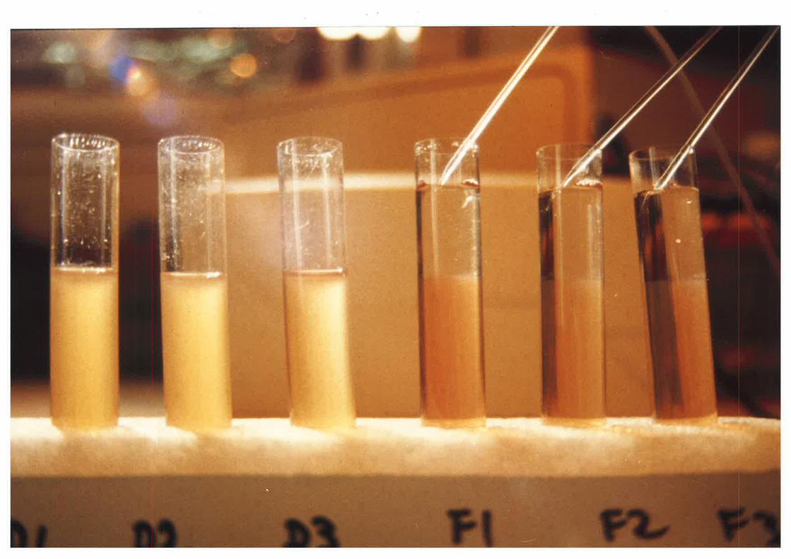
Figure 1.3
Figure 1.3 represenLs the rate of migration
of total sheep plasma lipoproteins wtren
centrifuged under the conditions deseribed in
the text (L.2.4.L). Complete recovery of the
Lipoproteins could only be achieved after a
40h ultrdcentrifugation period.
60
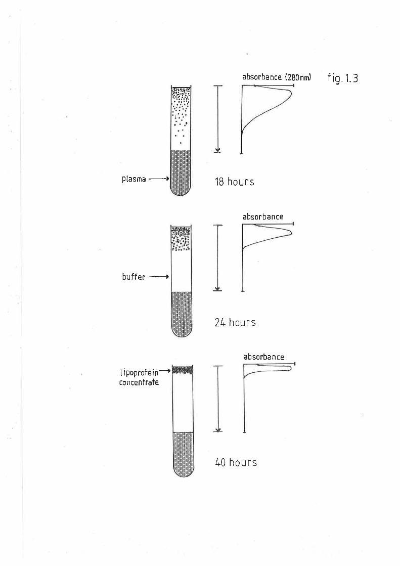
absorbance (2B0nm) f i9 1.3
$ffii
,I
Plasma
-)
buffe¡
lipoprotein+concentrafe
1B hours
2L hours
absorba n ce
I ,
absorba n ce
L
40 hours
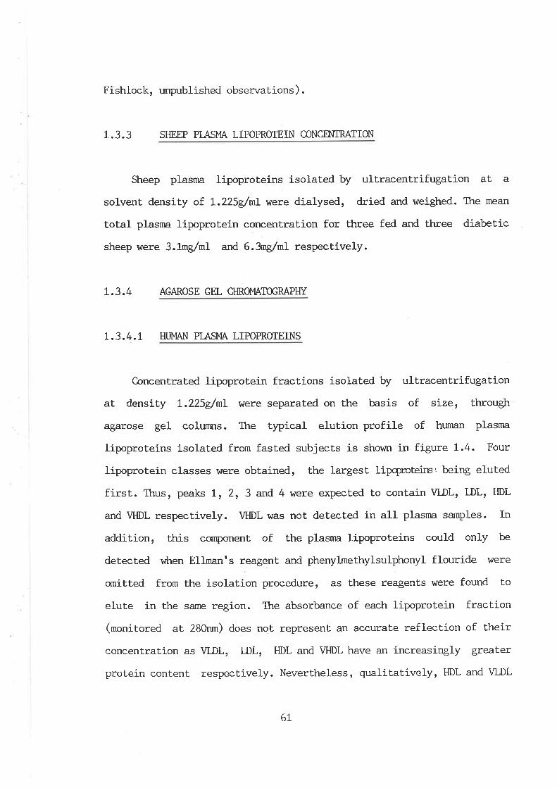
Fishlock, unpublished observat.ions) .
1.3.3 SHEEP PIASI',IA LIPOPRCIEIN CONCM{TRATION
Sheep plasma lipoproteins isolated by ultracentrifugation at a
solvent density of L.225glml were díalysed, dried and weighed. The mean
toÈal plasma lipoprotein concentration for three fed and three diabet.ic
sheep were 3.1mg/ml and 6.3rng/m1 respeclively.
L.3.4 AGAROSE GEL CI{ROMATOGRAPI{Y
L.3.4.I HUI'4AN PTASMA LIPOPROTEINS
Concentrated lipoprotein fract,ions isolated by ultracentrifugat.ion
at density L.225g/nL \Àrere separated on the basis of size, through
agarose gel colurnns. The typical elution profile of human plasma
lipoproteins isolated from fasted subjects is shown in figure 1.4. Four
lipoprotein classes r^¡ere obtained, the largest lipcprcteirs, being eluted
first. Thus, peaks L, 2, 3 and 4 were expected to contain VLDL, LDL, HDL
and VHDL respectively. VHDL was not detected in all plasnra sarnples. In
addition, this cornponent of the plasma lipoproteins could only be
detected v¡Lren Ellman's reagent and phenylmethylsulphonyl flouride \dere
omitted from the isolation procedure: âs these reagents were found to
elute in the same region. The absorbance of each lipoprotein fraction
(monitored at 280nm) does not represenL an accurate reflection of their
concentration as VLDL, DL, HDL and VHDL have an increasingly grealer
protein content respect.ively. Nevertheless, qualitat.ively, ÐL and VLDL
6L
Figure 1.4
Figure 1.4 shows the representative elution
profile of plasma lipoproteins from fasted
humans, fed sheep, fasted sheep and diabetic
sheep respectively, through agarose gel (5M)
sizeing colurrrs¡ âs described in section
L.2.4.4. Peaks I, II, III and IV are very low
densiLy, low density, high density and very
hígh density lipoproteins repectively.
62
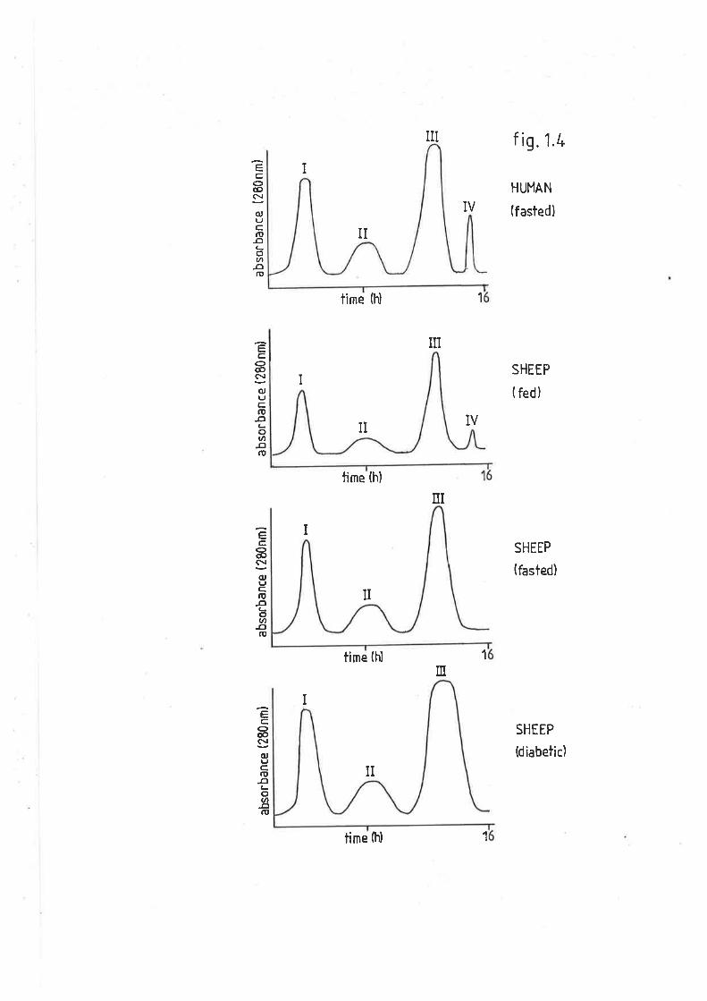
NI f ig.1.t-
HUMAN
(f asted)
SHEEP
( fed)
SHEIP
(fasted)
SHEEP
(diabetic)
Co@c\¡
(uTJcfI,
-cl(_otn
-oID
UI
I
L
o@ç\¡(uL'c,t!DLotn-oro
Ec,aç!
(uIJC,.DÐa-ov1
-ofo
Coæ(\¡(UtJc,.I,
-c¡a-oØ-orI,
I
II
time h)
II
time (h)
I
time (h)
II
IV
N
1
ilI
1
n
I
1time (tì)
appeared to be the major plasma lipoprotein components in fasted human
plasma. Peak II (I-nI-) may be more heterogeneous than the other
lipoprotein fractions as itts absorbance profile hTas much broader. The
total elution time for fasted human plasma lipoproteins \47as
approximately 16h.
I.3.4.2 STIEEP PTAsI.,IA LIPOPROTEINS
Agarose gel filtration of fed sheep dL.225glnl lipoprotein
concentrate gave an elution profile not unlike that seen for humans
(figure t.4). Ttre most striking feature was the significantly lower
lotal lipoprotein absorbance profile (per unit of plasma) ttran that
observed for humans. In addition VLDL and LDL did not appear to be as
quantilatively significant as that for human plasma. HDL I^Ias the
predominant lipoprotein. The VHDL fraction was not observed in all fed
sheep. Similar elution of the dL.225g/ml lipoprotej-n concentrate from
fasted sheep or alloxan diabetic wethers (figure 1.4) had a similar
profile with the exception that no VHDL was present in either treatment.
Each of the other fractions, and in particular HDL, appeared to be
elevated in metabolically stressed sheep.
1.3.5 AGAROSE cEL ELEGIROPHORESIS OF THE AGAROSE CHROI"r¡r*IÐGRAPHY
LIPOPROTEIN FRACTIONS
1 .3.5.1 HUI'4AN FRACTIONS
Each of the tubes containing individual lipoprotein peaks were
63
pooled and concentrated using pressure dialysis. Samples of the
concentraLed lipoproteins \,r/ere then separated by electrophoresis on
agarose gels (figure 1.5). Fractions 1, 2 and 3 had migrations of pre-
beta, beta and alpha respectively. TÏris corresponds with VLDLI LDL, and
HDL. It is apparent from the gels thaÈ each of the fractions \4tere
homogeneously distinct from the other lipoprotein classes. The VLDL
fraction fe-aLured tailing towards the beta region. Similarly, DL had a
degree of streaking towards Èhe pre-beta region. The HDL fraction had
two distinct bands vùrich overlapped. Ihe slower and less abr:ndant
component had an electrophoretic mobility símilar Èo that of the LDL
fraction.
L.3.5.2 SHEEP FRACTIONS
{garose ge1 electrophoretograms of plasnra, dl.225g/nL lipoprotein
concentrate and the respective lipoprotein fractions from fed, fasLed
and diabetic sheep are shown in figures 1.6a-1.6c respectively. Each of
the bands corresponded to those observed in the equivalent human
fractions. There \^Iere, however, Some slight differences in
electrophoretic mobility. The sheep VLDL fraction did not seem to be as
homogeneous as that observed in human plasma, as indicated by the
greater degree of tailing. Sheep LDL had a slightly greater mobility
than that. observed with the human LDL. C-onversely, the major sheep HDL
component had an electrophoretic mobility slightly less than the
equivalent. human HDL fraction.
Ttre VLDL fraction from fed, fasted and diabetic sheep differed-
Fasted sheep VLDL migrated more slowly than those either from diabetic
64
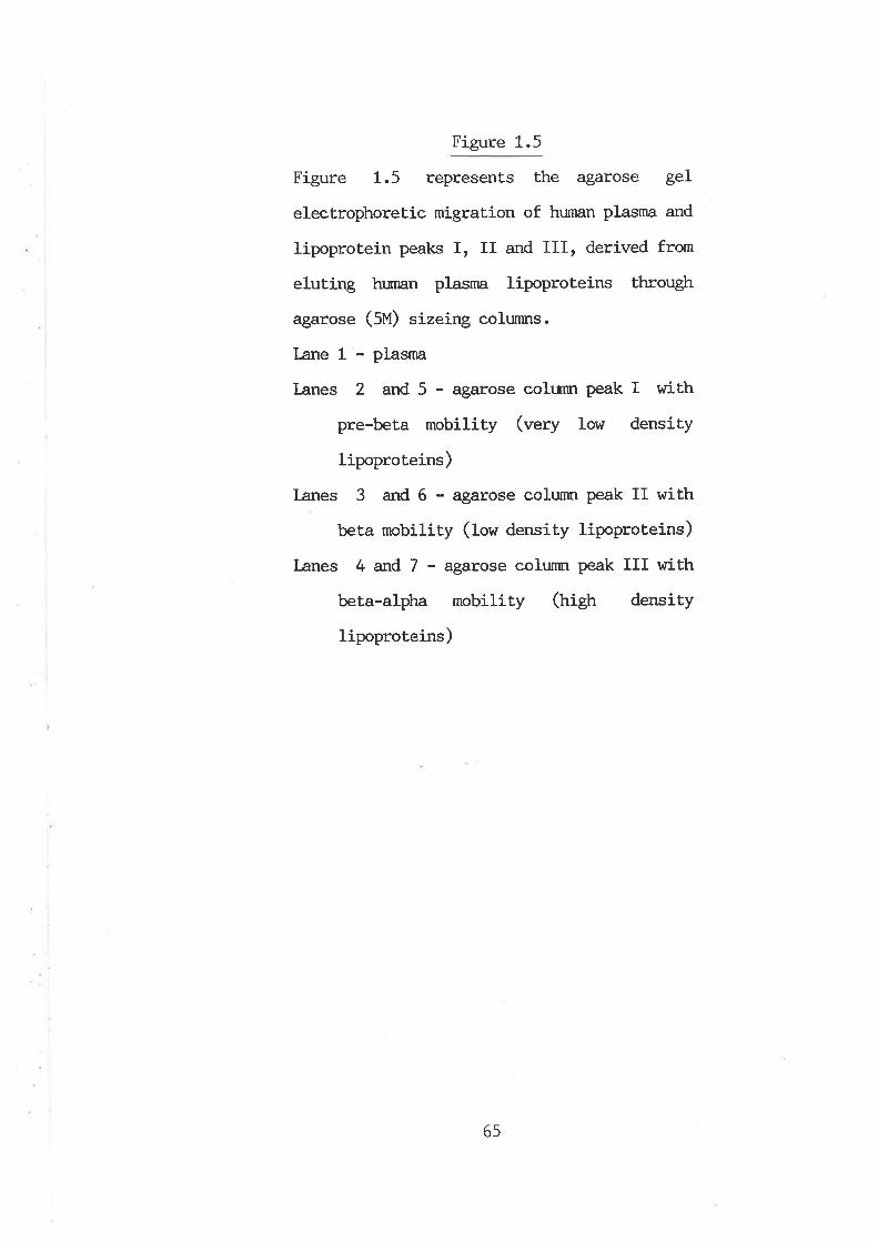
Figure 1.5
Figure 1.5 represents the agarose geI
electrophoretic migration of human plasnra and
lipoprotein peaks I, II and III, derived from
eluting hr¡nan plasma lipoproteins through
agarose (5M) sizeing colunnrs.
I¿ne 1 - plasma
lanes 2 and 5 - agarose colurnr peak I with
pre-beta mobility (very low density
lipoproteins)
Lanes 3 and 6 - agarose colurmr peak II with
beta mobility (low density lipoproteins)
I-anes 4 and 7 - agaxose colunm peak III with
beta-alpha mobiliuy (frigh densiuy
lipoproteins)
65
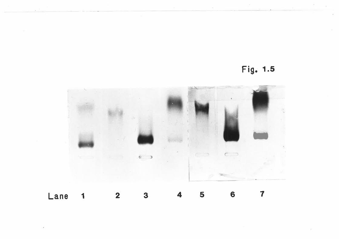
Fig. 1'5
þ$
5 ïta
¡B
2 3
üf - i$a' l
G)
Lane I 45 6 7
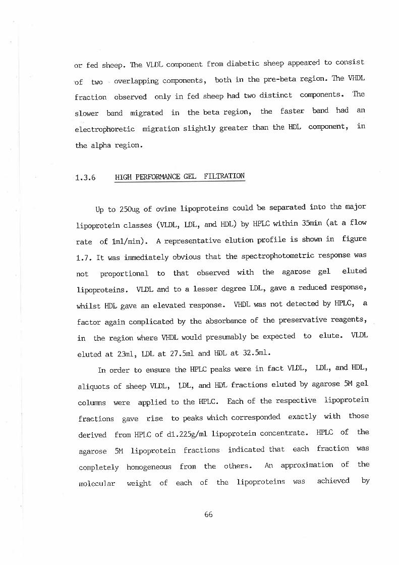
or fed sheep. The VLDL componenl from diabetic sheep appearerl to consist
'oftwo,overlappingcornponents'bothinthepre.belaregion.Tl.reVHDL
fraction obsen¡ed only in fed sheep had two distinct components' The
slower band migrated in the beta region, the fasLer band had an
electrophoretic migration slightly gtea1er than the HDL conponent, in
ühe alpha region.
1.3.6 HIGH PMFORI\,IANCE GH- FILTRATION
Up to 250ug of ovine lipoproteins could be separated into lhe major
lipoprotein classes (VDL, LDL, and HDL) by HPIC within 35min (at a flow
rate of 1ml/min). A representative elution profile is shor¿n in figure
I.7. Tt was inrnediately obvious that the spectrophotometric response I'ilas
not proportional to thab observed with the agarose gel eluted
lipoproteins. VLDL and to a lesser degree LDL, gave a reduced resPonset
viLrilst HDL gave an elevated response. VHDL was not detected by HPrc, a
factor again complícated by the absorbance of the presen¡ative reagentst
in the region vùtere VHDL would presumably be expected to elute' VLDL
eluted at 23rnl, LDL at. 27.5nL and HDL aL 32.5m1.
In order to ensure the HPLC pe-aks were in fact vLDL, DL, and HDLt
aliquots of sheep VLDL, LDL, and HDL fractions eluted by agarose 5M gel
colunns hrere applied to the HPLC. Each of the respective lipoprotein
fractions gave rise to peaks v¡hich corresponded exactly with t'hose
derived from HPLC of d1- .225g/nL lipoproLein concentrate. HPLC of t'he
agarose 5M lipoprotein fractions indicated that each fract'ion v/as
completely homogeneous from the others. An approximation of the
molecular weight of each of the lipoproteins \^las achieved by
66
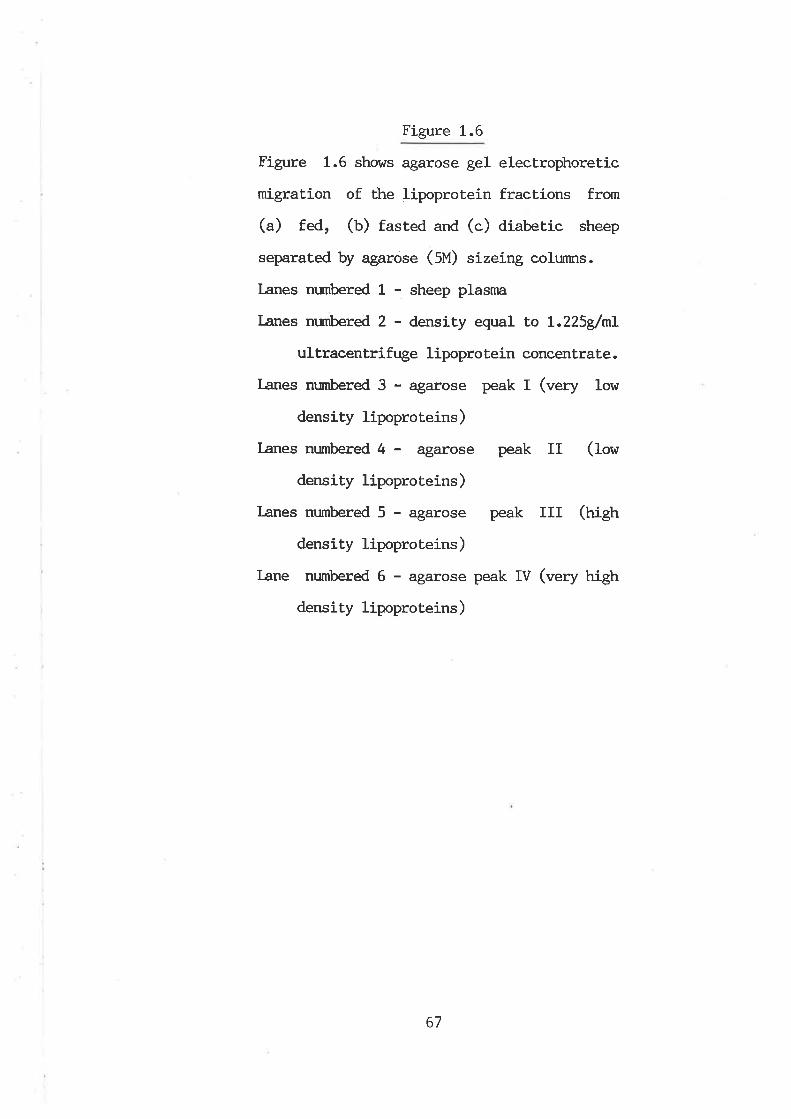
Figure 1.6
Figure 1.6 shows agarose gel electrophoretic
migration of the lipoprotein fractions from
(a) fed, (b) fasted and (c) diabetíc sheep
separated by agarose (5U) sizeing colunms.
Lanes nurnbered 1 - sheep plasnn
I¿nes numbered 2 - densit¡r equal to L.225g/ml
ultracentrifuge lipopro lein concentrate.
l¿nes nr:mbered 3 - agarose peak I (.rery low
densiÈy lipoproteins)
I-anes numbered 4 - agarose peak II (Iow
density lipoproteins)
Lanes numbered 5 - agarose peak III (trigtr
density lipoproteins)
lane numbered 6 - agarose peak IV (very high
density lipoproteins)
67
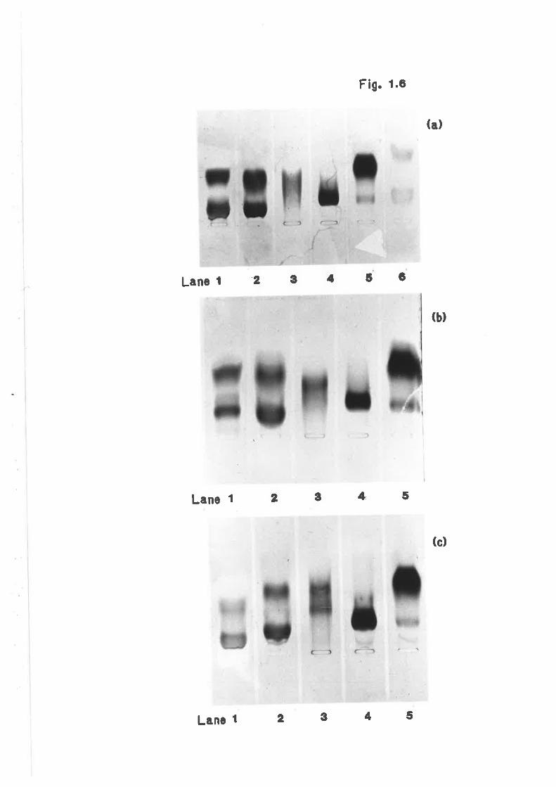
Fig. 1.e
Lanel 2 C 4 6 6
Lanel 2 g 4' 5
(a)
(b)
(c)
I
Lanc 1 2345
Figure 1.7
A representative elution profile of sheep
plasma Iipoproteins by high performance
liquid chroratography is shovrn in figure 1.7.
Peak 1 (nv--ZS.OmI) is the very large very 1ow
density lipoproteins. Peak II (Rv=27.5m1) and
Peak III (nv=SZ.lnI) are the low density and
high densíty lipoprotelns respectively.rArrepresentative elution profile ei fed sheep
68
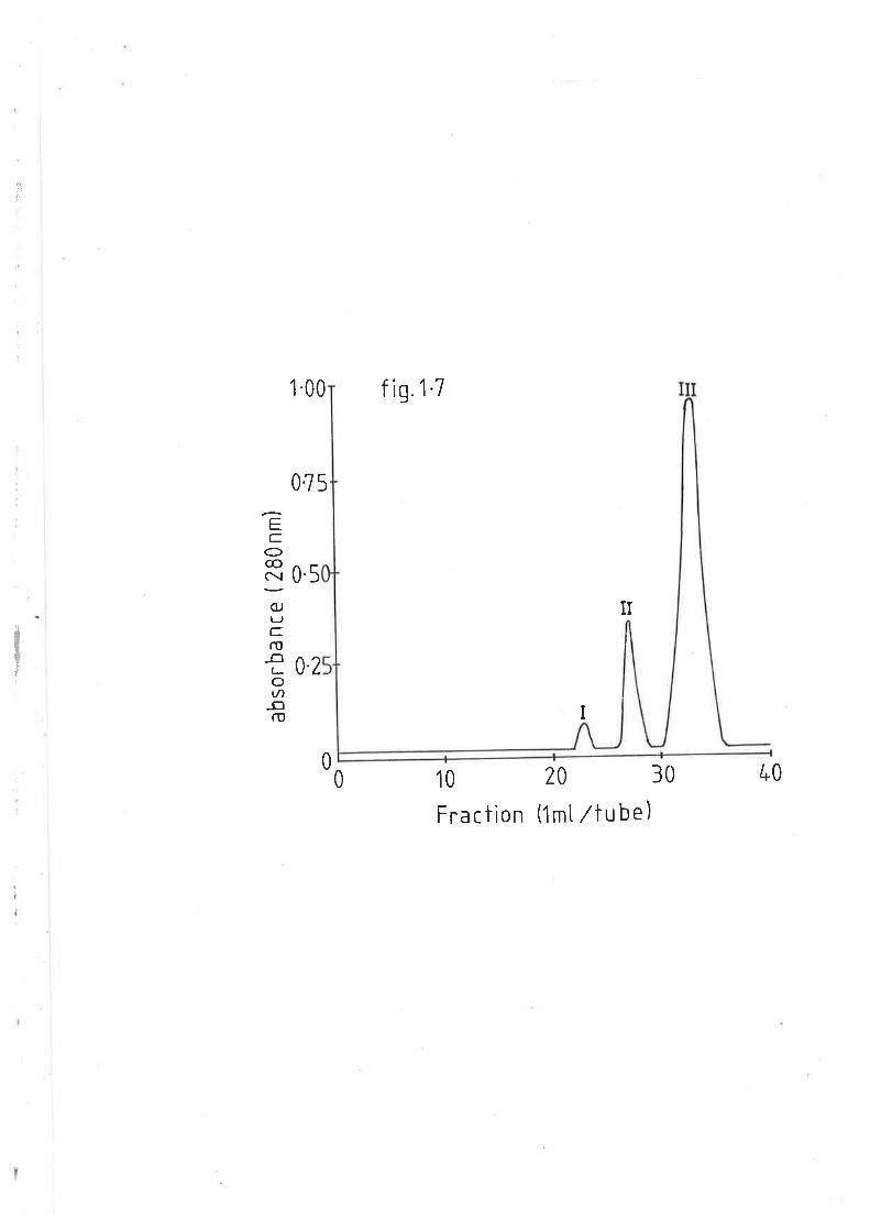
J,,
1-00 f ig.1'7
075
EC
O@:(UTJC.ru
_o(-ov)-oru
0.5
II
02
I
00 10
Fra c ti on
20 30
(1mt /tube)
40
dlll
t!
interpolating the reLention volumes of the lipoprotei-ns, with the
retenti-on volumes of proteins with lcrown molecular weights. Orrine VLDL
had an estimated molecular weight in the order of 117501000 dalüons. LDL
and HDL eluted at volumes v¡trich correspond to molecular weights of
approximately 8001000 and 3501000 daltons respectively.
L.3.7 SHEEP LIPOPROTEINS ISOIATED BY SERIAL T]LTRACENTRIFIJGATION
Serial centrifugation was also examined as a means of isolating
ovine VLDL, LDL, and HDL.
The lipoprotein fractions isolated by this Èechnique were eluted by
IIPLC and their electrophoretic mobility determined on agarose gels so as
to verify their identity. Fractions 1 (fractionl( 1.006g/ml), 2
(f .OOeg,/ml(fraction 2<L.O63g/m1) and 3 (1.0639/mLlfracLion 3(1 .2lOe/nL)
r,ùren eluted by HPLC gave rise to peaks with retent,ion volumes of 23rú,
27.5nL and 32.5mI respectively. These corresponded with bhe elution of
\iLDL, LDL, and HDL as earlier determined
HPI,C of the seríally isolated fractions indicated that there \,{as
little cross-contamination between groups. The VLDL fraction from
diabetic sheep showed less than 5% LDL and HDL components (determined on
an area percent absorbance of the total). Fraction 2 (tDL) from either
fed or diabetic sheep had on avêregêr less than 47" VLDL contamination
and the HDL fraction from either fed or diabetic sheep, had a combined
VLDL plus LDL content of approximately 3%.
Fract.ions 1, 2 and 3 yielded an expected electrophoretic mobility
pattern of pre-beta, beta and alpha respectively. However, the migration
of lipoproteins isolated from normal and alloxan diabetic aninals byI
il
69
rJlri
I
serial centrifugation differed (figure 1.8a-1.8c). In all instances,
VLDL, LDL and HDL from diabetic animals migrated further Lowards the
anode end than lhe equivalent fraction from fed animals. This was not
previously observed with the electrophoreti-c pattern of the lipoproteins
isolated by ge1 filtration (figures L.6a-L.6c). Other differences in the
nature of migration of the sheep lipoprotein fractions isolated by the
two techniques were observed. Ttre serial centrifugation HDL fraction was
considerably less homogeneous than the gel ehromatographed fraction as
indicated by the more diffuse banding. The migration of the slow and
fast components of HDL from fed sheep were reduced in the serially
isolated lipoproteins, as cornpared lo Ehe gel chromatographed fractions,
though this hras only obsenred in the slower component of HDL from
diabetic sheep.
1.3.8 CHEMICAL CHARACTRIZATIONS OF SHEEP LIPOPROTEINS
1.3.8.1 FED SHEEP
The major chemical corûponents of each of the classes of
Iipoproteins, namely VLDL, LDL and HDL for fed sheep is given in table
1.2. The VLDL were TAG rich \,rith 517. of the molecular cornplex being made
up of this lipid. The LDL were principally cornposed of cholesterol esLer
vilrilst. HDL were found to be essentialty of phospholipid composition.
Phospholipids were found to be the next major component of VLDL and LDL
respect.ively. Ckrolesterol, in all fractions was the smallest lipid
component, approximating 5"A of total lipoprotein lipids.
I
!
70
:
i
i
I
It,
TI
I
Figure 1.8
Figure 1.8 shows the electrophoretic
migration of (") very low density
lipoproteins, (b) low density lipoproteins
and (c) trigh density lipoproüeins isolated
by serial ultracentrifugation from fed and
diabetic sheep.
(a) I¿r¡es t, 2 and 6 represent very low
densíty lipoproteins from fed sheep. Lanes 3,
4 and 5 represent very low density
lipoproteins from diabetic sheep.
(b) Ianes 1, 2 and 3 represent lor^¡ density
lipoproLeins from fed sheep. lånes 4, 5 and 6
represent low density lipoproLeins from
diabetic sheep.
(") I-anes tr2 and 3 represent high density
lipoproteins from fed sheep. Lanes 4, 5 and 6
represent high density lipoproteins from
diabetic sheep.
Ili
7L
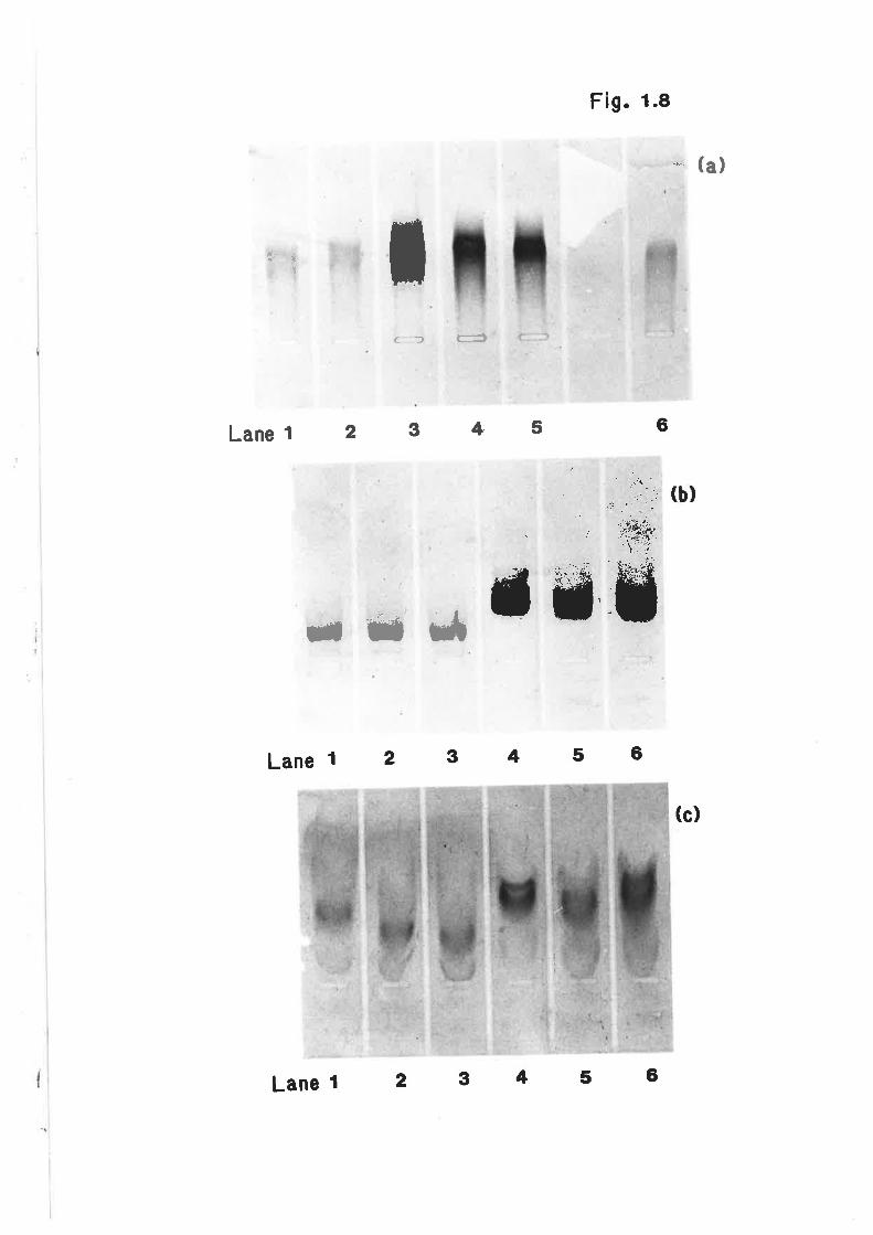
Fig. 1,8
6
(c)
;*'*
f F:,ï
'.¡: .,
rjc-)
". å,..
Lanel 2 3 4' 5
T*n5
Lanel 2 3 4 5 6
Lanel 2 3 4 5 6
Table 1.2
Table 1.2 lists the nnjor chemical components
of each of the lipoprotein classes from fed
and diabetic sheep. Very low density
lipoproteins were rich in triacylglyceride,
low density lipoproteins !ùere principally
composed of cholesterol ester and high
densíty Iípoproteins r¿trich were essentially
of phospLrolipid composíEion. The lipid and
proLein content of the lipoproteins differed
between treatments.
Itre results are for 3-6 anímals ín each treatment
72
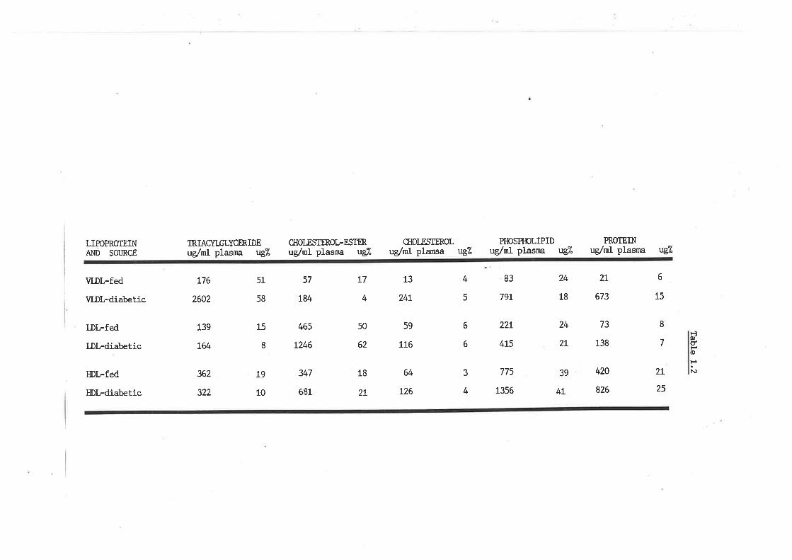
LIPOPRO1EINÆ.ID SOTJRCE
VLDL-fed
VLDL-diabetic
LDL-fed
LDl-diabetic
HDL-fed
HDl-diabetic
TRIACYI-GLYCERIDEug,/ml plasna u{/"
L76
2602
10
CIIOI^ESTROL-ESTERuglmI plasma É/"
CIIOLESTROLuglml plamsa u{A
PHOSPHOLIPIDuglml plasma vg?.
PROTE]Nug/m1 plasrna v{/"
2L
673
73
138
420
826
13
24t
59
116
64
t26
57
784
51
58
15
8
19
L39
t64
362
322
t7
4
50
62
18
2T
4
5
6
15
8
7
6
6
2l
25
24
18
24
2l
39
4L
83
79t
22r
4r5
465
t246
347
681
È0t-dtsoF¡.o3
4
775
1356
T.3.8.2 DIABE'TIC STIEEP
The chemical profile of the lipoproteins from alloxan diabet.ic
sheep differed considerably with respect to that frorn their fed
counterparts (tabte L.2). VLDL of diabetic sheep exhibited a greater TAG
content and pròtein content, Lhough a cholesterol ester content on1ry 257"
that of \ILDL from fed animals. Conversely, the diabetic LDL fraction had
a 507" lower TAG component and an elevated cholesterol ester component
v¡tren conrpa.red to LDL from fed sheep. The protein content re¡nained
unchanged. Similarly HDL from diabetic sheep had a TAG component only
hâtf as nnrch of that obserrzed in the same fraction from normal animals.
Ckrolesterol esters trere elevated to a lesser degree. The protein
cornponent in the HDL fracÈion from diabetic animals (257") may also have
been slightly elevated with respect to the equivalent fraction from fed
sheep (2IÐ.
L.3.9 PIASI.,IA LIPID PROFILE AND TI{E ROLE OF LIPOPROTEINS IN PTASMA
LIPID TRAI{SPORT
1.3.9.1 FED SHEEP
Table 1.3 sunrnarizes the plasma lipid profile and role of each of
the lipoprotein classes as mediators of plama lipids in fed and diabetic
sheep. In the former, phospholipids were the major plasma lipid G07").
TAG and cholesterol esters made up 2O%' and 357"i of the plasrna lipids
respectively, with cholesterol the other nrajor component (S7") .
VLDL, LDL, and HDL \^/ere responsible for the transport of LL7", 347"
73
Table 1.3
Table 1.3 sunrnarizes the plasma lipid profile
and role of each of the major llpoproteins
in transporting circulatory lipids in fed and
diabetic sheep.
Ihe resulÈs are for 3-6 anímals in each treatment.
Ttre molecular.weigþls used in the conversions are
TAG, 850 ;phospholipids, 750 ; choles terol ester, 651 ;
cholesÈeroLr42L.
74
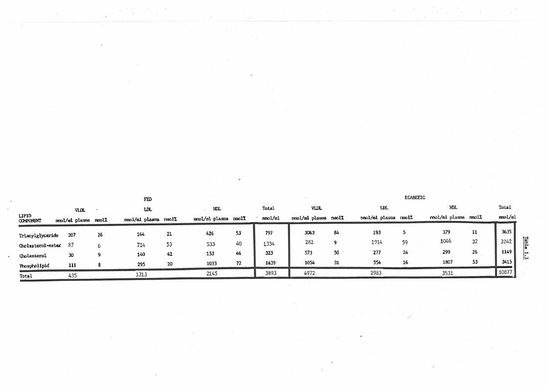
DIABEIICFU)
TDL
rmvdl plasoa rmllHDL
¡mVmt plaoa rroll
VIDL
r¡pVml plasna rollTotal
¡ræVoI
HDLTDL lotal
rmVolLIPIDCû.TPCNEITT
ll*æyþlyccrLde
CholesÈerol-ester
Cholesterol
Ihos$nlipld
Total
vuL
noVol plasa ræ11 n¡ol,/mt plasaa nrnlZ r¡rpI/ml ¡ræll
N7
ót
æ
111
435
26
6
9
I
t&7L4
140
295
1313
2L
53
42
20
426
533
153
1033
2145
53
@
46
72
tc
li3893
797
L334
323
1439
3063
282
573
1054
u9
50
31
193
L9t4
277
554
59
24
16
319
7046
299
1807
11
32
26
53
5
I+972 2983 3531
3ó35
3242
1149
3415
-to877
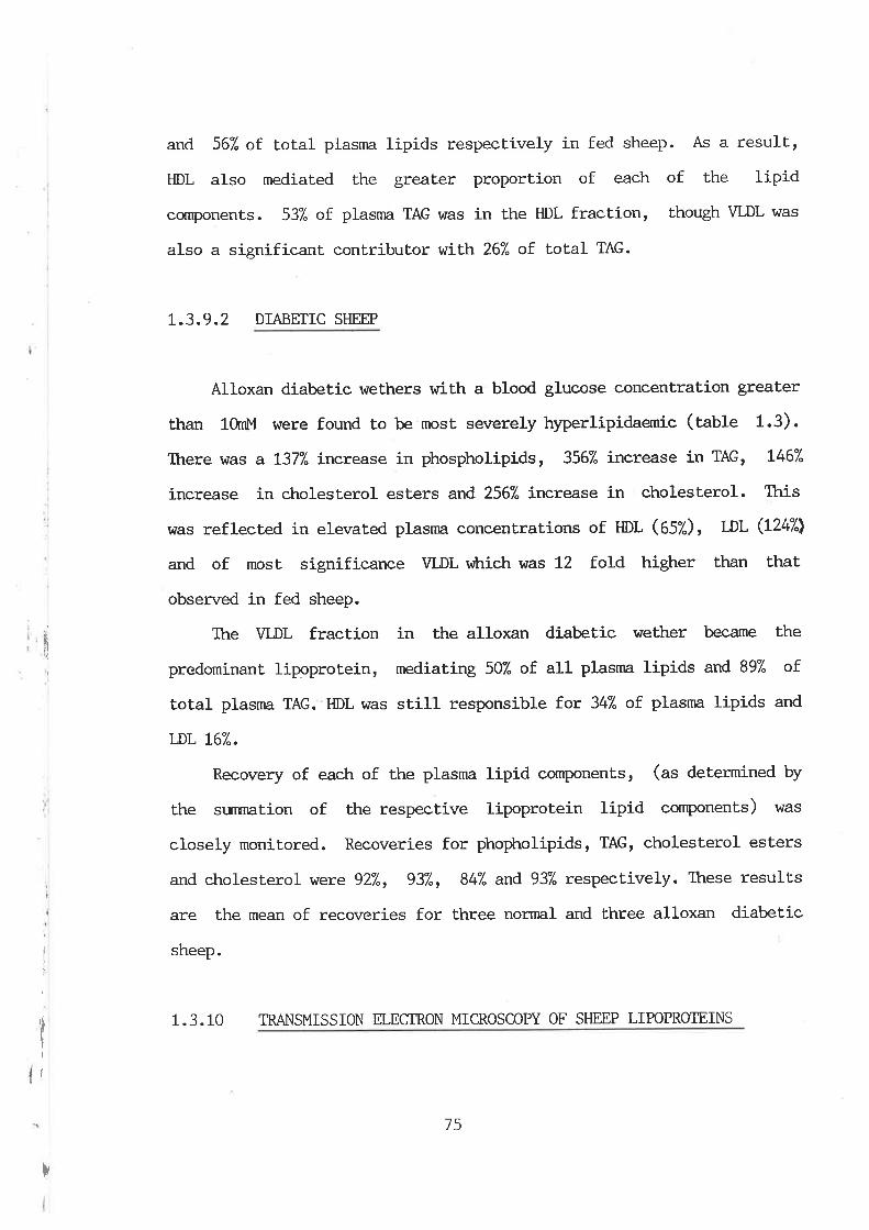
I
i
TI
i
and 56"/. of. total plasnra lipids respectively in fed sheep. As a result,
HDL also mediated the greater proportion of each of the lipid
cornponents. 537" of plasnn TAG was in the HDL fraction, though VLDL was
also a significant. contributor wiLh. 267" of total TAG.
L.3.9.2 DIABETIC SHEEP
Alloxan diabetic weÈhers with a blood glucose concentration greater
than 10rnl.,l were found to be most severely hyperlipidaemic (table 1.3).
There r{ras a L377" increase in phospholipids, 3567" incxease in TAG, L467"
i-ncrease in cholesterol esters and 256"/" increase in cholesterol. This
was refleeted in elevated plasnn concentrations of HDL (OSZ), DL (L247'l
and of most significance VLDL v¡trich was 12 fold higher than that
observed in fed sheep.
The VLDL fraction in the alloxan diabetic wether became the
predominant lipoprotein, mediaÈing 507" of. all plasma lipids and 89% of
total plasnra TAG. HDL was stifl responsible for 347" of. plasnn lipids and
T-DL L67".
Recovery of each of the plasma lipid cornponenLs, (as determined by
the sunnnation of the respective lipoprotein lipid components) I^/as
closely monitored. Recoveries for phopholipids, TAG, cholesterol esters
and cholesterol wexe 927", 937", 84% and 937" respectively. These resulLs
are the mean of recoveries for three normal and three alloxan diabetic
sheep.
1.3.10 TMNSMISSION ELEGTRON MICROSCOPY OF SFIEEP LIPOPROTEINS
r
75
Aliquots of sheep VLDL, LDL and HDL from both fed and diabetic
animals isolated by H.P.L.C. were viewed by Lransmission electron
microscopy. A mininmm of one hundred particles in each fraction v¡ere
approximated for their respective diameter based on an inlernal marker
of lanown díameter (3run), within the viewing chamber. Each particle was
grouped $dthin a 5nm classification for HDL and a lOnm group for LDL and
VLDL. Figure 1.9 shows the frequency of particle size in each of the
lipoprotein fractions from fed and diabetic animals. Figures 1.10a-1.10e
are representative micrographs of each of the major ovine lipoprotein
fractions. VLDL appeared as grey electron-transparent particles. Under
high magnification the finer strucLural details could be resolved.
Qualitatively in terms of size VLDL was the same in both fed and
diabetic animals (10-30nm), though quanLiLatively there \,rras a greater
frequency of snnller parLicles in the latter. Similarly, this trend was
obsen¡ed in the LDL component of fed and diabetic sheep plasma (fO-:Onm)
and viTas particularly notable in the HDL fraction of fed (5-20nrn) and
diabetic (S*n) animals. The LDL particles from fed animals appeared as
smooth sigmoidal particles, htËræs the equivalent fraction from diabetic
sheep e><hibited a rough surface, though these pa.rticles v/ere generally
spherical. HDL from both fed and diabetic aninnls \^Iere the most
homogeneous in terms of shape and appearance. Particles were spherieal,
but due to their small si-ze, differences in the nature of the particles
apart from size, \^ras difficult to determine.
76
Figure 1.9
Figure 1.9 shows Lhe size frequency of each
of the nnjor sheep plasma lipoprotein
fractions between fed and diabetic animals,
determined by transmission eleetron
microscopy.
77
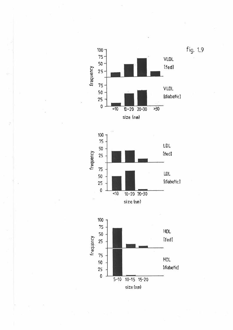
100
75
50
75
50
25
0
fis. 1.9
25L,C,(U
=trr(u(-
VLDL
lf edl
VLDL
[diabeticl
LDL
lfedl
LDL
ldiabeticl
HDL
lf edl
100
75
50
c¿>(U
g(U
.*L 7s
50
25
0
HDL
100
75
50!l ^?(U)çr(uLrF 75
s0
25
0
<10 1.20 20-30 >30
size (nm)
<10 10-20 20-30
size (nm)
5-10 10-15 15-?0
size (nm)
ldiabeticl
Figure 1.10a
Figure 1.10a represents negatively stained
very low density lipoproteins from fed sheep.
Particles r,rrere approximately 25-30nm.
top right - approxÍmate magnification 160,000
top left - approxirnate magnification 2501000
bottom figure - approximate magnification
10 x 250,000
78
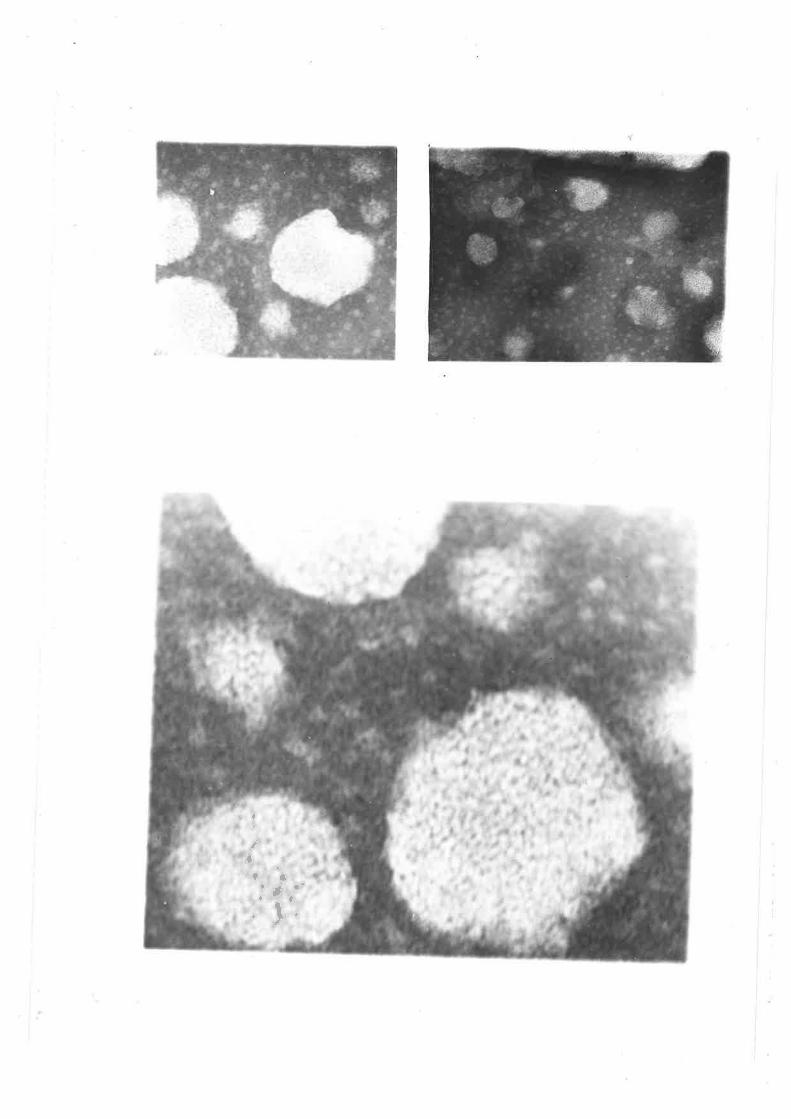
l
é*af'tt'
,' l'.
Figure 1.10b
Figure 1.10b represents negatively stained
very low densiLy lipoprotein pa.rticles from
diabetic sheep at 5 X 66'000 times
magnification. The majority of these
particles l^Iere approximately 10nm.
79
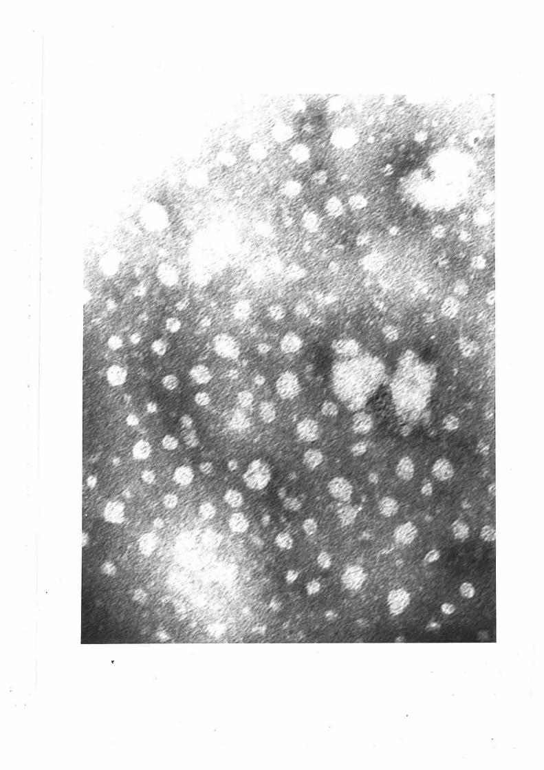
, :,:j"!..-À
,r ,l
',1
f
l
I
:
T:
'
Figure 1.10c
Figure 1.10c shows the low density
lipoproteins from both fed (top figure) and
diabetic sheep (bottom figure). These
particles from fed aninnls v¡ere aPProximately
10-20nm in size and synrnetrical ín shape. The
1ow density lipoproteins from diabetic sheep
had an approximate diameter of 1Orunr but in
conErast, their surface seemed less ordered.
top figure - approxímate magnification
10 x 250,000.
botÈom. figure - approximate nngnification
2 X 160,000
I
I
I
l
!
¡
i
III'
T'I
I
rI
80
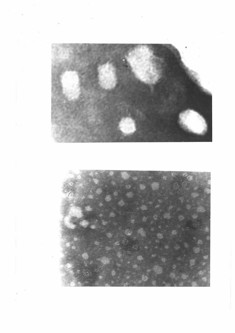
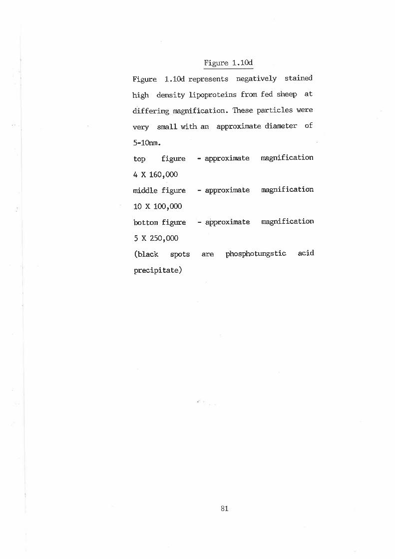
Figure 1.10d
Figure 1.10d represents negatively stained
high density lipoproteins from fed sheep at
differing nagnification. Tttese particles were
very srnall r,trith an approxinnte diameter of
5-10nm.
top figure - approximate magnification
4 X 160,000
middle figure - approxinate magnification
10 x 100,000
bottom figure - approximate magnification
5 X 250,000
(black spots are phosphotungstic acid
precípitate)
81
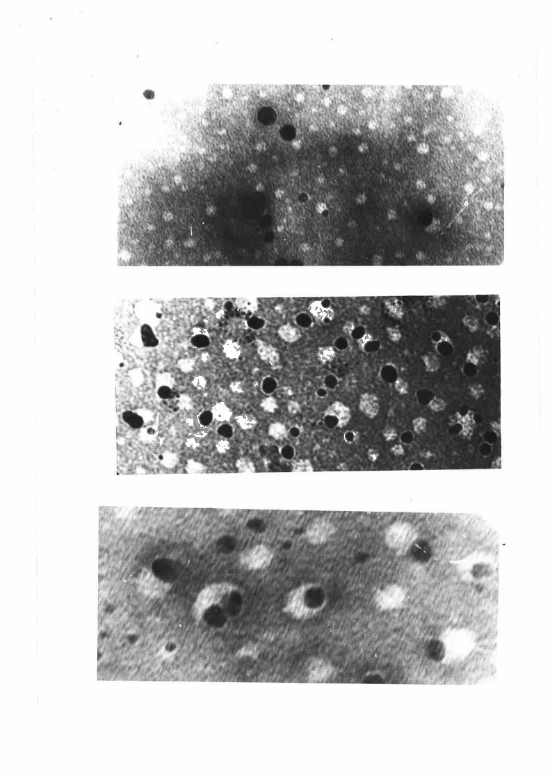
t
*F'.'..
t
It &a l )t
t ?\ql*t.
Ia ItË' It Ët-{F II
l|¡
'.t
l'
r{
'-*Jfb..
t
T (
T
dt..
P
f'
4lsi<,'
1t
lL
I
Figure 1.10e
Figure 1.10e shows negatively stained high
density lipoproteins from diabetic sheep'
Particles l^rere very synrnetrical rtrith an
approximate diameter of onlY 5nm.
top figure - approximate nngnification
13o,o0o
bottom figure - approximate rnagnification
5 X 130,000
(btack spots are phosphotungstic acid
precipitate)
I
r{
I
I
I
l
I
I
I
II
I
I
l
I
i
82
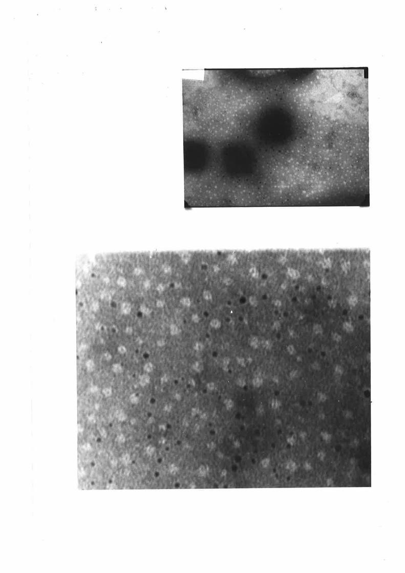
L.4 DISCUSSION
The total lipid composition of sheep plasma has been studied often
(Garton and Duncan L964, I-eaL L967, Nelson 7969), but the contribution
of the plasma lipoproteins to circulatory fats has received littleattention. All published data, either on vihole serum, plasma, or
isolated lipoproteins, agree that the Èotal lipid is very low vitren
compared to monogastric onnivores such as the rat or human. Nelson
(L973) has reported thât low levels of circulatory TAG are reflecLed ín
the virtual absence of VLDL, lrrith less Lhan 0.27" of plasma lipids in
this fraction. Similarly, l€at et al. (t976) attributed the contrib¡tion
of VI-DL mediating plasma lipids as being less than 57.. However, sheep
metabolically stressed as a result of fasting or diabetes are noL unlike
the rnonogastric species in that they exhibit accute hyperlipidaemia. tle
have previously s-hov¡n thât the elevation of circulatory TAG was in part
attributable to an increased hepatic release of this lipid (¡,ømo et. al.1983). Nonetheless TAG rapidly accunmlate in the liver of these animals,
suggesting that slmthesis far outweighs secretion. In view of this, the
first parL of this study had several objectives. The precise nature of
sheep plasma lipoprotêins re¡nained unclear and so initially, this thesis
was aimed at determining vitrether the parameters of size, electrophoretic
mobility and density by vihich human plasma lipoproteins are most
conrnonly isolated, can be applied to sheep. Having established the
appropriate methodologies, quantitative and qualitative analysis of
these particles in fed animals and their role in the
hypertriacylglyceridemia in diabetic sheep was determined.
Lipoproteins are prone to rapid physiochemical degradation and as
H
t
!
ð-1
such require expeditíous isolation, analysis and storage under strictly
controlled conditions. Degeneration or modification of their cornposit.ion
and structure, results through the act.ivity of several endogenous plasma
enzymes. The best characterized of these are lecithin cholesterol acyl
transferase (I,CAT) and lipoprotein lipase. To minimize the effects of
these enzymes, Ellnanrs reagent to irùribit LCAT, and phenylmethyl-
sulphonyl fluoride, to irùribit proteolyLic enzyrnes \,\tere added. Azide and
thimerosol were also included as bacteriocides, the latter having the
additional merit of irùribiting lipoprotein lipase (lee fgZA).
It has been lanoqrn for many years that lipoproteins are susceptible
to oxidative degradation (nay et al. 1954), catalyzed by heavy metals
(Scfrm et al. t978). Oxidation vras minimized by the addition of disodium
ethylenediamine tetraacetate to sequester heavy metal catalysts. Ïn
addition, isolated lipoprotein fractions r{rere nnintained at low
temperatures in chromic acid washed glassware, under high purity
nitrogen and in the dark. Analysis of lipoproteins for vùrich precautions
for lheir preservation have not been strictly adhered to, may be
considered as a futile exercise.
l'4any of the physical principles by vùrich lipoproteins are isolated
nay also bring about significant changes to their structure and
composition. For example, the quantitative loss of HDL apoprotein
subunits by prolonged ullracentrifugation has been well documented
(Scanu and Granda 1966), hrt little is lanown of the effects of high
pressure filtration on the lipoprotein molecules, vihen chronntographed
by HPIC. Thus, the process of isolating plasma lipoproteins is dependent
on the nature of an intended study. The techniques used, are dictated by
the balance of quantitative and qualitative recovery of the particles.
B4
Preliminary investigations in this laboratory in isolaLing bovine
lipoproteins at. a solvent density of 1.225g/nL, showed thåt a 24h-
ultracentrifugation Lime was sufficient to isolate all of the plasnra
lipoproteins. Ttris r^/as a substantial decrease compared with the 40h
required to float the plasnn lipoprotej-ns from rabbit, rhesus monkey or
humans under the same conditions (Rudel et aI. 1974). rn order toascertain the mininn:m period of ultracentrifugation required to achieve
full recovery of ovine plasma lipoproteins, the rate of migraÈion of
sudan black stained lipoproteins rrras monitored at..selected time
intervals. The results showed that to reduce ultracentrifugation time of
the lipoproteins, could only be achieved at. the expense of recovery.
Elution of the sheep tdL.225 lipoprotein concentrater through
agarose (5U) sizeing colunns gave rise to a profile, vrtrich qualitatively
lrlas similar to thât obsen¡ed for humans. Ttre generally smaller
absorbance profile of the ovine lipoproteins (per unit plasna),
suggested that this species had lower levels of total particles, vrtrich
was expected, in view of their low plasma lipid concentration. Ttre mean
concentration of plasma lipoproteins in adult hunnn males isapproxímately 8.5mg,/mf (Uatctr and Lees 1963), somevitrat higher tt¡,an the
ovine plasna concentration of 3.1mg/ml obtaíned in this study for fed
sheep. The sheep tdL.225 lipoproteinr elution profile also suggested
that although HDL was the nain ovine plasma lipoprotein, VLDL was also a
nnjor component of this speetrum (peak r, figure t.4), particularly ifone considers that due to Èhe low protein content of these particles,
absorbance at 28onm is low. The elution profile of the total sheep
plasma lipoproteins from fasted and diabetic aninals suggested that allof the major lipoprotein fractions had been elevated with respect to
85
their nornnl fed counterparts, although it. would appear that HDL \^/ere
the principal element of this increase.
Analysis of sheep plasna by agarose gel electrophoresis (figure
1.6) showed only two bands corresponding to beLa-protein (f,DI-) and
alpha-protein (mf) stained for lipid. The pre-beta band, v¡hich l,r7as a
characteristic of hunnn plasna, was not detected in adult sheep plasnn,
due principally to the overlap of the beta and alpha cornponents. The
electrophoretic mobility of the individual sheep lipoprotein fractions
(namely \ILDL, LDL and HDL) has not been previously reported. However,
electrophoretograms of peaks L, 2 and 3 from agarose gel filtration and
similarly that of fractions 1, 2 and 3 from serial ultracentrifugation
yietded bands v¡trich stained for lipid in the pre-beta (Vf¡I.), beta (t¡l)
and alpha (mI.) regions respectively. Ttreir migration clearly indicated
that VLDL, as well as the other rnajor classes of lipóproteins, I{rere
indeed present in sheep plasnn, and therefore in terms of mobility, like
that found in human plasma.
The tailing of the ÌúI-DL fractions towards the beta region and the
streaking of the IÐL fraction towards the pre-beta region, observed in
both human and ovine fractions (figures 1.5 and 1.6), reflects the
association of the two Iípoprotein classes. IÐL is formed by the process
of VLDL catabolism by lipoprotein lipase. Thus at any one time, plasma
will contain a heterogeneous mixture of partially metabolized VLDL (ot
intermediate density lipoprotein). Ovine LDL had a slight,ly fasLer
migration than human LDL, suggest.ing that sheep have a greater amount of
this lipoprotein of snraller size. Like humans, sheep HDL had two
distinct bands. The slower migrating fraction is HDL1, a subclass of the
HDL fraction, vùrich is arguably a low density lipoprotein, lhough by
86
tradit.ion is described as an HDL. The difference in electrophoret.ic
mobility of the faster HDL component between the two species, reiterates
the heterogeneity usually attributed to this lipoprotein class.
The electrophoretic patterns of ovine lipoproteins isolaLed by
either gel chromatography or seríal ultracentrifugation, yielded
Iipoprotein fractions r,*rich differed slightly in their rate or nature of
migration. These differenees may simply be a reflection of the mode of
plasma lipoprotein isolation, or in the stressed sheep, a response to
elevated levels of plasnn lipoproteins, or modification of the
lipoprotein particles. Nonetheless, from the reduced rnobility of VLDL
from fasted sheep and Èhe two overlapping components observed in
diabetic \ILDL isolated by gel filtration (figure 1.6), it appears that
there rnay be an accurrulation of a less dense tDL fraction, namely
inLermediate density lipoprotein. This would suggest that there could
exist a defect, in the catabolic processes of \ILDL metabolism in fasted
and alloxan diabetic sheep.
The migraLion of both cornponents of HDL from fed sheep, was reduced
in the serially isolated lipoproteins, as compared to Lhe gel
chronntographed fractions (figures 1.6 and 1.8). This was only obsenred
in the slower cornponent of HDL from diabetic sheep (figure 1.8c). In
addition, the banding of the serially isolated HDL lipoproteins r^rere
diffuse in comparison with the chromatographed HDL lipoproteins. It is
difficult to interpret these differences, however, the HDL fractions
from serially ultracentrifuged plasma may have undergone ctr,anges in
their physical characteristics, due to the prolonged ultracentrifugatíon
time required to isolate VLDL, LDL and finally HDL.
A very high density lipoprotein fraction \^/as found only on
87
occassions in fed wethers and ewes and was absent in the plasma of
fasted or alloxan diabetic sheep. As a result, for the purpose of this
study it. \4las not considered a major ovine lipoprotein and thus not
investigated further. GeI electrophoresis of VHDL yielded two distinct
bands (figure 1.6a), the slower corresponding to beta-protein and the
faster a little greater than the electrophoretic mobility of ovine HDL.
Agarose getr electrophoresis of diabetic sheep lipoproteins isolaÈed
by serial centrifugation migrated furÈher towards the anode conpared to
native VLDL, LDL and HDL. This is a characterístic of proteins v¡trich
have been glucosylated, in this instance as a result of the
hyperglycaemía associated with alloxan diabetes ín these animals.
HPI,C has only recently been utilized as a tool for sepa.rating lhe
major classes of lipoproteins. It holds particular promise in vastly
reducing the tíme usually required to achieve separation and hence
possibly particle degradation. The nnjor ovine lipoproteins in the
'd1.225 lipoproLein concentrate' were successfully sepa.rated by HPLC
within 35min, as compared to 16-18h by agarose gel filtration and 48-72h
by serial centrifugation.
HPLC analysis of the lipoprotein fractions isolated by agarose gel
filtration or serial ultracentrifugaLion detected a small degree (less
than 57") of cross-contamination in fractions L, 2 and 3 from the latter.As with the lipoproteins isolated by agarose chromatographyr HPLC
lipoproLein fractions were homogeneously distinct, with no overlap of
components. Another advantage of HPLC was the capacity to load, separate
and detect small quantities of lipoproteins, vfrrich should prove
particularly useful to the study of rumj-nant lipoproteins vùrose plasma
Iipoprotein content is low. However, the response of the FIPLC detector
88
lrras noL proport.ional to the concentration of plasma lipoproteins and in
particular, due to the extremely low protein content of VLDL, the
absorbance of this fraction \^ras poor. In the advent of improved
speclrophotometersr or by prestaining lipoproteins so that deLection is
not protein dependent (Busbee et al. 1931), this aspect should be
overcome. In addition, it was extremely difficult to obtain enough
individual lipoprotein rnaterial, without pooling equivalent fractions
from several elutions, to either chemically characterize or nnke subject
to electrophoresis, and thus its current application is somevrtnt
Iimited. Recently, t{PlC colunr¡s with the ability to quantitatively
determine all nnjor lipid cornponents (vùren used in conjunction with
light scattering detectors), have become conrnercially available.
An approximate rnolecular weight of each of the major ovine
lipoproteins was determined by referring the HPLC elution volume of each
of the lipoproteins, to the elution volume of proteins with lsrown
molecular weights. Sheep VIÐL, LDL and HDL had molecular weights
cornparíÈive Èo that for the equivalent fractions in manr that is
approximaueLy 2 million, 8001000 thousand and 3501000 thousand daltons
respectively.
Both gel filtrat.ion and serial ultracentrifugation yielded
homogeneously distinct fractions of each of the major classes of ovine
lipoproteins, with little or no overlap of components, as de¡nonstrated
by the respective electrophoreti.grams and HPLC elution profiles.
However, gel chromatographed lipoprotein fractions v¡ere very diluLe and
required concentrating prior to further analyses. Freeze drying, reverse
dialysis and pressure filtration \4rere investigated as means of
concentrating lipoprotein sarnples. Although qualitative recovery
89
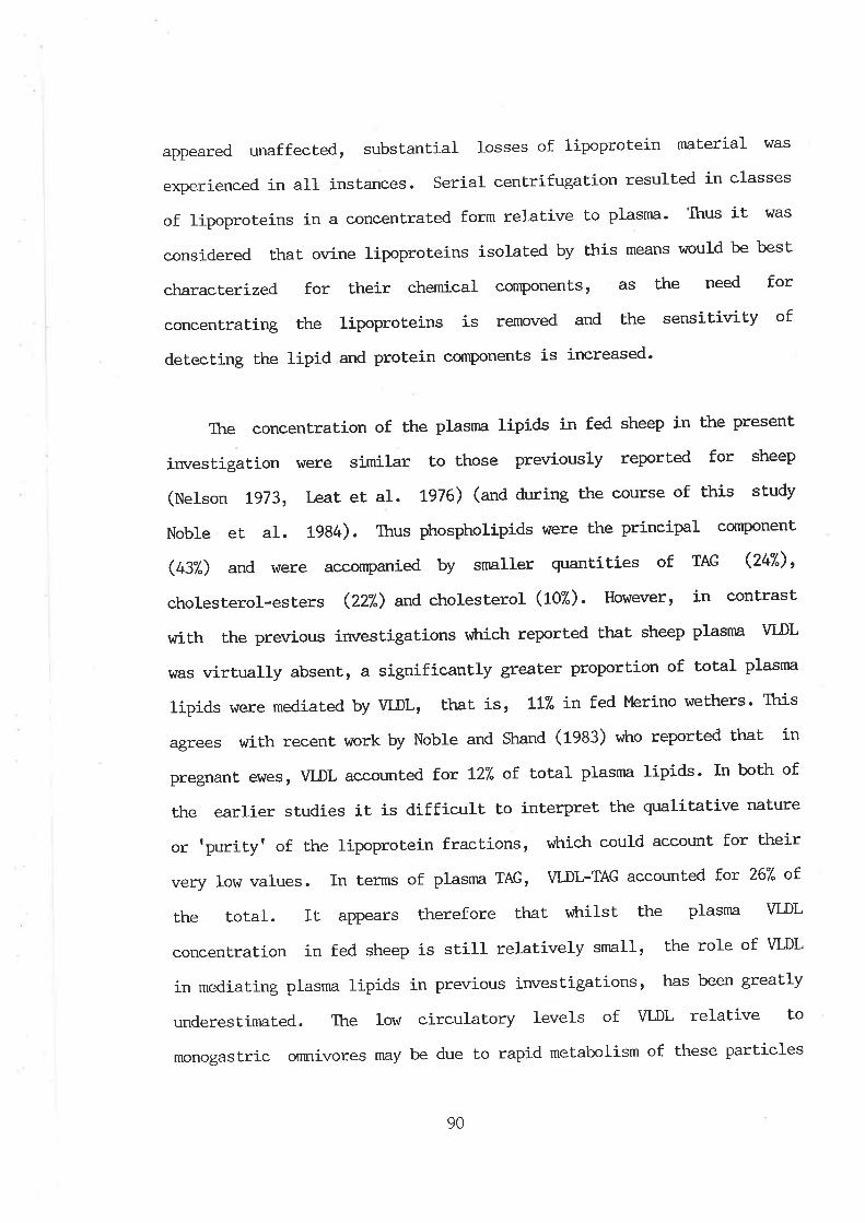
appeared unaffected, substantial losses of lipoprotein nraterial I4Ias
experienced in all instances. Serial centrifugation resulted in classes
of lipoprotej-ns in a concentrated form relative to plasnn. Thus it [^Ias
considered that ovine lipoproteins isolated by this means v¡ould be best
characterized for their chemical components, as Lhe need for
concentrating the lipoproLeins is removed and the sensitivity of
detecting the lipid and protein components is increased.
Ttre concentration of the plasrna lipids in fed sheep in the present
investigation vtere similar to those previously reported for sheep
(Nelson tg73, Leat et aI. tg76) (and during the course of this study
Noble et al. 1984). Ttrus phospholipids were the principal cornponent
(437") and \^¡ere accompanied by srnaller quantities of TAG (247"),
cholesterol-esters (22Ð and cholesterol (10Ð. However, ín contrast
hrith the previous investigations v*rich reported that sheep plasrna VLDL
was virtually absent, a signíficantty greater proportion of total plasrna
lipids were mediated by VLDLr that is , tl(" in fed Merino wethers' This
agrees with recent work by Noble and Shand (1933) viLro reported that in
pregnant e\^res, VLDL accounted fot t27" of total plasma lipids' In both of
the earlier studies it is difficult to interpret the qualitative nature
or 'purity' of Lhe lipoprotein fractions, vrhich could account for their
very low values. In terms of plasnra TAG, VI¡L-TAG accounted Í'or 267" of
the total. It appears therefore that r,ùrilst the plasma VLDL
concentration in fed sheep is still relatively small, the role of VLDL
in mediating plasma lipids in previous investigations, has been greatly
underestimaled. Ttre Iow circulatory levels of VLDL relative to
monogastric omnivores may be due to rapid metabolism of these particles
90
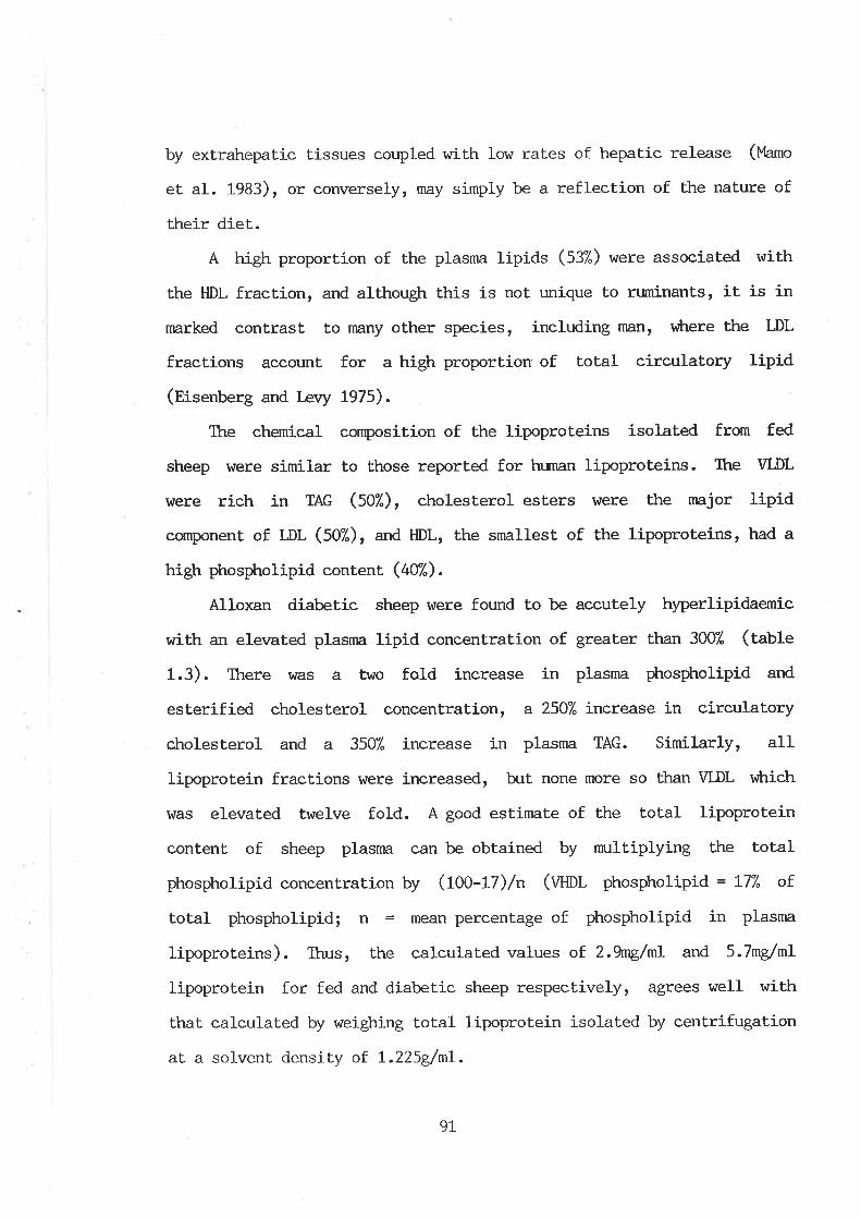
by extrahepatic tissues coupled with low rates of hepat.ic release (Uamo
et. al. 1983), or converselyr my simply be a reflection of the naLure of
their diet.
A high proportion of the plasma llpids (537") r^rere associated with
the HDL fraction, and althougþ this is not unique to ruminants, it is in
marked contrast to nnny other species, including man, l'¡trere the LDL
fractions account for a high proportion of Lotal circulatory lipid
(Eisenberg and Levy t975).
The chemical composition of the lipoproteins isolated from fed
sheep were similar to those reported for human lipoproteins. Ihe VLDL
\^rere rich in TAG (507.), cholesterol esters l,rtere the major lipid
component of LDL (507"), and HDL, the smallest of the lipoproteins, had a
high phospholipid content (407").
Alloxan diabetic sheep were found to be accutely hyperlipidae¡nic
with an elevated plasnn lipid concentration of greater than 3007. (table
1.3). There hras a two fold increase in plasma phospholipid and
esterified cholesterol concentration, a 2507" increase in circulatory
cLrolesterol and a 3507" i-ncrease in plasma TAG. Similarly, all
lipoprotein fractions hrere increased, ht none more so than VLDL vùrich
r4ras elevated twelve fold. A good estimate of the total lipoprotein
content of sheep plasma can be obtained by nmltiplying the total
phospholipid concentration by (tOO-tl)/n (Wnr phospholipid = L77" of
total phospholipid; n = mean percentage of phospholipid in plasma
lipoproteins). Thus, the calculated values of 2.9ng/nL and 5.7ng/nL
Iipoprotein for fed and diabetic sheep respectively, agrees well with
that calculated by weighing total lipoprotein isolated by centrifugation
at a solvent density oî L.225g/nL.
9L
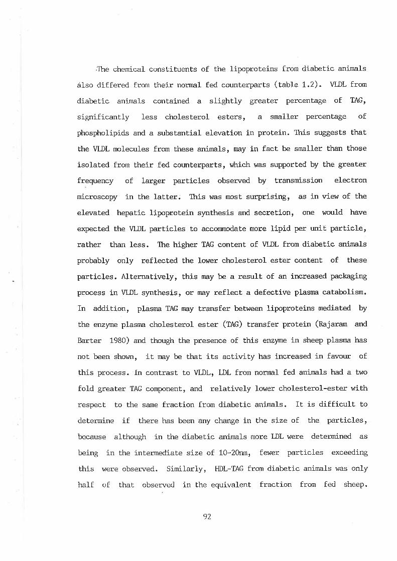
.The chemícal constituent,s of the lipoproteins from diabetic animals
álso differed from their normal fed counLerparts (table t.2). VLDL from
diabetic animals contained a slightly greater percenLage of TAG,
significant.ly less cholesterol esLers, a smaller percentage of
phospholipids and a substantial elevaLion in protein. This suggests that
the VLDL molecules from these animals, n:ø.y in fact be smaller than those
isolated from their fed counterparts, vrtrich was supported by the greater
frequency of larger particles obsen¡ed by transmission electron
microscopy in the latter. Ttris was most surprising, as in view of the
elevated hepatic lipoprotein synthesis and secret,ion, one would have
expected the VLDL particles to acconmodate more lipid per unit particle,
rather than less. The higher TAG content of VLDL from diabetic animals
probably only reflecLed the lower cholesterol ester content of lhese
particles. Alternatively, this may be a result of an increased packaging
process in VLDL synthesis, or rnay reflect a defective plasma catabolism.
In addition, plasma TAG nay transfer between lipoproteins mediated by
the enzyme plasma cholesterol ester (TAG) transfer protein (najaram and
Barter 1980) and though the presence of this enzyme in sheep plasma has
not been shown, it may be that its activity has increased in favour of
this process. In contrast to VLDL, L,DL from norrnal fed animals had a two
fold greater TAG component, and relatively lower cholesterol-ester with
respect to Lhe same fraction from diabetic animals. It is difficult to
determine if there has been any ctr,ange in the size of the particles,
because although in the diabetic aninrals more LDL were determined as
being in Lhe intermediate si-ze of 1-0-20r¡rn, fewer particles exceeding
this were observed. Similarly, HDL-TAG from diabetic animals was only
half of that observed in the equivalent fraction from fed sheep.
92
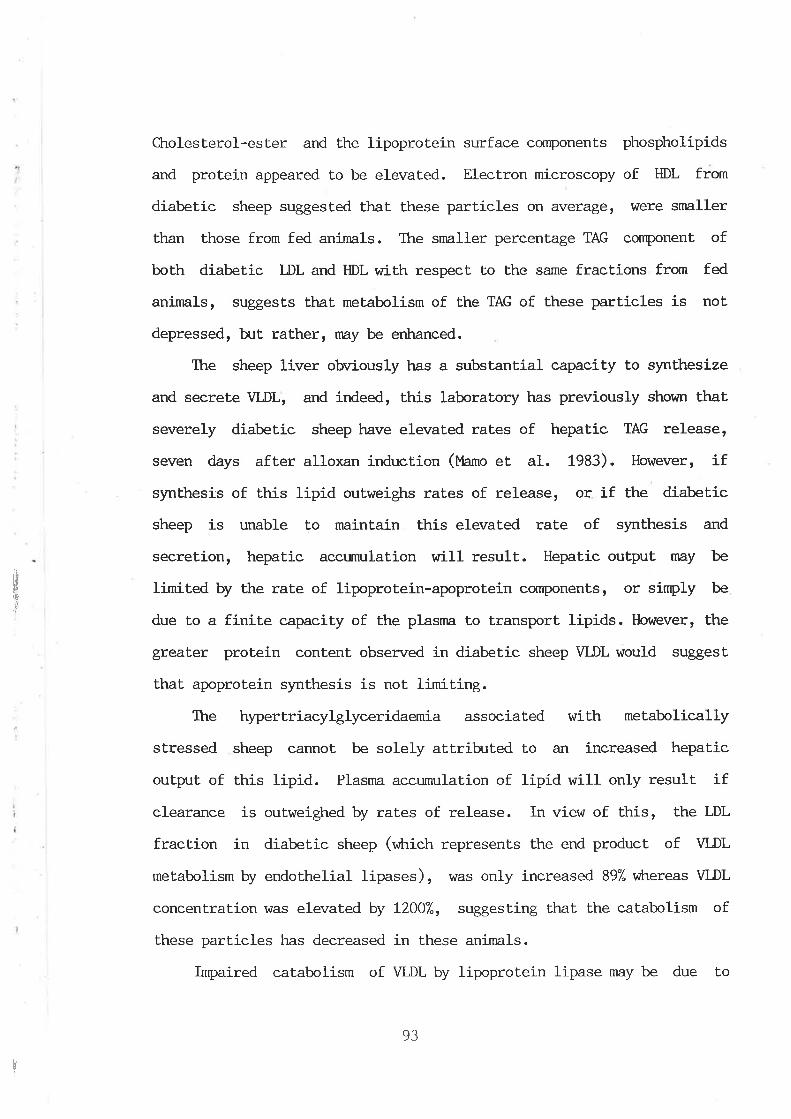
rin
'üI
Cholesterol-ester and the lipoprotein surface components phospholipids
and protein appeared to be elevated. Electron microscopy of HDL from
diabetic sheep suggested that these particles on average, were smaller
than those from fed animals. The snnller percentage TAG component of
both diabetic LDL and HDL with respect to the same fractions from fed
animals, suggests that metabolism of the TAG of these particles is not
depressed, but. rather, rnay be erh,anced.
Itre sheep liver obviously has a substantial capacity to synthesize
and secrete VLDL, and indeed, this laboratory has previously shown that
severely diabetic sheep have elevated rates of hepatic TAG release,
seven days after alloxan induction (I4amo et al. 1983). However, if
synthesis of this lipid outweigþs rates of release, or if the diabetíc
sheep is unable to maintain this elevated rate of synÈhesis and
secretion, hepatic accumulation will result. Hepatic output may be
limited by the rate of lipoprotein-apoprotein conrponents, or simply be
due to a finite capacity of the plasma to transport lipids. However, the
greater protein content obsenzed in diabetic sheep VLDL would suggest
that apoprotein synthesis is not limiting.
The hypertriacylglyceridae-rnia associated with metabolically
stressed sheep cannot be solely attributed to an increased hepatic
output of this lipid. Plasma accumulation of lipid will only result if
clearance is oulweighed by raLes of release. In view of this, the LDL
fraction in diabetic sheep (v¡:rictr represenLs the end product of VLDL
metabolism by endothelial lipases), \^ras only increased 897. vùrereas VLDL
concentration was elevated by L2OO7", suggest.ing that the catabolism of
these particles has decreased in these animals.
Impaired catabolism of VLDL by lipoprotein lipase may be due to
!
93
al
¡
reduced production and,/or act.ivity of the enzyme (chapter two) or a
physical or chemical modification of the substrate viLrich could prevent
binding and subsequent hydrolysis of the TAG. Ttre nature of the
irnpairment in the lat.ter nay be related to quantitative changes in the
apolipoprotein composition. Bar-On et al. (1976), reported thaL VLDL of
diabetic rats showed differences in their apoprotein C composition
(v¡ricfr aclivaLe/irhibit lipoprotein lipase) cornpared to WDL of non-
diabetic animals. More recently, WDL of diabetic origin were shown to
be deficient in apoprotein E (Bar-On et al. 1984). This apoprotein is
thought to play a role in the recognition of Lhe WDL particles or their
re¡rnants by peripheral tissues and liver (Innerarity and l"lahley 1978,
Shelb¡rne et al. 1980 and tlindler et al. 1980). In the latter study,
\ILDL isolated from the plasnn of diabetic rats and reinjected into
normal recipients had a significantly higher half life than the
corresponding VLDL of non-diabetic rats.
VLDL catabolism rnay also be impaired as a result of structural
alterations in the protein moiety brought about by increased
glucosylation of the lipoproteins in diabetes (Gonen et al. L98L,
Schleicher et al. 1981 and t{it.zum et al. L982). C,urtiss and [Jitzum
(f0aS¡ have demonstrated lhe non-enzymatic post-translational
glucosylation of the free amine of lysine residues of plasma
lipoproteins. They found that the majority of glucosylated proteins in
the lipoprotein fraction of density less than L.225dnL in
hyperglycaemic subject.s was in the TAG rich lipoproteins. In diabetic
subjects, apoproleins AI, AII, B, CI and E were all glucosylated. A
nurnber of studies (Gonen et al. 1981, Sasaki and Cottam L982arb and
Witzum et al. L982) have shown that extensive glucosylation of LDL
HI
tI
;
ï
94
.I¡ttf,
III
i
apoprotein B (greater than 407" of lysine residues) totally abolishes the
ability of LDL to be recognized by the LDL receptor.
There is evidence that the lysine residues of the various
apoproteins are required for various functional activities, including
receptor recognition (Weisgraber et al. 1978 and l4ahley et al. L979) t
enzyme interaction and activation (M:stíner et aI. L979 and Vainio et
al. 1983), lipid binding (Sparrow and C,otto L982) and the regulation of
cellular proliferation (NoeI et al. 1981). Thus glucosylation of the
various apoproteins could have a profound influence on the function of
that apoprotein.
Yamanroto et al. (1986) reported a significant reduction in the rate
of binding and degradation of glucosylated \II,DL in hunnn skin
fibroblasts compared to native VLDL. They srtggested that glucosylation
of apoprotein E results in the irnpairment of the receptor binding
capacity. This study also showed that glucosylated \ILDL on agarose gel
electrophoresis migrated further towards the anode, compared to native
VLDL. Similarly, âgarose gel electrophoresis of sheep lipoprotein
fractions from diabetic animals appeared to be glucosylated (figures
1.8a-1.8c) .
Finally, elevated plasma VLDL levels nny also be due to defective
hepat.ic VLDL synthesis in the firsl instance. Berry et al. (1981) showed
that severe insulin deficiency increased hexosamine incorpoiation into
VLDL in the perfused liver system.
The first part. of this study has shown that normal fed sheep
possess a lipoprotein compliment similar to that seen for other species,
in that all of the major lipoprotein fract.ions, namely VLDL, LDL and HDL
r
95
ü'8tf
are present. Previous investigations in this laboratory have also shown
that. sheep respond to metabolic stress (as a result of diabetes), by
increasing the slmthesis and release of lipoproteins. This has been
extended here, in that the nature of the hyperlipidaemia associated with
this i-ncrease has been deLermined and is reflected principally in the
\1LDL-TAG lipoprotein fraction. Ihe plasna eornpartment has a finite
capacity to transport TAG (and indeed all lipids) vùrich is regulated noÈ
only by secretion of these pa.rticles, ht also rates of clearance. Itre
catabolism of VLDL is generatly attríbuLed to two enzymes, lipoprotein
lipa.se and hepatic lipase and it may be that the activity of one or both
of these enzymes has diminished in diabetic sheep. In view of this, the
second part of this thesis was aimed at examining the catabolism of
\¡LDL-TAG by these enzymes in fed, diabetic and fasted sheep.
tII
I
r
96
rÌ
j
CHAPTER 2
2.L.L IMRODUCTION
In chapter one iL was shov¡n that the metabolic stress of diabetes
produced a substantial rise in sheep plasma lipids. ltris increase hlas
not uniform in all lipoproteins, but rather, there htas a
disproportionate elevation in VLDL-TAG, vihich is a reflection of Ehe
increased secretion of these particles from Ehe liver (rcmo et aI.
1933). However, the steady state concentration of plasma TAG, is also
critically regulated by the lipolylic rate of the tissues v¡hich utilize
TAG-fatty acids and therefore, this process has important implicationst
in terms of the hypertriacylglyceridaemia and hepatià accumulation of
this lipid seen in these animals. In man and other monogastric species
thus f.ax studied, there are essentially two enzymes involved in the
catabolism of circulating VLDL-TAG, namely lipoprotein lipase (fpl,) and
hepatic lipase (til-). LPL has been isolated in sheep and its biochemical
characterisLics determined. In swunary, it was found to be not unlike
that reported for other species (Ctegg 1981b and Vernon 1981). However,
there have only been few conrnunications of investigations concerned with
activity of this enzyrne in sheep under stressed conditions, namely
pregnancy and lactation (Vernon et al. 1980, 1981 and Smith and l,,lalsh
1934). Orrine HL has noL been previously reported and so the role of both
enzymes in the metabolism of VLDL-TAG in metabolically stressed sheep
are at present unlmown.
The second part of this project had several objectives, vùrich could
be divided into essentially biochemical and animal production related
I
97
aspects of TAG metabolism. Init.ially, the first part of this study \,tlas
aimed at establishing the presence of HL in sheep and thereafter, to
determine changes in the rates of TAG hydrolase activity of both LPL
and HL in fed, fasted and diabetic animals and correlate these with
differences in the plasma lipoprotein lipid profile. During the course
of these investigations, il became clear that such activities were
regulated by both steroidal and genetic factors and so the second part
of the results presented in this chapter are concerned with lipase
act,ivities in rams, castrates, el,ites and genetically tlean' and tobese'
sheep. The latter part of this sLudy is discussed in view of local
aninnl husbandary practices.
2.I.2 LIPOPROTEIN LIPASE AND HEPATIC LIPASE
LPL and HL are inrportant regulators of plasrna lipoprotein
concentrations and therefore are implicated in related disease states in
rnan such as atherogenesis. As such, both enzymes have enjoyed extensj-ve
investigation in monogastric animals. Some recent reviews of LPL and HL,
vÈrich emphasize various aspects of their synthesis, activity, mode of
regulation and metabolic significance, are listed (Robinson 1970, Scow
et al. 1976, Smith et al. L978, Tan 1978, Augustin and Greten t979,
Nilsson-Ekrle et al. 1980, Kinnunen et aI. 1983 and Breckenridge 1985).
This oven¡iew briefly describes the major characteristics attribr:ted to
these enzymes, wilh particular ernphasis on the catabolic processes of
VLDL-TAG within the plasma compartment.
9B
2.T.2.1. LIPOPROTEIN LIPASE
LPL is bound to the capillary endothelium (Pedersen et al. 1983) of
those cells v¡hich utilize plasma TAG fatty acids for oxidation such as
heart, lung and skeletal nn:scle (tWu et al. L976 and Gal et aL. 1982),
or resynthesis of TAG for storage such as adipose tissue or rnarlrnary
gland (Jansen et aI. L979, Clegg 1981a). In addition LPL is also a '
component of milk (Egelrud and Olivecrona L972) and iÈs presence in
macroptrages has also been dernonstrated (Kinnunen 1981).
LPL, v¡?rich r{ras referred to as clearing factor lipase in older
literature has been isolated and purified from a number of tissues and
species, and is thougþt to have an approxírnaÈe molecular weight of
341000-73,000 (Smith et aI. 1978 and Quinn et al. 1983). This enzyme Í-s
essentially a TAG hydrolase, showing highest rates of activity towards
TAG in large lipoproteins (nieUing and Higgins L974)r with preference
for the sn-l-position of the TAG moiety (Morley and Kuksis L972 and
Nilsson-ftrle et al. L974). To a lesser degree, LPL also exkribits
hydrolase activity towards diacylglycerides, monoacylglycerides and
phospholipids (Quinn et at. 1983 and Kinnunen et al- 1983).
Different forms of LPL exist, vilrich would seem to be a reflection
of their site of isolation. For example, a high molecular weight LPL
(69,250) appears to correspond to a low affinity enzyme from adipose
tissue (Xm=0.7ùn1"1 TAG in rats) and a low molecular weight form (:Z,OOO)I
to a cardiac high affinity enzyme (Xrn=0.07mM TAG) (Fielding et aI. t974,
L9l7 and, Fielding 1976). There are, however, inrnunological similarit,ies
between LPL from different tissues and species (Uiller and Gotto L982).
LPL requires the presence of apoproteín CII for expression of
99

hydrolase activity. This protein is a normal constituent of the TAG rich
lipoprotein fractions. The precise mechanism of apoprotein CII
activation re¡nains to be defined, though recently Kinnunen et al' (1933)
proposed a mechanism for this activati-on, namely, apoproLein cII accepts
the fa11y acyl groups from an enzyrne intermediate, and transfers these
to albumin. Maxinnrm activation is achieved in a tzL apoprotein molar
ratio with the enzyme (ckrung and scanu 1977 and Fielding L978) -
2.T.2.2 HEPATIC LIPASE
Hepatic lipase (fn) is similar to LPL, in that the enzyme is bound
to the capillary endothelium by electrostatic interaction wilh
nucopolysaccharides (Cheng et al. 1981). As the name suggests, Ehe liver
is thought to be the major source of this enzyme, although it is also
found on the plasma membranes of steroidogenic organs v¡hich utilize
lipoprotein cholesterol (Jansen eL aI. 1980a and Jansen and De Greef
1981). Ttrere are however, difficulties in deLermining the contribrtion
of these other tissues (noUerg el al. L964, I-a Rosa et aI. t972, Assmann
e! al. 1973 and Krauss et al. L974). In the liver, the enzyme is thought
to be synthesized by the parenchymal cells (Jansen et aI. L979) and
after secretion, binds to the hepatic endothelial cells that possess
receptors for this lipase. Ttre enzyme has also been located in coaLed
pits on the cell surface (Kinnunen and Virtanen 1980) '
HL has been isolated and purified and is reported to have an
apparent moleculan weight of 53,000 (Kuusi et al. L974 and Jensen and
Bensadoun 1981). HL has also been reported to be inirn:nologically
distinct from LPL (Huttunen et al. t975, T\¿u et al. 1984), although like
100
,,\lj.'j
LPL, HL is a serine-histidíne hydrolase (Kinnunen et al. 1983 ). til- does
not require any loown cofactor for activity, although apoprotein AII
enhances it's lipolytic act.ion (.lahn eL al. 1983).
2.L.3 ROLE OF LIPOPROTEIN LIPASE AND HEPATIC LIPASE IN TI{E
METABOLISM OF VLDL-TAG
The currently perceived physiological role of LPL and HL in Ëhe
catabolism of VLDL-TAG is depicted in figure (2.t). UPon entry into Èhe
plasna, VLDL is converted to the mature particle by the acquisition of
apoproteins from the large pool of circulating HDL. HDL are considered
to act as a plasrna reservoir for apoprotein CII (and other apoproLeins),
v¡trich transfers to newly secreted VLDL and chylomicron particles, ht
r'¡trich are returned to HDL during lipotysis of the core TAG. (Apoprotein
regulation of LPL and HL activity is discussed in chapter three). Ttre
TAG rich particles having attained a full conrpliment of apoproteins,
bind with LPL at the plasnn membrane of the varj-ous tissues containing
this enzyme, vrLrereby apoprotein stinmlated activation results. The TAG
core is progressively hydrolysed, resulting in the format,ion of smaller
intermediate (fOt) particles and eventually LDL part.icles. During this
process, apoproteins are lost (principally CII and CIII) or transferred
to smaller HDL2 particles (Patsch et al. L978, Eisenberg et al. L979 and
Tam et al. 1981). LPL will at different sites sequentially hydrolyze up
Lo 707" of VLDL-TAG. The rate-limiLing step in Lhe removal of circulating
plasna VLDL and chylomicron TAG, has been demonstrated to be the
hydrolysis of this lipid by this enzyme (Garfinkel et. al. L967, Huttunen
et. al. t976, Kompiang eL al. 1976 and Bensadoun and Kompiang L979) -
101
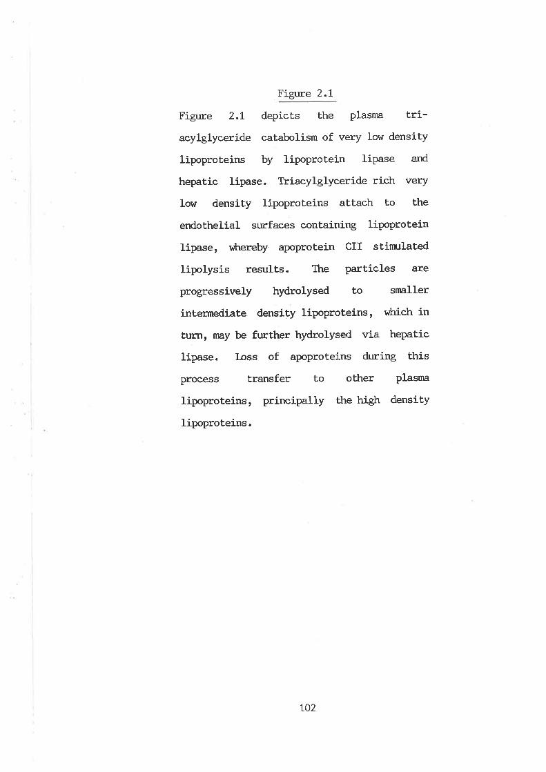
Figure 2.1
Figure 2.L depicts the plasma tri-
acylglyceride catabolism of very low density
lipoproteins by lipoprotein lipase and
hepatic lipase. Triacylglyceride rich very
low density lipoproteins aLtach to the
endothelial surfaces containing lipoprotein
lipase, tfrrereby apoprotein CII stimulated
lipolysis results. The particles are
progressively hydrolysed to smaller
intermediate density lipoproteins, v¡Lrich in
turn, rnay be further hydrolysed via hepatic
lípase. Loss of apoproteins during this
process transfer to other Plasma
lipoproteins, principally the high density
lipoproteins.
toz
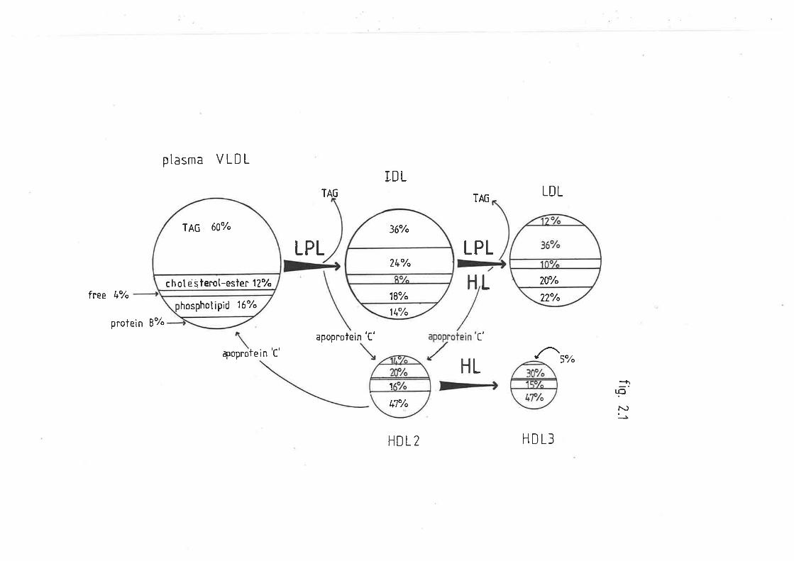
ptasma VLDLIDL
LPL
TA6
LPL
LDL
5"/"
TAG
Lf ree 4Y"
protein 87o
apoprolein 'C' c'
+0pr0 le in 'C'
L>+ :nF
l\)J
HDL2 HDL3
hosphotipid 16"/.
c h ol e s terot-esle¡' 127o
TAG 60'/"
14"/"
18"/"
36"/.
24"h
2?"/.
?0'h
Ël'/.
1
Although HL exkribits a potent TAG and phospholipase activity of
vihich the former is highest towards IDL, LDL and HDL (Musliner et aI'
\979a), the precise physiological role of HL, is at present, unl<nown.
The enzyme has been implicated in the clearance of renmant lipoproteins
(I,OI.) and HDL by the liverr âs administration of anti-Hl- antibodies
results in a rnarked accunn:lation of cholesterol and phospholipid in LDL
and HDL (Kuusi et al. IgTgb). That is, HL is now believed to catalyse
the further hydrolysis of IDL-TAG to produce LDL and by iLs cornbined TAG
and phospholipase activities to convert HDL2 into HDL3 (Kuusi et al'
tg7gb, Jansen et al. 1980b, Reardon et al. t982). A deficiency of this
enzyme in humans leads to an accunnrlation of IDL, an LDL erh'anced in TAG
and a pronounced elevation in HDL2 (Breckenridge eÈ aI. tgSZ). HL can
hydrolyze tri-, di- and monoacylglycerides and pholpholipids (I¿ Rosa et
aI. Ig72, Assman et al. L973, Ilaite and Sisson L973, 1974, Jansen and
Hulsnnnn Lgl4, 1975 and Ehrùrolm et aI. 1975b) and it is considered by
mnyr that this enzyrne has a distinct role in the melabolism of VLDL-TAG
(Grosser et al. 1981, lúrrase and Itakrta L98L and Goldberg et aL. L982),
vilrilst others believe this is not the case (tikkanen et al' 1985 and
Miller and C'otto L982).
2.L.4 POSTHEPARIN PTASMA LIPOPRCIEIN LIPASE AI{D HEPATIC LIPASE
Plasma under nornal circumstances contains liLtle or no TAG
hydrolase acLiviLy of any kind. Howeverr both LPL and HL are readily
released from their respective tissue plasnn mernbranes into the
circulatory system, by intravenous administration of heparin' The
interaction of this glycosaminoglycan with the enzymes, has suggested
103
that the binding of the enzymes to the endothelial cells may be due to
the presence of this type of compound on the cell me¡nbrane (Ootpfri-n
1935). Postheparin plasrna lipase activities have recej-ved much
investigation, as this provides a simple 'in-vivo-in-vitro' method of
assaying TAG hydrolase. Both LPL and HL have been purified and
characterized (Baginsky and Brown L977 and Clegg L979), vùrich has
enabled postheparin plasrna lipolytic activity to be readily resolved
inLo either of these conponents. Ttris is very importanLr âs their
activities invariably do not change in parallel in different metabolic
and pathological conditions (Krauss et aI. t974, ELrnholm et al. 1975a,
Greten et al. t976, L977, Klose et al. t977, l4ordasine et al. 1977 and
Nikkila et aI. Lg77). t{Lren measuring posthepa.rin plasnn LPL and HL
activity, it is now usual to determine total hydrolase activity, then to
irhibit either LPL or HL, and measure the activity of the remaining
enzyme. The activiÈy of the second enzlime is then determined by
difference. Most frequently, either sodium chloride or protamine
sulphate are used to irùribit LPL. Ttrese cornpounds quantitatively reverse
the apoprotein CII dependent activaüion of LPL, but they do not irhibit
any apoprotein CII independent activity, nor do they dissociate the
enzyme-substrate complexes (fietding and Fielding 1976). The NaCl effect
is an anionic dependent action (nietding and Fielding L976). In
addition, apoprotein CII is omitted from the assay. Alternativelyt
specific antibodies, or sodium dodecyl sulphate to inhibit HL can be
used to resolve the two enzymes in postheparin plasma (Krauss et al'
Lgl4, Huttunen et al. 1975 and Greten et aI. L976).
LO4
2.L.5 REGULATION OF LIPOPROTEIN LIPASE AND HEPATIC LIPASE
Investigations of LPL have shov¡n that activity correlates with its
site of isolat.ion to the metabolic and nuLritional state of the animal
(Beznnn et al. 1962, Garfinkel et al. L967, Austin and Nestel 1968 and
Cryer et al. tgTG). Ttris provides a means for distribrting the TAG falLy
acids Lo differenL tissues or organs, according to their metabolic
requirements. It is not surprising thereforer that LPL in heart and LPL
from adipose tissue are reciprocally related, depending on food intake
(Cryer et al. tg76). This, in addition to the differences in apparent
Km, would enable preferential saturation of the heart enzyrne r¡Lren
animals are on a plane of nutrition too low to support fat depostion
(Breckenridge 1985). Similarly, LPL activity in the nnnrnary gland is
increased during lactation (Scow et 41. L976 and Vernon and Flint L982).
The horrncnal and nutritional mechanisms v¡trich differentially regulate
activity in vivo, are however, poorly understood (Patten t970, Faergeman
and Havel Lg75, spooner et al. L979, Ashby and Robinson 1980, Bordeaux
et al. 1980 and Pedersen et aI. 1981). Insulin and glucocorticoids have
been shown to stinn:late adipose tissue LPL activity in both rnan and
aninrals by increasing the synthesis and secretion of the enz¡nne in vivo
(Garfinkel et al. 1976, Vydelingum et al. 1983 and Speake et al' 1985)'
HL has not been as widely investigated as LPL, presunmbly because
iLs precise physiological significance has not been resolved'
Nevertheless, as this enzyme is often associated with steroidogenic
organs, it is not surprising that the respective hormones contribute to
the regulation of this enzyme's activity. Administrat.ion of oestrogens
or androgens have been reported to decrease and increase, respectively,
105
the aclivity of postheparin plasnn HL (&rnnoh et al. L975a and
Applebaum et al. L917).
106
2.2 I.,IETHODS AND MATERIALS
2.2.L ANIMALS
Adult Merino wethers (35-60kg) and rams (AO-ASþ) were used. Each
sheep was housed individually and maintained on a diet of lucerne chaff
and pellets, with water available ad-libitum. Fasting animals were
studied after 72tl- of. food deprivation with water available ad-libitum.
Alloxan diabetes was induced by an íntravenous injection of sterile
alloxan (S0myç¡ into the jugular vein one week prior to
experimentation. A diabetic condition was confirmed by blood glucose
concentration greater than lùnl.'f. t{istar rats were weight and age matched
and maintained on laboratory chow with water available ad libitum.
Fasted rats were without food for 16h.
2.2.2 ACETONE PCI^IDER PREPARATIONS OF LIVER AT{D ADIPOSE TISSUE
For tissue sampling and powder preparations, animals ÍIere
slaughtered at approximately 0900h. Portions of liver and adipose
tissue (omental and perírenal) were inrnediately frozen in liquid
nitrogen and crushed. To 10g of tissue, 2ûnl of acetone was added and
blended on ice for lmin using a Polytron no. PCU2. The resulting
solut,ion was homogenized in a glass-teflon Potter-Elvehjem grinder for
1min. Four such sarçles were pooled and delipidated by suclion washing
r^r|th 40ùnl acetone and 20ùnl diethyl etherr ofl [lhratman nurnber 42
filters. The resulting powders were dried under vacuum at room
temperature. Powders \¡Iere stored at, -15oC-
L07
2.2.3 ADIPOSE LIPOPROTEIN LIPASE AND HEPATIC LIPASE ACET'ONE
POI^/DER ENZYME PREPARATIONS
39 of the respective acetone powder preparation vtas blended on ice
Ì^rith 6ûnl 5m1"1 NH4OH-NH4CI pH7.5 for 1min. The solulion r^Ias stirred
continuously for 3h at 4oC, after v¡trich samples were centrifuged at'
15,000 r.p.m. for 15min. The resulting supernatant was collected and
used as the source of TAG hydrolase inrnediately.
2.2.4 SHEEP AI{D RAT POSTÍIEPARIN PI,ASMA
Sheep post heparin blood samples for the TAG hydrolase (tpL and HL)
assay were drawn from the jugular vein and transferred to heparinized
tubes, 15min after injection of heparin (l-OOU/tg) into the jugular vein.
Rat postheparin blood sanples were collected from ether anaesthetized
rats through the abdominal aorta, 4min after heparin administration
(SOU/ZSOg) through the coûmon ili.ac vein. Plasrna l^ras separated by
centrifuging at 4oC at 3000 r.p.m. for 13nin and stored on ice. The
postheparin plasma LPL and HL assay was done inmediately.
2.2.5 LIPOPROTEIN LIPASE AND HEPATIC LIPASE ASSAY
LPL
modified
and HL TAG hydrolase activity were measured according to a
method of Nakai et aI. (L979). Substrate was made up of Lhe
following components per millilitre of assay mixture; lOtrnol of Glycerol
tri(flt¡ oleate (+SOOO d.p.*.), 60rng of bovine serum albumin (purified
and lyophilized), 3ùng gum arabic, 25umol anmonium sulphate, 40ug
108
phosphatidyl choline and 50ul rat or sheep serum (LPL assay only). (ac)-
Triolein and phospkr,atídyl choline were added to a 25"/" gum arabic
solution and sonicated four ti-mes for 2min at lmin intervals, in a cold
water bath, at a set,ting of 100 watts (Labsonic 1510' 9.5nrn probe) '
Albumin and annnonium sulphate were added Èo the emulsion and the pH
adjusted to 8.5 (unless stated otherwise) Uy ttre addition of KOH-
For the adipose LPL and liver HL enzyme prelnrations, the reaction
mixture contained 500u1 enzyme preparation and 500u1 of TAG substrate.
For total lipase (fpf plus HL) in postheparin plasrna, the assay
contained either 100u1 of postheparín plasnra and 400r¡l of 0.15M NaCl, or
20fu1 of plasnn and 3O0ul of 0.15M NaCl and 500u1 of substrate. Ttre
plasma HL assay contained the same components except thât' the 0.15M NaCI
was replaced with 2M NaCI or with 0.15M NaCI containing Zng/nL proÈamine
sulphate. In addition the HL assay did not contain serum. LPL activity
ÌÁ¡as determined by subtracting the activity in an assay containing 214
NaCl (HL) from the activity in an equivalent assay containing 0.15M NaCl
(fpl- plus HL). All assays v¡ere run at 37oC in a shaking water bath for
0-6ùnin. Each assay vras terminated and free fatty acids extracted
according to a modified Dole procedure as described by Kaplan (fgZO) '
Activity was counted in a Packard liquid scintillation counter (Tri Carb
460CD), with inbuilt corrections for quench and efficíency and
conversion of all act.ivity to d.p.m..
2.2.6 HEPARIN-SEPHAROSE AFFINITY CHROMATOGRAPFIY OF SHEEP LI\ÆR TISSUE
ENZYME AND POSTIIEPARIN PI"ASMA
affinity chromatography of the liver tissue enzyme homogenatetFor
109
1-3ml of extracL was loaded on heparin-sepharose Cl6B colunrts (:Ocm X
lcrn), pre-equilibrated wilh 0.15M NaCt-barbitone buffer solution (5il,
pH 7.5). The colurnns \Àrere sequentially eluted with 5ùnl of 0.15M, 25ml
of 0.45M, 5ûn1 of 0.7214 and 50 ml of 1.5!1 NaCl buffer solutions' A
maxinnrm of 5mI of sheep postheparin plasma was similarly eluted through
the colunrrs aL any one time.
2.2.7 ISOIATION OF VLDL FROM FED AT{D DIABETIC SHEEP
VLDL from fed and diabetic animals was isolated by centrifuging
sheep plasnn at a solvent density of 1.0063g/m1 as described in section
L.2.4.6.L Three anímals were used for each treatment and the samples
pooled. The concentration of VLDL-TAG in fed and diabetic fractions was
adjusted to 3ml4 with a NaCl diluenL such that the final concentration of
this salt was 0.15M.
2.2.8 I{YDROLYSIS OF VLDL-TAG FROM FED AND DIABETIC SHEEP IN
POSTHEPARIN PIASMA FROM FED SHEEP
For total rates of vL,DL hydrolysis (rpl ptus HL) in postheparin
plasma, the assay contained 300u1 of postheparin plasnn, 25ul rat serum
(heat inactivated at 6OoC for lûnin), 75ul 0.15M NaCl and 300u1 of TAG
adjusted VLDL. The postheparin plasma HL assay contained the same
components except that the 0.15M NaCl was replaced with 100u1 of 4M NaCl
and the assay did not contain rat serum. All assays hTere run at 37oC
in a shaking water bath for 0, 20 ot 4ûnin. Each assay was terminated
and free faLly acids extracted as described in section 2'2'5' NEFA
110
released as a result of lipase hydrolysis lrrere calculated by determining
total f.aLty acids at 2ûnin and 4Ornin and subtracting from this the
initial concenLration of unesLerified fatty acids at zeîo time. NEFA
\^/ere determined as described in seclion t.2.7. VLDL hydrolysis as a
result of LPL activity was determined by subtracting the activity in an
assay containing 4M NaCl (HL) from Ehe activity in an equivalent assay
containing 0.15M NaCl (f,PI, plus HL).
2.2.9 BI¡OD GLUCoSE, TRIACNGLYCEROL ATID NON-ESTM,IFIED FATTY ACIDS
Blood glucose, plasma TAG and NEFA were determined as described in
sections L.2.2, I.2.5.2 and L.2.7 respectively. Proteins were determined
using a modified Biuret method (ftznaU and Gill 7964).
Statistical evaluatj-on was by one way analysis of variance-
2.2.T0 MATERIAIS AT]D REAGM{TS
Ctremicals: Glycerol (f14c) trioleate (SOmci/nnnol) v/as
purchased from Amersham Australia Pty. Ltd.. Alloxan monohydrate \^las
purchased from Koch-Light Ltd., England. Bovine serum albumin (fraction
V, 997. pure) and L-alpha-pLrosptr,at.idyl choline (type1-EH) were purchased
from Sigma chemical company. Sodium heparin (fZOu/ne) and insulin
(Isophane) were purchased from C,onrnonwealth Serum I-aboratories
Australia. Ready Solv EP scintillation fluor vras purchased from Becknan
Instruments Inc., Australia. Heparin-sepharose CI-68 was purchased from
Pharmacia Pty. Ltd. Uppsala, Sweden.
LII
,.'I
Iilr¡li
2.3 RESI.JLTS
2.3.1. CÊIARACTERIZATION OF ACET'ONE POIIDER ENZYME HO}{OGENATES
The alkaline endothelial lipases LPL and HL, can be measured either
in homogenates from their respective tissuesr oÍ in postheparin plasma.
However, vilrilst LPL has been studied in sheep, HL in this species has
noL been previously reported. So to establish the identity of this
enzyme and its potential contribution to ovine heparin rel-easable plasna
TAG hydrolase activi.ty, acetone powder honngenates of sheep liver were
prepared and assayed for activity. Furthermore, so as to validate the
methods described herein and determine if conformity exísts with other
species, equivalent extracts from sheep adipose and rat adipose and
liver tissue were also assayed for TAG hydrolase activity.
2.3.L.L SHEEP AI{D RAT LIVM, ÐilRAgIS
Buffered extracts of sheep liver acetone powder preparations
exlribited a capacity to hydrolyze TAG. This activity was similar to the
HL activity of equivalent rat liver extracts, in that optimal acLivity
\^7as observed at pH 7 Lor sheep and pH 8 for rats (figurc 2.2). 75"/" of
the TAG hydroLyzing capacity of the sheep preparation I4Ias retained at
NaCI concentrations of up to 1.5M, though only 307" of lipolysis \,rlas
observed with the rat hepatic fraction at this level (figure 2.3).
Addition of heparin up to lOlunits/ml irùribited activity by
approximately 257" (figure 2.4). TAG hydrolase activity of the sheep
liver extract was linear with increasing triolein substrate up to 10rnM
3
rt2
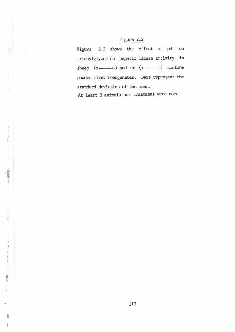
v
Figure 2.2
Figure 2.2 shows the effect of PH on
triacylglyceride hepaLic lipase activity in
sheep (o-o) and rat (+
-
+) acetone
povder liver homogenates. Bars represent the
standard deviation of the mean.
At least 3 animals per treatment were used
I
¿,
'i1'
I
rI
113

_ _ã
i.=
:<
¡a#l
!åL.
o/o
ðctiv
ity
ot, O
l\) LNO O
-l LN
u/ (¡ \¡ \o
r.O l\) l'\)
-E :t
I
:
Figure 2.3
Figure 2.3 shows .the effect of increasing
sodium chloride concentration on triacyl-
glyceride hepatic lipase aetivíty in sheep
(o--) and rat 1a- +) acetone Powder
liver homogenates. Bars represent the
standard deviation of Ehe mean for three experiments'
II
i
:
TI
I
rI
tL4
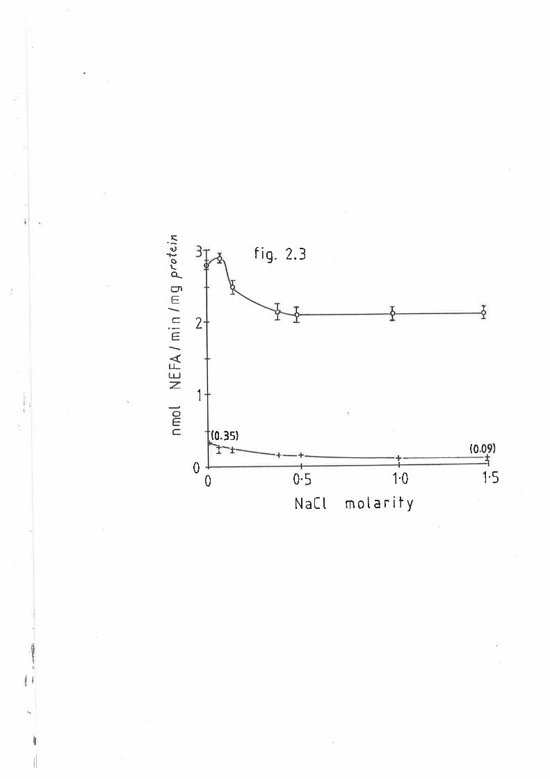
sc.)
oa_úrE
C.
E
LLlrJz.
oEC. (0.3s)
fig. 2.3
2
1
(0+
00 0.5
l.laC t
1.0
motarity1.5
I
I
{l
Figure 2.4
Figure 2.4 shows the effect of increasing
heparin per millilitre of the enzyme and
substrate mixture, on sheep triacylglyceride
hepatic lipase (o-o) and sheep adipose
Iipoprotein liPase (¡-r), from the
respective acetone powder hornogenates' Bars
represent standard deviation of the mean for
three experiments.
115
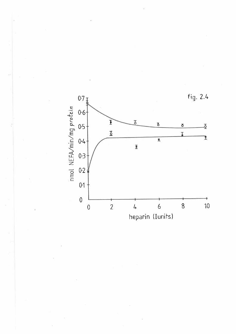
fig. 2.r.
0.6
0.7
0'/*
õõ
]t
T
ô0.5
.sa.,-þoÈ-ErrÉ.
C..E
IIlr IzoEC
Í
0.3
0.2
0l
010I6L20
heparin (lunits)
(figure 2.5). Activity, however, diminished rapidly with time, having a
biological half life of 3ùnin (figure 2.6). There \^Ias a small decline in
activity with increasing levels of serum (figure 2.1). Rates of TAG
hydrolysis \^rere significantly higher in sheep fractions, (approximately
2.8nmol NEFA released/min/mg protein) ttran that' of ta:L fractions
(approximately o.5nnrol/min/mg protein). Gradient NaCl elution of the
sheep liver enzyme preparation through heparin-sepharose affinity
colunms, resulted in a shouldered peak in the O.72V NaCl-barbitone
fraction (figure 2.8). TAG hydrolase aetivity was not however confined
to these peaks. 547" of total activity was not bound to the colunnrs, but
rather was eluted in the 0.15M NaCl to 0.45M NaCl wash fractions'
2.3.L.2 SHEEP AT{D RAT ADIPOSE ÐCTRACTS
Figures2.gand'2.lOshowtheeffectsofNaClandpHontheTAG
hydrolase activity of sheep and rat adipose tissue. Activity \'^Ias
progressively depressed with increasing concentration of Nacl and
in fact at 1.5M NaCl was cornpletely irùribited. Maxinnrm activity \^¡as
observed aL pH 7 and pti 8.5 for sheep and rat extracts respectively
(though act.ivity \^/as present over a wide pH range (6-10)). Addition of
heparin at 2lu/tube increased ovine TAG hydrolase acLivity by 1007"' but
no further increases were observed with further additions (figure 2'4)'
Activity \^7as linear with substraLe concentration up to 1ûnl4 triolein
(figure 2.5) and time up to 60 min (figure 2.6). Serum only narginally
stimulated LpL activity in these extracts (figure 2.1) - TAG hydrolase
activity per unit protein was roughly equivalent between sheep and rat
adipose fractions, that is, approximately 2nmol NEFA released/min/mg
tL6
Figure 2.5
Figure 2.5 shows the effect of increasing
concentrations of triolein substrate on the
sheep triacylglyceride hepatic lipase
(o-o) and sheep adipose lipoproteín
lipase (r-r), from the respective acetone
powder homogenates. Bars represent the
standard deviation of the mean, for three experiments.
LL7
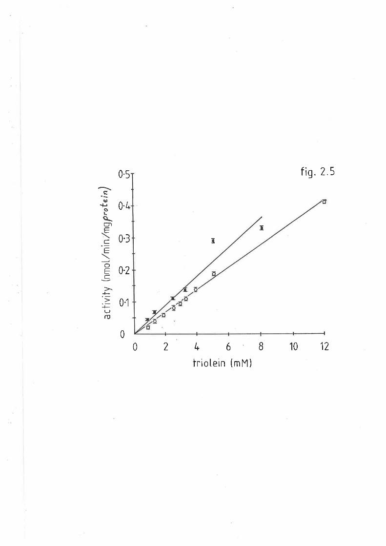
05
0.3
0-2
0l
fì9. 2 5
cs,+ro
\glC.\C.'=h
O
=s>LEtJrD
0.4
T
I
0
02 t.6810 12
trio Iein (m M )

Figure 2.6
Figure 2.6 shows the effecL of time on
sheep tr:iacylglyceride hepatic lipase
(o-o) and sheep adipose lipoprotein
Iipase (r-r), from the respective acetone
powder hornogixrates. Bars represent lhe
standard deviation of the mean for three experiments.
118
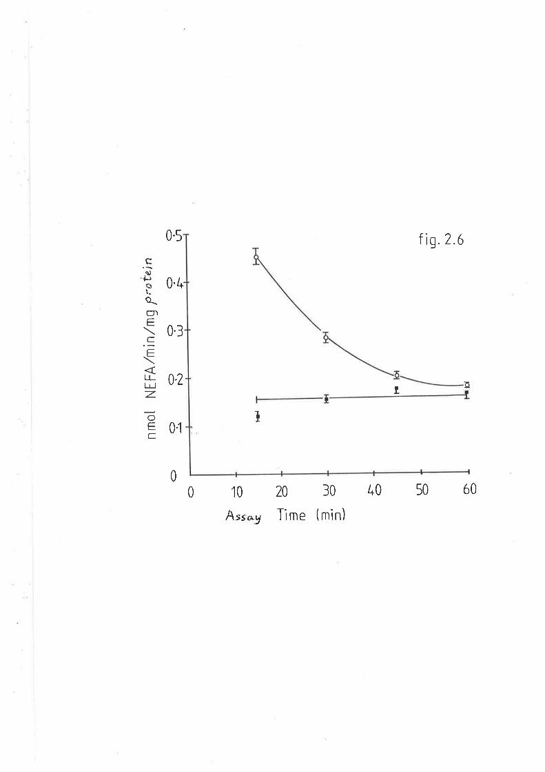
0.5 fig.? 6
T
0.4
cùJþôt_
a_gìE
=\LLlr I
z.
OE=
0.3
0.2
0t
oI
0
0 10 20 30 40 50 ó0
Ass*y Tìme (mìn)
Figure 2.7
Figure 2.7 shows the effect of increasing
levels of heat inacbivated exogenous sheep
or rat serum (as a source of apoprotein CII)
to sheep triacylglyceride hepatíc lipase
(o-o) and sheep adipose lipoprotein
Iipa.se (.-r), from the respecLive acetone
powder honrogenates. Bars represent the
standard deviaLion of the mean'for three experÍrnents'
LL9

0.5 f i9.2 7
10
"ss)
+.,oLa_úl-\
.c_
=ltlr I
z.O
=C.
0.4
0.3
02
0l
c
I
u
lróo/o SefUm
o
I
0I20
Figure 2.8
Figure 2.8 shows the sodium chloride gradient
elution of sheep acetone powder hepatic
homogenates through hepa.rin-sepharose
affinity colunns (---). Ttre corresponding
triacylglyceride hepatic lipase activity is
shown (x-x-x).
L20
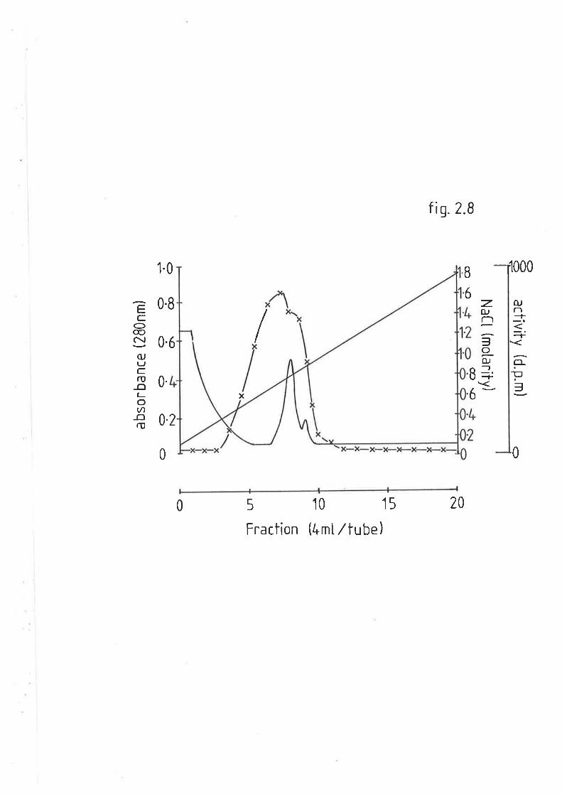
fis.2.B
,1000
OJn
\<
+F=
0
z.fUl-ì
=oOJ_,-+
B
6
1.0
0.8
0.6
B
6
IEC
O@c\¡
(UtJC.rU
-o(-ov)
-orI,
0.x
0.2
0
205
Fraction
10 1s
(4rntltube)0

Figure
sodium
Figure 2.9
2.9 shows the effect of i-ncreasing
chloride concentration on adipose
lipoprotein lipa.se in sheep (r-¡) and rat
(o-o) acetone powder homogenates. Bars
represent the standard deviation of the mean
for three eq)eriments in replicate.
a
Lzl
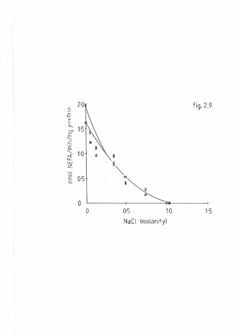
IT
I
5
2
1
"sa)
+Jù\o_glEC.'=
LLlr I
z.o=C-
1.0 t,
f is,2.e
1.5
0.5ff
0.5 10
Natt (motarity)
Í
0
0
Figure 2.10
Figure 2.10 shows the effect of pH on adipose
lipoproLein lipase in sheep (¡-r) and rat
(o-o ) acetone powder homogenates. Bars
represent the standard deviation of the mean
for three experíments in replicate.
L22
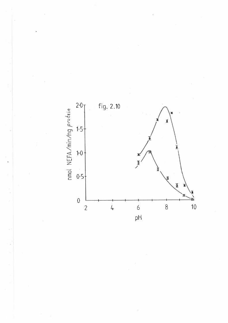
f iq. 2.102.0I
1
Ë(\)-þË)I
a_gìC.\=.E
lllr I
z.o
T
pH
a T
I
5
1.0
T
6l*?
= 0.s
E
T
10
0
rJqi.l
protein (at optinnLm pH).
2.3.2 POSTT]EPARIN PIÁSMA LIPASE ACTIVITY
2.3.2.t RAT POSTÍIEPARIN PTASMA
Prior to determining the TAG lipase activity in sheep posÈheparin
plasrna, the aetivities of LPL and HL were determined in rats as
described in sections 2.2.4 and 2.2.5. The method used initially r^ras
that described by Nakai et aI. (L979). Table 2.1 shows that in rats
fasted overnight, total TAG hydrolase activity was approximately
14.6umol NEFVmI plasma/h, vùrich was divided equally between LPL and HL.
However, preliminary investígations in this laboratory suggested that
extraction of the unesterified fatty acids was inadequate due to volume
fluctuations in the aqueous/solvent phases. tlaving increased the
specific activity of the triolein substrate and utilized the modified
DoIe extraction procedure (Xaplan 1970), higher rates of both LPL and HL
in raL postheparin plasnra were obtained (tabLe 2.1). Blanks with a lqrown
concentration of unesterified fatty acids were extracted under the same
c<.¡nditions to ascerlain that this process r^ras cornplete. Furthermore,
radioactivity in aliquots of the rDolet extract were determined before
and after elution of unesterified faLty acids through Biosil columns, to
determine v¡hether or not tri-di-or mono-acylglycerides hrere similarly
extracted. The results showed that the mean Lotal hydrolase activity
measured was nearly threefold greater than that previously determined.
The proportions of the two enzymes remained essentially unchanged, in
that LPL made up approximaLeLy 6A7" of the total activiLy.
II
Þ
123
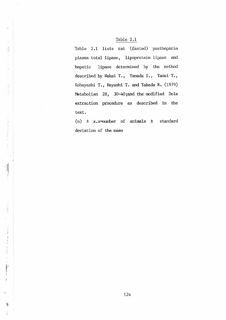
hl
iirt
Table 2.1
Table 2.L lists rat (fasted) postheparin
plasnra total lipase, Iipoprotein lipase and
hepatic lipase determined by the method
described by Nakai T., Yamada S., Tarnai T.,
Kobayashi T., Hayashi T. and Takeda R. (L979)
bbtabolism 28, 30-40;and the modified Dole
extraction procedure as described in the
text.
(") t x.x<umber of anirnals + standard
deviation of Ehe mean
T
I
i,
t24
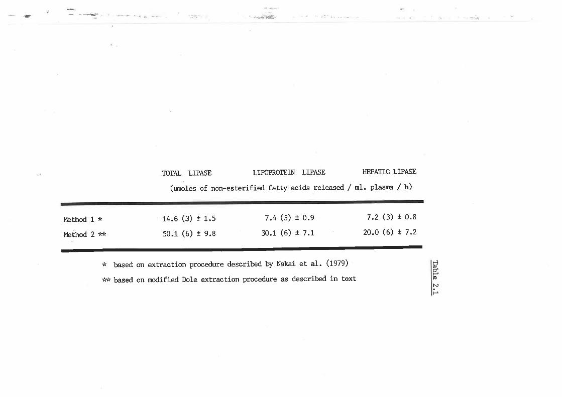
*-Æ-{=..=¿ãÊ.-
TTTAL LIPASE LIPOPROTETN LIPASE HEPATIC LIPASE
(umoles of non-esterified fatty acids released / nL. plasrna / h)
Merhod 1* 14.6 (3) t 1.5 7.4 (3) 10.9 7.2 (3) 10.8
t,rerhod 2 *.,^- 50.1 (6) t 9.8 30.1 (6) ! 7.L 20.0 (6) ! 7 -2
:k based on extraction procedure described by Nakai et aI. (fgZg)
:'c^' þ¿ssd on nrodified Dole extraction procedure as described in text
ËÞdHoN)
P
I
f,5I
2.3.2.2 SHEEP POSTT{EPARIN PIASMA
Intravenous adminisiration of heparin to sheep resulted in a rapíd
rise of TAG lipase activity viLrich was maximal 15min after injection (at
100u/kg). Approximately 707" of this activity was irùribitable by 1M NaCl
or protamine sulphate, w'ith no further change up to 3M NaCl (figure
z.fl). The NaCl resistant component of total TAG hydrolase activity
diminished with time (figure 2.t2). Hepa.rin-sepharose affinity
chrornatography resulted in the elution of two peaks (figure 2.t3). The
first in the 0.72M NaCl-barbitone fraction was considered to be 'salt
resistant' [L. The second peak in the 1.5M NaCl-buffer fraction is LPL.
Both LPL and HL in sheep postheparin plasnra had an alkaline pH optinn:m
of 8 and 9 respectively (figure 2.I4).
Having characterized both components of plasma TAG hydrolase
activity, namely LPL and HL, the effects of fasting and diabeLes \¡lere
determined.
2.3.2.3 POSTHEPARIN PTASMA LIPOPROTEIN LIPASE AND HEPATIC LIPASE IN
ÍII
I
FED. FASTED AND DIABETIC SHEEP
Total plasna TAG hydrolase, LPL and HL activities are shown in
table (Z.Za). In fed welhers total plasma TAG hydrolase activiLy rÀlas
significantly higher than lipolytic rates found in fasted (S17.) and
diabetic wethers (t78"/"). In normal fed wethers, LPL activity represented
approximately 70% of the total and in fasting wethers, this
proportionality was maintained, as an equi-valent reduction in both LPL
and HL \^ras obsen¡ed. However, it the diabetic animals, despite a
*
t25
I
Figure 2.11
Figure z.tL shows the effect of increasing
concentration of sodium chloride on sheep
posLheparin plasnn triacylglycerol lipase
activity. Bars represent the standard
deviation of the flêârlrfor three experíments
in replicaÈe.
L26
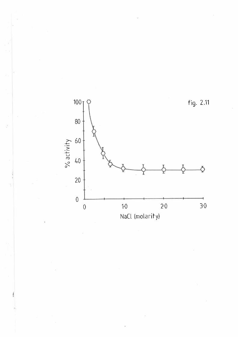
BO
100
20
f \9. 2.11
3.0
ã60.u-F(J
.: 40o\
0
0 1.0 2.0
Natt (motarit y)
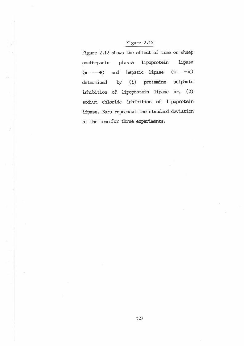
Figure 2.L2
Figure 2.L2 shows the effect of time on sheep
postheparin plasma lipoprotein lipase
(o o) and hepatic lipase 1¡-¡)deLermined by (1) protamine sulphate
irùribition of lipoprotein lipase otr Q)
sodium chloride irùribÍtion of lipoprotein
lipase. Bars represent the standard deviatíon
of the mean for three exPeriments.
L27
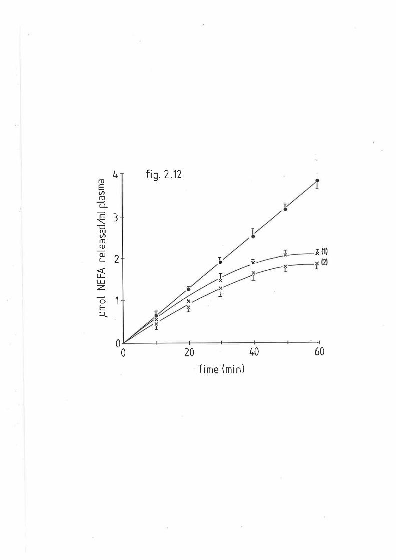
f mot
NE
FA
rel
ease
d,/m
l pla
srna
N)
LrJ
.FO
o
ç N) :, l\)
l\) O È + O
x-{
I
o\ o
x
_i =lD =.
=
F-
d xl
Figure 2.13
Figure 2.L3 represents the sodium chloride
gradient elution profile of sheep postheparin
plasrna through heparin-sepharose affinity
colunns. Ttre first peak in the 0-72Ì4 NaCl
fraction v/as identified as hepatic lipase.
Ttre peak eluting in the 1.5M NaCI region is
lipoprotein lipase.
t28
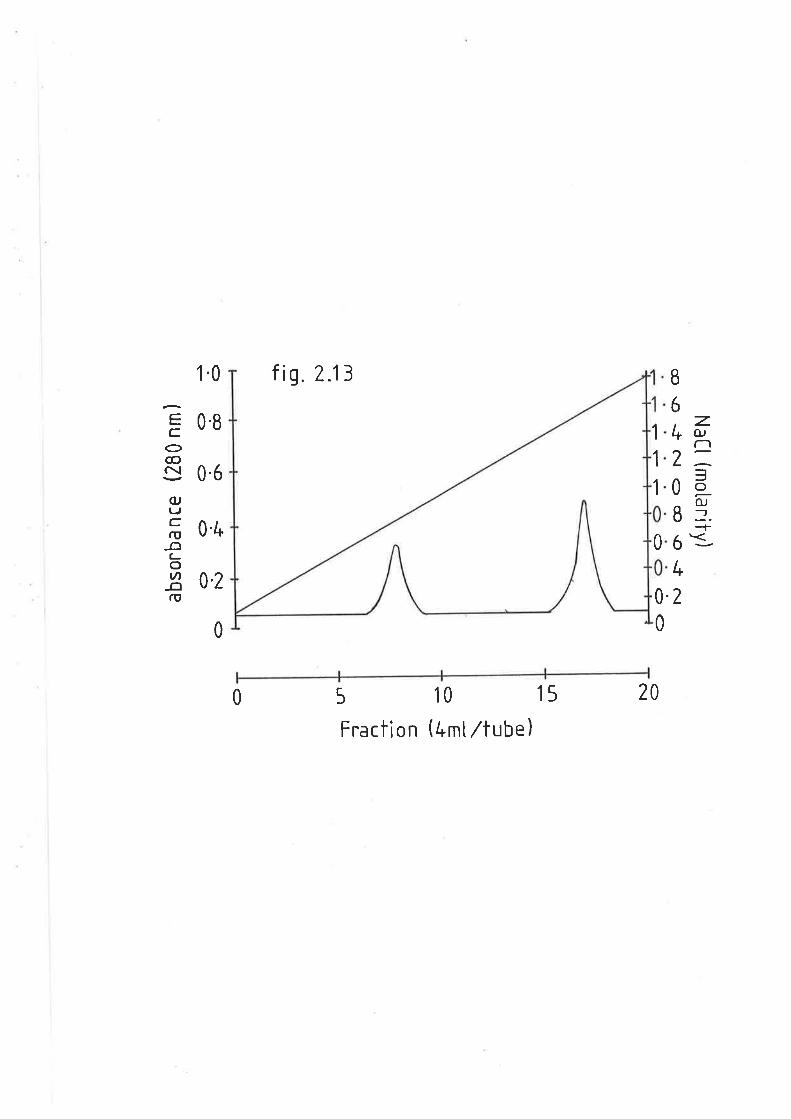
1.0 fi9.7.13 B
6.z-4 o-,
2==0l
83+6=-4
2
GCo@c\(uTJCrt,-ot-otA
-ctfit
0.8
0.6
0.4
0,2
1
1
1
0
0.
00
0 5 10 15
Fraction (4mtltube)20
Ei'gtre 2.1,4
Figure 2.14 shows the effect of pH on sheep
posLheparin plasma lipoprotein lipase
(o-o) and hepatic lipase (x-x). Bars
represent the standard deviation of the mean.
for three experiments.
t29
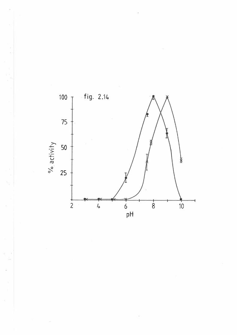
100 f iq. 2.U.
15
50
tIJrI,
-oo\ 25
8l*2 ó
pH
10
Table 2.2
Table 2.2 (a) lists sheep postheparin plasnn
total lipase, lipoprotein lipase and hepatic
lipase activities in fed r¿eEhers, fasted
wethers, diabeLic wethers an¿ (U) fe¿ e\^Ies
and rams.
(") t x.xx+tumber of animals + standard
deviatíon of the mean.
130
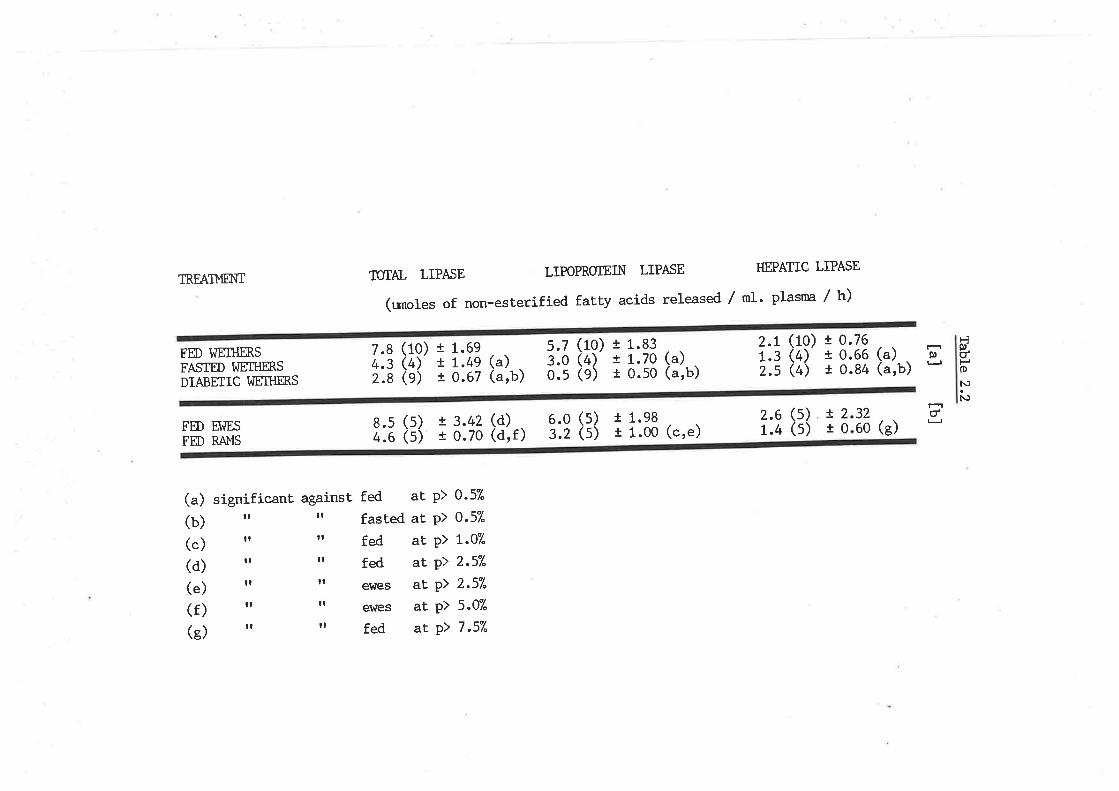
ÎUTAL LIPASE LIPOPROTEIN LIPASE HEPATIC LIPASETREAII.,IÐüI
(unoles of non-esterified fatty acids released / nl. plasrna / h)
ËÞúPoN)
N
å3 t.> E-84 (a,b) .-
2.1 (10) i 0.1.3 (4) t o.2.s (4) r o.
837050
s.7 (10) t 1.3.0 (4) t 1.0.s (e) t 0.
+g (a)67 (a'b)
7.8 (10) t 1.4.3 (4) t 1.2.8 (e) t o.
69FED WETHM,SFASTED WEII{M,SDIABETIC I^]ETIIB.S
a)arb)
((
! 3.42 (d)t 0.70 (d,f)
+lt1
(s)(s)
.98
.00 (c,e)2.6r.4
!2r0
(s)(s)
3260
núUFED ET{ES
FÐ RAI.,IS
(a) significant against fed at p) 0'57'
(b) rr rr fasted a! P) 0'57'
(.) rr I' fed aE P) 1'07"
(d) rr ' fed aE P> 2'57"
(.) rr rr ewes al P) 2'57"
(f) rt I' ewes at P) 5'07'
(g) rr I' fed at P) 7 '57"
6.03.2
8.s (s)4.6 (s) (s)
significant increase in HL activity (2OÐ, total activity was depressed
Lo 36"A of that seen in the normal fed state. This was due to the decline
in LPL activity, v¡trich was only O.5umol NEFA released/ml plasnn/hr or
207" of the total. The changes in heparin releasable TAG hydrolase
activity correlated with fluctuations in blood lipids. In a typical
alloxan diabetic wether, total activity !ùas reduced from 4'0umol
NEFA/mI/h to 2.1|tnoL/nL/h and plasma NEFA and TAG increased from
77tnoL/nL for the former, and 262umoL/mL fox the latter, to 1r258umol/ml
and 2r531umo1/ml respectively. Administration of exogenous insulin
restored activity to 7.6umo1 NEFvmI/h, and reduced plasnra NEFA to
t26tnoL/nL and TAG to 300umot/nL. subsequent withdrawal of the insulin
showed hydrolase activity was again reduced to 4.1umol NEFVmI/h, and
concentrations of plasma NEFA and TAG elevated to 685unrol/ml and
L962tnoL /ml resPectivelY .
2.3.2.4 POSTHEPARIN I{YDROLYSIS OF VLDL-TAG FROI'{ FED AI{D DIABEIIC
SI{EEP
VLDL was isolaÈed from fed and diabetic sheep and incubated with
postheparin plasma from fed animals to determine v¡hether the changes in
postheparin plasna lipase activity may have been a result of
physiochemical changes of these particles. Figure 2.15 shows rates of
NEFA released with time. VLDL-TAG from diabetic animals was hydrolyzed
aL a rate 280% greater than that of VLDL-TAG from fed sheep' Table 2'3
lists the contribution of both HL and LPL to this activity- LPL vùrich
comprised 947" of the total lipolyt.ic rate in VLDL substrate from fed
sheep, was increased 2.6 fold with diabetic VLDL substrate' Similarlyt
131
Figure 2.15
Figure 2.15 shows the rate of non-esterified
falty acids released from very low density
lipoproteins from fed and diabetic sheep,
wtren incubated with nonnal fed sheep
postheparin plasma. Bars represenL the
standard deviation of the rlêâll.
Three ar¡imals per treatment,
L32
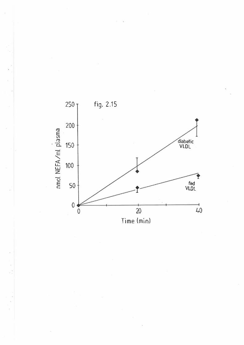
250 fig. 2.15
ruEv)ro.
O-
=tlU-Jz.oÉ.C
200
100
c150 VLDL
fêdVLDL50
0
1
0 20
Time (min)
40
Table 2.3
Table 2.3 lists the total lipa.se, lipoprotein
lipase and hepatic lipase activiEesofnornnl
fed sheep posthepa.rÍ-n plasma vitren incubated
v¡ith very low density lipoproteins from fed
and diabetic sheep.
(") t x=nrmber of anirnals + standard
deviation of the meån.
1-33
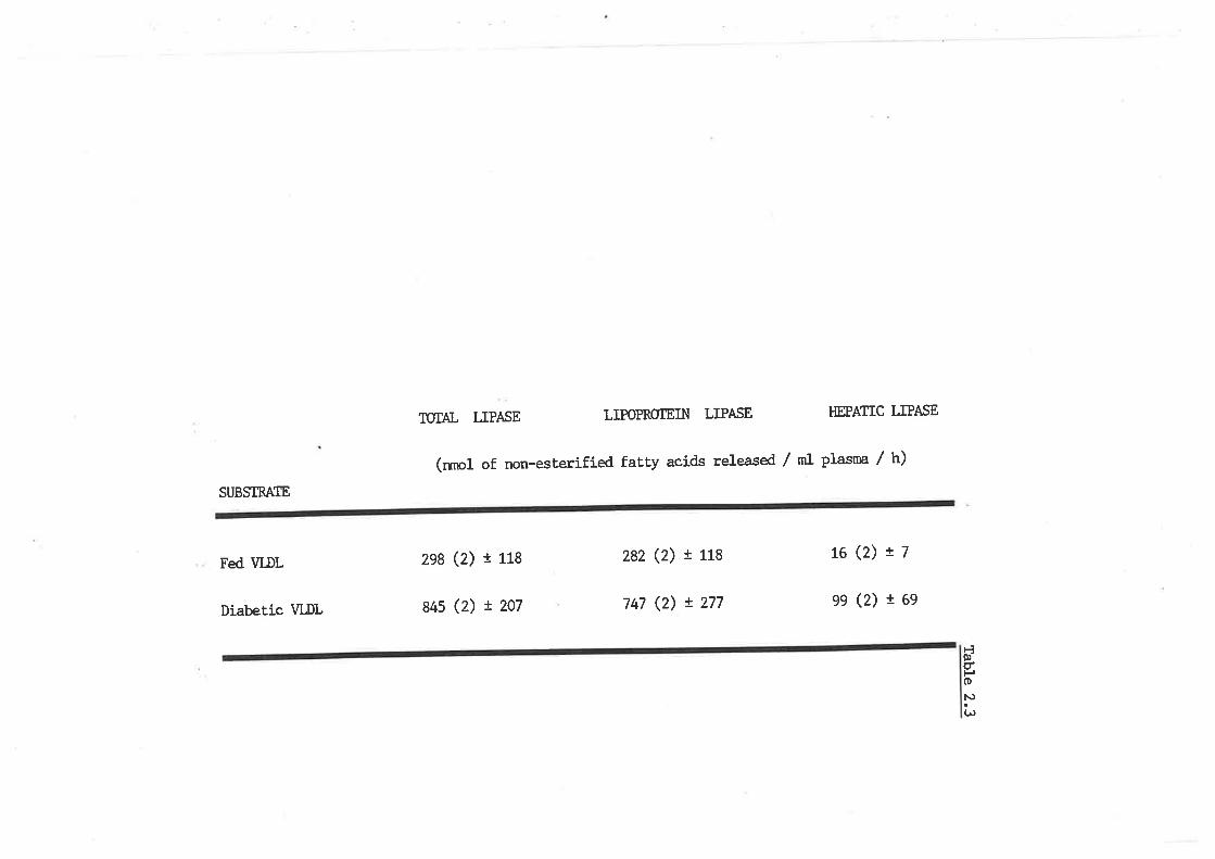
TTIIAL LIPA^SE L]POPROTEIN LIPASE HEPATIC LIPASE
(n¡rol of non-esterified fatty acids released / ml plasma / h)
SUBSTBATE
Fed VI-DL
Diabetic WDL
2e8 (2) I 118
84s (2) t 207
282 (2) r 118
747 (2) ! 277
L6(2)!7
ee (2) r 6e
ÊÂ)doN)(,
the HL component was also elevated in the latler, though substantially
more so (O.Z fold higher). As such, HL comprised L27" of the total
postheparin plasma activity, vúren incubated with wDL from diabetic
animals.
PIASMA LIPASE ASIIVITIES IN RAI.,IS, WEIHM.S AI{D2.3.2.5 POSTHEPAR]N
EI^IES
Table 2.2b depicts differences in the TAG hydrolase activity of
sheep postheparin plasma LPL and HL between el^lesr wethers and rams' Ev¡es
had a significantly greater total TAG hydroLyzing capacity OÐ and
conversely, rafns had a significantly lor¿er total TAG hydrolyzing
capacity (4LÐ than wethers. In ewes, the higher rates of lipolysis were
attributable solely to the LPL component, v¡trich was 887' higher than that
of rams. similarly LPL in wethers \^Ias 787. higþer than that of rams' HL
r^ras found to vary considerably in ewes and although activity of this
enzyme \^ras 467" higher than that of rams, this was not significant'
However, HL in rams \^ras significantly lower than fed wethers (33%)'
2.3.2.6 POSTI{EPARIN PTASI"IA LIPASE ASTIVITIES IN 'LEAI.I' AI{D 'OBESEI SHEEP
Heparin releasable plasma TAG hydrolase activities were determined
in ,Iean, (Merino) and 'obeset (Romney X Dorset X }4erino) sheep, to
ascertain vùrether different breeds of sheep, not subject to feed
restriction, are genetically predisposed to lipase activities and as
such, a polential degree of adiposity. Table 2.4 lists the total TAG
hydrolase activity, LPL and HL of pre-ruminant lambs from both groups'
t34
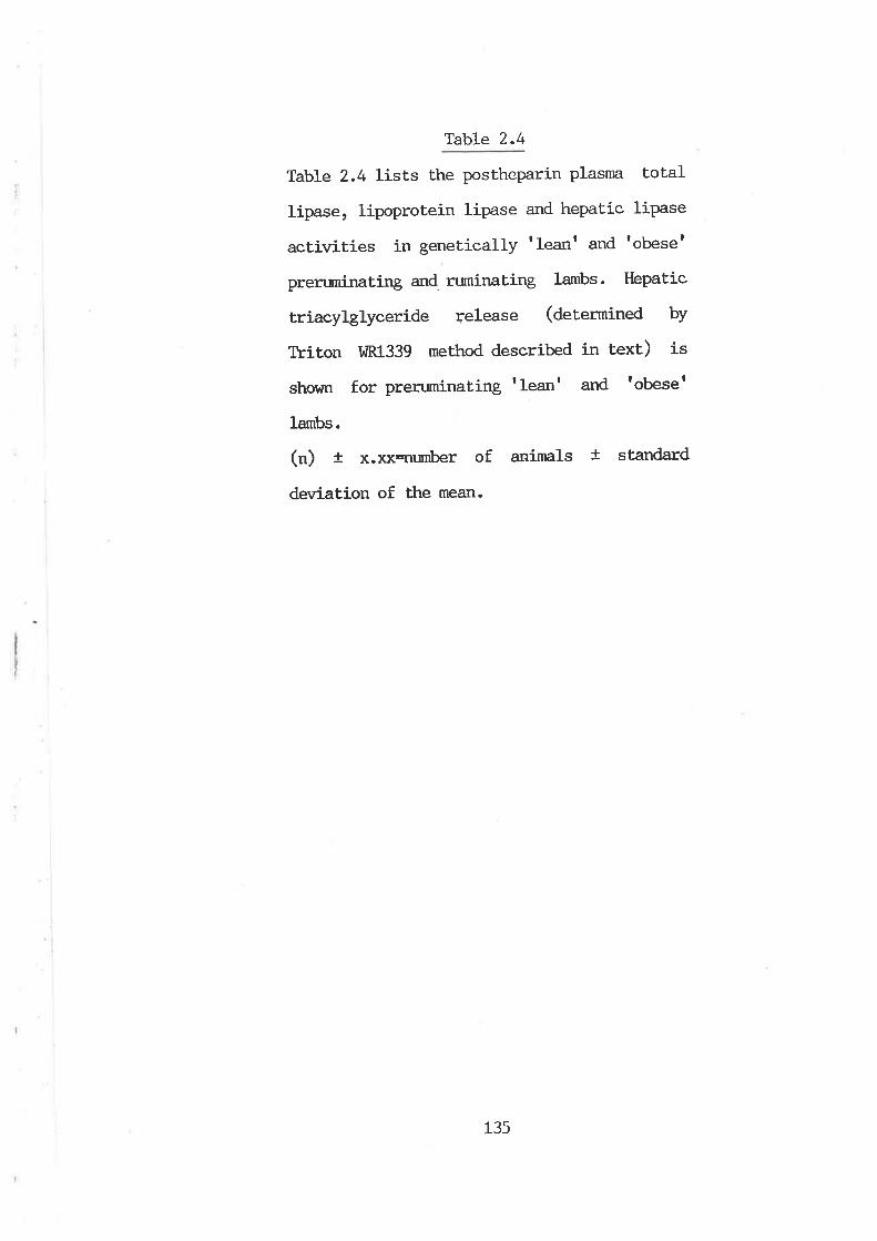
r
TabLe 2.4
Table 2.4 lists the postheparin plasnn toLal
lipase, lipoprotein lipa.se and hepatic lipase
activities in genetically tleant and tobese'
prenrminating and ruminaLing lambs. Hepatic
triacylglyceride release (determined by
Thiton IdR1339 method described in text) is
shown for prenminating tleant and tobeset
lambs.
(") + x.xxqu¡nber of animals t standard
deviation of the rllean.
135
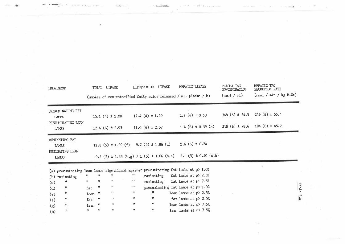
,t-
TREATT'{ENI
PRMUMINATING FAT
TAI',IBS
PRffi.UMINATING LEAt'l
tÁl"lBS
RI.MINATING FAT
IAI',IBS
RUMINATING LEATÌ
IAI',IBS
(a) prerurninating le¿n lambs significant against pren:minatíng fat(b) runinating rr rr rr It ruminating fat(") rr n rr rr It ruminating faL
(d) tt fat rt rr tt preruninating fat
(e) tt lean rr rr rr rr lean
(f ) tt fat rr rr rt It fat
(g) tt lean It tr rr ' lean
(h) rt rr rr It rr rr lean
TÛIAL LIPASE LIPOPROTEIN LIPASE HPATIC LIPASE
(unoles of non-esterified fatty acids released / m1' plasnra / h)
1s.1 (4) I 2.oo L2.4 (4) t 1.so 2.7 (4) r 0.s0 368 (6) t s4.s 24e (6) I ss.4
t2.4 (6) ! z.ss 11.0 (6) ! 2.57 1.4 (6) t 0.3e (a) 318 (6) r 78.6 1e4 (6) t 4s-2
11.S (s) I 1.3e (f) s.2 (s) t 1.s6 (d) 2.6 (6) ! 0.24
e.2 (s) r 1.33 (b,s) 7.1 (S) t 1.06 (b,e) 2.1 (5) t 0.50 (c,h)
PIASMA TAGCONGM{IRATION
(nmol / ml)
HPATTC TAGSECREIION RAIE
(rrnol / min / kg B.Wt)
Iambs
lambs
lambs
lambs
Iambs
larnbs
lambs
lambs
1.07"
2.s7"
7.s7"
t.07"
2.s7"
2.s7"
7.s7"
7.s%
ataÈ
atatatataE
at
p)p)
P>
p)p)p>
p)p)
lÉl0)
IF
.T8rü
l't
Total activity was 15umol NEFA released/ml plasna/h I'ot Lhe crossbreds',
and although total acLivity was only l0umot NffiA/ml/h in the Merino
lambs, this was not significantly different. Nonetheless, HL in the lean
merino sheep was significantly lower than their crossbred counterparts
(387").
Table 2.4 also shows the postheparin plasma total TAG hydrolase
activity, LPL and HL for both tleant and tfat' aninnls afLer weaning and
at a stage vùrere weight gain was at its highest' Total activity hras
significantly different. between these aninnls namely 11'70urnol NtrVml
plasnn/h and 9.07umol Nffivml/h for 'faL' and 'lean' groups
respectively. TLre lower rates of hydrolysis in Ehe lalter group vTere
solely attributable to LPL vùrich was 237" of. that obsen¡ed in the
crossbreds. Mean HL activity in the tleant animals !{as 2'04umol
NEf'Vml/h and significantly higher (25Ð than that in the 'fat' animals
(2. 56u¡nol NEFA/mf/h) .
TRIACNGLYCEROL SECRETION RATE IN PREI^iMNED.I-EAI.I' Æ{D IOBESEI
I
2.3.3.t
IA},IBS
Triton I,üR1339, vlas used to irhibit plasnn clearance of TAG and so
measure hepat.ic release of lipid in both tleant and 'obeset animals' to
see if this could be correlated with the higher rates of TAG lipolysis
observed in the latter grouP'
MeanplasmaTAGconcentrat.ionswere3lsumol/mlplasmaand
36gumo1/ml for 'leant and 'faLt lambs respect'ively (taule 2'4)' T?rere
I/\ras considerable variation, and results were not significantly
different. similarly, hepatic secretion of TAG was l-94nmol/min/kg body
!
136
weight and 249umol/min/kg body weight respectively and although in each
animal secretion was constant, (r greater lhan 0'96 over 4'5 hours)
there was considerable variation beLween aninrals'
2.3.3.2 TOXICITY OF TRITON I^1R1339
Inmediately after the TAG secretion studyt
returned to oPen grazing r^rithin a 2 acxe paddock'
Ehe animals \^Iere
þproxirnatelY two
ìI
lI
,ì
weeks after Tfiton administration, a number of sheep had lost weight and
generalbodycondition.Ttreydevelopedblackchappedpatchesonareas
vùrich l{ere generally elçosed, SUch as noser lips and rurnp and eventually
a ntunber of sheep died. Autopsies on each of the deceased lambs revealed
consumption of toxic rnaterial r^tas not the cause of death (as suggested
by the symptoms). There was, however' Sross hepatic cellular dannge'
r,¡?rich could not be further characterized'dliull
TI
I
r
L37
rrI
l
2.4 DISCUSSION
o¡¡ine LPL has been isolated, ctnracterized and shown to be similar
tothatreportedinotherspecies,i.thatitkrasanabsoluterequirement for apoprotein cII. The role of this enzyme in relation to
the hypertriacylglyceridaemia and hepatic accunnrlation of this lipid
extribited in diabetic sheep, has not however, been previously reported'
The activity of this enzyme in other species such as humans and rats
made diabetic, has been widely investigated, but both of these differ
from sheep, in that díabetes results in a reduced hepatic output of TAG' '
similarly, ovine HL has not been previously reported and the role of
this enzyme in the metabolism of WDL-TAG also remains to be defined'
Pr:rification of this enzyme was beyond the scope of this study, thougþ
an investigation of the catabolism of sheep VLDL-TAG would have been
incomplete without determining changes in itts activity' As such' this
section of this study had several objectivies. The first was aimed at
establishing if sheep liver possessed a TAG hydrolase activity v¡trich had
characteristics resembling membrane bound HL as reported for other
species. ltris \^ras achieved by extracting TAG lipolytic enzymes from
acetone powder preparations of sheep liver and comparing these \^rith
similar fractions from rat liver and published data on such fractions
for the Iatter (Jansen and Hulsmann L975 and Hulsmann et al' 1-977)'
Furthermore, to establish conformity with other species, equivalent
homogenates of sheep and rat adipose tissue exleibiting LPL act'ivity were
also character ized. Acetone powder homogenates have been a traditional
means where such enzymes and the characterisLics attributable to them'
may be identified without the need for further purification' such
ï
138
fractionsdohowever'representacnrdeproteinextract,vñichinaddition to LPL and HL, may contain other proteolytic enzyrnes active
under the described experimental conditions'
Acetone powder preparations of ovine liver contained TAG hydrolase
activity v¡hich rese¡nbled the membrane bound HL reported in other
species, in that activity \^Ias expressed under conditions of high salt
concentration (157" retarned). However, the sheep HL act'ivity did not
extribit the higher alkaline optinn:m usually associated with this enzyme
and as observed here for the equivalent fractions from rats' A nurnber of
Iiver lipases have been reported, including the me¡nbrane bound enzyme
v¡trich is NaCl resistant and has an alkaline opLimumr âs weII as a
lysosomal lipase, rlfrich is intribited by NaCI and has an acid optinral pH
(Assrnan et aI. tg73, Jenson et al. 1980 and Cordle et aI' 1933)'
Althougþ the presence of these activities have not been previously
reported in sheep líver, it is rnost probable that these extracLs contain
both enzymes, thus accounting for the lower pH optimum and 25% reduction
in acLivity at NaCI concentrations of 1.5M. Similarly, the reduction in
rat liver TAG lipolysis obsen¡ed urrder high NaCl concentrations' could
also be attributable to the Presence of these enzyÌnes' Ttre lower raLes
of TAG lipolysis per unit protein in ratsr compared to sheep is of
potential significance, in that this may suggest differences in the TAG
hydrolysing capacity of HL between species. Activity was linear with
triolein substrate concentratíons of up to 1ùnl'1, suggesting that the
sheepliverenzymehasalowaffinit'ythoughhighcapacityforthe
substrate. serum added to the assay had a depressing effect as reported
for ra¡ HL (I-a Rosa et al. t972 and. Kubo et. aI. 1980) and bovine HL
(C,ordle et aI. 1983). Gradient NaCl elution of the sheep liver enzyme
{r
L39
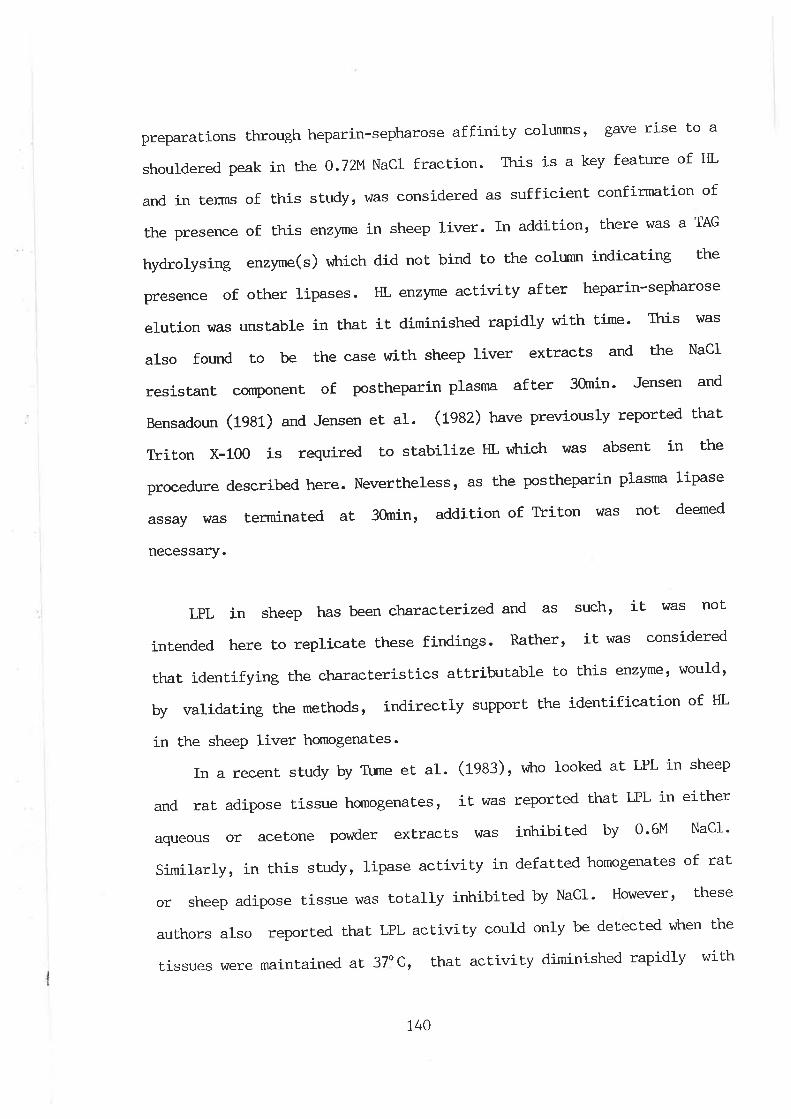
preparations through heparin-sepharose affinity colunrtst gave rise to a
shouldered peak in the 0.72V NaCl fraction. This is a key feature of HL
and in terms of this study, was considered as sufficient confirnration of
the presence of this enz)¡me in sheep liver. In addition, there was a TAG
hydrolysing enzyme(s) vitrich did not, bind to the colunn indicating Lhe
presence of other lipases. HL enzyme activity after heparin-sepharose
elution was unstable in that it diminished rapidly with time- Ttris \'tras
also found to be the case r,rith sheep liver extracts and the NaCI
resistant component of postheparin plasnn after 3ùnin' Jensen and
Bensadoun (1981) and Jensen et a1. (fgAZ) have previously reported that
Triton x-100 is required to stabilize HL v¡lnich IÁIas absent in the
procedure described here. Neverthelessr as the postheparin plasnm lipase
assay rdas terminated at 30min, addition of Triton
necessary.
hras not deemed
LPLinsheephasbeencharacterizedandassuch,itl^/asnot
intended here to replicate these findings. Rather, it was considered
that identifying the characteristics attributable to lhis enzyme, would'
by validating the methods, indirectly support the identification of HL
in the sheep liver honrogenat'es.
In a recent study by Tume et al. (1933), vùto looked at LPL in sheep
and rat adipose tissue homogenates, it was reported t'hat LPL in either
aqueous or acetone powder extracts $¡as irùribited by 0'6M NaCl'
similarly, in this study, lipase activity in defatted homogenates of rat
or sheep adipose t.issue was totally inhibited by NaCl' However, these
authors also reported that LPL activity could only be det'ected v¡tren the
tissues were maintained aL 37oC, that activity diminished rapidly with
140
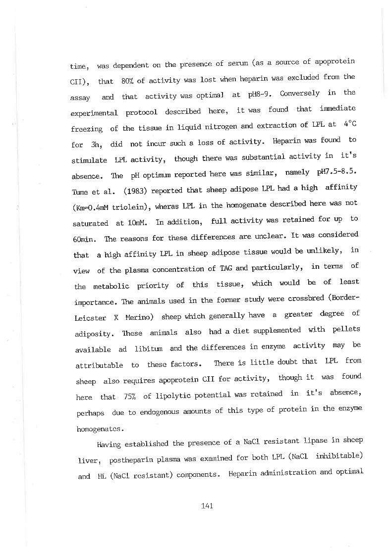
time, was dependent on the presence of serum (as a source of apoprotein
CII), that 807" of activity was lost vilren heparin was excluded from the
assayandthatactivitywasoptimalaLpH8-g.Converselyinthe
experimental protocol described here, it was found that inrnediaLe
freezing of the tissue in liquid nitrogen and extraction of LPL at 4oC
for 3h, did not incur such a loss of activity. Heparin was found to
stimulaüe LPL activity, though there was substantial activity in itts
absence. The pH optimum reported here was similar, namely pH7'5-8'5'
T\rme et aI. (1983) reported that sheep adipose LPL had a high affinity
(Xm=O.4nM triolein), r*reras LPL in the homogenate described here was not
saturated at 10mt"l. In addition, full activity was retained for up to
60rnin. The reasons for these differences are unclear' It was considered
that a high affinity LPL in sheep adipose tissue would be unlikelyt in
view of the plasnn concentration of TAG and particularly, j-n terms of
the metabolic priority of this tissue, vùrich would be of least
importance. The animals used in the former study hrere crossbred (norder-
l,eicster X },lerino) sheep vitrich generally have a greater degree of
adiposiLy. Inese animals also had a diet' supplemented with pellets
available ad libitum and the differences in enzyme activity may be
attributable to these factors. There is little doubt that LPL from
sheep also requires apoprotein cII for activity, though it' \/|Tas found
here that 757" of lipolytic potential was retained in it's absence'
perhaps due to endogenous amounts of this type of protein in the enzyme
homogenates.
Élaving established the presence of a NacI resistant lipase in sheep
liver, postheparin plasma was examined for both LPL (tlaCt irùribitable)
and HL (NaCI resistant) components. Heparin administraLion and opLimal
L4L
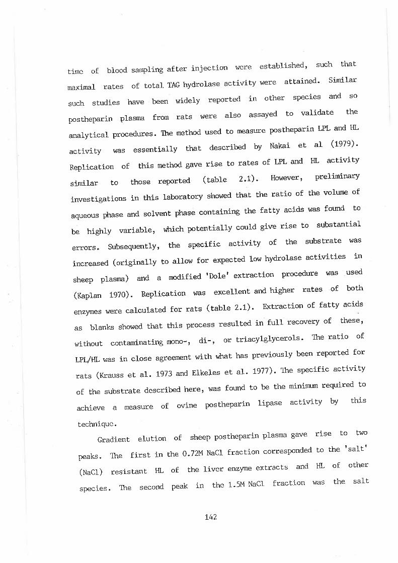
time of blood sarnpling arLex injection \i¡ere established' such that
maximal rates of total TAG hydrolase activity were attained' Similar
suchstudieshavebeenwidetyreportedinotherspeciesandSopostheparinplasnrafromratsv/erealsoassayedtovalidatethe
analytical procedures. The method used to measure postheparin LPL and HL
activityltTasessentiallythatdescribedbyNakaíetal(rozo¡.ReplicationofthismethodgaverisetoratesofLPLandHLactivity
similar to those reported (tabre 2.r). However, preliminary
investigations in this laboratory showed that Lhe ratio of the volume of
aqueousphaseandsolventp}rasecontainingthefattyacidswasfoundto
be highlY variablet
errors. SubsequentlY,
sheep plasnr,a)
(xaptan 1970).
rdaich potentially could give rise to substantial
the specific activity of the substrate hlas
increased(originallytoallowforexpectedlowhydrolaseactivitiest_n
HL of other
was the salt
and a modified 'Dolet extractíon procedure vtas used
Replication vras excellent and higher rates of both
enzymes were calculated for raLs (table 2.t). Extraction of fatty acids
as blanks shor^¡ed that this Process resulted in full recovery of theset
without contaminating mono-, di-' or triacylglycerols' The ratio of
LpL/Ín was in close agreement \/\¡,ith vftat has previously been reported for
rats(KraussetaL.tgT3andElkelesetaL.LSTT).T}respecificactivity
of the substrate described here, was found to be the minirrnrm required to
achieve a measure of ovine postheparin lipase activity by this
technique.
Gradient erulion of sheep postheparin prasma gave rise to two
The firs' in the o.Tz,MNacr fraction corresponded to the tsalttpeaks.
(uacr)
species
resistant HL
. The second
of the liver enzyme extracts and
peak in the 1.5|'1 NaCl fraction
L42
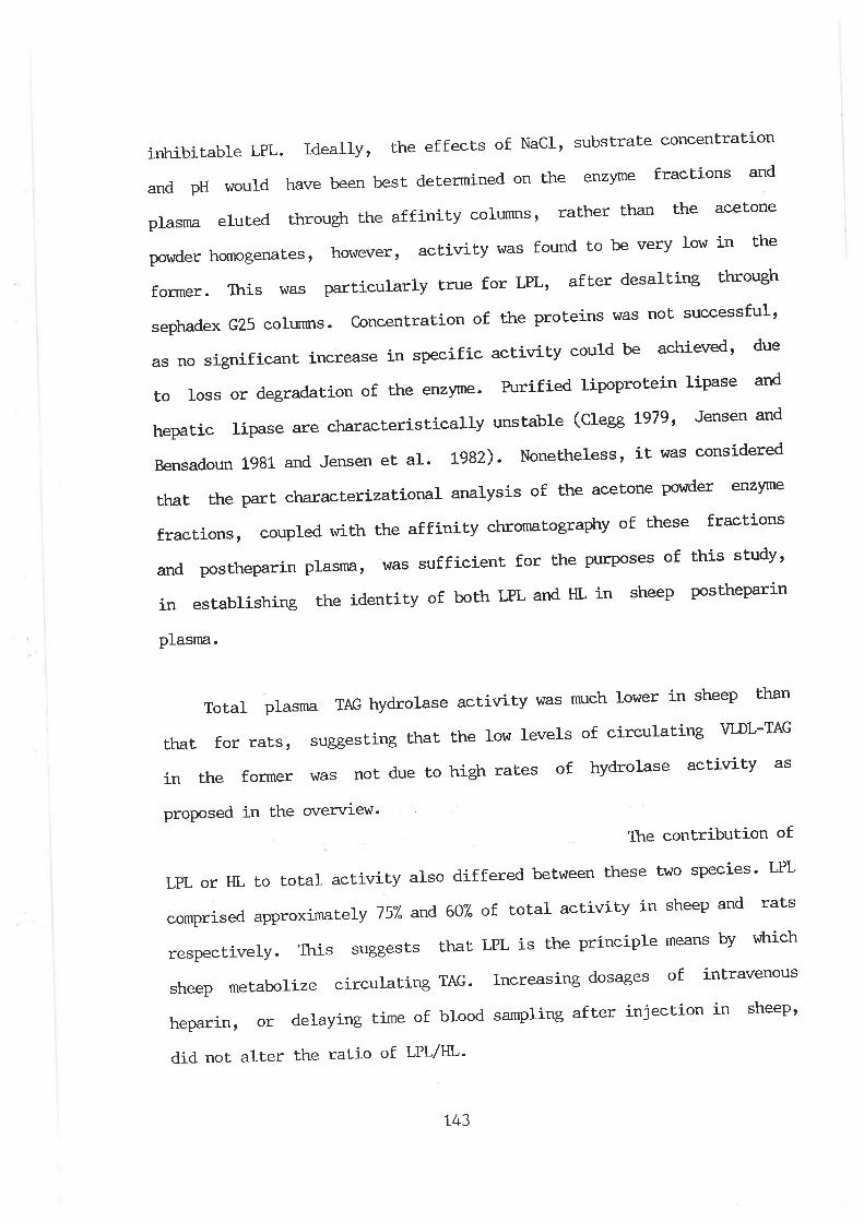
inhibitable LPL. Ideally, the effects of Nacl' substrate concentration
andpHwouldhavebeenbestdeterminedontheenzymefractionsand
plasmaelutedthroughtheaffinitycolunnrs,ratherthant'heacetone
powder homogenates, however, activity was found to be very low in t'he
former. TLris \^las particularly true for LPL, after desalting through
sephadex G25 colunms. concentration of the proteins was not successful'
as no significant increase in specific activity could be achieved' due
to loss or degradation of the enzyme. Rrrified lipoprotein Iipase and
hepatic ripa.se are characteristically unstabre (clegg L979, Jensen and
Bensadoun 1981 and Jensen et aI. Lg82). Nonetheless, it was considered
thât the part characterizational analysis of the acetone powder enzyme
fractions, coupled rnrith the affinity chromatography of these fractions
andpostheparinplasnm'v/assufficientforthepurposesofthisstudy'
in establishing the identity of both LPL and HL in sheep postheparin
plasma.
Total plasma TAG hydrolase activity was much lower in sheep than
ttìat for rats, suggesting that the low levels of circulating VLDL-TAG
in the former \^¡as not due to high rates of hydrolase activity as
proposed in the overview'
Ttre contribution of
LPL or HL to total activity also differed between Lhese two species' LPL
comprised approximate\y 757" and 60% of total activity in sheep and rats
respectivelY. This
sheep metabolize
suggests that LPL is lhe principle means by riLrich
circulating TAG. Increasing dosages of intravenous
heparinr or delaying time of blood sampling after injection in
did not alter the ratio of LPL/HL'
L43
sheept
InordertoestablishtheroleofLPLandHLinthe
hypertriacylglyceridaemia and hepati-c accumulation of this lipid in
metabolically stressed sheep, animals \^Iere deprived of food for 72h, ot
made diabeLic by intravenous administration of alloxan' Fasting for
three days resulted in a significant decrease (287") in total activityt
v¡lrich was due to a 471"decline in LPL and a 387' decline in HL' T?rus' in
addition to an erùnnced rate of release of vLDL-TAG by the liver in
these animals, the plasma accumulation was also attributable to a
decreased catabolism of these pa.rticles. Ttte decline in LPL activity was
probably a response to low plasma insulin levels (Steiner et aI' t975
and Bouchau et al. 19g1). Tüme et al. (19s3) have during the course of
this study also reported lowered LPL activity in powder preparations
from fasLed sheep adipose tissue. Although postheparin plasma LPL
activity has been reported to increase in fasted rats (Nakai et al'
LgTg), it is generally considered that this enzyme decreases in
activity, wtren the plane of nuLrition is below that' required to maintain
homeostasis (Bezman et. aI. t962, Garfinkel et aI. 1967, Austin and
Nestel 1968, lJing and Robinson 1968r Persson et al' L970r, Cryer et al'
Lg76, Taskinen and NikkiLa L979, Fried et al. 1983' I.asuncion and
Herrera 1983 and stam et ar. 1984). LPL from different tissues are often
reciprocal in their relationship to TAG lipolysis'
The rower rates of rAG hydrolysis in fasted sheep postheparin HL
activí-tylssimilar to thal reported for fasted rats (Jansen and Hulsmann
!974 and, Nakai et, aI. LgTg) or fasted cows (Liesnmn et al' 1984)' As
this enzyme is thought to be involved in the further hydrolysis of TAG
in VLDL rennants , iL was noL considered unusual t'haL the activity of
this enzyme paralleled that of LPL'
t44
Total TAG lipase \^/as significantly reduced in diabetic wethers v¡hen
compared to fed sheep (6/+7") or even fasted animals (357") ' Ttris \^Ias
despite a significant increase in HL compared to fed wethers (167") and a
L.g fold increase cornpared to fasted animals- The reason for this \^Ias
the very low LPL activity viLrich was only 87" of- that in fed sheep' (ftt
some anirnals LPL was below the threshold of detection)' In diabetic rats
and fnan, insulin has been shov¡n to be a critical regulaLor of LPL
(nittita et aI. t977 arñ. Stam eE al. lg84) and t'he results here \^rith
insulin stabilized sheep clearly showed that this is also true for this
species. The increase in HL in diabetic animals vTas however, extremely
surprising.AreciprocalinsulinregulationofHLl{Iasconsideredunlikely and in view of the lower HL activity seen in fasted sheep
(v¡trich also have lower concentrations of plasnra insuli-n) shown not to be
so. Ttris agrees with the recent report by t'furase and Inoue (fOaS¡' vilro
found that in perfused rat Iiversr HL was not an insulin dependent
enzyme. Similar results have just been published in a sÈudy of diabetic
dogs (f-tlter et al. 1985), vilrose postheparin HL activity \^Ias also
elevated against normals. Ttre relat.ionship between HL activity, plasnn
insulin and TAG concentration remains unclear. In humans, plasma HL
activity remains constant in either insulín dependent diabetes (pfeifer
et al 1983) or normotriacylglyceridaegic insulin dependent' diabetics
(Nit<tita et al. tgTl), though conversely, streptozot'ocin treated rats
exlribit a reduced HL activity (Nakâi et, al . L97g and SLam et al' 1984)'
It is difficult to perceive v,tkty HL increased in diabetic sheep' It may
simply be a physiological response to the elevated plasma TAG levels' as
a result of increased secretion and reduced LPL catabolismt or may
involve a cornplex hormonal regulation. The synthesis and secrelion of
t45
this protein as a result of diabetes may also be increased, t'hough such
a mechanism is difficult to perceive'
VLDL from both fed and diabetic animals hTere incubated with
postheparinplasmafromfedsheep,todetermineifchangesíneitheror
both the physical nature or chemical nature of these particles' may have
been responsible
observed between
for differences in rates of LPL and HL lipolysis
treatments. Higher rates of VLDL hydrolysis \Âlere
obtained in partieles isolated from diabetic anirnals as opposed to the
same fraction from nonnals. Ttris has not been previously reported and is
in contrast to vrhat Í-s generally considered to occur in diabetic
subjects. Radiolabelled VLDL and chylomicrons isolated from diabetic
rats and reinjected into normal recipients have been shown to have a
reduced rate of clearance (gar-on et al. 1984 and Levy et ar' 1985)'
These authors concluded that the longer plasnra half life of VLDL
particles may be related to qualitative changes ín the apoprotein
profile.However,thisqualificationmayalsobetrueforthehígher
rates of hydrolysis seen in this study. Radiolabelling lipoprotein
particlesandsubsequent'lymoniloringratesofclearanceinvivohasa
number of deficiencies vùrich must be considered' The labelling process
may modify the nature of the particle, such that in vivo lipolysis is
altered. In addiuion, there may be a considerable degree of t'ransfer of
the radiolabelled proteins or lipid cornponents between lipoprotein
fracLions,thusnotport'rayingtrueratesoflipolysis.Similarly,
although clearance may be impaired in diabetic subjects, this may
reflect. a reduction in the removal of lipoprotein re¡rnanLs produced as a
result of the lipolytic Process, rather ttran ref Iect a reduct'ion in the
activities of LPL and HL. The HL (NaCt resistant) cornponent of
r46
postheparin plasma lipase activity IÀIas very low in the WDL-TAG assay
from fed sheep, though based on the postheparin rates of lipolysis of
the triolein enmlsion, this activity was expected to represent only 257"
of total activity. Nonetheless, LPL and HL activities of fed sheep
postheparin plasma were clearly sLi-mulated v¡hen incubated vTith VLDL from
diabetic sheep, and wtrilst LPL remained the major cornponent of lipolytic
rate,quanti-tatively,HLactivityextribitedagreaterincrease.
Depressed LPL activity in diabetic sheep was probably caused by low
levels of plasma insulin, v¡hich sLinnrlates slmthesis and secretion of
LPL. In contrast, the increased rates of HL in these animals seemed to
reflect physiochemical modifications of the lipoproteins' Ttre higher
protein content of vtDL from fed sheep fnay account for this increaset or
perhaps qualitative changes in the apoprotein compliment' Alternatively'
glucosylation of these pa.rticles may have altered lipase activities (as
suggested in chaPter one).
HL is thought to have a physiological role in regulating VLDL-TAG
metabolism, as an impaired removal of these particles occurs after the
administration of HL specific antibodies (Grosser et al' 1981t Fhrrase
and rtak-rra 19g1 and Goldberg et al. Lg82). HL also plays a role in the
conversion of HDL2 type particles Eo the snraller HDI-3 type particles
(Van Tol et al. 1980 and Reardon et al L982). Specifically, HL removes
TAG and phospholipid from IDL, thereby increasing its density (Honrna et
al. 1935). A deficiency of this enzyme leads Lo an accunnrlat'ion of IDL'
an LDL enhanced in TAG and elevation in lÐL2 tyPe parlicles
(Breckenridge et al. tggz). Furthermore, Breckenri¿ge (t985) suggested
that HL may also promote Lhe formation of cholesterol-ester rich HDL
particles. \nith these postulated roles for this enzymet one can predict
L47
a change in the lipoprotein spectrum in diabetic sheep as a result of
an increase in HL activity. In chapter one it was observed that the VLDL
of diabetic origin had less TAG and was richer in cholesterol esters
hrith respect to the equivalent fraction from fed animals. Considering
LPL was depressed in diabetic sheep, this suggests that, HL I'\7as an
important enzyme in the metabolism of TAG rich VLDL. Applebaum-Bowden et
aI. (1935) recently reported in a population study of men and I'tToment
that HL r^7as directly correlated with VLDL-TAG concentration.
FurLhermore, as the VLDL fraction of diabetic sheep htas richer in
cholesterol-esters, HL rnay also be implicated in the formation of
cholesterol-esters, possibly from VLDL surface cholesterol' The LDL
from diabetic animals was enhanced in TAG and contained relatively Iess
cholesterol esters, but this does not necessarily mean that HL does not
hydrolyze VLDL(-IDL-LDL)-TAG, rather, this could reflect the virtual
absence of LPL activity. In other words, the catabolism of VLDL to LDL
in these animals may be solely attributable to HL action. similarlyt
Nozaki et al. (1986) showed that HL could be inversely correlated with
the cholesterol content of IDL, the phospholipid component of IDL and
the enzyme act,ivity was directly proportional to the ratio of HDL2/HDL3
cholesterol. The IÐL fraction in diabetic animals had a lower TAG
content and as a result, a greater protein per unit lipid ratior even
though these particles had a greater cholesterol ester cornpliment, in
comparison to HDL from fed animals. It. would apPear therefore, that
diabetic sheep had an HDL profile vihich resembled smaller HDL3 particles
("" supported by the frequency of snnller HDL particles observed by
Lransmission electron microscopy). The lipid cornplement of HDL from fed
and diabetic animals also correlated \n/ith HL activity in a manner such
L48
t?rat this enzyme may be implicated in the hydrolysis of lipoprotein TAG
and formation of lipoprotein cholesterol esters'
LpL and HL were determined in fed ewes, wethers and rams' Ttris
representsanexaminat'ionofpostheparinplasnnlipaseactivityin
animals actively producing oestrogens to animals producing androgenic
hormones. Total lipase activity was highest in e¡¡es (8'5umol NEFVmI
plasma/h) with wethers and rams extribiting 9t7" and 54% of' this activity
respectively. This trend l^/as seen in both the LPL and HL cornponent of
total Iipase activity. HL activity varied considerably in ewes and as a
result, \,rlas not significantly different from fed wethers or ramst even
though it was on average 837" greater than that of rams' Nonetheless rams
e>dribited an HL activity vùrich was significantlv lower (:37") úan that"--- --ctttistie(1979)
of werhers. It has been well esÞbìî;htd;-tÌtat wethers have a body
compositioncomparabletoewesrinthattheyhaveagreaterdegree
adiposity. The similar rates of LPL obsen¡ed in these animals' vitrich was
almost Lwofold higher than that of rams, showed that the process of
castration and hence, removal of androgenic synLhesis is the causative
agent for the greater degree of fat' deposition' similarlyt HL acLivity
progressively decreased from ewes, to wethers and rams reflecting its
association wiLh the metabolism of IDL-TAG produced as a result of LPL
lipolysis.oestrogensandandrogenstravebeenreport,edtodecreaseand
increase respectively, the activity of HL in postheparin plasma (frrnholm
etal.LgT5a,Tikkanenetal.LgSz,August'inandGreten|979and
þplebaum et al . Ig77, 1985), vfttereas the result's here indicate thaL the
exactoppositeoccursinsheep.Thehormonalregulationofthisenzyme
has not been widery researched and it is apparent that the
r49
oestrogen ic/androgenic conlrol of HL requires further investigation'
During the course of this study, access \^ras gained t'o sheep
designated as genetically 'lean' (Merino) and tobeset (l4erino X Ronmey X
Dorset), providing a natural extension of examining a different aspect
of sheep plasnra TAG metabolism. Postheparin plasma TAG lipase activiLies
were determined
see if these
adiposity. In preruminating lanbs, plasna TAG concent'ration
in preruminaring and postweanpr"!*ä1*ting) , to
could be correlated with heealicO ielease of TAG and
\riIaS
and318nmol/ml and 367nmo1/ml for 'leant and tfatr animals respectively
TAG hepatic secretion l^ras 194nmo1/min/k€ and 249rmol/nin/ke
respectively. These however, were not significantly different'
Similarly, total lipase acLivity wtrich was 337" higher in 'fat' type
animals than in 'leant, could only be attributed Lo a 577" lower HL in
the latter, as LPL in lean aninrals (v¡rictr was 727; that of fat lambs) ¿i¿
not differ significantly. Ttris was probably due to the considerable
degree of variation within grouPs. unfortunately, additional animals
hTere not available at the Lime of writing and furthermore, the same
animals could not be replicated, as Tfiton I'üR1339 has a long plasma half
life in sheep (Uamo et aI. 1933). Both LPL and HL did however, differ
between Lreatments in ruminating postweaned animals' LPL was 307" higher
and HL 257" higher in the 'fat' type lambs. The higher rates of
postheparin plasnn LPL activity seen in the crossbreds, reiterat'es the
relationship of this enzyme with adiposity. similarly, HL activity
positively correlated with LPL activity'
Total posLheparin lipase activity was lower in both'fac and lean'
animals after weaning, probably due to the decline in TAG rich milk' the
150
main dietary componenL of preruminating lambs. The differences in TAG
hydrolase aclivity, particularly LPL vùrich was the principal component
of this reducLion, between tleant and tfatr animals on an identical
plane of nutrit,ion, suggests that different breeds have a genetically
predetermined potential degree of adiposity. In terms of producing
Ieaner sheep in lieu of consumerhealth concelns, this avenue of research
has received little attention.
The toxic syrnptoms exhibited in Triton I^IR1339 treated sheep has not
been obsen¡ed in other such studies in this laboratory and has not been
previously documented (although cats rapidly extribit annaphylaxis v¡Lren
given intravenous Triton (Dr. David Topping, personal connnrnic¿tion))'
However, in the earlier studies, all sheep were housed indívidually in
sheltered pensr r,¡hereas the animals in this study \^Iere grazed openly' As
Èhe chapped black patches were observed only on exposed areas of skin
and in addition, as Triton I^IR1339 is a surfactant w?rich absorbs light
strongly in the ultraviolet region, it was considered that the sheep may
have suffered extensive sunburn as a result of the circulating Triton'
The loss of weight and subsequent death would have resulted from an
inability to eat, due to the extensive damage around the lip region'
It has been shown in chapter two that. sheep liver possesses a TAG
hydrolase comparable to the membrane bound endothelial HL reported for
other species. The regulation of this enzyme and LPL' are however,
inversely related, in that v¡trile LPL activity decreases as a result of
diabetes, HL activity is increased. In addition, this investigation has
shown that the VLDL particles undergo a physiochemical transformation
v¡trich aclually promotes the activities of these enzymes' It is most
151-
probable that such differences are reflected in the protein compliment
of these particles. Chapter three examines for the first time the
apoprotein profile of sheep plasma lipoproteins and determines
qualitative changes in the WDL, LDL and HDL apoprotein compliment as a
result of diabetes.
L52
GTAruER 3
3.1 INTRODUgTION
The hypertriacylglyceridaemia in metabolicatly stressed diabetic or
fasted sheep, is a result of an increased rate of hepatic secretion of
VLDL associated TAG and a decrease in the subsequent catabolism of these
particles by the endothelial lipases. Associated with these differences
¡'^rere ehanges in the physiochemical composition of Uhe lipoprot'eins t as
demonstrated by differences in their rate of electrophoretic migration
and protein/lipid ratio. Aproproteins are the means by vùrich the
metabolic processes of lipoprotein metabolism are directed and
controlled and so the differences in secretion and catabolism of these
pa.rticles are probably reflected in shifts in the apoprotein profile' At'
the start of this project, ovine apoproLeins had not been previously
reported, though recently, Forte et al. (1933) in a study of sheep lung
Iymph lipoproteins, described differences in the apoprotein profile of
LDL and HDL isolated from the plasma and lung lymph. Isolation and
quantificat.ion of all the sheep lipoprotein apoproteins was considered
to be beyond the aims of this investigation. Rather, this section of
this study viras concerTed with establishing the qualitative apoprotein
profile of the major sheep lipoprotein fractions between fed and
diabeLic animals and to see Lf differences between Lreatmentst
correlated with differences in TAG-VLDL metabolism. Apoprotein B can be
readily precipitated by established teckuriques and its concentration
determined. This protein is essential for the release of the TAG rich
VI-DL and so its content in these particles from both fed and diabetic
153
animals was also determined.
Apoproteins have been widely investigated particularly in
monogastric omnivores and many functions have been attribuled to
particular proteins. Thus, apoProteins are necessary for the secrelion
of TAG rich lipoproteÍ-ns from both the liver and intestine, they mediate
receptor binding to hepatic and extrahepatic tissues and regulate the
enzymes v¡trich promote or irihibit release and uptake of their lipid
cornponents. The subsequent uptake of the renu'tant lipoprotein pa'rticles'
v¡trich result from metabolism by peripheral tissues, is also dictated by
the apoprotein compliment, through specific apo-lipoprotein receptors'
In addition, apoproteins also play an irnportant structural role' In
conjunction with the polar head groups of the phospholipids and
unesterified cholesterol, they form the hydropLryllic shell surface of
the lipoprotein molecule (as discussed in Chapter one).
studies of ruminant apoproteins, are however, relatively few and
indeed the full compliment of ovine plasnn apoproteins has not been
previously reported. Limitations, defects or changes in the secreLion of
hepatic wDL, lipolysis of WDL-TAG and uptake of the rermant particles
aLtributable to an abnormal apoprotein profile, can thus only be
speculated uPonr based on the wealth of infornration available for
humans, rats and other monogastric animal species used principally as
models of man. These extensive investigat'ions of the structure and
funct.ion of the different apoproteins are sunmatized in a nurnber of
reviews (Hatctr and Lees 1968, Eisenberg and Levy L975, Jackson et al'
L976, Morrisett et al. tg77, Osborne and Brewet L977, Schaefer et al'
1978, Smith et al. Lg78, Eisenberg Iglg, Ëlavel 1980' Scanu and
l-andsberger 1980, Brewer Ig8!, Miller and Gotto t982, Kane 1983, Turpin
154
.'I
H'\&
"¡
and De Gennes 1983, Mahley et aI. 1984' Dolphin 1985 and Sparks and
Sparks 1985). It is noL intended here to provide an exknustive and
comprehensive treati-se, but raLher, to briefly sunrnarize the major
apoproteins and discuss Lheir role in relation to lipoprotein
metabolism, part.icularly TAG-rich VLDL-
3.1.1 HIJI\,IAN APOPROTEINS; STRUCTURE A}ID FT]NCTION
Table (3.1) lisùs the major hunran apoproteins, their sites of
biosynthesis, plasma lipoprotein distribution and ascribed function. The
apoproteins are very different in both structure and function, with
molecular weights some 60 fold different. Nevertheless, the apoproteins
can generally be divided inÈo those required for de novo synthesis of
lipoprotein particles, enzyme activation and receptor binding.
Apoproteins AI and AII consLitute greater than 907" of HDL protein.
Of this 657. is AI and 257" ATI. Human apoprotein AI is also present on
chylomicrons, but is rarely found in significant amounts on their
remnants, V[-DL, IDL or LDL. Both apoproteins have highly ordered
amphiphylic helical structures, viLrich are thought to be of critical
importance in the binding properties of all of lhe associated
apoproteins, wiLh the Iipid components of the lipoprotein particle'
(Jackson et al. Ig75). þoprotein AI is a single polypeptide of 243
amino acids and a molecular weight of 28,100, of vùrich several isoforms
are lmown to exist. It is synthesized in both the liver and intestine,
though littte is lcrown of their relative contributions and factors vfiich
regulate thern. Hepat.ic apoprotein AI is thought to enter the circulation
in associat.ion with nascent HDL part.icles viLrich have little or no core
ry
155
Table 3.1
Table 3.L lists the major hunran plasma
apoproteins, their sites of biosynthesist
plasma apoprotein distribution and ascribed
function.
ü'{ü
,-l
I
!
TI
I
3
156
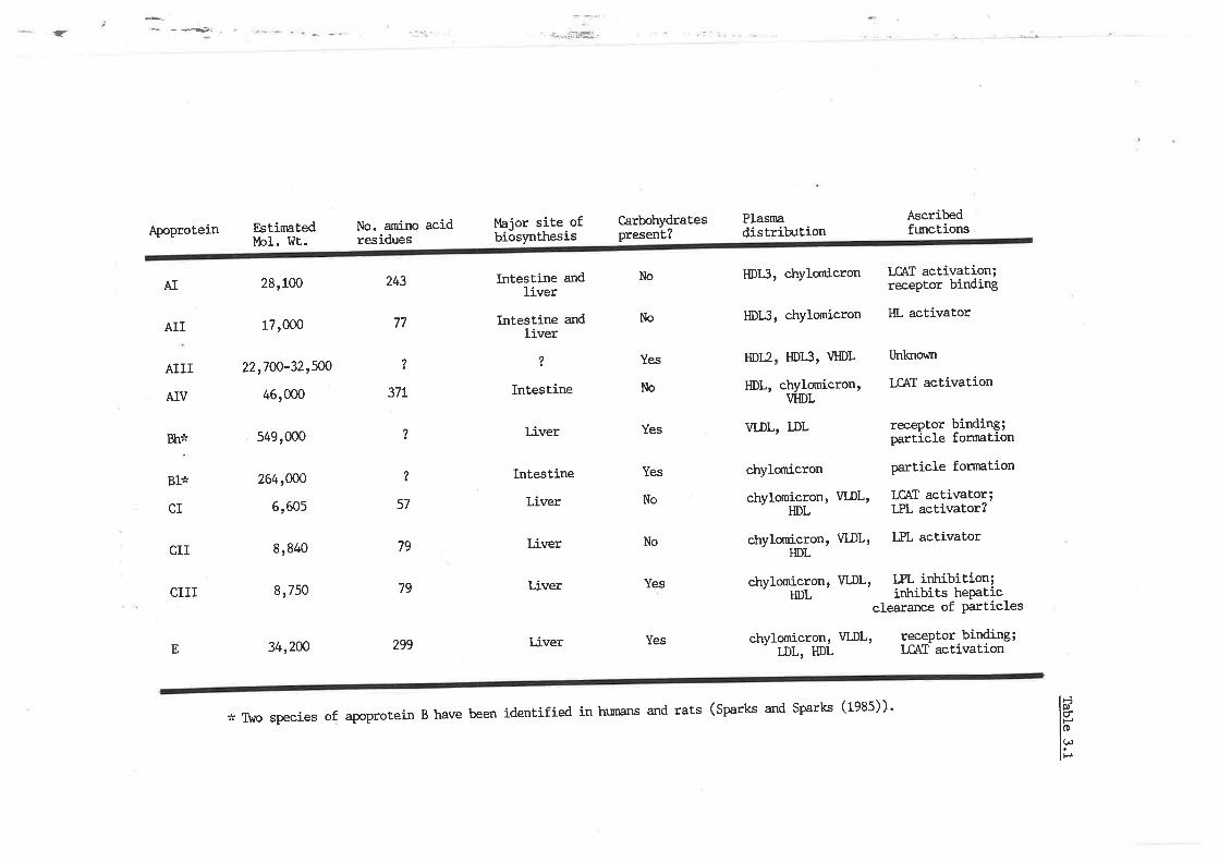
.< *--æ
Apoprotein
AIII
AIV
EstimaÈedMol. I^lt.
28,100
17,o0o
22r7oo-32,5@
46,000
549,000
264rcfo
6,605
8,840
8,750
34,200
No. amino acidresidues
l4ajor site ofbiosynthesis
Intestine andIiver
Intestine andliver
?
Intestine
Liver
Intestine
Liver
Liver
Liver
Liver
Carbohydratespresent?
No
No
Yes
No
Yes
Yes
No
No
Yes
Yes
Plasnadistribution
HDI3, chylornicron
HDLI, chylomicron
Ascribedftnctions
LCAT activation;receptor binding
HL activator
tlnlgrovn
I.CAT activation
receptor binding;particle formation
particle fornation
LCAI activator;LPL activator?
LPL activator
243
77
AI
AII
?
37t
?
57
79
79
299E
HDtz
ÐL'
HDLI, VHDL
chylomicron,VHDL
*iBl*
CI
CII
CIII
?
vL,DL, LDL
chylornicron
chylomicron' VIÐL,HDL
chylomicronr VI-DL'HDL
chylomicronr WDL' LFL intribition;' HDL irùibits hePaticclearance of Particles
chylønicron' WDL' lgcePtor.bil9ing;' LDL, HDL I,CAI activation
* T\¡o species of apoprotein B have been identified in ht¡nans and rats (sparks and sparks (1935))'
ilrl
,]
of cholesterol ester. As such, it is not surprising t'trat apoprotein AI
serves as a cofactor or activator of LCAT (fietaing et al' L972a)' This
mechanism is not, entirely understood, t'hough Soutar et aI. (rgzs)
showed that the degree of activation was dependenL on the fatty acid
composition of the phosphotipid substrate'
Aproprotein AII is a dimer (in nran and chirnpanzee) of 77 amino acids
withamolecularweightofLT'000.þolipoproteinAllhasbeenreported
to erùnnce the activity of til, (Jahn et aI. 1983) and may thus promote
further hydrolysis of IDL-TAG to form LDL particles' Apoprotein AII is
anantagonistofapoproteinAl,inthatitreducestheabilityof
thisproteintoactivateLCAT(Fieldingetal.L972a).
ApoproteinAlVisaprominant'componentofnewlysecreted
chylomicrons, although it is not found in significant amounts ín the
renrrantparticles'wDLorLDL.Therealsoappearstobeinterspecies
variation, as unlike human HDL this apoprotein is a major constituent of
ratHDL(}aahleyetal1984).þoproteinAlVissynthesizedalmost
exclusively by the liver and intestíne. Its amino acid sequence of 371
residues, means this protein has an apParent molecular weight of 46'000'
Like a nurnber of the apoproteins, Arv is a glycoprotein containing
carbohydrate prostheLic groups. Apoprotein AIV also activates LCATr but
to do so is onLy 25% t'hat' of apoprotein AI (Albers et al
I
i
I
I
its abilitY
1e84).
Apoprot.einAIII(vftichisalsoreferredtoasaPoProteinD)hasa
molecularweightofapproximat'ely22,7oo-32,5oodalt,ons.Itisa
glycoprotein conLaining 187" carbohydrate (Kostner L97/+, and Fielding et
al.L972a).TtresiteofsynthesisofapoAlllanditsfunctionareaSyet unresolved.
I
151
tII
1
Apoprotein E is the most. widely distributed protein unit' of
lipoproteins being a constituent of chylomicrons and their rennantst
WDL, DL, and HDL (Curry et al. Lg76 , Kushwaha et al. L977).
þoprotein E is composed of 299 amino acids, has a high arginine contenf
(L7Ð and a molecular weight of. 341200 daltons. Many isoforms have been
reported, though the molecular basis for the Presence of these is
unloown. þoprotein E has been extensively studied and as a result, a
number of functions have been ascribed to it. The major role of
apoprotein E appears to be its involvement in receptor mediated binding
and uptake of lipoproteins through either the apoprotein BrE (I-DL) or
apoprotein E receptors. It, is therefore a critical regulator of
cholesterol transport. Apoprotein E is also involved in the forrnation of
cholesterol ester-rich pa.rticlesr âs this protein also activates LCAT
(though its ability to do so is nmch less than apoprotein AI)'
Apoprotein E has been implicated in the processing of beta-VLDL'
irùribition of mitogenic stinnrlation of lymphocytes and in the metabolic
regulation of the central nervous systern (I/Iahley et al 1984). The liver
is thought to be the major sile of apoprotein E synt'hesis, although
recent.ly, a nurnber of peripheral tissues including the brain, adrenal,
spleen, ovary, kidney and muscle have also been shown to possess the
capacity to synthesize this protein. (nasu et al. 1981r Blue et al'
l-983, Driscoll and Getz 1-983, Boguski et al. 1984 and Reue et al' 1984) '
Apoprotein B is an obligatory structural cornponent of the TAG rich
lipoproteins, as demonstrated by subjects with abetalipoproteinaemia
(wLro cannot synthesize apoprotein n), rnrtrich feature a plasnn lipoprotein
component devoid of VLDL or chylomicrons respectively (C'otto et al . t91L
and Malloy and Kane 1982). Unlike the other apoproteins, apoprotein B
ì
158
does not transfer to other parLicles, but rather remains an integral
part of the lipoprotein. Tttus, apoproLein B is also a major cornponent
of the renmant chylomicron particles and the metabolic products of \ILDL
cataborism, namely rDL and subsequently LDL. rn rnanr apoprotein B
represent s L0-20% of chylomicron protein, 407" or. \rLDL protein and 90% or'
LDL proÈein. ApoproÈein B has proven inmensely difficutt to characLexize
due to its insolubility in aqueous buffers after lipid removal'
sensitiviÈy to oxidation and susceptability to cleavage by proteases'
Nevertheless,inrecentyearsProgresshasbeenmadeinits
characrerization. Ibne eE al. (fggO' 1983) and l4alloy et a1' (fgAf)
identífied two distinct forms of apoproÈein B, namely apoprotein 8100
(or Bh) wtrich is slmthesized by the liver and so is the obligatory
component of VI-DL and apoprotein 848 (or Bl) drích is synthesized by the
inÈestine and is thus usually associated with chylomicrons' In the rat
however, the liver synthesizes both apoproteins 8100 and 848 and both
forms of the apoprotein are associated with vLDL parLicles in this
species (sparks er al. 1981 and Bell-Qgint eE al. 1981). There have
been rnany estimates of the molecular weight of apoprotein Bt v¡trich
reflect the inherent difficulties associated with its characterization'
TÏregeneralconsensusofopinionnohl,isthatapoproteinBl@hasa
molecular weight of 5491000 and apoprotein 848'264t000' AproproÈein B
possesses some 4-87. carbohydrates (Sparks and Sparks 1935)' þoprotein
8100 is also critical for the receptor mediated uptake of LDL through
Èhe apoprotein BrE receptor, which is the means by r'ùrich the majority
(507" in man) of LDL are catabolized (Brown and Goldstein 1983)'
other molecular weight variants of apoproÈeín B
identifiedv¡hosemeÈabolícsignificanceremainunclear.
have been
such,
159
As
09r
ur urêlold Telol aq1 Jo %E? ,'{TaleuTxoldde dn se¿eu osTB IIIC uralo'rdody
.uoTfcB.rJ sTrtl uT urafo.Id Ie1o1 aql lo 7"0t otrdn asT'Iduoc ,¿{eu lcBJ
uT pue 1úlA lo fuauodmoc ) uralordode ¡oleru êt{1 sT IIIC uleloldody
.(tL6I.Te1aTê^EHPuB0¿6I.TBloEsouE-t)1¿18u1le4lce
uT .rolcBJoc leTtruêsse ue sT IID uralo.rddv 'êTcTf,'red sTql Jo luauodutoc
u1a1o.rd.Io[BlxesT(snr{lpuEuTê1o.rd1e1o1aq1Jo1"o]L,{laleurtxordde
s-t IIC ulelo.rdode t'lq'l^ uI 'a1e.rp'{qoqrec '{ue uTeluoc 1ou saoP
u1a1o.rd sTql IC urelotdode a>1-t1 '0t838 Jo lqBTê¡I f,BIncaTour PêleTncTBc E
qlrr\ sprcg ouTuIB 6L Jo uTet{c epllded'{1od e18uts B sT IIC uplo'rdody
'(çtol 're 1a :e1no5) 1y u¡e1o'rdode
usql 1ua1xa rassaT e o1 qSnoql IVDit sale TlcB u1e1o'rd sTql 'sPTce
ouTurB Lç qlTfr ç099 ,{1uo ¡o 1q31en leTncalou¡ e sstl Ic utelo:dodv
'ulalordodTT aq1 Jo êln1cru1s a{1 uT aTo; 1ue1'rodu:1 ue
,,{e1d osle .{eu: ,{aq1 os pue sp1dt1 o1 Eulpulq ueq$ a8ueqc TeuoTlsuLroJuoc
luect¡1u8ts sosnec sulelordode ¡o sse1c sTI{l 'a1c11;ed u1a1o'rdod11
êrl1 Jo 3u1pu1q lTqTquT '{eru su1e1o.rd asaql Jo êll¡os Jo acuasa'rd aq1 qtnoql
acuBreaTc palBTpaü .roldace¡ q1T¡l Pê Torrul ',{11ca'rTP 1ou alB su1a1o'rdode
D êtll . sau¡{zua ;o uo}lTQTrT¡T f,o uoTlB^T1ce aql qlTrl Pa^To^ur ',{lqeltaaur
êle f acuaq puE lDsTToqBlau¡ u1a1o.rdod11 Jo sassaco'rd cTToclBlBc êI{1
g1T¡l palgTcosse ,{11e.raua3 e¡e suleloldode D êI{l "lqH PuE suo:cpro1'{r¡c
ur saTlTluenb 1uec1¡1u31s uT punoJ osTB a'Ie r{eq1 rlo^aAoq tutalo'rd 1q111
Te1o1 aq1 Jo 7"or-ç au¡os dn a>1etu ,{eql uElll uf '(g¿6ilTP 1a ueuDlcrTÐ Pu€
gL6;- 'Te 1a TurnzTanry 'tL6I 'TB 1o ;aTIêrìÜPurA) uollnqtlluoc TBuTlseluT
êutos sT êf,aq1 qSnoqlle (.ran-¡1 aq1 Áq '{llueuTr:ope'rd pala'rcas ale '{attl
.TIIC pue IID r13 su-te1o'rdode se paleu8TsaP suralold 1q31an r'BTnceToÜ
¡\oTaq1ÁIau]Bu..lo.IAuTguBrllf,alÐosulalo.rdodeêo.Iqlaf,Ealattl.¡\aT^ê]sTt{lJoadocseqlpuoÁêqSTSuTelo:daseqlJouoTle]oPTsuoc
chylomicrons and 3-L07" in HDL (Brewer et al. tg74)' þoprot'ein CIII is a
singlepolypeptidechainofTgaminoacidresidueswithamolecular
weight of 81750. Ttrere are three isoforms of the protein v¡l'rich differ in
their sialic acid content. TLre precise metabolic role of apoprotein CIII
and the significance of the sialic acid heterogeneity is unclear'
Apoprotein CIII has a nonspecific inhibitory effect on LPL (Brown and
BaginskytgT|andKraussetaI.lgT3b).Shelburneetal.(1980)and
!üindler et aI. (1980) suggested that the presence of apoprotein GIII may
modulate the uptake of TAG rich rennants by hepatic receptors'
Aproprotein
ar. 1984).
CIII has also been demonstrated to activate LCAT (Jonas et
Inadditiontotheapoproteinsalreadydiscussed,thereareothers
of minor amounts, r'ilrose functions have not yet been elucidaLed'
3.1..2 METABOLISI.,I OF TRIACYI.GYCERIDE .RICH-LIPOPROTEINS; ROLE
OF APOPROTEINS
Theapoprotei.nregulationoflipoprot'einmetabolismthroughvarious
anabolic/catabolic processes requires a continual shift in the spectrum
oftheapoproteins.TÏrisisachievedinadirectedcyclingfashion'
vùrereby the apoproteins exchange between newly secreted lipoproteins and
exis|ingplasmalipoproteins.Ttrebindingorreleaseofspecific
apoproteinsfromthesurfaceofthepart,iclesismodulatedbythree
factors, the lipid binding properties of t'he part'icular apoprot'ein' the
composit'ionofthesurfacelipidsoftheli.poprot'einandalsothesize
of the particle. This relationship is bi-directional, as subsequently,
the nature and ratio of the surface lipids and the core lipid cornponents
T6L
are modulated by the activity of the apoprotein stimulated enzymes and
proteins, which mediate the exchange of the lipid cornponents'
þoprotein B is essential for both the secretion of TAG-rich VLDL
and for the uptake of the product of its metabolism, namely LDL, by the
apoprotein BrE (LDL) receptor. It does not however' apPear to be a major
regulatory factor in the catabolism of these particles. The synthesis
and secretion of VLDL has already been discussed in the general
ovenriew. Of prirnary interest now is v¡hether the genetic, dietary and
hormonal factors t¡hich affect hepatic TAG synthesis and release, mediate
this through the quantity of VLDL particles (apoprotein B secretion)
secreted.
Ttre newly secreted WDL are considered to be relatively inert, even
though they contain a full cornpliment of the apoproteins usually
associated with this pa.rticle (l"larsh and Sparks t979, t982, Rash et al'
1981 and BeII-Quint and Forte 1981), probably because the presence of
the C apoproteins are at a much lower proportion in nascent WDL' than
in plasnm VLDL (I{åmilton et al. Lg76). Ttre metabolic transformations
surrounding nascent VIÐL and the catabolic events vilrich transPose these
TAG rich part.icles to LDL are depicted in figure 3.1 Ttre newly
secreled particles attain a further conrplement of C and E apoproteins,
essentially from the large pool of plasma HDL. It should be realized
that the apoprotein contenL of VLDL varies depending on the protein in
question. Apoprotein B Per VLDL particle remains constant (one
apoprotein B molecule per lipoprotein particle) and does not differ
significantly from ttìât of an LDL particle (Eisenberg and l'evy t975),
because Lhis protein remains with the particle throughout its biological
catabolic life. Howevever, in contrasL, the concentration of apoproteins
L62
Figure 3.1
Figure 3.1 is a diagranntic representation of
the apoprotein regulated metabolic trans-
fornntions surrounding nascent very Iow
densiÈy lipoproteins and the catabolic events
which transpose these triacylglyceride rich
particles to low densiÈy lipoproteins, within
Ehe human plasma comPartment.
O- "poptotein AI
ï- .poprouein Arrr
lì- .poptotein 'B'
Â, - .poptotein C' ,
O'- apoprotein E
L63
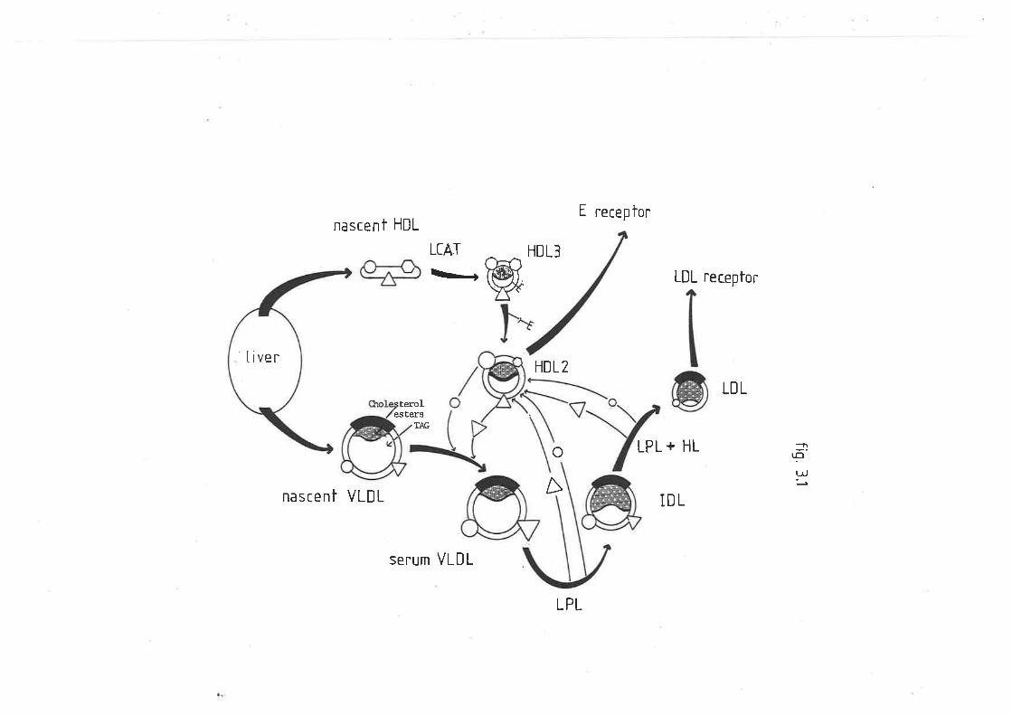
E receptornascent HDL
nascent VLDL
LIAT
\L+HDL3
w LDL receptor
HDL2
LDL
TAG
estersterol
SEIUM VLDL
o
A
LPL + HL
IDL
:nçt!
'tiver
LPL
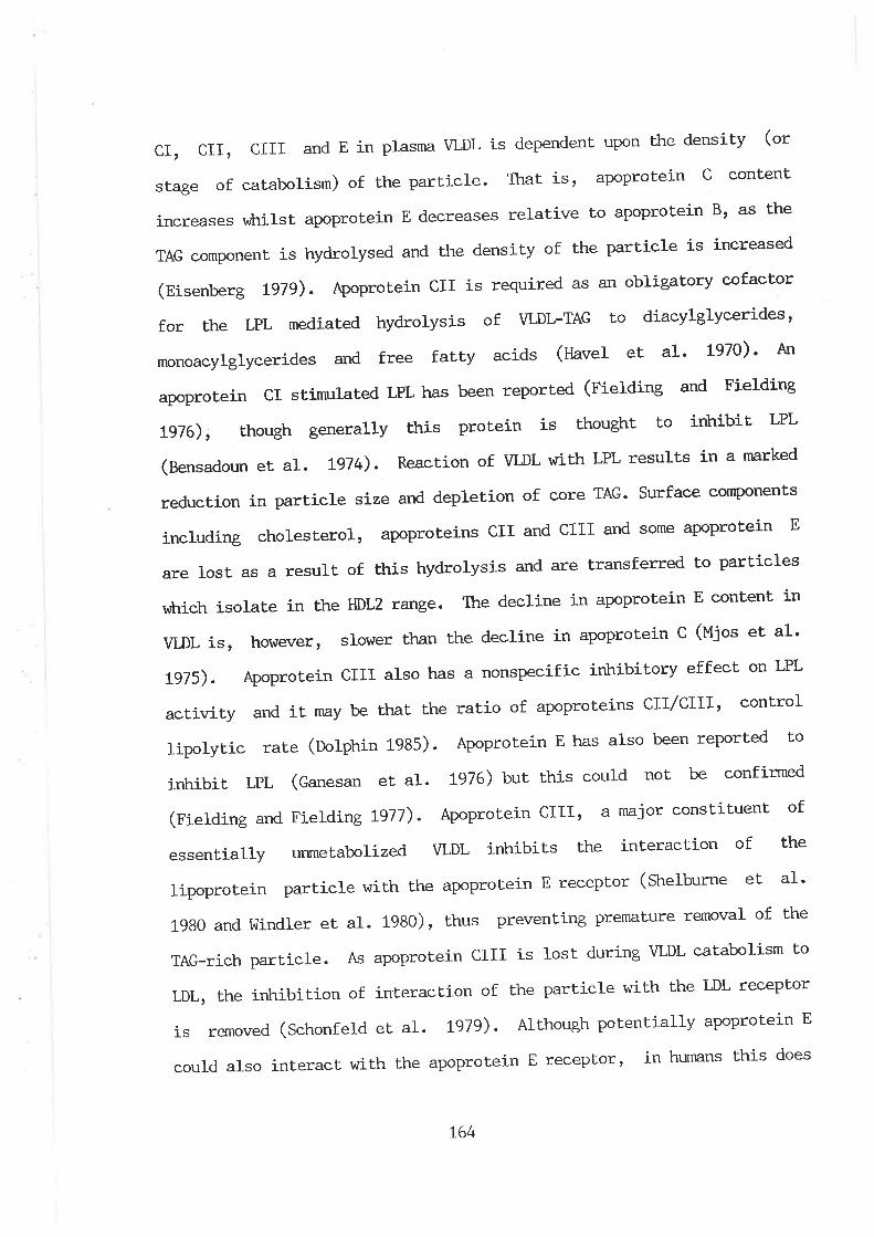
,97
seop sTql sueutnq ur t roldaca.r g u-ralo.rdode aql qlTll lcelê1ur osIP PInoc
g u.ralo.rdode ,{11er1uo1d qSnoqfTv '(øtol 'Te 1a PTêluoqcs) pa'rou:a: sr
roldacer TO.I aq1 q1T¡\ aTcTl.red aq1 Jo uoTlcele1uT 1o uoflTQTt{uT aq1 ''ICT
01 msTToqelec TOTA Sut.rnp f soT sT IIID uTalo.edode sy 'eTcT1;ed qcTf,-CVJ
or{1 Jo Te^ourêl a.rnleu;a.rd Surlua.rrard snql t (OgOt 'TP 1o f,aTPuTI{ pue 0867
-TE 1ê êr.unqToqs) .roldacar g uralo'rdode êq1 q1T¡\ aIcTtrled uralo:dod11
aql Jo uoTlcelê1uT aq1 slTqTqlT TCTA PêzTToqelarxun '{11e11uasse
Jo luanlTlsuoc :.ofeu e TIIID u-telo.rdody '(tt1t 3u1p1a1g pue 3utp1a1g)
pau¡.rTJuoc êq 1ou pTnoc srtp lnq (gtøt 'TB 1ê uesaueg) T¿T lTqTquT
03 pal.rodat uêaq osTB ssq g ulelotdody '(çgOf urqdloq) e1e.r c11'{1odr1
Iolluoc ,tttC/ttC su-¡alo.rdode Jo o-[18f, aq1 1sL{1 êq '{E¡x 1T Pue '{11zr11ce
1¿l-I uo lcaJJa Á.ro11qrr¡rt c1¡lcadsuou B ser{ osTB TIIC ulaloldody '(çtøt
.TE 1a soçhl) 3 ulalo.rdode u1 auTTcaP êq1 ueql lo¡loTs ';aaeatoq tsT TOLA
uT 1uêluoc g uTêlordode uT auTTcaP aql 'a8ue'r Z10H êq1 uT alETosT qcTtIA
sêTcT1aBd 01 pê.uaJSuB.I1 o.le pue stsr{1olp^{q sTql Jo lTnsal e sB lsoI ê;B
g ulalo.rdode a¡os pue IIfC Pue IIC surê1o:dode '1o.ra1sa1oqc 3u1pn1cu1
slueuodruoc êcBJ.rnS 'ÐVtr ê.toc ;o uoJlaTdap pue êzTs aTcTlred u1 uoTlcnpa'r
paì''x e uT sl1nsê.r .l¿1 r{1T¡\ -lffiA Jo uoTlceêU '(¡tøl 'TB 1a unopesuag)
-I¿11 lTqTqrT 01 lq8noql sT u1a1o:d sTql '{11erauat qtnorp | (grct
3u1p1a1g pue 3u-¡p1a1g) pelrodal uaeq ssq'I¿T Pê1BTnxT1s IC u1a1o'rdode
uV '(0¿6T 'TE 1o Tê E{) spTce tlllel aarJ PuE sepl:ec'{131'{ceouout
. sep1.rac,{1t1,{cerp 01 ÐVI-T(rIA Jo srsr{1o.rpr{q Pê1BTPêIü 1¿'I aql f,oJ
.rolc'Joc ,{role3tlqo ue se pa'r1nba'r sT IID utalo'rdody '(øtøl 3'requastg)
pasgêf,cuT sT aTcTl.red aqt ¡o .,{11suaP ar{1 pue pas,{1o'rp'{q s1 luauodruoc ÐVJ
aq1 sB 3g u-¡e1o:dode o1 aATlBTaf, Sasea;caP g u1a1o:dode lsTTtl$ sasee;cur
luaf,uoc C ulalo.rdode 's1 trerl¡ 'a1c1Ued aq1 Jo (urs11oqe1ec Jo a8els
f,o) Á11suep eq1 uodn luapuedap sT'ICT^ eurseld uT g Pue IIID .IID tIf,
not appear to be the case and factors goveming this preferential uptake
re¡nain unresolved. It nEy be that the apoprotein B molecule interferes
with the binding of Èhe apoprotein E and its receptor' There have been
numerous investigations into the apoprotein BrE recePtor pathwayt
díseussion of v¡hich is beyond the scope of this review.
165
3.2 METIIODS AND MATERIALS
3.2.L AT{I¡4ALS
Fed sheep and alloxan induced diabetic sheep were housed and
nnintained as already described (Section L'z't) '
The major lipoprotein fractions $rere isolated by serial
ultracentrifugation as described in section 1.2.4.6. The VLDL fraction
was washed by recentrifuging the lipoprotein concentrate for 20h at 20oC
with the 1.96M NaBr b¡ffer of solvent density 1.0063g/ml (unless sLated
otherr,.rise).
3.2.2 PROTEIN Ð(IRACTION
A modified procedure of the method described by cLram and l(nowles
(fgZA) designed for the delipidation of plasma lipoproteins from grossly
hypertriacylglyceridaemic subjects was used'
To 200uI of lipoprotein fract,ion in Eppendorf tubes 800ur of a
mixture of n-butanol/diethyl ether (1S:SS v/v) was added' The tubes were
mixed thoroughly by rotating end over end for 45-6ùnin at room
temperature. The samples were cenLrifuged for 5min and the organic
supernatant discarded. The process I4Ias repeated by the addition of
another 800uI of solvent. After aspirating the solvent phase, the lower
aqueous phase was kept under a gentle stream of high purity nitrogen for
5min Lo remove all traces of solvenL.
L66
3.2.3 APOPROTEIN B AND SOLUBLE APOPROTEIN DE'TERMINATION
Total protein content of each fraction was determined by the
modified Lov¡ry method described in sectj-on L.2.6- Selective
precipitation of apoprotein B was achieved by addition of iso-propanol,
as descríbed by Holnquist and Carlson (L977). Total soluble apoproteins
(ArC, and E) were then determined and the apoprotein B content
calculated by subtracting the soluble proteins from the total proteins'
Standard protein solutions of bovine serum albumin also contained t07"
isopropanol for the soluble protein assay.
3.2.4 SODII'M DODECYL SI.JLPHATE- POLYACRYI.AI\,IIDE GH-
sodium dodecyl sulphate-polyacrylamide gel electrophoresis (sDs-
PAGE) of the apoproteins was run on 180nm X 200fnn X 0.7nm or 180nrn X
200nm x 1.5nrn gels, r¿ith a t+% to 3o7" actyLamide gradient cont'aining 2.47"
crosslinker (bis-acrylamide), made up in a modified I¿enrnli
discontinuous buffer (0.:zsu Tris-HCl, pH 8.8, 0.1% sDS). Plates were
cast using a Pharmacia GSC-z gel casting apparatus. Each gel was
overlayed with 2-3crn of stacking gel (47. acryLamide, 2-77" crosslinker,
0.125M Tfis HCl, pH 6.8 , O.L"A SDS). Acrylamide gels were polymerised
with 107" anrnonium persulphate and NrNrNr,Nr -Tetramethylethylenediamine
(rnmo).
To each aliquot of protein exLract an equivalenL amount of l-aennnli
sample buffer, containing 0.01M Tfis-HCl, PH 8.0, 0.001 EDtfA' 17" SDS and
5% mercaptoethanol was added, thoroughly mixed and heated at 100".C for
5-10rnin. Glycerol (507. solution) was added to increase the density of
t67
the protein solution and bromophenol blue (0.1Ð was also added to
monitor the rate of electrophoretic migration.
If the protein concentration of Iipoprotein fractions llras low (less
than 5ug,/50uf ), samples \tlere concentrated under vaccum (fOOfpa) in a
Savant Speedvac centrifuge (approximately 1500 r.p.m.) at 30oC until a
suitable concenLration was achieved.
5-20ug of lipoprotein protein was added for each lane of the gel.
Each lane containing protein \{as separated by a lane containing buffer
only. Fach gel had a complete compliment of both high and low molecular
weight markers (section 3.2.5).
The SDS-acrylamide gradient gels were subject to electrophoresis in
0.05M Tris-HCI, 0.348M glycine, pH 8.3, 0.17" SDS' in a Pharrnacia GE
2/4LS water cooled, circulating tank. Constant voltage was nnintained at
100V until each sampte had migrated through the stacking gel and into
the separating gel. Voltage hras then increased (ISOV) and maintained for
a total of 1500Vh. The gels were carefully removed, fixed and stained in
0.L7" Coomassie Blue R-250 nnde up in methanol:acetic acid:water
(ZSO:100:650) for a minimum of 6h. Destaining took approxirnately L7}l^
r,rrith 3 X 1L changes of methanol:acetic acid:water (Z5O:100:650). Gels
hrere dried in a Bio Rad dual temperature slab gel dryer (model SE1125B)
at 60oC for 2-3h.
Nth order exponential regressÍ-on \,r¡as applied to the molecular
weight protein standards of each gel. Regression l47as better than
r'4.92. The Rf of each sample band was then interpolated according to
the standards, a molecular weight determined, and a tentative identity
assigned.
168
3.2.5 }4ATERIALS AND REAGENIS
Acrylamide and bis acrylamide (electrophoresis grade) and TEMED
(gg7" pure) were purchased from Signra Ckremical C,ompany. Low and high
molecular weight. markers were purchased from Bio-Rad Australia' Ttre low
molecular weight group contained lysozyme (141400) soybean trypsin
irùribitor (21,500), carbonic arihydrase (311000), ovalbumin (+5,OOO) t
bovine serum albumin (66,200) and phosphoryIase B (921500). Ttre high
molecular weight markers were bovine serum albumin, phosphorylase Bt
beta galactosidase (116,250) and myosin 2001000. The gel casting
apparatus, and electrophoretic tank were purchased from Pharmacis Pty.
Ltd., Uppsala, Sweden.
L69
3.3 RESULTS
3.3.1 APOPRCIEIN PROFII-E OF FED AND DIABE'TIC SHEEP
A number of gradient, gels vùrich differed in their thiclcress,
running time, amount of total protein and source of apoprotein \'\7ere runt
the results of v¡hich are tabulated in table 3.2. Apoprotein components
of particular lipoprotein fractions were not always deÈected in aII
acrylamide gels, principally because either the lipoprotein
concentration was very low, or the respective band was overlapped with
adjacent more abrndant proteins of similar molecular weight.
Nonetheless, a qualitative apoprotein profile of each of the major ovine
lipoprotein classes has been given. Ttre apolipoprotein distribrtion of
sheep plasma VLDL, LDL and HDL on SDS-PAGE (4-30"A) is shown in figures
3.2 and 3.3. The qualitative nature of the lipoprotein apoproteins
contained proteins vrith molecular weights similar to apoprotein Cts
(less than 11,000 daltons), apoprotein AII (151500 daltons), apoprotein
AIII (ZtrSOO daltons), apoprotein AI (25,500 daltons), apoprotein E
(33,500 daltons) apoprotein AIV (47'500 daltons) and apoprotein B
(greater than 65,000 daltons) (tabfe 3.2). The methods described did not
permit absolute identification of the apoproteins due to the
unavailability of suitable antisera. However, the respective apoproteins
vilrich are cofnmon to rat wDL, namely the B, c and E apoproteins, had
similar rates of migration and molecular weights within the given ranges
used to identify the ovine compliment (table 3.2' figures 3.2 and 3'3)'
Ttre major apoprotein components of vLDL on this basis from fed
sheep \^rere apoprotein B (molecular weight bands 60t000-465r000; Lhese
L70
Table 3.2
Table 3.2 lists the apoprotein compliment of
each of the major sheep plasma lipoproteins,
in both fed and diabetic animals. The
identiÈy of the apoproteins hrere based on
their molecular weight determined by sodium
dodecyl sulphate polyacrylamide gel
eleetrophoresis, and the apoprotein profíIe
of rat plasma very low density lipoproteins.
T7L
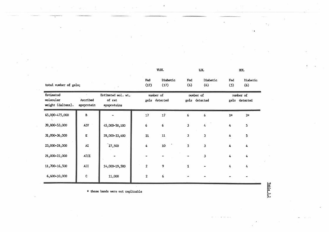
total nnber of gels;
Estimtedrclec¡¡laræigþt (daltons).
Estifiated ¡rcI. wt.of rat
apoproteins
¡¡¡mber ofgels detected
T7 L7
6 6
11
4 10
2
nunber ofgels detected
HDL
Di¡bettc
nr¡iber ofgels detected
1* 2*
4 5
VLDL lÐL
Fed
(17)Diabettc(17)
Di.abetic(6)
Fed
(s)(6)Fed
(6)
Ascribed
apoprotein
65,00û-475,000
39,800-53,000
31,@36r000
æ,00G-28,000
2Lr@22,W
11r70Þ16,500
6,600-10,000
5
4
4
4
11
6
4
3
6
3
3
5 3
3 4
4
4
419
6
2
IF
lrl¡"
AIV
E
AI
AIII
AII
c
B
45,@50,100
28,000-33,400
14,000-19,500
27,ffi
11,000
* these bands r¡ere not replicable
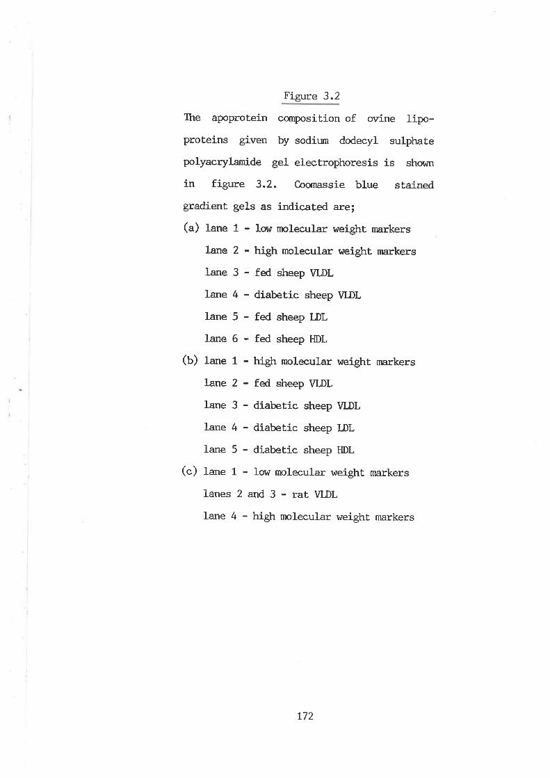
Figure 3.2
The apoprotein composition of ovine lipo-
proteins given by sodium dodecyl sulphate
polyacrylamide gel electrophoresis is shovrn
in figure 3.2. Coomassie blue stained
gradient gels as índicated are;
(a) lane 1 - low molecular weigþt markers
lane 2 - high mol-ecular weight markers
lane 3 - fed sheep VLDL
lane 4 - diabetic sheep \ILDL
lane 5 - fed sheep LDL
lane 6 - fed sheep HDL
(U) fane 1 - high molecular weight rnarkers
lane 2 - fed sheep VLDL
lane 3 - diabetic sheep VLDL
lane 4 - diabeÈic sheep LDL
lane 5 - diabetic sheep HDL
(c) lane 1 - low molecular weigþt markers
lanes 2 and 3 - rat \|LDL
Iane 4 - high molecular weight markers
L72
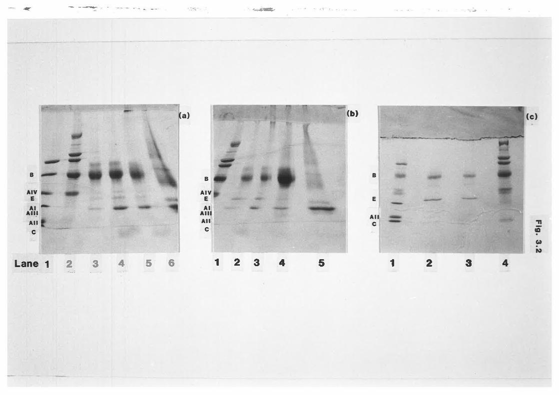
* ÈÉ.æ- ': 'rl
bl
i-jtËl.--ll'Ø'
..,.i,.'r.lf'29456 12 3 4 5 12 3
'¡,t.
,.!
ht
li
I
Figure 3.3
Ttre apoprotein composiÈion of ovine lipo-
proteins given by sodium dodecyl sulphate
polyacrylamide gel electrophoresí-s ís shown
in figure 3.3. Coornassie blue stained
gradient gels as indicated are;
(a) lane 1 - fed sheep VLDL
Lane2-ratVLDI
lane 3 - Iow molecular weight rnarkers
(b) lanes L and 5 - Low molec¡.¡lar weight
markers
lanes 2 and 6 - high molecular weight
markers
lane 3 - fed sheeP HDL
lane 4 - diabetie sheeP HDL
1.1
tI
L73
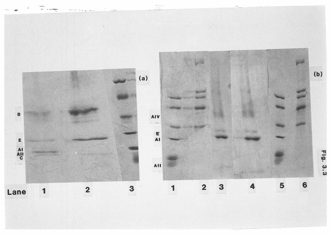
{
AIY:+
ËAI
Ail t,oi. a
frl
3 I 3 4I
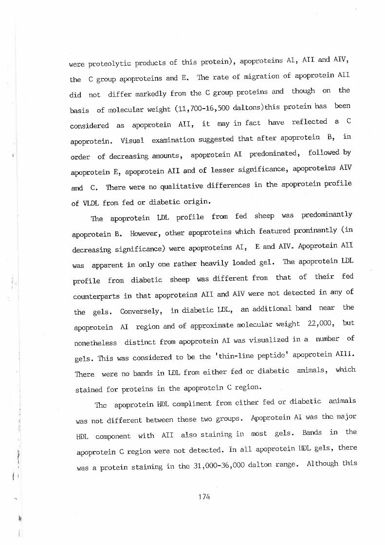
tïI
I
h/ere proteolyt,ic products of this protein), apoproteins AI' AII and AIV'
the c group apoproteins and E. The rate of migration of apoprotein AII
did not differ markedly from the c group proteins and though on t'he
basis of molecular weight, (11 1700-1¡61500 daltons)this protein has been
considered as apoprotein AII, it nny in fact have reflected a c
apoprotein. Visual examination suggested that after apoprotein Bt in
order of decreasing amounts, apoprotein AI predominated, followed by
apoprotein E, apoprotein AII and of lesser significancet apoProteins AIV
and C. Ttrere lrtere no qualitative differences in Ehe apoprotein profile
of VLDL from fed or diabetic origin'
Ttre apoprotein LDL profile from fed sheep v¡as predominantly
apoprotein B. However, other apoproteins v¡Lrich featured prominantly (in
decreasing significance) were apoproteins AI, E and AIV' Apoprotein AII
hras apparent ín only one rather heavily loaded gel' The apoprotein LDL
profile from diabetic sheep was different from that of bheir fed
counterparts in that apoproteins AII and AIV were not detected in any of
the gels. conversely, in diabetic LDL, an additional band near the
apoproLein AI region and of approximaLe molecular weighL 22t000t but
noneLheless distinct from apoprotein AI was visualized in a ntunber of
gels. This was considered to be the rthin-line peplide' apoprotein AIII'
There \^/ere no bands in LDL from either fed or diabetic animals, vùrich
stained for proteins in the apoprotein C region'
The apoprotein HDL compliment from either fed or diabetic animals
was not different beLween these two groups. Apoprotein AI was lhe major
HDL cornponent with AII also staining in most gels' Bands in the
apoprotein c region rlrere not detected. In all apoprotein HDL gels, there
was a protein staining in the 31,000-361000 dalton range' Although this
!
L74
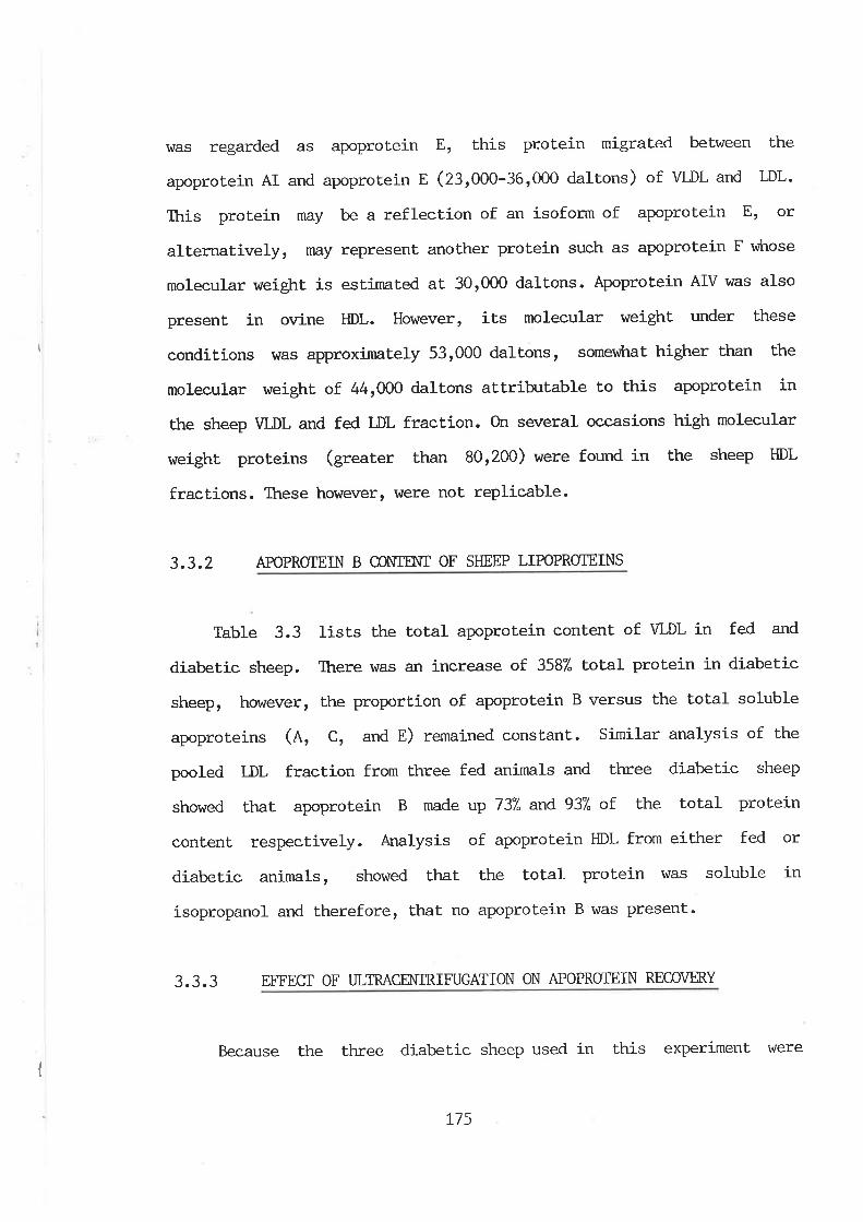
I
rdas regarded as apoprotein E, this protein migrated belween the
apoprotein AI and apoprotein f (231000-36'000 daltons) of VLDL and LDL.
This protein may be a reflection of an isoform of apoprotein Er or
alternatively, may represent another protein such as apoprotein F vfrrose
molecular weight is esLinnted at 301000 daltons. þoproteín AIV was also
present in ovine HDL. However, its molecular weight under these
conditions vras approxirnately 531000 dalbons, somev¡tnt higher than the
molecular weight of 441000 daltons attribrtable to this apoprotein in
the sheep VLDL and fed LDL fraction. On several occasions high molecular
weight proteins (greater than 801200) were found in the sheep HDL
fractions. These however, were not replicable.
3.3.2 APOPROTEIN B @[\TIN{I OF SHEEP LIPOPROTEÏNS
Table 3.3 lists the total apoprotein content of WDL in fed and
diabetic sheep. There was an increase of 3587" toüa1 protein in diabetic
sheep, however, the proportion of apoprotein B versus the total soluble
apoproteins (4, C, and E) remained constant. Similar analysis of the
pooled LDL fraction from three fed animals and three diabetic sheep
showed that apoprotein B made up 73"A and 937" of the total protein
content respectively. Analysis of apoprotein HDL from either fed or
diabetic animals, showed that the total protein \^/as soluble in
isopropanol and therefore, that no apoprotein B was present.
3.3.3 EFTEC.I OF I.]LTRACENTRIFUGATION ON APOPRCIEIN RECOVHìY
Because the three diabetic sheep used in this experiment wereII
L75

Tab1e 3.3
Table 3.3 lists the toLal apoproüein and
percentage apoprotein B content of e¿ch of
Ehe major sheep plasrna Iipoproteins from both
fed and diabetic animals.
(") + x.x=tumber of animals + the standard
deviation of the mean
L76
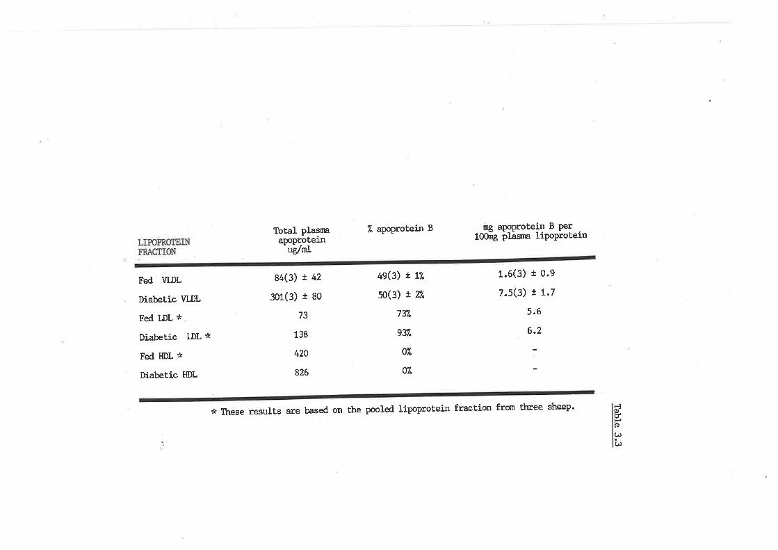
Total plasmaapoprotein
ug'/ml
84(3) ! 42
3ü(3) t 80
73
138
420
826
7. apoprotein B
49(3) r 17.
5o(3) ! T/"
7l/.
gl/"
f/"
07"
rng apoprotein B Per1@ þIãsrna lipoProtein
Fed \¡LDL
Diabetic VLDL
Fed LDL *
Diabetíc LDL *
Fed HDL *
Diabetic HDL
1.6(3) t o.e
7.s(3) r 1.7
5.6
6.2
* TÏrese results are based on the pooled Iípoprotein fraction from three sheep' Ëft)itHo(^)
(,
grossly hypertriacylglycerldaemic, recentrifugat.ion (20h) of the plasma
of these animals in the less than 1.0063g/ml solvent density range \¡/as
required to totally recover this fraction. An average of 267" of VLDL was
not recovered on the first, centrifugation (ZOt'r) in these aninnls- Total
plasma VLDL from fed sheep hras recovered with only one period of
centrifugat,ion.
To investigate the effects of prolonged ultracentrifugation on the
apoprotein content of these particles, the buffer overlay of density
1.0063 used to 'washt the VIÐL fraction was also assayed for total
protein, soluble protein and apoprotein B. Table 3.4 shows that
substantial losses of apoprotein resulted. Ttre ratio of apoprotein B
versus total (or sotuble) apoproteins was equivalent to that of the
intact particles, namely a 497" apoprotein B content. This being the
case, analysis of VLDL protein before and after 'washÍ-ngt, indicated
that in fed and diabetic sheep an 827" and 627" loss of apoprotein
componenLs occured aS a result of this procedure respectively.
L77
Table 3.4
Table 3.4 lists the recovery of very low
density lipoprotein apoproteins based on the
protein content of the 1.006g/mt buffer
overlay used to 'wash' the lipoprotein
fraction.
L78
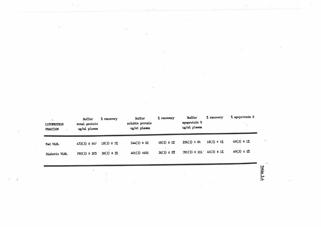
h¡ffer 1 recorerytotal proteinug/cl plåsoa
B¡ffersoluble proteln
uglol plåsoa
Z recoæry h¡fferapoprotein B
ug/ol Pro*
I recorerY. Z aPoProtein B
LIPOMÛIEIN
!ÎACÍTON
Fed VLDL 473(3)r167 18(3)t12 2tú(3)t82 18(3)t1Í 226(3)È84 18(3)t1Z 4e(3)t1l
rli¡beríc vr.DL n2(3) t zo5 3s(3) t 22, 401(3) t101 36(3) I 2É '391(3) t 111' a1(3) t 11 49(3) t 1tr
lÐIHl.D
h
3.4 DISCUSSION
This study represents the firsÈ examination of Lhe apoprotein
profile of all sheep plasma lipoproteins. Identity has been based on
rates of migration, molecular weights and homogeneity with the
documented apoprotein compliment of rat VLDL using SDS-PAGE'
Confirmation of these by other means, such as antibodies raised against
specific apoproteins, could not be done in this laboratory at Ehe time
of writing. Nevertheless, bearing in mind that this report represents
preliminary investigations, the sheep apoprotein profile wtrich has been
presented witl be discussed, in view of the role these proteins have in
lipoprotein TAG metabolism within the plasma compartment.
Ttre qualitative profile, in terms of the total spectrum of plasnn
apoproteins, strrns thatdreqare, similar to other species such as the rat
or humans, in that each of the major lsrown apoproteins, namely the A, B,
C and E group of proteins, were present. The distribution of these
between VLDL, LDL and HDL' did however, reveal sorne unusual features in
comparison to that. for man.
sheep vLDL contained proteins vilrich in terms of molecular weightt
corresponded with apoproteins AIr AII' AIV' B, C and E' In nnn the B and
c proteins are the major cornponents with AIr AII and AIV present only in
trace amounts. unfortunately, a gel density scanning device was not at
the disposal of this laboratory and so quantitation of these v/as not
possible. Nevertheless, visual examinaLion of the acrylamide gels,
clearly showed that apoprotein AI was the major component of sheep vLDL
after apoprotein B. Though of less significancer apoProteins Et AIII AIV
and C r^/ere also detected on a number of occassions ' Ttre protein
L79
recognised as apoprotein AII had a migration not greatly different from
the C group apoproteins and though iLs calculaLed molecular weight \^7as
equivalent to lhat usually associated with apoprotein AfI, this band may
in fact represent a c apoprotein. The A grouP of proteins are together
responsible for the activation of I-CAT, stimulation of hepatic lipase
and receptor binding and so the results presented here suggest that the
liver could play a major role in the catabolism of TAG rich VLDL
particles. In chapter two, it was seen that HL formed an integral part
of the me¡nbrane bound TAG lipase potential associated lÀrith plasnn and
furthermore, that this activity was significantly increased in diabetic
wethers. It may be that an increase in these components, or perhaps a
change in the ratio of particular apoproteins in diabetic vLDL promoted
this activity. Ttre higher rates of vLDL-TAG HL hydrolysis in particles
from diabetic animals also suggest that this may be an apoprot'ein
sÈimulated effect (perhaps increased apoprotein AII). Streptozotocin
induced diabetic rats, unlike lheir fed counterparts, also have an
apoprotein AIV component in their vLDL fraction, (gar-on et al' L976)
though these rats are associated with a lowered HL activit'y (Nat<ai et
aL. L979).
Another observation of potential interesL in the sheep VLDL
fraction was that the C group apoproteins did not appear to be of major
significance, as this class of apoproteins was detect'ed in relatively
few gels (207" total VLDL gels). If however, the protein designated as
apoprotein AII is in fact a c apoprotein, then their contribution to the
total protein spectrum will of course be much greater. Assuming that
this is the case, the results presented here suggest that the c
apoproteins have molecular weights considerably higher than that
180
reported in other species. A low apoprotein c content in sheep VLDL
could explain the relatively low postheparin plasma lipase activities,
(in comparison to rats or nnn) observed in these aninmls under fed
condirions. Alternatively, the apoprotein CII/CIII (Ct:Clt:CIII?) ratio
may be such that activity is not optimized. low levels of apoprotein
CIII in sheep VLDL, would also support an erùnnced hepatic contribution
to the metabolism of these parLiclesr âs the inhibitory affect of this
protein on the apoprotein BrE receptor would be reduced. Ttre tC'
apoproLeins \^rere more readily detected in diabetic VLDLT suggesting that
the low LPL activity in fed sheep hras not a result of deficient
apoprotein c in these particles. In facl, the higher rates of diabetic
sheep VLDL-TAG LPl-lipolysis, v¡hich was nearly three fold higþer than
the same fraction from fed aninnls, could reflect an improved
apoprotein C (perhaps greater CII) compliment. Alternatively, the
lipoproteins of diabetic sheep were physiochemically different from
those derived from normals, probabty a result of glucosylation, as
suggested by the greater rates of electrophoretic migration' Curtiss and
t{itzum (1985) have shov¡n that. in hyperglycaemic diabetic subjects'
apolipoproteins AI, AII, B, CI and E of the TAG rich lipoprotein
fraction were glucosylated and it is possible that the higher rates of
diabetic VLDL lipolysis may reflect an erùranced capacity for the
apoproteins to stimulate the lipase enzyfnes. However, the physiological
significance of apoprotein glucosylation will only'be lcrown, vùren it can
be demonstrated thât the apoprotein structural ckr'anges that accomPany
glucosylation, also lead to functional changes'
Apoprotein E, was not surprisingly, a readily detected prot,ein in
sheep VLDL. It's presence signifies that VLDL is metaboLized by the
181
apoprotein BrE (E?) receptor. It. would be interesting to determine
apoprotein CIII/E raLio in vLDL from both fed and diabetic aninals
view of the role of these proteins in regulating hepatic clearance
these part.icles.
the
in
of
Apoprotein B was the nrajor cornponent of both fed and diabetic sheep
VLDL, comprising 497" of the total protein compliment in each group' In
chapter one, it was shov¡n that the VLDL particles actually contaj-ned
more protein per unit lipid (table t.2). The ratio of apoproLein B to
total protein remained constant between fed and diabetic animals, and so
it could be calculated that there \,rras approxirnately 1.6mg of apoprotein
B per 100rng VLDL in fed aninnls, rilrich is similar to that found in rnan
(f-Smg/fOûng VLDL (Sparks and Sparks 1985)). In diabetic sheep, there
\,\7as nearly a 5 fold increase of this protein per unit wDLr thât is
7.5mg per 10ùng vIÐL. The secretion of these particles in met'abolically
stressed sheep was therefore not reduced or impaired by the rate of
synthesis of this obligatory protein.
It was hoped that the electrophoretic pattern of apoprot'ein B would
give an estinration of its molecular weight. However, in consequencet it
was considered that inclusion of Ellman's reagent in the centrifugation
procedure and extraction of lipid from VLDL with a butanol based solvent
mixture, may potentially promote cleavage of this protein into smaller
high molecular subfractions (Professor Julian Marsh personal
conrnunication). These replicable protein subunits (greater than 65'000)
r^rere at first. thought to be albumin binding to the lipoproteinst
however, the absence of such bands in the HDL fraction, vfuich on the
basis of the methods used here Lo isolate the lipoprotein fractions,
would contain the most. albumin, coupled with the absence of precipitable
L82
protein by isopropanol in this fraction, indicated tklat these bands were
not. albumin. If apoprotein B did not undergo any oxidation or cleavage,
then the approximated molecular weight of this protein in sheep VLDL and
LDL would be 661000-80,000' similar to that derived by Olofsson eL al'
(fOaO¡ v¡ho used a 57" SDS-PAGE system coupled with urea gel filtration'
The apoprotein LDL profile of sheep was again different from that
usually associated with nnn. In addition to proteins staining in the
apoprotein B and E region, there were others r'ihich on molecular weight
correlated hrith apoproteins AI, AII and AIV. Apoproteins AI and AIV are
minor components of human LDL and apoprotein AII is not usually
associated l^tith this fraction. The protein staining in the AII region
was only detected on one occassion, suggesting thaL this is only a minor
cornponent of sheep LDL. Alternativelry, if. this was a C apoprotein, then
this may simply reflect intermediate particles between the transition of
VLDL to LDL. Forte et al. (1933) did not detect any 'A' apoprot'eins in
ovine plasma LDL, though conversely, they did report that apoprotein AI
constituted 687. of lung Iymph LDL. Similar1y, in the results presented
here, this protein appeared to be the major componenL in sheep plasma
LDL after apoprotein B. The apoprotein AI and AIV conrponent of these
particles is readily associated with their high cholesterol esLer
content (qA1" in fed sheep and 60% in diabetic animals), both proteins
being activators of LCAT. LDL from diabetic sheep were shown to have a
higher content of cholesterol-esters and less TAG than this fraction
from fed animals. This may reflect an increased apoprotein AI and AIV
stimulated LCAT activity and the increased rate of hepaLic lipase in
diabetic sheep. Apoprotein AII v¡trich stimulates HL was not detected in
any of the gels for diabetic sheep and so if higher rates of hepatic
183
lipase are responsible for lower levels of LDL-TAG, then this would not
be an effect of this protein in this fracLion. C,onversely, this could be
a result of an increased apoproteín AII promoted actiwity of this
enzyme, in the precursors of LDL, that isr VLDL. In addition, the
diabetic LDL fraction featured a protein closely associated rtrith the
protein referred to as apoprotein AI, considered to be the thin line
peptide apoprotein AIII. This was not observed in the VLDL fraction of
these animals and so it can be concluded that this protein was acquired
during or after metabolism of the TAG conrponent. The function of this
protein is unclear, however, it, nray be that it too has the capaeity t'o
stinn:late HL and perhaps LCAT. It is difficult to perceive vùty this
protein vlas not. detected in any of the gels containing fed sheep DLt
but iL may be thåt this erçhasises the different catabolic pathways of
VL,DL between different treatments. The absence of the tCt apoproteins in
ovine LDL was probably a reflection of the same Process vùrich occurs in
man, namely the hydrolysis of TAG by LPL results in a decreased density
of these particles and subsequent loss of the C group apoproteins.
Apoprotein B was the major protein (727") of LDL from fed sheep,
however, it was not as significant as that usually attributable to man
1OO-0S7"). This again supports the possibility that the A grouP
apoproteins may be of greater significance in the former species.
C-onversely, as these results are the expression of pooled plasma from
three sheep, they may not Portray an accurate measure of apoprotein B in
this fraction. Nonetheless, these same sheep having been made diabetict
exkribited a much greater apoprotein B profiLe (92%)r ot conversely, a
lower soluble protein content. Ttre total protein per unit lipid of LDL
from fed and diabetic sheep did not differ significanLLy (87" and 7%
L84
respectively). Howeverr âs their respective metabolic Precursors had
less protein and more lipid for fed VLDL, or conversely more protein and
Iess lipid for diabetic VLDL, it can be inferred tkrat the metabolism of
the core lipid components Ì^/as greater in fed animals, than in the
diabetic ones. In view of the depressed LPL activity observed in the
Iatter, this was not surprising. The greater soluble protein content of
fed DL, suggests that during the catabolism of wDL, less of these
proteins are lost, than during the same Process rrith VLDL of diabetic
origin.
The apoprotein HDL profile of nnn is associated l{rith a high
apoprotein AI and AII conlent. This too, was tnre for ovine HDL' There
was, in addition, a protein sÈaini-ng at, 531000 referred to as apoprotein
AfV. Ttris was considerably higher than the approximated molecular weight
or 46,000 derived for this protein in the vLDL and LDL fracLions and may
reflecL an isoform of apoprotein AIV. Alternatively this protein rny be
another apoprotein such as H with a reported molecular weight of 54t000
(eolz et al. l-981-), but v¡trose function is unlstov¡n' Similarly, the
protein desiganted as HDL apoprotein E migrat'ed at a slightly slower
rate (36,000 daltons) than that of the same protein in VLDL and LDL
(:t,OOO-:3,000 daltons). Many j-soforms of this protein have been
reported, of vùrich this difference in migration may be a reflect'ion'
Alternatively, this may be apoprotein F vùrose function is also unlstown -
(Olofsson et aI. tg78). Should this in fact be so, then ovine HDL would
appear to have no aPoProtein E.
The c group apoproteins could not be deLected in the HDL from
either fed or diabetic origins based on the molecular weight groupings
used in this study. In view of the low levels of these proteins in wDL'
1,85
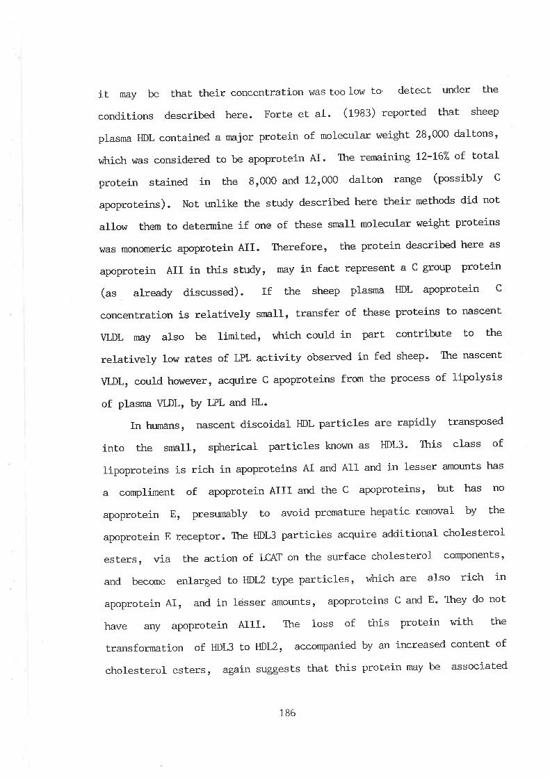
it may be that their concenlrat.ion \^/as too low to detect under the
conditions described here. Forte et al. (fggg) reported that sheep
plasma HDL contained a major protein of molecular weight 28t000 daltons,
r,¡trich was considered to be apoprotein AI. The remaining t2-L67" of total
protein stained in the 81000 and 121000 dalton range (possibly C
apoproteins). Not unlike the study described here their methods did not'
a1low them to determine if one of these snall molecular weight proteins
vras monomeric apoprotein AII. Therefore, the protein described here as
apoprotein AII in this study, may in fact represent a C group protein
(r" already discussed). If the sheep plasma HDL apoprotein C
concentration is relatively small, transfer of these proteins to nascent
VLDL rnay also be limited, vilrich could in part contribrte to the
relatively low rates of LPL activity observed in fed sheep- Ttre nascent
VLDL, could however, acquire C apoproteins from the process of lipolysis
of plasma VLDL' bY IIPL and HL.
In humans, nascent discoidal HDL particles are rapidly transposed
into the smaIl, spherical particles loown as HDL3. This class of
lipoproteins is rich in apoproLeins AI and AII and in lesser amounts has
a compliment of apoprotein AIII and the c apoproleins, but has no
apoprotein E, presumably to avoid premature hepatic removal by the
apoprotein E receptor. The HDl3 particles acquire additional cholesterol
esters, via the action of LCAT on the surface cholesterol componentst
and become enlarged to HDL2 type particles, vùrich are also rich in
apoprotein AI, and in lesser amounts, apoproteins c and E. They do noL
have any apoprotein AIII. The loss of this protein with the
transfornntion of HDI^3 to HDL2r accomPanied by an increased content of
cholesterol esters, again suggests that this protein may be associated
186
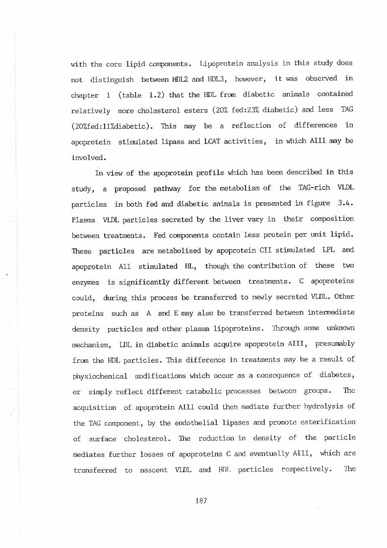
with the core lipid components. Lipoprotein analysis in this study does
not dist.inguish between HDL2 and HDL3, however, it was obsen¡ed in
chapter 1 (table L.2> Ltât the HDL from diabetic animals conlained
relatively more cholesterol esters (207" te¿2237" diabeLic) and less TAG
(ZO7"f.eAz117.diabetic). this may be a reflection of differences in
apoprotein stinmlated lipa.se and LC,AT activitíes, in vihich AIII nray be
involved.
In view of the apoprotein profile v¡hich has been described in this
study, a proposed pathway for the metabolism of the TAG-rich VLDL
particles in both fed and diabetic animals is presented in figure 3.4.
Plasma WDL particles secreted by the liver vary in their cornposition
between treatments. Fed components contain less protein per unit lipid.
These particles are metabolised by apoprotein CII stinmlated LPL and
apoprotein AII stinnrlated HL, though the contribution of these Lwo
enzymes is significantly different between treatments. C apoproteins
could, during this process be transferred to newly secreted VLDL. Other
proteins such as A and E may also be transferred between intermediate
densiLy particles and other plasma lipoproteins. Through some unktown
mechanism, DL in diabetic animals acquire apoprotein AIII, presumably
from the HDL part.icles. This difference in treaLments may be a result of
physiochemical modifications vùrich occur as a consequence of diabetes,
or simply reflect different catabolic processes between groups. The
acquisition of apoprotein AIII could then mediate further hydrolysis of
the TAG component, by the endothelial lipases and promote eslerification
of surface cholesterol. The reduct,ion in density of the particle
mediates further losses of apoproteins C and eventually AIIIr v¡trich are
transferred to nascent VLDL and HDL part.icles respectively. The
L87
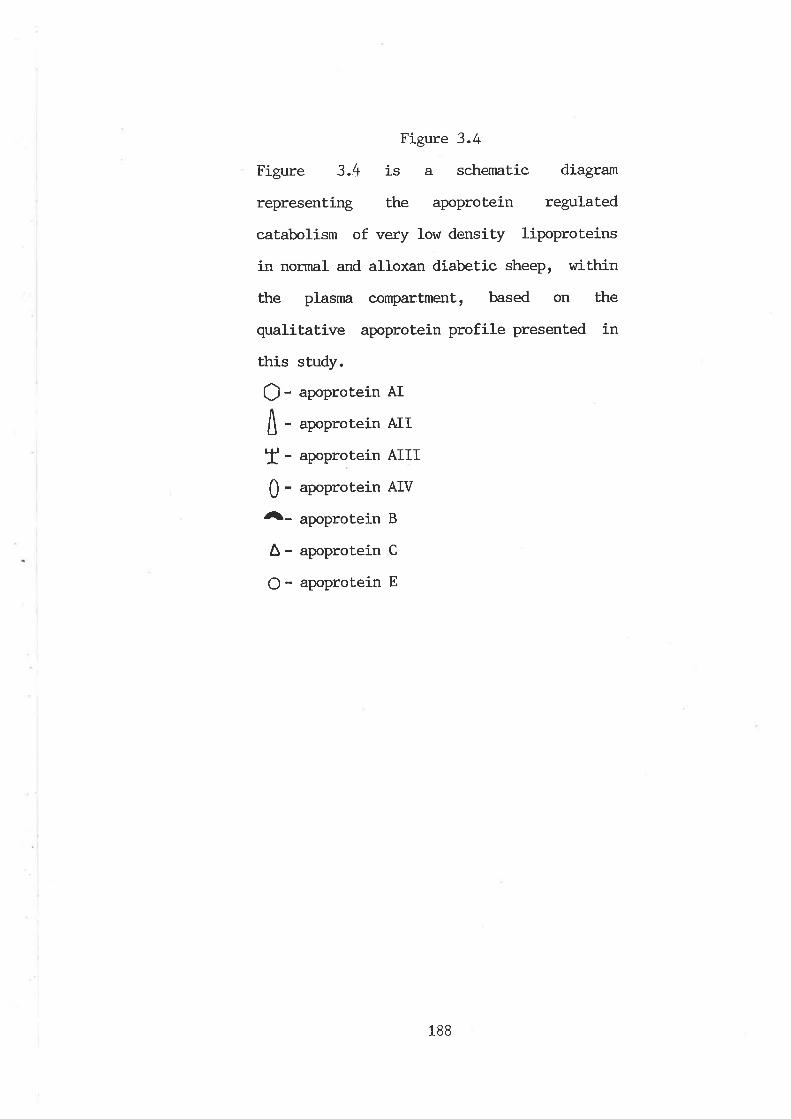
Figure 3.4
Figure 3.4 is a schernatic diagram
representing the apoprotein regulated
catabolism of very low density lipoproteins
in normal and alloxan diabetic sheep, within
the plasnn compartment, based on the
qualitative apoprotein profile presented in
this study.
O- "poptotein AI
A - .*ntotein A,II
Ï - .poptotein AIII
0 - upoptotein AIV
Â- "poprotein
B
A - apoprotein C
O - apoprotein E
188
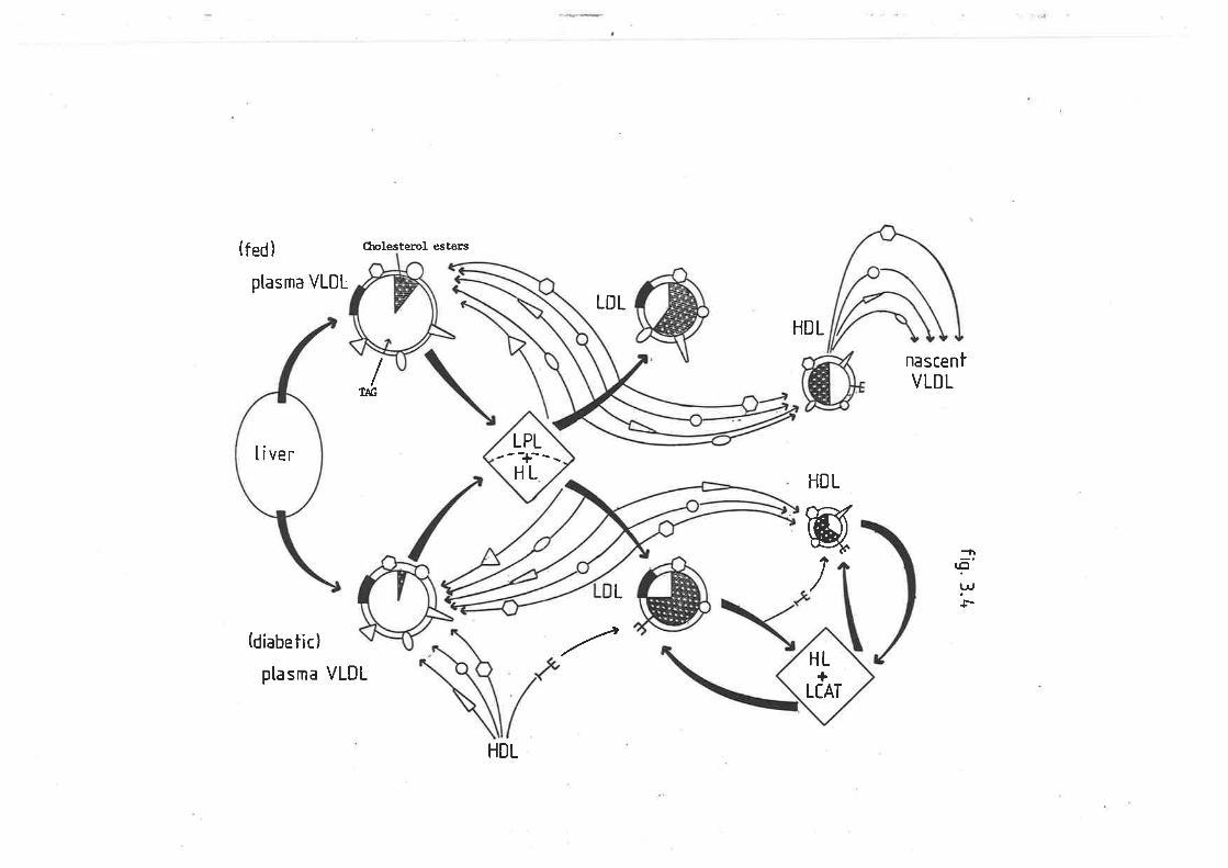
(fed)
ptasma VLDL
(diabetic)
ptasma VLDL
CholesEerol esters
LDL
HDL
/T¡G
nascentVLDL
HDL
wL /
l.c¡
l.^,
HL+
LCAT
---+---LPL
HLliver
HDL
a!
resulting LDL particles in the diabetic sheep will thus contain a
greater percentage of cholesterol esLers and lower amounts of TAG, than
the LDL components from fed animals (as was observed)' Ttte presence of
apoprotein AIII on HDL, results in part. hydrolysis of its TAG core and
esterification of surface cholesterol, vúrereby its subsequent reductj-on
in density (diabetic HDL have a higher protein per unit lipid content-
lable 1.2) causes a similar loss of apoprotein AIII, baek to VLDL, v¡trich
are acquiring this protein as a result of lipolysis'
Total protein and apoprotein B was determined
fraction used to trnrasht isolated VLDL: because
considerable amount of disagreement in the literature as to the benefits
of this procedure for re¡noving contaminating plasma proteins, versus the
poLential loss or degradation of apoproteins from the lipoprotein
particles. The results presented in this chapter showed t'hat
recentrifugation of sheep V[,DL resulted in a redistribut'ion of protein,
such that only 187. and 38% of total protein for fed and diabetic sheep
respectively, \Á¡as recovered with the tinlactt lipoprotein fraction at
the top of the ultracentrifuge tubes. Ttre ratio of soluble proteins
versus apoprotein B remained constanL (5L7":a97") indicat,ing that there
r^ras no contamination of plasma proteinsr or the unlikely possibilityt
that an equivalenL contaminat.j-on of soluble versus precipitable proteins
occurred. The reason vfrry diabetic VLDL underwent a smaller degree of
breakdown may be due to a protective factor of having a greater
concentration of lipids. It was concluded that the 'washing' procedure
for ovine VLDL was unnecessary, causing excessive loss and therefore
subsequent underestimation of the apoproteins'
l-n
there
the b-rffer
rennins a
ür¡]i
I
II
I
*
189
ilì¡l
I
rII
I
4 GM{ERAL DISCUSSION
Hepatic accunulation of triacylglyceride in sheep has been
associated with a nurnber of naturally occuring or clinically induced
pathological conditions. This was thought to be due to an inability to
maintain or increase hepatic release of this lipid in response to an
increased hepatic uptake of unesterified lalby acids. However, recently
\tre have reported that unlike other species such as the rat (Otway and
Robinson t967 ard Agius et al. 1981), Rhesus monkey (r'iser et al. L974)
and rabbit (Topping and T\:rner L976), the secretion of hepatic
triacylglyceride is elevated in fasted and diabetic animals (D'lamo et al.
1933). One of the first main findings of the work presented in this
thesis lfas the determination of the nature of the lipoproteins
mediating bhis increased secretion. It rsas found that under nornal fed
conditions, sheep have low levels of circulating plasnn triacyl-
glyceride, reflected in low levels of plasma very low density
lipoproteins. In stressed animalsr âs a result of diabetes, this
fraction was the major lipoprotein, transporting not only the majority
of triacylglyceride, but infact all plasma lipids. In addition, the very
low density Iipoprotein fraction contained less tipid per unit protein,
suggesting that limitations in the synthesis of this fraction vitas not
the causative agent for hepatic accumulation. Very low density
lipoproteins are the largest of the liver lipoproteins and it rnay be
that. if the hepatic sinusoids are surrounded by a fenestrated membrane,
secreLion could be retarded. Ttre much smaller low and high density
Iipoproteins would theoretically be free to pass Lhrough the lamina,
thus accounting for the lack of accumulat.ion of other lipid cornponents
in the liver. If secretion was impeded by this membrane, the increased
!
190
iI
hepat.ic release of
tpressuret induced
particles.
Diabetic and
very low density lipoproteins, might
effect. of increased hepatic synthesis
reflect a
of these
rÌ
;
fasted sheep l^/ere also grossly hypertriacyl-
glyceridaemic and it was considered that v¡hilst this reflected increased
hepatic secretion of this lipid, plasma accumulation would only occur if
clearance was not similarly increased, or impaired. Because of this, the
hyperlipidaemia in these animals may at some point reach a maximum, such
that hepatic release of further lipid is not possible and accumulation
results. The second part of this study was thus aimed at determining
rates of plasma triacylglyceride lipolysis. Ttiacylglyceride hydrolysis
in posLheparin plasnra was shown to F dependent on lipase activities
v¡trich rese¡nbled lipoprotein lipase and hepatic lipase from sheep adipose
ti-ssue and sheep liver respectively. Clearance of plasma
triacylglyceride lÀ/as infered in both fasted and diabetic animals' In
the former, this was due to decreased actiwity of both lipoprotein
Iipase and hepatic lipase, v¡hereas in the diabet'ic sheep, despite a
significant increase in hepatic lipase activity, lipolysis was decreased
due to the lipoprotein lipase component vitrich was severely depressed'
Ttre increase in hepatic lipase activity in diabetic sheep appeared to
reflecL physiochemical changes in the very low density lipoproteinsr âS
indicated by the greater rate of unesterified fatty acids released, vùten
this lipoprotein fraction from diabetic animals as opposed to that from
fed sheep, \^¡as incubated with postheparin plasma from fed animals'
Lipoprotein lipase act.ivity was also stimulated by modifications in Lhe
very low density lipoproteins, but an increase in activity l^/as not
realised in diabetic sheep, due to low plasma levels of insulin, vihich
þ
L9L
vùren administered exogenously, corçletely restored lipoprotein lipase
activity.
The secretj-on and subsequent catabolism of very low density
lipoprotein triacylglyceride is controlled by the apoprotein compliment
of the particle and so the higher rates of very low density lipoprotein
lipolysis ín particles isolated from diabeUic animals may have reflected
an improved apoprotein compliment. Similarly, deficiencies in the
quantity or quality of apoprotein B, rttrich is essential for the
secretion of very low density lipoproteins, nEy in Partr have
contributed to hepatic triacylglyceride accumulaLion' Ttre apoprotein
profile of the sheep plasnn lipoproteins presented in this studyr was
based principally on molecular weight as determined by sodium dodecyl
sulphate-polyacrylamide gel electrophoresis, and the apoprotein profile
of rat very low density lipoproteins. It was unfortunate thaL the recent
conrnercial availability of antisera to some of these proteins could not
be used to confirm identity, as the antibodies lüere raised in sheep in
the first, instance. Nevertheless, on the basis of the ascribed
apoprotein profile of Lhe sheep plasma lipoproteins presenLed in this
study, a postulated apoprolein regulated control of very low density
lipoprotein triacylglyceride lipolysis was made. Three main points were
raised. Firstly, the apoprotein 'A' conrpliment of the very low density
lipoproteins and low density lipoproteins suggest that in sheep, hepatic
lipase may play a major role in the catabolism of nascent very low
density lipoproteins. FurLhermore, greater hydrolase activity of
lipoprotein lipase and hepatic lipase, st.imulated by very low density
lipoprotein particles isolated from diabetic sheep, may have been due to
a greater apoprotein tCt and tAt compliment. Secondlyr apoprotein AIII
192
correlated \^rith parLicles vùrich contained a smaller component of
triacylglyceride and a greater fraction of cholesterol esters,
suggesting ttrat. this protein may stimulate Lhe endothelial lipases and
lecithin cholesterol acyl transferase. Ttre third main obsen¡ation
regarding the ovine apoproteins lrras that very low density lipoproteins
from diabetic animals actually contained more apoprotein B than
parÈic]es from normal fed sheep. Ttris suggested ttnt synthesis of this
protein r4ras not limiting hepatic secretion of these particles and
therefore, \¡ras not a causative agent for hepatic accumulation of
Lriacylglyceride. However, vùrether the nature of this protein remained
the same, cannot be determined until this protein can be isolated
intact.
Tkre main objecLives of this projecL revolved around lipoprotein
triacylglyceride metabolism in norural and stressed sheep. However,
during the course of this study, it was found that postheparin plasna
lipase activities differed according Lo gender. An examination of
differences in plasma lipolytic activities between sexes \^las a logical
exLension of the overall aims of this study and so lipoprotein lipase
and hepatic lipase were determined in rams, castrates and e\47es. The
results reflected differences in activities due to the loss of androgen
synthesis as a result of castrat.ion and converesly, the effects of
oestrogen production. The oestrogenic/androgenic conLrol mechanisms of
very low density lipoprotein triacylglyceride kinetics in other
species, is well defined for lipoprotein lipase, but in the case of
hepat.ic lipase, remains a somevùrat conLentious issue. Like other
species, lipoprotein lipase \iùas progressively higher in rams r wethers
and er,iles respectively, vilrich is reflected in a greater degree of
L93
adiposity in the latter. Similarly, hepatic lipase activity paralleled
that of lipoprotein lipase, indicating that the loss of androgen
synthesisr ot alternatively, the synthesis of oestrogens promotes
activity of this enzyme v¡hich is in contrast to that reported for rats.
It is apparent, therefore, that the steroidal regulation of hepatic
lipase requires further investigation. In view of the widespread
Australian farm practice of castration, the potential of producing a
slower growing animal with a greater body fat content means that this
process is undesirable.
I was fortunate in having gained access Eo sheep designated as
genetically tleant and tobeset, wtrich again provided a further extension
of lipoprotein triacylglyceride lipolysis and its relationship with fat
deposition. The results showed that if nutrition is not a limiting
factor, potential adiposity is in partr predetermined by differing rates
of lipoprotein lipase. Further investigations may in future provide a
rapid means of selecting animals v¡hich are generally leaner.
Figure 4 sunrnarises the current concepts of difficulties associaLed
with plasnra lipoprotein triacylglyceride metabolism in metabolically
stressed sheep, based on the results from ühis project.:.
Metabolically sLressed sheep obviously possess a tremendous
capacity to synthesize and secrete triacylglyceride-rich very low
density lipoproteins, in response to a large influx of unesterified
fat.ty acids. It is difficulL to conclude however, if this release
is impaired by a basal lamina surrounding the sinusoid, or by a
saturable capacity of the plasma cornpartmenL to accomodate lipids.
In diabetic animals, the quality of these very large, very low
density lipoproteins, in terms of their suitability as substrates
L94
Figure 4
Figure 4 is a diagrarnat,ic representation of
the problems associated with plasma very low
density lipoprotein triacylglyceride met-
abolism in metabolically stressed sheep.
195
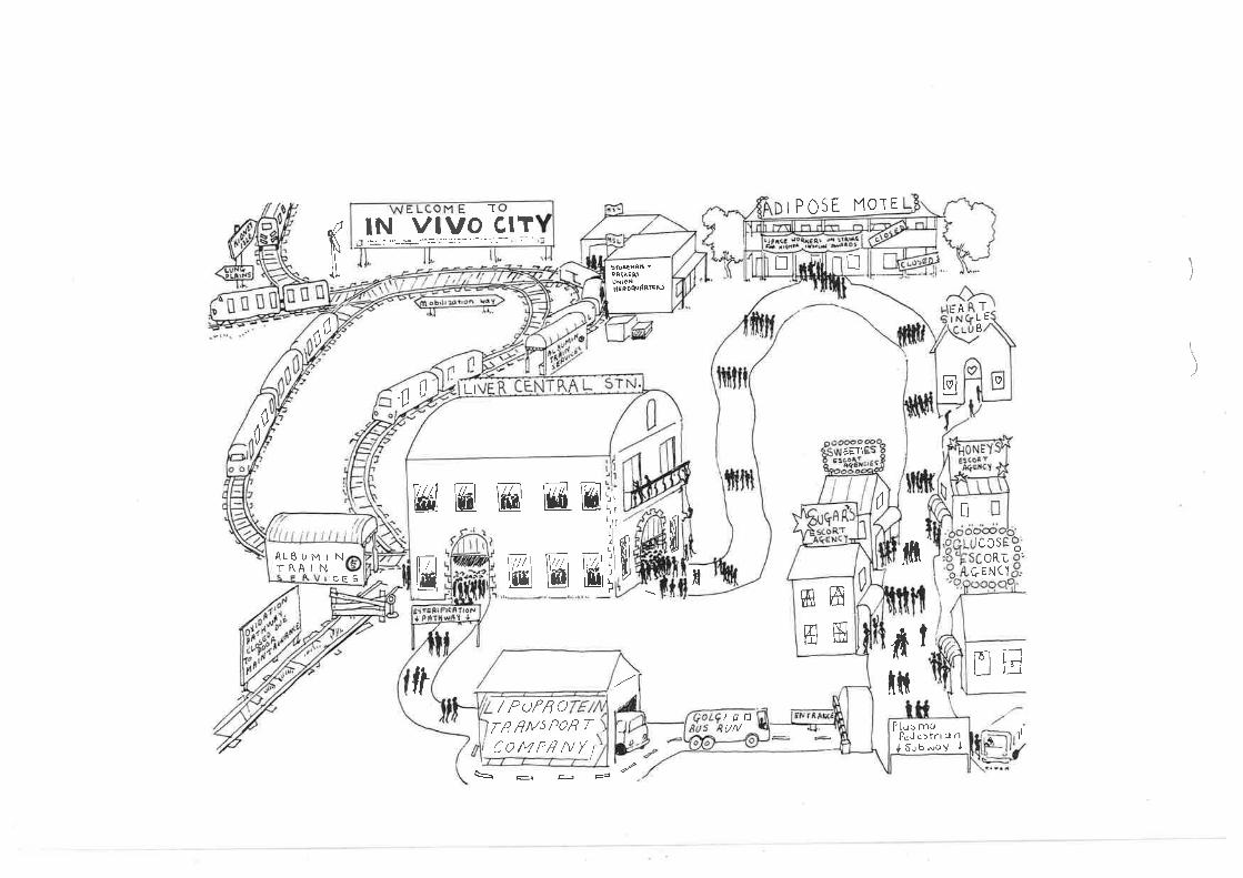
lN vlvo clT
E KJ Ki
ft
tP0 SE MOT E
l_ì
EETI
-T
fTLÈß
M
0w6l ffii ww L.
Lrft, \rLI¡lLl
JIJ
w lltKi w I
c0sOR¿
ÇENC'fr/
ftfril
INE
/Dp1i
S : tu é
\I
Plusmt¡Pc,l c: trr <¡ n{ Subwo! I
0
ffiawffi
nIQ If,E À'uNtoNHseDeu ßßßÀ)
/ PofßoT/l nMs P)n TCO/4PHNY
LItú
(,
0
cE S
AL6IJMINTRAIN
0
ç
UM
for lipoprotein lipase and hepatic lipase, is improved rn
comparison to these part.icles from fed sheep, as they are able to
stinn-rlate the activity of both endothelial lipases. This nny be due
to an improved protein lipid ratio, an improved apoprotein
compli-ment, or simply a resPonse to glucosylation of these
particles. Although hepatic lipase is elevated in diabetic animals
in response to these timprovementst, this is adverse to the needs
of the liver in diabetic sheep. Ttre triacylglyeeride fatry acids
are by this means returning to the liver, v¡hich is already trying
to remove them by increasing very low density lipoprotein
triacylglyceride secretion. Other extrahepatic tissues possessing
lipoprotein lipase are unable to utilize Lhe plasma
triacylglycerides, even though the nature of the particles means
thât lipolysis by this route is st.imulated, because of low levels
of plasma insulin concentration, v¡trich directly determines the
synthesis and secretion of this enzyme.
The objectives of this study have been achieved, but Iike many
scientific investigations, nnrch more work is needed to answer a number
of questions. Although sheep are able to increase rates of hepatic very
low density lipoprotein triacylglyceride release, accumulation of this
lipid in the liver continues. The possibility that a basal lamina
surrounds the hepatic sinusoid needs to be clarifiedr âs it may be that
this membrane inhribits the passage of the very large triacylglyceride
rich very low density lipoproteins. In addition, the sheep liver lipase
described in the defatted liver extracts, needs to be purified and itts
biochemical properties characterj-sed, to determine if this is infact the
L96
same as hepatic lipase in other species. Promotion of plasma lipase
activity by very low density lipoproteins from diabelic animals
contradicLs current conceptions as to problems associated with the
clearance of plasma very low density lipoproteins in hunnn diabetic
subjects. Because very low density lipoprotein lipolysis and subsequent
formation of low density lipoprotein is directly associated wit'h
atherogenesis (u*ricfr is the nnjor complication and cause of death in
human diabetes), the implicatíons of timprovingt a lipoprotein particle
in terms of its substrate potential are very important. The ovine
apoprotein investigation reported here, is in a sense preliminary. Ifuch
more work is required to purify and identify these componenLs, so that
the processes of sheep very low density lipoprotein triacylglyceride
metabolism can be elucidated. Results presented in this study raised a
nurnber of additional aspects of sheep, or rather ruminant
triacylgyceride metabolism. Steroidal regulation of plasma lipases
remains to be defined and in terms of body conrposition and current
animal production practices, genetic regulation of lipase activity has
received little attention.
t97
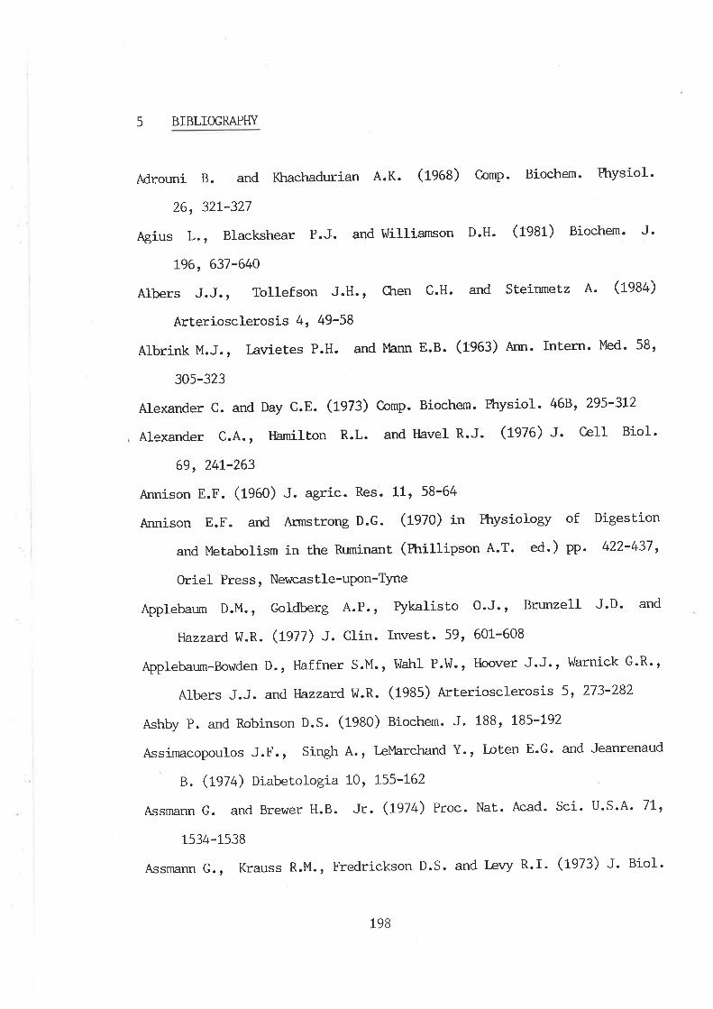
5 BIBLIOGRAPFIY
Adrouni B. and Khachadurian A.K. (1968) Comp. Biochem. Physiol.
26, 32L-327
Agius L., Blackshear P.J. and !üilliamson D.H. (fgAf ) Biochem. J'
196,637-640
Albers J.J., Tollefson J.H., CLren C.H. and steinmetz A. (1984)
Arteriosclerosis 4, 49-58
Albrink M.J., lavietes P.H. and Mann E.B. (1963) Ann. Intern' Med' 58,
305-323
Alexander c. and Day c.E. (1973) Comp. Biochem. Physiol - 468, 295-3L2
Alexander C.4., Hamilton R.L. and Havel R.J. (1976) J. C-eIl Biol'
69,24r-263
Annison E.F. (1960) J. agrie- Res. 11, 58-64
Annison E.F. and Armstrong D.G. (fgZO) in Physiology of Digestion
and Metabolism in the Ruminant (Phillipson A.T. ed.) Pp. 422-437,
Oriel Press, Newcastle-uPon-TYne
þplebaum D.M., Goldberg 4.P., Pykalisto 0.J., Brunzell J.D. and
tlazzard !ü.R. (L977) J. Clin. Invest. 59, 601-608
Applebaum-Bowden D., Haffner S.M.r llahl P.W., Hoover J.J., Warnick G'R"
Albers J.J. and llazzard,w.R. (1935) Arteriosclerosis 5, 273-282
Ashby p. and Robinson D.S. (1980) Biochem. J. 188, 185-192
Assimacopoulos J.F., Singh 4., LeMarchand Y., I-oten E.G. and Jeanrenaud
B. (1974) Diabetologia 1-0, L55-L62
Assmann G. and Brewer H.B. Jr. (t974) Proc. Nat. Acad. Sci. u.s.A' 7L'
1534-1538
Assmann G., Krauss R.M., Fredrickson D.S. and l-evy R.I. (1-973) J. BioI.
198
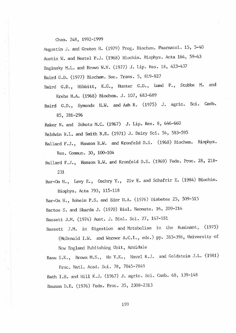
CLrem. 248, L992'I999
Augustin J. and Greten H. (tots¡ Prog. Biochem. Pharmacol. 15r 5-40
Ansrin tl. and Nestel P.J. (1968) Biochim. Biophys. Acta L6t+r 59-63
Baginsky M.L. and Brown td.V. (t977) J. Lip. Res. 18' 423-437
Baird G.D. (1977) Bioche¡n. Soc. Trans. 5, 819-827
Baird G.D., Hibbitt, K.G-, Hunter G.D', I-und P' ' St'ubbs M' and
Krebs H.A. (1968) Biochem. J- LO7' 683-689
Baird G.D., Symonds H.I^1. and Ash R. (1975) J. agric. Sci. Camb.
85, 28t-296
Baker N. and Schotz M.C. (19eÐ J- Lip. Res' 8' 646-660
Baldwin R.L. and SmiLh N.E. (tglt) J. Dairy Sci. 54, 583-595
Ballard F.J., tlanson R.tl. and t(ronfeld D.S. (1963) Biochem. Biophys'
Res. C.onnn¡n. 30, 100-104
Ballard F.J., Hanson R.tl. and Kronfeld D.S. (1969) Fedn. Proc. 28r 218-
23t
Bar-On H., Ièvy E., Oschry Y., Ziv E. and Schafrir E. (1984) Biochim'
Biophys. Acta 793, tl5-tL8
Bar-On H., Roheim P.s. and Eder H.A. (1976) Diabetes 25' 509-515
Bartos S. and Skarda J. (1970) Biol. Neonate. 16, 209-214
Bassett J.M. (L974) Aust. J. Biol- Scí. 27, L67-L8I
Basset.t. J.M. in Digestion and Metabolism in the Ruminant, (1975)
(l,,leDonald I.I,J. and l,üarner A.C.I., eds.) pp. 383-398r University of
New England Publishing Unit, Armidale
Basu S.K., Brown M.S., Ho Y.K., tlavel R.J. and Goldstein J.L. (1981)
Proc. Natl. Acad. Sci. 78, 7845-1849
Barh I.H. and Hill K.J. (L967) J. agric. Sci. Camb. 68, 1-39-148
Bauman D.E. (Ple) Fedn. Proc. 35, 2308-23L3
L99
Baxter G.p. and !üallace C.C. (fgfe) J. Amer. Chem. Soc.38, 70-105
BelI A.!ü. (L979) Prog. Lip. Res. 18, LI7-L64
Bell A.w. and Thornpson G.E. (L979) Am. J. Physiol. 237, 8309-E315
BeIl F.P. (1978) Prog. Lip. Res. L7, 207-243
Bell-Quinr J. and Forte T. (1931) Biochim. Biophys. Acta. 663' 83-98
Bell-Quinr. J., Forte T. and Graham P. (1981) Biochem. Biophys-
Res. C-onrnun . 99, 700-706
Bensadoun A., Ehntrolm C., Steinberg D. and Brown Il.V. (L974) J.
Biol. Chem. 249, 2220-2227
Bensadoun A. and Kompiang I. P. (1979) Fedn. Proc., 38, 2622'2626
Bergnnn E.N. (1963) Arn. J. Physiol. 2I5, 865-873
Bergnran E.N. (L973) Cornell Vet. 63, 34L-382
Bergrnan E.N. (L97L) J. Dairy Sci. 54, 936-9t+8
Bergman E.N., Havel R.J. , !üolfe B.M. and Bohmer T. (fgZf ) ¡. Clin.
Invest. 50, 1831-1839
Bergmeyer H.U. and Bernt E. (L974) in Methods of Enzynratic Analysis
(Bergmeyer H.U. ed.) vol.3, pp.1205-12L5, Academic Press, New York,
I-ondon
Berry E.M., Ziv E. and Bar-On H. (fgAf) Diabetologia 2L, 402-408
Beynen 4.C., Haagsman H.P., Van Golde L.M.G. and Geelen M.J.H.
(fgef) Biochim. Biophys. Acta 665' L-7
Bezman 4., Felr.s J.M. and Élavel R.J. (tgøZ) l. Lip. Res. 3, 427-43t
Bird M.I. and I,Jilliams M.A. (t982) J. Nutr. LLz, 2267-2278
Blix G., Tiselius A. and Svensson H. (tg+t) J. Biol. Ctrem. L37, 485'494
Blue M.L., I^/illiams D.L., Zucker S., Kkran S.A. and Blum C.B. (fga:)
Proc. NatI. Acad. Sci. USA 80' 283-287
Bobek P., CLrorvathova V. and Ozdin L. (1981) Physiol. bohemoslov.
200
30, 35-40
Boberg J., Carlson L.A. and NormeII L. (tgï+) Life Sci . 3, LOIL-1019
Boguski M.S., Elshourbagy N., Taylor J.M. and Gordon J.I. (1984) Proc.
Nat.l. Acad. Sci. 81 , 5O2L-5025
Bordeaux 4.M., Giudicelli Y., Rebourcet M.C., Nordnmnn J. and Nordmann
R. (1980) Biochem. Biophys. Res. Conrnun. 95, 2L2-219
Bottcher C.J.F., van Gent C.M. and Fries C., (1961) AnaI. CLrim
Þ,cLa 24, 203-204
Bouchat J.C., Doize F. and Paquay R. (1931) Reprod. Nutr. Dev.
2L, 69-8L
Breckenridge Il.C. (1985) Can J. Biochem. Cell Biol. 63, 890-897
Breckenridge W.C., Little J.4., Alaupovic P, [,/ang C.S. Kuksis At
Kakis G., Lindgren F. and Gardiner G. (L982) Atherosclerosis 45,
L6L-L79
Brewer H.B.Jr. (1981) Klin. Idochenschr. 59, 1023-1035
Brewer H.B.Jr., Schulnnn R., Herbert P., Ronan R. and !üekrrly K.
(L974) J. Biol. Ckrem. 249, 4975-4984
Brocknan R.P. (fVZa¡ C,an. Vet. J. L9, 55-62
Brown I^j.V. and Baginsky M.L. (tglZ) Biochem Biophys. Res. Conmun.
46,375-382
Brown M.S. and Goldstein J.L. (fg8:) J. CIin. Invest. 72, 743-741
Brurnby P.E., Anderson M., T\rckley B. and Storry J.E. (1975) Biochem. J.
L46, 609-6L5
Busbee D.L. Payne D.M., Jasheway D.W., Carlisle S. and Lacko A.G. (fggf )
Clin. Ctrem. 27, 2052-2058
Campbell E.A. (fg6:) Res. Vet. Sci. 4, 56-63
Carlson L.A. and l^ladstrom L.B., (fgSg) Clin. Ckrim. Acta. 4, 197-205
20L
Oram B.E. and Knowles B.R. (tgle) J. Lip. Res. L7, L76-L8L
chen T.c., Bradley [J.4., Gor.ro A.M.Jr. and Morrisett J.D. (rgzg) Fnes
l,ett. L04, 236-240
C,kren Z. and Danon A. (L979) Biochem. Pharm. 28, 267-27L
clreng c.F., oosta G.M., Bensadoun A. and Rosenberg R.D. (1981) J. Biol.
Ckrem. 256, L2893-L2898
crrristie t{.ü/. (L979) Prog. Lip- Res - t7, 245-277
Ckrung J and Scanu A.M. (L977) J. Biol Chem. 252, 4202-4209
Clrurch D.C. (L976) in Digestive Physiology and Nutrition of Ruminants
(Church D.C. ed.) vot. 1, pp. 280-3II, Corvallis.
claude A. (1970) J. CelI BioL. 47, 745-766
Clegg R.A. (1979) Biochem. Soc. Tlans. 75, 1053-1054
Clegg R.A. (fg8fa) Biochim. Biophys. Acta 664, 397-408
Clegg R.A. (1981b) Comp. Biochem. Physiol. 698' 585-591
C-ordle S.R., Yeaman S.J. and Clegg R.A. (fgg:) Biochim. Biophys. Acta
753,2L3-219
Cryer 4., Riley S.E., t{illiams E.R. and Robinson D.S. (tgl') Clin. Sci.
Mol. Med. 50, 2L3-221
Curry M.D., llcConathy W.J., Alaupovic P., Ledford J.H., Popovic M.
(t976) Biochim. Biophys. Acta 439, 413-425
Curt.iss L.K. and lJitzum J.L. (1985) Diabetes 34, 452-46t
David H. (1964) in Sutxnicroscopic Ortho- and Pathomorthology of the
Liver (David H. ed) pp. L69-280, Pergamon Press, Oxford
Davis R.A. and Boogaerts J.R. (ßgz) J. Blol. ctrem. 257, t0908-10913
De Jong J.J.B. and l4arsh J.B. (fg68) J. Biol. Ckrem. 243, L92-L99
Dolphin, P.J. (1935) Can. J. Biochem. CelI. Biol. 63' 850-869
Driscoll D.M. and Getz G.S. (1983) Circulation 68(SuppL.), t7
202
Dryerre H. and Robertson A. (L94L) J- Physiol- 99, 443-453
Durrington P.N., Newton R.S., I^Teinstein D.B. and steinberg D.
(ßez) J. clin. rnvesL. 70, 63-73
Edelstein c., Kezdy F.J., Scanu A.M. and shen B.l,ü. (1979) J. Lip.
Res. 20, L43-I53
Edwards P.4., LemongelloD. andFogelmanA.M. (L979) J. Lip. Res.
20, 2-7
Egelrud T. and Olivecrona T. (tglZ) J. Biol. Ckrem. 247, 62L2-L7
Ehrùrolm C., Heaf D.J., Kaijser L., Kinnunen P.K.J. and C,arlson L'A'
(1971) Atherosclerosis 27, 35-39
Ehntrolm C., Huttunen J.K., Kinnunen P.J., Meittinen T.A. and Nikkila
E.A. (fgZSa) N. Eng. J. Med. 292, L3L4-L3L7
Ehnholm C., SLraw t/., Greten H. and Brown !ü.V. (1975b) J. Biol. Gre¡n'
250, 6756-676L
Eisenberg S. (L979) Prog. Biochem Pharnncol L5, t39-L65
Eisenberg S. and Ler4y R.I. (1975) Adv. Lip. Res. 13, 1-89
Eisenberg S., Patsch J.R., Sparrow J.T., C'otto A.M. and Olivecrona T.
(L979) J. Biol. Ckrem. 254, L2603-12608
Elkeles R., Hambley J. and Bird D. (t977) Horm. Met. Res. 9' 63-65
Faergeman O. and tlavel R.J. (1975) J. CIin. Invest. 55' 1210-18
Fielding C.J. (L976) BiochemisLry 15, 879-884
Fielding C.J. (1973) in Disturbances in Lipid and Lipoprotein Metabolism
(Dietschy J.M., Gotto A.M. and Ontko J.A. eds.) pp. 83-89 Arnerican
Physiological Society, Bethesda, M.D.
Fielding C.J. and Fielding P.E. (L976) J. Lip. Res. 17, 248-256
Fielding C.J. and Fielding P.E. (1977) in Ckrolesterol Metabolism and
Lipolyt.ic enzymes (Polonovski J. ed.) pp. 165-L72, Masson, New York
203
Fielding c.J. and Higgins J.M. (1974) BiochemisLry L3, 4324'4329
Fielding, c.J., shore v.G. and Fielding P.E. (L972a) Biochem. Biophys.
Res. Conrnun. 46, L493'L498
Fielding C.J., Shore V.G. and Fielding P.E. (L972b) Biochim. Biophys.
Ãcta 27O, 513-518
Fielding P.E., shore v.G. and Fielding c.J. (L974) Biochemistry,
L3, 4318-4232
Fielding P.E., Shore V.G. and Fielding C.J. (L977) Biochemistry L6,
1896-1900
Fiser R.H., Denniston J.C., Rindsig R.B. and BeiseM.R. (L974) J. Nutr.
L04,223-226
FleLcher M.J. (1968) Clin. Chim. ÃcLa 22, 393-397
Folch J., I-ees M., and sloane-stanley G.H. (1957) J. Biol. Cl:run. 226,
497-509
Ford E.J.H. (1962) J. Agric. Sci. 59, 67-75
Forte T. and Nichols A.V. (1972) ndv. Lip. Res.I0, L-4L
Forte T.M., Cross C.E., Gunther R.A. and Kramer G.C. (1933) J. Lip. Res.
24, t358-L367
Fredrickson D.s. and Gordon R.s. Jr. (1958) Physiol. Rev. 38, 585-630
Fried S.K., Hilt J.O., Nickel M. and DiGirolamo M. (fgA:) J. Nutr. LL3'
1861-1869
Fritz I.B. (fg6f) Physiol. Rev. 41, 52-L29,
Furman R.H., Sanbar s.s., Alaupovic P., Bradford R.H. and Howard
R.P. (1964) J. Iåb. Clin. Med. 63, 193-204
Gal s. , Bassett D.J.P. , Flamosh M. and Élamosh P. (1982) Biochim.
Biophys. Acta 7L3, 222-229
Ganesan D., Bass H.8., l,lcConathy I,r7.T. and Alaupovic P. (L976) Metabolism
204
25, LL89-LL95
Garfinkel 4.S., Baker N and Schotz M.C. (L967) J. Lip Res. 8, 274-280
Garfinkel 4.S., Nilsson-Ekrle P. and Schotz M.C. (1976) Biochim. Biophys.
Acta 424, 264-273
Garton G.A. and Duncan I,ü.R.H. (fg6+) Biochem. J. 92, 472-475
Geelen M.J.H., Harris R.4., Beynen A.C. and },IcCune S.A. (1980) Diabetes
29, LOO6-1O22
Ge¡rmell R.T. and He¿th T. (tSlZ¡ AnaÈ. Rec. L72, 52-70
Gitlin D., Cornwell D.G., Nakasato D., Oncley J.L., Hughes [r7.L.Jr. and
Janeway C.A. (1958) J. Clín. Invest. 37, t72-L84
Glascock R.F. and Vüelch V.A. (f974) J. Dairy Sci. 57, L364't370
Glaumann H., Bergstrand A. and Eri-csson J.L.E. (tglS) J. CeIl. Biol. 64,
356-377
Gliclanan R.M. , Green P.H.R., Lees R.S. and Tall A. (1978) N. EngI. J.
Med. 299, L424-L427
Glomset J.A., Parker F., Tjaden M. and I^lilliams R.H. (L962) Biochim.
Biophys. Acta 58, 398-406
Gofman J.W., Lindgren F.T., Elliott H. (L949) J. Biol. Chem. L79, 973-
979
Goldberg I.J., te N.4., Paterniti J.R.Jr., Ginsberg H.N-, Lindgren F.T.
and Brown W.V. (fqAZ) J. Clin. Invest. 70, L|84-LL92
Gonen 8., Baenziger J. Schonfeld G., Jacobson D. and Farrar P.
(1981) Diabetes 30, 875-878
Gooden J.M., Fraser R., Bosanquet H.G. and Bickerstaffe R. (1979)
Aust. J. Biol. Sci. 32, 533-542
Gordon T., C.astelli W.P., Hjortland M.C., Kannel I^1.8. and Dawber
205
T.R. (L977) Am J. Med. 62, 707-7t4
GoLto 4.M., tevyR.I., JohnK., FredricksonD.S.(L97L) N. Eg1. J
Med. 284, 813-818
Green P.H.R., Gliclcnan R.M. (1981) J. Lipid. Res. 22, LL53-LL73
Greten H. and Augustin J. (L979) Athero. Rev. 5, 9L-L24
Greten H., De Grella R., Klose G., Rascher I{., de Gennes J-L. and
Gjone E. (tglø) J. Lip. Res. 17, 203-2L0
GreÈen H., l¿ible V., Zipperle G. and Augustin J.r (tSll¡
Atherosclerosis, 26, 563-572
Grosser J., Schrecker O. and Greten H. (fggf) J. Lip. Res. 22, 437-442
Grubb D.J. and Jones A.L. (L97L) Anat. Record 170, 75-80
tlaagsman H.P. and Van Golde L.M.G. (fOSf¡ Arch. Bíochem. Biophys.
2O8, 395-402
tlamilton R.L. (tglZ) Adv. Exp. Med. Biol . 26, 7-24
Hamilton R.L., Regen D.M., Gray M.E. and LeQuire V.S. I¿b. Invest.
(L961) 16, 305-319
tlamilton R.L., Williams M.C., Fielding C.J. and FIaveI R.J. (fgZe ) ¡.
Clin. Invest. 58, 667-680
Hanson R.t{. and Ballard F.J. (L967) Biochem J. 105' 529'536
Hanson R.!ü. and Ballard F.J. (fgAA) Biochem J. 108, 705-7L3
Harano D.Y., DePalma R.G. and Miller M. (fS0O¡ Proc. Soc. Exp. Biol.
MeC. 131, 9t3-9L7
Flarfoot, C.G. (fgZA) Prog. Lip. Res. L7, 2L-54
Harrison M.F. (1953) Biochem. J. 55, 204-2Ll
Harrison F.A. and l-eat i,/.M.F. (1-975) Proc. Nutr. Soc. 34, 203-2LO
tlartree E.F. (1972) Analyt.. Biochem. 48, 422-427
Hatch F.T. and l-ees R.S. (fg08) Adv. Lip. Res. 6, 1-68
206
Havel R.J. (fgeO) Ann. N.Y. Acad. Sci. 348, t6-29
FIaveI R.J., Felts J.M. and Van Duyne C.M. (L962) J. Lip. Res. 3, 297-308
Havel R.J., Fielding C.J., Olivecrona T. r Shore V.G., Fielding
P.E. and Egelrud T. (1973) Biochemislry 12, 1828-1833
Havel R.J. and Kane J.P. (L975) Fed. Proc. 34, 2250-2257
Havel R.J., Shore V.G., Shore B. and Bier D.M. (fgZO) Circ. Res-
27, 595-600
Heath T.J. and Hill L.N. (1969) Aust. J. biol. Scí. 22, L0L5-IO29
Heímberg M., Dunkerley A. and Brown T.O. (1966) Biochim. Biophys.
Ãcta I25, 252-264
Heimberg 14., C'oh H.J., Klausner H.J., Soler-Argilaga C., I^leinstein I-
and tr{ilcox H.G. (1973) in Disturbances i-n Lipid and Lipoprotein
Metabolism (Dietschy J.M., Gotto A.l'4. and Ontko J.A. eds.) pp. 251-
267, Amex. Physiol. Soc., Bethseda, MD.
Heimberg M., Van Harken D.R. and Brown T.O. (L967) Biochim. Biophys.
Acta 137, 435-445
Heimberg M., tr{einstein I. and Kohout M. (fg0g) J. Biol . CÍ:rel,;r. 244, 5131-
5139
Heimberg M., trTeinstein I., Klausner H. and lüatkins M.L. (L962) Am. J.
Physiol. 202, 353-358
Heimberg M., !üilcox H.G., Dunn G.D., Woodside I,Í.F., Breen K.J. and
Soler-Argilaga C. (tgl+) in Regulation of Hepat.ic Metabolism
(Linquist F. and Tygstrup N. eds.) pp. LI9-L43, |tunksgaard,
Copenhagen
Heiss G., Johnson N.J., Reiland S., Davis C.E. and Tyroler H-4.
(fgAO) Circulation, 62 (Suppl. IV), LL6-L36
Henderson G.D., Read L.C. and Snoswell A.M. (ßgZ) Biochim. Biophys.
207
d$¡
Acra 710, 236-24t
Hertelendy F. and Kipnis D.M. (L973) Endocrin. 92, 402-4LO
Holmquist L. and C,arlson K. (1977) Biochim. Biophys. Acta 493, 4OO-409
Holt P.R. and Dominguez A.A. (1980) Am. J. Physiol. 238, G453-G457
Hon¡rna Y., Nakaya N., Nakarmrra H. and Goto Y. (1985) Artery L3, I9-3L
Hood R.L., Thornpson E.H. and Allen C.E. (tOlZ¡ Int. J. Biochem. 31 598-
606
ttuang M.T. and llilliams M.A. (1930) Am. J. Physiol. 238, V+99-E505
Hulsmann W.C., Oerle¡nans M.C. and Geelhoed-Mieras M.M. (1977) Biochem.
Biophys. Res. Connnrn. 79, 784-788
Huttunen J.K., Ehrùrolm C., Kekki M. and Nikkila E. A. (t0lø¡
Clin. Sci. Mol. Med. 50, 249-260
Huttunen J.K., Ehrùro1m C. Kinnunen P.K.J. and Nikkila E.A. (1975)
Clin. Ckrim. Acta 63, 335-47
Inraizumi K., tlavel R.J., Fainaru M., Vigne J.L. (1978) J.Lip.Res . t9
1038-1046
Ingle D.L., Bauman D.E. and Garrigus U.S. (tglZa) J. Nutr. LOz, 609-6L6
Ingle D.L., Bauman D.E. and Garrigus U.S. (tglZU) J. Nutr. LO2, 6L7-624
Innerarity T.L. and lufahley R.l,/. (1978) Biochem. 17, L44O't447
It,zhaki R.F. and Gill D.M. (L964) Anal. Biochem. 9, 4OL-4L0
Jackson, R.L. Morriset J.D., Gotto A.M.Jr, and Segrest J.P. (fgZS) Uof .
CeIl. Biochem. 6, 43-50
Jackson R.L., Morrisett J.D. and Gotto A.M.Jr. (tglí) Physiol. Rev. 56,
259-3t6
Jackson H.D., Taylor J.4., Hatcher B.l,l. and Carter J.M. (1964) Arch.
Biochem. Biophys. 105, 575-581-
Jahn C.E. Osborne J.C.Jr. Schaefer E.J. and Brewer H.B.Jr. (fgg:) Eur.
208
#iÈ).î
J. Biochem. 131, 25-29
Jansen H. and De Greef trd.J. (1981) Biochem J. L96' 739-745
Jansen H, Garfinkel 4.S., T\,rru J.S., Nikazy J. and Schotz M.C. (1978)
Biochim. Biophys. Acta 531' LO9'LL4
Jansen H., Kalknan C., Zonneveld A.J. and Hulsmann i{.C. (fgZg) FEBS
lêtt. 98, 299-302
Jansen H. and Hulsnnnn tl.C. (L974) Biochim. Biophys. Acta 3691 387'396
Jansen H. and Hulsmann VJ.C. (1975) Biochim. Biophys. Acta 398, 337-346
Jansen H, Kallcnan C., Birkentr,ager J.C. and Hulsmann tr'7.C. (1980a) FEBS
l€t.t,. , Llz, 30-34
Jansen H, van Tol A. and Hulsmann I,ü.C. (fgSOU) Biochem. Biophys. Res.
C,onrnun. 92, 53'59
Jarrett I.G., Potter B.J. and Packlram A. (1956) Aust- J. Exp. Biol' Med'
Sci. 34, 133-t42
Jensen G.L., Baly D.L., Brannon P.M. and Bensadoun A. (1930) J. Biol.
Ckrem. 255, LLL4L-LIL49
Jensen G.L. and Bensadoun A. (1981) Anal. Biochem. IL3, 246-252
Jensen G.L., Daggy B. and Bensadoun A. (1982) Biochim. Biophys. AcLa
7t0, 464-470
Johnston J.M. (1970) in Comprehensive Biochemistry (Florkin M. and
stotz E.H. eds.) vot. 18 pp. 1-18, Elsevier, Amsterdam
Jonas 4., Sweeny S.A. and Herbert P.N. (1984) J. Biol. Ckrem. 259, 6369'
6375
Jones A. (1981) Biochim. Biophys- Acta 427, 325-336
Jones 4.L., Ruderman N.B. and Herrera M.G. (1967) J. Lip. Res. 8r 429-
446
Kalopissis 4.D., Griglio S. and Le Liepvre X. QSAZ) Biochim. Biophys'TI
I
þ
209
tI
Acta. 7LL, 33-39
Kalopissis 4.D., Griglio S., Malewiak M.I. and Rozen R. (1980) Biochim.
Bicphys. Acta 620, LLI-tlg
Kane J.P. (L973) Anal. Biochem. 53, 350-364
Kane J.P. (fg8:) Ann. Rev. Physiol. 45, 637'50
Kane J.P., tlardman D.A.and Paulus H.E. (fgAO) Proc. Natl. Acad. Sei.77,
2465-2469
Kane J.P., Chen G.C., tlamilton R.L., tlardnen D.4., I4alloy M.J. and tlavel
R.J. (1983) Arteriosclerosis, 3, 47-56
Kap1an A. (1970) AnaI. Biochem. 33, 218-225
Katz M.L. and Bergman E.N. (1969) Am. J. Physiol. 216, 953-960
Kempen H.J.M. (fgeO) J. Lip. Res. 21, 67t-680
Khachadurian 4.K., Adrouni B. and Yacoubian H. (1966) J. Lip. Res. 7,
427-436
Kinnunen P.K.J. (fgaf) Med. BioI. 55, L87-L9t
Kinnunen P.K.J., Virtanen J.A. and Vaineo P. (fgA:) Athero. Rev. tL, 65-
106
Kinnunen P.K.J. and Virtanen t (1980) Atherosclerosis V, (C'otto A.M,
Smith L.C. and Allen B. eds.) pp 383-386, Springer-Verlag, Berlin
Heidelberg, New York
Kirkeby K. (f900) Scand. J. clin. I^ab. Invest. 18, 437-442
Klose G., I,/indelband J., I{eizel A. and Greton H. (L977) Err. J. Clin.
Invest. 7, 557-562
Kochen J., [4arinetti G.V. and stotz E. (1959) J. Lip. Res. 1, L/+7-L49
Kohout M., Kohoutova B. and Heimberg M. (197L) J. Biol. Ckrem. 246'
5067-5074
Kompiang I.P., Bensadoun A. and Yang M.l^l.ln. (fgZ6) ¡. Lip. Res. 17,
it!,1
l
II
I
2to
!
498-505
Kosrner G. (Lg74) Scand. J. Clin. Lab. Invest. 33 (Suppl.) tg'zt
Koundakjian p.p. and Snoswell A.M. (1970) Biochem J. tlg, 49-57
Krauss R.M., Herbert P.N., l.evy R.I. and Fredrickson D.s. (L973a) Circ..
Res. 33, 403-4LL
I(rauss R.M. Ievy R.I. and Fredrickson D.S. (L974) J. CIin. Invest., 54,
tL07-LL24
Krauss R.M., t/indmueller É{.G., Levy R.I. and Fredrickson D.S. (1973b)
J. Lip. Res. 14, 286-295
Krebs H.A. (1966) Vet. Res. 78, t87-t92
Kubo M., MaLsuzawa Y., Sudo H., Ishikawa K., Yannrnoto A. and Tanri S.
(1980) J. Biochem. 88, 905-908
Kushwaha R.S., lfazzard W.R., I^tahl P.l^/. and Hoover J.J. (L977) Ann.
Intern. Med. 86, 509-516
Kuusi T., Kinnunen P.K.J., Ehnholm C. and Nikkila E.A. (tglga) FEBS
l,ett. 98, 3t4-L8
Kuusi T., Ki-nnunen P.K.J. and Nikkita E.A. (1979b) FEBS I-ett. 104' 384-
388
lå Rosa J.C., Ievy R.I., Herbert P., I-¡:x s.E. and Fredrickson D.s.
(1-970) Biochem. Biophys. Res. Conrnun. 4L, 57'62
I-a Rosa J.C., I-evy R.I.,Ilindnn:eller H.G. and Fredrickson D.S. (tglZ) l.
Lip. Res. 13, 356-363
l-ascelles 4.K., Ëlardwick D.C., Linzell J.L. and Mepham T.B. (tg1+)
Biochem. J. 92, 36'42
lasuncion M.A. and Herrera E. (fgg:) Biochem J. 2L0, 639-643
I-eat I,r].M.F. (tg1l) J. Agric. Sci. Camb. 69, 24L-246
Lear I,J.M.F. and Flarrison F.A. (L974) Q. J. Exp. Physiol. 59, 131-139{
zLL
Leat. I,ü.M.F., Kubasek F.O.T. and Buttress N. (t976) Quart. J. Exp.
Phys. 6L, L93-202
Lee D.M. (L976) in Iow Density Lipoproteins (Day C.E. and l-ecy R.S.
eds.) pp.t-47, Plenum Press, New York
Leng R.A. (1965) Adv. vet. Sci. 6, 433-44L
l,eng R.A. (fgZOa) Adv. vet. Sci. 14, 209-260
I"eng R.4., Steel J.t{. and Ilick J.R. (1967) Biochem. J. 103, 785'790
leng R.A. (1970b) in Physiology of Digestion and Metabolism in the
Ruminant, (nrittipson A.T. ed.) pp.406-42I, Oriel Press, Newcastle-
upon-Tyne
Ievy E., Shafrir 8., Ziv E. and Bar-On H. (1935) Biochim. Biophys. Acta
834, 376-385
Levy R.I. and Fredrickson D.S. (f00S¡ J. Clin. Invest. 44, 426-44L
Liesman J., Emery R.S., Gerloff B. and Herdt T. (fSA+¡ C-an. J. Anim.
Sci. 64 (Suppl.), ZqA-Z+g
Lindgren F.T. (1975) in Analysis if Lipids and Lipoproteins (Perkins
E.G. ed.) pp.204-224, American Oil Grernists Soc. Ckrampaign,
Illinois U.S.A.
Lindsay D.B. (1970) in Physiology of Digestion and ùletabolism in the
Ruminant (nriUpson A.T. ed.) pp. 438-45L, Oriel Press, Newcastle-
upon-Tyne
Irrcas C.C. and Ridout. J.H. (1967) Prog. Ckrem. Fats Other Lipids 10' 1-
150
lue G., T\rrpin G. and De Gennes J.L. (fgg:) Ann. Med. Interne. L34r 695-
toL
Irrthman J. and Jonson G. (t972) Acta Vet. Scand. 13, LL2-II7
Macheboeuf M. (tgZga) Bull. Soc. Ctrim. Biol. 11, 268-293
2r2
Macheboeuf M. (L929b) BuIl. Soc. Ctrim. Biol- 11' 485-503
l,4cCormick, E.C., Cornwell D.G. and Brown J.B. (fg6O) J. Lip. Res. 1,
221-228
ÙbGarry J.D. and Fosrer D.!ü. (1930) Ann. Rev. Biochem. 49r 395'420
tr{ahtey R.l,J. (tgaz) Med. Clin. North Am. 66, 375-402
It4ahley R.[f.¡ Innerarity T.L., RalI S.C.Jr., !üeisgraber K.H. (1984) J.
Lip. Res ., 25, t277-L294
lvfahley R.W., Innerarity T.L., Weisgraber K.H. and Okr S.Y. (fgZg) ¡.
Clin. Invest. 64, 743-50
l4alloy M.J. and t(ane J.P. (f0aZ¡ Med. CIin. N. Am- 66, 469-84
I,lalloy M.J., Kane J.P., Hardman D.4., tlamilton R.L. and Dalal K.B.
(fOar¡ J. CIin. rnvest. 67, t44L-7450
Mamo J.C.L., Snoswell A.M. and Topping D.L. (fga:) Biochim. Biophys.
Acta 753, 272-275
Manns E. (L972) Res. Vet. Sci. 13, L40-L45
Marsh J.B. and Sparks C.E. (7979) J. Clin. InvesL. 64, t229-L237
Marsh J.B. and Sparks c.E. (1982) Proc. Soc. Exp.Biol. Med. 170' 178-81
Martin R.J., !üilson L.L., C,owan R.L. and sink J.D. (t913) J. Anim. sci.
36, 101-106
Mayes P.A. and Felts J.M. (L967) Nature 2L5, 7L6-7L8
Miller J.P. and Got.to A.M.Jr. (fggZ) in C-omprehensive Biochemistry
(Neuberger A. and Van Deenen L.L.M. eds.) VoL. L9B/pL.2 pp.419-506'
Elsevier Sci. Pubt. Co., Amsterdam, N.Y- and London
MiIIs G.L., Lane P.A. and ldeech P.K. (1984) in I-aboratory techniques in
Biochemistry and Molecular Biology,-a guidebook to lipoprotein
technique (Burdon R.H. and van Knippenberg P.H. eds.) Elsevier
Press New York, London, Amsterdam
2t3
Mills G.L. and Taylor C.E. (tglt) Comp. Biochem Physiol. 40B, 489-501
Mjos 0.D., Faergernan O., gamilton R.L. and Havel R.J. (L975) J. CIin.
Invest. 56, 603-615
Mooney R.A. and l-ane M.D. (1931) J. BioI. Ckrem. 256, LL724-LL733
Mordasini R., Frey F., Flury I^/., Klose G. and Greten H. (7977) New Engl.
J. I'led. 297, 1362-L366
Morley N. and Kuksis A. (1972) J. Biol. Ckrem. 247, 6389-6393
Morley N.H., Kuksis A., Buchnea D. and Myher J.J. (1975) J. Biol. Ckrem.
25O, 34L4-34L8
Morris B. (1963a) J. Physiol. (ron¿on) 168, 564-583
tbrris B. (1963b) J. Physiol. (ton¿on) 168, 584-598
lbrris B. and C-ourtice F.C. (1955) Q. J. Fxp. Physiol. 40' L27-L37
Morrisett J.D., Jackson R.L. and Gotto A.M.Jr. (tgll) Biochim. Biophys.
ÃeLa 472, 93-133
ItuIler D.L., Saudek C.D. and þplebaum-Bowden D. (1985) Metabolism 34,
25L-254
lturase T. and Inoue S. (1985) Metabolism 34, 531-534
ltrrase T. and ltakura H. (fggf) Atherosclerosis 39, 293-300
Itusliner T.4., Herbert P.N. and Clrurch E.C. (tglga) Biochim. Biophys.
Acta 573, 501-509
ltusliner T.4., Herbert P.N. and Kingston M.J. (t979b) Biochim. Biophys.
Acta 575, 277-288
Nakai T., Yamada S., Tamai T., Kobayashi T., Hayashi T- and Takeda R
(L979) Merabolism 28, 30-40
Nelson G.J. (fg6g) Comp. Biochem. Physiol. 30, 7t5'725
Nelson G.J. (1972) in Blood Lipids and Lipoproteins: Quantitation,
C-omposition and Metabolism, Ililey-Interscience, New York and london
2t4
Nelson G.J. (L973) Cornp. Biochem. Physiol. 468, 8'J--9L
New M.I., Roberts T.N., Bierman E.L. and Reader G.G. (1963) J. Arner.
Diet. Ass. 12, 208-2L2
Nilsson-frrle P., Garfinkel A.s. and schotz M.c. (L974) Lipids 9, 548-553
Nilsson-Eh}e P., Garfinkel A.S. and Schotz M.C. (fgaO) Ann. Rev.
Biochem. 49, 667-693
Nikkila E.A. (L974) in Regulation of Hepatic Metabolism (Ilndquist F.
and Tygstrup N. eds.) pp. 360-378, I4unksgaard, Coperùr,agen
Nikkila E.4., Huttenen J.K. and Ehntrolm C. (1977) Diabetes 26, LL-21
Nob1e, R.C. (1978) Prog. Lip. Res. L7, 55-91
Noble R.C. and Shand J.H. (1983) BioI. Neonate 44, L0-20
Noble R.C. Shand J.H., C,alvert D.T., Clegg R.A. and Ckristie !ü.!1. (fSe+¡
J. Sci. Food Agric. 35, 1083-1091
Noble R.C., Steele I,l. and Moore J.H. (tglt) Lipids 6, 26-34
Noel J.G., Hui D.Y., Blankenship D.T. and tlarmony J.A.K. (roar¡ Biochim.
Biophys. Acta 665, 519-530
Nozaki S., Kubo M., Sudo H., MatsuzawaY. and Tarui S. (1986) Metabolism
35, 53-58
Olofsson, S.O., Bostrom K., Svanberg U. and Bondjers G- (1980)
Biochemistry L9, 1059-64
Olofsson S.O., |4cC,onathy I,l.J. and Alaupovic P. (1978) BiochemisLty L7,
LO32-36
onrko J.A. (tglZ) J. BioI. C*rcn. 247, 1788-1800,
Osborne, J.C., and Brewer H.B.Jr. (1977) Adv. Protein CLrem. 31, 253'337
Otway S. and Robinson D.S. (L967) J. Physiot. L90, 32L-332
Palmer J.F., Cooper C. and Shipley R.A. (1978), Biochem J. L72, 2L9-226
Palmquist D.L. and Mattos I{. (1973) J. Dairy Sci. 61' 561-565
2L5
Patsch t{., Franz S. and Schonfeld G. (1983) J. Clin Invest. TL, LL6L-
LI74
Patsch J.R., Gotto A.M.Jr., Olivecrona T and Eisenberg S (1978) Froc.
Natl. Acad. Sci. U.S.A. 75, 45t9-4523
ParLen R.L. (1970) J. Biol. Ckrem. 245, 5577-84
Patterson D.S.P. (f900) Res. vet. Sci. 7, 484-492
Payne E. and Þfasters C.J. (197t) Int. J. Biochem. 2, 349-367
Pedersen M.8., t{olf L.E., and Schotz M.C. (1931) Biochim. Biophys. Acta
666, t9L-t97
pedersen M.8., Cohen M. and Schotz M.C. (f9AS¡ J. Lip. Res. 24, 5L2-52L
Pelech S.L., Pritchard P.H., Bríndley D.N. and Vance D.E (1983) -1. eiot.
Ckre¡n. , 258, 6782-6788
Pennington R.J. (L952) Biochem. J. 51, 25t-258
Perk K. Crespi H.L. (1954) J. Phys. Ckrern. 58, 841-846
Perk K. and I¡bI K. (fgSg) Am. J. Vet. Res. 20, 989-999
Perk K. and lobl K. (fgAO) Br. Vet. J. 116, t67-L74
Persson B., Hood 8., Angervall G. (fgZO) Acta Med. Scand. L88, 225-229
Peters J.A. and Smith L.M. (Lg64) Biochem J. 92, 379.385
Pethick D.[r7. (1975) Honours Thesis, UniversíLy of Adelaide
Pethick D.l,/. and Lindsay D.B. (tggza) Br. J. Nutr. 48, 3L9-328
Perhick D.[,/. and Lindsay D.B. (1982b) Br. J. Nutr. 48, 549'563
Per,hiek D.w., Lindsay D.8., Barker P.J. and Northrop A.J. (1981) Br. J.
Nutr. 46, 97-LL0
Pethick D.W., Lindsay D.8., Barker P.J. and Northrop A.J. (fgA:) Br. J.
Nutr. 49, L29-t43
Pfeifer M.A. , Brunzell J.D., Best J.D., Judzewitsch R.G., tlalter J.B.
and Porte D.Jr. (fg8:) Diabetes 32, 525'53t
216
PoLz 8., Inftrrm H. and Kostner G.M. (fggf) Artery, 9' 305-15
erinn D., Shirai K. and Jackson R.L. (1933) Prog. Lip. Res . 22, 35-78
Radding C.M. and Sreinberg D. (1960) J. Clin. InvesL. 39, 1560-1569
Rajaram O.V. and Barter P.J. (1980) Biochim. Biophys. Acta 620, 483-488
Ray 8.R., Davisson E.O. and Crespi H.L. (1954) J. Phys. Crrem. 58r 841-
846
Rash J.M., Rothblat G.H. and sparks c.E. (1931) Bichim Biophys. Acta
666,294-298
Read L.C. (L976) Honours Thesis, University of Adelaide
Reardon M.F., Sakai H. and Steiner G. (LOSZ¡ Arteriosclerosis 2, 396'402
Reid R.L. (ro0a¡ Adv. ver. sci. 12, L63-228
Reid I.M., Collins R.A. and BAird G.D. (L976) Proc. Nutr. Soc. 35, tLzA'
1134
Reid I.M., Harrison R.D. and Collins R.A. (tglla) l. Comp. Path. 87t
253-265
Reid I.M., Baird G.D. and Heitzman R.J. (1977b) J. Agric. Sci. (C"*U.)
89, 3L9-325
Rerup C.C. (fgZO) Pharnracol. Rev. 22' 485-???
Reue K.L., Quon D.H., O'Donnell K.4., Dizikes G.J., Fareed G'C' and
l-usis A.J. (fg8+) J. Biol. Ckrem. 259, 2LOO-2L07
Risser T.R., Raven G.M. and Reaven E.P. (fgZA) Arn. J. Physiol . 234,
8277-828t
Robínson D.S. (1970) in C-omprehensive Biochemistry, (Florkin M. and
Stotz E. eds.) Vot.tg pp.51-116, Elsevier PubI. Co. Essex.
Rudel L.L., I-ee J.4., Morris M.D. and Felt.s J.M. (1914) Biochem J. L39,
89-95
Sasaki J. and Cottam G.L. (ßAZa) Biochim. Biophys. Res. Connn:n. 104'
2L7
977-983
Sasaki J. and Cottam G.L. (1982b) Biochim. Biophys Acta. 7L3, t99-2O7
Scanu A.M. and Granda J.L. (1966) in Prog. in Clin. Path. (Stefanini M.
ed.) pp. 398-418, Grune and Stratton, N.Y.
Scanu A.M. and Landsberger F.R. (1980) Ann. N.Y. Acad. Sci. 348, L-436
Schaefer E.J., Eisenberg S. and Lerry R.I. (1978) J. Lip. Res. 19, 667-
687
Schleicher E., Deufel T. and tleiland O.H. (1981) FEBS l€tt. t29, L-4
Schonfeld G., Patsch tl., Pfleger 8., tlitztum J.L. and l,/eidnran S.l,ü.
(L979) J. Ctin. Invest. 64, L288-t297
Schuh J, Fairclough G.F. and Haschemeyer R.H. (1978) Proc. Natl. Acad.
Sci. U.S.^. 75r, 3173-3177
Schultz L.H. (1963) J. Dairy Sci. 51, lI33-L140
Schulrz L.H. (L97t) J. Dairy Sci. 54, 962-973
Scott T.l,¡. (197L) in Biochemistry Methodology of Lipids (Jihnson A.R.
and Davenport J.B. eds.) pp. 459-483, lüiley Interscience, New York
Scott T.l^1. and Cook L.J. (1975) in Digestion and MeLabolism in the
Ruminant (McDonald I.l4l.M. and Warner A.C.I. eds.) pp.510-523,
University of New England publishing unit
Scow R.VJ., Blanchette-Mackie E.J and Smith L.C. (L976) Circ. Res. 39,
I49-L62
Segrest J.P., Jackson R.L., l4orrisett. J.D. and Golto A.M.Jr. (1974) FEBS
l€rr. 38, 247-253
Shelburne F., Flanks J., Meyers tnl. and Quarfordt S. (fggO) J. CIin.
InvesL. 65, 652-658.
Shen B.tl., Scanu A.M. and Kezdy F.J. (1977) Proc. NatI. Acad. Sci.
u.s.A. , 74, 937-841
2L8
Sinnnonds I,ü.J. (tglZ) in Blood Lipids and Lipoproteins (Nelson G.J.
ed. ) pp. 705-143, I,'Iiley-Interscience, New York
Smith R.M. and Osborne-!úkrite I,,I.S. (L973) Biochem. J. L36, 279-293
Smith R.M., Osborne-l,lhite !ü.S. and Russell G.R. (L967) Biochem. J. tO4,
44t-449
Smith L.C., PownaII, H.J. and Gotto A.M.Jr. (1973) Ann. Rev. Biochem.
47, 75I-777
Smith R.t{. and !üalsh A. (fgZS) Res. Vet. Sci. 19, 230-232
Smith R.l^l. and tlalsh A. (1934) Res. Vet. Sci. 37, 320-323
Soler-Argilaga C., Infante R. and PolonovskL J. (f973) giochin. Biophys.
Acta, 326, L67-L73
Soutar 4.K., Garner C.W., Baker H.N., Spa.rrow J.T., Jackson R.L. r Gotto
A.M. and Smith L.C. (tglS) BiochemisLry L4, 3057-64
Sparks C.E., tlnatiuk O. and Marsh J.B. (fg8f) Can. J. Biochem. 59, 693-
699
Sparks J.D. and Sparks C.E. (1985) Adv. Lip. Res. 21, I-46
Sparrow J.T. and Gotto A.M. Jr. (1982) CRC Crit. Rec. Biochem. 13, 87-
LO7
Speake 8.K., Parkin S.M. and Robinson D.S. (fVaS¡ Biochem. Soc. Trans.
t3, 1,40
Spector A. (197I) in Biochemistry and Pharmacology of Free Fatty Acids
(Holmes I,rI.L. and BorLz I,ü.1'1. eds.) S. Karger, New York,
Spooner P.M., Ckrernick S.S., Garrison M.M. and Scow R.O. (1979) J. Biol.
C'lrem. 254, IO02l-29
Stam H., Schoonderwoerd K., Breeman I{. and Hulsmann I^/.C. (fgg+) Horm.
Met. Res. 16, 293-297
Stamler J. (L979) in Nutrition, Lipids and Coronary Artery Disease (tevy
2r9
R.I., Rifkind 8.M., Dennis B.H. eds.) pp. 25-88, Raven Press, N.Y.
Steiner G., Haynes F.J., Yoshino G., Vranic M. (fgg+) Am. J. Physiol.
246, EL87-8L92
Steiner G. and Ilse I,ü.K. (f0af¡ Can. J. Biochem. 59, 637-64L
Steiner G., Poapst M. and Davidson J.K. (1975) Diabetes 24, 263-27I
Steiner G. and Reardon M.F. (fggZ) in Lipoprotein Kinetics and Modeling
(Bernnn M., Grr:ndy S.M. and Howard B.V. eds.) pp. 237-244, Academic
Press, New York
Steiner G. and Reardon M.F. (1983) Metabolism 32, 342-347
Steiner G. and Streja D. (I977a) Atherosclerosis (¡erf:-n) 4, I29-L32
Steiner G. and Streja D. (tgln) Atherosclerosis (¡ertin) 4, 7Ll-779
Steiner G., Vranic M. (L982) Int. J. obes. 6 (Suppl.), LL7-L24
Streja D., Kallai M.A. and Steiner G. (L977) Metabolism 26, t333-1344
Tall A.R. and Smafl D.M. (1973) N. Eng. J. Med. 299, L232-L236
Tam S.-P., Dory L. and Rubinstein D. (1931) J. Lip. Res. 22, 64L-65L
Tan M.H. (fgZg) Can. Med. Assoc. J. 118 675-676 and 679-680
Taskinen M.R. and Nikkila E.A. (f979) Atherosclerosis 32, 289-299
Taylor J.A. and Jackson H.D. (1968) Biochem. J. 106, 289-292
Thompson G.E. and Darling K.F. (1975) Res. Vet. Sci. L8, 325-327
Thompson G.E., Gardner J.t{. and Bell A.l,J. (1975) Q. J. Exp. Physiol.
60, L07-L2r
Thompson G.E., Manson Il., Clarke P.L. and Bell A.!/. (1978) Q. J. Exp.
Physiol. 63, L89-L99
Tikkanen M.J., Kuusi T., Nikkila E.A. and Sane T. (fg8:) FEBS lêtt. 181,
L60-L64
Tikkanen M.J., Nikkila E.4., Kuusi T. and Sipinen S. (1982) J. CIin.
Endocr. Metab. 54, LlI3-LlL7
220
Topping D.L. and Mayes P.A. (1972) Biochem. J. 126, 295-3IL
Topping D.L. and Mayes P.A. (fgAZ) Biochem. J. 204, 433-439
Topping D.L. and Targ M.E. (1-975) Horm. Res. 5, L29-L3l
Topping D.L. and Ttrrner D.M. (nle) Biochem. Med. 16, L6-20
Trenkle A. and Kuhlmeier K.V. (1966) J. Anim. Sci. 25, LILI-LLL'
Tnlloch 8.R., Dyat K. and Fraser T.R. (tglz) Diabetologia 8, 267-272
TÛme R.K., Ihornton R.F. and Johnson G.tl. (1933) Aust.. J. Biol. Sci. 36,
4t-48
Î:rpin L.G. and De Gerures J.L. (1933) Ann. Med. Interne. t34, 695-70L
T\¡ru J.S., Garfinkel A.S. and Schotz M.C. (L976) Atherosclerosis 24, LL9-
L28
T\,rrr J.S, Garfinkel A.S. and Schotz M.C. (1984) Biochim. Biophys. Acta
792, 330-337
Vainio P., Virtanen J.4., Kinnunen P.K.J. , Gotto A.M.Jr., Sparrow J.T.
pattus F., Bougis P. and Verger R. (1983) J. Biol. Ckrem. 258, 5477-
5482
Van Harken D.R., Brown T.O. and Heimberg M. (L967) Lipids 2, 23L-238
Van tlarken D.R., Dixon C.l{. and Heirnberg M. (f000¡ J. BioI. Cl:rern. 244,
2278-2285
Van Tol 4., Van GenL T. and Jansen H. (fgAO) Biochem. Biophys. Res.
Comnnrn. 94, 101-108
varnam G.c.E., Jeacock 1"1.K. and shepers D.A.L. (rgzg) Res. vet. sci.24,
t24-L25
Vernon R.G. (tglg) Int. J. Biochem. 1-0, 57-60
Vernon R.G. (fgAf) in Lipid Metabolism in Ruminant Animals (Christie
I^1.1,,1. ed.) pp. 272-362, Pergamon Press, Oxford.
vernon R.G., Ctegg R.A. and Flint D.J. (1930) Biochem. Soc. Trans. 370
22L
Vernon R.G., Clegg R.A. and Flint D.J. (1981) Biochem. J.2O0r 3O7-3L4
Vernon R.G., Clegg R.A. and Kissebah A.H. (1983) Am. J. trysLoL. 245,
EIZL-EL3T
Vernon R.G. and Flint D.J. (L982) Sym. Zool. Soc. London 5L, LL9-L45
Vydelingum N., Drake R.L., Etienne J. and Kissebah A.H. (1983) Am. J.
Physiol. 245, EL2L-E131
tüaite M. and Sisson P. (L973) J. Biol. Ckrem. 248, 7985-7992
tlaite M. and Sisson P. (tSl+¡ J. Biol. Ckrem. 249, 64OL-6405
t{eigand E., Young J.t7. and }bGilliard A.D. (1972) J. Dairy Sci. 55' 589-
597
trTeisgraber K.H., Innerarity T.L. and I'lahley R.lü. (1978) J. Biol. CLrem.
253, 9053-9062
tlest C.E. and Passey R.F. (L967) Bioehem J. L02, 58-64
Iilindler 8., Ckrao Y. and Havel R.J. (fggO) ¡. Biol. Ckrem. 255, 5475-
5480
!/indmueller H.G., Herbert P.N., Levy R.I. (tglS) J.Lip. Res. 14, 215-223
tling D.R. and Robinson D.S. (1968) Biochem. J. 106, 667'676
I{itzum J.L., }4alroney E.M., Branks M.J., Fisher M., Elam R. and Steinberg
D. (tgeZ) Diaberes 31, 283-29L
I{oodside id.F. and Heimberg M.G., Herbert P.N., l,evy R.I. (1973) J.Lip.
Res. 14, 215-?
hToodside I¡].F. and Heimberg M. (L972) Israel J. Med. Sci. 8, 309-316
I,Jright P.L., Smith K.F., Day I,J.A. and Fraser R. (fg8:) Anat. Rec. 206,
385-390
Yamamoto M., Ranganathan S. and Kottke B.A. (fgg0) Biochem. Biophys.
Acta 875, 4L0-4L3
Yang Y.T. and Baldwin R.L. (tglSa) J. Dairy. Sci. 56, 350-365
222
Yang Y.T. and Baldwin R.L. (1973b) J. Dairy. Sci. 56, 366-374
Year Book of Australia, Ar:stralian Bureau of Stabistics, C,anberra
Australia L970-L984, (printed by C.J. Ihompson Ar¡stralia.)
Young J.[^7., Thorp S.L. and De l¡men H.Z. (1969) Biochem. J. L14r 83-88
223
Related Documents