ORIGINAL PAPER Plant regeneration via somatic embryogenesis of Camptotheca acuminata in temporary immersion system (TIS) Yantree Devi Sankar-Thomas Katja Saare-Surminski Reinhard Lieberei Received: 6 September 2007 / Accepted: 11 July 2008 / Published online: 26 July 2008 Ó Springer Science+Business Media B.V. 2008 Abstract The present study describes a protocol for plant regeneration via somatic embryogenesis in temporary immersion system (TIS) for Camptotheca acuminata. Somatic embryos were induced by cultur- ing hypocotyl segments from 14-day-old in vitro grown C. acuminata seedlings in TIS. Hypocotyl segments were placed in culture vessels modified with a mechanical device to support the fixation of explants. Cultures were maintained under a 16 h photoperiod with a light intensity of 60 lmol m -2 s -1 PPF at 25 ± 1°C. After 16 weeks of incubation embryogenic calli were formed above the edge of the mechanical device in the basal Murashige and Skoog (MS) medium containing 35 g l -1 sucrose and without hormonal supplementation. For plantlet regeneration, somatic embryos at cotyledonary stage were cultured in three different concentrations of 6-benzylamino-purine (0.5, 1.0 and 1.5 mg l -1 BAP) and in plant growth regulator (PGR) free medium. In general, 0.5 mg l -1 BAP was found to be the most effective concentration for growth and development of Camptotheca embryos in TIS. Conversion of somatic embryos into plantlets was also successfully achieved on sterile substrates moistened with 0.5 mg l -1 BAP. Plantlets derived from cotyledonary embryos were rooted in vitro with 0.5 mg l -1 indole-3-butyric acid (IBA) before transfer to ex vitro conditions. Keywords Camptotheca acuminata Á Somatic embryogenesis Á Temporary immersion system Abbreviations BAP 6-Benzylamino-purine CPT Camptothecin DVS Dual vessel system IBA Indole-3-butyric acid MS Murashige and Skoog’s (1962) medium PGR Plant growth regulator PPF Photosynthetic photon flux TIS Temporary immersion system SV Sand and vermiculite SSV Sand, soil and vermiculite Introduction This paper reports on the induction of somatic embryogenesis in Camptotheca acuminata in TIS and the in vitro conversion of embryos into plantlets in TIS and on sterile substrates. Finally, we compared the survival rates of rooted plantlets derived from both systems under ex vitro conditions. C. acuminata Decaisne (Nyssaceae) is a deciduous and endangered Y. D. Sankar-Thomas (&) Á K. Saare-Surminski Á R. Lieberei Department of Crop Science and Plant Ecology, Biocentre Klein Flottbek and Botanical Garden, University of Hamburg, Ohnhorststr. 18, 22609 Hamburg, Germany e-mail: [email protected] 123 Plant Cell Tiss Organ Cult (2008) 95:163–173 DOI 10.1007/s11240-008-9428-3

Welcome message from author
This document is posted to help you gain knowledge. Please leave a comment to let me know what you think about it! Share it to your friends and learn new things together.
Transcript
ORIGINAL PAPER
Plant regeneration via somatic embryogenesisof Camptotheca acuminata in temporary immersionsystem (TIS)
Yantree Devi Sankar-Thomas ÆKatja Saare-Surminski Æ Reinhard Lieberei
Received: 6 September 2007 / Accepted: 11 July 2008 / Published online: 26 July 2008
� Springer Science+Business Media B.V. 2008
Abstract The present study describes a protocol for
plant regeneration via somatic embryogenesis in
temporary immersion system (TIS) for Camptotheca
acuminata. Somatic embryos were induced by cultur-
ing hypocotyl segments from 14-day-old in vitro
grown C. acuminata seedlings in TIS. Hypocotyl
segments were placed in culture vessels modified with
a mechanical device to support the fixation of explants.
Cultures were maintained under a 16 h photoperiod
with a light intensity of 60 lmol m-2 s-1 PPF at
25 ± 1�C. After 16 weeks of incubation embryogenic
calli were formed above the edge of the mechanical
device in the basal Murashige and Skoog (MS) medium
containing 35 g l-1 sucrose and without hormonal
supplementation. For plantlet regeneration, somatic
embryos at cotyledonary stage were cultured in three
different concentrations of 6-benzylamino-purine (0.5,
1.0 and 1.5 mg l-1 BAP) and in plant growth regulator
(PGR) free medium. In general, 0.5 mg l-1 BAP was
found to be the most effective concentration for growth
and development of Camptotheca embryos in TIS.
Conversion of somatic embryos into plantlets was also
successfully achieved on sterile substrates moistened
with 0.5 mg l-1 BAP. Plantlets derived from
cotyledonary embryos were rooted in vitro with
0.5 mg l-1 indole-3-butyric acid (IBA) before transfer
to ex vitro conditions.
Keywords Camptotheca acuminata �Somatic embryogenesis �Temporary immersion system
Abbreviations
BAP 6-Benzylamino-purine
CPT Camptothecin
DVS Dual vessel system
IBA Indole-3-butyric acid
MS Murashige and Skoog’s (1962) medium
PGR Plant growth regulator
PPF Photosynthetic photon flux
TIS Temporary immersion system
SV Sand and vermiculite
SSV Sand, soil and vermiculite
Introduction
This paper reports on the induction of somatic
embryogenesis in Camptotheca acuminata in TIS
and the in vitro conversion of embryos into plantlets
in TIS and on sterile substrates. Finally, we compared
the survival rates of rooted plantlets derived from both
systems under ex vitro conditions. C. acuminata
Decaisne (Nyssaceae) is a deciduous and endangered
Y. D. Sankar-Thomas (&) � K. Saare-Surminski �R. Lieberei
Department of Crop Science and Plant Ecology, Biocentre
Klein Flottbek and Botanical Garden, University
of Hamburg, Ohnhorststr. 18, 22609 Hamburg, Germany
e-mail: [email protected]
123
Plant Cell Tiss Organ Cult (2008) 95:163–173
DOI 10.1007/s11240-008-9428-3
tree species native to southern China. It has gained
great attention in recent years because of its anti-cancer
indole alkaloid camptothecin (CPT), which was iden-
tified by Wall et al. (1966). CPT is known for its
inhibitory activity against tumour cells and the Human
Immunodeficiency Virus (HIV) (Priel et al. 1991).
Currently, two semi-synthetic derivatives, Topotecan
(TPT) and Irinotecan (CPT-11), are widely used as a
standard treatment against ovarian and colorectal
cancer (Cunningham et al. 1998; Douillard et al.
2000). Thus CPT is becoming increasingly important
and it is a valuable starting material for the production
of TPT and CPT-11 (Maliepaard et al. 2001). The
yields of CPT from field trees vary widely and depend
on factors that are difficult to control. For instance,
plant diseases such as leaf spots and root rot, which are
some of the major fungal diseases that can limit the
cultivation of Camptotheca plants (Li et al. 2005) and
diminish the production of CPT. Currently, the com-
bination of a high demand for CPT and its low
concentration in free-grown plants has led to a number
of strategies to gain plants or cell lines that could
produce higher amounts of CPT. Therefore, the
production of in vitro-grown plants to obtain its natural
products may be an attractive alternative. Several
attempts have been made to produce CPT from cell
suspension (Sakato and Misawa 1974; Wiedenfeld
et al. 1997) however, the low yield limits this approach
(Lorence et al. 2004). Cultures of differentiated tissues
and the selection of highly desirable clonally propa-
gated plants may be an alternative source for the
production of CPT. A successful protocol for the in
vitro cultures of C. acuminata could open the door for
large-scale production and at the same time supersedes
the extraction of CPT from plants of wild populations.
Until now only a few reports on in vitro culture
systems of C. acuminata have been published using
shoot buds (Jain and Nessler 1996; Li and Liu 2001,
2005; Wang et al. 2006) and plant regeneration
through organogenesis (Wang et al. 2006). To our
knowledge there is no report on the in vitro culture of
C. acuminata using TIS. Hence, the purpose of this
study was to develop a rapid and reproducible
protocol for the regeneration of C. acuminata in
TIS, a technique that allows mass production of
differentiated material within a short period of time
and which is also considered to be a practical
and efficient method for in vitro commercialisation
(Aitken-Christie 1991; Berthouly and Etienne 2005).
Materials and methods
Seed germination
C. acuminata seeds were obtained from four geo-
graphical locations: Baton Rouge, Louisiana (USA)
and from the provinces Guangdong, Sichuan and
Jiangsu (China) (Fig. 1a, b), respectively.
Seeds with pericarp were washed in a 5% (v/v)
Triton X-100 (Biomol GmbH, Hamburg) detergent
solution for 3 min then thoroughly rinsed with sterile
water until foam free. The seeds were then dipped
into a 70% ethanol (v/v) solution for 1 min. There-
after, they were transferred into a 20% (v/v) sodium
hypochlorite solution (Clorox� containing 5.0%
Chlorine), for 10 min, gently agitated once or twice
during this time. Subsequently, they were rinsed four
times with sterile distilled water. The zygotic
embryos were aseptically removed from the testa
(Fig. 1c) and then from the endosperm (Fig. 1d) and
10 embryos were placed in each vessel (Weckglass�,
Ø 100 9 h 69 mm) containing 100 ml PGR-free MS
medium (Murashige and Skoog 1962). Culture
medium for the zygotic embryos was supplemented
with 30 g l-1 sucrose, 6 g l-1 Phytoagar (Duchefa
Biochemie B.V., Netherlands) and the pH was
adjusted to 5.8 before being autoclaved at 121�C
and 103 KPa for 20 min. All cultures were main-
tained under a 16-h photoperiod provided by Philips
TLD 58 W/840 fluorescent lamps, with a light
intensity of 60 lmol m-2 s-1 PPF at 25 ± 1�C.
Somatic embryogenesis induction in PGR-free
medium in TIS
Hypocotyls of 14-day-old seedlings (Fig. 2a) with a
height of about 20–40 mm were dissected and used as
primary explants. For the induction of somatic
embryogenesis, ten hypocotyl segments of 2–3 mm
thick (Fig. 2b) from each seedling were cultured in
200 ml full-strength hormone free MS medium
containing various concentrations of sucrose (20,
25, 30 and 35 g l-1). Each treatment consisted of
three replicates. It should be noted that each seedling
represents a single genotype. Explants were cultured
in two different types of TIS vessels, the Dual Vessel
System (DVS, Fig. 2c) and the RITA� (Fig. 2d).
The RITA� vessel, which was developed by
CIRAD (Alvard et al. 1993) is made up of two
164 Plant Cell Tiss Organ Cult (2008) 95:163–173
123
compartments. Explants are rested in the upper
compartment, while the lower one holds the medium.
The DVS consists of two separate vessels, the
medium reservoir and the explants vessel. The two
chambers are connected by silicone and glass tubes,
which allowed the medium transfer. Explants were
cultured in both, the standard (unmodified) and
modified DVS. The modified DVS consisted of a
silicone ring and a sieve, which was placed on a
stainless steel ring to prevent direct contact between
explants and the liquid layer that remained in the
culture chamber after each immersion cycle. Conse-
quently, this modification served at the same time as
an orientation device for the explants. The Immersion
cycle was 1 min every 6 h throughout the study
unless otherwise indicated.
Plant regeneration through somatic
embryogenesis in TIS
To accelerate maturation, embryogenic calli induced
in PGR-free medium containing 35 g l-l sucrose in
TIS were removed and plated on the same medium
gelled with 6 g l-1 Phytoagar. After 4 weeks of
maturation on solid medium, embryos at cotyledon-
ary stage were selected and recultured in DVS and
RITA� in medium fortified with 0.5, 1.0, 1.5 mg l-1
BAP and in PGR-free medium. A total of 25 embryos
were placed in each vessel and all treatments were
conducted nine times. After 6 weeks in TIS, regen-
erants were visually evaluated. Only embryos grown
in 0.5 mg l-1 BAP and PGR-free medium were
scored and classified in the three morphological
categories, adnated, deformed and normal growth.
Growth and development were evaluated by record-
ing the conversion rate in percentage, the numbers of
leaves and roots of regenerants.
Plant regeneration through somatic
embryogenesis on sterilised substrates
For the in vitro culture on sterile substrates a total of
10 cotyledonary embryos were dislodged from the
solid culture medium and cultured directly into glass
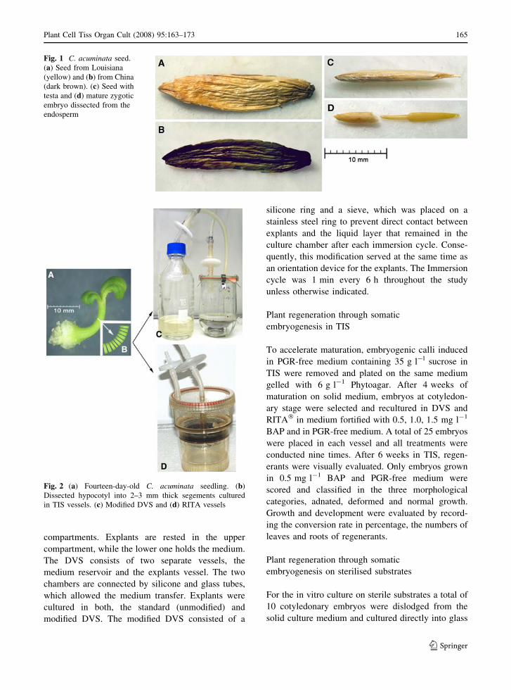
Fig. 1 C. acuminata seed.
(a) Seed from Louisiana
(yellow) and (b) from China
(dark brown). (c) Seed with
testa and (d) mature zygotic
embryo dissected from the
endosperm
Fig. 2 (a) Fourteen-day-old C. acuminata seedling. (b)
Dissected hypocotyl into 2–3 mm thick segements cultured
in TIS vessels. (c) Modified DVS and (d) RITA vessels
Plant Cell Tiss Organ Cult (2008) 95:163–173 165
123
jars (Weckglass�) of the four different substrates with
five replicates: (1) a mixture of sand and vermiculite
(SV, 1:2 v/v); (2) sand, soil and vermiculite (SSV,
1:2:2 v/v/v); (3) pure sand and (4) pure soil. All
substrates were moistened with MS medium supple-
mented with 0.5 mg l-1 BAP before being autoclaved
twice within 48 h at 121�C for 45 min. Cultures were
visual evaluated under aseptic condition on the 4th,
8th and 12th week. Growth, development and survival
rate of regenerants were recorded.
In vitro rooting in TIS and on sterile substrates
Rooting in TIS was achieved by subculturing all
regenerated plantlets for 4 weeks in a half strength
MS medium supplemented with 0.5 mg l-1 IBA plus
20 g l-1 sucrose, while those on sterile substrates were
moistened with the same medium but without sucrose.
After the third in vitro evaluation on the 12th
week, rooted plantlets derived from both systems
were rinsed in tap water and transferred without any
further pre-treatment directly into well-drained trays
(30 9 45 cm) containing non-sterile commercial
sowing soil (Gartenkrone� Aussaaterde, Zeus
GmbH). The mean day and night temperatures in
the greenhouse were 24 ± 2�C and 19 ± 2�C,
respectively. Plantlets were kept covered in the trays
for the first 10 days before being exposed to green-
house conditions and 3 weeks later the survival rate
of plantlets was recorded.
Statistical analyses
All experiments were repeated at least three times
and the statistical significance was determined by
ANOVA at a 5% level of probability.
Results
Somatic embryogenesis induction in PGR-free
medium in TIS
Hypocotyl segments cultured in PGR-free MS medium
containing 20, 25, 30 and 35 g l-1 sucrose in the
unmodified DVS remained green during the first
2 weeks. After 3 weeks in culture explants gradually
became brown and died. No callus formation was
observed among these explants in neither of the four
sucrose concentrations used. Callus induction was also
not observed in PGR-free medium containing 20, 25
and 30 g l-1 sucrose in the RITA vessel. Among the
same treatments in the modified DVS explants cultured
in medium supplemented with 20 and 25 g l-1 sucrose
showed no callus formation, while those in medium
with 30 g l-1 sucrose showed a tendency for callus
formation with a mean of 40% (4 ± 1.53) but then
retarded. Successful callus formation was observed
4 weeks after inoculation only on segments cultured in
medium fortified with 35 g l-1 sucrose in the modified
DVS. Similar responses were observed in the RITA�
vessels in medium supplemented with 35 g l-1
sucrose. An average of 80% eight out of ten segments
in DVS (8 ± 1.53) and 60% six out of ten in RITA�
(6 ± 1.0) formed callus, upon which embryogenic
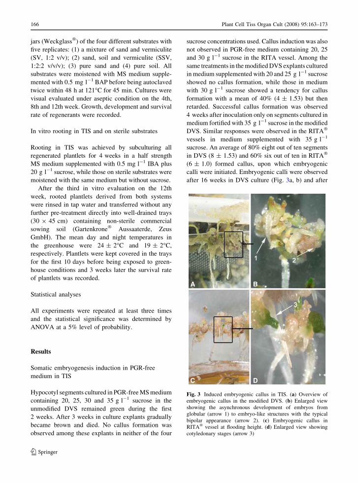
calli were initiated. Embryogenic calli were observed
after 16 weeks in DVS culture (Fig. 3a, b) and after
Fig. 3 Induced embryogenic callus in TIS. (a) Overview of
embryogenic callus in the modified DVS. (b) Enlarged view
showing the asynchronous development of embryos from
globular (arrow 1) to embryo-like structures with the typical
bipolar appearance (arrow 2). (c) Embryogenic callus in
RITA� vessel at flooding height. (d) Enlarged view showing
cotyledonary stages (arrow 3)
166 Plant Cell Tiss Organ Cult (2008) 95:163–173
123
12 weeks in RITA� (Fig. 3c, d). Calli in DVS were
friable and showed a translucent to yellow–green
appearance, while those in RITA� were soft and light
brownish. Proembryos protruded as green dots on the
surface of the callus mass. The development of somatic
embryos was asynchronous, and several stages of
embryos from globular to embryo-like structures with
a typical bipolar appearance and cotyledonary stage
were present at the same time in both vessels. Embryo
maturation into cotyledonary stage was very slow in
TIS but when plated on solid PGR-free medium
proembryos matured into well-developed cotyledon-
ary embryos within 4 weeks. Cotyledonary embryos
selected from the solid media were then used for
plantlet conversion in TIS.
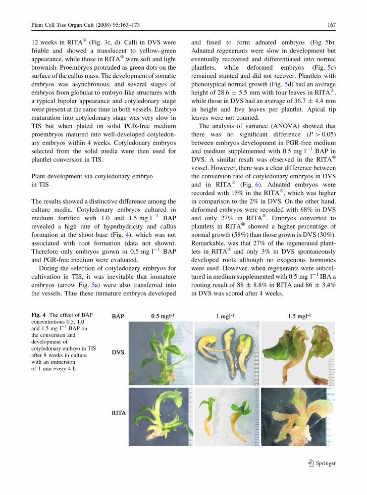
Plant development via cotyledonary embryo
in TIS
The results showed a distinctive difference among the
culture media. Cotyledonary embryos cultured in
medium fortified with 1.0 and 1.5 mg l-1 BAP
revealed a high rate of hyperhydricity and callus
formation at the shoot base (Fig. 4), which was not
associated with root formation (data not shown).
Therefore only embryos grown in 0.5 mg l-1 BAP
and PGR-free medium were evaluated.
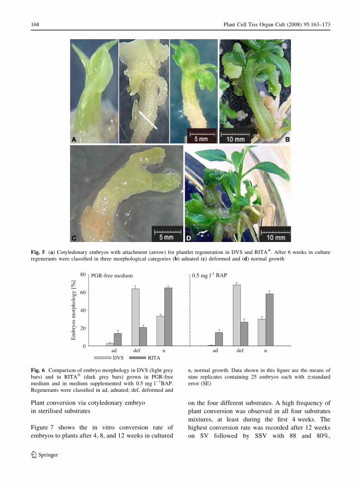
During the selection of cotyledonary embryos for
cultivation in TIS, it was inevitable that immature
embryos (arrow Fig. 5a) were also transferred into
the vessels. Thus these immature embryos developed
and fused to form adnated embryos (Fig. 5b).
Adnated regenerants were slow in development but
eventually recovered and differentiated into normal
plantlets, while deformed embryos (Fig. 5c)
remained stunted and did not recover. Plantlets with
phenotypical normal growth (Fig. 5d) had an average
height of 28.6 ± 5.5 mm with four leaves in RITA�,
while those in DVS had an average of 36.7 ± 4.4 mm
in height and five leaves per plantlet. Apical tip
leaves were not counted.
The analysis of variance (ANOVA) showed that
there was no significant difference (P [ 0.05)
between embryos development in PGR-free medium
and medium supplemented with 0.5 mg l-1 BAP in
DVS. A similar result was observed in the RITA�
vessel. However, there was a clear difference between
the conversion rate of cotyledonary embryos in DVS
and in RITA� (Fig. 6). Adnated embryos were
recorded with 15% in the RITA�, which was higher
in comparison to the 2% in DVS. On the other hand,
deformed embryos were recorded with 68% in DVS
and only 27% in RITA�. Embryos converted to
plantlets in RITA� showed a higher percentage of
normal growth (58%) than those grown in DVS (30%).
Remarkable, was that 27% of the regenerated plant-
lets in RITA� and only 3% in DVS spontaneously
developed roots although no exogenous hormones
were used. However, when regenerants were subcul-
tured in medium supplemented with 0.5 mg l-1 IBA a
rooting result of 88 ± 8.8% in RITA and 86 ± 3.4%
in DVS was scored after 4 weeks.
Fig. 4 The effect of BAP
concentrations 0.5, 1.0
and 1.5 mg l-1 BAP on
the conversion and
development of
cotyledonary embryo in TIS
after 8 weeks in culture
with an immersion
of 1 min every 4 h
Plant Cell Tiss Organ Cult (2008) 95:163–173 167
123
Plant conversion via cotyledonary embryo
in sterilised substrates
Figure 7 shows the in vitro conversion rate of
embryos to plants after 4, 8, and 12 weeks in cultured
on the four different substrates. A high frequency of
plant conversion was observed in all four substrates
mixtures, at least during the first 4 weeks. The
highest conversion rate was recorded after 12 weeks
on SV followed by SSV with 88 and 80%,
Fig. 5 (a) Cotyledonary embryos with attachment (arrow) for plantlet regeneration in DVS and RITA�. After 6 weeks in culture
regenerants were classified in three morphological categories (b) adnated (c) deformed and (d) normal growth
Em
bryo
s m
orph
olog
y [%
]
0.5 mg l-1 BAPPGR-free medium
ad def n ad def n0
20
40
60
80
DVS RITA
Fig. 6 Comparison of embryo morphology in DVS (light grey
bars) and in RITA� (dark grey bars) grown in PGR-free
medium and in medium supplemented with 0.5 mg l-1BAP.
Regenerants were classified in ad, adnated; def, deformed and
n, normal growth. Data shown in this figure are the means of
nine replicates containing 25 embryos each with ±standard
error (SE)
168 Plant Cell Tiss Organ Cult (2008) 95:163–173
123
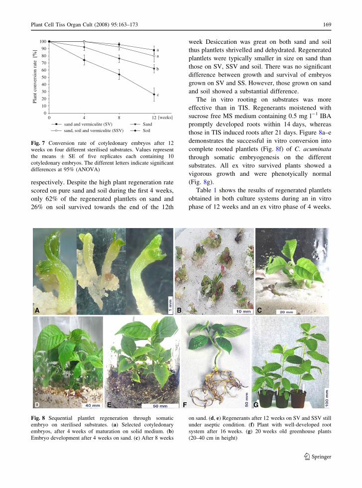
respectively. Despite the high plant regeneration rate
scored on pure sand and soil during the first 4 weeks,
only 62% of the regenerated plantlets on sand and
26% on soil survived towards the end of the 12th
week Desiccation was great on both sand and soil
thus plantlets shrivelled and dehydrated. Regenerated
plantlets were typically smaller in size on sand than
those on SV, SSV and soil. There was no significant
difference between growth and survival of embryos
grown on SV and SS. However, those grown on sand
and soil showed a substantial difference.
The in vitro rooting on substrates was more
effective than in TIS. Regenerants moistened with
sucrose free MS medium containing 0.5 mg l-1 IBA
promptly developed roots within 14 days, whereas
those in TIS induced roots after 21 days. Figure 8a–e
demonstrates the successful in vitro conversion into
complete rooted plantlets (Fig. 8f) of C. acuminata
through somatic embryogenesis on the different
substrates. All ex vitro survived plants showed a
vigorous growth and were phenotyically normal
(Fig. 8g).
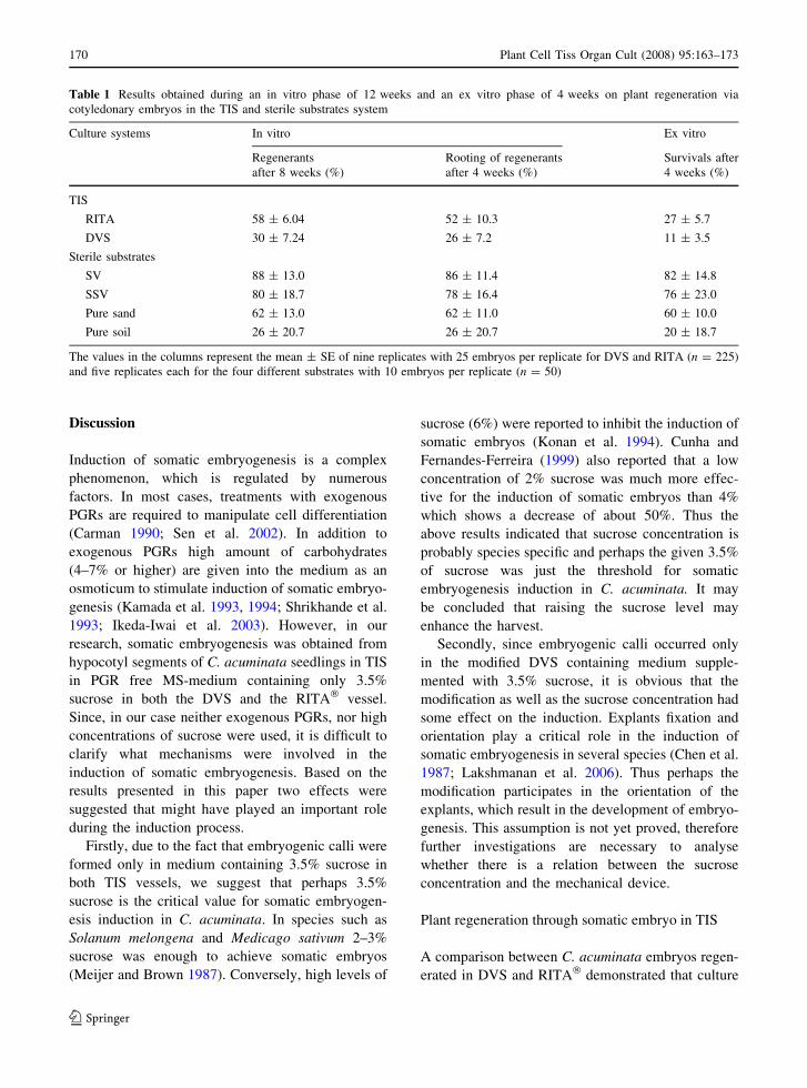
Table 1 shows the results of regenerated plantlets
obtained in both culture systems during an in vitro
phase of 12 weeks and an ex vitro phase of 4 weeks.
aa
b
c
[weeks]0 4 8 12
Plan
t con
vers
ion
rate
[%
]
0
10
20
30
40
50
60
70
80
90
100
sand and vermiculite (SV)sand, soil and vermiculite (SSV)
SandSoil
Fig. 7 Conversion rate of cotyledonary embryos after 12
weeks on four different sterilised substrates. Values represent
the means ± SE of five replicates each containing 10
cotyledonary embryos. The different letters indicate significant
differences at 95% (ANOVA)
Fig. 8 Sequential plantlet regeneration through somatic
embryo on sterilised substrates. (a) Selected cotyledonary
embryos, after 4 weeks of maturation on solid medium. (b)
Embryo development after 4 weeks on sand. (c) After 8 weeks
on sand. (d, e) Regenerants after 12 weeks on SV and SSV still
under aseptic condition. (f) Plant with well-developed root
system after 16 weeks. (g) 20 weeks old greenhouse plants
(20–40 cm in height)
Plant Cell Tiss Organ Cult (2008) 95:163–173 169
123
Discussion
Induction of somatic embryogenesis is a complex
phenomenon, which is regulated by numerous
factors. In most cases, treatments with exogenous
PGRs are required to manipulate cell differentiation
(Carman 1990; Sen et al. 2002). In addition to
exogenous PGRs high amount of carbohydrates
(4–7% or higher) are given into the medium as an
osmoticum to stimulate induction of somatic embryo-
genesis (Kamada et al. 1993, 1994; Shrikhande et al.
1993; Ikeda-Iwai et al. 2003). However, in our
research, somatic embryogenesis was obtained from
hypocotyl segments of C. acuminata seedlings in TIS
in PGR free MS-medium containing only 3.5%
sucrose in both the DVS and the RITA� vessel.
Since, in our case neither exogenous PGRs, nor high
concentrations of sucrose were used, it is difficult to
clarify what mechanisms were involved in the
induction of somatic embryogenesis. Based on the
results presented in this paper two effects were
suggested that might have played an important role
during the induction process.
Firstly, due to the fact that embryogenic calli were
formed only in medium containing 3.5% sucrose in
both TIS vessels, we suggest that perhaps 3.5%
sucrose is the critical value for somatic embryogen-
esis induction in C. acuminata. In species such as
Solanum melongena and Medicago sativum 2–3%
sucrose was enough to achieve somatic embryos
(Meijer and Brown 1987). Conversely, high levels of
sucrose (6%) were reported to inhibit the induction of
somatic embryos (Konan et al. 1994). Cunha and
Fernandes-Ferreira (1999) also reported that a low
concentration of 2% sucrose was much more effec-
tive for the induction of somatic embryos than 4%
which shows a decrease of about 50%. Thus the
above results indicated that sucrose concentration is
probably species specific and perhaps the given 3.5%
of sucrose was just the threshold for somatic
embryogenesis induction in C. acuminata. It may
be concluded that raising the sucrose level may
enhance the harvest.
Secondly, since embryogenic calli occurred only
in the modified DVS containing medium supple-
mented with 3.5% sucrose, it is obvious that the
modification as well as the sucrose concentration had
some effect on the induction. Explants fixation and
orientation play a critical role in the induction of
somatic embryogenesis in several species (Chen et al.
1987; Lakshmanan et al. 2006). Thus perhaps the
modification participates in the orientation of the
explants, which result in the development of embryo-
genesis. This assumption is not yet proved, therefore
further investigations are necessary to analyse
whether there is a relation between the sucrose
concentration and the mechanical device.
Plant regeneration through somatic embryo in TIS
A comparison between C. acuminata embryos regen-
erated in DVS and RITA� demonstrated that culture
Table 1 Results obtained during an in vitro phase of 12 weeks and an ex vitro phase of 4 weeks on plant regeneration via
cotyledonary embryos in the TIS and sterile substrates system
Culture systems In vitro Ex vitro
Regenerants
after 8 weeks (%)
Rooting of regenerants
after 4 weeks (%)
Survivals after
4 weeks (%)
TIS
RITA 58 ± 6.04 52 ± 10.3 27 ± 5.7
DVS 30 ± 7.24 26 ± 7.2 11 ± 3.5
Sterile substrates
SV 88 ± 13.0 86 ± 11.4 82 ± 14.8
SSV 80 ± 18.7 78 ± 16.4 76 ± 23.0
Pure sand 62 ± 13.0 62 ± 11.0 60 ± 10.0
Pure soil 26 ± 20.7 26 ± 20.7 20 ± 18.7
The values in the columns represent the mean ± SE of nine replicates with 25 embryos per replicate for DVS and RITA (n = 225)
and five replicates each for the four different substrates with 10 embryos per replicate (n = 50)
170 Plant Cell Tiss Organ Cult (2008) 95:163–173
123
vessels have a great influence on plant development.
In this study a higher percentage of well-developed
plantlets was achieved in RITA� than in DVS. Similar
results were reported on the development of somatic
embryos in RITA� with different plant species such as
Musa spp. (Alvard et al. 1993), Hevea brasiliensis
(Etienne et al. 1997), Citrus deliciosa (Cabasson et al.
1997) and Coffea arabica (Etienne-Barry et al. 1999).
Cabasson et al. (1997) asserted that the temporary
immersion in RITA� promotes development and
conversion of somatic embryos. This is probably
due to the design of the vessel. Thus the upper
chamber is overlaid with open-pores polyurethane
foam in which embryos are able to support their
geotropism easier than in DVS without such adhering
possibility. During immersion and emersion embryos
in RITA� were able to obtain a positive geotropism
while those in DVS were disrupted until they reached
a certain height and biomass. This could be one reason
why 27% of the Camptotheca regenerants in RITA�
developed roots without any additional exogenous
PGR. Hence, the results in this experiment provide
clear evidence that the RITA� vessels are more
suitable and effective for embryo-to-plantlet devel-
opment. These vessels were developed for culturing
embryogenic cells and embryos (Afreen et al. 2002).
However, comparing the two vessels the DVS is
easier to handle especially in terms of medium
exchange, which is simply done by disconnecting
the medium vessel without exposing the culture to the
environment outside the vessel like in RITA�. This
may increase the risk of contamination.
In vitro plant regeneration on sterilised substrates
To acclimatise regenerated plantlets to greenhouse
conditions is one of the most critical phases in the
whole micropropagation process. Plantlets are forced
to change from being heterotrophic to autotrophic
and in most cases a considerable number of micro-
propagated plantlets did not survive the ex vitro
conditions. In vitro grown plants typically showed a
low photosynthetic efficiency due to malfunctioning
of stomata and the lack of epicuticular wax, so that
plants are predetermined to dehydration (Preece and
Sutter 1991; Hazarika 2006). Thus the consideration
of the substrate experiment was not to compare the
conversion rate of cotyledonary embryos in the two
culture system (TIS and on sterilised substrates). It
was an approach to compare the survival rate of
regenerated plantlets derived from TIS with those
from derived from sterile substrates upon exposure to
greenhouse conditions. A similar experiment on
substrates was conducted by Jayasankar et al.
(2001) who achieved a high conversion rate of
grapevine via somatic embryos on sand and soil
overlaid with sand but only a few embryos regener-
ated into plantlets due to inadequate moisture and
perhaps lack of nutrient because only tap water was
used to irrigate the substrates. In contrast to seeds,
somatic embryos have to develop without seed coat
and maternal tissue that normally supplies nutrition
during germination (Gray and Purohit 1991). Thus
the high survival rate obtained in this study is
probably due to extensive autoclaving of substrates as
well as the irrigation with MS medium supplemented
with 0.5 mg l-1 BAP during the regeneration phase,
which facilitated the maturity of cotyledonary
embryos.
From the eighth week on we shifted from full
strength to half strength MS medium without sucrose
to allow plantlets to become partially autotrophic.
This reduced the stress and allowed plantlets to adapt
better to ex vitro conditions (Purohit et al. 1995).
Rooting plants in in vitro is considered to be labour-
intensive and expensive (Hazarika 2003) but it was
apparent that rooting plantlets on sterile substrates
irrigated with medium containing 0.5 mg l-1 IBA
without sucrose promoted well-developed roots
within 2 weeks and guaranteed a higher survival rate
when transfer to ex vitro conditions.
Acknowledgements The authors thank Professor Zhijun Liu,
School of Renewable Natural Resources Louisiana State
University Baton Rouge, LA 70803 USA for providing
Camptotheca acuminata seeds for this research and Rainer
Thomas for the technical assistance the critical reading and
comments on this manuscript.
References
Afreen F, Zobayed SMA, Kozai T (2002) Photoautotrophic
culture of Coffea arabusta somatic embryos: development
of a bioreactor for large-scale plantlet conversion from
cotyledonary embryos. Ann Bot (Lond) 90:21–29.
doi:10.1093/aob/mcf151
Aitken-Christie J (1991) Automation. In: Debergh PC, Zim-
merman RH (eds) Micropropagation technology and
application. Kluwer Academic Publishers, Dordrecht,
pp 363–388
Plant Cell Tiss Organ Cult (2008) 95:163–173 171
123
Alvard D, Cote F, Teisson C (1993) Comparison of methods of
liquid medium culture for banana micropropagation.
Effects of temporary immersion of explants. Plant Cell
Tissue Organ Cult 32(1):55–60. doi:10.1007/BF00040116
Berthouly M, Etienne H (2005) Temporary immersion system:
a new concept for use liquid medium in mass propagation.
In: Hvoslef-Eide AK, Preil W (eds) Liquid culture sys-
tems for in vitro plant propagation. Springer Verlag,
Berlin, pp 165–195
Cabasson C, Alvard D, Dambier D, Ollitrault P, Teisson C
(1997) Improvement of Citrus somatic embryo develop-
ment by temporary immersion. Plant Cell Tissue Organ
Cult 50:33–37. doi:10.1023/A:1005896725780
Carman JG (1990) Embryogenic cells in plant tissue cultures:
occurrence and behaviour. In Vitro Cell Dev Biol
26(8):746–753. doi:10.1007/BF02623615
Chen THH, Marowitch J, Thompson BG (1987) Genotypic
effects on somatic embryogenesis and plant regeneration
from callus cultures of alfalfa. Plant Cell Tissue Organ
Cult 8:73–81. doi:10.1007/BF00040734
Cunha A, Fernandes-Ferreira M (1999) Influence of medium
parameters on somatic embryogenesis from hypocotyl
explants of flax (Linum usitatissimum L.): effect of carbon
source, total inorganic nitrogen and balance between ionic
forms and interaction between calcium and zeatin. J Plant
Physiol 155(4–5):591–597
Cunningham D, Pyrhonen S, James RD, Punt CJ, Hickish TF,
Heikkila R et al (1998) Randomised trial of irinotecan
plus supportive care versus supportive care alone after
fluorouracil failure for patients with metastatic colorectal
cancer. Lancet 352:1413–1418. doi:10.1016/S0140-
6736(98)02309-5
Douillard JY, Cunningham D, Roth AD, Navarro M, James
RD, Karasek P et al (2000) Irinotecan combined with
fluorouracil compared with fluorouracil alone as first-line
treatment for metastatic colorectal cancer: a multicentre
randomised trial. Lancet 355:1041–1047. doi:10.1016/
S0140-6736(00)02034-1
Etienne HM, Lartaud M, Michaux-Ferriere N, Carron MP,
Berthouly M, Teisson C (1997) Improvement of somatic
embryogenesis in Hevea brasiliensis (Mull. Arg.) using
the temporary immersion technique. In Vitro Cell Dev
Biol 33:81–87
Etienne-Barry D, Bertrand B, Vasquez N, Etienne H (1999)
Direct sowing of Coffea arabica somatic embryos mass-
produced in a bioreactor and regeneration of plant. Plant
Cell Rep 199:111–119. doi:10.1007/s002990050720
Gray DJ, Purohit A (1991) Somatic embryogenesis and
development of synthetic seed technology. Crit Rev Plant
Sci 10:33–61
Hazarika BN (2003) Acclimatisation of tissue-culture plants.
Curr Sci 85(12):1704–1712
Hazarika BN (2006) Morpho-physiological disorders in in vitro
culture of plants. Sci Hortic 108(2):105–120
Ikeda-Iwai M, Umehara M, Satoh S, Kamada H (2003) Stress-
induced somatic embryogenesis in vegetative tissue of
Arabidopsis thaliana. Plant J 24:109–114
Jain KJ, Nessler CL (1996) Clonal propagation of Camptothecaacuminata through shoot bud culture. Plant Cell Tissue
Organ Cult 44:229–233
Jayasankar S, Van Aman M, Li Z, Gray DJ (2001) Direct
seeding of grapevine somatic embryos and regeneration of
plants. In Vitro Cell Dev Biol 37:476–479
Kamada H, Ishikawa K, Saga H, Harada H (1993) Induction of
somatic embryogenesis in carrot by osmotic stress. Plant
Tissue Cult Lett 10:38–44
Kamada H, Tachikawa Y, Saitou T, Harada H (1994) Heat
stresses induction of carrot somatic embryogenesis. Plant
Tissue Cult Lett 11(3):229–232
Konan NK, Sangwan RS, Sangwan BS (1994) Somatic
embryogenesis from cultured mature cotyledons of cas-
sava (Manihot esculenta Crantz). Identification of
parameters influencing the frequency of embryogenesis.
Plant Cell Tissue Organ Cult 37(2):91–102. doi:10.1007/
BF00043602
Lakshmanan P, Geijskes R, Wang L, Elliott A, Grof C, Berding
N et al (2006) Developmental and hormonal regulation of
direct shoot organogenesis and somatic embryogenesis in
sugarcane (Saccharum spp. interspecific hybrids) leaf
culture. Plant Cell Rep 25(10):1007–1015. doi:10.1007/
s00299-006-0154-1
Li Z, Liu Z (2001) Micropropagation of Camptotheca acumi-nata decaisne from axillary buds, shoot tips, and seed
embryos in a tissue culture system. In Vitro Cell Dev Biol
37:84–88
Li Z, Liu Z (2005) Plant regeneration from leaf petiole in
Camptotheca acuminata. In Vitro Cell Dev Biol
41(3):262–265
Li S, Zhang Z, Cain A, Wang B, Long M, Taylor J (2005)
Antifungal activity of camptothecin, trifolin, and hy-
peroside isolated from Camptotheca acuminata. J Agric
Food Chem 53(1):32–37. doi:10.1021/jf0484780
Lorence A, Medina-Bolivar F, Nessler CL (2004) Campto-
thecin and 10-hydroxy-camptothecin from Camptothecaacuminata hairy roots. Plant Cell Rep 22:437–441.
doi:10.1007/s00299-003-0708-4
Maliepaard M, van Gastelen MA, Tohgo A, Hausheer FH, van
Waardenburg RCAM, de Jong LA et al (2001) Circum-
vention of breast cancer resistance protein (BCRP)-mediated resistance to camptothecins in vitro using non-
substrate drugs or the BCRP inhibitor GF120918. Clin
Cancer Res 7:935–941
Meijer EGM, Brown DCW (1987) Role of exogenous reduced
nitrogen and sucrose in rapid high frequency somatic
embryogenesis in Medicago sativa. Plant Cell Tissue
Organ Cult 10(1):11–19. doi:10.1007/BF00037492
Murashige T, Skoog F (1962) A revised medium for rapid
growth and bio assays with tobacco tissue cultures.
Physiol Plant 15:473–497. doi:10.1111/j.1399-
3054.1962.tb08052.x
Preece JE, Sutter EG (1991) Acclimatisation of micropropa-
gated plants to the greenhouse and field. In: Debergh PC,
Zimmerman RH (eds) Micropropagation. Kluwer Aca-
demic Publishers, Dordrecht, pp 71–93
Priel E, Showalter SD, Blair DG (1991) Inhibition of human
immunodeficiency virus (HIV-1) replication by non-
cytotoxic doses of camptothecin, a topoisomerase I
inhibitor. AIDS Res Hum Retroviruses 7:65–72
Purohit SD, Tak K, Kukda G (1995) In vitro propagation of
Boswellia serrata Roxb. Biol Plant 37:335–340
172 Plant Cell Tiss Organ Cult (2008) 95:163–173
123
Sakato K, Misawa M (1974) Effects of chemical and physical
condition of growth of Camptotheca acuminata cell cul-
ture. Agric Biol Chem 38(3):491–497
Sen J, Kalia S, Guha-Mukherjee S (2002) Level of endogenous
free amino acids during various stages of culture of Vignamungo (L.) Hepper somatic embryogenesis, organogene-
sis and plant regeneration. Curr Sci 82(4):429–433
Shrikhande M, Thengane SR, Mascarenhas AF (1993) Somatic
embryogenesis and plant regeneration in Azadirachtaindica A. Juss. In Vitro Cell Dev Biol 29:38–42
Wall ME, Wani MC, Cook CE, Palmer KH, McPhail AT, Sim
GA (1966) Plant antitumor agents. I. Isolation and
structure of camptothecin, a novel alkaloidal leukemia and
tumor inhibitor from Camptotheca acuminata. J Am
Chem Soc 88:3888–3890. doi:10.1021/ja00968a057
Wang H, Zu Y, Wang W, Wu S, Dong F (2006) Establishment
of Camptotheca acuminata regeneration from leaf
explants. Biol Plant 50(4):725–728. doi:10.1007/s10535-
006-0116-z
Wiedenfeld H, Furmanowa M, Roeder E, Guzewska J,
Gustowski W (1997) Camptothecin and 10-hydroxycam-
ptothecin in callus and plantlets of Camptotheca acuminata.
Plant Cell Tissue Organ Cult 49:213–218. doi:10.1023/
A:1005704429339
Plant Cell Tiss Organ Cult (2008) 95:163–173 173
123
Related Documents


