Welcome message from author
This document is posted to help you gain knowledge. Please leave a comment to let me know what you think about it! Share it to your friends and learn new things together.
Transcript
PLANT HEALTHRESEARCH AND PRACTICE
RUSSIAN-ENGLISH JOURNAL
DECEMBER 4/26/2018IS
SN 2
306-
9767
КАРАНТИН РАСТЕНИЙНАУКА И ПРАКТИКА
РУССКО-АНГЛИЙСКИЙ ЖУРНАЛ
ДЕКАБРЬ 4/26/2018
ВИДЫ РОДА HELIANTHUS L. В ЕДИНОМ ПЕРЕЧНЕ КАРАНТИННЫХ ОБЪЕКТОВ ЕВРАЗИЙСКОГО ЭКОНОМИЧЕСКОГО СОЮЗА стр. 2
«НОВОГОДНИЕ ЕЛКИ» КАК УГРОЗА РАСПРОСТРАНЕНИЯ ВРЕДНЫХ И ПАТОГЕННЫХ ЛЕСНЫХ ОРГАНИЗМОВ стр. 9
БАКТЕРИОЗЫ – ВОЗБУДИТЕЛИ БОЛЕЗНЕЙ ЗЕРНОБОБОВЫХ КУЛЬТУР И РАЗРАБОТКА МЕТОДОВ ИХ ДИАГНОСТИКИ стр. 28
ЭКСПЕДИЦИОННЫЕ ИССЛЕДОВАНИЯ ВРЕДИТЕЛЕЙ ЮЖНОЙ ЧАСТИ КОРЕЙСКОГО ПОЛУОСТРОВА стр. 57
SPECIES OF THE HELIANTHUS L. GENUS IN THE COMMON LIST OF QUARANTINE OBJECTS OF THE EURASIAN ECONOMIC UNION page 6
“CHRISTMAS TREES” AS A THREAT TO THE SPREAD OF PATHOGENIC FOREST PESTS page 14
BACTERIAL DISEASE PATHOGENS OF GRAIN LEGUMES AND DEVELOPMENT OF METHODS FOR THEIR DIAGNOSTICS page 34
EXPEDITIONARY STUDIES OF PESTS IN THE SOUTHERN PART OF THE KOREAN PENINSULA page 61
Тел./факс: 8 (499) 707-22-27
Журнал «Карантин растений. Наука и практика» зарегистрирован в Федеральной службе по надзору в сфере связи, информационных технологий и массовых коммуникаций (Роскомнадзор), свидетельство о регистрации ПИ № ФС77-52594 от 25 января 2013 г. Учредитель: ООО «Успех», выпускается по заказу Федерального государственного бюджетного учреждения «Всероссийский центр карантина растений» (ФГБУ «ВНИИКР»)Издатель: ООО «У-Строй»Адрес редакции: 115551, г.Москва, Шипиловский проезд, дом 39, корпус 2, этаж 15, помещение 53, комнаты 1, 3Номер отпечатан в ООО «Юнион Принт», г. Нижний Новгород, Окский съезд, д. 2, тел.: (831) 416-01-68
Дата выхода ч.12.2018 г. Тираж 3000 экземпляров. Подписной индекс 70195 в Каталоге Агентства «Роспечать»
«КАРАНТИН РАСТЕНИЙ. НАУКА И ПРАКТИКА» ДВУЯЗЫЧНЫЙ НАУЧНЫЙ ЖУРНАЛ №4 (26) 2018 г.
Зиновьева Светлана Георгиевна+7 967 294 90 61
Главный редактор:А.Я. Сапожников, директор ФГБУ «ВНИИКР»
Шеф-редактор: Светлана Зиновьева, начальник отдела по связям с общественностью и СМИ ФГБУ «ВНИИКР»
Выпускающий редактор: Ольга Лесных e-mail: [email protected]
Редакционная коллегия журнала «Карантин растений. Наука и практика»:
Швабаускене Ю.А. — заместитель Руководителя Россельхознадзора
Долженко В.И. — академик РАН, доктор сельскохозяйственных наук, заместитель директора Всероссийского НИИ защиты растений
Надыкта В.Д. — академик РАН, доктор технических наук, директор Всероссийского НИИ биологической защиты растений
Орлинский А.Д. — доктор биологических наук, научный советник ЕОКЗР
Павлюшин В.А. — академик РАН, доктор биологических наук, директор Всероссийского НИИ защиты растений
Санин С.С. — академик РАН, доктор биологических наук, профессор, заведующий отделом Всероссийского НИИ фитопатологии
Мартин Уорд — Генеральный директор ЕОКЗР
Ханну Кукконен — директор подразделения фитосанитарного надзора, EVIRA (Финляндия)
Сагитов А.О. — доктор биологических наук, Генеральный директор ТОО «Казахский НИИ защиты и карантина растений»
Сорока С.В. — кандидат сельскохозяйственных наук, директор РУП «Институт защиты растений» НАН Республики Беларусь
Джалилов Ф.С. — доктор биологических наук, профессор, заведующий лабораторией защиты растений МСХА им. К.А. Тимирязева
Абасов М.М. — доктор биологических наук, заместитель директора ФГБУ «ВНИИКР»
Шероколава Н.А. — заместитель директора ФГБУ «ВНИИКР», вице-президент ЕОКЗР
Добровольская О.Б. — кандидат биологических наук, заместитель директора ФГБУ «ВНИИКР»
Камаев И.О. — кандидат биологических наук, старший научный сотрудник научно-экспериментального отдела ФГБУ «ВНИИКР»
РЕДАКЦИЯ: Волкова Е.М. — кандидат биологических наук, заведующая лабораторией сорных растений
Волков О.Г. — начальник отдела биометода
Кулинич О.А. — доктор биологических наук, начальник отдела лесного карантина
Приходько Ю.Н. — кандидат сельскохозяйственных наук, начальник научно-методического отдела фитопатологии
Скрипка О.В. — кандидат биологических наук, ведущий научный сотрудник лаборатории микологии
Усачева С.Е. — переводчик отдела фитосанитарных рисков и международного взаимодействия
Быков И.И. — переводчик отдела фитосанитарных рисков и международного взаимодействия
Беломестнова А.А. — переводчик отдела фитосанитарных рисков и международного взаимодействия
Красовский Г.С. — переводчик отдела фитосанитарных рисков и международного взаимодействия
Дизайн и верстка: Роман Солоха
Корректоры: Татьяна АртемьеваОльга Тренева
Менеджер по подписке и дистрибуции: Павел Сафронов+7 903 505 33 23
4| 26| 2018 1КАРАНТИН РАСТЕНИЙ. НАУКА И ПРАКТИКА
Виды рода Helianthus L. в Едином перечне карантинных объектов Евразийского экономического союза
Д.Л. Белкин, зам. начальника Испытательного лабораторного центра ФГБУ «ВНИИКР»
Ю.Ю. Кулакова, начальник научно-экспериментального отдела ФГБУ «ВНИИКР»
2
СОДЕРЖАНИЕ CONTENTSpecies of the Helianthus L. Genus in the Common List of Quarantine Objects of the Eurasian Economic UnionD.L. Belkin, Deputy Head of the Testing Laboratory Center of FGBU “VNIIKR”Y.Y. Kulakova, Head of the Research and Testing Department of FGBU “VNIIKR”
6«Новогодние елки»
как угроза распространения вредных и патогенных лесных организмовО.А. Кулинич, Всероссийский центр карантина растений (ФГБУ «ВНИИКР»)
А.Г. Щуковская, Всероссийский центр карантина растений (ФГБУ «ВНИИКР»)Е.Н. Арбузова, Всероссийский центр карантина растений (ФГБУ «ВНИИКР»)
Н.И. Козырева, Институт проблем экологии и эволюции (ИПЭЭ РАН)
9Особо опасные возбудители болезней
косточковых культур рода Candidatus Phytoplasma spp.Г.Н. Бондаренко, старший научный сотрудник – начальник Испытательного
лабораторного центра ФГБУ «ВНИИКР»И.Г. Башкирова, агроном лаборатории анализа ГМО
Испытательного лабораторного центра ФГБУ «ВНИИКР»
18Бактериозы – возбудители болезней зернобобовых культур
и разработка методов их диагностикиЕ.В. Каримова, старший научный сотрудник НМОФ ФГБУ «ВНИИКР»
И.М. Игнатьева, научный сотрудник лаборатории бактериологии ИЛЦ ФГБУ «ВНИИКР»
28Неповирус кольцевой пятнистости малины Raspberry ringspot virus –
новый карантинный организм, включенный в список ЕАЭСЮ.Н. Приходько, ведущий научный сотрудник НМОФ ФГБУ «ВНИИКР»
К.О. Тихонова, научный сотрудник НМОФ ФГБУ «ВНИИКР»
41Анализ возможностей формирования очагов карантинных объектов –
филлоксеры и ценхруса длинноколючкового на территории Белгородской области
А.Н. Мирошников, ведущий энтофитопатолог отдела карантина растений ФГБУ «Белгородская МВЛ»
Л.П. Скотникова, начальник отдела карантина растений ФГБУ «Белгородская МВЛ»
52Экспедиционные исследования вредителей
южной части Корейского полуостроваИ.О. Камаев, старший научный сотрудник
научно-экспериментального отдела ФГБУ «ВНИИКР»Г.С. Красовский, переводчик отдела фитосанитарных рисков
и международного взаимодействия ФГБУ «ВНИИКР»
57
“Christmas trees” as a �reat to the Spread of Pathogenic Forest PestsO.A. Kulinich, All-Russian Plant Quarantine Center (FGBU “VNIIKR”)A.G. Shchukovskaya, All-Russian Plant Quarantine Center (FGBU “VNIIKR”)E.N. Arbuzova, All-Russian Plant Quarantine Center (FGBU “VNIIKR”)N.I. Kozyreva, Institute of Ecology and Evolution Russian Academy of Sciences, Moscow
14Particularly Dangerous Pathogens of Candidatus Phytoplasma spp. Genus for Stone Fruit CropsG.N. Bondarenko, Senior Researcher, Head of the Laboratory and Testing Center of FGBU “VNIIKR”I.G. Bashkirova, Agronomist of the GMO Analysis Laboratory of the Laboratory and Testing Center of FGBU “VNIIKR”
23Bacterial Disease Pathogens of Grain Legumes and Development of Methods for �eir DiagnosticsE.V. Karimova, Senior Researcher of the RMPD of FGBU “VNIIKR”I.M. Ignatyeva, Researcher of the Bacteriology Laboratory of the LTC of FGBU “VNIIKR”
34Raspberry Ringspot Virus – New Quarantine Organism, Included in the EEU ListY.N. Prikhodko, Leading Researcher of NMOF FGBU “VNIIKR”K.O. Tikhonova, Researcher of NMOF FGBU “VNIIKR”
47�e Analysis of Outbreak Formation Possibilities of Quarantine Pests: Phylloxera (Dactylosphaira vitifoliae) and Spiny Burr Grass (Cenchrus longispinus) in the Belgorod Region, RussiaA.N. Miroshnikov, Leading Entophytopatologist of the Plant Quarantine Department of FGBU “Belgorodskaya MVL”L.P. Skotnikova, Head of Plant Quarantine Department of FGBU “Belgorodskaya MVL”
55Expeditionary Studies of Pests in the Southern Part of the Korean PeninsulaI.O. Kamayev, Senior Researcher of the Research and Testing Department FGBU “VNIIKR”G.S. Krasovsky, Translator of Phytosanitary Risk and International Cooperation Department FGBU “VNIIKR”
61
4| 26| 20182 КАРАНТИН РАСТЕНИЙ. НАУКА И ПРАКТИКА
Аннотация. В статье приведены сведения о географическом распро-странении подсолнечника калифор-нийского и подсолнечника реснитча-того, их краткое морфологическое описание и возможные пути распро-странения.
Ключевые слова. Карантин расте-ний, Helianthus L., подсолнечник кали-форнийский, подсолнечник реснитча-тый, распространение, морфология, местообитания, вредоносность.
Helianthus L. или Подсолнечник, – сложный в таксономическом отно-шении род американского проис-хождения, относящийся к семейству Сложноцветные (Asteraceae Bercht. & J. Presl) и объединяющий, по раз-ным оценкам, до 110 однолетних и многолетних видов (Анащенко, 1974; Schilling, 2006; Schilling, Heiser, 1981; Schilling, Panero, 2011). В со-став флоры России включают 8 ви-дов рода Подсолнечник (Анащенко, 1974; Баркалов и др., 1992; Бочкин, 2003; Васильченко, 1959; Маевский, 2014; Майоров, 2004; Майоров и др., 2012; Протопопова, 1994; Скворцов, 1973; Сырейщиков, 1910; Терентье-ва, 2002; Ульянова, 1998), из которых наиболее распространенными явля-ются H. lenticularis Douglas ex Lindl. (подсол нечник сорнополевой) и H. tuberosus L. (подсолнечник клубне-носный).
Среди всех видов подсолнеч-ников в мировом объеме встре-чаются как ценные культурные (H. annuus L., H. tuberosus L. и др.)
ВИДЫ РОДА HELIANTHUS L. В ЕДИНОМ ПЕРЕЧНЕ КАРАНТИННЫХ ОБЪЕКТОВ ЕВРАЗИЙСКОГО ЭКОНОМИЧЕСКОГО СОЮЗА
УДК 632.3.01/.08, 632.2
Д.Л. Белкин, зам. начальника Испытательного лабораторного центра ФГБУ «ВНИИКР»Ю.Ю. Кулакова, начальник научно-экспериментального отдела ФГБУ «ВНИИКР»
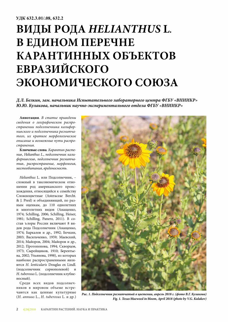
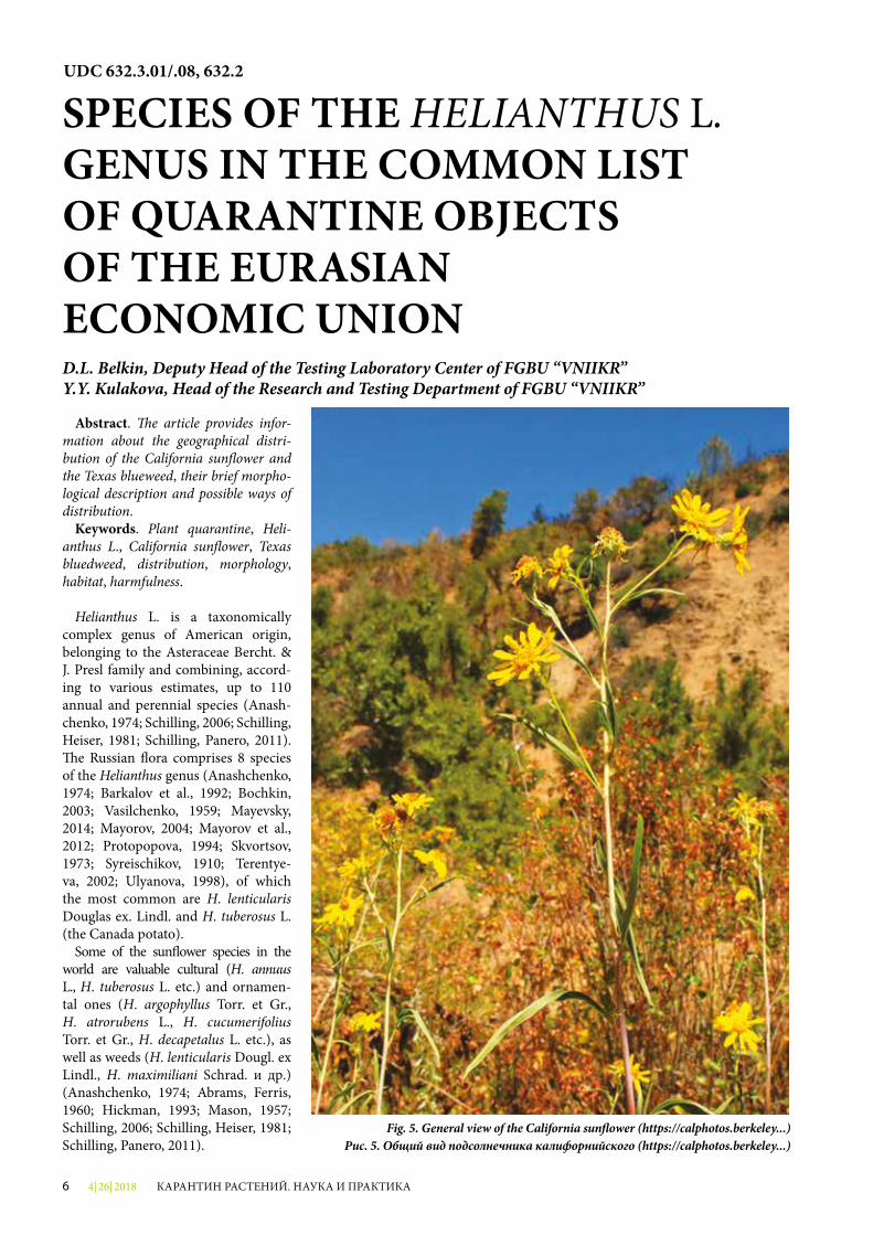
Рис. 1. Подсолнечник реснитчатый в цветении, апрель 2018 г. (фото В.Г. Кулакова)Fig. 1. Texas blueweed in bloom, April 2018 (photo by V.G. Kulakov)
4| 26| 2018 3КАРАНТИН РАСТЕНИЙ. НАУКА И ПРАКТИКА
и декоративные (H. argophyllus Torr. et Gr., H. atrorubens L., H. cucumerifolius Torr. et Gr., H. decapetalus L. и др.), так и сор-ные растения (H. lenticularis Dougl. ex Lindl., H. maximiliani Schrad. и др.) (Анащенко, 1974; Abrams, Ferris, 1960; Hickman, 1993; Ma-son, 1957; Schilling, 2006; Schil-ling, Heiser, 1981; Schilling, Panero, 2011).
В Единый перечень карантинных объектов Евразийского экономи-ческого союза (ЕАЭС) включены Helianthus ciliaris DC. (подсолнеч-ник реснитчатый) и Helianthus californicus DC. (подсолнечник кали-форнийский), которые отсутствуют на территории Российской Федера-ции (РФ) и Евразийского экономи-ческого союза.
Подсолнечник реснитчатый (He-lianthus ciliaris DC.) – многолетнее травянистое растение (рис. 1), совре-менный ареал которого охватывает юго-западные районы США (штаты Аризона, Калифорния, Колорадо, Иллинойс, Канзас, Небраска, Нева-да, Новая Мексика, Оклахома, Техас, Юта), Мексику (штаты Чиуауа, Ко-ауила, Дуранго, Сан-Луис-Потоси,
Сонора, Тамаулипас) и отдельные районы Австралии (штаты Новый Южный Уэльс и Квинсленд), в кото-рых является заносным сорным рас-тением (Abrams, Ferris, 1960; Mason, 1957; Schilling, 2006; Schilling, Heiser, 1981; Schilling, Panero, 2011).
Подсолнечник реснитчатый легко образует гибриды с другими вида-ми, например с Helianthus laciniatus, что приводит к образованию поли-плоидных клонов внутри общего ареала видов.
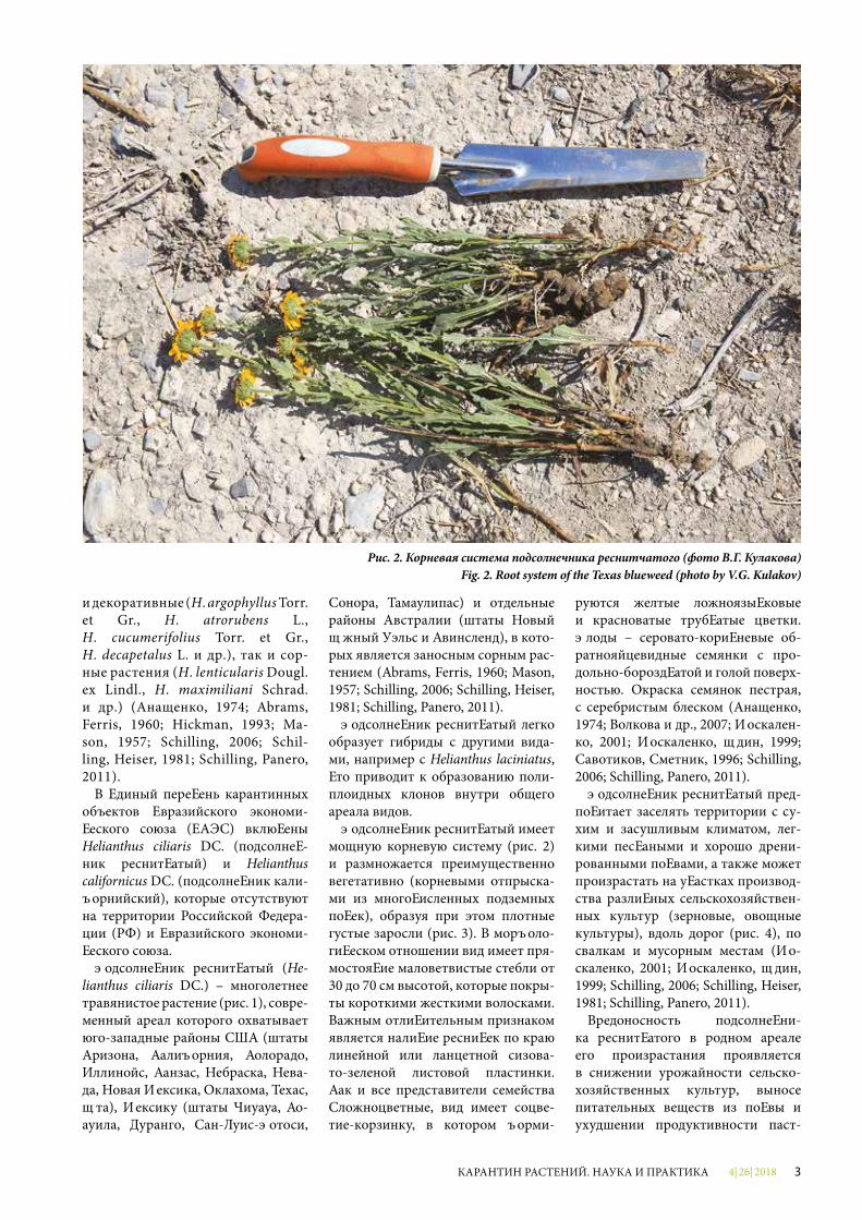
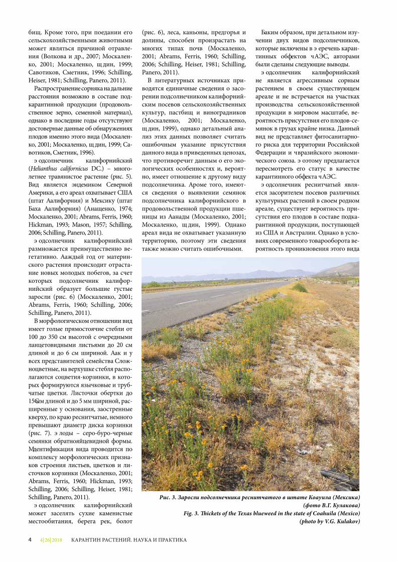
Подсолнечник реснитчатый имеет мощную корневую систему (рис. 2) и размножается преимущественно вегетативно (корневыми отпрыска-ми из многочисленных подземных почек), образуя при этом плотные густые заросли (рис. 3). В морфоло-гическом отношении вид имеет пря-мостоячие маловетвистые стебли от 30 до 70 см высотой, которые покры-ты короткими жесткими волосками. Важным отличительным признаком является наличие ресничек по краю линейной или ланцетной сизова-то-зеленой листовой пластинки. Как и все представители семейства Сложноцветные, вид имеет соцве-тие-корзинку, в котором форми-
руются желтые ложноязычковые и красноватые трубчатые цветки. Плоды – серовато-коричневые об-ратнояйцевидные семянки с про-дольно-бороздчатой и голой поверх-ностью. Окраска семянок пестрая, с серебристым блеском (Анащенко, 1974; Волкова и др., 2007; Москален-ко, 2001; Москаленко, Юдин, 1999; Савотиков, Сметник, 1996; Schilling, 2006; Schilling, Panero, 2011).
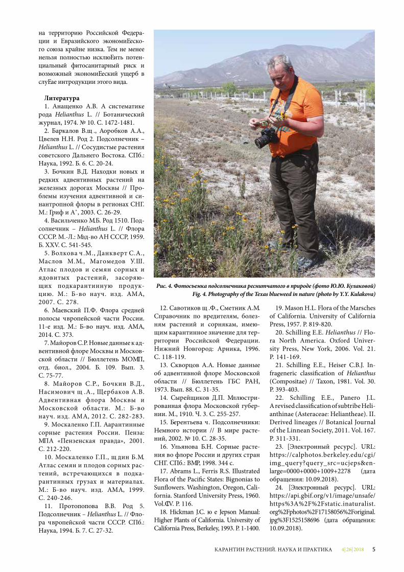
Подсолнечник реснитчатый пред-почитает заселять территории с су-хим и засушливым климатом, лег-кими песчаными и хорошо дрени-рованными почвами, а также может произрастать на участках производ-ства различных сельскохозяйствен-ных культур (зерновые, овощные культуры), вдоль дорог (рис. 4), по свалкам и мусорным местам (Мо-скаленко, 2001; Москаленко, Юдин, 1999; Schilling, 2006; Schilling, Heiser, 1981; Schilling, Panero, 2011).
Вредоносность подсолнечни-ка реснитчатого в родном ареале его произрастания проявляется в снижении урожайности сельско-хозяйственных культур, выносе питательных веществ из почвы и ухудшении продуктивности паст-
Рис. 2. Корневая система подсолнечника реснитчатого (фото В.Г. Кулакова)Fig. 2. Root system of the Texas blueweed (photo by V.G. Kulakov)
4| 26| 20184 КАРАНТИН РАСТЕНИЙ. НАУКА И ПРАКТИКА
бищ. Кроме того, при поедании его сельскохозяйственными животными может являться причиной отравле-ния (Волкова и др., 2007; Москален-ко, 2001; Москаленко, Юдин, 1999; Савотиков, Сметник, 1996; Schilling, Heiser, 1981; Schilling, Panero, 2011).
Распространение сорняка на дальние расстояния возможно в составе под-карантинной продукции (продоволь-ственное зерно, семенной материал), однако в последние годы отсутствуют достоверные данные об обнаружениях плодов именно этого вида (Москален-ко, 2001; Москаленко, Юдин, 1999; Са-вотиков, Сметник, 1996).
Подсолнечник калифорнийский (Helianthus californicus DC.) – много-летнее травянистое растение (рис. 5). Вид является эндемиком Северной Америки, а его ареал охватывает США (штат Калифорния) и Мексику (штат Баха Калифорния) (Анащенко, 1974; Москаленко, 2001; Abrams, Ferris, 1960; Hickman, 1993; Mason, 1957; Schilling, 2006; Schilling, Panero, 2011).
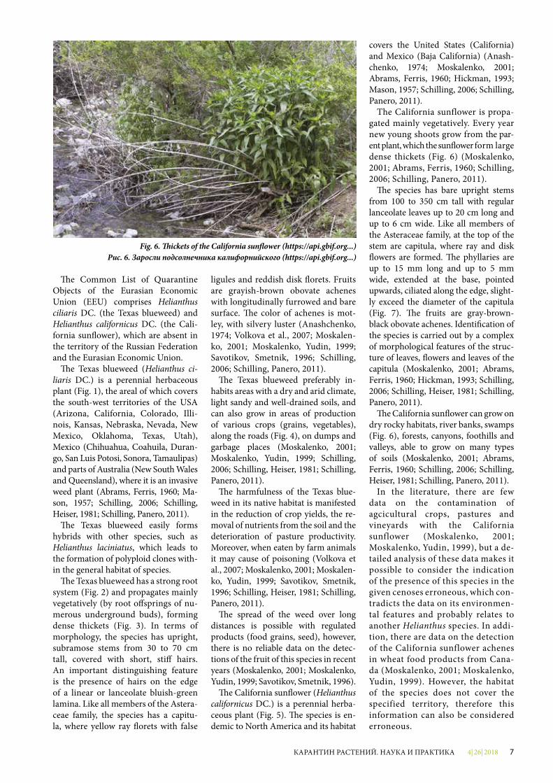
Подсолнечник калифорнийский размножается преимущественно ве-гетативно. Каждый год от материн-ского растения происходит отраста-ние новых молодых побегов, за счет которых подсолнечник калифор-нийский образует большие густые заросли (рис. 6) (Москаленко, 2001; Abrams, Ferris, 1960; Schilling, 2006; Schilling, Panero, 2011).
В морфологическом отношении вид имеет голые прямостоячие стебли от 100 до 350 см высотой с очередными ланцетовидными листьями до 20 см длиной и до 6 см шириной. Как и у всех представителей семейства Слож-ноцветные, на верхушке стебля распо-лагаются соцветия-корзинки, в кото-рых формируются язычковые и труб-чатые цветки. Листочки обертки до 15 мм длиной и до 5 мм шириной, рас-ширенные у основания, заостренные кверху, по краю реснитчатые, немного превышают диаметр диска корзинки (рис. 7). Плоды – серо-буро-черные семянки обратнояйцевидной формы. Идентификация вида проводится по комплексу морфологических призна-ков строения листьев, цветков и ли-сточков корзинки (Москаленко, 2001; Abrams, Ferris, 1960; Hickman, 1993; Schilling, 2006; Schilling, Heiser, 1981; Schilling, Panero, 2011).
Подсолнечник калифорнийский может заселять сухие каменистые местообитания, берега рек, болот
(рис. 6), леса, каньоны, предгорья и долины, способен произрастать на многих типах почв (Москаленко, 2001; Abrams, Ferris, 1960; Schilling, 2006; Schilling, Heiser, 1981; Schilling, Panero, 2011).
В литературных источниках при-водятся единичные сведения о засо-рении подсолнечником калифорний-ским посевов сельскохозяйственных культур, пастбищ и виноградников (Москаленко, 2001; Москаленко, Юдин, 1999), однако детальный ана-лиз этих данных позволяет считать ошибочным указание присутствия данного вида в приведенных ценозах, что противоречит данным о его эко-логических особенностях и, вероят-но, имеет отношение к другому виду подсолнечника. Кроме того, имеют-ся сведения о выявлении семянок подсолнечника калифорнийского в продовольственной продукции пше-ницы из Канады (Москаленко, 2001; Москаленко, Юдин, 1999). Однако ареал вида не охватывает указанную территорию, поэтому эти сведения также можно считать ошибочными.
Таким образом, при детальном изу-чении двух видов подсолнечников, которые включены в Перечень каран-тинных объектов ЕАЭС, авторами были сделаны следующие выводы.
Подсолнечник калифорнийский не является агрессивным сорным растением в своем существующем ареале и не встречается на участках производства сельскохозяйственной продукции в мировом масштабе, ве-роятность присутствия его плодов-се-мянок в грузах крайне низка. Данный вид не представляет фитосанитарно-го риска для территории Российской Федерации и Евразийского экономи-ческого союза. Поэтому предлагается пересмотреть его статус в качестве карантинного объекта ЕАЭС.
Подсолнечник реснитчатый явля-ется засорителем посевов различных культурных растений в своем родном ареале, существует вероятность при-сутствия его плодов в составе подка-рантинной продукции, поступающей из США и Австралии. Однако в усло-виях современного товарооборота ве-роятность проникновения этого вида
Рис. 3. Заросли подсолнечника реснитчатого в штате Коауила (Мексика) (фото В.Г. Кулакова)
Fig. 3. �ickets of the Texas blueweed in the state of Coahuila (Mexico) (photo by V.G. Kulakov)
4| 26| 2018 5КАРАНТИН РАСТЕНИЙ. НАУКА И ПРАКТИКА
на территорию Российской Федера-ции и Евразийского экономическо-го союза крайне низка. Тем не менее нельзя полностью исключить потен-циальный фитосанитарный риск и возможный экономический ущерб в случае интродукции этого вида.
Литература1. Анащенко А.В. К систематике
рода Helianthus L. // Ботанический журнал, 1974. № 10. C. 1472-1481.
2. Баркалов В.Ю., Коробков А.А., Цвелев Н.Н. Род 2. Подсолнечник – Helianthus L. // Сосудистые растения советского Дальнего Востока. СПб.: Наука, 1992. Т. 6. С. 20-24.
3. Бочкин В.Д. Находки новых и редких адвентивных растений на железных дорогах Москвы // Про-блемы изучения адвентивной и си-нантропной флоры в регионах СНГ. М.: Гриф и К˚, 2003. С. 26-29.
4. Васильченко И.Т. Род 1510. Под-солнечник – Helianthus L. // Флора СССР. М.-Л.: Изд-во АН СССР, 1959. Т. XXV. С. 541-545.
5. Волкова Е.М., Данкверт С.А., Маслов М.И., Магомедов У.Ш. Атлас плодов и семян сорных и ядовитых растений, засоряю-щих подкарантинную продук-цию. М.: Т-во науч. изд. КМК, 2007. С. 278.
6. Маевский П.Ф. Флора средней полосы Европейской части России. 11-е изд. М.: Т-во науч. изд. КМК, 2014. С. 373.
7. Майоров С.Р. Новые данные к ад-вентивной флоре Москвы и Москов-ской области // Бюллетень МОИП, отд. биол., 2004. Т. 109. Вып. 3. С. 75-77.
8. Майоров С.Р., Бочкин В.Д., Насимович Ю.А., Щербаков А.В. Адвентивная флора Москвы и Московской области. М.: Т-во науч. изд. КМК, 2012. С. 282-283.
9. Москаленко Г.П. Карантинные сорные растения России. Пенза: ИПК «Пензенская правда», 2001. С. 212-220.
10. Москаленко Г.П., Юдин Б.И. Атлас семян и плодов сорных рас-тений, встречающихся в подка-рантинных грузах и материалах. М.: Т-во науч. изд. КМК, 1999. С. 240-246.
11. Протопопова В.В. Род 5. Подсол нечник – Helianthus L. // Фло-ра Европейской части СССР. СПб.: Наука, 1994. Т. 7. С. 27-32.
12. Савотиков Ю.Ф., Сметник А.И. Справочник по вредителям, болез-ням растений и сорнякам, имею-щим карантинное значение для тер-ритории Российской Федерации. Нижний Новгород: Арника, 1996. С. 118-119.
13. Скворцов А.К. Новые данные об адвентивной флоре Московской области // Бюллетень ГБС РАН, 1973. Вып. 88. С. 31-35.
14. Сырейщиков Д.П. Иллюстри-рованная флора Московской губер-нии. М., 1910. Ч. 3. С. 255-257.
15. Терентьева Е. Подсолнечники: Немного истории // В мире расте-ний, 2002. № 10. С. 28-35.
16. Ульянова Т.Н. Сорные расте-ния во флоре России и других стран СНГ. СПб.: ВИР, 1998. 344 с.
17. Abrams L., Ferris R.S. Illustrated Flora of the Paci�c States: Bignonias to Sun�owers. Washington, Oregon, Cali-fornia. Stanford University Press, 1960. Vol. IV. P. 116.
18. Hickman J.C. �e Jepson Manual: Higher Plants of California. University of California Press, Berkeley, 1993. P. 1-1400.
19. Mason H.L. Flora of the Marsches of California. University of California Press, 1957. P. 819-820.
20. Schilling E.E. Helianthus // Flo-ra North America. Oxford Univer-sity Press, New York, 2006. Vol. 21. P. 141-169.
21. Schilling E.E., Heiser C.B.J. In-frageneric classi�cation of Helianthus (Compositae) // Taxon, 1981. Vol. 30. P. 393-403.
22. Schilling E.E., Panero J.L. A revised classi�cation of subtribe Heli-anthinae (Asteraceae: Heliantheae). II. Derived lineages // Botanical Journal of the Linnean Society, 2011. Vol. 167. P. 311-331.
23. [Электронный ресурс]. URL: https://calphotos.berkeley.edu/cgi/img_query?query_src=ucjeps&en-large=0000+0000+1009+2278 (дата обращения: 10.09.2018).
24. [Электронный ресурс]. URL: https://api.gbif.org/v1/image/unsafe/https%3A%2F%2Fstatic.inaturalist.org%2Fphotos%2F17158056%2Foriginal.jpg%3F1525158696 (дата обращения: 10.09.2018).
Рис. 4. Фотосъемка подсолнечника реснитчатого в природе (фото Ю.Ю. Кулаковой)Fig. 4. Photography of the Texas blueweed in nature (photo by Y.Y. Kulakova)
4| 26| 20186 КАРАНТИН РАСТЕНИЙ. НАУКА И ПРАКТИКА
Abstract. �e article provides infor-mation about the geographical distri-bution of the California sun�ower and the Texas blueweed, their brief morpho-logical description and possible ways of distribution.
Keywords. Plant quarantine, Heli-anthus L., California sun�ower, Texas bluedweed, distribution, morphology, habitat, harmfulness.
Helianthus L. is a taxonomically complex genus of American origin, belonging to the Asteraceae Bercht. & J. Presl family and combining, accord-ing to various estimates, up to 110 annual and perennial species (Anash-chenko, 1974; Schilling, 2006; Schilling, Heiser, 1981; Schilling, Panero, 2011). �e Russian �ora comprises 8 species of the Helianthus genus (Anashchenko, 1974; Barkalov et al., 1992; Bochkin, 2003; Vasilchenko, 1959; Mayevsky, 2014; Mayorov, 2004; Mayorov et al., 2012; Protopopova, 1994; Skvortsov, 1973; Syreischikov, 1910; Terentye-va, 2002; Ulyanova, 1998), of which the most common are H. lenticularis Douglas ex. Lindl. and H. tuberosus L. (the Canada potato).
Some of the sun�ower species in the world are valuable cultural (H. annuus L., H. tuberosus L. etc.) and ornamen-tal ones (H. argophyllus Torr. et Gr., H. atrorubens L., H. cucumerifolius Torr. et Gr., H. decapetalus L. etc.), as well as weeds (H. lenticularis Dougl. ex Lindl., H. maximiliani Schrad. и др.) (Anashchenko, 1974; Abrams, Ferris, 1960; Hickman, 1993; Mason, 1957; Schilling, 2006; Schilling, Heiser, 1981; Schilling, Panero, 2011).
UDC 632.3.01/.08, 632.2
SPECIES OF THE HELIANTHUS L. GENUS IN THE COMMON LIST OF QUARANTINE OBJECTS OF THE EURASIAN ECONOMIC UNIOND.L. Belkin, Deputy Head of the Testing Laboratory Center of FGBU “VNIIKR”Y.Y. Kulakova, Head of the Research and Testing Department of FGBU “VNIIKR”
Fig. 5. General view of the California sun�ower (https://calphotos.berkeley...)Рис. 5. Общий вид подсолнечника калифорнийского (https://calphotos.berkeley...)
4| 26| 2018 7КАРАНТИН РАСТЕНИЙ. НАУКА И ПРАКТИКА
�e Common List of Quarantine Objects of the Eurasian Economic Union (EEU) comprises Helianthus ciliaris DC. (the Texas blueweed) and Helianthus californicus DC. (the Cali-fornia sun�ower), which are absent in the territory of the Russian Federation and the Eurasian Economic Union.
�e Texas blueweed (Helianthus ci-liaris DC.) is a perennial herbaceous plant (Fig. 1), the areal of which covers the south-west territories of the USA (Arizona, California, Colorado, Illi-nois, Kansas, Nebraska, Nevada, New Mexico, Oklahoma, Texas, Utah), Mexico (Chihuahua, Coahuila, Duran-go, San Luis Potosi, Sonora, Tamaulipas) and parts of Australia (New South Wales and Queensland), where it is an invasive weed plant (Abrams, Ferris, 1960; Ma-son, 1957; Schilling, 2006; Schilling, Heiser, 1981; Schilling, Panero, 2011).
�e Texas blueweed easily forms hybrids with other species, such as Helianthus laciniatus, which leads to the formation of polyploid clones with-in the general habitat of species.
�e Texas blueweed has a strong root system (Fig. 2) and propagates mainly vegetatively (by root o�springs of nu-merous underground buds), forming dense thickets (Fig. 3). In terms of morphology, the species has upright, subramose stems from 30 to 70 cm tall, covered with short, sti� hairs. An important distinguishing feature is the presence of hairs on the edge of a linear or lanceolate bluish-green lamina. Like all members of the Astera-ceae family, the species has a capitu-la, where yellow ray �orets with false
ligules and reddish disk �orets. Fruits are grayish-brown obovate achenes with longitudinally furrowed and bare surface. �e color of achenes is mot-ley, with silvery luster (Anashchenko, 1974; Volkova et al., 2007; Moskalen-ko, 2001; Moskalenko, Yudin, 1999; Savotikov, Smetnik, 1996; Schilling, 2006; Schilling, Panero, 2011).
�e Texas blueweed preferably in-habits areas with a dry and arid climate, light sandy and well-drained soils, and can also grow in areas of production of various crops (grains, vegetables), along the roads (Fig. 4), on dumps and garbage places (Moskalenko, 2001; Moskalenko, Yudin, 1999; Schilling, 2006; Schilling, Heiser, 1981; Schilling, Panero, 2011).
�e harmfulness of the Texas blue-weed in its native habitat is manifested in the reduction of crop yields, the re-moval of nutrients from the soil and the deterioration of pasture productivity. Moreover, when eaten by farm animals it may cause of poisoning (Volkova et al., 2007; Moskalenko, 2001; Moskalen-ko, Yudin, 1999; Savotikov, Smetnik, 1996; Schilling, Heiser, 1981; Schilling, Panero, 2011).
�e spread of the weed over long distances is possible with regulated products (food grains, seed), however, there is no reliable data on the detec-tions of the fruit of this species in recent years (Moskalenko, 2001; Moskalenko, Yudin, 1999; Savotikov, Smetnik, 1996).
�e California sun�ower (Helianthus californicus DC.) is a perennial herba-ceous plant (Fig. 5). �e species is en-demic to North America and its habitat
covers the United States (California) and Mexico (Baja California) (Anash-chenko, 1974; Moskalenko, 2001; Abrams, Ferris, 1960; Hickman, 1993; Mason, 1957; Schilling, 2006; Schilling, Panero, 2011).
The California sunflower is propa-gated mainly vegetatively. Every year new young shoots grow from the par-ent plant, which the sun�ower form large dense thickets (Fig. 6) (Moskalenko, 2001; Abrams, Ferris, 1960; Schilling, 2006; Schilling, Panero, 2011).
�e species has bare upright stems from 100 to 350 cm tall with regular lanceolate leaves up to 20 cm long and up to 6 cm wide. Like all members of the Asteraceae family, at the top of the stem are capitula, where ray and disk �owers are formed. �e phyllaries are up to 15 mm long and up to 5 mm wide, extended at the base, pointed upwards, ciliated along the edge, slight-ly exceed the diameter of the capitula (Fig. 7). �e fruits are gray-brown-black obovate achenes. Identi�cation of the species is carried out by a complex of morphological features of the struc-ture of leaves, �owers and leaves of the capitula (Moskalenko, 2001; Abrams, Ferris, 1960; Hickman, 1993; Schilling, 2006; Schilling, Heiser, 1981; Schilling, Panero, 2011).
�e California sun�ower can grow on dry rocky habitats, river banks, swamps (Fig. 6), forests, canyons, foothills and valleys, able to grow on many types of soils (Moskalenko, 2001; Abrams, Ferris, 1960; Schilling, 2006; Schilling, Heiser, 1981; Schilling, Panero, 2011).
In the literature, there are few data on the contamination of agcicultural crops, pastures and vineyards with the California sunflower (Moskalenko, 2001; Moskalenko, Yudin, 1999), but a de-tailed analysis of these data makes it possible to consider the indication of the presence of this species in the given cenoses erroneous, which con-tradicts the data on its environmen-tal features and probably relates to another Helianthus species. In addi-tion, there are data on the detection of the California sunflower achenes in wheat food products from Cana-da (Moskalenko, 2001; Moskalenko, Yudin, 1999). However, the habitat of the species does not cover the specified territory, therefore this information can also be considered erroneous.
Fig. 6. �ickets of the California sun�ower (https://api.gbif.org...)Рис. 6. Заросли подсолнечника калифорнийского (https://api.gbif.org...)

4| 26| 20188 КАРАНТИН РАСТЕНИЙ. НАУКА И ПРАКТИКА
�us, in a detailed study of two He-lianthus species, which are included in the Common List of Quarantine Ob-jects of the EEU, the authors made the following conclusions.
�e California sun�ower is not an aggressive weed plant in its existing habitat and is absent in the areas of agricultural production on a global scale, the probability of the presence of its achenes in the plant products is extremely low. �is species does not pose a pest risk to the territory of the Russian Federation and the Eurasian Economic Union. �erefore, it is pro-posed to revise its status as a quarantine object of the EEU.
�e Texas blueweed is a weed of crops of di�erent cultivated plants in their native habitat, there is a possibi-lity of the presence of its fruits in regulat-ed products imported from the United States and Australia. However, in the conditions of modern trade turnover, the probability of introduction of this species into the territory of the Russian Federation and the Eurasian Economic Union is extremely low. Nevertheless, the potential pest risk and possible eco-nomic damage in the case of introduc-tion of this species cannot be complete-ly excluded.
References1. Anashchenko A.V. To the taxo-
nomy of the Helianthus L. genus // Botanical Magazine, 1974. № 10. P. 1472-1481.
2. Barkalov V.Y., Korobkov A.A., Tsvelev N.N. Genus 2. Sunflower – Helianthus L. / Vascular plants of the Soviet Far East. SPb.: Science, 1992. Vol. 6. P. 20-24.
3. Bochkin V.D. Findings of new and rare adventive plants on the railways of Moscow // Problems of studying adventive and synanthropic flora in the CIS regions. M.: Grif i K°, 2003. P. 26-29.
4. Vasilchenko I.T. Genus 1510. Sunflower – Helianthus L. // Flora of the USSR. M.-L.: Publishing house of the USSR Academy of Sciences, 1959. Vol. XXV. P. 541-545.
5. Volkova E.M., Dankvert S.A., Maslov M.I., Magomedov U.S. Atlas of fruits and seeds of weeds and poi-sonous plants, contaminating regu-lated products. M.: KMK Scientific Publishing Partnership, 2007. P. 278.
6. Mayevsky P.F. Flora of the middle zone of the European part of Russia.
11th ed. M.: KMK Scientific Publish-ing Partnership, 2014. P. 373.
7. Mayorov S.R. New data to the ad-ventive flora of Moscow and Moscow region // Bulletin of the MSN, Biol. Dep., 2004. Vol. 109. Issue 3. P. 75-77.
8. Mayorov S.R., Bochkin V.D., Na-simovich Y.A., Shcherbakov A.V. Ad-ventive flora of Moscow and Moscow region. M.: KMK Scientific Publish-ing Partnership, 2012. P. 282-283.
9. Moskalenko G.P. Quaran-tine weed plants of Russia. Pen-za: Penzenskaia Pravda PPC, 2001. P. 212-220.
10. Moskalenko G.P., Yudin B.I. Atlas of seeds and fruits of weed plants found in quarantined car-goes and materials. M.: KMK Sci-entific Publishing Partnership, 1999. P. 240-246.
11. Protopopova V.V. Genus 5. Sun-flower – Helianthus L. // Flora of the European part of the USSR. SPb.: Sci-ence, 1994. Vol. 7. P. 27-32.
12. Savotikov Y.F., Smetnik A.I. The reference book on pests, plant diseas-es and weed plants of quarantine im-portance for the territory of the Rus-sian Federation. Nizhny Novgorod: Arnika, 1996. P. 118-119.
13. Skvortsov А.К. New data on the adventive flora of the Moscow region // Bulletin of the MBG RAS, 1973. Issue 88. P. 31-35.
14. Syreishchikov D.P. Illustrated flora of the Moscow province. М., 1910. P. 3. P. 255-257.
15. Terentyeva E. Sunflowers: A little history // In a World of Plants, 2002. № 10. P. 28-35.
16. Ulyanova T.N. Weeds in the flo-ra of Russia and other CIS countries. SPb.: VIR, 1998. 344 p.
17. Abrams L., Ferris R.S. Illus-trated Flora of the Pacific States: Bignonias to Sunflowers. Washing-ton, Oregon, California. Stanford University Press, 1960. Vol. IV. P. 116.
18. Hickman J.C. The Jepson Manual: Higher Plants of Califor-nia. University of California Press, Berkeley, 1993. P. 1-1400.
19. Mason H.L. Flora of the Mar-sches of California. University of California Press, 1957. P. 819-820.
20. Schilling E.E. Helianthus // Flo-ra North America. Oxford Univer-sity Press, New York, 2006. Vol. 21. P. 141-169.
21. Schilling E.E., Heiser C.B.J. Infrageneric classification of He-lianthus (Compositae) // Taxon, 1981. Vol. 30. P. 393-403.
22. Schilling E.E., Panero J.L. A revised classification of sub-tribe Helianthinae (Asteraceae: Helianthe ae). II. Derived lineages // Bota nical Journal of the Linnean Society, 2011. Vol. 167. P. 311-331.
23. [Electronic resource]. URL: https://calphotos.berkeley.edu/cgi/img_query?query_src=ucjeps&en-l a r g e = 0 0 0 0 + 0 0 0 0 + 1 0 0 9 + 2 2 7 8 (reference date: 10.09.2018).
24. [Electronic resource]. URL: https://api.gbif.org/v1/image/unsafe/https%3A%2F%2Fstatic.inatural-ist.org%2Fphotos%2F17158056%-2Foriginal.jpg%3F1525158696 (refer-ence date: 10.09.2018).
Fig. 7. In�orescence of the California sun�ower (https://calphotos.berkeley...)Рис. 7. Соцветие подсолнечника калифорнийского (https://calphotos.berkeley...)
4| 26| 2018 9КАРАНТИН РАСТЕНИЙ. НАУКА И ПРАКТИКА
Аннотация. Представлен краткий анализ возможного заноса различных карантинных вредных организмов с «рождественскими деревьями» и свежесрезанными ветками хвойных пород. Приведен перечень карантин-ных вредных организмов (насекомых, нематод, фитопатогенных грибов), которые могут быть занесены с ана-лизируемой продукцией на террито-рию Российской Федерации из различ-ных стран мира.
Ключевые слова. «Рождественские деревья», свежесрезанные ветки хвой-ных пород, занос, карантинные вред-ные организмы, вредители, сосновая стволовая нематода, возбудители грибных болезней, интродукция, ущерб, Россия, РФ.
Традиционным новогодним укра-шением каждого дома являются но-вогодние елки, также известные как «рождественские деревья», и свеже-срезанные ветки хвойных пород, ис-
пользуемые как основной материал в флористическом новогоднем и рож-дественском декоре.
Однако нарядные хвойные деревья и их срезанные ветки являются не толь-ко источником праздничного настро-ения, но и потенциальной угрозой за-носа различных вредных организмов, которые могут представлять угрозу как для здоровья человека, так и для окружающей среды, особенно если эти рождественские деревья были завезе-ны издалека.
В предновогодний период Ин-тернет пестрит предложения-ми о поставке презентабельных «новогодних елей» из-за рубежа. В данной статье представлен краткий анализ возможности за-носа карантинных вредных орга-низмов с новогодней продукцией и степени угрозы для хвойных насаждений на территории РФ.
По данным Федеральной таможен-ной службы, за три последних года
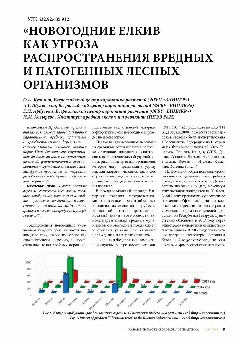
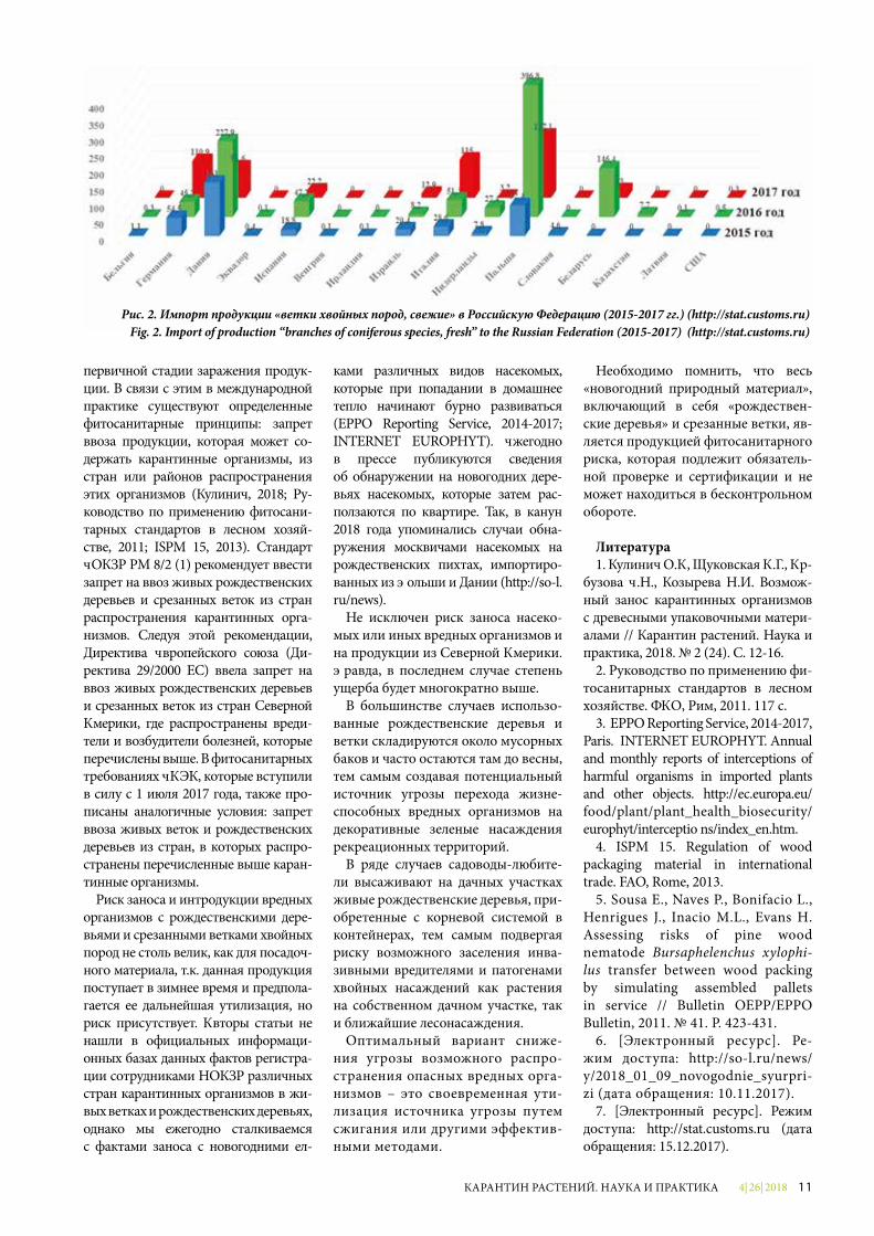
(2015-2017 гг.) продукция по коду ТН ВЭД 0604202000 «рождественские де-ревья, свежие» была экспортирована в Российскую Федерацию из 13 стран мира (http://stat.customs.ru). Это Бе-ларусь, Бельгия, Канада, США, Да-ния, Испания, Латвия, Нидерланды, Польша, Германия, Италия, Арме-ния, Эстония (рис. 1).
Наибольший объем поставок «рож-дественских деревьев» из-за рубежа приходится на Данию и Польшу (соот-ветственно 905,2 и 928,8 т), максимум этих поставок приходится на 2016 год. В 2017 году произошло существенное снижение объема импорта «рожде-ственских деревьев» из этих стран и увеличился объем поставляемой про-дукции из Республики Беларусь. Суще-ственно обновился в 2017 году пере-чень стран – экспортеров «рождествен-ских деревьев». В 2017 году появились новые страны-экспортеры – Эстония и Армения. Следует отметить, что если поставки «рождественских деревьев»,
УДК 632.92:635.912
«НОВОГОДНИЕ ЕЛКИ» КАК УГРОЗА РАСПРОСТРАНЕНИЯ ВРЕДНЫХ И ПАТОГЕННЫХ ЛЕСНЫХ ОРГАНИЗМОВО.А. Кулинич, Всероссийский центр карантина растений (ФГБУ «ВНИИКР»)А.Г. Щуковская, Всероссийский центр карантина растений (ФГБУ «ВНИИКР»)Е.Н. Арбузова, Всероссийский центр карантина растений (ФГБУ «ВНИИКР»)Н.И. Козырева, Институт проблем экологии и эволюции (ИПЭЭ РАН)
Рис.1. Импорт продукции «рождественские деревья» в Российскую Федерацию (2015-2017 гг.) (http://stat.customs.ru)Fig. 1. Import of products “Christmas trees” to the Russian Federation (2015-2017) (http://stat.customs.ru)
4| 26| 201810 КАРАНТИН РАСТЕНИЙ. НАУКА И ПРАКТИКА
хотя и в ограниченном объеме, в 2015-2016 гг. осуществлялись даже из Се-верной Америки (Канады и США), то в 2017 году импорта данной продукции из этих стран не было. Возможно, это связано с новыми фитосанитарными требованиями ЕАЭС, которые всту-пили в силу в июле 2017 года. Согласно этим требованиям, импорт посадочно-го материала хвойных пород, включая срезанные ветки и рождественские де-ревья, запрещен ввиду фитосанитар-ной угрозы для хвойных насаждений Российской Федерации.
В отличие от рождественских де-ревьев, свежие ветки хвойных пород импортируются из гораздо большего числа стран. По данным Федеральной таможенной службы, за три последних года (2015-2017 гг.) в Российскую Фе-дерацию продукция по коду ТН ВЭД 0604204000 «ветки хвойных деревьев, свежие» была поставлена из 22 стран мира (http://stat.customs.ru). Крупней-шими странами-поставщиками были Польша, Дания, Беларусь, Италия, Гер-мания (рис. 2).
В 2015 году «ветки хвойных деревь-ев» на территорию РФ были постав-лены из 12 стран мира – не только из Европы, но и из Южной Америки.
Как и в случае с «рождественскими деревьями», основными экспортерами веток хвойных пород являются Поль-ша, Дания, Германия. За три указан-ных года наибольший объем поставок приходился на 2016 год. В 2017 году наблюдалось снижение количества поставляемой продукции, однако и в этом году в число основных стран-по-ставщиков входили Польша (187,1 т), Италия (115 т), Германия (110,9 т), Да-ния (91,6 т), Испания (22,2 т). Поставки веток хвойных, хоть и в минимальном количестве, были осуществлены также из США.
Ниже проведена оценка фитосани-тарного риска при возможном заносе на территорию РФ вредных организ-мов с рождественскими деревьями и ветками хвойных пород из различных стран мира.
Наибольший фитосанитарный риск представляет продукция «рож-дественские деревья» и «срезанные ветки хвойных пород», ввезенная из Северной Америки (США и Канада). С американскими живыми (срублен-ными) елями, соснами и пихтами в Россию могут быть завезены вреди-тели, входящие в Перечень карантин-ных организмов, отсутствующих на
территории РФ и Евразийского эко-номического союза (ЕАЭС). Среди насекомых это несколько видов ли-стоверток родов Choristoneura и Acleris. В зимнее время они могут присутство-вать в стадии яиц на ветках дерева. Жуки-короеды могут скрываться под корой веток и ствола хвойных деревь-ев. Это лубоеды рода Dendroctonus и несколько видов короедов, относя-щихся к роду Ips. Особую опасность представляют жуки рода Dendroctonus, наносящие большой вред лесным хвойным массивам в Канаде и США и, согласно анализу фитосанитарного риска, представляющие наибольшую угрозу хвойным насаждениям в РФ.
Сосновая стволовая нематода Bursaphelenchus xylophilus относится к числу наиболее патогенных видов ор-ганизмов, широко распространенных на североамериканском континенте. Местные хвойные породы устойчивы к данному патогену, а вот большин-ство европейских и азиатских хвойных восприимчивы к нему. В случае зара-жения восприимчивого растения-хо-зяина гибель дерева может произойти в течение одного сезона. Нематоды отлично выживают в стволе, ветках дерева, коре и просто в древесине и при благоприятных условиях (в случае контакта зараженного и незараженно-го материала) могут самостоятельно перемещаться из зараженной древеси-ны в здоровые деревья (Sousa, 2011).
К числу патогенов, которые могут присутствовать на ветках рождествен-ских деревьев, следует отнести возбу-дителей следующих грибных болезней: рака (ожога) стволов и ветвей сосны (Atropellis piniphila, A. pinicola), который в зимнее время сохраняется в апотеци-ях на стволе и ветках или в виде мице-лия под корой веток и ствола; корич-невого пятнистого ожога хвои сос-ны Lecanosticta acicola (Mycosphaerella dearnessii) – патоген перезимовывает в виде пучков спорогенного мицелия в стромах (группа пикнид) на отмер-шей хвое и ветках; веретеноподобной ржавчины сосны (Cronartium fusiforme) и западной галлоподобной ржавчины сосны (Endocronartium harknessii) – в зимнее время патоген сохраняется в виде галлов на стволе и ветках со-сен; желтой кольцевой гнили хвой-ных пород Coniferiporia weirii (Phellinus weirii); ржавчины хвои ели Chrysomyxa arctostaphyli. Данные грибные патоге-ны, так же как и сосновая стволовая нематода B. xylophilus, распространены
в Северной Америке и входят в переч-ни карантинных организмов многих стран мира.
Как показывает таможенная ста-тистика, «рождественские деревья» и срезанные ветки не импортируются в Россию из стран Азии, однако ниже мы также провели оценку фитосани-тарного риска при возможном заносе опасных организмов из ближайших азиатских стран.
Из стран Восточной Азии (Китая, Кореи и Японии) с посадочным ма-териалом и «рождественскими дере-вьями», включая срезанные ветки, также могут быть завезены: сосно-вая стволовая нематода B. xylophilus, можжевельниковый паутинный клещ Oligonychus perditus (может находиться на хвое новогодних деревьев); возбу-дитель коричневого ожога хвои сосны Mycosphaerella gibsonii (в зимнее время в виде стром на отмершей хвое пора-женных веток); возбудитель коричне-вого пятнистого ожога хвои L. acicоla.
Наибольшие поставки продукции «рождественские деревья» и «срезан-ные ветки» в РФ осуществляются из европейских стран. В целом фауна вредителей и возбудителей болезней в европейской части России и в Евро-пе сходна. Однако ряд вредоносных организмов, которые уже проникли в Европу из Америки и наносят зна-чительный ущерб лесонасаждениям, пока отсутствует на территории Рос-сии. Все они могут быть завезены с данным видом продукции в зимнее время. Это опять же сосновая ство-ловая нематода B. xylophilus, которая может быть завезена с хвойными дере-вьями или ветками из Португалии или Испании; возбудитель коричневого пятнистого ожога хвои L. acicola, вы-зывающий заболевание, широко рас-пространенное в странах Евросоюза (Австрии, Хорватии, Словении, Чехии, Франции, Германии, Швейцарии, Лат-вии и Литве). Предполагаемый ущерб при интродукции этих организмов на территории РФ оценивается в десятки миллиардов долларов.
Ввозимая в страну продукция про-ходит фитосанитарную экспертизу со стороны НОКЗР, в России ее осущест-вляет Россельхознадзор. Инспектор визуально проверяет «рождественские деревья» и «срезанные ветки» на нали-чие вредителей или болезней. Однако далеко не всегда их можно выявить при визуальном осмотре и даже при лабораторной экспертизе, особенно на
4| 26| 2018 11КАРАНТИН РАСТЕНИЙ. НАУКА И ПРАКТИКА
первичной стадии заражения продук-ции. В связи с этим в международной практике существуют определенные фитосанитарные принципы: запрет ввоза продукции, которая может со-держать карантинные организмы, из стран или районов распространения этих организмов (Кулинич, 2018; Ру-ководство по применению фитосани-тарных стандартов в лесном хозяй-стве, 2011; ISPM 15, 2013). Стандарт ЕОКЗР PM 8/2 (1) рекомендует ввести запрет на ввоз живых рождественских деревьев и срезанных веток из стран распространения карантинных орга-низмов. Следуя этой рекомендации, Директива Европейского союза (Ди-ректива 29/2000 EC) ввела запрет на ввоз живых рождественских деревьев и срезанных веток из стран Северной Америки, где распространены вреди-тели и возбудители болезней, которые перечислены выше. В фитосанитарных требованиях ЕАЭК, которые вступили в силу с 1 июля 2017 года, также про-писаны аналогичные условия: запрет ввоза живых веток и рождественских деревьев из стран, в которых распро-странены перечисленные выше каран-тинные организмы.
Риск заноса и интродукции вредных организмов с рождественскими дере-вьями и срезанными ветками хвойных пород не столь велик, как для посадоч-ного материала, т.к. данная продукция поступает в зимнее время и предпола-гается ее дальнейшая утилизация, но риск присутствует. Авторы статьи не нашли в официальных информаци-онных базах данных фактов регистра-ции сотрудниками НОКЗР различных стран карантинных организмов в жи-вых ветках и рождественских деревьях, однако мы ежегодно сталкиваемся с фактами заноса с новогодними ел-
ками различных видов насекомых, которые при попадании в домашнее тепло начинают бурно развиваться (EPPO Reporting Service, 2014-2017; INTERNET EUROPHYT). Ежегодно в прессе публикуются сведения об обнаружении на новогодних дере-вьях насекомых, которые затем рас-ползаются по квартире. Так, в канун 2018 года упоминались случаи обна-ружения моск вичами насекомых на рождественских пихтах, импортиро-ванных из Польши и Дании (http://so-l.ru/news).
Не исключен риск заноса насеко-мых или иных вредных организмов и на продукции из Северной Америки. Правда, в последнем случае степень ущерба будет многократно выше.
В большинстве случаев использо-ванные рождественские деревья и ветки складируются около мусорных баков и часто остаются там до весны, тем самым создавая потенциальный источник угрозы перехода жизне-способных вредных организмов на декоративные зеленые насаждения рекреационных территорий.
В ряде случаев садоводы-любите-ли высаживают на дачных участках живые рождественские деревья, при-обретенные с корневой системой в контейнерах, тем самым подвергая риску возможного заселения инва-зивными вредителями и патогенами хвойных насаждений как растения на собственном дачном участке, так и ближайшие лесонасаждения.
Оптимальный вариант сниже-ния угрозы возможного распро-странения опасных вредных орга-низмов – это своевременная ути-лизация источника угрозы путем сжигания или другими эффектив-ными методами.
Необходимо помнить, что весь «новогодний природный материал», включающий в себя «рождествен-ские деревья» и срезанные ветки, яв-ляется продукцией фитосанитарного риска, которая подлежит обязатель-ной проверке и сертификации и не может находиться в бесконтрольном обороте.
Литература1. Кулинич О.А, Щуковская А.Г., Ар-
бузова Е.Н., Козырева Н.И. Возмож-ный занос карантинных организмов с древесными упаковочными матери-алами // Карантин растений. Наука и практика, 2018. № 2 (24). С. 12-16.
2. Руководство по применению фи-тосанитарных стандартов в лесном хозяйстве. ФАО, Рим, 2011. 117 с.
3. EPPO Reporting Service, 2014-2017, Paris. INTERNET EUROPHYT. Annual and monthly reports of interceptions of harmful organisms in imported plants and other objects. http://ec.europa.eu/food/plant/plant_health_biosecurity/europhyt/interceptio ns/index_en.htm.
4. ISPM 15. Regulation of wood packaging material in international trade. FAO, Rome, 2013.
5. Sousa E., Naves P., Bonifacio L., Henrigues J., Inacio M.L., Evans H. Assessing risks of pine wood nematode Bursaphelenchus xylophi-lus transfer between wood packing by simulating assembled pallets in service // Bulletin OEPP/EPPO Bulletin, 2011. № 41. P. 423-431.
6. [Электронный ресурс]. Ре-жим доступа: http://so-l.ru/news/ y/2018_01_09_novo godnie_syur pri-zi (дата обращения: 10.11.2017).
7. [Электронный ресурс]. Режим доступа: http://stat.customs.ru (дата обращения: 15.12.2017).
Рис. 2. Импорт продукции «ветки хвойных пород, свежие» в Российскую Федерацию (2015-2017 гг.) (http://stat.customs.ru)Fig. 2. Import of production “branches of coniferous species, fresh” to the Russian Federation (2015-2017) (http://stat.customs.ru)
4| 26| 201812 КАРАНТИН РАСТЕНИЙ. НАУКА И ПРАКТИКА
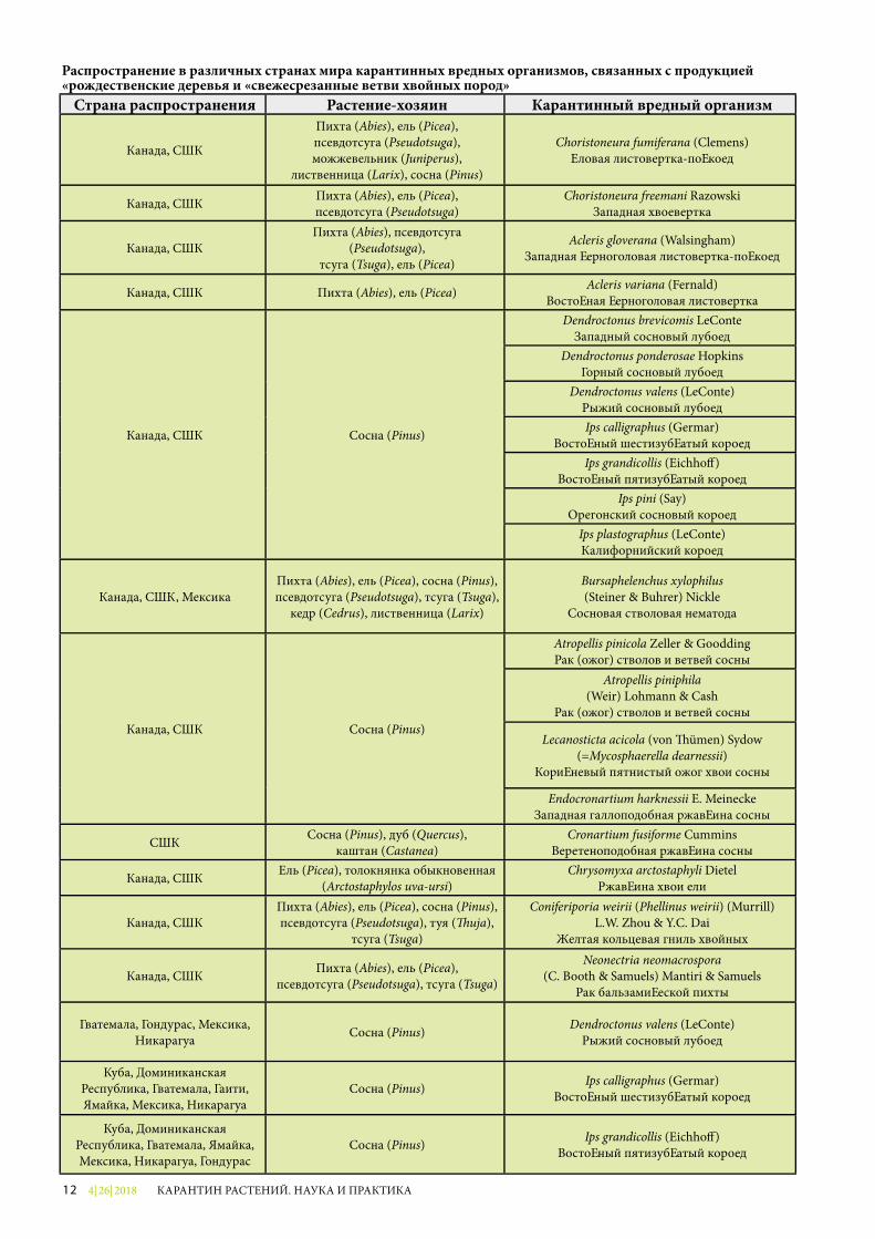
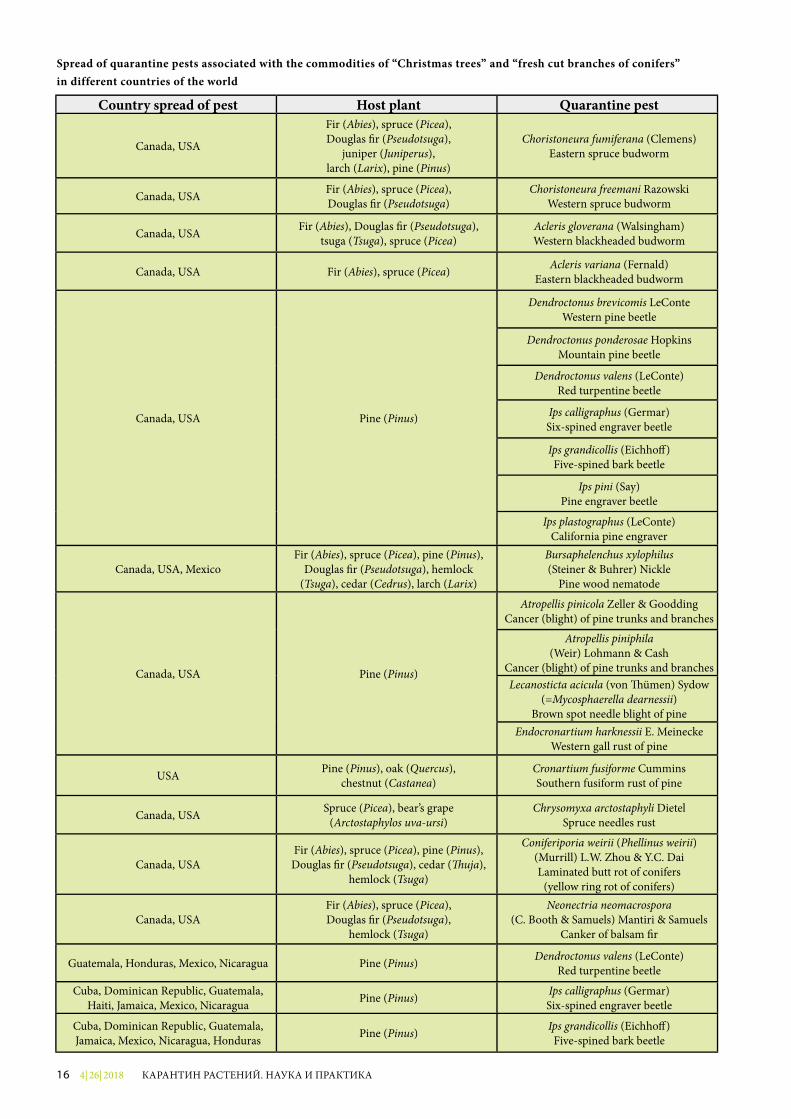
Распространение в различных странах мира карантинных вредных организмов, связанных с продукцией «рождественские деревья и «свежесрезанные ветви хвойных пород»
Страна распространения Растение-хозяин Карантинный вредный организм
Канада, США
Пихта (Abies), ель (Picea), псевдотсуга (Pseudotsuga),можжевельник (Juniperus),
лиственница (Larix), сосна (Pinus)
Choristoneura fumiferana (Clemens) Еловая листовертка-почкоед
Канада, США Пихта (Abies), ель (Picea), псевдотсуга (Pseudotsuga)
Choristoneura freemani Razowski Западная хвоевертка
Канада, СШАПихта (Abies), псевдотсуга
(Pseudotsuga), тсуга (Tsuga), ель (Picea)
Acleris gloverana (Walsingham) Западная черноголовая листовертка-почкоед
Канада, США Пихта (Abies), ель (Picea) Acleris variana (Fernald) Восточная черноголовая листовертка
Канада, США Сосна (Pinus)
Dendroctonus brevicomis LeConte Западный сосновый лубоед
Dendroctonus ponderosae Hopkins Горный сосновый лубоед
Dendroctonus valens (LeConte) Рыжий сосновый лубоедIps calligraphus (Germar)
Восточный шестизубчатый короедIps grandicollis (Eichho�)
Восточный пятизубчатый короедIps pini (Say)
Орегонский сосновый короедIps plastographus (LeConte) Калифорнийский короед
Канада, США, МексикаПихта (Abies), ель (Picea), сосна (Pinus), псевдотсуга (Pseudotsuga), тсуга (Tsuga),
кедр (Cedrus), лиственница (Larix)
Bursaphelenchus xylophilus (Steiner & Buhrer) Nickle
Сосновая стволовая нематода
Канада, США Сосна (Pinus)
Atropellis pinicola Zeller & Goodding Рак (ожог) стволов и ветвей сосны
Atropellis piniphila (Weir) Lohmann & Cash
Рак (ожог) стволов и ветвей сосны
Lecanosticta acicola (von �ümen) Sydow (=Mycosphaerella dearnessii)
Коричневый пятнистый ожог хвои сосны
Endocronartium harknessii E. Meinecke Западная галлоподобная ржавчина сосны
США Сосна (Pinus), дуб (Quercus),каштан (Castanea)
Cronartium fusiforme Cummins Веретеноподобная ржавчина сосны
Канада, США Ель (Picea), толокнянка обыкновенная (Arctostaphylos uva-ursi)
Chrysomyxa arctostaphyli Dietel Ржавчина хвои ели
Канада, СШАПихта (Abies), ель (Picea), сосна (Pinus),псевдотсуга (Pseudotsuga), туя (�uja),
тсуга (Tsuga)
Coniferiporia weirii (Phellinus weirii) (Murrill) L.W. Zhou & Y.C. Dai
Желтая кольцевая гниль хвойных
Канада, США Пихта (Abies), ель (Picea), псевдотсуга (Pseudotsuga), тсуга (Tsuga)
Neonectria neomacrospora(C. Booth & Samuels) Mantiri & Samuels
Рак бальзамической пихты
Гватемала, Гондурас, Мексика, Никарагуа Сосна (Pinus) Dendroctonus valens (LeConte)
Рыжий сосновый лубоед
Куба, Доминиканская Республика, Гватемала, Гаити, Ямайка, Мексика, Никарагуа
Сосна (Pinus) Ips calligraphus (Germar) Восточный шестизубчатый короед
Куба, Доминиканская Республика, Гватемала, Ямайка, Мексика, Никарагуа, Гондурас
Сосна (Pinus) Ips grandicollis (Eichho�) Восточный пятизубчатый короед
4| 26| 2018 13КАРАНТИН РАСТЕНИЙ. НАУКА И ПРАКТИКА
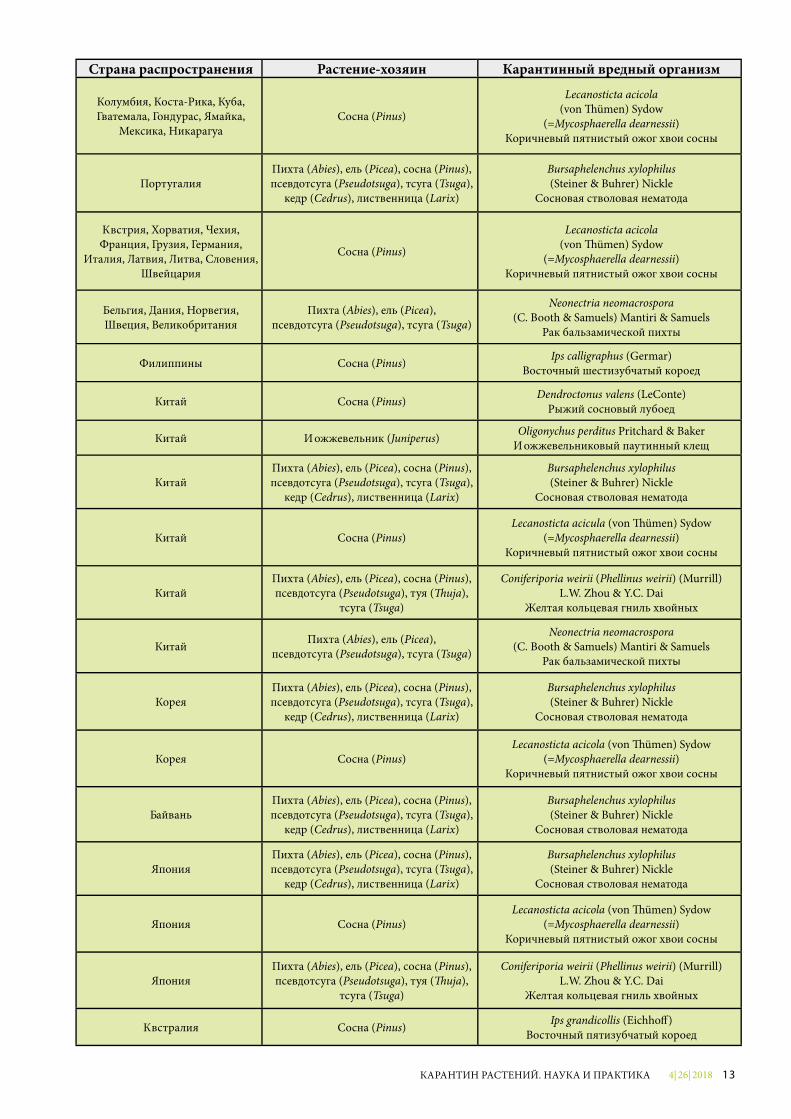
Страна распространения Растение-хозяин Карантинный вредный организм
Колумбия, Коста-Рика, Куба, Гватемала, Гондурас, Ямайка,
Мексика, НикарагуаСосна (Pinus)
Lecanosticta acicola(von �ümen) Sydow
(=Mycosphaerella dearnessii)Коричневый пятнистый ожог хвои сосны
ПортугалияПихта (Abies), ель (Picea), сосна (Pinus), псевдотсуга (Pseudotsuga), тсуга (Tsuga),
кедр (Cedrus), лиственница (Larix)
Bursaphelenchus xylophilus(Steiner & Buhrer) Nickle
Сосновая стволовая нематода
Австрия, Хорватия, Чехия, Франция, Грузия, Германия,
Италия, Латвия, Литва, Словения, Швейцария
Сосна (Pinus)
Lecanosticta acicola(von �ümen) Sydow
(=Mycosphaerella dearnessii)Коричневый пятнистый ожог хвои сосны
Бельгия, Дания, Норвегия, Швеция, Великобритания
Пихта (Abies), ель (Picea), псевдотсуга (Pseudotsuga), тсуга (Tsuga)
Neonectria neomacrospora(C. Booth & Samuels) Mantiri & Samuels
Рак бальзамической пихты
Филиппины Сосна (Pinus) Ips calligraphus (Germar) Восточный шестизубчатый короед
Китай Сосна (Pinus) Dendroctonus valens (LeConte)Рыжий сосновый лубоед
Китай Можжевельник (Juniperus) Oligonychus perditus Pritchard & Baker Можжевельниковый паутинный клещ
КитайПихта (Abies), ель (Picea), сосна (Pinus), псевдотсуга (Pseudotsuga), тсуга (Tsuga),
кедр (Cedrus), лиственница (Larix)
Bursaphelenchus xylophilus(Steiner & Buhrer) Nickle
Сосновая стволовая нематода
Китай Сосна (Pinus)Lecanosticta acicula (von �ümen) Sydow
(=Mycosphaerella dearnessii)Коричневый пятнистый ожог хвои сосны
КитайПихта (Abies), ель (Picea), сосна (Pinus),псевдотсуга (Pseudotsuga), туя (�uja),
тсуга (Tsuga)
Coniferiporia weirii (Phellinus weirii) (Murrill) L.W. Zhou & Y.C. Dai
Желтая кольцевая гниль хвойных
Китай Пихта (Abies), ель (Picea), псевдотсуга (Pseudotsuga), тсуга (Tsuga)
Neonectria neomacrospora(C. Booth & Samuels) Mantiri & Samuels
Рак бальзамической пихты
КореяПихта (Abies), ель (Picea), сосна (Pinus), псевдотсуга (Pseudotsuga), тсуга (Tsuga),
кедр (Cedrus), лиственница (Larix)
Bursaphelenchus xylophilus(Steiner & Buhrer) Nickle
Сосновая стволовая нематода
Корея Сосна (Pinus)Lecanosticta acicola (von �ümen) Sydow
(=Mycosphaerella dearnessii)Коричневый пятнистый ожог хвои сосны
ТайваньПихта (Abies), ель (Picea), сосна (Pinus), псевдотсуга (Pseudotsuga), тсуга (Tsuga),
кедр (Cedrus), лиственница (Larix)
Bursaphelenchus xylophilus (Steiner & Buhrer) Nickle
Сосновая стволовая нематода
ЯпонияПихта (Abies), ель (Picea), сосна (Pinus), псевдотсуга (Pseudotsuga), тсуга (Tsuga),
кедр (Cedrus), лиственница (Larix)
Bursaphelenchus xylophilus(Steiner & Buhrer) Nickle
Сосновая стволовая нематода
Япония Сосна (Pinus)Lecanosticta acicola (von �ümen) Sydow
(=Mycosphaerella dearnessii)Коричневый пятнистый ожог хвои сосны
ЯпонияПихта (Abies), ель (Picea), сосна (Pinus),псевдотсуга (Pseudotsuga), туя (�uja),
тсуга (Tsuga)
Coniferiporia weirii (Phellinus weirii) (Murrill) L.W. Zhou & Y.C. Dai
Желтая кольцевая гниль хвойных
Австралия Сосна (Pinus) Ips grandicollis (Eichho�) Восточный пятизубчатый короед
4| 26| 201814 КАРАНТИН РАСТЕНИЙ. НАУКА И ПРАКТИКА
Abstract. A brief analysis of possible introduction of various quarantine pests with Christmas trees and freshly cut co-niferous branches is presented. �e list of quarantine pests (insects, nematodes, phy-topathogenic fungi), which can enter the territory of the Russian Federation with the analyzed products from di�erent coun-tries of the world, is given.
Keywords. Christmas trees, freshly cut coniferous branches, entry, quarantine pests, pests, pine stem nematode, fungal pathogens, introduction, damage, Russia, RF.
�e traditional Christmas decorations of each house are “Christmas trees” and fresh cut branches of conifers, used as the main material in the �oral Christmas and new year decor.
However, decorated conifers and cut branches are not only a source of festive mood, but also a potential threat of intro-duction of various pests that can be harm-ful to both human health and the environ-ment, especially if these Christmas trees were imported from afar.
In the new year period, the Internet is full of proposals for the supply of pre-sentable “Christmas trees” from abroad. �is article presents a brief analysis of the possible introduction of quarantine pests with new year products and the degree of threat to so¥wood in the territory of the Russian Federation.
According to the Federal customs service, over the last three years (2015-2017) pro-ducts under the FEACN code 0604202000 “Christmas trees, fresh” were exported to the Russian Federation from 13 countries (http://stat.customs.ru). �ese are Belarus, Belgium, Canada, USA, Denmark, Spain, Latvia, Netherlands, Poland, Germany, Ita-ly, Armenia, Estonia (Fig. 1).
�e largest volume of deliveries of “Christmas trees” from abroad accounts for Denmark and Poland (respectively 905.2 and 928.8 t), the maximum of these supplies accounts for 2016. In 2017, there was a signi�cant decrease in the volume of imports of “Christmas trees” from these
countries, and the volume of pro ducts supplied from the Republic of Belarus increased. �e list of “Christmas trees” exporting countries was signi�cantly updated in 2017. In 2017, new export-ing countries – Estonia and Armenia – appeared. It should be noted that if the supply of “Christmas trees”, although in a limited amount, in 2015-2016 was carried out even from North America (Cana-da and the United States), then in 2017 there was no import of these products from these countries. Perhaps this is due to the new phytosanitary requirements of the EAEU, which came into e�ect in July 2017. According to these requirements, the import of conifer planting material, including cut coniferous branches and Christmas trees, is prohibited due to the phytosanitary threat to the conifers of the Russian Federation.
Unlike Christmas trees, fresh bran-ches of conifers are imported from a much larger number of countries. According to the Federal customs service, over the last three years (2015-2017) products under the FEACN code 0604204000 “branches of coniferous trees, fresh” were exported to the Russian Federation from 22 countries (http://stat.customs.ru). �e largest sup-plier countries were: Poland, Denmark, Belarus, Italy, Germany (Fig. 2).
In 2015, “branches of coniferous trees” on the territory of the Russian Federation were delivered from 12 countries not only from Europe but also from South America.
As in the case of “Christmas trees”, the main exporters of coniferous branches are Poland, Denmark, Germany. When com-paring the volume of deliveries to Russia of these products for three years, the largest volume accounts for 2016. In 2017, there is a decrease in the number of products supplied, however, Poland (187.1 t), Italy (115 t), Germany (110.9 t), Denmark (91.6 t), Spain (22.2 t) are among the priority coun-tries. Deliveries of coniferous branches, though in a minimum quantity, were also made from the United States.
Below is an assessment of phytosanitary risk in case of possible introduction of pests with Christmas trees and branches of conifers from around the world into the territory of the Russian Federation.
�e greatest phytosanitary risk is posed by Christmas trees and cut branches im-ported from North America (USA and Canada). With the American live (felled) spruces, pines and �rs to Russia pests en-tering the list of the quarantine objects absent in the territory of the Russian Fede-ration and the Eurasian economic Union (EAEU) can be introduced. Among the insects there are several species of leaf rol-ler moths of the Choristoneura and Acleris. In winter, they may be present in the egg stage on the branches of the tree. Bark beet les can hide under the bark of branches and trunks of coniferous trees. �ese are the bark beetles of the Dendroctonus ge-nus and several bark beetle species of the Ips genus. Particular risk is posed by the beetles of the Dendroctonus genus, which does much harm to coniferous forests in Canada and the United States and, ac-cording to the phytosanitary risk analysis, represents the greatest threat to coniferous plantations in Russia.
Pine wood nematode Bursaphelenchus xylophilus is one of the most pathogenic species, widely distributed in the North American continent. Local conifers spe-cies are resistant to this pathogen, but most European and Asian conifers are susceptible to it. In case of infestation of a susceptible host plant, the tree can die within one season. Nematodes easily survive in the trunk, tree branches, bark and in the wood and, under favorable conditions (in the case of infested and uninfested material contact), they can move independently from infested wood to healthy trees (Sousa, 2011).
Among the pathogens that may be pre-sent on the branches of Christmas trees, should include the following pathogens of fungal diseases: cancer (blight) of the trunks and branches of pine (Atropellis pinip hila, A. pinicola), in the winter time
UDC 632.92:635.912
“CHRISTMAS TREES” AS A THREAT TO THE SPREAD OF PATHOGENIC FOREST PESTSO.A. Kulinich, All-Russian Plant Quarantine Center (FGBU “VNIIKR”)A.G. Shchukovskaya, All-Russian Plant Quarantine Center (FGBU “VNIIKR”)E.N. Arbuzova, All-Russian Plant Quarantine Center (FGBU “VNIIKR”)N.I. Kozyreva, Institute of Ecology and Evolution Russian Academy of Sciences, Moscow
4| 26| 2018 15КАРАНТИН РАСТЕНИЙ. НАУКА И ПРАКТИКА
pest remains in apothecia on stem and branches or in the form of mycelium under the bark of branches and trunk; brown spot needle blight of pine Leca-nosticta acicola (Mycosphaerella dear-nessii), the pathogen overwinters in the form of beams sporogenous mycelium in stromach (pycnidia) in dead needles and branches; Southern fusiform rust of pine (Cronartium fusiforme) and western gall rust of pine (Endocronartium harknessii) – in winter, the pathogen persists in the form of galls on the trunk and branches of pine trees; laminated butt rot of coni-fers Coniferiporia weirii (Phellinus weirii); broom rust of spurce Chrysomyxa arcto-staphyli. �ese fungal pathogens, as well as pine wood nematode B. xylophilus, are common in North America and are in-cluded in the lists of quarantine objects of many countries of the world.
Customs statistics show that “Christmas trees” and cut branches are not imported to Russia from Asian countries, but below we have also assessed the phytosanitary risk of possible introduction of pests from the nearest Asian countries.
From East Asian countries (China, Ko-rea and Japan) with planting material and “Christmas trees”, including cut branches, can also be imported: Pine wood nema-tode (pine wilt disease) B. xylophilus, juni-per spider mite Oligonychus perditus (can be on the needles of Christmas trees); the brown needle blight of pine Mycosphae-rella gibsonii (in winter in the form of a stromas on dead needles of infected bran-ches); the brown spot of pine (L. acicоla).
�e largest deliveries of products “Christ-mas trees” and cut branches in Russia are made from European countries. In gener-al, the pests in the European part of Russia and Europe is similar. However, a number of pests that have already entered into Eu-rope from America and cause signi�cant damage to forest plantations do not yet exist in Russia. All of them can be entered with this species of product in the winter. �is is again a pine wood nematode (B. xyloph-ilus), which can be imported from the co-niferous trees or branches from Portugal or Spain; the brown spot needle blight of pine (L. acicula), which causes the disease wide-spread in the European Union (Austria, Croatia, Slovenia, the Czech Republic, France, Germany, Switzerland, Latvia and Lithuania). �e estimated damage caused by the introduction of these organisms on the territory of the Russian Federation amounts to tens of billions of dollars.
�e products imported into the country are subject to phytosanitary examination
by the NPPO, in Russia it is carried out by the Rosselkhoznadzor. �e inspector visually checks the “Christmas trees” and branches for presence of pests or diseas-es. However, not always they can be de-tected during visual inspection and even during laboratory examination, especial-ly at the initial stage of contamination of products. In this regard, there are certain phyto sanitary principles in the interna-tional practice: prohibition of import of products that may contain quaran-tine objects from countries (or areas) of distribution of these objects (Kulinich, 2018; Guide to the implementation of phytosanitary standards in forestry, 2011; ISPM 15, 2013). �e EPPO standard PM 8/2 (1) recommends a ban on the im-port of live Christmas trees and branch-es from the countries where quarantine organisms are spread. Following this recommendation, the European Council Directive (Directive 29/2000 EC) banned the importation of live Christmas trees and branches from North American countries where pests listed above are spread. �e estimated damage caused by the introduction of these pests in Russia is estimated at tens of billions of dollars. �e phytosanitary requirements of the EEC, which entered into force on July 1, 2017, also prescribe similar conditions: prohi-bition of importation of live branches and Christmas trees from countries where quarantine objects listed above are spread.
�e risk of introduction with such pro-ducts (Christmas trees and live branches of coniferous trees) is not so great in com-parison with the planting material, be-cause it comes in the winter, and its further disposal is expected, but the risk is present. �e authors of the article did not �nd in the o¦cial information databases the facts of registration of quarantine objects in live branches and Christmas trees by the NPPO sta� of di�erent countries, but eve-ry year we are faced with the facts of intro-duction of various species of insects into the house with Christmas trees, which be-gin to develop rapidly in warm conditions (EPPO Reporting Service, 2014-2017; INT ERNET EUROPHYT). Every year, the press publishes information about the detection of insects on Christmas trees that spread around the apartment. So, on the eve of 2018, there were cases of detec-tion of insects by Moscow residents on Christmas �rs imported from Poland and Denmark (http://so-l.ru/news).
�e risk of introduction of insects or other pests from North America is not excluded. However, in the latter case,
the degree of damage will be many times higher.
In most cases, used Christmas trees and branches are stored near garbage cans and o¥en remain there until spring, thus creating a potential source of threat of transition of viable pests on decorative green plantings of recreational territories.
In some cases, Amateur gardeners plant in suburban areas live Christmas trees acquired with the root system in containers, thereby exposing the risk of possible settlement of invasive pests and pathogens of coniferous plantations as plants in their own suburban area, and the nearest forest plantations.
�e best option to reduce the threat of the possible spread of pests is the time-ly disposal of the source of the threat by burning or other e�ective methods.
It should be remembered that all “new year’s natural material”, including “Christmas trees” and cut branches, is a product of phytosanitary risk, which is subject to mandatory inspection and cer-ti�cation and cannot be in uncontrolled circulation.
References1. Kulinich O.A., Shchukovskaya A.G.,
Arbuzova E.N., Kozyreva N.I. Possible in-troduction of quarantine pests with wood packaging materials // Plant Quarantine. Science and practice, 2018. № 2 (24). P. 12-16.
2. Guide to the implementation of phy-tosanitary standards in forestry. FAO, Rome, 2011. 117 p.
3. EPPO Reporting Service, 2014-2017, Paris. INTERNET EUROPHYT. Annual and monthly reports of interceptions of harmful organisms in imported plants and other objects. http://ec.europa.eu/food/plant/plant_health_biosecurity/eu-rophyt/interceptio ns/index_en.htm.
4. ISPM 15. Regulation of wood packag ing material in international trade. FAO, Rome, 2013.
5. Sousa E., Naves P., Bonifacio L., Hen-rigues J., Inacio M.L., Evans H. Assessing risks of pine wood nematode Bursaphe-lenchus xylophilus transfer between wood packing by simulating assembled pallets in service // Bulletin OEPP/EPPO Bulle-tin, 2011. № 41. P. 423-431.
6. [Electronic resource]. Access mode: http://so-l.ru/news/y/2018_01_09_novogodnie_syurprizi (access date: 10.11.2017).
7. [Electronic resource]. Access mode: http://stat.customs.ru (access date: 15.12.2017).
4| 26| 201816 КАРАНТИН РАСТЕНИЙ. НАУКА И ПРАКТИКА
Country spread of pest Host plant Quarantine pest
Canada, USA
Fir (Abies), spruce (Picea), Douglas �r (Pseudotsuga),
juniper (Juniperus),larch (Larix), pine (Pinus)
Choristoneura fumiferana (Clemens) Eastern spruce budworm
Canada, USA Fir (Abies), spruce (Picea), Douglas �r (Pseudotsuga)
Choristoneura freemani Razowski Western spruce budworm
Canada, USA Fir (Abies), Douglas �r (Pseudotsuga), tsuga (Tsuga), spruce (Picea)
Acleris gloverana (Walsingham) Western blackheaded budworm
Canada, USA Fir (Abies), spruce (Picea) Acleris variana (Fernald) Eastern blackheaded budworm
Canada, USA Pine (Pinus)
Dendroctonus brevicomis LeConte Western pine beetle
Dendroctonus ponderosae Hopkins Mountain pine beetle
Dendroctonus valens (LeConte) Red turpentine beetle
Ips calligraphus (Germar) Six-spined engraver beetle
Ips grandicollis (Eichho�) Five-spined bark beetle
Ips pini (Say) Pine engraver beetle
Ips plastographus (LeConte) California pine engraver
Canada, USA, MexicoFir (Abies), spruce (Picea), pine (Pinus),
Douglas �r (Pseudotsuga), hemlock (Tsuga), cedar (Cedrus), larch (Larix)
Bursaphelenchus xylophilus (Steiner & Buhrer) Nickle
Pine wood nematode
Canada, USA Pine (Pinus)
Atropellis pinicola Zeller & Goodding Cancer (blight) of pine trunks and branches
Atropellis piniphila (Weir) Lohmann & Cash
Cancer (blight) of pine trunks and branchesLecanosticta acicula (von �ümen) Sydow
(=Mycosphaerella dearnessii)Brown spot needle blight of pine
Endocronartium harknessii E. Meinecke Western gall rust of pine
USA Pine (Pinus), oak (Quercus),chestnut (Castanea)
Cronartium fusiforme Cummins Southern fusiform rust of pine
Canada, USA Spruce (Picea), bear’s grape (Arctostaphylos uva-ursi)
Chrysomyxa arctostaphyli Dietel Spruce needles rust
Canada, USAFir (Abies), spruce (Picea), pine (Pinus),Douglas �r (Pseudotsuga), cedar (�uja),
hemlock (Tsuga)
Coniferiporia weirii (Phellinus weirii) (Murrill) L.W. Zhou & Y.C. Dai Laminated butt rot of conifers
(yellow ring rot of conifers)
Canada, USAFir (Abies), spruce (Picea), Douglas �r (Pseudotsuga),
hemlock (Tsuga)
Neonectria neomacrospora(C. Booth & Samuels) Mantiri & Samuels
Canker of balsam �r
Guatemala, Honduras, Mexico, Nicaragua Pine (Pinus) Dendroctonus valens (LeConte) Red turpentine beetle
Cuba, Dominican Republic, Guatemala, Haiti, Jamaica, Mexico, Nicaragua Pine (Pinus) Ips calligraphus (Germar)
Six-spined engraver beetle
Cuba, Dominican Republic, Guatemala, Jamaica, Mexico, Nicaragua, Honduras Pine (Pinus) Ips grandicollis (Eichho�)
Five-spined bark beetle
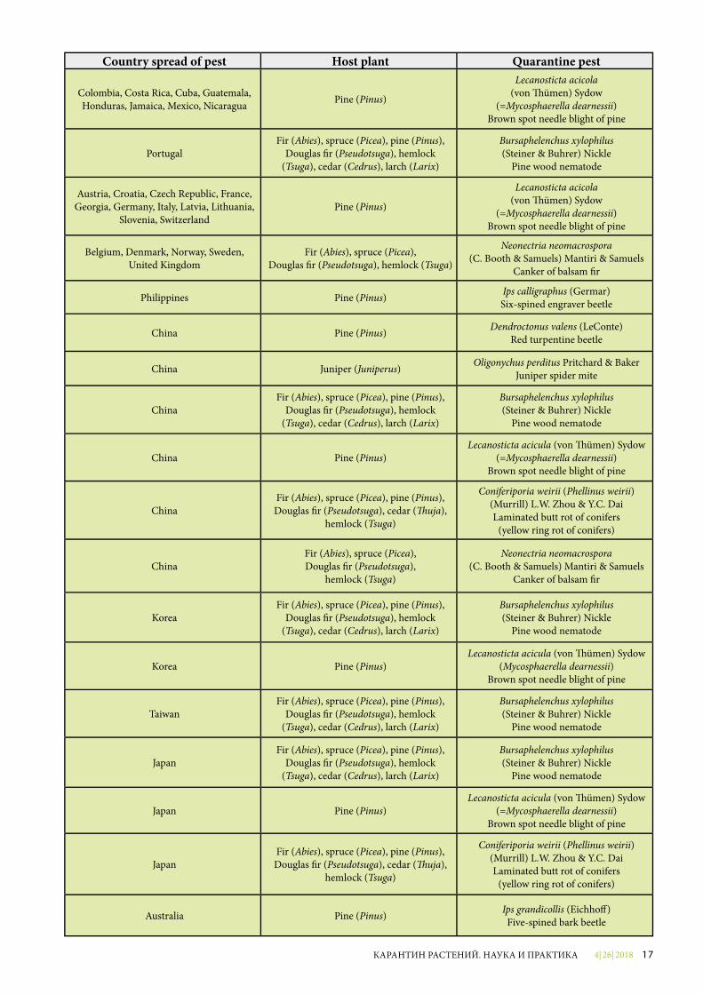
Spread of quarantine pests associated with the commodities of “Christmas trees” and “fresh cut branches of conifers” in different countries of the world
4| 26| 2018 17КАРАНТИН РАСТЕНИЙ. НАУКА И ПРАКТИКА
Country spread of pest Host plant Quarantine pest
Colombia, Costa Rica, Cuba, Guatemala, Honduras, Jamaica, Mexico, Nicaragua Pine (Pinus)
Lecanosticta acicola(von �ümen) Sydow
(=Mycosphaerella dearnessii)Brown spot needle blight of pine
PortugalFir (Abies), spruce (Picea), pine (Pinus),
Douglas �r (Pseudotsuga), hemlock (Tsuga), cedar (Cedrus), larch (Larix)
Bursaphelenchus xylophilus(Steiner & Buhrer) Nickle
Pine wood nematode
Austria, Croatia, Czech Republic, France, Georgia, Germany, Italy, Latvia, Lithuania,
Slovenia, SwitzerlandPine (Pinus)
Lecanosticta acicola(von �ümen) Sydow
(=Mycosphaerella dearnessii)Brown spot needle blight of pine
Belgium, Denmark, Norway, Sweden, United Kingdom
Fir (Abies), spruce (Picea), Douglas �r (Pseudotsuga), hemlock (Tsuga)
Neonectria neomacrospora(C. Booth & Samuels) Mantiri & Samuels
Canker of balsam �r
Philippines Pine (Pinus) Ips calligraphus (Germar) Six-spined engraver beetle
China Pine (Pinus) Dendroctonus valens (LeConte) Red turpentine beetle
China Juniper (Juniperus) Oligonychus perditus Pritchard & Baker Juniper spider mite
ChinaFir (Abies), spruce (Picea), pine (Pinus),
Douglas �r (Pseudotsuga), hemlock (Tsuga), cedar (Cedrus), larch (Larix)
Bursaphelenchus xylophilus(Steiner & Buhrer) Nickle
Pine wood nematode
China Pine (Pinus)Lecanosticta acicula (von �ümen) Sydow
(=Mycosphaerella dearnessii)Brown spot needle blight of pine
ChinaFir (Abies), spruce (Picea), pine (Pinus),Douglas �r (Pseudotsuga), cedar (�uja),
hemlock (Tsuga)
Coniferiporia weirii (Phellinus weirii)(Murrill) L.W. Zhou & Y.C. Dai Laminated butt rot of conifers
(yellow ring rot of conifers)
ChinaFir (Abies), spruce (Picea), Douglas �r (Pseudotsuga),
hemlock (Tsuga)
Neonectria neomacrospora(C. Booth & Samuels) Mantiri & Samuels
Canker of balsam �r
KoreaFir (Abies), spruce (Picea), pine (Pinus),
Douglas �r (Pseudotsuga), hemlock (Tsuga), cedar (Cedrus), larch (Larix)
Bursaphelenchus xylophilus(Steiner & Buhrer) Nickle
Pine wood nematode
Korea Pine (Pinus)Lecanosticta acicula (von �ümen) Sydow
(Mycosphaerella dearnessii)Brown spot needle blight of pine
TaiwanFir (Abies), spruce (Picea), pine (Pinus),
Douglas �r (Pseudotsuga), hemlock (Tsuga), cedar (Cedrus), larch (Larix)
Bursaphelenchus xylophilus (Steiner & Buhrer) Nickle
Pine wood nematode
JapanFir (Abies), spruce (Picea), pine (Pinus),
Douglas �r (Pseudotsuga), hemlock (Tsuga), cedar (Cedrus), larch (Larix)
Bursaphelenchus xylophilus(Steiner & Buhrer) Nickle
Pine wood nematode
Japan Pine (Pinus)Lecanosticta acicula (von �ümen) Sydow
(=Mycosphaerella dearnessii)Brown spot needle blight of pine
JapanFir (Abies), spruce (Picea), pine (Pinus),Douglas �r (Pseudotsuga), cedar (�uja),
hemlock (Tsuga)
Coniferiporia weirii (Phellinus weirii) (Murrill) L.W. Zhou & Y.C. Dai Laminated butt rot of conifers
(yellow ring rot of conifers)
Australia Pine (Pinus) Ips grandicollis (Eichho�) Five-spined bark beetle
4| 26| 201818 КАРАНТИН РАСТЕНИЙ. НАУКА И ПРАКТИКА
УДК 632.911.2:632.913.1
ОСОБО ОПАСНЫЕ ВОЗБУДИТЕЛИ БОЛЕЗНЕЙ КОСТОЧКОВЫХ КУЛЬТУР РОДА CANDIDATUS PHYTOPLASMA SPP.Г.Н. Бондаренко, старший научный сотрудник – начальник Испытательного лабораторного центра ФГБУ «ВНИИКР»И.Г. Башкирова, агроном лаборатории анализа ГМО Испытательного лабораторногоцентра ФГБУ «ВНИИКР»
Аннотация. В статье пред-ставлен обзор особо опасных видов фитоплазм – возбудителей болез-ней косточковых культур. Среди них фитоплазма хлоротическо-го скручивания листьев абрикоса (Candidatus Phytoplasma prunorum), фитоплазма X-болезни персика (Ca. Phytoplasma pruni) и возбуди-тель «ведьминых метел» миндаля (Ca. Phytoplasma phoenicium), про-никновение и распространение ко-торых стоит брать во внимание. В работе представлены данные о географическом распространении, растениях-хозяевах и универсаль-ных методах диагностики фито-патогенов.
Ключевые слова. Фитоплазма, косточковые культуры, плодовые
насаждения, фитосанитарный контроль, посадочный материал.
На протяжении последних трех лет в России наблюдался как спад, так и подъем производства плодов косточковых культур (сливы, виш-ни, черешни, абрикосов, персиков и других) в хозяйствах всех кате-горий. Для получения большего объема продукции организована программа развития и увеличения площадей плодовых насаждений. Нужно отметить, что отечествен-ные питомники и маточники не справляются с запланированны-ми объемами агропроизводите-лей (Башкирова и др., 2018). Как правило, посадочный материал поступает из стран Европы, где
распространена фитоплазма хло-ротического скручивания листьев абрикоса (Candidatus Phytoplasma prunorum). Если рассматривать другие рынки, то возможен ввоз из Северной Америки и из некоторых стран Азии, где широко распро-странены такие опасные организ-мы, как фитоплазма X-болезни пер-сика и возбудитель «ведьминых ме-тел» миндаля Ca. Phytoplasma pruni. Проникновение исследуемых фито-плазм в латентном состоянии с им-портным посадочным материалом представляется возможным из-за отсутствия у них регуляционного статуса. В связи с этим в работе уде-ляется внимание географическому распространению, растениям-хозя-евам и универсальным методам ди-
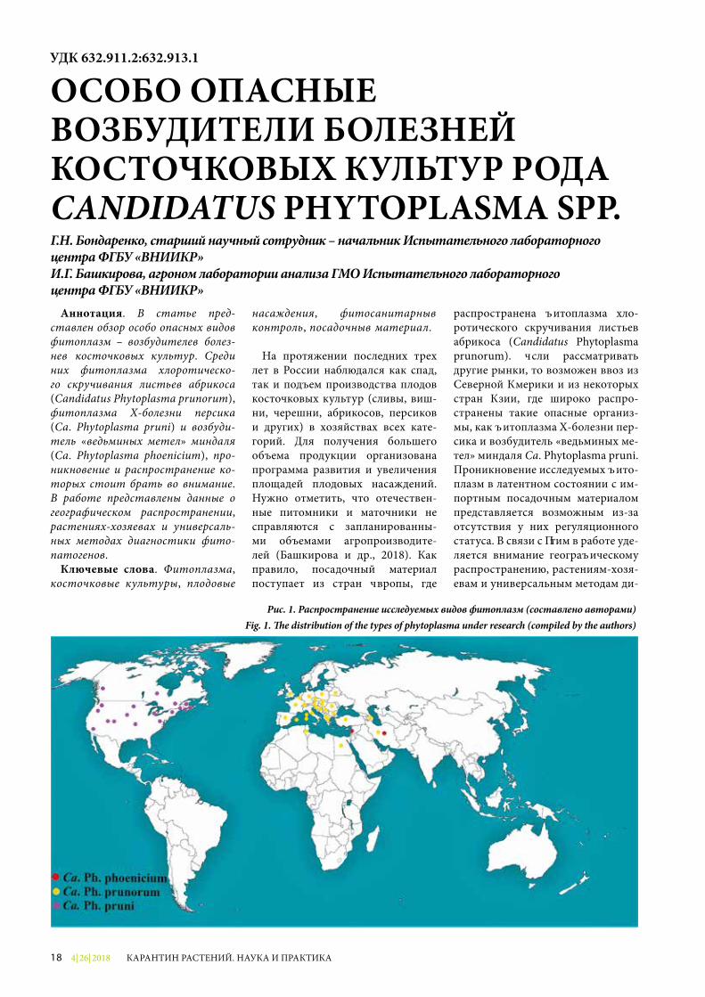
Рис. 1. Распространение исследуемых видов фитоплазм (составлено авторами)Fig. 1. �e distribution of the types of phytoplasma under research (compiled by the authors)
4| 26| 2018 19КАРАНТИН РАСТЕНИЙ. НАУКА И ПРАКТИКА
агностики группы фитопатогенов рода Ca. Phytoplasma.
Для применения определенных ме-тодов диагностики необходимо по-нимать таксономическое положение возбудителей болезней косточковых культур. Фитоплазмы принадлежат к царству Bacteria, типу Firmicutes, клас-су Mollicutes, отделу Acholeplasmatales, семейству Acholeplasmataceae, роду Candidatus Phytoplasma.
Фитоплазма хлоротического скру-чивания листьев абрикоса – Ca. Phytoplasma prunorum (Seemüller and Schneider) – принадлежит к группе Apple proliferation (16SrX). Обще-принятые названия и сокращения: Apricot chlorotic leafroll, European stone fruit yellows phytoplasma (ESFY).
Фитоплазма X-болезни пер-сика – Ca. Phytoplasma pruni (IRPCM, 2004), относится к группе X-disease (16SrIII). Общепринятые
названия: Peach X-disease, Peach yellow leafroll phytoplasma, Western X-disease phytoplasma.
Возбудитель такого заболева-ния, как «ведьмины метлы» мин-даля – Ca. Phytoplasma phoenicium (Verdin et al., 2003) – по генети-ческим признакам входит в ма-лоизученную группу Pigeon pea witches’ broom (16SrIX). Общепри-нятые названия и сокращения: Phytoplasma phoenicium, Witches’ broom of almond, Almond witches’ broom phytoplasma, AlWB (https://www.cabi.org; https://www.eppo.int; Bertaccini et al., 2014; Hodgetts, Dickinson, 2010; Zhao et al., 2010).
Если рассматривать вид Ca. Ph. prunorum, то он инфициру-ет большое количество растений (табл. 1). В основном поражает растения рода Prunus: абрикос обыкновенный (P. armeniaca), персик обыкновенный (P. persica) и сливу китайскую (P. salicina). Патоген был обнаружен и на ди-ких видах рода Prunus: терне (P. spinosa) и черемухе поздней (P. serotina). Более устойчивыми видами являются: слива домаш-няя (P. domestica), алыча обык-новенная (P. cerasifera), черешня (P. avium) и вишня обыкновенная (P. cerasus). Экспериментальным растением-хозяином является барвинок розовый (Catharanthus roseus). Установлено, что фито-плазму переносят листоблошка
КультураФитоплазмы
Ca. Ph. prunorum
Ca. Ph. pruni
Ca. Ph. phoenicium
Абрикос обыкновенный (Prunus armeniaca)
Основной + -
Черешня (Prunus avium) + Основной -Слива домашняя (Prunus domestica)
+ + -
Миндаль обыкновенный (Prunus dulcis)
+ + Основной
Персик обыкновенный (Prunus persica)
Основной Основной Основной
Нектарин (Prunus persica var. nectarina)
- - Основной
Слива китайская (Prunus salicina)
Основной Основной -
Терн (Prunus spinosa) + - -Алыча культурная (Prunus cerasifera)
+ - -
Черемуха антипка (Prunus mahaleb)
+ - -
Слива узколистная (Prunus angustifolia)
- + -
Вишня обыкновенная (Prunus cerasus)
- Основной -
Черемуха виргинская (Prunus virginiana)
- + -
Дикий миндаль (Prunus scoparia)
- - +
Таблица 1 Растения – хозяева рода Prunus для фитоплазм (https://www.eppo.int; https://www.cabi.org)
Рис. 2. Симптомы повреждения Ca. Ph. prunorum – побурение среднего слоя коры после зимы (фото G. Morvan, INRA, Montfavet)
Fig. 2. Symptoms of Ca. Ph. prunorum damage – the middle layer of the bark a�er the winter (photo by G. Morvan, INRA, Montfavet)
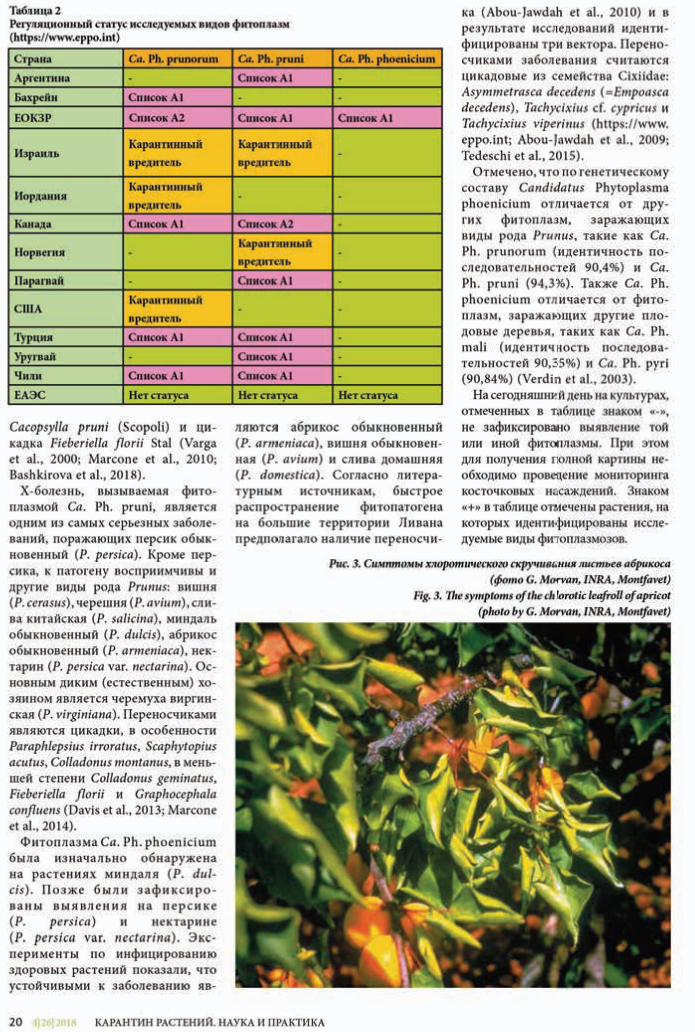
в директиве ЕС
ЕОКЗР.
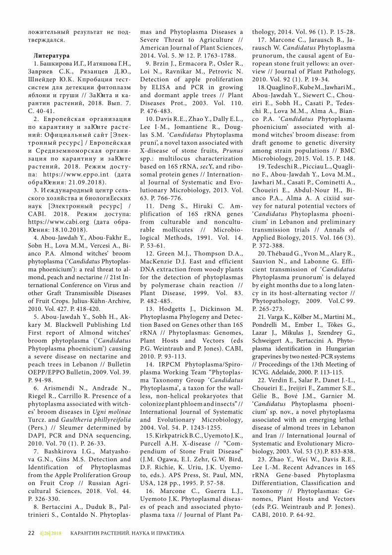
4| 26| 201822 КАРАНТИН РАСТЕНИЙ. НАУКА И ПРАКТИКА
ложительный результат не под-тверждался.
Литература1. Башкирова И.Г., Матяшова Г.Н.,
Завриев С.К., Рязанцев Д.Ю., Шнейдер Ю.А. Апробация тест-сис тем для детекции фитоплазм яблони и груши // Защита и ка-рантин растений, 2018. Вып. 7. С. 40-41.
2. Европейская организация по карантину и защите расте-ний: Официальный сайт [Элек-тронный ресурс] / Европейская и Средиземноморская органи-зация по карантину и защите растений, 2018. Режим дост у-па: https://www.eppo.int (дата обращения: 21.09.2018).
3. Международный центр сель-ского хозяйства и биологических наук [Электронный ресурс] / CABI. 2018. Режим доступа: https://www.cabi.org (дата обра-щения: 18.10.2018).
4. Abou-Jawdah Y., Abou-Fakhr E., Sobn H., Lova M.M., Vercesi A., Bi-anco P.A. Almond witches’ broom phytoplasma (‘Candidatus Phytoplas-ma phoenicium’): a real threat to al-mond, peach and nectarine // 21st In-ternational Conference on Virus and other Graft Transmissible Diseases of Fruit Crops. Julius-Kühn-Archive, 2010. Vol. 427. P. 418-420.
5. Abou-Jawdah Y., Sobh H., Ak-kary M. Blackwell Publishing Ltd First report of Almond witches’ broom phytoplasma (‘Candidatus Phytoplasma phoenicium’) causing a severe disease on nectarine and peach trees in Lebanon // Bulletin OEPP/EPPO Bulletin, 2009. Vol. 39. P. 94-98.
6. Arismendi N., Andrade N., Riegel R., Carrillo R. Presence of a phytoplasma associated with witch-es’ broom diseases in Ugni molinae Turcz. and Gaultheria phillyreifolia (Pers.) // Sleumer determined by DAPI, PCR and DNA sequencing, 2010. Vol. 70 (1). P. 26-33.
7. Bashkirova I.G., Matyasho-va G.N., Gins M.S. Detection and Identi fication of Phytoplasmas from the Apple Proliferation Group on Fruit Crop // Russian Agri-cultural Sciences, 2018. Vol. 44. P. 326-330.
8. Bertaccini A., Duduk B., Pal-trinieri S., Contaldo N. Phytoplas-
mas and Phytoplasma Diseases a Severe Threat to Agriculture // American Journal of Plant Sciences, 2014. Vol. 5. № 12. P. 1763-1788.
9. Brzin J., Ermacora P., Osler R., Loi N., Ravnikar M., Petrovic N. Detection of apple proliferation by ELISA and PCR in growing and dormant apple trees // Plant Diseases Prot., 2003. Vol. 110. P. 476-483.
10. Davis R.E., Zhao Y., Dally E.L., Lee I-M., Jomantiene R., Doug-las S.M. ‘Candidatus Phyto plasma pruni’, a novel taxon associated with X-disease of stone fruits, Prunus spp.: multilocus characteriz ation based on 16S rRNA, secY, and ribo-somal protein genes // Internation-al Journal of Systematic and Evo-lutionary Microbiology, 2013. Vol. 63. P. 766-776.
11. Deng S., Hiruki C. Am-plification of 16S rRNA genes from culturable and noncultu-rable mollicutes // Microbio-logical Methods, 1991. Vol. 14. P. 53-61.
12. Green M.J., Thompson D.A., MacKenzie D.J. East and efficient DNA extraction from woody plants for the detection of phytoplasmas by polymerase chain reaction // Plant Disease, 1999. Vol. 83. P. 482-485.
13. Hodgetts J., Dickinson M. Phytoplasma Phylogeny and Detec-tion Based on Genes other than 16S rRNA // Phytoplasmas: Genomes, Plant Hosts and Vectors (eds P.G. Weintraub and P. Jones). CABI, 2010. P. 93-113.
14. IRPCM Phytoplasma/Spiro-plasma Working Team “Phytoplas-ma Taxonomy Group ‘Candidatus Phytoplasma’, a taxon for the wall-less, non-helical prokaryotes that colonize plant phloem and insects” // International Journal of Systematic and Evolutionary Microbiology, 2004. Vol. 54. P. 1243-1255.
15. Kirkpatrick B.C., Uyemoto J.K., Purcell A.H. X-disease // “Com-pendium of Stone Fruit Disease” (J.M. Ogawa, E.I. Zehr, G.W. Bird, D.F. Richie, K. Uriu, J.K. Uyemo-to, eds.). APS Press, St. Paul, MN, USA, 128 pp., 1995. P. 57-58.
16. Marcone C., Guerra L.J., Uyemoto J.K. Phytoplasmal diseas-es of peach and associated phyto-plasma taxa // Journal of Plant Pa-
thology, 2014. Vol. 96 (1). P. 15-28.17. Marcone C., Jarausch B., Ja-
rausch W. Candidatus Phyto plasma prunorum, the causal agent of Eu-ropean stone fruit yellows: an over-view // Journal of Plant Pathology, 2010. Vol. 92 (1). P. 19-34.
18. Quaglino F., Kube M., Jawhari M., Abou-Jawdah Y., Siewert C., Chou-eiri E., Sobh H., Casati P., Tedes-chi R., Lova M.M., Alma A., Bian-co P.A. ‘Candidatus Phytoplasma phoenicium’ associated with al-mond witches’ broom disease: from draft genome to genetic diversity among strain populations // BMC Microbiology, 2015. Vol. 15. P. 148.
19. Tedeschi R., Picciau L., Quagli-no F., Abou‐Jawdah Y., Lova M.M., Jawhari M., Casati P., Cominetti A., Choueiri E., Abdul-Nour H., Bi-anco P.A., Alma A. A cixiid sur-vey for natural potential vectors of ‘Candidatus Phytoplasma phoeni-cium’ in Lebanon and preliminary transmission trials // Annals of Applied Biology, 2015. Vol. 166 (3). P. 372-388.
20. Thébaud G., Yvon M., Alary R., Sauvion N., and Labonne G. Effi-cient transmission of ‘Candidatus Phytoplasma pru no rum’ is delayed by eight months due to a long laten-cy in its host-alternating vector // Phytopathology, 2009. Vol. 99. P. 265-273.
21. Varga K., Kölber M., Martini M., Pondrelli M., Ember I., Tökes G., Lazar J., Mikulas J., Szendrey G., Schweigert A., Bertaccini A. Phyto-plasma identi�cation in Hungarian grapevines by two nested-PCR systems // Proceedings of the 13th Meeting of ICVG. Adelaide, 2000. P. 113-115.
22. Verdin E., Salar P., Danet J.-L., Choueiri E., Jreijiri F., Zammer S.E., Gélie B., Bové J.M., Garnier M. ‘Candidatus Phytoplasma phoeni-cium’ sp. nov., a novel phytoplasma associated with an emerging lethal disease of almond trees in Lebanon and Iran // International Journal of Systematic and Evolutionary Micro-biology, 2003. Vol. 53 (3).P. 833-838.
23. Zhao Y., Wei W., Davis R.E., Lee I.-M. Recent Advances in 16S rRNA Gene-based Phytoplasma Differentiation, Classification and Taxonomy // Phytoplasmas: Ge-nomes, Plant Hosts and Vectors (eds P.G. Weintraub and P. Jones). CABI, 2010. P. 64-92.
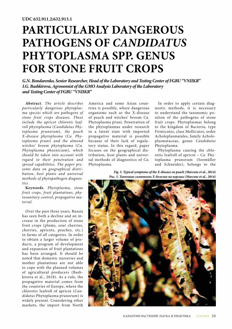
4| 26| 2018 23КАРАНТИН РАСТЕНИЙ. НАУКА И ПРАКТИКА
Abstract. The article describes particularly dangerous phytoplas-ma species which are pathogens of stone fruit crops diseases. These include the apricot chlorotic leaf-roll phytoplasma (Candidatus Phy-toplasma prunorum), the peach X-disease phytoplasma (Ca. Phy-toplasma pruni) and the almond witches’ broom phytoplasma (Ca. Phytoplasma phoenicium), which should be taken into account with regard to their penetration and spread capabilities. The paper pre-sents data on geographical distri-bution, host plants and universal methods of phytopathogen diagnos-tics.
Keywords. Phytoplasma, stone fruit crops, fruit plantations, phy-tosanitary control, propagative ma-terial.
Over the past three years, Russia has seen both a decline and an in-crease in the production of stone fruit crops (plums, sour cherries, cherries, apricots, peaches, etc.) in farms of all categories. In order to obtain a larger volume of pro-ducts, a program of development and expansion of fruit plantations has been arranged. It should be noted that domestic nurseries and mother plantations are not able to cope with the planned volumes of agricultural producers (Bash-kirova et al., 2018). As a rule, the propagative material comes from the countries of Europe, where the chlorotic leafroll of apricot (Can-didatus Phytoplasma prunorum) is widely present. Considering other markets, the import from North
America and some Asian coun-tries is possible, where dangerous organisms such as the X-disease of peach and witches’ broom Ca. Phyto plasma pruni. Penetration of the phytoplasmas under research in a latent state with imported propagative material is possible because of their lack of regula-tory status. In this regard, paper focuses on the geographical dis-tribution, host plants and univer-sal methods of diagnostics of Ca. Phytoplasma.
In order to apply certain diag-nostic methods, it is necessary to understand the taxonomic po-sition of the pathogens of stone fruit crops. Phytoplasmas belong to the kingdom of Bacteria, type Firmicutes, class Mollicutes, order Acholeplasmatales, family Achole-plasmataceae, genus Candidatus Phytoplasma.
Phytoplasma causing the chlo-rotic leafroll of apricot – Ca. Phy-toplasma prunorum (Seemüller and Schneider), belongs to the
UDC 632.911.2:632.913.1
PARTICULARLY DANGEROUS PATHOGENS OF CANDIDATUS PHYTOPLASMA SPP. GENUS FOR STONE FRUIT CROPSG.N. Bondarenko, Senior Researcher, Head of the Laboratory and Testing Center of FGBU “VNIIKR”I.G. Bashkirova, Agronomist of the GMO Analysis Laboratory of the Laboratory and Testing Center of FGBU “VNIIKR”
Fig. 5. Typical symptoms of the X-disease on peach (Marcone et al., 2014)Рис. 5. Типичные симптомы X-болезни на персике (Marcone et al., 2014)
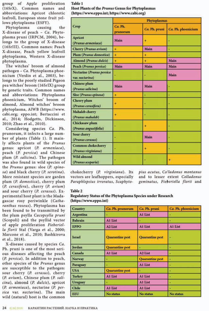
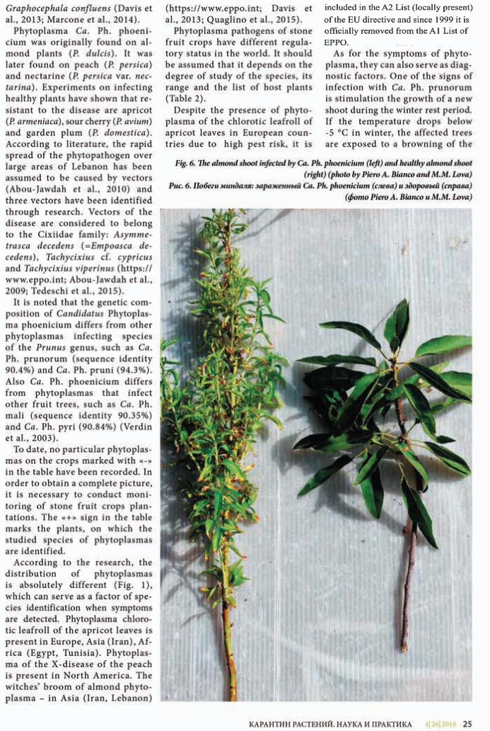
included in the A2 List (locally present) of the EU directive and since 1999 it is officially removed from the A1 List of EPPO.
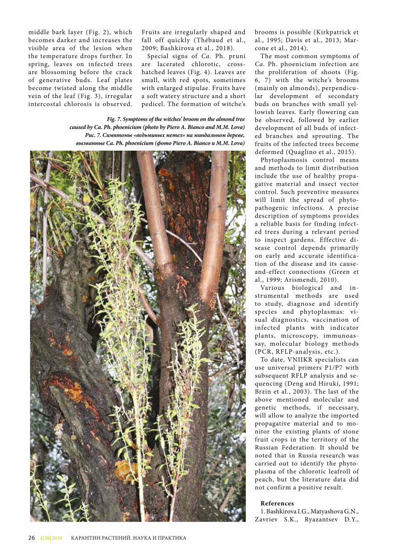
4| 26| 201826 КАРАНТИН РАСТЕНИЙ. НАУКА И ПРАКТИКА
middle bark layer (Fig. 2), which becomes darker and increases the visible area of the lesion when the temperature drops further. In spring, leaves on infected trees are blossoming before the crack of generative buds. Leaf plates become twisted along the middle vein of the leaf (Fig. 3), irregular intercostal chlorosis is observed.
Fruits are irregularly shaped and fall off quickly (Thébaud et al., 2009; Bashkirova et al., 2018).
Special signs of Ca. Ph. pruni are lacerated chlorotic, cross-hatched leaves (Fig. 4). Leaves are small, with red spots, sometimes with enlarged stipulae. Fruits have a soft watery structure and a short pedicel. The formation of witchе’s
brooms is possible (Kirkpatrick et al., 1995; Davis et al., 2013; Mar-cone et al., 2014).
The most common symptoms of Ca. Ph. phoenicium infection are the proliferation of shoots (Fig. 6, 7) with the witchе’s brooms (mainly on almonds), perpendicu-lar development of secondary buds on branches with small yel-lowish leaves. Early flowering can be observed, followed by earlier development of all buds of infect-ed branches and sprouting. The fruits of the infected trees become deformed (Quaglino et al., 2015).
Phytoplasmosis control means and methods to limit distribution include the use of healthy propa-gative material and insect vector control. Such preventive measures will limit the spread of phyto-pathogenic infections. A precise description of symptoms provides a reliable basis for finding infect-ed trees during a relevant period to inspect gardens. Effective di-sease control depends primarily on early and accurate identifica-tion of the disease and its cause-and-effect connections (Green et al., 1999; Arismendi, 2010).
Various biological and in-strumental methods are used to study, diagnose and identify species and phytoplasmas: vi-sual diag nostics, vaccination of infected plants with indicator plants, microscopy, immunoas-say, molecular biology methods (PCR, RFLP-analysis, etc.).
To date, VNIIKR specialists can use universal primers P1/P7 with subsequent RFLP analysis and se-quencing (Deng and Hiruki, 1991; Brzin et al., 2003). The last of the above mentioned molecular and genetic methods, if necessary, will allow to analyze the imported propagative material and to mo-nitor the existing plants of stone fruit crops in the territory of the Russian Federation. It should be noted that in Russia research was carried out to identify the phyto-plasma of the chlorotic leafroll of peach, but the literature data did not confirm a positive result.
References1. Bashkirova I.G., Matyashova G.N.,
Zavriev S.K., Ryazantsev D.Y.,
Fig. 7. Symptoms of the witchеs’ broom on the almond treecaused by Ca. Ph. phoenicium (photo by Piero A. Bianco and M.M. Lova)
Рис. 7. Симптомы «ведьминых метел» на миндальном дереве,вызванные Ca. Ph. phoenicium (фото Piero A. Bianco и M.M. Lova)
4| 26| 2018 27КАРАНТИН РАСТЕНИЙ. НАУКА И ПРАКТИКА
Shneyder Y.A. Testing systems for the detection of apple and pear phytoplasmas // Plant protection and quarantine, 2018. Issue 7. P. 40-41.
2. European and Mediterranean Plant Protection Organization: Official site [Electronic resource] / European and Mediterranean Plant Protection Organization, 2018. Access mode: https://www.eppo.int (access date: 21.09.2018).
3. Centre for Agriculture and Bioscience International [Elec-tronic resource] / CABI, 2018. Ac-cess mode: https://www.cabi.org (access date: 18.10.2018).
4. Abou-Jawdah Y., Abou-Fakhr E., Sobn H., Lova M.M., Vercesi A., Bianco P.A. Almond witches’ broom phytoplasma (‘Candidatus Phytoplasma phoenicium’): a real threat to almond, peach and nec-tarine // 21st International Con-ference on Virus and other Graft Transmissible Diseases of Fruit Crops. Julius-Kühn-Archive, 2010. Vol. 427. P. 418-420.
5. Abou-Jawdah Y., Sobh H., Ak-kary M. Blackwell Publishing Ltd First report of Almond witches’ broom phytoplasma (‘Candidatus Phytoplasma phoenicium’) caus-ing a severe disease on nectarine and peach trees in Lebanon // Bul-letin OEPP/EPPO Bulletin, 2009. Vol. 39. P. 94-98.
6. Arismendi N., Andrade N., Riegel R., Carrillo R. Presence of a phytoplasma associated with witches’ broom diseases in Ugni molinae Turcz. and Gaultheria phillyreifolia (Pers.) // Sleumer determined by DAPI, PCR and DNA sequencing, 2010. Vol. 70 (1). P. 26-33.
7. Bashkirova I.G., Matya-shova G.N., Gins M.S. Detection and Identification of Phytoplas-mas from the Apple Proliferation Group on Fruit Crop // Russian Ag-ricultural Sciences, 2018. Vol. 44. P. 326-330.
8. Bertaccini A. , Duduk B. , Paltr inieri S. , Contaldo N. Phytoplasmas and Phytoplas-ma Dise ases a Severe Threat to Agri culture // American Journal of Plant Sciences, 2014. Vol. 5. № 12. P. 1763-1788.
9. Brzin J., Ermacora P., Osler R., Loi N., Ravnikar M., Petrovic N.
Detection of apple proliferation by ELISA and PCR in growing and dormant apple trees // Plant Diseases Prot., 2003. Vol. 110. P. 476-483.
10. Davis R.E., Zhao Y., Dal-ly E.L., Lee I-M., Jomantiene R., Douglas S.M. ‘Candidatus Phyto-plasma pruni’, a novel taxon as-sociated with X-disease of stone fruits, Prunus spp.: multilocus characterization based on 16S rRNA, secY, and ribosomal pro-tein genes // International Jour-nal of Systematic and Evolutiona-ry Microbiology, 2013. Vol. 63. P. 766-776.
11. Deng S., Hiruki C. Amplifi-cation of 16S rRNA genes from cul-turable and nonculturable molli-cutes // Microbiological Methods, 1991. Vol. 14. P. 53-61.
12. Green M.J., Thompson D.A., MacKenzie D.J. East and effi-cient DNA extraction from woody plants for the detection of phyto-plasmas by polymerase chain reac-tion // Plant Disease, 1999. Vol. 83. P. 482-485.
13. Hodgetts J., Dickinson M. Phytoplasma Phylogeny and De-tection Based on Genes other than 16S rRNA // Phytoplasmas: Ge-nomes, Plant Hosts and Vectors (eds P.G. Weintraub and P. Jones). CABI, 2010. P. 93-113.
14. IRPCM Phytoplasma/Spiro-plasma Working Team “Phyto-plasma Taxonomy Group ‘Candi-datus Phytoplasma’, a taxon for the wall-less, non-helical prokaryotes that colonize plant phloem and insects” // International Jour-nal of Systematic and Evolutio-nary Microbiology, 2004. Vol. 54. P. 1243-1255.
15. Kirkpatrick B.C., Uyemoto J.K., Purcell A.H. X-disease // “Com-pendium of Stone Fruit Disease” (J.M. Ogawa, E.I. Zehr, G.W. Bird, D.F. Richie, K. Uriu, J.K. Uyemoto, eds.). APS Press, St. Paul, MN, USA, 128 pp., 1995. P. 57-58.
16. Marcone C., Guerra L.J., Uyemoto J.K. Phytoplasmal di-seases of peach and associated phytoplasma taxa // Journal of Plant Pathology, 2014. Vol. 96 (1). P. 15-28.
17. Marcone C., Jarausch B., Ja-rausch W. Candidatus Phytoplas-ma prunorum, the causal agent of
European stone fruit yellows: an ove r v i e w / / Jou r n a l o f P l ant Pat h o l o g y, 2 0 1 0 . Vol . 9 2 ( 1 ) . P. 1 9 - 3 4 .
18. Quaglino F., Kube M., Jawhari M., Abou-Jawdah Y., Siewert C., Choueiri E., Sobh H., Casati P., Tedeschi R., Lova M.M., Alma A., Bianco P.A. ‘Candidatus Phytoplas-ma phoenicium’ asso ciated with almond witches’ broom disease: from draft genome to genetic di-versity among strain populations // BMC Microbiology, 2015. Vol. 15. P. 148.
19. Tedeschi R., Picciau L., Quagli-no F., Abou‐Jawdah Y., Lova M.M., Jawhari M., Casati P., Cominetti A., Choueiri E., Abdul-Nour H., Bian-co P.A., Alma A. A cixiid survey for natural potential vectors of ‘Can-didatus Phytoplasma phoenicium’ in Lebanon and preliminary trans-mission trials // Annals of Ap-plied Biology, 2015. Vol. 166 (3). P. 372-388.
20. Thébaud G., Yvon M., Alary R., Sauvion N., and Labonne G. Effi-cient transmission of ‘Candidatus Phytoplasma prunorum’ is delayed by eight months due to a long la-tency in its host-alternating vector // Phytopathology, 2009. Vol. 99. P. 265-273.
21. Varga K., Kölber M., Martini M., Pondrelli M., Ember I., Tökes G., Lazar J., Mikulas J., Szendrey G., Schweigert A., Bertaccini A. . Phyto-plasma identi�cation in Hungarian grapevines by two nested-PCR sys-tems // Proceedings of the 13th Meeting of ICVG. Adelaide, 2000. P. 113-115.
22. Verdin E., Salar P., Danet J.-L., Choueiri E., Jreijiri F., Zammer S.E., Gélie B., Bové J.M., Garnier M. ‘Candidatus Phytoplasma phoeni-cium’ sp. nov., a novel phytoplasma associated with an emerging lethal disease of almond trees in Leba-non and Iran // International Jour-nal of Systematic and Evolution-ary Microbiology, 2003. Vol. 53 (3). P. 833-838.
23. Zhao Y., Wei W., Davis R.E., Lee I.-M. Recent Advances in 16S rRNA Gene-based Phytoplasma Differentiation, Classification and Taxonomy // Phytoplasmas: Ge-nomes, Plant Hosts and Vectors (eds P.G. Weintraub and P. Jones). CABI, 2010. P. 64-92.
4| 26| 201828 КАРАНТИН РАСТЕНИЙ. НАУКА И ПРАКТИКА
Аннотация. Российская Федерация экспортирует зерно зернобобовых культур в страны, в фитосанитар-ных требованиях которых часто прописано обязательное отсут-ствие возбудителей бактериаль-ных болезней зернобобовых культур. В данной статье кратко рассмотрены биолого-морфологические особенности возбудителей основных бактериаль-ных болезней зернобобовых культур, растения-хозяева, симптомы, ущерб и методы диагностики возбудителей.
Ключевые слова. Curtobacterium �accumfaciens pv. �accumfaciens, Pseudo-monas savastanoi pv. phaseolicola, Xantho-monas axonopodis pv. phaseoli, зерно-бобовые культуры, бактерия, возбуди-тель болезни.
В последние годы внимание к зер-нобобовым культурам возросло в связи с необходимостью ликвидации белкового дефицита в питании и корм-лении сельскохозяйственных живот-ных. Зерно зернобобовых культур – высокопитательный и концентриро-ванный продукт и корм для сельско-хозяйственных животных. Основная часть запасаемого зернобобовыми белка создается за счет азота воздуха в результате симбиотической деятельно-сти клубеньковых бактерий.
Необходимость всестороннего ана-лиза возбудителей бактериальных бо-лезней зернобобовых культур, вклю-чающего в себя анализ вероятности их проникновения на территорию Российской Федерации с различными видами продукции, анализ вероят-ности акклиматизации в конкретных регионах России с учетом специфики абиотических и биотических факто-ров, анализ вероятности разносторон-него влияния на экономику России, анализ состояния окружающей среды
и анализ социальной обстановки на территории страны определяет необ-ходимость разработки современных методов диагностики бактериозов.
Контроль распространения вред-ных организмов играет важную роль в охране сельского хозяйства и окру-жающей среды. Актуальность данной темы в том, что РФ экспортирует зерно зернобобовых в разные страны, ко-торые предъявляют фитосанитарные требования, в частности, отсутствие возбудителей бактериальных болезней в зерне зернобобовых на семенные цели.
Основные бактериальные болезни зернобобовых вызывают возбудите-ли ржаво-бурой пятнистости листьев фасоли Curtobacterium �accumfaciens pv. �accumfaciens (рис. 1), угловатой бактериальной пятнистости фасоли
Pseudomonas savastanoi pv. phaseolicola (рис. 2) и бактериального ожога фасо-ли Xanthomonas axonopodis pv. phaseoli (рис. 3).
Данные патогены вызывают сни-жение урожая и качества зерна зер-нобобовых культур и являются очень важными с точки зрения экономики. Контроль бактериозов возможен при использовании свободных от возбуди-телей болезней семян и соблюдении се-вооборота. Эффективные химические средства защиты в настоящее время отсутствуют. Интродукция фитопато-генных бактерий в новые регионы мо-жет произойти с семенным материа-лом (главным образом фасоли и сои), зараженным бактериальной инфекци-ей в латентном состоянии, что может вызвать серьезные проблемы при про-изводстве зернобобовых культур.
УДК 632.3.01/.08
БАКТЕРИОЗЫ – ВОЗБУДИТЕЛИ БОЛЕЗНЕЙ ЗЕРНОБОБОВЫХ КУЛЬТУР И РАЗРАБОТКА МЕТОДОВ ИХ ДИАГНОСТИКИЕ.В. Каримова, старший научный сотрудник НМОФ ФГБУ «ВНИИКР»И.М. Игнатьева, научный сотрудник лаборатории бактериологии ИЛЦ ФГБУ «ВНИИКР»
Рис. 1. Симптомы C. �accumfaciens pv. �accumfaciens (фото Harveson et al., 2015)Fig. 1. Symptoms of C. �accumfaciens pv. �accumfaciens (photo by Harveson et al., 2015)
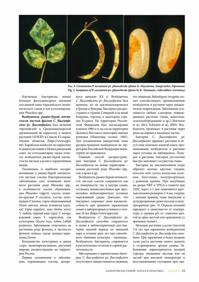
4| 26| 2018 29КАРАНТИН РАСТЕНИЙ. НАУКА И ПРАКТИКА
Изучаемые бактериозы имеют большое фитосанитарное значение для южной зоны Евразийского эконо-мического союза и зон культивирова-ния Phasoleus spp.
Возбудитель ржаво-бурой пятни-стости листьев фасоли C. �accumfa-ciens pv. �accumfaciens был включен Европейской и Средиземноморской организацией по карантину и защите растений (ЕОКЗР) в Список А2 каран-тинных объектов (https://www.eppo.int). Карибская комиссия по карантину и защите растений и Межафриканский совет по фитосанитарии также отно-сят возбудителя ржаво-бурой пятни-стости листьев фасоли к карантинным видам.
Основными и наиболее воспри-имчивыми к ржаво-бурой пятнисто-сти листьев фасоли (бактериальному заболеванию сои) хозяевами явля-ются растения рода Phaseolus spp., в особенности фасоль обыкновен-ная Phaseolus vulgaris, фасоль огнен-но-красная P. coccineus, фасоль луно-видная P. lunatus, горох обыкновенный Pisum sativum, вигна угловатая (адзу-ки) Vigna angularis, маш (бобы мунг) V. radiata, черный маш (урд) V. mungo, коровий горох V. unguiculata, соя культурная Glycine max, лобия Lablab purpureus. Заболевание отмечалось на растениях рода Ipomoea, в частности Ipomoea violacea, также ложная мари-хуана Zornia.
Большинство культурных и диких форм вышеперечисленных растений широко распространено на террито-рии России.
Первые упоминания о заболева-нии, поражающем фасоль, датиру-
ются началом XX в. Возбудитель C. �accumfaciens pv. �accumfaciens был выявлен, но не акклиматизировался в Греции и Венгрии. Бактериоз распро-странен в странах Северной и Южной Америки, Европы, в некоторых стра-нах Африки. На территории Россий-ской Федерации был зафиксирован в начале 1980-х гг. на сое на территории Дальнего Востока и некоторых южных регионах (Никитина, Щелко, 1982). Для установления конкретной зоны распространения возбудителя на тер-ритории Российской Федерации мони-торинг не проводился.
Главный способ распростране-ния бактерии C. �accumfaciens pv. �accumfaciens на новые территории – семена растений рода Phaseolus spp., сои, гороха и др.
Возбудитель ржаво-бурой пятнисто-сти листьев фасоли сохраняется как на поверхности, так и внутри семян, оставаясь жизнеспособным при чрез-вычайно неблагоприятных условиях окружающей среды. Доказано, что бактериоз сохраняет свою жизнеспо-собность при хранении зараженных семян в лабораторных условиях в тече-ние 24 лет (https://www.eppo.int).
Возбудитель C. �accumfaciens pv. �accumfaciens способен сохраняться в почве в неблагоприятный для бак-терий зимний период по меньшей мере в течение двух лет при севообо-роте «бобовые культуры – пшеница». Возбудитель бактериоза сохраняется в растительных остатках и сорной рас-тительности.
Информация о переносчиках бакте-рии C. �accumfaciens pv. �accumfaciens отсутствует, однако имеются сведения,
что нематода Meloidogyne incognita мо-жет способствовать проникновению возбудителя в растение через механи-ческие повреждения. Заболевание уси-ливается любым фактором, повреж-дающим растение (люди, животные, сельхозоборудование и др.) (Harveson et al., 2011; Schwartz et al., 2005). Воз-будитель проникает в растение через раны на корнях и наземных частях.
Бактерия C. �accumfaciens pv. �accumfaciens заражает растение в от-сутствие капельно-жидкой влаги, про-никновение возбудителя в растение через устьица не наблюдалось. Попа-дая в растение, бактерия достаточно быстро заполняет сосудистую ткань.
Бактерия на мясопептонном агаре образует желтые, круглые, гладкие, плоские или слегка выпуклые коло-нии, блестящие, полупрозрачные, с ровными краями. При инкубации на средах NBY и YPGA в темноте при 23 ºС через 2-3 дня появляются круг-лые колонии размером 2-4 мм, гладкие, с целыми краями, чаще выпуклые и полупрозрачные, реже плоские и полу-прозрачные (рис. 4). Окраска колоний варьирует в зависимости от темпера-туры и уровня рН от сливочно-жел-той до ярко-желтой или оранжевой, со временем темнеет.
Молодые растения фасоли (высотой 5-8 см) при заражении возбудителем C. �accumfaciens pv. �accumfaciens поги-бают. При заражении в более поздние фазы роста растение может выжить и сформировать зрелые семена. За-болевание характеризуется потерей тургора, увяданием листьев или их частей при высокой температуре и восстановлением состояния при сни-
Рис. 2. Симптомы P. savastanoi pv. phaseolicola (фото К. Науманна, Ашерслебен, Германия)Fig. 2. Symptoms of P. savastanoi pv. phaseolicola (photo by K. Naumann, Ashersleben, Germany)
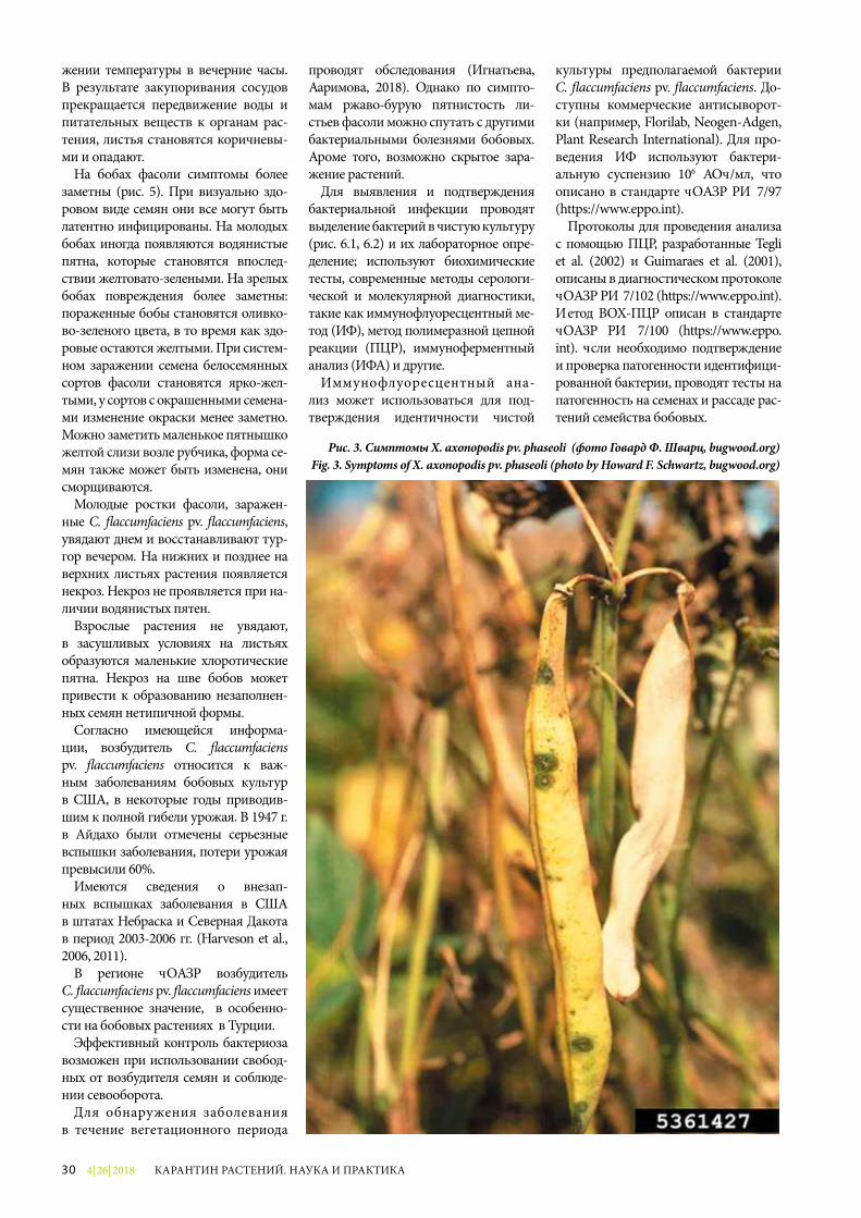
4| 26| 201830 КАРАНТИН РАСТЕНИЙ. НАУКА И ПРАКТИКА
жении температуры в вечерние часы. В результате закупоривания сосудов прекращается передвижение воды и питательных веществ к органам рас-тения, листья становятся коричневы-ми и опадают.
На бобах фасоли симптомы более заметны (рис. 5). При визуально здо-ровом виде семян они все могут быть латентно инфицированы. На молодых бобах иногда появляются водянистые пятна, которые становятся впослед-ствии желтовато-зелеными. На зрелых бобах повреждения более заметны: пораженные бобы становятся оливко-во-зеленого цвета, в то время как здо-ровые остаются желтыми. При систем-ном заражении семена белосемянных сортов фасоли становятся ярко-жел-тыми, у сортов с окрашенными семена-ми изменение окраски менее заметно. Можно заметить маленькое пятнышко желтой слизи возле рубчика, форма се-мян также может быть изменена, они сморщиваются.
Молодые ростки фасоли, заражен-ные C. �accumfaciens pv. �accumfaciens, увядают днем и восстанавливают тур-гор вечером. На нижних и позднее на верхних листьях растения появляется некроз. Некроз не проявляется при на-личии водянистых пятен.
Взрослые растения не увядают, в засушливых условиях на листьях образуются маленькие хлоротические пятна. Некроз на шве бобов может привести к образованию незаполнен-ных семян нетипичной формы.
Согласно имеющейся информа-ции, возбудитель C. �accumfaciens pv. �accumfaciens относится к важ-ным заболеваниям бобовых культур в США, в некоторые годы приводив-шим к полной гибели урожая. В 1947 г. в Айдахо были отмечены серьезные вспышки заболевания, потери урожая превысили 60%.
Имеются сведения о внезап-ных вспышках заболевания в США в штатах Небраска и Северная Дакота в период 2003-2006 гг. (Harveson et al., 2006, 2011).
В регионе ЕОКЗР возбудитель C. �accumfaciens pv. �accumfaciens имеет существенное значение, в особенно-сти на бобовых растениях в Турции.
Эффективный контроль бактериоза возможен при использовании свобод-ных от возбудителя семян и соблюде-нии севооборота.
Для обнаружения заболевания в течение вегетационного периода
проводят обследования (Игнатьева, Каримова, 2018). Однако по симпто-мам ржаво-бурую пятнистость ли-стьев фасоли можно спутать с другими бактериальными болезнями бобовых. Кроме того, возможно скрытое зара-жение растений.
Для выявления и подтверждения бактериальной инфекции проводят выделение бактерий в чистую культуру (рис. 6.1, 6.2) и их лабораторное опре-деление; используют биохимические тесты, современные методы серологи-ческой и молекулярной диагностики, такие как иммунофлуоресцентный ме-тод (ИФ), метод полимеразной цепной реакции (ПЦР), иммуноферментный анализ (ИФА) и другие.
Иммунофлуоресцентный ана-лиз может использоваться для под-тверждения идентичности чистой
культуры предполагаемой бактерии C. �accumfaciens pv. �accumfaciens. До-ступны коммерческие антисыворот-ки (например, Florilab, Neogen-Adgen, Plant Research International). Для про-ведения ИФ используют бактери-альную суспензию 106 КОЕ/мл, что описано в стандарте ЕОКЗР РМ 7/97 (https://www.eppo.int).
Протоколы для проведения анализа с помощью ПЦР, разработанные Tegli et al. (2002) и Guimaraes et al. (2001), описаны в диагностическом протоколе ЕОКЗР РМ 7/102 (https://www.eppo.int). Метод ВОХ-ПЦР описан в стандарте ЕОКЗР РМ 7/100 (https://www.eppo.int). Если необходимо подтверждение и проверка патогенности идентифици-рованной бактерии, проводят тесты на патогенность на семенах и рассаде рас-тений семейства бобовых.
Рис. 3. Симптомы X. axonopodis pv. phaseoli (фото Говард Ф. Шварц, bugwood.org)Fig. 3. Symptoms of X. axonopodis pv. phaseoli (photo by Howard F. Schwartz, bugwood.org)
4| 26| 2018 31КАРАНТИН РАСТЕНИЙ. НАУКА И ПРАКТИКА
Бактериальный ожог фасоли X. axonopodis pv. phaseoli впервые был описан в 1893 г., возбудитель заболевания был выделен в чистую культуру и идентифицирован в 1897 г. Смитом (Zaumeyer, 1930).
X. axonopodis pv. phaseoli можно раз-делить на три группы. X. axonopodis pv. phaseoli var. fuscans выделяет диффузный коричневый пигмент в чистой культуре, выделение кото-рого можно увеличить тирозином (Goodwin and Sopher, 1994). Данный пигмент является результатом секре-ции и окисления гомогенистической кислоты в катаболическом пути ти-розина. X. axonopodis pv. phaseoli var. indica выделяет диффузный корич-невый пигмент в чистой культуре, выделение которого нельзя увели-чить тирозином (Bradbury, 1986). X. axonopodis pv. phaseoli не выделяет коричневого пигмента в культуре.
Данный бактериоз поражает мно-гие виды Phaseolus: горох (Pisum sativum), люпин многолистный (Lupinus polyphyllus), гиацинто-вые бобы (Lab lab purpureus). Расы X. axonopodis pv. phaseoli были за-регистрированы на вигне зонтич-ной (Vigna umbellata, P. calcaratus), вигне борцелистной (Vigna aconiti-folia), маше, или бобах мунг (V. radiata, P. aureus), урде, или чер-ном маше (Vigna mungo) (Bradbury, 1986). Opio et al. (1996) обнаружили восемь рас X. axonopodis pv. phaseoli на P. acuti folius.
Возбудитель бактериального ожо-га фасоли X. axonopodis pv. phaseoli распространен по всему миру – в большинстве районов, где выращи-вают зернобобовые культуры, за ис-ключением засушливых тропических районов.
О возбудителе заболевания сооб-щалось из некоторых штатов США, многих стран Северной, Централь-ной и Латинской Америки, Азии и тихоокеанских островов.
Возбудитель бактериального ожога фасоли X. axonopodis pv. phaseoli яв-ляется карантинным вредным орга-низмом и входит в Список А2 ЕОКЗР, встречается локально или широко в ряде стран, производящих зерно-бобовые культуры.
X. axonopodis pv. phaseoli сохраняет-ся на поверхности и внутри семян, на зараженных растительных остатках, поверхности растений-хозяев и дру-гих растений (Saettler, 1991).
Семена являются основным путем распространения возбудителя при заражении новых регионов и играют большую роль в сохранении жизне-способности X. axonopodis pv. phaseoli.
Заражение цветочных почек и моло-дых стручков может привести к пере-носу X. axonopodis pv. phaseoli через со-судистую систему к формирующимся семенам, что вызывает внутреннюю инфекцию (Aggour et al., 1989). Расте-ния, выросшие из зараженных семян, имеют повреждения семядолей, узлов и первичных листьев.
Возбудитель X. axonopodis pv. phaseoli развивается на поверхности расте-ний-хозяев и других растений (Cafati, Saettler, 1980). Рост возбудителя на поверхности растений-хозяев способ-ствует развитию популяции, способ-ной при благоприятных условиях вы-зывать заражение растений. Бактерии проникают в растения через устьица или поранения, затем перемещаются в межклеточные пространства, при-водя к постепенному растворению сердцевины стебля. Иногда бактерия из паренхимной ткани проникает в проводящую систему растения, где продолжает свое развитие в форме трахеобактериоза, вызывая систем-ную инфекцию.
Признаки болезни проявляются на всех надземных органах растения.
При заражении молодого боба се-мена сгнивают, сморщиваются или деформируются. При проникновении бактерий через ножку на рубчике се-мени можно наблюдать изменение окраски, появление желтоватых или коричневых пятен на семенной обо-лочке. Иногда заражение семян может быть бессимптомным.
При посеве семян, зараженных воз-будителем бактериального ожога фа-соли X. axonopodis pv. phaseoli, часть проростков погибает. Всходы имеют частично или полностью разрушен-ную точку роста, поэтому семядоли остаются склеенными и растение по-гибает. Данный симптом известен как «змеиная головка».
При заражении в поле на настоя-щих листьях образуются точковид-ные, разрастающиеся до крупных водянистые пятна красно-бурого цвета с узким желтым ободком. По-вреждения темнеют, листья быстро некротизируются и опадают. Рас-тения, пораженные возбудителем X. axonopodis pv. phaseoli, выглядят обожженными, что является харак-
терным отличительным признаком бактериального ожога фасоли.
На бобах инфекция проявляется на любом участке в виде малень-ких водянистых пятен, которые постепенно увеличиваются. Иногда пятна окружены четко выраженной узкой красно-коричневой или кир-пично-красной каймой.
Бактериальный ожог фасоли X. axo nopodis pv. phaseoli вызывает значительные потери урожая зерно-бобовых в странах умеренного, тро-пического и субтропического кли-мата. Широкая распространенность возбудителя, способность вызы-вать значительные потери урожая, трудности, связанные с борьбой, распространение семенами и др. факторы способствовали тому, что X. axonopodis pv. phaseoli считается одним из наиболее важных патоге-нов зернобобовых культур во всем мире (Irigoyen, Garbagnoli, 1997).
Большие потери урожая наблю-даются при раннем заражении растений. Это связано с преждев-ременной дефолиацией, которая уменьшает доступную область фо-тосинтеза, препятствует трансло-кации и снижает количество и раз-мер семян. В результате поражения семян и бобов снижается качество продукции.
На степень вредоносности так-же влияет фаза роста культуры в момент заражения бактерио-зом: чем раньше произошло зара-жение, тем больших потерь уро-жая можно ожидать.
Бактерия X. axonopodis pv. phaseoli – грамотрицательная аэробная под-вижная палочка размером 0,4-0,9 х 0,6-2,6 мкм с одним полярным жгу-тиком. В культуре на сложной пи-тательной среде или среде, содер-жащей тирозин, X. axonopodis pv. phaseoli var. fuscans выделяет диф-фузный коричневый пигмент.
Для обнаружения заболевания в течение вегетационного периода проводят обследования. Однако по симптомам бактериальный ожог фасоли можно спутать с другими бактериальными заболеваниями бобовых. Кроме того, возможно скрытое заражение растений.
Для выявления и подтверждения бактериальной инфекции прово-дится выделение бактерий в чистую культуру и их лабораторное опреде-ление (Игнатьева, Каримова, 2018).
4| 26| 201832 КАРАНТИН РАСТЕНИЙ. НАУКА И ПРАКТИКА
Для выявления и идентификации X. axonopodis pv. phaseoli используют биохимические тесты, современные методы серологической и молеку-лярной диагностики, такие как им-мунофлуоресцентный метод (ИФ), иммуноферментный анализ (ИФА), метод полимеразной цепной реакции (ПЦР) и другие.
Реакцию непрямой иммунофлуорес-ценции проводят с коммерческим на-бором фирмы Loewe (Германия).
ИФА ((DAS)-ELISA) проводят с помо щью коммерческих наборов, выпускаемых фирмами Loewe Biochemica GmbH (Германия), ADGEN Phytodiagnostics, Neogen Eu-rope Ltd. (Великобритания), согласно инструк циям производителей.
Бактерия Pseudomonas savastanoi pv. phaseolicola – возбудитель углова-той бактериальной пятнистости фасо-ли – является важным бактериальным патогеном фасоли обыкновенной.
Основными и наиболее восприим-чивыми к Pseudomonas savastanoi pv. phaseolicola растениями-хозяевами яв-ляются растения рода Phaseolus spp., в особенности фасоль обыкновенная Phaseolus vulgaris.
По данным EPPO Global Database, возбудитель Pseudomonas savastanoi pv. phaseolicola включен в перечни ка-рантинных вредных организмов в та-ких странах, как Бразилия, Парагвай, Уругвай, Израиль и Бахрейн, а в Чили объект включен в список ограниченно распространенных карантинных объ-ектов на территории государства.
Внешние признаки заболевания подобны симптомам бактериального ожога фасоли Xanthomonas axonopodis pv. phaseoli и сильно варьируют в зави-симости от условий среды. При посеве зараженных семян бактериоз развива-ется системно. Иногда инфекционный процесс развивается очень быстро и выражается в хлорозе и увядании нижних листьев растений, приводя-щих к их преждевременному усыха-нию. Листья пораженных растений желтоватые, верхние листья дефор-мированные и мозаичные. Такие рас-тения увядают в дневное время суток, но независимо от этого могут продол-жить свое развитие и успевают образо-вать семена с зараженным зародышем.
Локальное развитие заболевания проявляется в виде единичных бурых пятен на семядолях или многочис-ленных мелких (1-4 мм) маслянистых пятен на настоящих листьях, которые
впоследствии некротизируются. Наи-более характерный признак бактерио-за фасоли – формирование широкого (1-3 см) бледно-зеленого ореола во-круг пятна, появляющегося в резуль-тате выделения патогеном токсинов. Ореол всегда предшествует появле-нию бактериальной пятнистости. На стеблях образуются продольные мас-лянистые полосы с красно-бурой кай-мой, которые у молодых растений ча-сто растрескиваются. На бобах обра-зуется много мелких округлых пятен, по периферии которых со временем появляется бурая кайма.
Семена недоразвитые, желтова-тые или кремовые, с низкой всхоже-стью. На семенах могут появляться характерные мелкие бесформенные язвочки, подобные повреждениям насекомых. Из всех пораженных ча-стей растения вытекает белый бакте-риальный экссудат.
Бактериальная инфекция сохраня-ется в семенах и пораженных расти-тельных остатках до их разложения. В почве погибает быстро. В расте-ние может проникать через устьица и ранки.
Импорт зерна бобовых на семен-ные цели – основной путь распро-странения заболевания. Бактерии локализуются преимущественно между семенной кожурой и семя-долями, а также на поверхности се-мян. В сильно пораженных семенах бактерии иногда обнаруживаются в тканях семядолей.
Было описано множество процедур диагностики Pseudomonas savastanoi pv. phaseolicola, но ни одна из них не явля-ется общепринятой.
Для выявления и подтверждения бактериальной инфекции проводится выделение бактерий в чистую куль-туру и их лабораторное определение. После 4-5 дней инкубации на среде МТ колонии Pseudomonas savastanoi pv. phaseolicola беловато-кремовые, плоские, округлые, 4,5-5 мм в диаме-тре. На среде MSP колонии округлые, светло-желтые, куполообразные (ле-ван-положительные), блестящие, про-дуцируют флуоресцирующий пигмент.
Перспективным направлением идентификации патогена является анализ на основе ПЦР. Быстрый и высокоспецифичный метод может быть использован как для выявле-ния возбудителя в экстракте семян (с использованием предваритель-ного выделения ДНК), так и для
прямого анализа бактериальных смывов и идентификации суспензий чистых культур.
Исследование, основанное на ана-лизе ПЦР, способствует проведению быстрой и достоверной диагностики Pseudomonas savastanoi pv. phaseolicola.
Сотрудники ФГБУ «ВНИИКР» разрабатывают современные высо-кочувствительные методы выявле-ния и идентификации возбудителей ржаво-бурой пятнистости листьев фасоли Curtobacterium �accumfaciens pv. �accumfaciens, угловатой бак-териальной пятнистости фасоли Pseudomonas savastanoi pv. phaseolicola и бактериального ожога фасоли Xanthomonas axonopodis pv. phaseoli, что позволит проводить лаборатор-ную экспертизу подкарантинного материала при импорте его в РФ (Иг-натьева, Каримова, 2018). При про-ведении лабораторных валидацион-ных испытаний используют штаммы из бактериальной коллекции ФГБУ «ВНИИКР», насчитывающей более трехсот единиц (рис. 7.1, 7.2).
Существенное влияние на достовер-ность результатов диагностики ока-зывает выбор праймеров и оптими-зация состава ПЦР-смеси. Различные наборы для выделения ДНК харак-теризуются определенным уровнем чувствительности. Диагностические тест-системы различаются по степени взаимодействия праймеров с реакци-онной смесью. Для корректной работы диагностического набора необходимо проводить валидацию тест-системы для определения аналитической чув-ствительности (рис. 8), аналитической специфичности, селективности, по-вторяемости и воспроизводимости. Важно также, чтобы тест-система была удобной для проведения стандартной лабораторной экспертизы, а приготов-ление реакционной смеси занимало как можно меньше времени.
Литература1. Aggour A.R., Coyne D.P., Vidaver A.K.,
Eskridge K.M. Transmission of the common blight pathogen in bean seed // Journal of the American Society of Horticultural Sciences, 1989. № 114. Р. 1002-1008.
2. Bradbury J.F. Guide to Plant Pathogenic Bacteria. Wallingford, UK: CAB International, 1986.
3. CABI Author(s) Sun Suli; Zhi Ye; Zhu Zhen Dong; Jin Jing; Duan Can Xing; Wu Xiao Fei; Wang Xiao Ming.
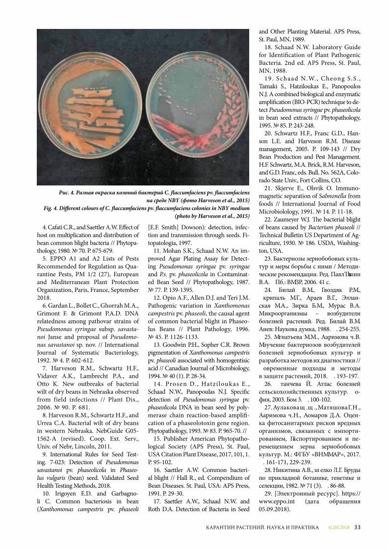
4| 26| 2018 33КАРАНТИН РАСТЕНИЙ. НАУКА И ПРАКТИКА
4. Cafati C.R., and Saettler A.W. E�ect of host on multiplication and distribution of bean common blight bacteria // Phytopa-thology, 1980. № 70. Р. 675-679.
5. EPPO A1 and A2 Lists of Pests Recommended for Regulation as Qua-rantine Pests, PM 1/2 (27), European and Mediterranean Plant Protection Organization, Paris, France, September 2018.
6. Gardan L., Bollet C., Ghorrah M.A., Grimont F. & Grimont P.A.D. DNA relatedness among pathovar strains of Pseudomonas syringae subsp. savasta-noi Janse and proposal of Pseudomo-nas savastanoi sp. nov. // Internatio nal Journal of Systematic Bacteriology, 1992. № 4. Р. 602-612.
7. Harveson R.M., Schwartz H.F., Vidaver A.K., Lambrecht P.A., and Otto K. New outbreaks of bacterial wilt of dry beans in Nebraska observed from field infections // Plant Dis., 2006. № 90. Р. 681.
8. Harveson R.M., Schwartz H.F., and Urrea C.A. Bacterial wilt of dry beans in western Nebraska. NebGuide G05-1562-A (revised). Coop. Ext. Serv., Univ. of Nebr, Lincoln, 2011.
9. International Rules for Seed Test-ing. 7-023: Detection of Pseudomonas savastаnoi pv. phaseolicola in Phaseo-lus vulgaris (bean) seed. Validated Seed Health Testing Methods, 2018.
10. Irigoyen E.D. and Garbagno-li C. Common bacteriosis in bean (Xanthomonas campestris pv. phaseoli
[E.F. Smith] Dowson): detection, infec-tion and transmission through seeds. Fi-topatologia, 1997.
11. Mohan S.K., Schaad N.W. An im-proved Agar Plating Assay for Detect-ing Pseudomonas syringae pv. syringae and P.s. pv. phaseolicola in Contaminat-ed Bean Seed // Phytopathology, 1987. № 77. Р. 139-1395.
12. Opio A.F., Allen D.J. and Teri J.M. Pathogenic variation in Xanthomonas campestris pv. phaseoli, the causal agent of common bacterial blight in Phaseo-lus Beans // Plant Pathology, 1996. № 45. Р. 1126-1133.
13. Goodwin P.H., Sopher C.R. Brown pigmentation of Xanthomonas campestris pv. phaseoli associated with homogentisic acid // Canadian Journal of Microbiology, 1994. № 40 (1). Р. 28-34.
14. Prosen D. , Hatzi loukas E. , Schaad N.W., Panopoulas N.J. Speci�c detection of Pseudomonas syringae pv. phaseolicola DNA in bean seed by poly-merase chain reaction-based ampli�-cation of a phaseolotoxin gene region. Phytopathology, 1993. № 83. Р. 965-70. //
15. Publisher American Phytopatho-logical Society (APS Press), St. Paul, USA Citation Plant Disease, 2017, 101, 1. P. 95-102.
16. Saettler A.W. Common bacteri-al blight // Hall R., ed. Compendium of Bean Diseases. St. Paul, USA: APS Press, 1991. Р. 29-30.
17. Saettler A.W., Schaad N.W. and Roth D.A. Detection of Bacteria in Seed
and Other Planting Material. APS Press, St. Paul, MN, 1989.
18. Schaad N.W. Laboratory Guide for Identi�cation of Plant Pathogenic Bacteria. 2nd ed. APS Press, St. Paul, MN, 1988.
19. S chaad N.W., Cheong S .S . , Tamaki S., Hatziloukas E., Panopoulos N.J. A combined biological and enzymatic ampli�cation (BIO-PCR) technique to de-tect Pseudomonas syringae pv. phaseolicola in bean seed extracts // Phytopathology, 1995. № 85. Р. 243-248.
20. Schwartz H.F., Franc G.D., Han-s on L.E. and Harveson R.M. Disease management, 2005. P. 109-143 // Dry Bean Production and Pest Management. H.F. Schwartz, M.A. Brick, R.M. Harveson, and G.D. Franc, eds. Bull. No. 562A, Colo-rado State Univ., Fort Collins, CO.
21. Skjerve E., Olsvik O. Immuno-magnetic separation of Salmonella from foods // International Journal of Food Microbiolology, 1991. № 14. Р. 11-18.
22. Zaumeyer W.J. �e bacterial blight of beans caused by Bacterium phaseoli // Technical Bulletin US Department of Ag-riculture, 1930. № 186. USDA, Washing-ton, USA.
23. Бактериозы зернобобовых куль-тур и меры борьбы с ними / Методи-ческие рекомендации. Ред. Павлюшин В.А. СПб.: ВИЗР, 2006. 41 с.
24. Билай В.И., Гвоздяк Р.И., Скрипаль И.Г., Краев В.Г., Эллан-ская И.А., Зирка Т.И., Мурас В.А. Микроорганизмы – возбудители болезней растений. Ред. Билай В.И. Киев: Наукова думка, 1988. С. 254-255.
25. Игнатьева И.М., Каримова Е.В. Изучение бактериозов возбудителей болезней зернобобовых культур и разработка методов их диагностики // Современные подходы и методы в защите растений, 2018. С. 193-197.
26. Станчева Й. Атлас болезней сельскохозяйственных культур. Со-фия, 2003. Том 3. С. 100-102.
27. Кулакова Ю.Ю., Матяшова Г.Н., Каримова Е.Н., Комаров Д.А. Оцен-ка фитосанитарных рисков вредных организмов, связанных с импорти-рованием, экспортированием и пе-ремещением зерна зернобобовых культур. М.: ФГБУ «ВНИИКР», 2017. С. 161-171, 229-239.
28. Никитина К.В., Щелко Л.Г. Труды по прикладной ботанике, генетике и селекции, 1982. № 71 (3). С. 86-88.
29. [Электронный ресурс]. https://www.eppo.int (дата обращения 05.09.2018).
Рис. 4. Разная окраска колоний бактерий C. �accumfaciens pv. �accumfaciens на среде NBY (фото Harveson et al., 2015)
Fig. 4. Di�erent colours of C. �accumfaciens pv. �accumfaciens colonies in NBY medium (photo by Harveson et al., 2015)
4| 26| 201834 КАРАНТИН РАСТЕНИЙ. НАУКА И ПРАКТИКА
Abstract. The Russian Federa-tion exports grain legume products to countries that have phytosanitary requirements for bacterial disease pathogen absence. The article brief-ly considers the biological features of bacterial pathogens of grain legumes, host plants, symptoms, damage and methods of diagnostics.
Keywords. Curtobacterium flac-cumfaciens pv. flaccumfaciens, Pseu-domonas savastanoi pv. phaseolicola, Xanthomonas axonopodis pv. phaseo-li, grain legumes, bacterium, disease pathogen.
Seeds of grain legumes are a highly nutritious and concentrated product and an important component of feed for farm animals. The high content of protein in grain legumes is ob-tained by fixation of air nitrogen as a result of the symbiotic activity of nodule bacteria.
The development of modern diag-nostics methods is required due to the need for a comprehensive analy-sis of bacterial pathogens of grain le-gumes, including the analysis of the probability of their introduction into the territory of the Russian Federa-tion with different types of products from various countries, the analysis of the probability of establishment in specific regions of Russia, taking into account the specificity of abiotic and biotic factors, the analysis of the probability of a versatile impact on the economy of Russia, the analysis of the state of the environment and the analysis of the social situation in its territory.
Pest spread control plays an im-portant role in the protection of ag-riculture and the environment. This
issue is relevant because the Russian Federation exports seeds of grain le-gumes to different countries that im-pose phytosanitary requirements, in particular, the absence of pathogens of bacterial diseases on leguminous grain for seed purposes.
The main bacterial diseases of grain legumes are caused by the bac-terial tan spot of bean Curtobacteri-um flaccumfaciens pv. flaccumfaciens (Fig. 1), the angular leaf spot of bean Pseudomonas savastanoi pv. phaseo-licola (Fig. 2), and the bacterial blight of bean Xanthomonas axono-podis pv. phaseoli (Fig. 3).
These pathogens reduce crop yields and quality of seeds of grain legumes which makes them eco-nomically significant. Control of bacterial diseases is possible when using pathogen-free seeds and crop rotation. Effective chemical protec-tion means against these pathogens are not currently available. The in-troduction of these bacteria into new regions may occur with infected seed (mainly beans and soybeans), and the infection may be latent, which could cause serious problems in grain legume production.
These bacterial diseases are of phytosanitary significance for the countries of the Eurasian Economic Union, first of all, for the cultivation area of Phasoleus spp.
The pathogen C. flaccumfaciens pv. flaccumfaciens was included into the European and Mediterra-nean Plant Protection Organiza-tion (EPPO) A2 List of quarantine objects (https://www.eppo.int). The bacterial tan spot of bean has quar-antine status also for the Caribbe-an Plant Protection Commission
(CPPC) and Inter-African Phy-tosanitary Council (IAPSC).
The main and most susceptible host plants to the bacterial tan spot of bean are plants of the Phaseo-lus spp. genus, especially bush bean Phaseolus vulgaris, runner bean P. coccineus, Lima bean P. lunatus, field pea Pisum sativum, adzuki bean Vigna angularis, mung bean V. radia-ta, black gram V. mungo, black-eye pea V. unguiculata, soybean Glycine max, Australian pea Lablab purpu-reus. The disease has been reported on the plants of Ipomoea genus, in particular Ipomoea violacea, as well as Zornia.
The majority of cultivated and wild forms of the above-mentioned plants are widely spread in Russia.
The first mention of bean disease dates back to the early 20th century. The pathogen C. flaccumfaciens pv. flaccumfaciens has been identified but has not established in Greece and Hungary. Bacteriosis is common in the Americas, Europe and some African countries. On the territo-ry of the Russian Federation it was reported in the beginning of 1980 on the soybean in the Far East and some southern regions (Nikitina, Shchelko, 1982), which, however, requires confirmation by modern methods.
The main pathway of C. flaccumfa-ciens pv. flaccumfaciens to new terri-tories is the seeds of beans, soybeans, peas and other plants.
The pathogen of the bacterial tan spot of bean preserves both on the surface and inside the seeds, remain-ing viable under extremely adverse environmental conditions. Bacterio-sis has been shown to remain via-
UDC 632.3.01/.08
BACTERIAL DISEASE PATHOGENS OF GRAIN LEGUMES AND DEVELOPMENT OF METHODS FOR THEIR DIAGNOSTICSE.V. Karimova, Senior Researcher of the RMPD of FGBU “VNIIKR”I.M. Ignatyeva, Researcher of the Bacteriology Laboratory of the LTC of FGBU “VNIIKR”
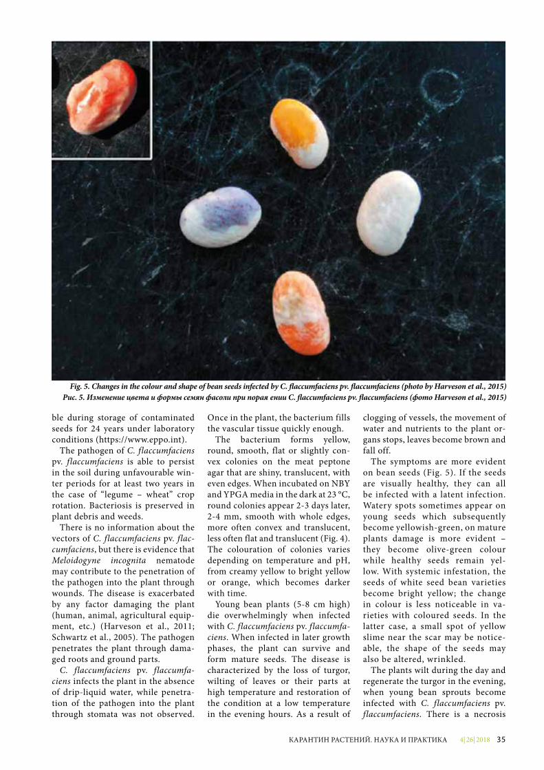
4| 26| 2018 35КАРАНТИН РАСТЕНИЙ. НАУКА И ПРАКТИКА
ble during storage of contaminated seeds for 24 years under laboratory conditions (https://www.eppo.int).
The pathogen of C. flaccumfaciens pv. flaccumfaciens is able to persist in the soil during unfavourable win-ter periods for at least two years in the case of “legume – wheat” crop rotation. Bacteriosis is preserved in plant debris and weeds.
There is no information about the vectors of C. flaccumfaciens pv. flac-cumfaciens, but there is evidence that Meloidogyne incognita nematode may contribute to the penetration of the pathogen into the plant through wounds. The disease is exacerbated by any factor damaging the plant (human, animal, agricultural equip-ment, etc.) (Harveson et al., 2011; Schwartz et al., 2005). The pathogen penetrates the plant through dama-ged roots and ground parts.
C. flaccumfaciens pv. flaccumfa-ciens infects the plant in the absence of drip-liquid water, while penetra-tion of the pathogen into the plant through stomata was not observed.
Once in the plant, the bacterium fills the vascular tissue quickly enough.
The bacterium forms yellow, round, smooth, flat or slightly con-vex colonies on the meat peptone agar that are shiny, translucent, with even edges. When incubated on NBY and YPGA media in the dark at 23 °C, round colonies appear 2-3 days later, 2-4 mm, smooth with whole edges, more often convex and translucent, less often flat and translucent (Fig. 4). The colouration of colonies varies depending on temperature and pH, from creamy yellow to bright yellow or orange, which becomes darker with time.
Young bean plants (5-8 cm high) die overwhelmingly when infected with C. flaccumfaciens pv. flaccumfa-ciens. When infected in later growth phases, the plant can survive and form mature seeds. The disease is characterized by the loss of turgor, wilting of leaves or their parts at high temperature and restoration of the condition at a low temperature in the evening hours. As a result of
clogging of vessels, the movement of water and nutrients to the plant or-gans stops, leaves become brown and fall off.
The symptoms are more evident on bean seeds (Fig. 5). If the seeds are visually healthy, they can all be infected with a latent infection. Watery spots sometimes appear on young seeds which subsequently become yellowish-green, on mature plants damage is more evident – they become olive-green colour while healthy seeds remain yel-low. With systemic infestation, the seeds of white seed bean varieties become bright yellow; the change in colour is less noticeable in va-rieties with coloured seeds. In the latter case, a small spot of yellow slime near the scar may be notice-able, the shape of the seeds may also be altered, wrinkled.
The plants wilt during the day and regenerate the turgor in the evening, when young bean sprouts become infected with C. flaccumfaciens pv. flaccumfaciens. There is a necrosis
Fig. 5. Changes in the colour and shape of bean seeds infected by C. �accumfaciens pv. �accumfaciens (photo by Harveson et al., 2015)Рис. 5. Изменение цвета и формы семян фасоли при поражении C. �accumfaciens pv. �accumfaciens (фото Harveson et al., 2015)
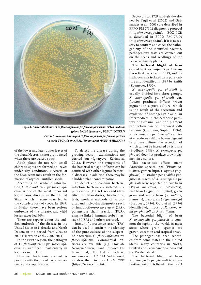
4| 26| 201836 КАРАНТИН РАСТЕНИЙ. НАУКА И ПРАКТИКА
of the lower and later upper leaves of the plant. Necrosis is not pronounced when there are watery spots.
Adult plants do not wilt, small chlorotic spots are formed on leaves under dry conditions. Necrosis at the bean seam may result in the for-mation of atypical, unfilled seeds.
According to available informa-tion, C. flaccumfaciens pv. flaccumfa-ciens is one of the most important leguminous diseases in the United States, which in some years led to the complete loss of crops. In 1947, in Idaho, there have been serious outbreaks of the disease, and yield losses exceeded 60%.
There are reports about the sud-den outbreak of the disease in the United States in Nebraska and North Dakota in the period from 2003 to 2006 (Harveson et al., 2006, 2011).
In the EPPO region, the pathogen of C. flaccumfaciens pv. flaccumfa-ciens is significant, particularly in legumes in Turkey.
Effective bacteriosis control is possible with the use of bacteria-free seeds and crop rotation.
To detect the disease during the growing season, examinations are carried out (Ignatyeva, Karimova, 2018). However, the symptoms of the bacterial tan spot of bean can be confused with other legume bacteri-al diseases. In addition, there may be a hidden plant contamination.
To detect and confirm bacterial infection, bacteria are isolated in a pure culture (Fig. 6.1, 6.2) and iden-tified in laboratories; biochemical tests, modern methods of serolo-gical and molecular diagnostics such as immunofluorescence assay (IFA), polymerase chain reaction (PCR), enzyme-linked immunosorbent as-say (ELISA) and others are used.
Immunofluorescence assay (IFA) can be used to confirm the identity of the pure culture of the suspect-ed bacterium C. flaccumfaciens pv. f laccumfaciens. Commercial an-tisera are available (e.g. Florilab, Neogen-Adgen, Plant Research In-ternational). For IFA a bacterial suspension of 106 CFU/ml is used, as described in EPPO PM 7/97 (https://www.eppo.int).
Protocols for PCR analysis develo-ped by Tegli et al. (2002) and Gui-maraes et al. (2001) are described in EPPO PM 7/102 diagnostic protocol (https://www.eppo.int). BOX-PCR is described in EPPO RM 7/100 (https://www.eppo.int). If it is neces-sary to confirm and check the patho-genicity of the identified bacteria, pathogenicity tests are carried out on the seeds and seedlings of the Fabaceae family plants.
The bacterial blight of bean caused by X. axonopodis pv. phaseo-li was first described in 1893, and the pathogen was isolated in a pure cul-ture and identified in 1897 by Smith (Zaumeyer, 1930).
X. axonopodis pv. phaseoli is usually divided into three groups. X. axonopodis pv. phaseoli var. fuscans produces diffuse brown pigment in a pure culture, which is the result of the secretion and oxi dation of homogenistic acid, an intermediate in the catabolic path-way of tyrosine; and the pigment production can be increased with tyrosine (Goodwin, Sopher, 1994). X. axonopodis pv. phaseoli var. in-dica produces a di�use brown pigment in a pure culture, the secretion of which cannot be increased by tyrosine (Bradbury, 1986). X. axonopodis pv. phaseoli does not produce brown pig-ment in a culture.
�is bacteriosis affects many Phaseolus species, pea (Pisum sa-tivum), garden lupin (Lupinus poly-phyllus), Australian pea (Lablab pur-pureus). Races of X. axonopodis pv. phaseoli were reported on rice bean (Vigna umbellata, P. calcaratus), mat bean (Vigna aconitifolia), green gram and mung bean (V. radiata, P. aureus), black gram (Vigna mungo) (Bradbury, 1986). Opio et al. (1996) identified eight races of X. axonopo-dis pv. phaseoli on P. acutifolius.
The bacterial blight of bean X. axonopodis pv. phaseoli is com-mon throughout the world, in most areas where grain legumes are grown, except in arid tropical areas.
The pathogen has been report-ed from some states in the United States, many countries in North, Central and Latin America, Asia and the Pacific Islands.
The bacterial blight of bean X. axonopodis pv. phaseoli is a qua-ran tine pest and is listed in the EPPO
Fig. 6.1. Bacterial colonies of C. �accumfaciens pv. �accumfaciens on YPGA medium (photo by I.M. Ignatyeva, FGBU “VNIIKR”)
Рис. 6.1. Колонии бактерий C. �accumfaciens pv. �accumfaciensна среде YPGA (фото И.М. Игнатьевой, ФГБУ «ВНИИКР»)
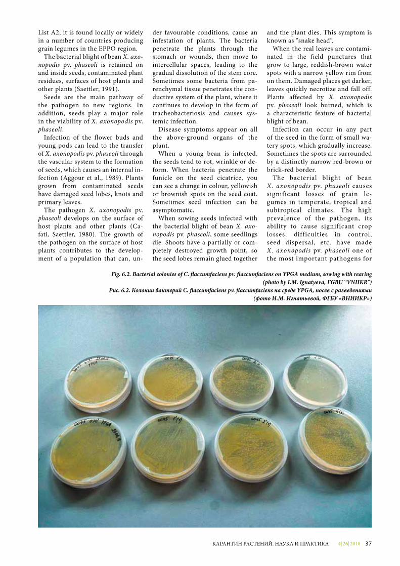
4| 26| 2018 37КАРАНТИН РАСТЕНИЙ. НАУКА И ПРАКТИКА
List A2; it is found locally or widely in a number of countries producing grain legumes in the EPPO region.
The bacterial blight of bean X. axo-nopodis pv. phaseoli is retained on and inside seeds, contaminated plant residues, surfaces of host plants and other plants (Saettler, 1991).
Seeds are the main pathway of the pathogen to new regions. In addition, seeds play a major role in the viability of X. axonopodis pv. phaseoli.
Infection of the flower buds and young pods can lead to the transfer of X. axonopodis pv. phaseoli through the vascular system to the formation of seeds, which causes an internal in-fection (Aggour et al., 1989). Plants grown from contaminated seeds have damaged seed lobes, knots and primary leaves.
The pathogen X. axonopodis pv. phaseoli develops on the surface of host plants and other plants (Ca-fati, Saettler, 1980). The growth of the pathogen on the surface of host plants contributes to the develop-ment of a population that can, un-
der favourable conditions, cause an infestation of plants. The bacteria penetrate the plants through the stomach or wounds, then move to intercellular spaces, leading to the gradual dissolution of the stem core. Sometimes some bacteria from pa-renchymal tissue penetrates the con-ductive system of the plant, where it continues to develop in the form of tracheobacteriosis and causes sys-temic infection.
Disease symptoms appear on all the above-ground organs of the plant.
When a young bean is infected, the seeds tend to rot, wrinkle or de-form. When bacteria penetrate the funicle on the seed cicatrice, you can see a change in colour, yellowish or brownish spots on the seed coat. Sometimes seed infection can be asym ptomatic.
When sowing seeds infected with the bacterial blight of bean X. axo-nopodis pv. phaseoli, some seedlings die. Shoots have a partially or com-pletely destroyed growth point, so the seed lobes remain glued together
and the plant dies. This symptom is known as “snake head”.
When the real leaves are contami-nated in the field punctures that grow to large, reddish-brown water spots with a narrow yellow rim from on them. Damaged places get darker, leaves quickly necrotize and fall off. Plants affected by X. axonopodis pv. phaseoli look burned, which is a characteristic feature of bacterial blight of bean.
Infection can occur in any part of the seed in the form of small wa-tery spots, which gradually increase. Sometimes the spots are surrounded by a distinctly narrow red-brown or brick-red border.
The bacterial blight of bean X. axonopodis pv. phaseoli causes significant losses of grain le-gumes in temperate, tropical and subtropical climates. The high prevalence of the pathogen, its ability to cause significant crop losses, difficulties in control, seed dispersal, etc. have made X. axonopodis pv. phaseoli one of the most important pathogens for
Fig. 6.2. Bacterial colonies of C. �accumfaciens pv. �accumfaciens on YPGA medium, sowing with rearing (photo by I.M. Ignatyeva, FGBU “VNIIKR”)
Рис. 6.2. Колонии бактерий C. �accumfaciens pv. �accumfaciens на среде YPGA, посев с разведениями (фото И.М. Игнатьевой, ФГБУ «ВНИИКР»)
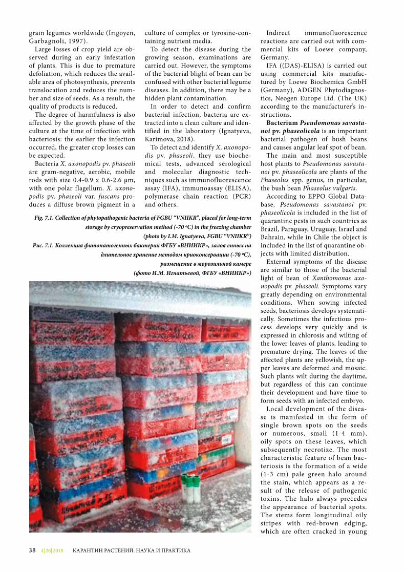
4| 26| 201838 КАРАНТИН РАСТЕНИЙ. НАУКА И ПРАКТИКА
grain legumes worldwide (Irigoyen, Garbagnoli, 1997).
Large losses of crop yield are ob-served during an early infestation of plants. This is due to premature defoliation, which reduces the avail-able area of photosynthesis, prevents translocation and reduces the num-ber and size of seeds. As a result, the quality of products is reduced.
The degree of harmfulness is also affected by the growth phase of the culture at the time of infection with bacteriosis: the earlier the infection occurred, the greater crop losses can be expected.
Bacteria X. axonopodis pv. phaseoli are gram-negative, aerobic, mobile rods with size 0.4-0.9 x 0.6-2.6 µm, with one polar flagellum. X. axono-podis pv. phaseoli var. fuscans pro-duces a diffuse brown pigment in a
culture of complex or tyrosine-con-taining nutrient media.
To detect the disease during the growing season, examinations are carried out. However, the symptoms of the bacterial blight of bean can be confused with other bacterial legume diseases. In addition, there may be a hidden plant contamination.
In order to detect and confirm bacterial infection, bacteria are ex-tracted into a clean culture and iden-tified in the laboratory (Ignatyeva, Karimova, 2018).
To detect and identify X. axonopo-dis pv. phaseoli, they use bioche-mical tests, advanced serological and molecular diagnostic tech-niques such as immunofluorescence assay (IFA), immunoassay (ELISA), polymerase chain reaction (PCR) and others.
Indirect immunofluorescence reac tions are carried out with com-mercial kits of Loewe company, Germany.
IFA ((DAS)-ELISA) is carried out using commercial kits manufac-tured by Loewe Biochemica GmbH (Germany), ADGEN Phytodiagnos-tics, Neogen Europe Ltd. (The UK) according to the manufacturer’s in-structions.
Bacterium Pseudomonas savasta-noi pv. phaseolicola is an important bacterial pathogen of bush beans and causes angular leaf spot of bean.
The main and most susceptible host plants to Pseudomonas savasta-noi pv. phaseolicola are plants of the Phaseolus spp. genus, in particular, the bush bean Phaseolus vulgaris.
According to EPPO Global Data-base, Pseudomonas savastanoi pv. phaseolicola is included in the list of quarantine pests in such countries as Brazil, Paraguay, Uruguay, Israel and Bahrain, while in Chile the object is included in the list of quarantine ob-jects with limited distribution.
External symptoms of the disease are similar to those of the bacterial light of bean of Xanthomonas axo-nopodis pv. phaseoli. Symptoms vary greatly depending on environmental conditions. When sowing infected seeds, bacteriosis develops systemati-cally. Sometimes the infectious pro-cess develops very quickly and is expressed in chlorosis and wilting of the lower leaves of plants, leading to premature drying. The leaves of the affected plants are yellowish, the up-per leaves are deformed and mosaic. Such plants wilt during the daytime, but regardless of this can continue their development and have time to form seeds with an infected embryo.
Local development of the disea-se is manifested in the form of single brown spots on the seeds or numerous, small (1-4 mm), oily spots on these leaves, which subsequently necrotize. The most characteristic feature of bean bac-teriosis is the formation of a wide (1-3 cm) pale green halo around the stain, which appears as a re-sult of the release of pathogenic toxins. The halo always precedes the appearance of bacterial spots. The stems form longitudinal oily stripes with red-brown edging, which are often cracked in young
Fig. 7.1. Collection of phytopathogenic bacteria of FGBU “VNIIKR”, placed for long-term storage by cryopreservation method (-70 ºС) in the freezing chamber
(photo by I.M. Ignatyeva, FGBU “VNIIKR”)Рис. 7.1. Коллекция фитопатогенных бактерий ФГБУ «ВНИИКР», заложенных на
длительное хранение методом криоконсервации (-70 ºС), размещение в морозильной камере
(фото И.М. Игнатьевой, ФГБУ «ВНИИКР»)
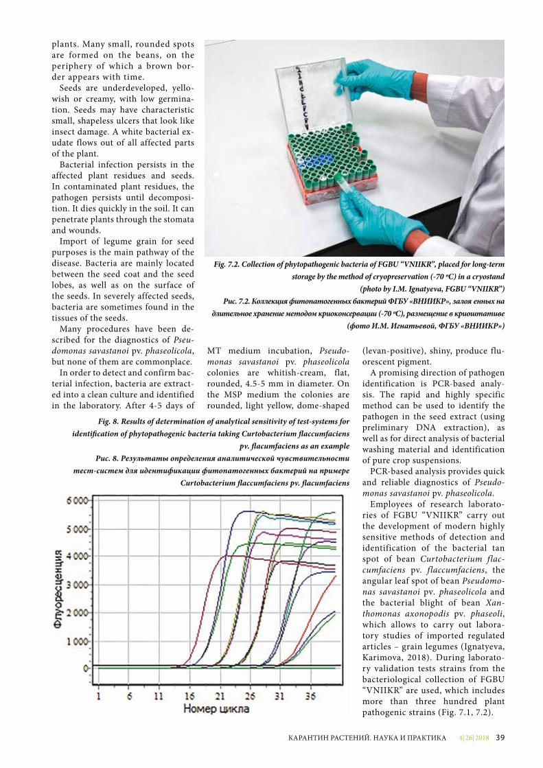
4| 26| 2018 39КАРАНТИН РАСТЕНИЙ. НАУКА И ПРАКТИКА
plants. Many small, rounded spots are formed on the beans, on the periphery of which a brown bor-der appears with time.
Seeds are underdeveloped, yello-wish or creamy, with low germina-tion. Seeds may have characteristic small, shapeless ulcers that look like insect damage. A white bacterial ex-udate flows out of all affected parts of the plant.
Bacterial infection persists in the affected plant residues and seeds. In contaminated plant residues, the pathogen persists until decomposi-tion. It dies quickly in the soil. It can penetrate plants through the stomata and wounds.
Import of legume grain for seed purposes is the main pathway of the disease. Bacteria are mainly located between the seed coat and the seed lobes, as well as on the surface of the seeds. In severely affected seeds, bacteria are sometimes found in the tissues of the seeds.
Many procedures have been de-scribed for the diagnostics of Pseu-domonas savastanoi pv. phaseolicola, but none of them are commonplace.
In order to detect and confirm bac-terial infection, bacteria are extract-ed into a clean culture and identified in the laboratory. After 4-5 days of
MT medium incubation, Pseudo-monas savastanoi pv. phaseolicola colonies are whitish-cream, flat, rounded, 4.5-5 mm in diameter. On the MSP medium the colonies are rounded, light yellow, dome-shaped
(levan-positive), shiny, produce flu-orescent pigment.
A promising direction of pathogen identification is PCR-based analy-s is. The rapid and highly specific method can be used to identify the pathogen in the seed extract (using preliminary DNA extraction), as well as for direct analysis of bacterial washing material and identification of pure crop suspensions.
PCR-based analysis provides quick and reliable diagnostics of Pseudo-monas savastanoi pv. phaseolicola.
Employees of research laborato-ries of FGBU “VNIIKR” carry out the development of modern highly sensitive methods of detection and identification of the bacterial tan spot of bean Curtobacterium flac-cumfaciens pv. f laccumfaciens, the angular leaf spot of bean Pseudomo-nas savastanoi pv. phaseolicola and the bacterial blight of bean Xan-thomonas axonopodis pv. phaseoli, which allows to carry out labora-tory studies of imported regulated articles – grain legumes (Ignatyeva, Karimova, 2018). During laborato-ry validation tests strains from the bacteriological collection of FGBU “VNIIKR” are used, which includes more than three hundred plant pathogenic strains (Fig. 7.1, 7.2).
Fig. 7.2. Collection of phytopathogenic bacteria of FGBU “VNIIKR”, placed for long-term storage by the method of cryopreservation (-70 ºС) in a cryostand
(photo by I.M. Ignatyeva, FGBU “VNIIKR”)Рис. 7.2. Коллекция фитопатогенных бактерий ФГБУ «ВНИИКР», заложенных на
длительное хранение методом криоконсервации (-70 ºС), размещение в криоштативе (фото И.М. Игнатьевой, ФГБУ «ВНИИКР»)
Fig. 8. Results of determination of analytical sensitivity of test-systems for identi�cation of phytopathogenic bacteria taking Curtobacterium �accumfaciens
pv. �acumfaciens as an exampleРис. 8. Результаты определения аналитической чувствительности
тест-систем для идентификации фитопатогенных бактерий на примере Curtobacterium �accumfaciens pv. �acumfaciens
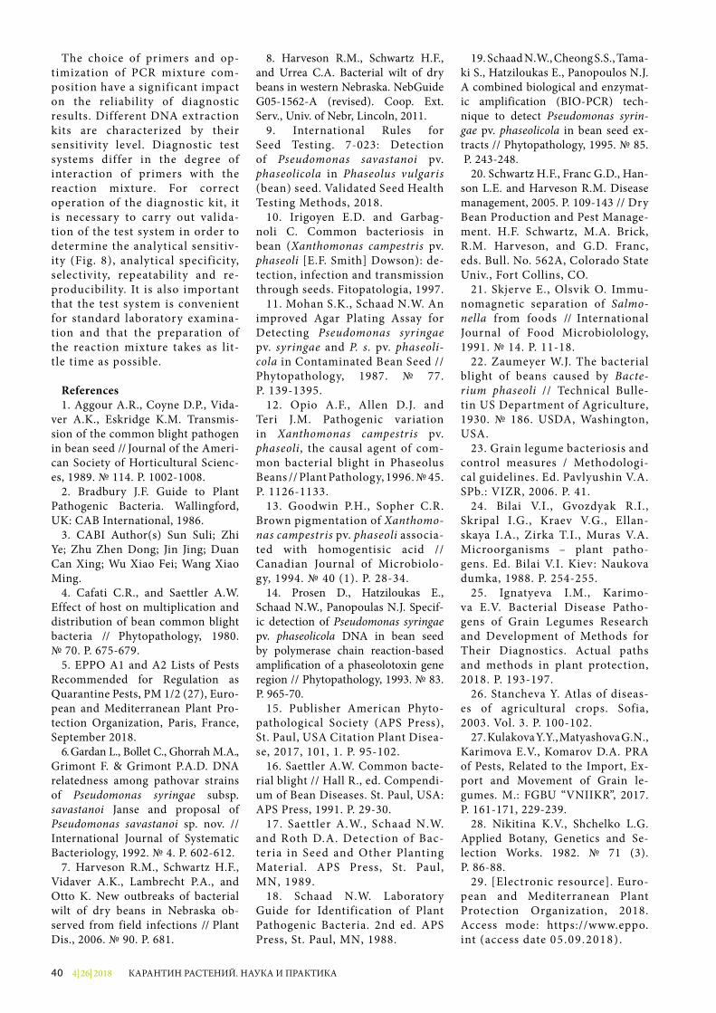
4| 26| 201840 КАРАНТИН РАСТЕНИЙ. НАУКА И ПРАКТИКА
The choice of primers and op-timization of PCR mixture com-position have a significant impact on the reliability of diagnostic results. Different DNA extraction kits are characterized by their sensitivity level. Diagnostic test systems differ in the degree of interaction of primers with the reaction mixture. For correct opera tion of the diagnostic kit, it is necessary to carry out valida-tion of the test system in order to determine the analytical sensitiv-ity (Fig. 8), analytical specificity, selectivity, repeatability and re-producibility. It is also important that the test system is convenient for standard laboratory examina-tion and that the preparation of the reaction mixture takes as lit-tle time as possible.
References1. Aggour A.R., Coyne D.P., Vida-
ver A.K., Eskridge K.M. Transmis-sion of the common blight pathogen in bean seed // Journal of the Ameri-can Society of Horticultural Scienc-es, 1989. № 114. Р. 1002-1008.
2. Bradbury J.F. Guide to Plant Pathogenic Bacteria. Wallingford, UK: CAB International, 1986.
3. CABI Author(s) Sun Suli; Zhi Ye; Zhu Zhen Dong; Jin Jing; Duan Can Xing; Wu Xiao Fei; Wang Xiao Ming.
4. Cafati C.R., and Saettler A.W. Effect of host on multiplication and distribution of bean common blight bacteria // Phytopathology, 1980. № 70. Р. 675-679.
5. EPPO A1 and A2 Lists of Pests Recommended for Regulation as Quarantine Pests, PM 1/2 (27), Euro-pean and Mediterranean Plant Pro-tection Organization, Paris, France, September 2018.
6. Gardan L., Bollet C., Ghorrah M.A., Grimont F. & Grimont P.A.D. DNA relatedness among pathovar strains of Pseudomonas syringae subsp. savastanoi Janse and proposal of Pseudomonas savastanoi sp. nov. // International Journal of Systematic Bacteriology, 1992. № 4. Р. 602-612.
7. Harveson R.M., Schwartz H.F., Vidaver A.K., Lambrecht P.A., and Otto K. New outbreaks of bacterial wilt of dry beans in Nebraska ob-served from field infections // Plant Dis., 2006. № 90. Р. 681.
8. Harveson R.M., Schwartz H.F., and Urrea C.A. Bacterial wilt of dry beans in western Nebraska. NebGuide G05-1562-A (revised). Coop. Ext. Serv., Univ. of Nebr, Lincoln, 2011.
9. International Rules for Seed Testing. 7-023: Detection of Pseudo monas savastanoi pv. phaseolicola in Phaseolus vulgaris (bean) seed. Validated Seed Health Testing Methods, 2018.
10. Irigoyen E.D. and Garbag-noli C. Common bacteriosis in bean (Xanthomonas campestris pv. phaseoli [E.F. Smith] Dowson): de-tection, infection and transmission through seeds. Fitopatologia, 1997.
11. Mohan S.K., Schaad N.W. An improved Agar Plating Assay for Detecting Pseudomonas syringae pv. syringae and P. s. pv. phaseoli-cola in Contaminated Bean Seed // Phytopathology, 1987. № 77. Р. 139-1395.
12. Opio A.F., Allen D.J. and Teri J.M. Pathogenic variation in Xanthomonas campestris pv. phaseoli, the causal agent of com-mon bacterial blight in Phaseolus Beans // Plant Pathology, 1996. № 45. Р. 1126-1133.
13. Goodwin P.H., Sopher C.R. Brown pigmentation of Xanthomo-nas campestris pv. phaseoli associa-ted with homogentisic acid // Canadian Journal of Microbiolo-gy, 1994. № 40 (1). Р. 28-34.
14. Prosen D., Hatziloukas E., Schaad N.W., Panopoulas N.J. Specif-ic detection of Pseudomonas syringae pv. phaseolicola DNA in bean seed by polymerase chain reaction-based ampli�cation of a phaseolotoxin gene region // Phytopathology, 1993. № 83. Р. 965-70.
15. Publisher American Phyto-pathological Society (APS Press), St. Paul, USA Citation Plant Disea-se, 2017, 101, 1. P. 95-102.
16. Saettler A.W. Common bacte-rial blight // Hall R., ed. Compendi-um of Bean Diseases. St. Paul, USA: APS Press, 1991. Р. 29-30.
17. Saettler A.W., Schaad N.W. and Roth D.A. Detection of Bac-teria in Seed and Other Planting Material. APS Press, St. Paul, MN, 1989.
18. Schaad N.W. Laboratory Guide for Identification of Plant Pathogenic Bacteria. 2nd ed. APS Press, St. Paul, MN, 1988.
19. Schaad N.W., Cheong S.S., Tama-ki S., Hatziloukas E., Panopoulos N.J. A combined biological and enzymat-ic amplification (BIO-PCR) tech-nique to detect Pseudomonas syrin-gae pv. phaseolicola in bean seed ex-tracts // Phytopathology, 1995. № 85. Р. 243-248.
20. Schwartz H.F., Franc G.D., Han-son L.E. and Harveson R.M. Disease management, 2005. P. 109-143 // Dry Bean Production and Pest Manage-ment. H.F. Schwartz, M.A. Brick, R.M. Harveson, and G.D. Franc, eds. Bull. No. 562A, Colorado State Univ., Fort Collins, CO.
21. Skjerve E., Olsvik O. Immu-nomagnetic separation of Salmo-nella from foods // International Journal of Food Microbiolology, 1991. № 14. Р. 11-18.
22. Zaumeyer W.J. The bacterial blight of beans caused by Bacte-rium phaseoli // Technical Bulle-tin US Department of Agriculture, 1930. № 186. USDA, Washington, USA.
23. Grain legume bacteriosis and control measures / Methodologi-cal guidelines. Ed. Pavlyushin V.A. SPb.: VIZR, 2006. P. 41.
24. Bilai V.I., Gvozdyak R.I., Skripal I.G., Kraev V.G., Ellan-skaya I.A., Zirka T.I., Muras V.A. Microorganisms – plant patho-gens. Ed. Bilai V.I. Kiev: Naukova dumka, 1988. P. 254-255.
25. Ignatyeva I.M., Karimo-va E.V. Bacterial Disease Patho-gens of Grain Legumes Research and Development of Methods for Their Diagnostics. Actual paths and methods in plant protection, 2018. P. 193-197.
26. Stancheva Y. Atlas of diseas-es of agricultural crops. Sofia, 2003. Vol. 3. P. 100-102.
27. Kulakova Y.Y., Matyashova G.N., Karimova E.V., Komarov D.A. PRA of Pests, Related to the Import, Ex-port and Movement of Grain le-gumes. M.: FGBU “VNIIKR”, 2017. P. 161-171, 229-239.
28. Nikitina K.V., Shchelko L.G. Applied Botany, Genetics and Se-lection Works. 1982. № 71 (3). P. 86-88.
29. [Electronic resource]. Euro-pean and Mediterranean Plant Protection Organization, 2018. Access mode: https://www.eppo.int (access date 05 .09.2018) .
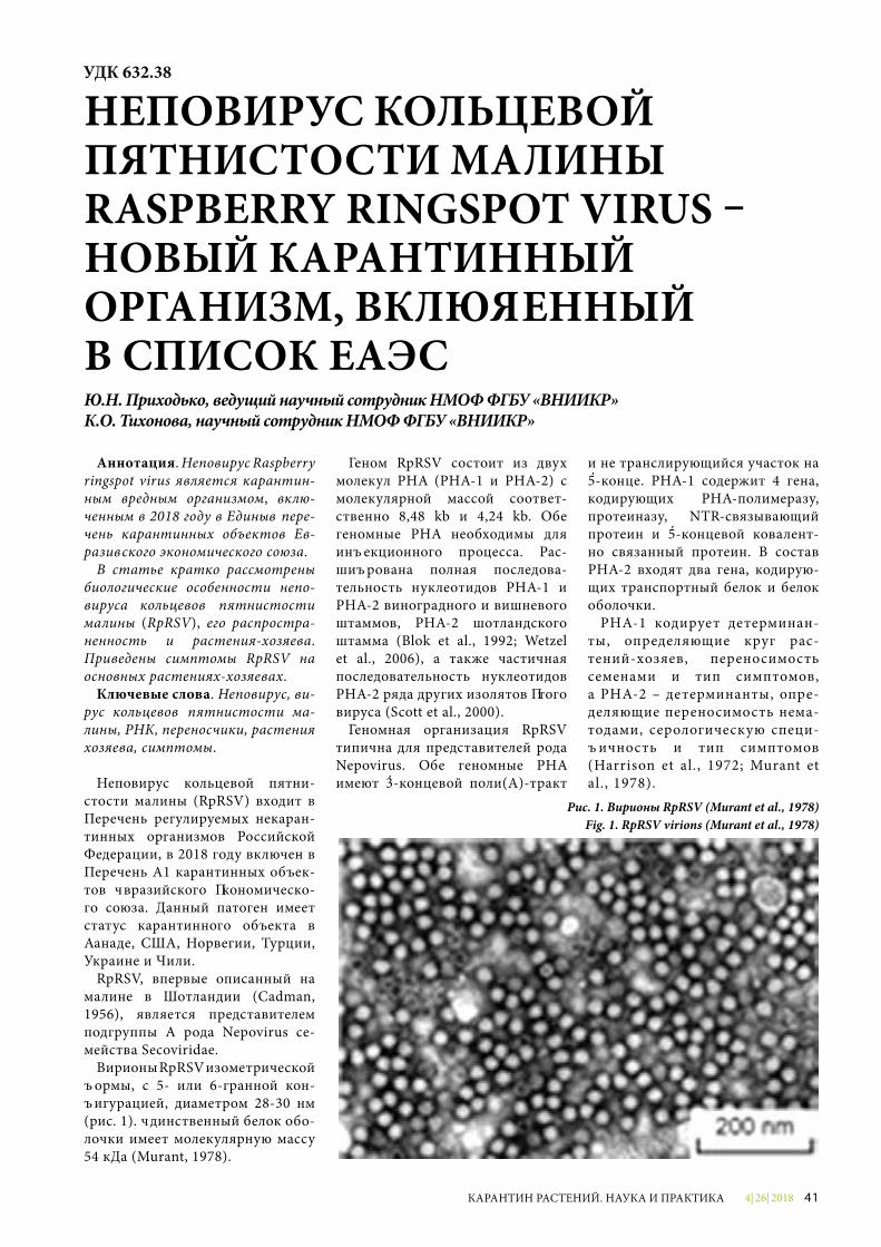
4| 26| 2018 41КАРАНТИН РАСТЕНИЙ. НАУКА И ПРАКТИКА
Аннотация. Неповирус Raspberry ringspot virus является карантин-ным вредным организмом, вклю-ченным в 2018 году в Единый пере-чень карантинных объектов Ев-разийского экономического союза.
В статье кратко рассмотрены биологические особенности непо-вируса кольцевой пятнистости малины (RpRSV), его распростра-ненность и растения-хозяева. Приведены симптомы RpRSV на основных растениях-хозяевах.
Ключевые слова. Неповирус, ви-рус кольцевой пятнистости ма-лины, РНК, переносчики, растения хозяева, симптомы.
Неповирус кольцевой пятни-стости малины (RpRSV) входит в Перечень регулируемых некаран-тинных организмов Российской Федерации, в 2018 году включен в Перечень А1 карантинных объек-тов Евразийского экономическо-го союза. Данный патоген имеет статус карантинного объекта в Канаде, США, Норвегии, Турции, Украи не и Чили.
RpRSV, впервые описанный на малине в Шотландии (Cadman, 1956), является представителем подгруппы А рода Nepovirus се-мейства Secoviridae.
Вирионы RpRSV изометрической формы, с 5- или 6-гранной кон-фигурацией, диаметром 28-30 нм (рис. 1). Единственный белок обо-лочки имеет молекулярную массу 54 кДа (Murant, 1978).
Геном RpRSV состоит из двух молекул РНК (РНК-1 и РНК-2) с молекулярной массой соответ-ственно 8,48 kb и 4,24 kb. Обе геномные РНК необходимы для инфекционного процесса. Рас-шифрована полная последова-тельность нуклеотидов РНК-1 и РНК-2 виноградного и вишневого штаммов, РНК-2 шотландского штамма (Blok et al., 1992; Wetzel et al., 2006), а также частичная последовательность нуклеотидов РНК-2 ряда других изолятов этого вируса (Scott et al., 2000).
Геномная организация RpRSV типична для представителей рода Nepovirus. Обе геномные РНК имеют 3-концевой поли(А)-тракт
и не транслирующийся участок на 5-конце. РНК-1 содержит 4 гена, кодирующих РНК-полимеразу, протеиназу, NTR-связывающий протеин и 5-концевой ковалент-но связанный протеин. В состав РНК-2 входят два гена, кодирую-щих транспортный белок и белок оболочки.
РНК-1 кодирует детерминан-ты, определяющие круг рас-тений-хозяев, переносимость семенами и тип симптомов, а РНК-2 – детерминанты, опре-деляющие переносимость нема-тодами, серологическую специ-фичность и тип симптомов (Harrison et al., 1972; Murant et al., 1978).
УДК 632.38
НЕПОВИРУС КОЛЬЦЕВОЙ ПЯТНИСТОСТИ МАЛИНЫ RASPBERRY RINGSPOT VIRUS – НОВЫЙ КАРАНТИННЫЙ ОРГАНИЗМ, ВКЛЮЧЕННЫЙ В СПИСОК ЕАЭСЮ.Н. Приходько, ведущий научный сотрудник НМОФ ФГБУ «ВНИИКР»К.О. Тихонова, научный сотрудник НМОФ ФГБУ «ВНИИКР»
Рис. 1. Вирионы RpRSV (Murant et al., 1978)Fig. 1. RpRSV virions (Murant et al., 1978)
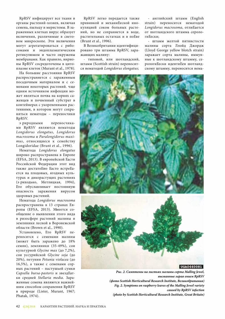
4| 26| 201842 КАРАНТИН РАСТЕНИЙ. НАУКА И ПРАКТИКА
RpRSV инфицирует все ткани и органы растений-хозяев, включая семена, пыльцу и меристемы. В за-раженных клетках вирус образует включения, различимые в свето-вом микроскопе. Эти включения могут агрегатироваться с рибо-сомами и эндоплазматическим ретикулюмом и часто окружены мембранами. Как правило, вирио-ны RpRSV сосредоточены в цито-плазме клеток (Murant et al., 1978).
На большие расстояния RpRSV распространяется с зараженным посадочным материалом и с се-менами некоторых растений. Еще одним источником инфекции мо-жет являться почва на корнях са-женцев и почвенный субстрат в контейнерах с укорененными рас-тениями, в котором могут сохра-няться нематоды – переносчики RpRSV.
Природными переносчика-ми RpRSV являются нематоды Longidorus elongatus, Longidorus macrosoma и Paralongidorus maxi-mus, относящиеся к семейству Longidoridae (Brunt et al., 1996).
Нематода Longidorus elongatus широко распространена в Европе (EFSA, 2013). В европейской части Российской Федерации этот вид также достаточно часто встреча-ется на плодовых, ягодных куль-турах и дикорастущих растениях (Приходько, Метлицкая, 1994), что обуславливает постоянную опасность заражения вирусом здоровых растений.
Нематода Longidorus macrosoma распространена в 13 странах Ев-ропы (EFSA, 2013). Имеется со-общение о выявлении этого вида в ризосфере растений малины и земляники лесной в Воронежской области (Brown et al., 1990).
Установлено, что RpRSV пе-реносится с семенами малины (может быть заражено до 18% семян), земляники (35-49%), сои культурной Glycine max (до 7,2%), сои уссурийской Glycine soja (до 20%), петунии Petunia violacea (до 16,5%), а также с семенами сор-ных растений – пастушьей сумки Capsella bursa-pastoris и звездчат-ки средней Stellaria media. Зара-женные семена являются важней-шим способом сохранения RpRSV в природе (Lister, Murant, 1967; Phatak, 1974).
RpRSV легко передается также прививкой и механической ино-куляцией соком больных расте-ний, но не сохраняется в воде, растительных остатках и в почве (Brunt et al., 1996).
В Великобритании идентифици-ровано три штамма RpRSV, зара-жающих малину:
- типовой, или шотландский, штамм (Scottish strain) переносит-ся нематодой Longidorus elongatus;
- английский штамм (English strain) переносится нематодой Longidorus macrosoma, отличается от шотландского штамма сероло-гически;
- штамм желтой пятнистости малины сорта Ллойд Джордж (Lloyd George yellow blotch strain) заражает сорта малины, иммун-ные к шотландскому штамму, се-рологически идентичен шотланд-скому штамму, переносится нема-
Рис. 2. Симптомы на листьях малины сорта Malling Jewel,вызванные заражением RpRSV
(фото Scottish Horticultural Research Institute, Великобритания)Fig. 2. Symptoms on raspberry leaves of the Malling Jewel variety
caused by RpRSV infection(photo by Scottish Horticultural Research Institute, Great Britain)
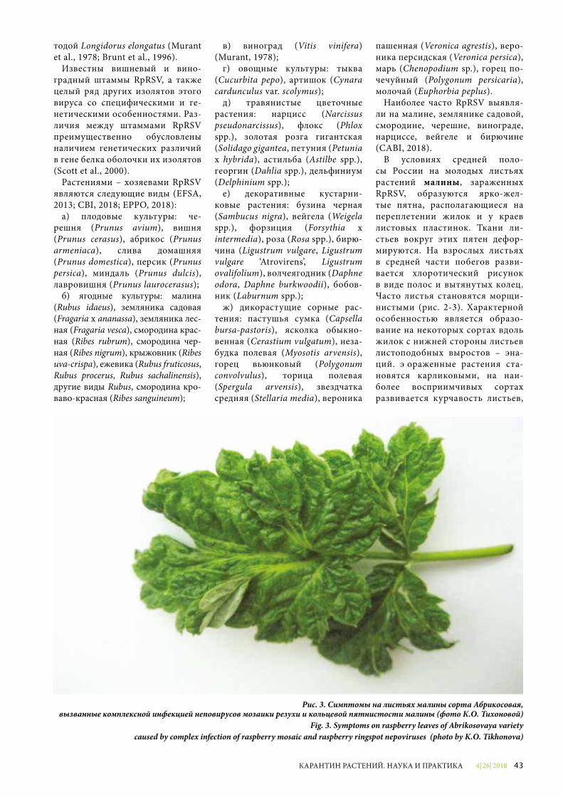
4| 26| 2018 43КАРАНТИН РАСТЕНИЙ. НАУКА И ПРАКТИКА
тодой Longidorus elongatus (Murant et al., 1978; Brunt et al., 1996).
Известны вишневый и вино-градный штаммы RpRSV, а также целый ряд других изолятов этого вируса со специфическими и ге-нетическими особенностями. Раз-личия между штаммами RpRSV преимущественно обусловлены наличием генетических различий в гене белка оболочки их изолятов (Scott et al., 2000).
Растениями – хозяевами RpRSV являются следующие виды (EFSA, 2013; CBI, 2018; EPPO, 2018):
а) плодовые культуры: че-решня (Prunus avium), вишня (Prunus cerasus), абрикос (Prunus armeniaca), слива домашняя (Prunus domestica), персик (Prunus persica), миндаль (Prunus dulcis), лавровишня (Prunus laurocerasus);
б) ягодные культуры: малина (Rubus idaeus), земляника садовая (Fragaria x ananassa), земляника лес-ная (Fragaria vesca), смородина крас-ная (Ribes rubrum), смородина чер-ная (Ribes nigrum), крыжовник (Ribes uva-crispa), ежевика (Rubus fruticosus, Rubus procerus, Rubus sachalinensis), другие виды Rubus, смородина кро-ваво-красная (Ribes sanguineum);
в) виноград (Vitis vinifera) (Murant, 1978);
г) овощные культуры: тыква (Cucurbita pepo), артишок (Cynara cardunculus var. scolymus);
д) травянистые цветочные растения: нарцисс (Narcissus pseudonarcissus), флокс (Phlox spp.), золотая розга гигантская (Solidago gigantea, петуния (Petunia x hybrida), астильба (Astilbe spp.), георгин (Dahlia spp.), дельфиниум (Delphinium spp.);
е) декоративные кустарни-ковые растения: бузина черная (Sambucus nigra), вейгела (Weigela spp.), форзиция (Forsythia x intermedia), роза (Rosa spp.), бирю-чина (Ligustrum vulgare, Ligustrum vulgare ‘Atrovirens’, Ligustrum ovalifolium), волчеягодник (Daphne odora, Daphne burkwoodii), бобов-ник (Laburnum spp.);
ж) дикорастущие сорные рас-тения: пастушья сумка (Capsella bursa-pastoris), ясколка обыкно-венная (Cerastium vulgatum), неза-будка полевая (Myosotis arvensis), горец вьюнковый (Polygonum convolvulus), торица полевая (Spergula arvensis), звездчатка средняя (Stellaria media), вероника
пашенная (Veronica agrestis), веро-ника персидская (Veronica persica), марь (Chenopodium sp.), горец по-чечуйный (Polygonum persicaria), молочай (Euphorbia peplus).
Наиболее часто RpRSV выявля-ли на малине, землянике садовой, смородине, черешне, винограде, нарциссе, вейгеле и бирючине (CABI, 2018).
В условиях средней поло-сы России на молодых листьях растений малины, зараженных RpRSV, образуются ярко-жел-тые пятна, располагающиеся на переплетении жилок и у краев листовых пластинок. Ткани ли-стьев вокруг этих пятен дефор-мируются. На взрослых листьях в средней части побегов разви-вается хлоротический рисунок в виде полос и вытянутых колец. Часто листья становятся морщи-нистыми (рис. 2-3). Характерной особенностью является образо-вание на некоторых сортах вдоль жилок с нижней стороны листьев листоподобных выростов – эна-ций. Пораженные растения ста-новятся карликовыми, на наи-более восприимчивых сортах развивается курчавость листьев,
Рис. 3. Симптомы на листьях малины сорта Абрикосовая, вызванные комплексной инфекцией неповирусов мозаики резухи и кольцевой пятнистости малины (фото К.О. Тихоновой)
Fig. 3. Symptoms on raspberry leaves of Abrikosovaya variety caused by complex infection of raspberry mosaic and raspberry ringspot nepoviruses (photo by K.O. Tikhonova)
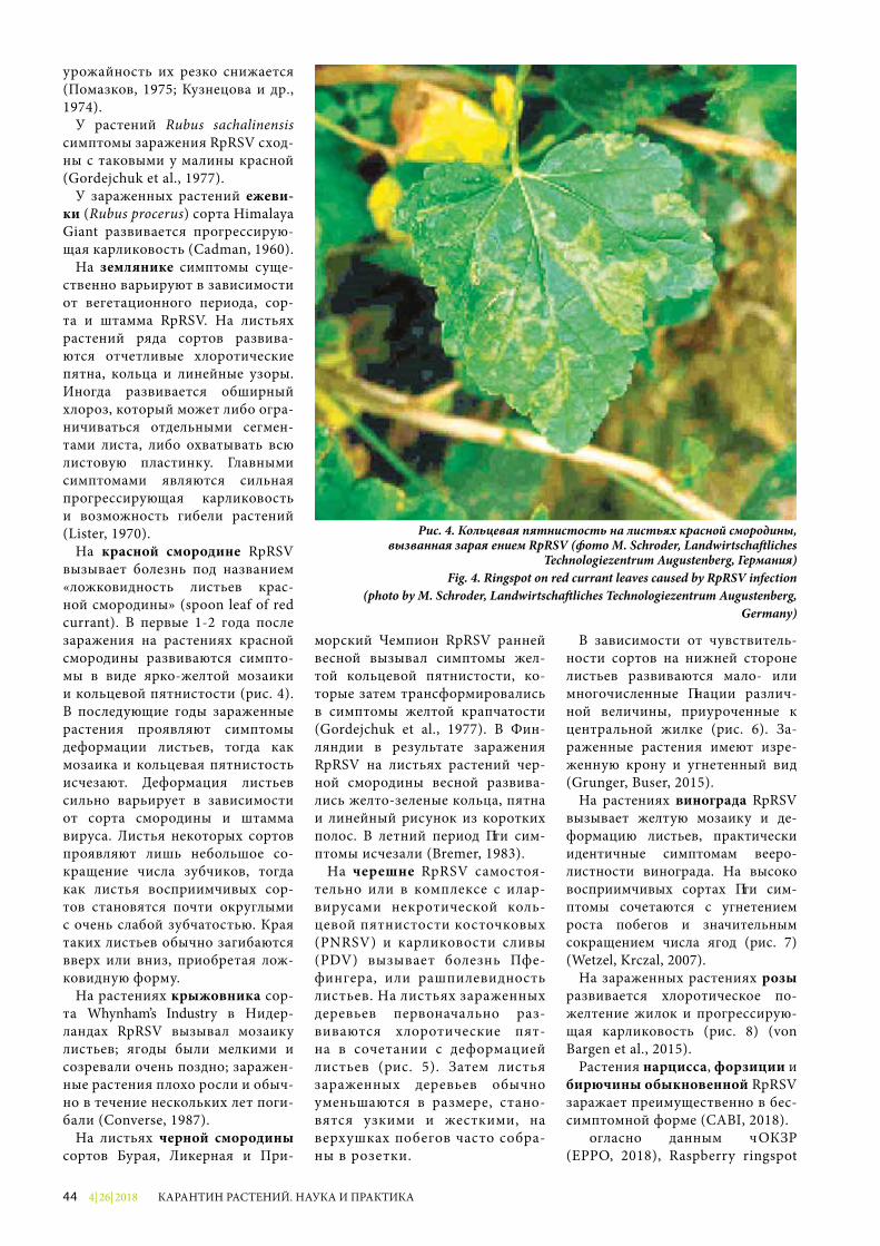
4| 26| 201844 КАРАНТИН РАСТЕНИЙ. НАУКА И ПРАКТИКА
урожайность их резко снижается (Помазков, 1975; Кузнецова и др., 1974).
У растений Rubus sachalinensis симптомы заражения RpRSV сход-ны с таковыми у малины красной (Gordejchuk et al., 1977).
У зараженных растений ежеви-ки (Rubus procerus) сорта Himalaya Giant развивается прогрессирую-щая карликовость (Cadman, 1960).
На землянике симптомы суще-ственно варьируют в зависимости от вегетационного периода, сор-та и штамма RpRSV. На листьях растений ряда сортов развива-ются отчетливые хлоротические пятна, кольца и линейные узоры. Иногда развивается обширный хлороз, который может либо огра-ничиваться отдельными сегмен-тами листа, либо охватывать всю листовую пластинку. Главными симптомами являются сильная прогрессирующая карликовость и возможность гибели растений (Lister, 1970).
На красной смородине RpRSV вызывает болезнь под названием «ложковидность листьев крас-ной смородины» (spoon leaf of red currant). В первые 1-2 года после заражения на растениях красной смородины развиваются симпто-мы в виде ярко-желтой мозаики и кольцевой пятнистости (рис. 4). В последующие годы зараженные растения проявляют симптомы деформации листьев, тогда как мозаика и кольцевая пятнистость исчезают. Деформация листьев сильно варьирует в зависимости от сорта смородины и штамма вируса. Листья некоторых сортов проявляют лишь небольшое со-кращение числа зубчиков, тогда как листья восприимчивых сор-тов становятся почти округлыми с очень слабой зубчатостью. Края таких листьев обычно загибаются вверх или вниз, приобретая лож-ковидную форму.
На растениях крыжовника сор-та Whynham’s Industry в Нидер-ландах RpRSV вызывал мозаику листьев; ягоды были мелкими и созревали очень поздно; заражен-ные растения плохо росли и обыч-но в течение нескольких лет поги-бали (Converse, 1987).
На листьях черной смородины сортов Бурая, Ликерная и При-
морский Чемпион RpRSV ранней весной вызывал симптомы жел-той кольцевой пятнистости, ко-торые затем трансформировались в симптомы желтой крапчатости (Gordejchuk et al., 1977). В Фин-ляндии в результате заражения RpRSV на листьях растений чер-ной смородины весной развива-лись желто-зеленые кольца, пятна и линейный рисунок из коротких полос. В летний период эти сим-птомы исчезали (Bremer, 1983).
На черешне RpRSV самостоя-тельно или в комплексе с илар-вирусами некротической коль-цевой пятнистости косточковых (PNRSV) и карликовости сливы (PDV) вызывает болезнь Пфе-фингера, или рашпилевидность листьев. На листьях зараженных деревьев первоначально раз-виваются хлоротические пят-на в сочетании с деформацией листьев (рис. 5). Затем листья зараженных деревьев обычно уменьшаются в размере, стано-вятся узкими и жесткими, на верхушках побегов часто собра-ны в розетки.
В зависимости от чувствитель-ности сортов на нижней стороне листьев развиваются мало- или многочисленные энации различ-ной величины, приуроченные к центральной жилке (рис. 6). За-раженные растения имеют изре-женную крону и угнетенный вид (Grunger, Buser, 2015).
На растениях винограда RpRSV вызывает желтую мозаику и де-формацию листьев, практически идентичные симптомам вееро-листности винограда. На высоко восприимчивых сортах эти сим-птомы сочетаются с угнетением роста побегов и значительным сокращением числа ягод (рис. 7) (Wetzel, Krczal, 2007).
На зараженных растениях розы развивается хлоротическое по-желтение жилок и прогрессирую-щая карликовость (рис. 8) (von Bargen et al., 2015).
Растения нарцисса, форзиции и бирючины обыкновенной RpRSV заражает преимущественно в бес-симптомной форме (CABI, 2018).
Согласно данным ЕОКЗР (EPPO, 2018), Raspberry ringspot
Рис. 4. Кольцевая пятнистость на листьях красной смородины,вызванная заражением RpRSV (фото M. Schroder, Landwirtscha�liches
Technologiezentrum Augustenberg, Германия)Fig. 4. Ringspot on red currant leaves caused by RpRSV infection
(photo by M. Schroder, Landwirtscha�liches Technologiezentrum Augustenberg, Germany)
4| 26| 2018 45КАРАНТИН РАСТЕНИЙ. НАУКА И ПРАКТИКА
virus в настоящее время зареги-стрирован в следующих странах:
Европа: Албания, Беларусь, Болгария, Великобритания, Гер-мания, Греция, Ирландия, Ита-лия, Латвия, Люксембург, Нидер-ланды, Норвегия, Португалия, Российская Федерация, Сербия, Турция, Финляндия, Франция, Чехия, Швейцария.
Азия: Казахстан.Включение RpRSV в Перечень
карантинных объектов, отсут-ствующих на территории ЕАЭС, вызывает большие сомнения.
В Российской Федерации RpRSV был широко распростра-нен в 50-70-е годы прошлого века (Помазков, 1975; Кузнецова и др., 1974), что было взаимосвязано с интродукцией зараженных кло-нов малины английской селекции, а также смородины и крыжовника голландской селекции. В после-дующие годы, в связи с широким внедрением в производство сортов ягодных культур отечественной селекции и системы производ-ства оздоровленного посадоч-ного материала, распространен-ность этого вируса в нашей стра-не несколько снизилась, однако о его выявлении по-прежнему име-ются многочисленные сообщения в отечественной литературе.
Так, RpRSV был выявлен при обследовании насаждений ма-лины в Московской, Ленин-градской и Самарской областях (Приходько, 2002). RpRSV вы-являли методом ИФА в коллек-ционных насаждениях красной смородины научных учреждений в Московской области и в ана-логичных насаждениях черной смородины в Московской, Ор-ловской и Самарской областях (Суркова, 1994). Распростра-ненность RpRSV в коллекцион-ных насаждениях крыжовника также была оценена как высо-кая (Приходько, Суркова, 1994). В результате проведенного серо-мониторинга RpRSV был выявлен в коллекционных насаждениях сирени и чубушника научных учреждений в городах Москвы и Подмосковья (Мельникова, При-ходько, 2006). В Российской Фе-дерации RpRSV выявляли также на розе (Келдыш, 1994) и георги-не (Шатило, 1991). В коллекции
ГБС РАН (г. Москва) заражен-ность RpRSV растений некоторых сортов розы достигала 41% (Чер-вякова, Келдыш, 1994).
Согласно последним данным, RpRSV был выявлен на малине в Московской, Рязанской и Брян-ской областях соответственно в 19,0, 8,2 и 49,5% образцов из 216, 44 и 78 протестированных.
В Московской области встре-чае мость RpRSV на пяти основ-ных сортах малины варьировала от 17,5 до 29,4% (Упадышев и др., 2014; Тихонова, 2016; Тихонова и др., 2016). При совместном за-ражении RpRSV и вирусом ку-стистой карликовости малины (RBDV) продуктивность некото-рых сортов малины снижалась до 44% (Тихонова и др., 2016).
В Республике Беларусь RpRSV широко распространен в насаж-де ни ях Института садоводства. Так, девять протестированных сортов черной смородины были заражены этим вирусом на 100%. Зараженность сортов красной смородины варьировала от 5 до 100%, а зараженность мали-ны составила в среднем 29,3% (Valasevich, Kolbanova, 2010).
Необходимо отметить, что все эти результаты были получены с использованием метода имму-ноферментного анализа (ИФА) без дополнительной идентифи-кации выявленных изолятов ме-тодом ПЦР.
Для уточнения фитосанитарно-го статуса RpRSV в Российской Федерации необходимо проведе-ние его мониторинга на всех ос-новных растениях-хозяевах.
В связи с импортом большо-го количества растений – хозяев RpRSV существует постоянная опасность проникновения новых штаммов этого вируса на терри-торию Российской Федерации, а наличие нематод-переносчиков обуславливает возможность его быстрого распространения в су-ществующих насаждениях.
Для предотвращения проник-новения RpRSV на территорию Российской Федерации должны практиковаться следующие фито-санитарные меры:
- посадочный и семенной мате-риал всех растений-хозяев дол-жен быть свободен от RpRSV;
- саженцы плодовых, ягодных, декоративных культур и виногра-да должны происходить из зон, мест и (или) участков производ-ства, свободных от RpRSV;
- посадочный материал, импор-тируемый из ЕОКЗР, должен быть выращен в соответствии с обще-признанными схемами сертифи-кации для косточковых плодовых культур, ягодных культур и вино-града;
- растения с открытой корневой системой должны быть свободны от почвы; при ввозе растений в контейнерах почвенный субстрат должен быть проверен на отсут-ствие нематод-переносчиков.
Для обеспечения этих мер не-обходимо практиковать предот-грузочное обследование участков производства, досмотр в пункте пропуска и лабораторное иссле-дование образцов на наличие RpRSV.
На территории Российской Фе-дерации должны соблюдаться сле-дующие фитосанитарные меры:
- систематические обследова-ния насаждений растений – хозя-ев RpRSV с отбором и лаборатор-ным исследованием образцов;
- выращивание семенного и по-садочного материала только в ме-стах производства, свободных от RpRSV;
- предпосадочное исследование почвы на отсутствие нематод-пе-реносчиков.
Литература1. Келдыш М.А. Особенности рас-
пространения неповирусов в антро-погенных экосистемах // Бюллетень ГБС РАН, 1994. Вып. 169. С. 132-136.
2. Кузнецова А.А., Келдыш М.А., Помазков Ю.И. Анализ некоторых изолятов курчавости малины // Промышленная культура малины в РСФСР. М., 1974. С. 77-82.
3. Мельникова Н.Н. Вирусные болезни древесных и декоратив-ных культур и разработка мер борьбы с ними // Автореф. дисс. … канд. с.-х. наук. М., 2005. 23 с.
4. Мельникова Н.Н., Приходько Ю.Н. Вирусные болезни древесных де-коративных культур в Подмос-ковье // Агроэкологические аспек-ты устойчивого развития АПК. Тезисы докл. науч. конф. Брянск, 2006. С. 47-50.
4| 26| 201846 КАРАНТИН РАСТЕНИЙ. НАУКА И ПРАКТИКА
5. Помазков Ю.И. Вирусные бо-лезни плодовых и ягодных куль-тур в Нечерноземной зоне // Ав-тореф. дисс. … д. с.-х. наук. М., 1975. 34 с.
6. Приходько Ю.Н. Распростра-ненность вирусных и фитоплаз-менных болезней на плодовых и ягодных культурах европейской части России // Доклады на от-четной сессии 2002 г. секции са-доводства и виноградарства от-деления растениеводства РАСХН. М., 2002. С. 209-219.
7. Приходько Ю.Н., Суркова О.Ю. Вирусные болезни крыжовника в средней полосе России // Плодо-водство и ягодоводство России / Сб. науч. тр. ВСТИСП, 1994. С. 124-134.
8. Приходько Ю.Н., Метлиц кая К.В. Нематоды – переносчики фито-патогенных вирусов плодовых и ягодных культур // Вестник РАСХН, 1994. № 1. С. 27-29.
9. Суркова О.Ю. Анализ рас-пространения вредоносности, этио-логии вирусных и вирусоподоб-ных болезней красной и черной смородины и разработка мер борьбы с ними в средней полосе России // Автореф. дис. … канд. с.-х. наук. М., 1994. 20 с.
10. Тихонова К.О. Распростра-ненность, вредоносность вирус-ных болезней и эффективные методы оздоровления малины // Автореф. дис. … канд. с.-х. наук. М., 2016. 20 с.
11. Тихонова К.О., Упадышев М.Т., Метлицкая К.В. О вредоносности вирусов на малине и оздоров-лении от них // Плодоводство и ягодоводство России / Сб. науч. тр. ВСТИСП, 2015. Т. XXXIII. С. 349-353.
12. Тихонова К.О., Упадышев М.Т., Метлицкая К.В. Распространен-ность и вредоносность вирусов ма-лины и современные способы ее оз-доровления // Сб. науч. тр. ВСТИСП, 2016. Т. XXXXI. С. 234-238.
13. Упадышев М.Т., Метлицкая К.В., Тихонова К.О., Евдокименко С.Н. О распространенности вирусных болезней малины в Центральном регионе России // Плодоводство и ягодоводство России / Сб. науч. тр. ВСТИСП, 2014. Т. XXXVII. Ч. 2. С. 184-190.
14. Червякова О.Н., Келдыш М.А. Вирусы непогруппы на предста-
вителях родов Rosa и Sorbus // Бюллетень ГБС, 1994. Вып. 170. С. 124-129.
15. Шатило В.И. Вирусные бо-лезни георгин и оздоровление от них методом культуры тканей // Автореф. дис. … канд. биол. наук. М., 1991. 21 с.
16. Blok V.C., Wardell J., Jolly C.A., Manoukian A., Robinson D.J., Edwards M.L., Mayo M.A. The nucleotide sequence of RNA-2 of Raspberry ringspot nepovirus // Journal of General Virology, 1992. Vol. 73 (9). P. 2189-2194.
17. Bremer K. Viral diseases occurring on Ribes species in Finland // Annales Agriculturae Fenniae, 1983. Vol. 22. P. 104-109.
18. Brown D.J.F., Taylor C.E., Choleva B., Romanenko N.D. The occurrence of Longidoridae (Nema-toda: Dorylaimida) in western USSR with further comments on longi-dorid nematodes in Europe and the Mediterranean basin // Nemato-logia Mediterranea, 1990. Vol. 18. P. 199-207.
19. Brunt A.A., Crabtree K., Dall-witz M.J., Gibbs A.J., Watson L. and Zurcher E.J. Raspberry ringspot nepovirus // Plant Viruses Online: Descriptions and Lists from the VIDE Database.Version: 20th Au-gust 1996. http://biology.anu.edu.au/Groups/MES/vide.
20. CABI Crop Protection Compendium. http://www.cabi.org/cpc/datasheet?/2018.
21. Cadman C.H. Studies on the etiology and mode of spread of Scottish raspberry leaf curl disease // Journal of Horticultural Science, 1956. Vol. 31. P. 111-118.
22. Cadman C.H. Studies on the relationship between soil-borne viruses of the ringspot type occurring in Britain and continental Europe // Virology, 1960. Vol. 11. P. 653-664.
23. Converse R.H. Virus diseases of small fruits // USDA Agricultural Handbook, 1987. № 631. 277 s.
24. EPPO Global Database, 2018.25. Gordejchuk O.G., Krylov O.V.,
Kyloba L.V., Samonina I.N. Zentralblatt fur Bakteriologia, Parasi tenkurde, Infectionskrank-hei ten und Hygiene, 1977. Vol. 132. P. 686-707.
26. Grunder J.M., Buser A. Viroses du cerisier (Prunus avium) causes
par des nematodes vecteurs // Station de recherché Agroscope, Switzerland, 2015. 4 s.
27. Harrison B.D., Murant A.F., Mayo M.A. Evidence for two functional RNA species in Raspberry ringspot virus // Journal of General Virology, 1972. Vol. 16. P. 339-348.
28. Lister R.M. Soilborne viruses as pathogens in strawberry // Virus diseases of small fruits and grapevines, a handbook (Ed. by Frazier N.W.), 1970. P. 34-36.
29. Lister R.M., Murant A.F. Seed-transmission of nematode-borne viruses // Annals of Applied Biology, 1967. Vol. 59. P. 49-62.
30. Phatak H.C. Seed-borne plant viruses-identification and diagnosis in seed health testing // Seed Sci. Technol., 1974. Vol. 2. P. 3-155.
31. Scientif ic opinion on the r isk to plant health posed by Arabis mosaic virus, Raspberr y r ingspot virus, Strawberr y latent r ingspot virus and Tomato black r ing virus to the EU territor y with the identif icat ion and evaluation of r isk reduction options // EFSA Journal, 2013. Vol. 11. P. 1-83.
32. Scott S.W., Zimmerman M.T., Jones A.T., Le Gall O. Differences between the coat protein amino acid sequences of English and Scottish serotypes of Raspberry r ingspot v irus exposed on the sur face of v irus par t ic les / / Virus Research, 2000. Vol . 68 (2) . P. 119-126.
33. Valasevich N., Kolbanova E. Occurrence of small fruit viruses in Belarus // 21st Int. Conf. Virus and other Transmissible Diseases of Fruit Crops / Julius-Kuhn-Archiv, 2010. Vol. 427. P. 129-132.
34. von Bargen S., Demiral R., Büt-tner C. First detection of Raspberry ringspot virus in mosaic diseased hybrid roses in Germany // New Disease Reports, 2015. Vol. 32. P. 18-19.
35. Wetzel T., Ebel R., Moury B., Le Gall O., Endisch S., Reustle G.M., Krczal G. Sequence analysis of grapevine isolates of Raspberry ringspot nepovirus // Archives of Virology, 2006. Vol. 151. P. 599-606.
36. Wetzel T., Krczal G. Molecular biology of Raspberry ringspot virus // Plant Viruses, 2007. Vol. 1. P. 45-51.
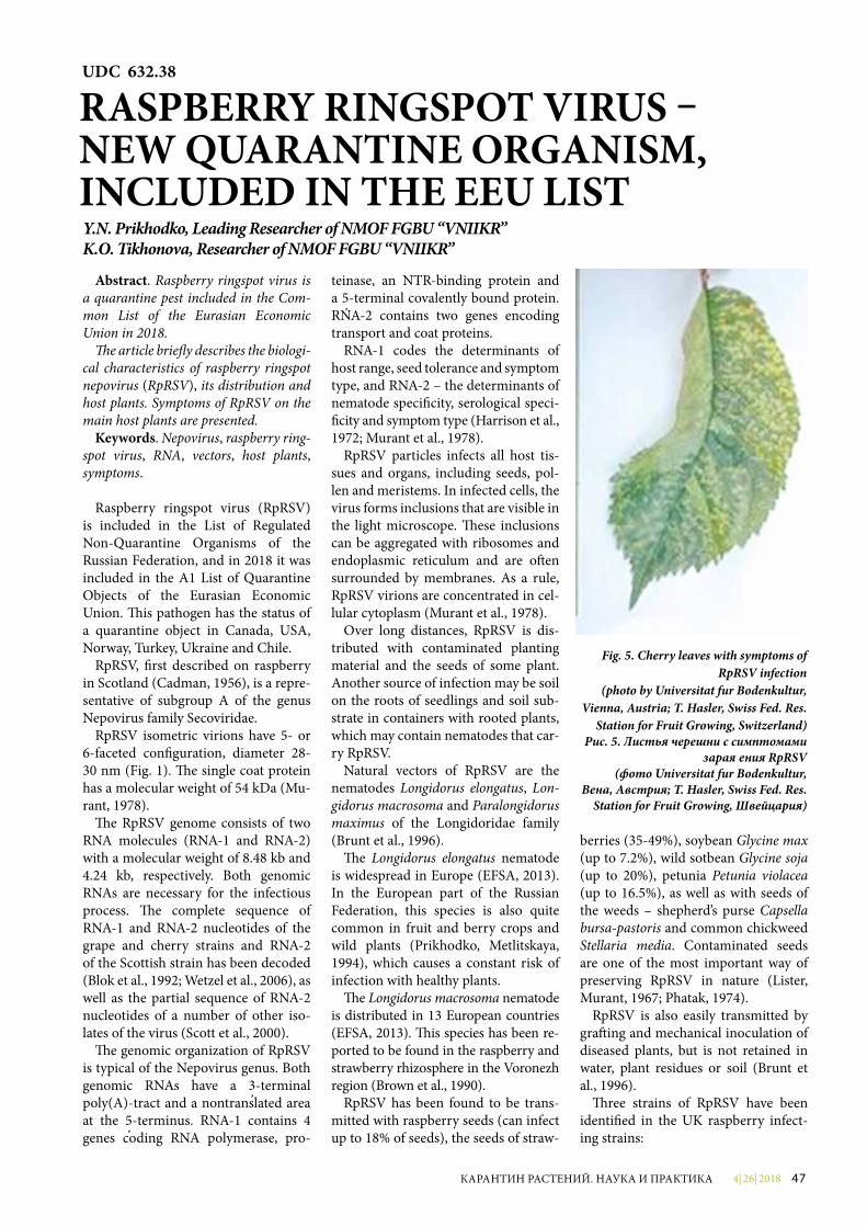
4| 26| 2018 47КАРАНТИН РАСТЕНИЙ. НАУКА И ПРАКТИКА
Abstract. Raspberry ringspot virus is a quarantine pest included in the Com-mon List of the Eurasian Economic Union in 2018.
�e article brie�y describes the biologi-cal characteristics of raspberry ringspot nepovirus (RpRSV), its distribution and host plants. Symptoms of RpRSV on the main host plants are presented.
Keywords. Nepovirus, raspberry ring-spot virus, RNA, vectors, host plants, symptoms.
Raspberry ringspot virus (RpRSV) is included in the List of Regulated Non-Quarantine Organisms of the Russian Federation, and in 2018 it was included in the A1 List of Quarantine Objects of the Eurasian Economic Union. �is pathogen has the status of a quarantine object in Canada, USA, Norway, Turkey, Ukraine and Chile.
RpRSV, �rst described on raspberry in Scotland (Cadman, 1956), is a repre-sentative of subgroup A of the genus Nepovirus family Secoviridae.
RpRSV isometric virions have 5- or 6-faceted con�guration, diameter 28-30 nm (Fig. 1). �e single coat protein has a molecular weight of 54 kDa (Mu-rant, 1978).
�e RpRSV genome consists of two RNA molecules (RNA-1 and RNA-2) with a molecular weight of 8.48 kb and 4.24 kb, respectively. Both genomic RNAs are necessary for the infectious process. �e complete sequence of RNA-1 and RNA-2 nucleotides of the grape and cherry strains and RNA-2 of the Scottish strain has been decoded (Blok et al., 1992; Wetzel et al., 2006), as well as the partial sequence of RNA-2 nucleotides of a number of other iso-lates of the virus (Scott et al., 2000).
�e genomic organization of RpRSV is typical of the Nepovirus genus. Both genomic RNAs have a 3-terminal poly(A)-tract and a nontranslated area at the 5-terminus. RNA-1 contains 4 genes coding RNA polymerase, pro-
teinase, an NTR-binding protein and a 5-terminal covalently bound protein. RNA-2 contains two genes encoding transport and coat proteins.
RNA-1 codes the determinants of host range, seed tolerance and symptom type, and RNA-2 – the determinants of nematode speci�city, serolo gical speci-�city and symptom type (Harrison et al., 1972; Murant et al., 1978).
RpRSV particles infects all host tis-sues and organs, including seeds, pol-len and meristems. In infected cells, the virus forms inclusions that are visible in the light microscope. �ese inclusions can be aggregated with ribosomes and endoplasmic reticulum and are o¥en surrounded by membranes. As a rule, RpRSV virions are concentrated in cel-lular cytoplasm (Murant et al., 1978).
Over long distances, RpRSV is dis-tributed with contaminated planting material and the seeds of some plant. Another source of infection may be soil on the roots of seedlings and soil sub-strate in containers with rooted plants, which may contain nematodes that car-ry RpRSV.
Natural vectors of RpRSV are the nematodes Longidorus elongatus, Lon-gidorus macrosoma and Paralongidorus maximus of the Longidoridae family (Brunt et al., 1996).
�e Longidorus elongatus nematode is widespread in Europe (EFSA, 2013). In the European part of the Russian Federation, this species is also quite common in fruit and berry crops and wild plants (Prikhodko, Metlitskaya, 1994), which causes a constant risk of infection with healthy plants.
�e Longidorus macrosoma nematode is distributed in 13 European countries (EFSA, 2013). �is species has been re-ported to be found in the raspberry and strawberry rhizosphere in the Voronezh region (Brown et al., 1990).
RpRSV has been found to be trans-mitted with raspberry seeds (can infect up to 18% of seeds), the seeds of straw-
berries (35-49%), soybean Glycine max (up to 7.2%), wild sotbean Glycine soja (up to 20%), petunia Petunia violacea (up to 16.5%), as well as with seeds of the weeds – shepherd’s purse Capsella bursa-pastoris and common chickweed Stellaria media. Contaminated seeds are one of the most important way of preserving RpRSV in nature (Lister, Murant, 1967; Phatak, 1974).
RpRSV is also easily transmitted by gra¥ing and mechanical inoculation of diseased plants, but is not retained in water, plant residues or soil (Brunt et al., 1996).
�ree strains of RpRSV have been identi�ed in the UK raspberry infect-ing strains:
Fig. 5. Cherry leaves with symptoms of RpRSV infection
(photo by Universitat fur Bodenkultur, Vienna, Austria; T. Hasler, Swiss Fed. Res.
Station for Fruit Growing, Switzerland)Рис. 5. Листья черешни с симптомами
заражения RpRSV(фото Universitat fur Bodenkultur,
Вена, Австрия; T. Hasler, Swiss Fed. Res. Station for Fruit Growing, Швейцария)
UDC 632.38
RASPBERRY RINGSPOT VIRUS – NEW QUARANTINE ORGANISM, INCLUDED IN THE EEU LISTY.N. Prikhodko, Leading Researcher of NMOF FGBU “VNIIKR”K.O. Tikhonova, Researcher of NMOF FGBU “VNIIKR”
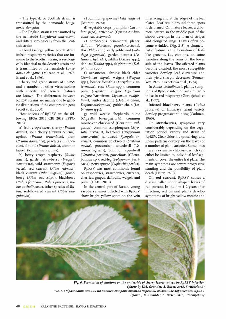
4| 26| 201848 КАРАНТИН РАСТЕНИЙ. НАУКА И ПРАКТИКА
Fig. 6. Formation of enations on the underside of cherry leaves caused by RpRSV infection (photo by J.M. Grunder, A. Buser, 2015, Switzerland)
Рис. 6. Образование энаций на нижней стороне листьев черешни, вызванное заражением RpRSV (фото J.M. Grunder, A. Buser, 2015, Швейцария)
- �e typical, or Scottish strain, is transmitted by the nematode Longi-dorus elongatus;
- �e English strain is transmitted by the nematode Longidorus macrosoma and di�ers serologically from the Scot-tish strain;
- Lloyd George yellow blotch strain infects raspberry varieties that are im-mune to the Scottish strain, is serologi-cally identical to the Scottish strain and is transmitted by the nematode Longi-dorus elongatus (Murant et al., 1978; Brunt et al., 1996).
Cherry and grape strains of RpRSV and a number of other virus isolates with speci�c and genetic features are known. �e di�erences between RpRSV strains are mainly due to gene-tic distinctions of the coat protein gene (Scott et al., 2000).
Host species of RpRSV are the fol-lowing (EFSA, 2013; CBI, 2018; EPPO, 2018):
a) fruit crops: sweet cherry (Prunus avium), sour cherry (Prunus cerasus), apricot (Prunus armeniaca), plum (Prunus domestica), peach (Prunus per-sica), almond (Prunus dulcis), common laurel (Prunus laurocerasus);
b) berry crops: raspberry (Rubus idaeus), garden strawberry (Fragaria xananassa), wild strawberry (Fragaria vesca), red currant (Ribes rubrum), black currant (Ribes nigrum), goose-berry (Ribes uva-crispa), blackberry (Rubus fruticosus, Rubus procerus, Ru-bus sachalinensis), other species of Ru-bus, red-�owered currant (Ribes san-guineum);
c) common grapevine (Vitis vinifera) (Murant, 1978);
d) vegetable crops: pumpkin (Cucur-bita pepo), artichoke (Cynara cardun-culus var. scolymus);
e) herbaceous ornamental plants: da�odil (Narcissus pseudonarcissus), �ox (Phlox spp.), early goldenrod (Soli-dago gigantean), garden petunia (Pe-tunia x hybrida), astilba (Astilbe spp.), dahlias (Dahlia spp.), delphinium (Del-phinium spp.);
f) ornamental shrubs: black elder (Sambucus nigra), weigela (Weigela spp.), border forsythia (Forsythia x in-termedia), rose (Rosa spp.), common privet (Ligustrum vulgare, Ligustrum vulgare ‘Atrovirens’, Ligustrum ovalifo-lium), winter daphne (Daphne odora, Daphne burkwoodii), golden chain (La-burnum spp.);
g) wild weeds: shepherd’s purse (Capsella bursa-pastoris), common mouse-ear chickweed (Cerastium vul-gatum), common scorpiongrass (Myo-sotis arvensis), bearbind (Polygonum convolvulus), sandweed (Spergula ar-vensis), common chickweed (Stellaria media), procumbent speedwell (Ve-ronica agrestis), common speedwell (Veronica persica), goosefoots (Cheno-podium sp.), red-leg (Polygonum persi-caria), petty spurge (Euphorbia peplus).
RpRSV was most commonly found on raspberries, strawberries, currants, cherries, grapes, da�odils, weigels and privet (CABI, 2018).
In the central part of Russia, young raspberry leaves infected with RpRSV show bright yellow spots on the vein
interlacing and at the edges of the leaf plates. Leaf tissue around these spots is deformed. On mature leaves, a chlo-rotic pattern in the middle part of the shoots develops in the form of stripes and elongated rings. Leaves o¥en be-come wrinkled (Fig. 2-3). A characte-ristic feature is the formation of leaf-like growths, i.e., enations, on some varieties along the veins on the lower side of the leaves. �e a�ected plants become dwarfed, the most susceptible varieties develop leaf curvature and their yield sharply decreases (Pomaz-kov, 1975; Kuznetsova et al., 1974).
In Rubus sachalinensis plants, symp-toms of RpRSV infection are similar to those in red raspberry (Gordejchuk et al., 1977).
Infested blackberry plants (Rubus procerus) of Himalaya Giant variety develop progressive stunting (Cadman, 1960).
On strawberries, symptoms vary considerably depending on the vege-tation period, variety and strain of RpRSV. Clear chlorotic spots, rings and linear patterns develop on the leaves of a number of plant varieties. Sometimes there is extensive chlorosis, which can either be limited to individual leaf seg-ments or cover the entire leaf plate. �e main symptoms are severe progressive stunting and the possibility of plant death (Lister, 1970).
On red currant, RpRSV causes a disease called spoon-shaped leaves of red currant. In the �rst 1-2 years a¥er infection, red currant plants develop symptoms of bright yellow mosaic and
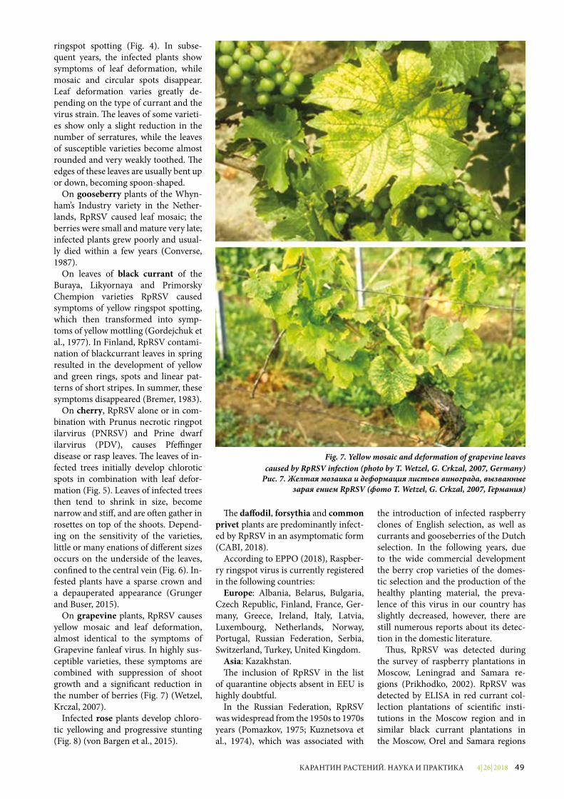
4| 26| 2018 49КАРАНТИН РАСТЕНИЙ. НАУКА И ПРАКТИКА
ringspot spotting (Fig. 4). In subse-quent years, the infected plants show symptoms of leaf deformation, while mosaic and circular spots disappear. Leaf deformation varies greatly de-pending on the type of currant and the virus strain. �e leaves of some varieti-es show only a slight reduction in the number of serratures, while the leaves of susceptible varieties become almost rounded and very weakly toothed. �e edges of these leaves are usually bent up or down, becoming spoon-shaped.
On gooseberry plants of the Whyn-ham’s Industry variety in the Nether-lands, RpRSV caused leaf mosaic; the berries were small and mature very late; infected plants grew poorly and usual-ly died within a few years (Converse, 1987).
On leaves of black currant of the Buraya, Likyornaya and Primorsky Chempion varieties RpRSV caused symptoms of yellow ringspot spotting, which then transformed into symp-toms of yellow mottling (Gordejchuk et al., 1977). In Finland, RpRSV contami-nation of blackcurrant leaves in spring resulted in the development of yellow and green rings, spots and linear pat-terns of short stripes. In summer, these symptoms disappeared (Bremer, 1983).
On cherry, RpRSV alone or in com-bination with Prunus necrotic ringpot ilarvirus (PNRSV) and Prine dwarf ilarvirus (PDV), causes Pfe¦nger disea se or rasp leaves. �e leaves of in-fected trees initially develop chlorotic spots in combination with leaf defor-mation (Fig. 5). Leaves of infected trees then tend to shrink in size, become narrow and sti�, and are o¥en gather in rosettes on top of the shoots. Depend-ing on the sensitivity of the varieties, little or many enations of di�erent sizes occurs on the underside of the leaves, con�ned to the central vein (Fig. 6). In-fested plants have a sparse crown and a depauperated appearance (Grunger and Buser, 2015).
On grapevine plants, RpRSV causes yellow mosaic and leaf deformation, almost identical to the symptoms of Grapevine fanleaf virus. In highly sus-ceptible varieties, these symptoms are combined with suppression of shoot growth and a signi�cant reduction in the number of berries (Fig. 7) (Wetzel, Krczal, 2007).
Infected rose plants develop chloro-tic yellowing and progressive stunting (Fig. 8) (von Bargen et al., 2015).
�e da¡odil, forsythia and common privet plants are predominantly infect-ed by RpRSV in an asymptomatic form (CABI, 2018).
According to EPPO (2018), Raspber-ry ringspot virus is currently registered in the following countries:
Europe: Albania, Belarus, Bulgaria, Czech Republic, Finland, France, Ger-many, Greece, Ireland, Italy, Latvia, Luxembourg, Netherlands, Norway, Portugal, Russian Federation, Serbia, Switzerland, Turkey, United Kingdom.
Asia: Kazakhstan.�e inclusion of RpRSV in the list
of quarantine objects absent in EEU is highly doubtful.
In the Russian Federation, RpRSV was widespread from the 1950s to 1970s years (Pomazkov, 1975; Kuznetsova et al., 1974), which was associated with
the introduction of infected raspberry clones of English selection, as well as currants and gooseberries of the Dutch selection. In the following years, due to the wide commercial development the berry crop varieties of the domes-tic selection and the production of the healthy planting material, the preva-lence of this virus in our country has slightly decreased, however, there are still numerous reports about its detec-tion in the domestic literature.
�us, RpRSV was detected during the survey of raspberry plantations in Moscow, Leningrad and Samara re-gions (Prikhodko, 2002). RpRSV was detected by ELISA in red currant col-lection plantations of scienti�c insti-tutions in the Moscow region and in similar black currant plantations in the Moscow, Orel and Samara regions
Fig. 7. Yellow mosaic and deformation of grapevine leavescaused by RpRSV infection (photo by T. Wetzel, G. Crkzal, 2007, Germany)
Рис. 7. Желтая мозаика и деформация листьев винограда, вызванные заражением RpRSV (фото T. Wetzel, G. Crkzal, 2007, Германия)

4| 26| 201850 КАРАНТИН РАСТЕНИЙ. НАУКА И ПРАКТИКА
Fig. 8. Symptoms caused by RpRSV infection on a rose(photo by S. von Bargen et al., 2015, Germany)
Рис. 8. Симптомы, вызванные заражением RpRSV, на розе(фото S. von Bargen et al., 2015, Германия)
(Surkova, 1994). �e prevalence of RpRSV in gooseberry collection plan-tations was also assessed as high (Pri-khodko, Surkova, 1994). As a result of seromonitoring, RpRSV was found in the collection plantations of lilac and mock orange of scienti�c institu-tions in Moscow and Moscow region (Melnikova, Prikhodko, 2006). In the Russian Federation, RpRSV was also detected on roses (Keldysh, 1994) and dahlias (Shatilo, 1991). In the GBS RAS collection (Moscow), the infection of RpRSV in some rose varieties reached 41% (Chervyakova and Keldysh, 1994).
According to the latest data, RpRSV was detected on raspberry in the Mos-cow, Ryazan and Bryansk regions re-spectively in 19.0, 8.2 and 49.5% of the samples from 216, 44 and 78 tested. In the Moscow region, RpRSV abundance in the �ve main raspberry varieties ranged from 17.5 to 29.4% (Upadyshev et al., 2014; Tikhonova, 2016; Tikhono-va et al., 2016). When RpRSV and Raspberry bushy dwarf virus (RBDV) were co-infected, the productivity of some raspberry varieties decreased to 44% (Tikhonova et al., 2016).
In the Republic of Belarus, RpRSV is widespread in the plantations of the Institute of Horticulture. �us, nine black currant varieties tested were 100% infected with the virus. Infection of red currant varieties ranged from 5 to 100%, while raspberry infection averag ed 29.3% (Valasevich and Kol-banova, 2010).
It should be noted that all these results were obtained using enzyme-linked immunoassay (ELISA) without addi-tional identi�cation of the detected iso-lates by PCR.
In order to clarify the phytosanitary status of RpRSV in the Russian Federa-tion, it is necessary to monitor it on all main host plants.
Due to the import of a large number of host plants of RpRSV there is a con-stant danger of entry of new strains of the virus to the territory of the Russian Federation, and the presence of nema-tode vectors makes it possible to spread it quickly in existing plantations.
In order to prevent the entry of RpRSV to the territory of the Russian Federation, the following phytosani-tary measures should be practiced:
- planting and seed material of all host plants should be free from RpRSV;
- plants for planting of fruit, berry, ornamental crops and grapes should
originate from areas, locations and/or production sites free of RpRSV;
- planting material imported from the EPPO region should be grown in accordance with generally accepted cer-ti�cation schemes for stone fruit crops, berries and grapes;
- plants with open root systems should be soil free; when importing plants in containers, the soil substrate should be checked for absence of nematode vec-tors.
To ensure these measures, pre-ship-ment inspection of production sites, entry point inspection and laboratory testing of samples for RpRSVs should be practiced.
�e following phytosanitary measures should be followed in the Russian Fede-ration:
- systematic examination of host plan-tations of RpRSV with sampling and laboratory testing of samples;
- cultivation of seed and planting ma-terial only in production areas free from RpRSV;
- pre-planting soil testing for the ab-sence of nematode vectors.
References1. Keldysh M.A. Peculiarities of
spreading of nepoviruses in anthro-pogenic ecosystems // Bulletin of GBS RAS, 1994. Issue 169. P. 132-136.
2. Kuznetsova A.A., Keldysh M.A., Pomazkov Y.I. Analysis of some raspberry leaf curl isolates // Indus-trial raspberry culture in the RSFSR. М., 1974. P. 77-82.
3. Melnikova N.N. Viral diseases of woody and ornamental crops and development of measures to control them // Synopsis of thesis, PhD in Agriculture. М., 2005. 23 p.
4. Melnikova N.N., Prikhodko Y.N. Viral diseases of wood ornamental cultures in the Moscow region // Agroecological aspects of sus-tainable development of agro-in-dustrial complex. Theses of scien-tific conference report. Bryansk.
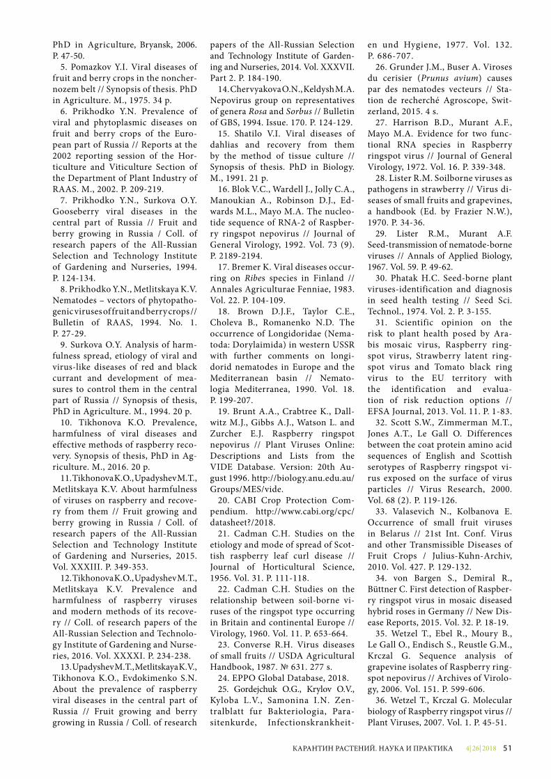
4| 26| 2018 51КАРАНТИН РАСТЕНИЙ. НАУКА И ПРАКТИКА
PhD in Agriculture, Bryansk, 2006. P. 47-50.
5. Pomazkov Y.I. Viral diseases of fruit and berry crops in the noncher-nozem belt // Synopsis of thesis. PhD in Agriculture. М., 1975. 34 p.
6. Prikhodko Y.N. Prevalence of viral and phytoplasmic diseases on fruit and berry crops of the Euro-pean part of Russia // Reports at the 2002 reporting session of the Hor-ticulture and Viticulture Section of the Department of Plant Industry of RAAS. М., 2002. P. 209-219.
7. Prikhodko Y.N., Surkova O.Y. Gooseberry viral diseases in the cent ral part of Russia // Fruit and berry growing in Russia / Coll. of research papers of the All-Russian Selection and Technology Institute of Gardening and Nurseries, 1994. P. 124-134.
8. Prikhodko Y.N., Metlitskaya K.V. Nematodes – vectors of phytopatho-genic viruses of fruit and berry crops // Bulletin of RAAS, 1994. No. 1. P. 27-29.
9. Surkova O.Y. Analysis of harm-fulness spread, etiology of viral and virus-like diseases of red and black currant and development of mea-sures to control them in the central part of Russia // Synopsis of thesis, PhD in Agriculture. М., 1994. 20 p.
10. Tikhonova K.O. Prevalence, harmfulness of viral diseases and effective methods of raspberry reco-very. Synopsis of thesis, PhD in Ag-riculture. М., 2016. 20 p.
11. Tikhonova K.O., Upadyshev M.T., Metlitskaya K.V. About harmfulness of viruses on raspberry and recove-ry from them // Fruit growing and berry growing in Russia / Coll. of research papers of the All-Russian Selection and Technology Institute of Gardening and Nurseries, 2015. Vol. XXXIII. P. 349-353.
12. Tikhonova K.O., Upadyshev M.T., Metlitskaya K.V. Prevalence and harmfulness of raspberry viruses and modern methods of its recove-ry // Coll. of research papers of the All-Russian Selection and Technolo-gy Institute of Gardening and Nurse-ries, 2016. Vol. XXXXI. P. 234-238.
13. Upadyshev M.T., Metlitskaya K.V., Tikhonova K.O., Evdokimenko S.N. About the prevalence of raspberry viral diseases in the central part of Russia // Fruit growing and berry growing in Russia / Coll. of research
papers of the All-Russian Selection and Technology Institute of Garden-ing and Nurseries, 2014. Vol. XXXVII. Part 2. P. 184-190.
14. Chervyakova O.N., Keldysh M.A. Nepovirus group on representatives of genera Rosa and Sorbus // Bulletin of GBS, 1994. Issue. 170. P. 124-129.
15. Shatilo V.I. Viral diseases of dahlias and recovery from them by the method of tissue culture // Synop sis of thesis. PhD in Biology. М., 1991. 21 p.
16. Blok V.C., Wardell J., Jolly C.A., Manoukian A., Robinson D.J., Ed-wards M.L., Mayo M.A. The nucleo-tide sequence of RNA-2 of Raspber-ry ringspot nepovirus // Journal of General Virology, 1992. Vol. 73 (9). P. 2189-2194.
17. Bremer K. Viral diseases occur-ring on Ribes species in Finland // Annales Agriculturae Fenniae, 1983. Vol. 22. P. 104-109.
18. Brown D.J.F., Taylor C.E., Choleva B., Romanenko N.D. The occurrence of Longidoridae (Nema-toda: Dorylaimida) in western USSR with further comments on longi-dorid nematodes in Europe and the Mediterranean basin // Nemato-logia Mediterranea, 1990. Vol. 18. P. 199-207.
19. Brunt A.A., Crabtree K., Dall-witz M.J., Gibbs A.J., Watson L. and Zurcher E.J. Raspberry ringspot nepovirus // Plant Viruses Online: Descriptions and Lists from the VIDE Database. Version: 20th Au-gust 1996. http://biology.anu.edu.au/Groups/MES/vide.
20. CABI Crop Protection Com-pendium. http://www.cabi.org/cpc/datasheet?/2018.
21. Cadman C.H. Studies on the etiology and mode of spread of Scot-tish raspberry leaf curl disease // Journal of Horticultural Science, 1956. Vol. 31. P. 111-118.
22. Cadman C.H. Studies on the relationship between soil-borne vi-ruses of the ringspot type occurring in Britain and continental Europe // Virology, 1960. Vol. 11. P. 653-664.
23. Converse R.H. Virus diseases of small fruits // USDA Agricultural Handbook, 1987. № 631. 277 s.
24. EPPO Global Database, 2018.25. Gordejchuk O.G., Krylov O.V.,
Kyloba L.V., Samonina I.N. Zen-tral blatt fur Bakteriologia, Para-siten kurde, Infectionskrankheit-
en und Hygiene, 1977. Vol. 132. P. 686-707.
26. Grunder J.M., Buser A. Viroses du cerisier (Prunus avium) causes par des nematodes vecteurs // Sta-tion de recherché Agroscope, Swit-zerland, 2015. 4 s.
27. Harrison B.D., Murant A.F., Mayo M.A. Evidence for two func-tional RNA species in Raspberry ringspot virus // Journal of General Virology, 1972. Vol. 16. P. 339-348.
28. Lister R.M. Soilborne viruses as pathogens in strawberry // Virus di-seases of small fruits and grapevines, a handbook (Ed. by Frazier N.W.), 1970. P. 34-36.
29. Lister R.M., Murant A.F. Seed-transmission of nematode-borne viruses // Annals of Appli ed Biology, 1967. Vol. 59. P. 49-62.
30. Phatak H.C. Seed-borne plant viruses-identification and diagnosis in seed health testing // Seed Sci. Technol., 1974. Vol. 2. P. 3-155.
31. Scientific opinion on the risk to plant health posed by Ara-bis mosaic virus, Raspberry ring-spot virus, Strawberry latent ring-spot virus and Tomato black ring virus to the EU territory with the identification and evalua-tion of risk reduction options // EFSA Journal, 2013. Vol. 11. P. 1-83.
32. Scott S.W., Zimmerman M.T., Jones A.T., Le Gall O. Differences between the coat protein amino acid sequences of English and Scottish serotypes of Raspberry ringspot vi-rus exposed on the surface of virus particles // Virus Research, 2000. Vol. 68 (2). P. 119-126.
33. Valasevich N., Kolbanova E. Occurrence of small fruit viruses in Belarus // 21st Int. Conf. Virus and other Transmissible Diseases of Fruit Crops / Julius-Kuhn-Archiv, 2010. Vol. 427. P. 129-132.
34. von Bargen S., Demiral R., Büttner C. First detection of Raspber-ry ringspot virus in mosaic diseased hybrid roses in Germany // New Dis-ease Reports, 2015. Vol. 32. P. 18-19.
35. Wetzel T., Ebel R., Moury B., Le Gall O., Endisch S., Reustle G.M., Krczal G. Sequence analysis of grapevine isolates of Raspberry ring-spot nepovirus // Archives of Virolo-gy, 2006. Vol. 151. P. 599-606.
36. Wetzel T., Krczal G. Molecular biology of Raspberry ringspot virus // Plant Viruses, 2007. Vol. 1. P. 45-51.
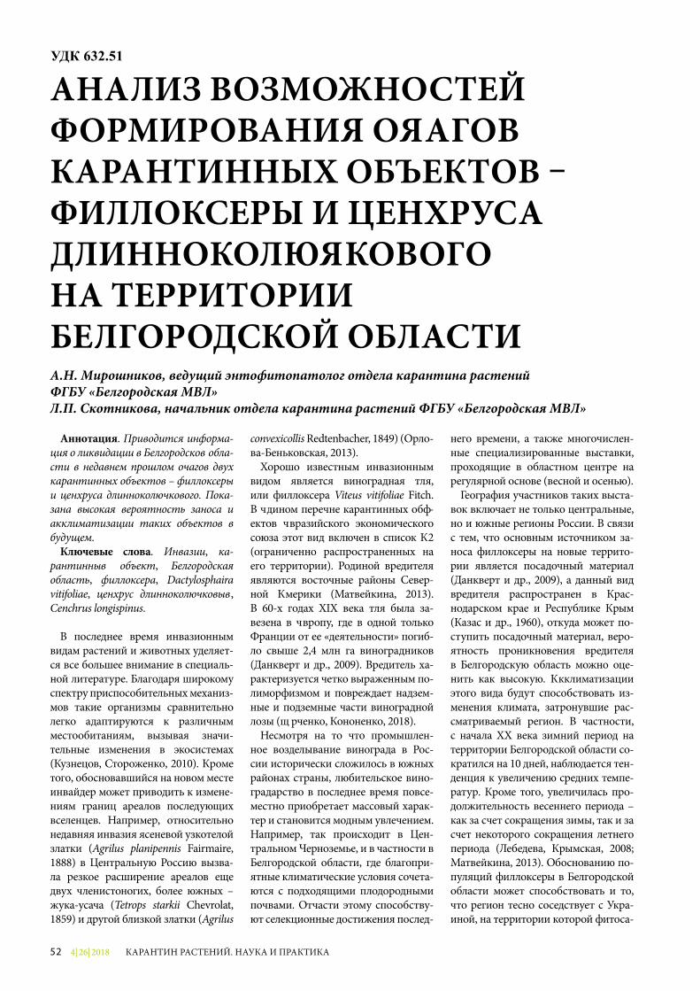
4| 26| 201852 КАРАНТИН РАСТЕНИЙ. НАУКА И ПРАКТИКА
Аннотация. Приводится информа-ция о ликвидации в Белгородской обла-сти в недавнем прошлом очагов двух карантинных объектов – филлоксеры и ценхруса длинноколючкового. Пока-зана высокая вероятность заноса и акклиматизации таких объектов в будущем.
Ключевые слова. Инвазии, ка-рантинный объект, Белгородская область, филлоксера, Dactylosphaira vitifoliae, ценхрус длинноколючковый, Cenchrus longispinus.
В последнее время инвазионным видам растений и животных уделяет-ся все большее внимание в специаль-ной литературе. Благодаря широкому спектру приспособительных механиз-мов такие организмы сравнительно легко адаптируются к различным местообитаниям, вызывая значи-тельные изменения в экосистемах (Кузнецов, Стороженко, 2010). Кроме того, обосновавшийся на новом месте инвайдер может приводить к измене-ниям границ ареалов последующих вселенцев. Например, относительно недавняя инвазия ясеневой узкотелой златки (Agrilus planipennis Fairmaire, 1888) в Центральную Россию вызва-ла резкое расширение ареалов еще двух членистоногих, более южных – жука-усача (Tetrops starkii Chevrolat, 1859) и другой близкой златки (Agrilus
convexicollis Redtenbacher, 1849) (Орло-ва-Беньковская, 2013).
Хорошо известным инвазионным видом является виноградная тля, или филлоксера Viteus vitifoliae Fitch. В Едином перечне карантинных объ-ектов Евразийского экономического союза этот вид включен в список А2 (ограниченно распространенных на его территории). Родиной вредителя являются восточные районы Север-ной Америки (Матвейкина, 2013). В 60-х годах XIX века тля была за-везена в Европу, где в одной только Франции от ее «деятельности» погиб-ло свыше 2,4 млн га виноградников (Данкверт и др., 2009). Вредитель ха-рактеризуется четко выраженным по-лиморфизмом и повреждает надзем-ные и подземные части виноградной лозы (Юрченко, Кононенко, 2018).
Несмотря на то что промышлен-ное возделывание винограда в Рос-сии исторически сложилось в южных районах страны, любительское вино-градарство в последнее время повсе-местно приобретает массовый харак-тер и становится модным увлечением. Например, так происходит в Цен-тральном Черноземье, и в частности в Белгородской области, где благопри-ятные климатические условия сочета-ются с подходящими плодородными почвами. Отчасти этому способству-ют селекционные достижения послед-
него времени, а также многочислен-ные специализированные выставки, проходящие в областном центре на регулярной основе (весной и осенью).
География участников таких выста-вок включает не только центральные, но и южные регионы России. В связи с тем, что основным источником за-носа филлоксеры на новые террито-рии является посадочный материал (Данкверт и др., 2009), а данный вид вредителя распространен в Крас-нодарском крае и Республике Крым (Казас и др., 1960), откуда может по-ступить посадочный материал, веро-ятность проникновения вредителя в Белгородскую область можно оце-нить как высокую. Акклиматизации этого вида будут способствовать из-менения климата, затронувшие рас-сматриваемый регион. В частности, с начала XX века зимний период на территории Белгородской области со-кратился на 10 дней, наблюдается тен-денция к увеличению средних темпе-ратур. Кроме того, увеличилась про-должительность весеннего периода – как за счет сокращения зимы, так и за счет некоторого сокращения летнего периода (Лебедева, Крымская, 2008; Матвейкина, 2013). Обоснованию по-пуляций филлоксеры в Белгородской области может способствовать и то, что регион тесно соседствует с Укра-иной, на территории которой фитоса-
АНАЛИЗ ВОЗМОЖНОСТЕЙ ФОРМИРОВАНИЯ ОЧАГОВ КАРАНТИННЫХ ОБЪЕКТОВ – ФИЛЛОКСЕРЫ И ЦЕНХРУСА ДЛИННОКОЛЮЧКОВОГО НА ТЕРРИТОРИИ БЕЛГОРОДСКОЙ ОБЛАСТИ
УДК 632.51
А.Н. Мирошников, ведущий энтофитопатолог отдела карантина растений ФГБУ «Белгородская МВЛ»Л.П. Скотникова, начальник отдела карантина растений ФГБУ «Белгородская МВЛ»
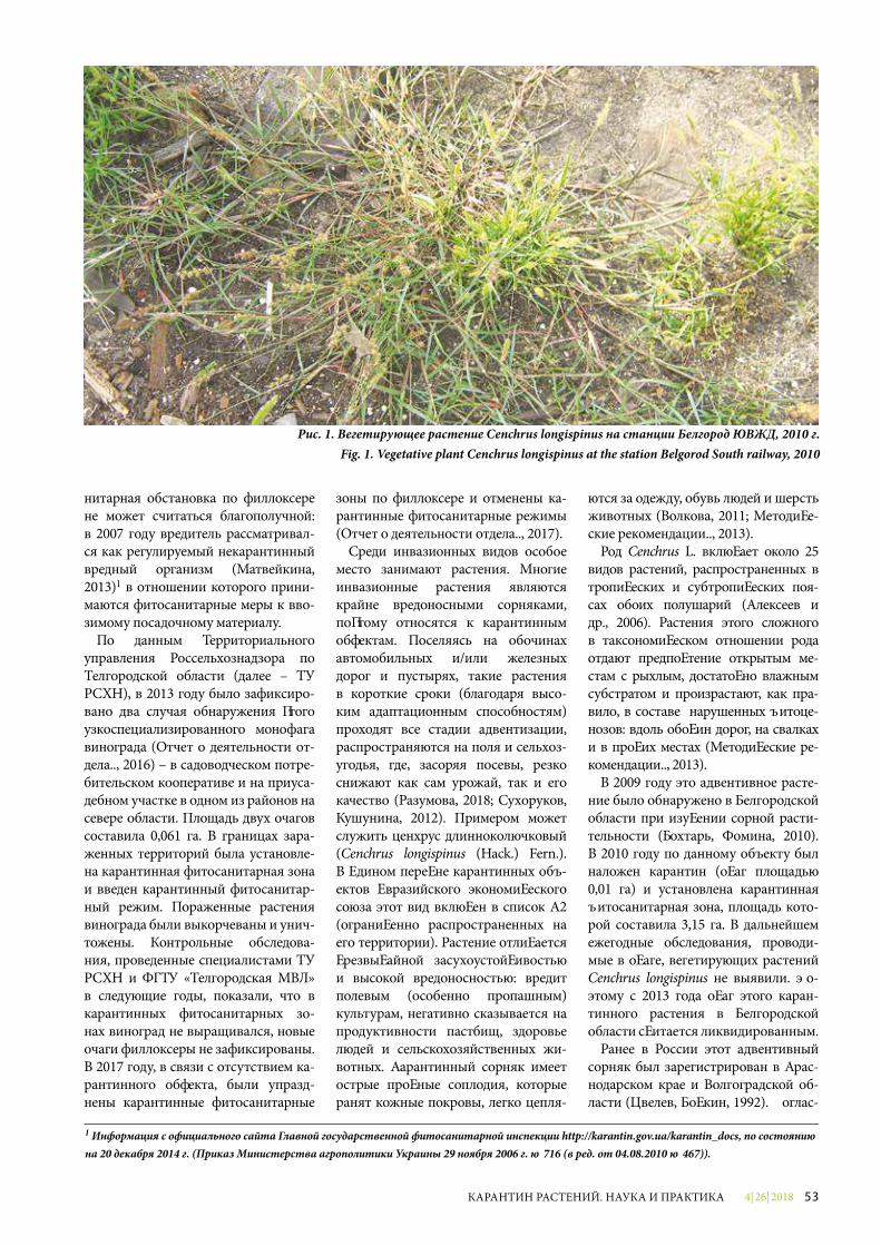
4| 26| 2018 53КАРАНТИН РАСТЕНИЙ. НАУКА И ПРАКТИКА
нитарная обстановка по филлоксере не может считаться благополучной: в 2007 году вредитель рассматривал-ся как регулируемый некарантинный вредный организм (Матвейкина, 2013)1 в отношении которого прини-маются фитосанитарные меры к вво-зимому посадочному материалу.
По данным Территориального управления Россельхознадзора по Белгородской области (далее – ТУ РСХН), в 2013 году было зафиксиро-вано два случая обнаружения этого узкоспециализированного монофага винограда (Отчет о деятельности от-дела.., 2016) – в садоводческом потре-бительском кооперативе и на приуса-дебном участке в одном из районов на севере области. Площадь двух очагов составила 0,061 га. В границах зара-женных территорий была установле-на карантинная фитосанитарная зона и введен карантинный фитосанитар-ный режим. Пораженные растения винограда были выкорчеваны и унич-тожены. Контрольные обследова-ния, проведенные специалистами ТУ РСХН и ФГБУ «Белгородская МВЛ» в следующие годы, показали, что в карантинных фитосанитарных зо-нах виноград не выращивался, новые очаги филлоксеры не зафиксированы. В 2017 году, в связи с отсутствием ка-рантинного объекта, были упразд-нены карантинные фитосанитарные
зоны по филлоксере и отменены ка-рантинные фитосанитарные режимы (Отчет о деятельности отдела.., 2017).
Среди инвазионных видов особое место занимают растения. Многие инвазионные растения являются крайне вредоносными сорняками, поэтому относятся к карантинным объектам. Поселяясь на обочинах автомобильных и/или железных дорог и пустырях, такие растения в короткие сроки (благодаря высо-ким адаптационным способностям) проходят все стадии адвентизации, распространяются на поля и сельхоз-угодья, где, засоряя посевы, резко снижают как сам урожай, так и его качество (Разумова, 2018; Сухоруков, Кушунина, 2012). Примером может служить ценхрус длинноколючковый (Cenchrus longispinus (Hack.) Fern.). В Едином перечне карантинных объ-ектов Евразийского экономического союза этот вид включен в список А2 (ограниченно распространенных на его территории). Растение отличается чрезвычайной засухоустойчивостью и высокой вредоносностью: вредит полевым (особенно пропашным) культурам, негативно сказывается на продуктивности пастбищ, здоровье людей и сельскохозяйственных жи-вотных. Карантинный сорняк имеет острые прочные соплодия, которые ранят кожные покровы, легко цепля-
ются за одежду, обувь людей и шерсть животных (Волкова, 2011; Методиче-ские рекомендации.., 2013).
Род Cenchrus L. включает около 25 видов растений, распространенных в тропических и субтропических поя-сах обоих полушарий (Алексеев и др., 2006). Растения этого сложного в таксономическом отношении рода отдают предпочтение открытым ме-стам с рыхлым, достаточно влажным субстратом и произрастают, как пра-вило, в составе нарушенных фитоце-нозов: вдоль обочин дорог, на свалках и в прочих местах (Методические ре-комендации.., 2013).
В 2009 году это адвентивное расте-ние было обнаружено в Белгородской области при изучении сорной расти-тельности (Тохтарь, Фомина, 2010). В 2010 году по данному объекту был наложен карантин (очаг площадью 0,01 га) и установлена карантинная фитосанитарная зона, площадь кото-рой составила 3,15 га. В дальнейшем ежегодные обследования, проводи-мые в очаге, вегетирующих растений Cenchrus longispinus не выявили. По-этому с 2013 года очаг этого каран-тинного растения в Белгородской области считается ликвидированным.
Ранее в России этот адвентивный сорняк был зарегистрирован в Крас-нодарском крае и Волгоградской об-ласти (Цвелев, Бочкин, 1992). Соглас-
Рис. 1. Вегетирующее растение Cenchrus longispinus на станции Белгород ЮВЖД, 2010 г.Fig. 1. Vegetative plant Cenchrus longispinus at the station Belgorod South railway, 2010
1 Информация с официального сайта Главной государственной фитосанитарной инспекции http://karantin.gov.ua/karantin_docs, по состоянию на 20 декабря 2014 г. (Приказ Министерства агрополитики Украины 29 ноября 2006 г. № 716 (в ред. от 04.08.2010 № 467)).
4| 26| 201854 КАРАНТИН РАСТЕНИЙ. НАУКА И ПРАКТИКА
но данным Национального доклада о карантинном фитосанитарном состоянии территории Российской Федерации, за последние годы (2016-2017 гг.) очаги ценхруса обнаружены также в Ставропольском крае и Рес-публике Крым (Национальный док-лад.., 2016, 2018). По состоянию на 1 января текущего года очаги данного карантинного сорняка в России за-регистрированы на площади свыше 19 га, а суммарная площадь установ-ленных карантинных фитосанитар-ных зон превышает 720 га (Нацио-нальный доклад.., 2018).
Обнаружение филлоксеры и ценхруса не только в администра-тивных подразделениях Южного и Северо-Кавказского федераль-ных округов, но и в Центральной России (Белгородская область) свидетельствует о потенциально высокой вероятности проникно-вения карантинных объектов в более северные регионы с после-дующей акклиматизацией. В связи с этим представляется совершенно обоснованным проведение целого комплекса карантинных фитосани-тарных мероприятий. Кроме того, своевременное информирование населения о потенциально опасных сорняках и беспозвоночных будет способствовать их раннему обна-ружению и, следовательно, суще-ственному сокращению расходов на борьбу с ними.
Литература1. Алексеев Ю.В., Клинкова Г.Ю.,
Лактионов А.П. и др. Флора Нижнего
Поволжья. Т. 1. М.: Товарищество научных изданий КМК, 2006. 435 с.
2. Волкова Е.М. Опасный сорняк осваивает новые регионы России // Защита и карантин растений, 2011. № 8. С. 30-32.
3. Данкверт С.А., Маслов М.И., Магомедов У.Ш., Мордкович Я.Б. (науч. ред.) Вредные организмы, имеющие карантинное фитосани-тарное значение для Российской Фе-дерации. Воронеж: Научная книга, 2009. С. 153.
4. Казас И.А., Горкавенко А.С., Пойченко В.М. Филлоксера и меры борьбы с ней. Симферополь: Крым-издат, 1960. 230 с.
5. Кузнецов В.Н., Стороженко С.Ю. Инвазии насекомых в наземные эко-системы Дальнего Востока России // Российский журнал биологических инвазий, 2010. № 1. С. 12-18.
6. Лебедева М.Г., Крымская О.В. Проявление современных клима-тических изменений в Белгород-ской области // Научные ведомости БелГУ. Серия: Естественные науки, 2008. № 3 (43). С. 188-195.
7. Матвейкина Е.А. Эффективность инсектицидов при защите виноград-ников от листовой формы филлоксе-ры в условиях Южного берега Кры ма / Е.А. Матвейкина, Е.П. Странишев-ская // «Магарач». Виноградарство и виноделие, 2013. № 2. С. 15-17.
8. Методические рекоменда-ции по выявлению и идентифи-кации ценхруса малоцветково-го (Cenchrus pauciflorus Benth.) и близких к нему видов. М.: ФГБУ «ВНИИКР», 2013. 60 с.
9. Национальный доклад о каран-тинном фитосанитарном состоянии территории Российской Федерации в 2016 году. М.: Росинформагротех, 2016. 55 с.
10. Национальный доклад о каран-тинном фитосанитарном состоянии территории Российской Федерации в 2017 году. М., 2018. 25 с.
11. Орлова-Беньковская М.Я. Tetrops starkii (Coleoptera: Cerambycidae) и Agrilus convexicollis (Coleoptera: Buprestidae) – вредители ясеня, со-путствующие ясеневой изумрудной узкотелой златке / VII чтения памя-ти О.А. Катаева: Материалы меж-дународной конференции «Вреди-тели и болезни древесных растений России». Санкт-Петербург, 25-27 нояб ря 2013 г. СПб.: СПбГЛТУ, 2013. С. 67-68.
12. Разумова Е.В. О возможности натурализации ипомеи плющевидной на территории Воронежской области // Защита и карантин растений, 2018. № 9. С. 40-42.
13. Сухоруков А.П., Кушунина М.А. Новые данные по адвентивной фракции флоры Белгородской об-ласти // Научные ведомости БелГУ. Серия: Естественные науки, 2012. № 21 (140). С. 40-46.
14. Тохтарь В.К., Фомина О.В. Ценх-рус длинноколючковый – еще один американский «гость» Центрально-го Черноземья // Защита и карантин растений, 2010. № 12. С. 26-27.
15. Цвелев Н.Н., Бочкин В.Д. О новых и редких для Краснодарского края ад-вентивных растениях // Бюлл. МОИП., отд. биол., 1992. Т. 97. № 5. С. 99-106.
16. Юрченко Е.Г., Кононенко С.В. Биоэкологические особенности ли-стовой филлоксеры на виноградни-ках Тамани и поиск эффективных инсектицидов в борьбе с ней / Науч-ные труды СКФНЦСВВ, 2018. Т. 18. С. 91-96.
17. Отчет о деятельности отдела надзора в области карантина расте-ний ТУ РСХН по Белгородской обла-сти в 2016 году.
18. Отчет о деятельности отдела надзора в области карантина расте-ний ТУ РСХН по Белгородской обла-сти в 2017 году.
19. http://www.aif.ru/archive/ 1704032.
20. https://altapress.ru/potrebitel/story/umnaya-kultura-kak-reshit-problemi-vozdelivaniya-vinograda-na-altae-108591.
Рис. 2. А.Н. Мирошников проводит энтомологическую экспертизуFig. 2. A.N. Miroshnikov is carrying out an entomological expertise
4| 26| 2018 55КАРАНТИН РАСТЕНИЙ. НАУКА И ПРАКТИКА
Abstract. Information on the recent elimi nation of two quarantine pest out-breaks in the Belgorod region: phylloxera and Spiny burr grass, is provided. �e high probability of introduction and acclimatiza-tion of such objects in the future is shown.
Keywords. Invasion, quarantine ob-ject, Belgorod region, phylloxera, Dac-tylosphaira vitifoliae, Spiny burr grass, Cenchrus longispinus.
Recently, invasive species of plants and animals get more and more attention in the specialist literature. Due to a wide range of adaptive mechanisms, such or-ganisms are relatively easy to adapt to dif-ferent habitats, causing signi�cant changes in ecosystems (Kuznetsov, Storozhenko, 2010). In addition, the new location of the invader can result in changes of the habitat boundaries of subsequent immi-grants. For example, the relatively recent invasion of the emerald ash borer (Agrilus planipennis Fairmaire, 1888) into Central Russia caused a vast expansion of the areas of two more arthropods, more southern: longhorn beetle (Tetrops starkii Chevrolat, 1859) and other close borer species (Agri-lus convexicollis Redtenbacher, 1849) (Or-lova-Benkovskaya, 2013).
A well-known invasive species is the vine louse or the grapevine louse Viteus vitifoliae Fitch. In the Common list of quarantine objects of the Eurasian Eco-nomic Union, this type is included into the list A2 (locally presented pests). �e place of origin of the pest is the Eastern parts of North America (Matveykina, 2013). In the 60s of the XIX century,
the aphid was brought into Europe, and in France alone, more than 2.4 million hecta res of vineyards died due to its ac-tivity (Dankvert et al., 2009). �e pest is characterized by a clear polymorphism and damages the above-ground and underground parts of the grape-vine (Yurchenko, Kononenko, 2018).
Despite the fact that the industrial cul-tivation of grapes in Russia has histori-cally developed in the southern regions of the country, amateur viticulture in recent years has become a common and fashion able hobby. For example, this is typical for the Central Black Earth Re-gion, and in particular in the Belgorod region, where favorable climatic condi-tions are combined with suitable fertile soils. �is is partly a result of the recent selection achievements, as well as nu-merous specialized exhibitions which are held regularly in the regional center (in spring and autumn).
�e participants of such exhibitions are from not only Central but also Southern regions of Russia. Due to the fact that the main source of introduction of phylloxe-ra into new territories is planting material (Dankvert et al., 2009), and this type of the pest is distributed in Krasnodar Krai and the Republic of Crimea (Kazas et al., 1960), from where the planting material can arrive, the probability of the pest in-troduction into the Belgorod region can be estimated as high. Climate change af-fecting the region will contribute to the ac-climatization of this species. In particular, since the beginning of the XX century, the winter period in the Belgorod region has
decreased by 10 days, and there is a ten-dency of average temperatures increasing. In addition, the duration of the spring has increased, due to the reduction of winter and summer periods (Lebedev, Krym-skaya, 2008; Matveykina, 2013). �e es-tablishment of phylloxera populations in the Belgorod region may also be facilitated by the fact that the region is closely adja-cent to Ukraine, where the phytosanitary situation with respect to phylloxera cannot be considered safe: in 2007, the pest was considered as a regulated non-quarantine pest, in respect of which phytosanitary measures to the imported planting mate-rial are taken (Matveykina, 2013)1.
According to the Regional O¦ce of Rosselkhoznadzor for the Belgorod region (hereina¥er – Rosselkhoznadzor Regio-nal O¦ce), in 2013, two cases of detection of this highly specialized monophage of grapes were recorded (Report on the ac-tivities of the Department.., 2016): in a horticultural consumer cooperative and in a homestead land in one of the districts in the northern part of the region. �e area of the two outbreaks was 0.061 ha. Within the boundaries of the infected areas, a quaran-tine phytosanitary zone was established and a quarantine phytosanitary regime was introduced. �e a�ected grape plants were grubbed up and destroyed. Control surveys conducted by the experts of Ros-selkhoznadzor Regional O¦ce and FGBU “Belgorodskaya MVL” in the following years, demonstrated that the grapes were not grown in the phytosanitary quaran-tine zones and new outbreaks of phyllox-era were not recorded. In 2017, due to the
UDC 632.51
THE ANALYSIS OF OUTBREAK FORMATION POSSIBILITIES OF QUARANTINE PESTS: PHYLLOXERA (DACTYLOSPHAIRA VITIFOLIAE) AND SPINY BURR GRASS (CENCHRUS LONGISPINUS) IN THE BELGOROD REGION, RUSSIAA.N. Miroshnikov, Leading Entophytopatologist of the Plant Quarantine Department of FGBU “Belgorodskaya MVL”L.P. Skotnikova, Head of Plant Quarantine Department of FGBU “Belgorodskaya MVL”
1 Information from the o£cial website of the Main State Phytosanitary Inspection http://karantin.gov.ua/karantin_docs, as of December 20, 2014 (Order of the Ministry of agrarian policy of Ukraine, November 29, 2006 № 716, as amended from 04.08.2010 № 467)).
4| 26| 201856 КАРАНТИН РАСТЕНИЙ. НАУКА И ПРАКТИКА
absence of the quarantine object, quaran-tine phytosanitary zones for phylloxera were abolished and quarantine phytosan-itary regimes were abolished (Report on the activities of the Department.., 2017).
Plants play a special role among invasive species. Many invasive plants are extremely harmful weeds, so they belong to quaran-tine objects. Establishing on the roadsides and/or near railways and waste grounds, such plants within a short period of time (due to high adaptive abilities) pass adven-tive stages and spread on �elds and farm-lands, where their contamination of crops extremely reduce the quantity and quality of the harvest (Razumova, 2018; Sukho-rukov, Kushunina, 2012). An example is the Spiny burr grass (Cenchrus longispi-nus (Hack.) Fern.). In the Common list of quarantine objects of the Eurasian Eco-nomic Union, this type is included into the list A2 (locally presented pests). �e plant is characterized by extreme drought resis-tance and high harmfulness: it damag es �eld (especially tilled) crops, a�ects the productivity of pastures, human health and farm animals. Quarantine weed has sharp, strong collective fruits, which can injure skin, easily cling to clothes, shoes and animal hair (Volkova, 2011; Methodi-cal guidelines.., 2013).
�e genus Cenchrus L. includes about 25 species of plants distributed in tropical and subtropical zones of both hemispheres (Alekseev et al., 2006). Plants of this taxo-nomically complex genus prefer open are-as with a loose, fairly moist substrate and grow, as a rule, in disrupted phytocenoses: along the roadsides, in land�lls and other places (Methodical guidelines.., 2013).
In 2009, this adventive plant was found in the Belgorod region during the studying of the weed vegetation (Tokhtar, Fomina, 2010). In 2010, this object was quaran-tined (outbreak area of 0.01 hectares) and a quarantine phytosanitary zone was estab-lished, the area of which was 3.15 hectares. In the future, annual surveys conducted in the outbreak did not reveal any vegetating plants Cenchrus longispinus. �erefore, since 2013, the outbreak of this quarantine plant in the Belgorod region is considered to be eliminated.
Earlier in Russia, this adventive weed was registered in Krasnodar and Volgo-grad regions (Tsvelev, Bochkin, 1992). According to the National report on the quarantine phytosanitary situation of the territory of the Russian Federation, in recent years (2016-2017) outbreaks of Spiny burr grass have also been found in the Stavropol region and the Republic of
Crimea (National report.., 2016, 2018). As of January, 1 of the current year, the outbreaks of this quarantine weed in Russia are registered within the area of more than 19 hectares, and the total area of the established quarantine phytosani-tary zones exceeds 720 hectares (Natio-nal report.., 2018).
�e detection of the grapevine louse and the spiny burr grass in the administ-rative units of the Southern and North-ern Caucasus Federal districts and in Central Russia too (Belgorod region) indicates a potentially high introduction probability of the quarantine object to the Northern regions and its subsequent acclimatization. In this regard, it seems quite reasonab le to carry out a whole range of quarantine phytosanitary mea-sures. In addition, timely informing the public about potentially dangerous weeds and invertebrates will contribute to their early detection and, therefore, signi�cantly lower costs of their control.
References1. Alekseev Y.V., Klinkova G.Y., Lak-
tionov A.P et al. Flora of �e Lower Volga region. Vol. 1. М.: Association of scientif-ic publications KMK, 2006. 435 p.
2. Volkova E.M. Dangerous weed comes to new regions of Russia // Plant Protection and quarantine, 2011. No. 8. P. 30-32.
3. Dankvert S.A., Maslov M.I., Ma-gomedov U.S., Mordkovich Y.B. (sc. ed.). Pests of quarantine phytosanitary signi�cance for the Russian Federation. Voronezh: Nauchnaya kniga, 2009. P. 153.
4. Casas I.A., Gorkavenko A.S., Boi-chenko V.M. Phylloxera and the cont-rol measures. Simferopol: Krymizdat, 1960. P. 230.
5. Kuznetsov V.N., Storozhenko S.Y. Insect invasions in terrestrial ecosys-tems of the Russian Far East // Russian journal of biological infestations, 2010. No. 1. P. 12-18.
6. Lebedeva M.G., Krymskaya O.V. The appearance of modern climatic changes in the Belgorod region // Scientific Bulletin of BelGU. Series: Natural Sciences, 2008. № 3 (43). P. 188-195.
7. Matveykina E.A. �e e�ectiveness of insecticides in the protection of vine-yards from the leaf form of phylloxera in the southern coast of Crimea / E.A. Mat-veykina, E.P. Stranishevskaya // “Maga-rach”. Viticulture and winemaking, 2013. No. 2. P. 15-17.
8. Guidelines for the identi�ca-tion of low-�owered Spiny burr grass (Cenchrus pauci�orus Benth.) and spe-cies close to it. М.: FGBU “VNIIKR”. 2013. 60 pages.
9. National report on the quarantine phytosanitary situation of the territory of the Russian Federation in 2016. М.: Rosinformagrotech, 2016. 55 pages.
10. National report on the quarantine phytosanitary situation of the territory of the Russian Federation in 2017. M., 2018. 25 pages.
11. Orlova-Benkovskaya M.Y. Tetrops starkii (Coleoptera: Cerambycidae), and Agrilus convexicollis (Coleoptera: Buprestidae) ash pests associated with emerald ash borer / Readings VII in the memory of O.A. Kataev: Materials of the international conference “Pests and diseases of woody plants of Russia”. St. Petersburg, 25-27 November 2013. SPb.: SPBGLTU, 2013. P. 67-68.
12. Razumova E.V. On the Possibility of the Morning Glory Naturalization in the Voronezh Region // Plant Protection and Quarantine, 2018. No. 9. P. 40-42.
13. Sukhorukov A.P., Kushunina M.A. New data on the adventive fraction of the Belgorod region �ora // Scienti�c Bulletin of BelGU. Series: Natural Sci-ences, 2012. № 21 (140). P. 40-46.
14. Tokhtar V.K., Fomina O.V. Spiny burr grass is another American “guest” in the Central Black Earth Region // Protection and quarantine of plants, 2010. No. 12. P. 26-27.
15. Tsvelev N.N., Bochkin V.D. About new and rare adventive species of the Krasnodar region plants // Bull. Mos-cow society of naturalists, Biology dep. 1992. Vol. 97. No. 5. P. 99-106.
16. Yurchenko E.G., Kononenko S.V. Bioecological features of leaf phylloxe-ra in Taman vineyards and the search for e�ective insecticides for control / Scienti�c works of SKFNTSVV, 2018. Vol. 18. P. 91-96.
17. A report on the activities of the De-partment of surveillance in the sphere of plant quarantine TU Rosselkhoznadzor for the Belgorod region in 2016.
18. A report on the activities of the surveillance Department in the sphere of plant quarantine TU Rosselkhoznadzor for the Belgorod region in 2017.
19. http : / /www.ai f . ru/archive /1704032.
20. https://altapress.ru/potrebitel/story/umnaya-kultura-kak-resh it-problemi-vozdelivaniya-vinograda-na-altae-108591.
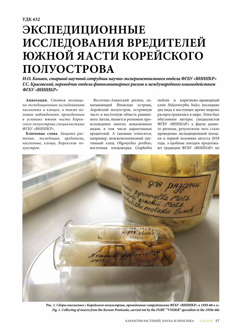
4| 26| 2018 57КАРАНТИН РАСТЕНИЙ. НАУКА И ПРАКТИКА
Аннотация. Статья посвяще-на экспедиционным исследованиям насекомых и клещей, а также по-левым наблюдениям, проведенным в условиях южной части Корей-ского полуострова специалистами ФГБУ «ВНИИКР».
Ключевые слова. Защита рас-тений, экспедиция, вредители, насекомые, клещи, Корейский по-луостров.
Восточно-Азиатский регион, ох-ватывающий Японские острова, Корейский полуостров, островную часть и восточную область равнин-ного Китая, является регионом про-исхождения многих инвазионных видов, в том числе карантинных вредителей. К таковым относятся, например, можжевельниковый пау-тинный клещ Oligonychus perditus, восточная плодожорка Grapholita
molesta и коричнево-мраморный клоп Halyomorpha halys; последние два вида в настоящее время широко распространились в мире. Этим был обусловлен интерес специалистов ФГБУ «ВНИИКР» к фауне данно-го региона, результатом чего стало проведение экспедиционной поезд-ки в первой половине августа 2018 года. Подобные поездки продолжа-ют традиции ФГБУ «ВНИИКР» по
УДК 632
ЭКСПЕДИЦИОННЫЕ ИССЛЕДОВАНИЯ ВРЕДИТЕЛЕЙ ЮЖНОЙ ЧАСТИ КОРЕЙСКОГО ПОЛУОСТРОВАИ.О. Камаев, старший научный сотрудник научно-экспериментального отдела ФГБУ «ВНИИКР»Г.С. Красовский, переводчик отдела фитосанитарных рисков и международного взаимодействия ФГБУ «ВНИИКР»
Рис. 1. Сборы насекомых с Корейского полуострова, проведенные сотрудниками ФГБУ «ВНИИКР» в 1950-60-е гг.Fig. 1. Collecting of insects from the Korean Peninsula, carried out by the FGBU “VNIIKR” specialists in the 1950s-60s
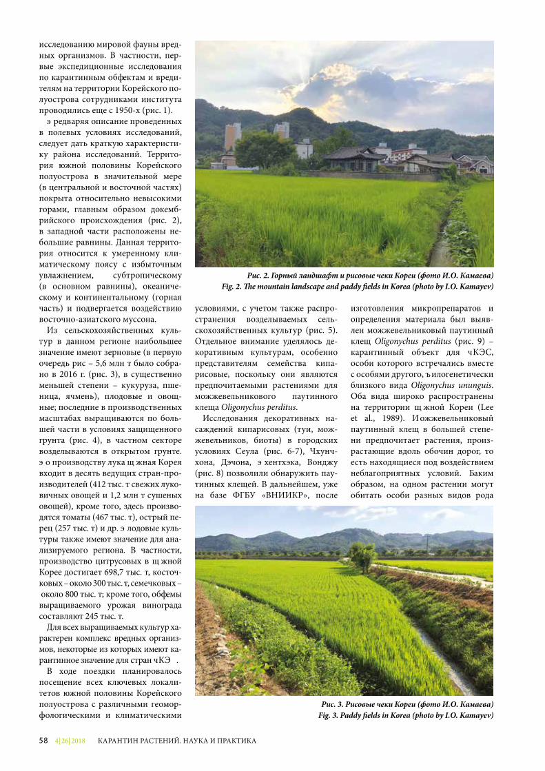
4| 26| 201858 КАРАНТИН РАСТЕНИЙ. НАУКА И ПРАКТИКА
исследованию мировой фауны вред-ных организмов. В частности, пер-вые экспедиционные исследования по карантинным объектам и вреди-телям на территории Корейского по-луострова сотрудниками института проводились еще с 1950-х (рис. 1).
Предваряя описание проведенных в полевых условиях исследований, следует дать краткую характеристи-ку района исследований. Террито-рия южной половины Корейского полуострова в значительной мере (в центральной и восточной частях) покрыта относительно невысокими горами, главным образом докемб-рийского происхождения (рис. 2), в западной части расположены не-большие равнины. Данная террито-рия относится к умеренному кли-матическому поясу с избыточным увлажнением, субтропическому (в основном равнины), океаниче-скому и континентальному (горная часть) и подвергается воздействию восточно-азиатского муссона.
Из сельскохозяйственных куль-тур в данном регионе наибольшее значение имеют зерновые (в первую очередь рис – 5,6 млн т было собра-но в 2016 г. (рис. 3), в существенно меньшей степени – кукуруза, пше-ница, ячмень), плодовые и овощ-ные; последние в производственных масштабах выращиваются по боль-шей части в условиях защищенного грунта (рис. 4), в частном секторе возделываются в открытом грунте. По производству лука Южная Корея входит в десять ведущих стран-про-изводителей (412 тыс. т свежих луко-вичных овощей и 1,2 млн т сушеных овощей), кроме того, здесь произво-дятся томаты (467 тыс. т), острый пе-рец (257 тыс. т) и др. Плодовые куль-туры также имеют значение для ана-лизируемого региона. В частности, производство цитрусовых в Южной Корее достигает 698,7 тыс. т, косточ-ковых – около 300 тыс. т, семечковых – около 800 тыс. т; кроме того, объемы выращиваемого урожая винограда составляют 245 тыс. т.
Для всех выращиваемых культур ха-рактерен комплекс вредных организ-мов, некоторые из которых имеют ка-рантинное значение для стран ЕАЭС.
В ходе поездки планировалось посещение всех ключевых локали-тетов южной половины Корейского полуострова с различными геомор-фологическими и климатическими
условиями, с учетом также распро-странения возделываемых сель-скохозяйственных культур (рис. 5). Отдельное внимание уделялось де-коративным культурам, особенно представителям семейства кипа-рисовые, поскольку они являются предпочитаемыми растениями для можжевельникового паутинного клеща Oligonychus perditus.
Исследования декоративных на-саждений кипарисовых (туи, мож-жевельников, биоты) в городских условиях Сеула (рис. 6-7), Чхунч-хона, Дэчона, Пхентхэка, Вонджу (рис. 8) позволили обнаружить пау-тинных клещей. В дальнейшем, уже на базе ФГБУ «ВНИИКР», после
изготовления микропрепаратов и определения материала был выяв-лен можжевельниковый паутинный клещ Oligonychus perditus (рис. 9) – карантинный объект для ЕАЭС, особи которого встречались вместе с особями другого, филогенетически близкого вида Oligonychus ununguis. Оба вида широко распространены на территории Южной Кореи (Lee et al., 1989). Можжевельниковый паутинный клещ в большей степе-ни предпочитает растения, произ-растающие вдоль обочин дорог, то есть находящиеся под воздействием неблагоприятных условий. Таким образом, на одном растении могут обитать особи разных видов рода
Рис. 2. Горный ландшафт и рисовые чеки Кореи (фото И.О. Камаева)Fig. 2. �e mountain landscape and paddy �elds in Korea (photo by I.O. Kamayev)
Рис. 3. Рисовые чеки Кореи (фото И.О. Камаева)Fig. 3. Paddy �elds in Korea (photo by I.O. Kamayev)
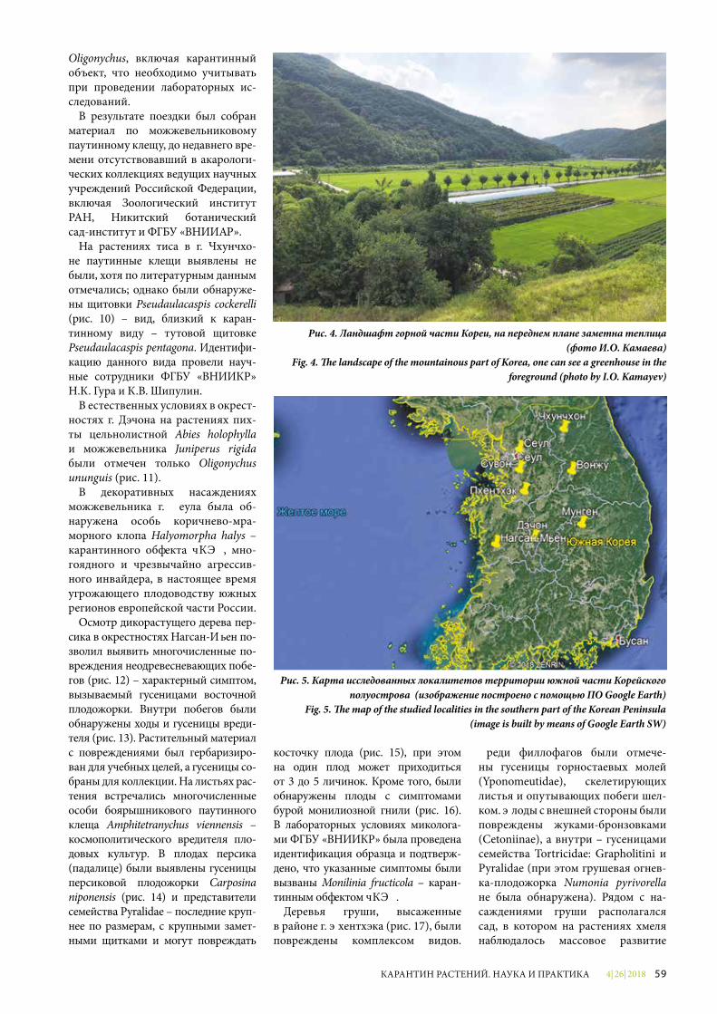
4| 26| 2018 59КАРАНТИН РАСТЕНИЙ. НАУКА И ПРАКТИКА
Рис. 4. Ландшафт горной части Кореи, на переднем плане заметна теплица (фото И.О. Камаева)
Fig. 4. �e landscape of the mountainous part of Korea, one can see a greenhouse in the foreground (photo by I.O. Kamayev)
Рис. 5. Карта исследованных локалитетов территории южной части Корейского полуострова (изображение построено с помощью ПО Google Earth)
Fig. 5. �e map of the studied localities in the southern part of the Korean Peninsula (image is built by means of Google Earth SW)
Oligonychus, включая карантинный объект, что необходимо учитывать при проведении лабораторных ис-следований.
В результате поездки был собран материал по можжевельниковому паутинному клещу, до недавнего вре-мени отсутствовавший в акарологи-ческих коллекциях ведущих научных учреждений Российской Федерации, включая Зоологический институт РАН, Никитский ботанический сад-институт и ФГБУ «ВНИИКР».
На растениях тиса в г. Чхунчхо-не паутинные клещи выявлены не были, хотя по литературным данным отмечались; однако были обнаруже-ны щитовки Pseudaulacaspis cockerelli (рис. 10) – вид, близкий к каран-тинному виду – тутовой щитовке Pseudaulacaspis pentagona. Идентифи-кацию данного вида провели науч-ные сотрудники ФГБУ «ВНИИКР» Н.А. Гура и А.В. Шипулин.
В естественных условиях в окрест-ностях г. Дэчона на растениях пих-ты цельнолистной Abies holophylla и можжевельника Juniperus rigida были отмечен только Oligonychus ununguis (рис. 11).
В декоративных насаждениях можжевельника г. Сеула была об-наружена особь коричнево-мра-морного клопа Halyomorpha halys – карантинного объекта ЕАЭС, мно-гоядного и чрезвычайно агрессив-ного инвайдера, в настоящее время угрожающего плодоводству южных регионов европейской части России.
Осмотр дикорастущего дерева пер-сика в окрестностях Нагсан-Мьен по-зволил выявить многочисленные по-вреждения неодревесневающих побе-гов (рис. 12) – характерный симптом, вызываемый гусеницами восточной плодожорки. Внутри побегов были обнаружены ходы и гусеницы вреди-теля (рис. 13). Растительный материал с повреждениями был гербаризиро-ван для учебных целей, а гусеницы со-браны для коллекции. На листьях рас-тения встречались многочисленные особи боярышникового паутинного клеща Amphitetranychus viennensis – космополитического вредителя пло-довых культур. В плодах персика (падалице) были выявлены гусеницы персиковой плодожорки Carposina niponensis (рис. 14) и представители семейства Pyralidae – последние круп-нее по размерам, с крупными замет-ными щитками и могут повреждать
косточку плода (рис. 15), при этом на один плод может приходиться от 3 до 5 личинок. Кроме того, были обнаружены плоды с симптомами бурой монилиозной гнили (рис. 16). В лабораторных условиях миколога-ми ФГБУ «ВНИИКР» была проведена идентификация образца и подтверж-дено, что указанные симптомы были вызваны Monilinia fructicola – каран-тинным объектом ЕАЭС.
Деревья груши, высаженные в районе г. Пхентхэка (рис. 17), были повреждены комплексом видов.
Среди филлофагов были отмече-ны гусеницы горностаевых молей (Yponomeutidae), скелетирующих листья и опутывающих побеги шел-ком. Плоды с внешней стороны были повреждены жуками-бронзовками (Cetoniinae), а внутри – гусеницами семейства Tortricidae: Grapholitini и Pyralidae (при этом грушевая огнев-ка-плодожорка Numonia pyrivorella не была обнаружена). Рядом с на-саждениями груши располагался сад, в котором на растениях хмеля наблюдалось массовое развитие
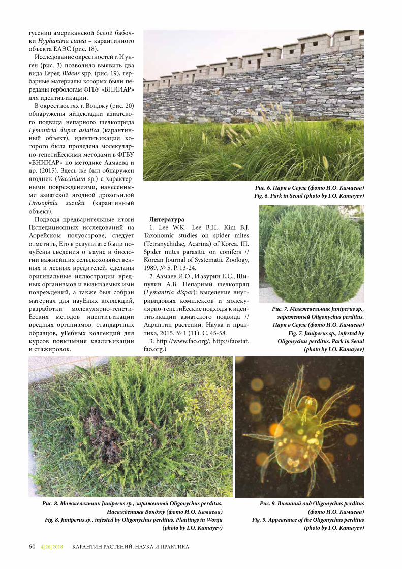
4| 26| 201860 КАРАНТИН РАСТЕНИЙ. НАУКА И ПРАКТИКА
Рис. 6. Парк в Сеуле (фото И.О. Камаева)Fig. 6. Park in Seoul (photo by I.O. Kamayev)
гусениц американской белой бабоч-ки Hyphantria cunea – карантинного объекта ЕАЭС (рис. 18).
Исследование окрестностей г. Мун-ген (рис. 3) позволило выявить два вида черед Bidens spp. (рис. 19), гер-барные материалы которых были пе-реданы гербологам ФГБУ «ВНИИКР» для идентификации.
В окрестностях г. Вонджу (рис. 20) обнаружены яйцекладки азиатско-го подвида непарного шелкопряда Lymantria dispar asiatica (карантин-ный объект), идентификация ко-торого была проведена молекуляр-но-генетическими методами в ФГБУ «ВНИИКР» по методике Камаева и др. (2015). Здесь же был обнаружен ягодник (Vaccinium sp.) с характер-ными повреждениями, нанесенны-ми азиатской ягодной дрозофилой Drosophila suzukii (карантинный объект).
Подводя предварительные итоги экспедиционных исследований на Корейском полуострове, следует отметить, что в результате были по-лучены сведения о фауне и биоло-гии важнейших сельскохозяйствен-ных и лесных вредителей, сделаны оригинальные иллюстрации вред-ных организмов и вызываемых ими повреждений, а также был собран материал для научных коллекций, разработки молекулярно-генети-ческих методов идентификации вредных организмов, стандартных образцов, учебных коллекций для курсов повышения квалификации и стажировок.
Рис. 7. Можжевельник Juniperus sp., зараженный Oligonychus perditus.
Парк в Сеуле (фото И.О. Камаева)Fig. 7. Juniperus sp., infested by
Oligonychus perditus. Park in Seoul (photo by I.O. Kamayev)
Рис. 8. Можжевельник Juniperus sp., зараженный Oligonychus perditus. Насаждения в Вонджу (фото И.О. Камаева)
Fig. 8. Juniperus sp., infested by Oligonychus perditus. Plantings in Wonju (photo by I.O. Kamayev)
Литература1. Lee W.K., Lee B.H., Kim B.J.
Taxonomic studies on spider mites (Tetranychidae, Acarina) of Korea. III. Spider mites parasitic on conifers // Korean Journal of Systematic Zoology, 1989. № 5. P. 13-24.
2. Камаев И.О., Мазурин Е.С., Ши-пулин А.В. Непарный шелкопряд (Lymantria dispar): выделение внут-ривидовых комплексов и молеку-лярно-генетические подходы к иден-тификации азиатского подвида // Карантин растений. Наука и прак-тика, 2015. № 1 (11). С. 45-58.
3. http://www.fao.org/; http://faostat.fao.org.)
Рис. 9. Внешний вид Oligonychus perditus (фото И.О. Камаева)
Fig. 9. Appearance of the Oligonychus perditus (photo by I.O. Kamayev)
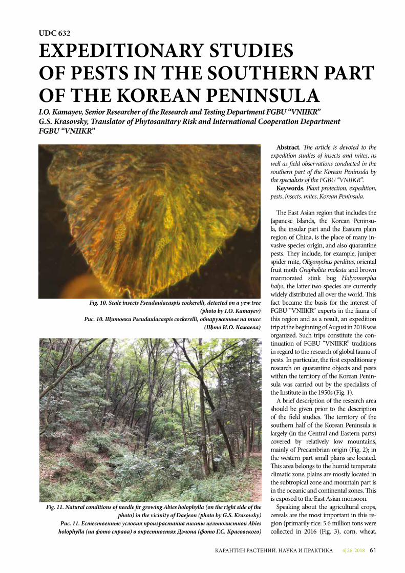
4| 26| 2018 61КАРАНТИН РАСТЕНИЙ. НАУКА И ПРАКТИКА
Abstract. �e article is devoted to the expedition studies of insects and mites, as well as �eld observations conducted in the southern part of the Korean Peninsula by the specialists of the FGBU “VNIIKR”.
Keywords. Plant protection, expedition, pests, insects, mites, Korean Peninsula.
�e East Asian region that includes the Japanese Islands, the Korean Peninsu-la, the insular part and the Eastern plain region of China, is the place of many in-vasive species origin, and also quarantine pests. �ey include, for example, juniper spider mite, Oligonychus perditus, oriental fruit moth Grapholita molesta and brown marmorated stink bug Halyomorpha halys; the latter two species are currently widely distributed all over the world. �is fact became the basis for the interest of FGBU “VNIIKR” experts in the fauna of this region and as a result, an expedition trip at the beginning of August in 2018 was organized. Such trips constitute the con-tinuation of FGBU “VNIIKR” traditions in regard to the research of global fauna of pests. In particular, the �rst expeditionary research on quarantine objects and pests within the territory of the Korean Penin-sula was carried out by the specialists of the Institute in the 1950s (Fig. 1).
A brief description of the research area should be given prior to the description of the �eld studies. �e territory of the southern half of the Korean Peninsula is largely (in the Central and Eastern parts) covered by relatively low mountains, mainly of Precambrian origin (Fig. 2); in the western part small plains are located. �is area belongs to the humid temperate climatic zone, plains are mostly located in the subtropical zone and mountain part is in the oceanic and continental zones. �is is exposed to the East Asian monsoon.
Speaking about the agricultural crops, cereals are the most important in this re-gion (primarily rice: 5.6 million tons were collected in 2016 (Fig. 3), corn, wheat,
Fig. 11. Natural conditions of needle �r growing Abies holophylla (on the right side of the photo) in the vicinity of Daejeon (photo by G.S. Krasovsky)
Рис. 11. Естественные условия произрастания пихты цельнолистной Abies holophylla (на фото справа) в окрестностях Дэчона (фото Г.С. Красовского)
UDC 632
EXPEDITIONARY STUDIES OF PESTS IN THE SOUTHERN PART OF THE KOREAN PENINSULAI.O. Kamayev, Senior Researcher of the Research and Testing Department FGBU “VNIIKR”G.S. Krasovsky, Translator of Phytosanitary Risk and International Cooperation Department FGBU “VNIIKR”
Fig. 10. Scale insects Pseudaulacaspis cockerelli, detected on a yew tree (photo by I.O. Kamayev)
Рис. 10. Щитовки Pseudaulacaspis cockerelli, обнаруженные на тисе (фото И.О. Камаева)
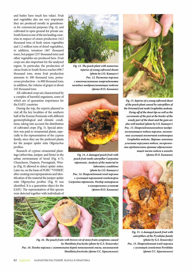
4| 26| 201862 КАРАНТИН РАСТЕНИЙ. НАУКА И ПРАКТИКА
Fig. 12. �e peach plant with numerous injuries of young so�wood shoots
(photo by I.O. Kamayev)Рис. 12. Растение персика
с многочисленными повреждениями молодых неодревесневающих побегов
(фото И.О. Камаева)
Fig. 13. Injuries of a young so�wood shoot of the peach plant caused by caterpillars of
the Oriental fruit moth Grapholita molesta. Dying-o� of the shoot tips as well as the
excrements of the pest at the border of the woody part of the shoot and the gum are
also well marked (photo by I.O. Kamayev)Рис. 13. Повреждения молодого неодре-весневающего побега персика, вызван-ные гусеницей восточной плодожорки
Grapholita molesta. Хорошо заметны усыхание верхушки побега, экскремен-
ты вредителя на границе одревеснева-ющей части побега и камедь
(фото И.О. Камаева)
and barley have much less value). Fruit and vegetables also are very important: they are produced mostly in greenhous-es for commercial purposes (Fig. 4), and cultivated in open ground for private use. South Korea is one of the ten leading coun-tries in respect of onion production (412 thousand tons of fresh onion vegetables and 1.2 million tons of dried vegetables), in addition, tomatoes (467 thousand tons), hot pepper (257 thousand tons) and other vegetables are produced here. Fruit crops are also important for the analyzed region. In particular, the production of citrus fruits in South Korea reaches 698.7 thousand tons, stone fruit production amounts to 300 thousand tons, poma-ceous production – to 800 thousand tons; in addition, the volume of grapes is about 245 thousand tons.
All cultivated crops are characterized by a complex of harmful organisms, some of which are of quarantine importance for the EAEU countries.
During the trip, the experts planned to visit all the key localities of the southern half of the Korean Peninsula with di� erent geomorphological and climatic condi-tions, taking into account the distribution of cultivated crops (Fig. 5). Special atten-tion was paid to ornamental plants, espe-cially to the representatives of the cypress family, since they are the preferred plants for the juniper spider mite Oligonychus perditus.
Research of cypress ornamental plant-ings (arborvitae, juniper, and biota) in the urban environment of Seoul (Fig. 6-7), Chuncheon, Daejeon, Pyeongtaek, Won-ju (Fig. 8) allowed to detect spider mites. Later on, on the basis of FGBU “VNIIKR”, a¥er creating micropreparations and iden-ti�cation of the material the juniper spider mite Oligonychus perditus (Fig. 9) was identi�ed. It is a quarantine object for the EAEU. �e representatives of this species were detected together with individuals of
Fig. 14. A damaged peach fruit with peach fruit moth caterpillar Carposina niponensis. Analysis of the material in
laboratory conditions (photo by I.O. Kamayev)
Рис. 14. Поврежденный плод персика с гусеницей персиковой плодожорки
Carposina niponensis. Разбор материала в камеральных условиях
(фото И.О. Камаева)
Fig. 15. A damaged peach fruit with caterpillars of the Pyralidae family
(photo by G.S. Krasovsky)Рис. 15. Поврежденный плод персика
с гусеницей семейства Pyralidae (фото Г.С. Красовского)
Fig. 16. �e peach fruits with brown rot of stone fruits symptoms caused by Monilinia fructicola (photo by G.S. Krasovsky)
Рис. 16. Плоды персика с симптомами бурой монилиозной гнили, вызываемой Monilinia fructicola (фото Г.С. Красовского)
4| 26| 2018 63КАРАНТИН РАСТЕНИЙ. НАУКА И ПРАКТИКА
the other phylogenetically close species, Oli-gonychus ununguis. Both species are widely distributed throughout South Korea (Lee et al., 1989). �e juniper spider mite mostly prefers plants that grow along the roadsides, which, therefore, are under the in�uence of adverse conditions. �us, one plant can have individuals of di�erent species of the Oligony chus genus, including the quarantine object, which must be taken into account during laboratory studies.
As a result of the trip, the juniper spider mite material which had been absent until re-cently in the acarological collections of lead-ing Russian institutions including Zoological Institute of RAS, Nikitsky Botanical Garden, and FGBU “VNIIKR”, has been collected.
�e mites were not detected on yew plants in Chuncheon, although, the literature sources have data about the mite presence there, yet a lot of scale insects Pseudaula-caspis cockerelli were detected. �is species is close to a quarantine one: white peach scale Pseudaulacaspis pentagona. �e iden-ti�cation of this species was conducted by N.A. Gura and A.V. Shipulin, the researchers of the FGBU “VNIIKR”.
Individuals of Oligonychus ununguis species were detected in natural conditions in the vicinity of Daejeon on needle �r Abies holophylla and juniper Juniperus rigi-da plants (Fig. 11).
In the ornamental plantations of juniper in Seoul, an individual of the brown-mar-morated stink bug Halyomorpha halys was found. �is quarantine object for EAEU is a polyphagous and extremely aggressive in-vader, which currently poses a risk to fruit cultivation in the southern regions of the Eu-ropean part of Russia.
During the examination of a wild peach tree in the vicinity of Nagsan-Myeon the spe-cialists found numerous injuries of so¥wood shoots (Fig. 12), which is a typical symptom caused by the oriental fruit moth caterpillars. Inside the shoots galleries of the pest cater-pillars were found (Fig. 13). �e damaged plant material was herborized for training purposes, and the caterpillars were collected. �e plant leaves had numerous individuals of sweet-cherry spider mite Amphitetrany-chus viennensis, a cosmopolitan pest of fruit crops. In peach fruits (windfalls), caterpillars of the peach fruit moth Carposina niponensis (Fig. 14) and representatives of the Pyralidae family were detected. �e latter are larger in size, with a large noticeable pinacula and can damage the fruit stone (Fig. 15); in one fruit there can be from 3 to 5 larvae. In addition, the fruits with the symptoms of brown rot of stone fruits were found (Fig. 16). In the labo-ratory, the mycologists of FGBU “VNIIKR”
Fig. 17. Pear trees plantings in the area of Pyeongtaek (photo by G.S. Krasovsky)Рис. 17. Деревья груши, насаждения в районе Пхентхэка (фото Г.С. Красовского)
Fig. 18. �e caterpillars of the Fell webworm Hyphantria cunea (photo by G.S. Krasovsky)Рис. 18. Гусеницы американской белой бабочки Hyphantria cunea
(фото Г.С. Красовского)
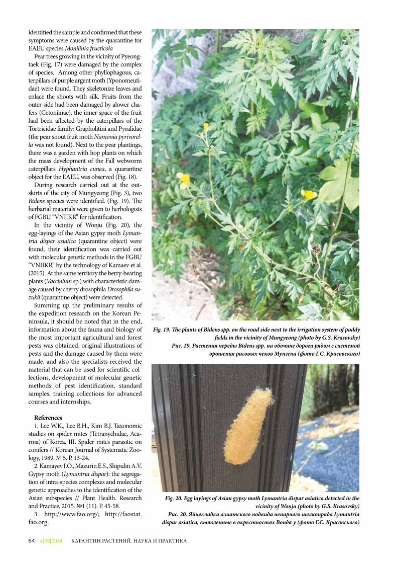
4| 26| 201864 КАРАНТИН РАСТЕНИЙ. НАУКА И ПРАКТИКА
identi�ed the sample and con�rmed that these symptoms were caused by the quarantine for EAEU species Monilinia fructicola
Pear trees growing in the vicinity of Pyeong-taek (Fig. 17) were damaged by the complex of species. Among other phyllophagous, ca-terpillars of purple argent moth (Yponomeuti-dae) were found. �ey skeletonize leaves and enlace the shoots with silk. Fruits from the outer side had been damaged by аlower cha-fers (Cetoniinae), the inner space of the fruit had been a�ected by the caterpillars of the Tortricidae family: Grapholitini and Pyralidae (the pear snout fruit moth Numonia pyrivorel-la was not found). Next to the pear plantings, there was a garden with hop plants on which the mass development of the Fall webworm caterpillars Hyphantria cunea, a quarantine object for the EAEU, was observed (Fig. 18).
During research carried out at the out-skirts of the city of Mungyeong (Fig. 3), two Bidens species were identi�ed. (Fig. 19). �e herbarial materials were given to herbologists of FGBU “VNIIKR” for identi�cation.
In the vicinity of Wonju (Fig. 20), the egg-layings of the Asian gypsy moth Lyman-tria dispar asiatica (quarantine object) were found, their identi�cation was carried out with molecular genetic methods in the FGBU “VNIIKR” by the technology of Kamaev et al. (2015). At the same territory the berry-bearing plants (Vaccinium sp.) with characteristic dam-age caused by cherry drosophila Drosophi la su-zukii (quarantine object) were detected.
Summing up the preliminary results of the expedition research on the Korean Pe-ninsula, it should be noted that in the end, information about the fauna and biology of the most important agricultural and forest pests was obtained, original illustrations of pests and the damage caused by them were made, and also the specialists received the material that can be used for scienti�c col-lections, development of molecular genetic methods of pest identi�cation, standard samples, training collections for advanced courses and internships.
References1. Lee W.K., Lee B.H., Kim B.J. Taxonomic
studies on spider mites (Tetranychidae, Aca-rina) of Korea. III. Spider mites parasitic on conifers // Korean Journal of Systematic Zoo-logy, 1989. № 5. P. 13-24.
2. Kamayev I.O., Mazurin E.S., Shipulin A.V. Gypsy moth (Lymantria dispar): the segrega-tion of intra-species complexes and molecular genetic approaches to the identi�cation of the Asian subspecies // Plant Health. Research and Practice, 2015. №1 (11). P. 45-58.
3. http://www.fao.org/; http://faostat.fao.org.
Fig. 19. �e plants of Bidens spp. on the road side next to the irrigation system of paddy �elds in the vicinity of Mungyeong (photo by G.S. Krasovsky)
Рис. 19. Растения череды Bidens spр. на обочине дороги рядом с системой орошения рисовых чеков Мунгена (фото Г.С. Красовского)
Fig. 20. Egg layings of Asian gypsy moth Lymantria dispar asiatica detected in the vicinity of Wonju (photo by G.S. Krasovsky)
Рис. 20. Яйцекладки азиатского подвида непарного шелкопряда Lymantria dispar asiatica, выявленные в окрестностях Вонджу (фото Г.С. Красовского)
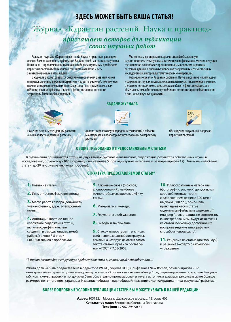
Журнал «Карантин растений. Наука и практика» зарегистрирован в Федеральной службе по надзору в сфере связи, информационных технологий и массовых коммуникаций (Роскомнадзор), свидетельство о регистрации ПИ № ФС77-52594 от 25 января 2013 г. Учредитель: ООО «Успех», выпускается по заказу Федерального государственного бюджетного учреждения «Всероссийский центр карантина растений» (ФГБУ «ВНИИКР»)Издатель: ООО «У-Строй»Адрес редакции: 115551, г.Москва, Шипиловский проезд, дом 39, корпус 2, этаж 15, помещение 53, комнаты 1, 3Номер отпечатан в ООО «Юнион Принт», г. Нижний Новгород, Окский съезд, д. 2, тел.: (831) 416-01-68
Дата выхода ч.12.2018 г. Тираж 3000 экземпляров. Подписной индекс 70195 в Каталоге Агентства «Роспечать»
«КАРАНТИН РАСТЕНИЙ. НАУКА И ПРАКТИКА» ДВУЯЗЫЧНЫЙ НАУЧНЫЙ ЖУРНАЛ №4 (26) 2018 г.
Зиновьева Светлана Георгиевна+7 967 294 90 61
Главный редактор:А.Я. Сапожников, директор ФГБУ «ВНИИКР»
Шеф-редактор: Светлана Зиновьева, начальник отдела по связям с общественностью и СМИ ФГБУ «ВНИИКР»
Выпускающий редактор: Ольга Лесных e-mail: [email protected]
Редакционная коллегия журнала «Карантин растений. Наука и практика»:
Швабаускене Ю.А. — заместитель Руководителя Россельхознадзора
Долженко В.И. — академик РАН, доктор сельскохозяйственных наук, заместитель директора Всероссийского НИИ защиты растений
Надыкта В.Д. — академик РАН, доктор технических наук, директор Всероссийского НИИ биологической защиты растений
Орлинский А.Д. — доктор биологических наук, научный советник ЕОКЗР
Павлюшин В.А. — академик РАН, доктор биологических наук, директор Всероссийского НИИ защиты растений
Санин С.С. — академик РАН, доктор биологических наук, профессор, заведующий отделом Всероссийского НИИ фитопатологии
Мартин Уорд — Генеральный директор ЕОКЗР
Ханну Кукконен — директор подразделения фитосанитарного надзора, EVIRA (Финляндия)
Сагитов А.О. — доктор биологических наук, Генеральный директор ТОО «Казахский НИИ защиты и карантина растений»
Сорока С.В. — кандидат сельскохозяйственных наук, директор РУП «Институт защиты растений» НАН Республики Беларусь
Джалилов Ф.С. — доктор биологических наук, профессор, заведующий лабораторией защиты растений МСХА им. К.А. Тимирязева
Абасов М.М. — доктор биологических наук, заместитель директора ФГБУ «ВНИИКР»
Шероколава Н.А. — заместитель директора ФГБУ «ВНИИКР», вице-президент ЕОКЗР
Добровольская О.Б. — кандидат биологических наук, заместитель директора ФГБУ «ВНИИКР»
Камаев И.О. — кандидат биологических наук, старший научный сотрудник научно-экспериментального отдела ФГБУ «ВНИИКР»
РЕДАКЦИЯ: Волкова Е.М. — кандидат биологических наук, заведующая лабораторией сорных растений
Волков О.Г. — начальник отдела биометода
Кулинич О.А. — доктор биологических наук, начальник отдела лесного карантина
Приходько Ю.Н. — кандидат сельскохозяйственных наук, начальник научно-методического отдела фитопатологии
Скрипка О.В. — кандидат биологических наук, ведущий научный сотрудник лаборатории микологии
Усачева С.Е. — переводчик отдела фитосанитарных рисков и международного взаимодействия
Быков И.И. — переводчик отдела фитосанитарных рисков и международного взаимодействия
Беломестнова А.А. — переводчик отдела фитосанитарных рисков и международного взаимодействия
Красовский Г.С. — переводчик отдела фитосанитарных рисков и международного взаимодействия
Дизайн и верстка: Роман Солоха
Корректоры: Татьяна АртемьеваОльга Тренева
Менеджер по подписке и дистрибуции: Павел Сафронов+7 903 505 33 23
PLANT HEALTHRESEARCH AND PRACTICE
RUSSIAN-ENGLISH JOURNAL
DECEMBER 4/26/2018IS
SN 2
306-
9767
КАРАНТИН РАСТЕНИЙНАУКА И ПРАКТИКА
РУССКО-АНГЛИЙСКИЙ ЖУРНАЛ
ДЕКАБРЬ 4/26/2018
ВИДЫ РОДА HELIANTHUS L. В ЕДИНОМ ПЕРЕЧНЕ КАРАНТИННЫХ ОБЪЕКТОВ ЕВРАЗИЙСКОГО ЭКОНОМИЧЕСКОГО СОЮЗА стр. 2
«НОВОГОДНИЕ ЕЛКИ» КАК УГРОЗА РАСПРОСТРАНЕНИЯ ВРЕДНЫХ И ПАТОГЕННЫХ ЛЕСНЫХ ОРГАНИЗМОВ стр. 9
БАКТЕРИОЗЫ – ВОЗБУДИТЕЛИ БОЛЕЗНЕЙ ЗЕРНОБОБОВЫХ КУЛЬТУР И РАЗРАБОТКА МЕТОДОВ ИХ ДИАГНОСТИКИ стр. 28
ЭКСПЕДИЦИОННЫЕ ИССЛЕДОВАНИЯ ВРЕДИТЕЛЕЙ ЮЖНОЙ ЧАСТИ КОРЕЙСКОГО ПОЛУОСТРОВА стр. 57
SPECIES OF THE HELIANTHUS L. GENUS IN THE COMMON LIST OF QUARANTINE OBJECTS OF THE EURASIAN ECONOMIC UNION page 6
“CHRISTMAS TREES” AS A THREAT TO THE SPREAD OF PATHOGENIC FOREST PESTS page 14
BACTERIAL DISEASE PATHOGENS OF GRAIN LEGUMES AND DEVELOPMENT OF METHODS FOR THEIR DIAGNOSTICS page 34
EXPEDITIONARY STUDIES OF PESTS IN THE SOUTHERN PART OF THE KOREAN PENINSULA page 61
Тел./факс: 8 (499) 707-22-27
Related Documents