Abstract To test the hypothesis of a planning dysfunc- tion in schizophrenia using a precise temporal definition, the readiness potential (RP), a negative cortical wave preceding self-initiated movements and reflecting motor preparation processes, was studied in patients under sta- ble medication and in controls. The supplementary motor area (SMA), known to be involved in the generation of the RP, has also been implicated in movement selection (fixed versus free) and complexity (single versus se- quence). This is the first study using RP for the assess- ment of the influence of these factors on motor prepara- tion in schizophrenics. Our results show that schizo- phrenics’ RP amplitude is significantly lower than in controls at central and contralateral electrodes. However, RP amplitude increases with task difficulty in both groups, offering important new insight into classical SMA hypo- activation in schizophrenics performing motor tasks. To- pographic analysis shows that RP amplitude is, for both groups, significantly higher in sequence than in single movements at fronto-central sites and higher for free than for fixed movements at centro-parietal sites. Finally, RP onset occurs significantly later in schizophrenics than in controls. These results support the view of a motor- preparation and decision-making dysfunction in schizo- phrenia. They are interpreted within the framework of a fronto-striatal disorder in this disease. Key words Electrophysiology · Schizophrenia · Readiness potential · Motor planning · Frontal Cortex Abbreviations EEG Electroencephalography · ERP event related potentials · MI primary motor cortex · MRCPs movement-related cortical potentials · NS’ negative slope · RP readiness potential · SMA supplementary motor area · PFC prefrontal cortex · EOG electrooculogram Introduction Planning dysfunction in schizophrenia Dysfunction of dopamine (DA)-modulated cortico-sub- cortical loops has been supposed to mediate part of the physiopathology of schizophrenia (Swerdlow and Koob 1987; Robbins 1990; Grace 1991, 1993; Gray et al. 1991; Weinberger 1993a, b; Frith 1992; Deutsch et al. 1993; Gray and Joseph, 1995). This disease can partly be con- sidered as a glutamate-mediated disruption in the balance between DA afferents to frontal cortex and nucleus acc- umbens (NAc) (Tassin et al. 1982; Carlsson 1990; Tassin 1995). A fronto-striatal dysfunction in schizophrenia should be revealed in planning tasks, because loops be- tween frontal cortex and basal ganglia are supposed to constitute the neurobiological substrate of planning, whether cognitive, motor or language-related. This hy- pothesis has been partially supported by brain imaging studies using positron emission tomography (PET) and functional magnetic resonance imaging (fMRI). Indeed, Günther et al. (1994) have used PET to show that during performance of simple and complex sequenc- es of finger movements, both medicated and neuroleptic- free negative schizophrenics have significantly lower re- gional glucose metabolism in frontal regions and higher- than-normal metabolism in the thalamus and basal gan- glia. In the same finger-to-thumb opposition task, Schrö- der et al. (1995) used fMRI to show that schizophrenics have lower activation of sensorimotor cortices and SMA than normals. Hypofrontality is also reported in cogni- tive planning tasks known to involve the frontal lobes (Weinberger et al. 1986, 1992; Goldberg and Weinberger J.-C. Dreher ( ✉ ) · J.-P. Banquet · Y. Burnod Laboratoire de Neurosciences et modélisation, INSERM U 483, University of Paris VI Jussieu, 9 quai St Bernard, F-75005 Paris e-mail: [email protected] W. Trapp · M. Keil · W. Günther Laboratory of Neurophysiology, Department of Psychiatry, Nervenklinik, D-96049 Bamberg, Germany W. Günther Ludwig-Maximilian Universität, Nussbaumstr. 7, Psychiatrische Universitätsklinik, D-80336 Munich, Germany Exp Brain Res (1999) 124:200–214 © Springer-Verlag 1999 RESEARCH ARTICLE J.-C. Dreher · W. Trapp · J.-P. Banquet · M. Keil W. Günther · Y. Burnod Planning dysfunction in schizophrenia: impairment of potentials preceding fixed/free and single/sequence of self-initiated finger movements Received: 11 July 1997 / Accepted: 16 July 1998

Welcome message from author
This document is posted to help you gain knowledge. Please leave a comment to let me know what you think about it! Share it to your friends and learn new things together.
Transcript
Abstract To test the hypothesis of a planning dysfunc-tion in schizophrenia using a precise temporal definition,the readiness potential (RP), a negative cortical wavepreceding self-initiated movements and reflecting motorpreparation processes, was studied in patients under sta-ble medication and in controls. The supplementary motorarea (SMA), known to be involved in the generation ofthe RP, has also been implicated in movement selection(fixed versus free) and complexity (single versus se-quence). This is the first study using RP for the assess-ment of the influence of these factors on motor prepara-tion in schizophrenics. Our results show that schizo-phrenics’ RP amplitude is significantly lower than incontrols at central and contralateral electrodes. However,RP amplitude increases with task difficulty in both groups,offering important new insight into classical SMA hypo-activation in schizophrenics performing motor tasks. To-pographic analysis shows that RP amplitude is, for bothgroups, significantly higher in sequence than in singlemovements at fronto-central sites and higher for freethan for fixed movements at centro-parietal sites. Finally,RP onset occurs significantly later in schizophrenics thanin controls. These results support the view of a motor-preparation and decision-making dysfunction in schizo-phrenia. They are interpreted within the framework of afronto-striatal disorder in this disease.
Key words Electrophysiology · Schizophrenia ·Readiness potential · Motor planning · Frontal Cortex
Abbreviations EEG Electroencephalography ·ERP event related potentials · MI primary motor cortex ·MRCPs movement-related cortical potentials ·NS’ negative slope · RP readiness potential ·SMA supplementary motor area ·PFC prefrontal cortex · EOG electrooculogram
Introduction
Planning dysfunction in schizophrenia
Dysfunction of dopamine (DA)-modulated cortico-sub-cortical loops has been supposed to mediate part of thephysiopathology of schizophrenia (Swerdlow and Koob1987; Robbins 1990; Grace 1991, 1993; Gray et al. 1991;Weinberger 1993a, b; Frith 1992; Deutsch et al. 1993;Gray and Joseph, 1995). This disease can partly be con-sidered as a glutamate-mediated disruption in the balancebetween DA afferents to frontal cortex and nucleus acc-umbens (NAc) (Tassin et al. 1982; Carlsson 1990; Tassin1995). A fronto-striatal dysfunction in schizophreniashould be revealed in planning tasks, because loops be-tween frontal cortex and basal ganglia are supposed toconstitute the neurobiological substrate of planning,whether cognitive, motor or language-related. This hy-pothesis has been partially supported by brain imagingstudies using positron emission tomography (PET) andfunctional magnetic resonance imaging (fMRI).
Indeed, Günther et al. (1994) have used PET to showthat during performance of simple and complex sequenc-es of finger movements, both medicated and neuroleptic-free negative schizophrenics have significantly lower re-gional glucose metabolism in frontal regions and higher-than-normal metabolism in the thalamus and basal gan-glia. In the same finger-to-thumb opposition task, Schrö-der et al. (1995) used fMRI to show that schizophrenicshave lower activation of sensorimotor cortices and SMAthan normals. Hypofrontality is also reported in cogni-tive planning tasks known to involve the frontal lobes(Weinberger et al. 1986, 1992; Goldberg and Weinberger
J.-C. Dreher () · J.-P. Banquet · Y. BurnodLaboratoire de Neurosciences et modélisation, INSERM U 483,University of Paris VI Jussieu, 9 quai St Bernard, F-75005 Parise-mail: [email protected]
W. Trapp · M. Keil · W. GüntherLaboratory of Neurophysiology, Department of Psychiatry, Nervenklinik, D-96049 Bamberg, Germany
W. GüntherLudwig-Maximilian Universität, Nussbaumstr. 7, Psychiatrische Universitätsklinik, D-80336 Munich, Germany
Exp Brain Res (1999) 124:200–214 © Springer-Verlag 1999
R E S E A R C H A RT I C L E
J.-C. Dreher · W. Trapp · J.-P. Banquet · M. KeilW. Günther · Y. Burnod
Planning dysfunction in schizophrenia: impairmentof potentials preceding fixed/free and single/sequenceof self-initiated finger movements
Received: 11 July 1997 / Accepted: 16 July 1998

1988; Weinberger and Berman 1988; Buchsbaum et al.1992; Sullivan et al. 1994). However, hypofrontality hasnot been reported systematically in schizophrenia insome verbal fluency tasks, in which schizophrenics’ per-formance matched with controls (Frith et al. 1995). Thisdiscordance cannot only be attributed to heterogeneity inthe schizophrenic population, but also to heterogeneityin the controls. Indeed, compared with their normal ho-mozygotic twins, schizophrenics do show decreasedfrontal cortex activation while performing the Wisconsincard-sorting test, even if performance per se did not dif-fer between twins (Berman et al. 1992). In fact, it cannow be reliably argued that motor and cognitive tasksthat depend on frontal lobe function produce depressedfrontal lobe activation in schizophrenic patients – partic-ularly those with negative signs (Guy et al. 1986; Kemaliet al. 1987; Braff et al. 1991; Wolkin et al. 1992; Ingvar1995; Malla et al. 1995; Spence et al. 1997). Importantly,this hypofrontality would not be a long-term effect ofneuroleptics or of illness chronicity (Andreasen et al.1992).
In addition to these deficits revealed by PET andfMRI, there is a large variety of event-related potential(ERP) abnormalities in schizophrenic patients: N100,N200, mismatch negativity (MMN), P300, N400, RP andcontingent negative variation (CNV) have all been shownto be smaller than in controls (Pritchard 1986; Rockstrohet al. 1989; Cohen 1991; Wagner et al. 1996). In particu-lar, attenuation of the late CNV component (which in-cludes but is not restricted to motor preparation) inschizophrenics may be related to their frequent perturba-tion in the initiation and execution of goal-directed ac-tions. Further support for this assumption comes fromthe low amplitude of the readiness potential (RP) thatprecedes voluntary single-finger movements (Timsit-Bert-hier 1973; Timsit-Berthier et al. 1978; Chiarenza 1985;Westphal et al. 1986; Singh et al. 1992; Karaman et al.1997) and from the hypoactivation of contralateral motorareas, as revealed by electroencephalograph (EEG)-map-ping methods (Günther et al. 1991; Günther, 1992). Theseresults could either support the hypothesis of a specificcortical disturbance (such as in the prefrontal cortex,PFC) or of a more generalized cortical dysfunction (Javitt1995).
Premovement potentials in normals
Source(s) of the RP
Movement-related cortical potentials (MRCPs) are gen-erated prior to, during and after the execution of self-paced voluntary movements. They are recorded by back-averaging EEG activity time-locked to muscle activationonset. Human volitional self-initiated movements are pre-ceded by two premovement potentials: the RP, or Be-reitschaftspotential, and the negative slope (NS’) (Korn-huber and Deecke 1965). The RP and NS’ extend, re-spectively, from –2 s to –0.5 s and from –0.5 to –60 ms
prior to movement onset (Shibasaki et al. 1980). Fourhypotheses have been proposed to explain the origins ofthe RP and NS’ that are generated prior to unilateral handmovements. Two hypotheses implicate only one struc-ture for the generation of both RP and NS’: bilateral acti-vation of either SMA (Tarkka et al. 1994; MacKinnon etal. 1996), or primary motor cortex (MI) (Cheyne andWeinberg 1989; Kristeva et al. 1991; Bötzel et al. 1993;Toro et al. 1993; Böcker et al. 1994). The two alternativehypotheses implicate SMA and MI, but assume differenttime courses of activation. The first hypothesis statesthat the RP originates bilaterally from both SMAs andreflects neural activity associated with the preparation ofmovement, whereas the NS’ potential is generated by thecontralateral MI (Deecke et al. 1976, 1987; Boschert1983; Tanji et al. 1985, 1994; Deecke 1990; Lang et al.1990, 1991; Jahanshahi et al. 1995). The second hypoth-esis proposes that RP and NS’ are both generated by bi-lateral activation of MI and SMA with an equivalenttime-course (Ikeda 1992, 1993; Rektor et al. 1994).
It is now admitted that SMA is activated during thegeneration of the RP, but temporal precedence or simul-taneity of SMA and MI activation remains an open ques-tion. As noticed by Praamstra et al. (1995), simultaneousactivity of the SMA and MI does not necessarily excludethe possibility that the SMA subserves a supramotor func-tion. This study will not directly address the question ofRP localization. However, a comparison of RP variationswith PET and fRMI results, according to groups (nor-mals and schizophrenics) and parameters (mode of move-ment selection and complexity), should indicate whether:(1) the reported cerebral activation changes are related toevents that occur during motor preparation; and (2) themain generators of the RP vary with groups and/or theparameters explored (complexity and mode of movementselection).
SMA functions
The SMA has been implicated in complex versus simplemotor tasks, internal versus external motor initiation, andfreely selected versus fixed (memory-guided) movements.We briefly review arguments supporting these differenthypotheses.
Complexity factor: is the RP amplitude largerfor sequential than for single movements?
In the generation of a sequence of finger movements,Lang et al. (1989) used EEG to show that both the nega-tivity and regional density of the inward-directed currentflow significantly increased over the frontocentral mid-line (SMA) for complex versus simple sequences, where-as no differences were observed over either the ipsi- orcontralateral MI. They interpreted this result as an in-crease in SMA activation and no change in MI activationwith task complexity. Some ERP studies also supported
201

the view that sequential movements cause higher RP am-plitudes than single movements (Benecke et al. 1985;Lang et al. 1988, 1989; Simonetta et al. 1991); othershave not (Ikeda et al. 1993; Praamstra et al. 1995).
The effect of complexity has also been reported inPET studies for which greater SMA activation occurredduring execution of a learned motor sequence than dur-ing simple repetitive movements (Orgogozo and Larsen1979; Roland et al. 1980, 1982; Deiber et al. 1991;Shibasaki et al. 1993). More recently, a positive correla-tion between regional cerebral blood flow (rCBF) andsequence complexity was found in the controlateral ros-tral SMA (pre-SMA) and associated pallido-thalamic loop(Boecker et al. 1998).
Electrophysiological studies in monkeys showed thatneural activities related to motor preparation are found inMI, SMA and pre-SMA (Evarts et al. 1984; Lecas et al.1986; Riehle and Requin 1989). However, with morecomplex preparatory processes, only SMA and pre-SMAneurons were involved (Tanji et al. 1980). Furthermore,Tanji et al. (1994) found some pre-SMA and SMA neu-rons that were exclusively active relative to a particularordering of forthcoming memory-guided movements, andothers that were preferentially active during the intervalbetween two specific movements. These neurons wouldtherefore contribute a signal regarding the order of forth-coming sequential movements and help to retrieve ap-propriate action according to a memorized order.
These results can be interpreted as the expression of agradient of activation from the pre-SMA, particularly ac-tive in tasks requiring temporal sequencing of multiplemovements, to MI, mostly involved in the execution ofthe motor task itself. Thus, they support the hypothesisof a higher RP for sequential than for single movements.Furthermore, the time course and slope of the first com-ponent of the RP in humans closely resembles the pre-paratory related activity recorded from cells in the SMAof behaving monkeys, whereas the second component(NS’) resembles the movement-related activity charac-teristic of a large number of MI neurons (Tanji et al.1985; Alexander and Crutcher 1990; Romo and Schultz1992).
Is the RP amplitude larger for internallythan externally triggered movements?
The most reproducible result concerning the role of theSMA, in particular its rostral part (pre-SMA), is its in-volvement in internally versus externally triggered move-ments (Libet et al. 1982; Goldberg 1985; Passingham1987, 1993; Deiber et al. 1991; Frith et al. 1991; Wise etal. 1991; Playford et al. 1992; Tanji et al. 1994; Jahan-shahi et al. 1995). Distinct cerebral structures are expect-ed to control these different types of movement. Gold-berg (1985) has proposed that a medial premotor system(SMA) mediates self-initiated movements, whereas a lat-eral system (centered on the lateral premotor cortex) me-diates externally triggered movements. The lateral pre-
motor cortex receives inputs from the inferior parietallobule and subcortical inputs from the cerebellum viadistinct areas of the thalamus; the SMA receives inputsfrom the superior parietal lobule and subcortical inputsfrom the basal ganglia. These two routes to MI couldthus subserve distinct motor functions. This hypothesisseems to be confirmed by experiments with monkeys thathave shown that: (1) during the pre-movement period,the SMA neurons are particularly activated in self-initi-ated versus externally triggered movements whereas theopposite is true for premotor cortex neurons (Mushiakeet al. 1990, 1991); and (2) the removal of the medial pre-motor cortex (SMA) impairs self-initiated movements,but affects tone-paced performance to a lesser extent(Thaler et al. 1995). Conversely, monkeys with lateralpremotor lesions are less impaired in making self-initiat-ed versus externally triggered movements (Thaler et al.1995). Furthermore, performance of animals with lesionsof the medial premotor cortex is improved when externalcues prompt the retrieval of an appropriate action (Chenet al. 1995). These results in monkeys are in agreementwith results in Parkinson’s disease, showing that, relativeto self-initiated movements, SMA deficits are not asmarked when external cues are provided to guide move-ments (Flowers 1976; Stern et al. 1983; Cunnington et al.1995).
Despite these findings, anatomical studies question theconcept of two routes to action (Matelli et al. 1989; Dari-an-Smith et al. 1990) and, instead, suggest a continuousfunctional gradient. Similarly, PET studies making thedistinction between internally and externally generatedmovements support the concept of a medial premotorsystem, but do not show evidence of a lateral system. In-deed, in self-initiated movements, there is a clear activa-tion not only of the SMA but also of the dorsolateral pre-frontal cortex (DLPFC), anterior cingulate and lateralpremotor cortex (Deiber et al. 1991; Frith et al. 1991;Papa et al. 1991; Playford et al. 1992; Jahanshahi et al.1995; Rao et al. 1997). The contralateral thalamus andputamen have also been shown to be significantly acti-vated in self-initiated movements (relative to rest) innormals (Jahanshahi et al. 1995; Rao et al. 1997).
Influence of the movement selection mode:is the RP amplitude larger for free than fixed movements?
In the RP paradigm, all the movements are self-initiatedand thus do not require an external cue. Therefore, thedistinction between internally versus externally generat-ed movements is recast as a distinction between fixed(i.e. repetition of the same movements for all trials) andfree movements (i.e. freely chosen movements for all tri-als). Fixed movements can also be considered memoryguided (reproduction), whereas free movements implyself generation. In normal subjects, RP amplitude is larg-er preceding free than fixed single movements of a joy-stick or presses of a button (Praamstra et al. 1995; Tougeet al. 1995).
202
203
This study investigates whether the mode of move-ment selection, which has been shown by PET andEEG to influence SMA activity in normals, also modu-lates the RP in schizophrenics. In addition, single ver-sus sequential movements will be tested in order to ex-amine: (1) the implications for the frontal cortex duringcomplex movements; (2) purported planning dysfunc-tion in schizophrenia; and (3) the relative and interac-tive effects of mode of selection and complexity. Addi-tive effects of complexity and mode of movement se-lection could make an SMA contribution to the RP eas-ier to identify. More specifically, we expected to find:(1) significantly lower RP amplitudes in schizophrenicsversus controls for all tasks and particularly for thefree-sequence task (which requires more planning); (2)a larger RP in both groups for free versus fixed move-ments and for sequences versus single movements; and(3) longer RP latencies in schizophrenics than in con-trols for all tasks, reflecting motor preparation impair-ment in schizophrenia.
Materials and methods
A similar experiment has previously been performed with normalsubjects (Praamstra et al. 1995). Our paradigm was approved bythe ethics commission of the Medical University of München (Lud-wig-Maximilian Universität). All subjects gave their informed con-sent prior to inclusion in the study.
Subjects
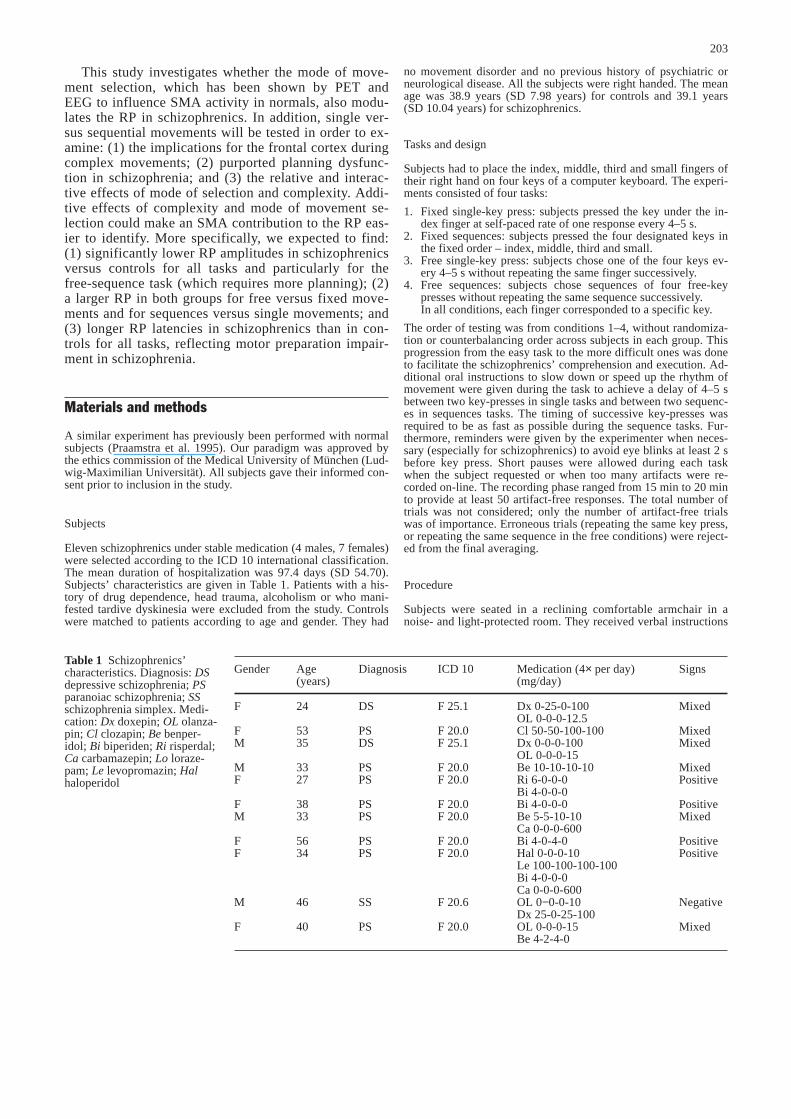
Eleven schizophrenics under stable medication (4 males, 7 females)were selected according to the ICD 10 international classification.The mean duration of hospitalization was 97.4 days (SD 54.70).Subjects’ characteristics are given in Table 1. Patients with a his-tory of drug dependence, head trauma, alcoholism or who mani-fested tardive dyskinesia were excluded from the study. Controlswere matched to patients according to age and gender. They had
no movement disorder and no previous history of psychiatric orneurological disease. All the subjects were right handed. The meanage was 38.9 years (SD 7.98 years) for controls and 39.1 years(SD 10.04 years) for schizophrenics.
Tasks and design
Subjects had to place the index, middle, third and small fingers oftheir right hand on four keys of a computer keyboard. The experi-ments consisted of four tasks:
1. Fixed single-key press: subjects pressed the key under the in-dex finger at self-paced rate of one response every 4–5 s.
2. Fixed sequences: subjects pressed the four designated keys inthe fixed order – index, middle, third and small.
3. Free single-key press: subjects chose one of the four keys ev-ery 4–5 s without repeating the same finger successively.
4. Free sequences: subjects chose sequences of four free-keypresses without repeating the same sequence successively.In all conditions, each finger corresponded to a specific key.
The order of testing was from conditions 1–4, without randomiza-tion or counterbalancing order across subjects in each group. Thisprogression from the easy task to the more difficult ones was doneto facilitate the schizophrenics’ comprehension and execution. Ad-ditional oral instructions to slow down or speed up the rhythm ofmovement were given during the task to achieve a delay of 4–5 sbetween two key-presses in single tasks and between two sequenc-es in sequences tasks. The timing of successive key-presses wasrequired to be as fast as possible during the sequence tasks. Fur-thermore, reminders were given by the experimenter when neces-sary (especially for schizophrenics) to avoid eye blinks at least 2 sbefore key press. Short pauses were allowed during each taskwhen the subject requested or when too many artifacts were re-corded on-line. The recording phase ranged from 15 min to 20 minto provide at least 50 artifact-free responses. The total number oftrials was not considered; only the number of artifact-free trialswas of importance. Erroneous trials (repeating the same key press,or repeating the same sequence in the free conditions) were reject-ed from the final averaging.
Procedure
Subjects were seated in a reclining comfortable armchair in anoise- and light-protected room. They received verbal instructions
Table 1 Schizophrenics’ characteristics. Diagnosis: DSdepressive schizophrenia; PSparanoiac schizophrenia; SSschizophrenia simplex. Medi-cation: Dx doxepin; OL olanza-pin; Cl clozapin; Be benper-idol; Bi biperiden; Ri risperdal;Ca carbamazepin; Lo loraze-pam; Le levopromazin; Halhaloperidol
Gender Age Diagnosis ICD 10 Medication (4× per day) Signs(years) (mg/day)
F 24 DS F 25.1 Dx 0-25-0-100 MixedOL 0-0-0-12.5
F 53 PS F 20.0 Cl 50-50-100-100 MixedM 35 DS F 25.1 Dx 0-0-0-100 Mixed
OL 0-0-0-15M 33 PS F 20.0 Be 10-10-10-10 MixedF 27 PS F 20.0 Ri 6-0-0-0 Positive
Bi 4-0-0-0F 38 PS F 20.0 Bi 4-0-0-0 PositiveM 33 PS F 20.0 Be 5-5-10-10 Mixed
Ca 0-0-0-600F 56 PS F 20.0 Bi 4-0-4-0 PositiveF 34 PS F 20.0 Hal 0-0-0-10 Positive
Le 100-100-100-100Bi 4-0-0-0Ca 0-0-0-600
M 46 SS F 20.6 OL 0−0-0-10 NegativeDx 25-0-25-100
F 40 PS F 20.0 OL 0-0-0-15 MixedBe 4-2-4-0

For topographical analysis, comparisons of the peak of NS’amplitude were confined to electrodes Fz, Cz, Pz, C1 and C3. Theinterval from –500 ms to 0 ms, corresponding to the NS’ was se-lected as the main epoch for statistical analysis. MANOVA of thepeak of NS’ amplitude was conducted with group as the between-subjects variable, and mode of movement selection and movementcomplexity as the within-subjects factor.
To further quantify temporal differences in the shape of the RPacross groups and conditions, mean amplitudes were calculatedfor successive time windows (–1000 ms to –900 ms, –900 ms to–800 ms, –800 ms to –700 ms, –700 ms to –600 ms, –600 ms to–500 ms, –500 to 0 ms and 0 ms to 500 ms). The time windowsare numbered from 1 to 7. MANOVA of RP amplitude was per-formed for each time window at electrodes Fz, Cz, Pz, C1 and C3with group, mode of selection and complexity as variables.
In all of the analyses of amplitude, the three following one-sid-ed hypotheses were tested: (1) amplitudes are greater in normalsthan in schizophrenics; and for both groups that: (2) sequence in-duces greater amplitudes than single movements; and (3) free-se-lected movements induce greater amplitudes than fixed movements.
Analysis of latencies
MANOVA of RPO was conducted at Cz, with group (schizophren-ics versus controls) as a between-subjects variable, and with modeof movement selection (fixed versus free conditions) and com-plexity (single movement versus sequence) as within-subjects fac-tors. MANOVA of NSO was conduced at Cz, with group (schizo-phrenics versus controls) as a between-subjects variable, and withmode of movement selection (fixed versus free conditions) andcomplexity (single movement versus sequence) as within-subjectsfactors.
We tested the two one-sided hypotheses: (1) RPO starts soonerin controls than in schizophrenics; and (2) NSO starts sooner incontrols than schizophrenics. For all tasks, the significance levelwas set at P<0.05.
Results
Task performance
Schizophrenics generally presented difficulties at the be-ginning of the tasks but were able to press the keys cor-rectly after a few minutes of training. Visual inspection ofthe EMG recordings revealed no substantial differencesin strength and speed of finger movements between pa-tients and controls. On-line control of sequence execu-tion was done by monitoring the fingers. When repeti-tion of the same finger occurred in a sequence, the ex-perimenter instructed the subject not to press the samefinger twice in one sequence.
General wave form and distribution of the RP
The general wave form of the grand mean RP at Cz foreach group and condition is given in Fig. 1. Maps ofRPO, NSO and NS’ peak are displayed in the bottom pan-el of Fig. 1. The general wave form of the RP does notdiffer in normals across the various conditions. In schizo-phrenics, the RP rises much more slowly than in con-trols, has lower amplitudes and is less structured. Themore striking characteristics of schizophrenic RPs are:(1) delayed onset and low amplitudes and (2) persistence
204
read by the experimenter. For the free-selection tasks, the instruc-tions stressed that movements should be chosen arbitrarily, i.e.,without use of the same finger across two successive trials. Fur-thermore, it was expressly requested that subjects change sequenc-es from trial to trial to avoid short cyclic repetition of the same se-quences. Subjects had to practice each type of movement a fewtimes to ensure that they made brisk movements at the desiredrate. They were instructed to fixate on a square (1 cm2) on the cen-ter of a screen, placed 1 m from their eyes. Subjects were given en-couragement by the experimenter particularly given the difficultyexperienced by the schizophrenics to concentrate on fixating on thesquare.
Data acquisition and apparatus
The EEG activity was recorded with Ag/AgCl electrodes from 10positions of the 10–20 system (Fz, Cz, Pz, C1, F3, C3, P3, F4, C4,P4). Electrodes were attached using collodion after careful cleaningof the skin. Horizontal and vertical electrooculograms (EOG) wererecorded from the outer canthi of both eyes and from sites aboveand below the two eyes to control for eye-movement artifacts. Addi-tionally, artifacts were controlled using electrodes at Fp1 and Fp2.Linked electrodes attached to both earlobes served as reference. Bi-polar EMG was recorded using Ag/AgCl electrodes attached to theflexor side of the right forearm. The EEG was filtered with a timeconstant of 5 s and a low-pass filter set at 30 Hz. Signals were digi-tized on-line with a sampling frequency of 128 Hz per channel, adynamic range of ±150 µV, and a resolution of 7.8 ms per samplepoint (400 points). Electrode impedance was kept below 5 kΩ. Datawere stored on the hard disk of the EEG brain-mapping system andanalyzed off-line with Evoked Potential Software.
Data analyses
To define point 0 of a trial, movement-onset triggers were manual-ly placed where the EMG signal began to rise. Additionally, trig-gers sent at each key press were used when the EMG was notclearly visible. The interval (for averaging the sweeps) selected tostudy the RP was 2 s before point 0 and 1.2 s after (7.8×400 ms).RP curves were obtained by averaging at least 50 artifact-freesweeps. For sequences, the onset triggers were placed at the be-ginning of the first EMG activity. Baseline was corrected from–2000 ms to –1900 ms.
RP onset (RPO) latency was defined as the beginning of thenegative potential rise. According to Shibasaki et al. (1980), nega-tive slope (NS’) can be defined in normal MRCPs as a late negativeslope (prior to actual motor potential) that is steeper than the earlierRP slope and contralateral to the moving hand. Functionally, NS’seems to indicate a terminal increase of lateral premotor system ac-tivation prior to movement onset. Peak of NS’ was quantified as themaximum negative amplitude prior to movement onset. Negativeslope onset (NSO) was defined at the point were NS’ began to rise(approximately 500 ms before EMG activity). It has to be noted thatNSO results have to be interpreted with caution, given that the pointwere NS’ begins to rise is often difficult to determine, especially inschizophrenics. Amplitudes of the early RP were calculated at Czand C1 at NSO. Motor preparation was supposed to be reflected bythe RPO and early RP amplitude; decision-making was supposed tobe reflected by NS’ onset and peak of NS’ amplitude.
Statistical analysis
Analysis of amplitudes
Multivariate analysis of variance (MANOVA) of the early RP am-plitude (amplitude of NS’ at the time of its onset) was confined toelectrodes Cz and C1, with group (schizophrenics versus controls)as a between-subjects variable, and mode of movement selection(fixed versus free conditions) and movement complexity (singlemovement versus sequence) as within-subjects factors.

205
(as in controls) of a monotonically increasing amplitudethat emerges at Cz as the task becomes progressivelymore complex (from single-fixed, single-free, sequence-fixed to sequence-free).
For both groups, in addition to these effects that occurbefore movement, there are differences between singlemovements and sequences that appear during the move-ment execution: with sequential, but not with single mo-
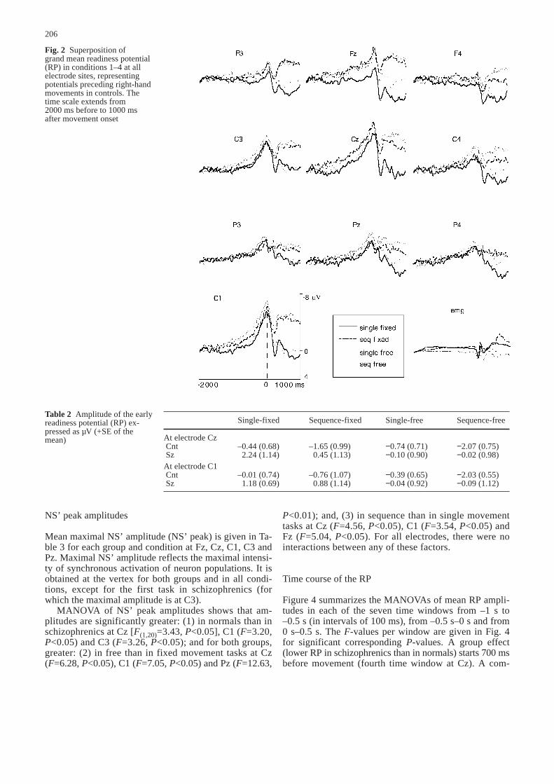
vements, the RP is followed by a performance-relatednegativity. Classically, when a large degree of corticalcontrol has to be exercised during a movement, negativi-ty is not resolved immediately after movement onset but,rather, is maintained throughout the movement. This sus-tained negativity has been named the movement-moni-toring potential (Deecke et al. 1984). Distribution of theRP is displayed in Figs. 2 and 3 and conforms to generalcharacteristics established in previous studies.
Amplitudes of the early RP
Analysis of the amplitude of the early RP (correspondingto the amplitude of NS’ at the time of its onset) was per-formed at electrodes Cz and C1 (see Table 2). MANO-VAs of the early RP show that the amplitudes are signifi-cantly greater in controls than in schizophrenics at Cz[F(1,20)=3.99, P<0.05], and in both groups, greater at Czfor sequences than for single movements [F(1,20)=4.49,P<0.05] and greater at C1 for free than for fixed move-ments [F(1,20)=3.82, P<0.05]. There was no interactionbetween any of the factors for the amplitude of the earlyRP analysis.
ReadinessPotentialOnset
NegativeSlopeOnset
Time of NegativeSlope peak
-1469.49 ms -1511.40 ms -1414.10 ms -1496.48 ms
-424.11 ms -335.33 ms -343.86 ms -289.17 ms
-73.99 ms -51.26 ms -44.87 ms -61.91 ms
0
0- 2000 1000
-8
Grand meanmaps at:
time (ms)
Amplitude (µV)
Single-fixed Single-free Seq-fixed Seq-free
-1000
Single- f ixed
Single- f ree
4
Seq- f ree
single fixed
seq fixed
single free
seq free
+10 µV
-10 µV
-826.94 ms -1282.72 ms -1150.62 ms -1080.32 ms
-367.29 ms -402.80 ms -310.48 ms -371.05 ms
-93.16 ms -74.44 ms -88.19 ms -61.92 ms
0
0- 2000 1000
-8
time (ms)
Single-fixed Single-free Seq-fixed Seq-free
-1000
Fig. 1 On the left: Top: superposition of grand mean readiness po-tential (RP) of single-fixed, single-free, sequence-fixed, sequence-free conditions, representing potentials preceding right-hand mo-vements recorded at electrode Cz in controls. The time scale ex-tends from 2000 ms before to 1000 ms after movement onset. Bot-tom: grand mean maps of controls in the four conditions displayedat the time of readiness potential onset (RPO), negative slope on-set (NSO) and peak of negative slope. Note the increased recruit-ment of the supplementary motor area (SMA) with time and diffi-culty of the task. On the right: Top: superposition of grand meanRP of single-fixed, single-free, sequence-fixed, sequence-free con-ditions, representing potentials preceding right-hand movementsrecorded at electrode Cz in schizophrenics. Note: (1) their lowerand more noisy RP amplitudes than in controls; and (2) the con-servation of an increased SMA recruitment as a gradient of taskcomplexity. Bottom: grand mean maps of schizophrenics in thefour conditions displayed at the time of RPO, NSO and peak ofnegative slope. Even if the pattern of activation is more diffusethan in controls, a gradient of SMA activation appears when timeand difficulty of the task increase
206
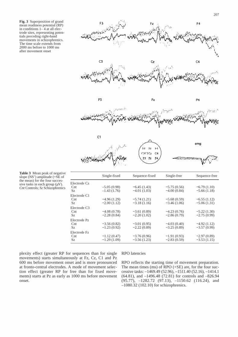
NS’ peak amplitudes
Mean maximal NS’ amplitude (NS’ peak) is given in Ta-ble 3 for each group and condition at Fz, Cz, C1, C3 andPz. Maximal NS’ amplitude reflects the maximal intensi-ty of synchronous activation of neuron populations. It isobtained at the vertex for both groups and in all condi-tions, except for the first task in schizophrenics (forwhich the maximal amplitude is at C3).
MANOVA of NS’ peak amplitudes shows that am-plitudes are significantly greater: (1) in normals than inschizophrenics at Cz [F(1,20)=3.43, P<0.05], C1 (F=3.20,P<0.05) and C3 (F=3.26, P<0.05); and for both groups,greater: (2) in free than in fixed movement tasks at Cz(F=6.28, P<0.05), C1 (F=7.05, P<0.05) and Pz (F=12.63,
P<0.01); and, (3) in sequence than in single movementtasks at Cz (F=4.56, P<0.05), C1 (F=3.54, P<0.05) andFz (F=5.04, P<0.05). For all electrodes, there were nointeractions between any of these factors.
Time course of the RP
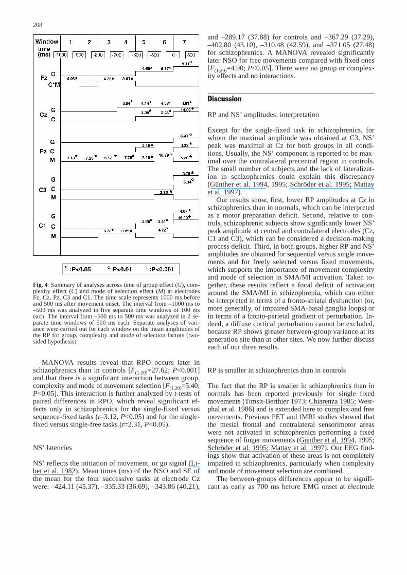
Figure 4 summarizes the MANOVAs of mean RP ampli-tudes in each of the seven time windows from –1 s to–0.5 s (in intervals of 100 ms), from –0.5 s–0 s and from0 s–0.5 s. The F-values per window are given in Fig. 4for significant corresponding P-values. A group effect(lower RP in schizophrenics than in normals) starts 700 msbefore movement (fourth time window at Cz). A com-
Fig. 2 Superposition of grand mean readiness potential(RP) in conditions 1–4 at allelectrode sites, representing potentials preceding right-handmovements in controls. Thetime scale extends from 2000 ms before to 1000 ms after movement onset
Table 2 Amplitude of the earlyreadiness potential (RP) ex-pressed as µV (+SE of themean)
Single-fixed Sequence-fixed Single-free Sequence-free
At electrode CzCnt –0.44 (0.68) –1.65 (0.99) −0.74 (0.71) −2.07 (0.75)Sz 2.24 (1.14) 0.45 (1.13) −0.10 (0.90) −0.02 (0.98)
At electrode C1Cnt –0.01 (0.74) –0.76 (1.07) −0.39 (0.65) −2.03 (0.55)Sz 1.18 (0.69) 0.88 (1.14) −0.04 (0.92) −0.09 (1.12)
207
plexity effect (greater RP for sequences than for singlemovements) starts simultaneously at Fz, Cz, C1 and Pz600 ms before movement onset and is more pronouncedat fronto-central electrodes. A mode of movement selec-tion effect (greater RP for free than for fixed move-ments) starts at Pz as early as 1000 ms before movementonset.
RPO latencies
RPO reflects the starting time of movement preparation.The mean times (ms) of RPO (+SE) are, for the four suc-cessive tasks: –1469.49 (52.96), –1511.40 (52.16), –1414.1(64.81), and –1496.48 (72.81) for controls and –826.94(95.77), –1282.72 (97.13), –1150.62 (116.24), and–1080.32 (102.10) for schizophrenics.
Fig. 3 Superposition of grandmean readiness potential (RP)in conditions 1– 4 at all elec-trode sites, representing poten-tials preceding right-handmovements in schizophrenics.The time scale extends from2000 ms before to 1000 ms after movement onset
Table 3 Mean peak of negativeslope (NS’) amplitude (+SE ofthe mean) for the four succes-sive tasks in each group (µV).Cnt Controls; Sz Schizophrenics
Single-fixed Sequence-fixed Single-free Sequence-free
Electrode CzCnt –5.05 (0.98) −6.45 (1.43) −5.75 (0.56) −6.79 (1.10)Sz –1.43 (1.76) −4.01 (1.03) −4.00 (0.84) −5.66 (1.18)
Electrode C1Cnt −4.96 (1.29) −5.74 (1.21) −5.68 (0.59) −6.55 (1.12) Sz −2.00 (1.12) −3.18 (1.16) −3.46 (1.06) −5.06 (1.31)
Electrode C3Cnt −4.08 (0.78) −3.61 (0.89) −4.23 (0.76) −5.22 (1.30)Sz −2.28 (0.84) −2.20 (1.02) −2.86 (0.79) −2.75 (0.99)
Electrode PzCnt −3.56 (0.82) −3.01 (0.95) −4.03 (0.40) −4.92 (1.12)Sz −1.23 (0.92) −2.22 (0.89) −3.25 (0.89) −3.57 (0.99)
Electrode FzCnt −1.12 (0.47) −3.76 (0.96) −1.91 (0.93) −2.97 (0.89)Sz −1.29 (1.09) −3.56 (1.23) −2.83 (0.59) −3.53 (1.15)
208
MANOVA results reveal that RPO occurs later inschizophrenics than in controls [F(1,20)=27.62; P<0.001]and that there is a significant interaction between group,complexity and mode of movement selection [F(1,20)=5.40;P<0.05]. This interaction is further analyzed by t-tests ofpaired differences in RPO, which reveal significant ef-fects only in schizophrenics for the single-fixed versussequence-fixed tasks (t=3.12, P<0.05) and for the single-fixed versus single-free tasks (t=2.31, P<0.05).
NS’ latencies
NS’ reflects the initiation of movement, or go signal (Li-bet et al. 1982). Mean times (ms) of the NSO and SE ofthe mean for the four successive tasks at electrode Czwere: –424.11 (45.37), –335.33 (36.69), –343.86 (40.21),
and –289.17 (37.88) for controls and –367.29 (37.29),–402.80 (43.10), –310.48 (42.59), and –371.05 (27.48)for schizophrenics. A MANOVA revealed significantlylater NSO for free movements compared with fixed ones[F(1,20)=4.90; P<0.05]. There were no group or complex-ity effects and no interactions.
Discussion
RP and NS’ amplitudes: interpretation
Except for the single-fixed task in schizophrenics, forwhom the maximal amplitude was obtained at C3, NS’peak was maximal at Cz for both groups in all condi-tions. Usually, the NS’ component is reported to be max-imal over the contralateral precentral region in controls.The small number of subjects and the lack of lateralizat-ion in schizophrenics could explain this discrepancy(Günther et al. 1994, 1995; Schröder et al. 1995; Mattayet al. 1997).
Our results show, first, lower RP amplitudes at Cz inschizophrenics than in normals, which can be interpretedas a motor preparation deficit. Second, relative to con-trols, schizophrenic subjects show significantly lower NS’peak amplitude at central and contralateral electrodes (Cz,C1 and C3), which can be considered a decision-makingprocess deficit. Third, in both groups, higher RP and NS’amplitudes are obtained for sequential versus single move-ments and for freely selected versus fixed movements,which supports the importance of movement complexityand mode of selection in SMA/MI activation. Taken to-gether, these results reflect a focal deficit of activationaround the SMA/MI in schizophrenia, which can eitherbe interpreted in terms of a fronto-striatal dysfunction (or,more generally, of impaired SMA-basal ganglia loops) orin terms of a fronto-parietal gradient of perturbation. In-deed, a diffuse cortical perturbation cannot be excluded,because RP shows greater between-group variance at itsgeneration site than at other sites. We now further discusseach of our three results.
RP is smaller in schizophrenics than in controls
The fact that the RP is smaller in schizophrenics than innormals has been reported previously for single fixedmovements (Timsit-Berthier 1973; Chiarenza 1985; West-phal et al. 1986) and is extended here to complex and freemovements. Previous PET and fMRI studies showed thatthe mesial frontal and contralateral sensorimotor areaswere not activated in schizophrenics performing a fixedsequence of finger movements (Günther et al. 1994, 1995;Schröder et al. 1995; Mattay et al. 1997). Our EEG find-ings show that activation of these areas is not completelyimpaired in schizophrenics, particularly when complexityand mode of movement selection are combined.
The between-groups differences appear to be signifi-cant as early as 700 ms before EMG onset at electrode
Fig. 4 Summary of analyses across time of group effect (G), com-plexity effect (C) and mode of selection effect (M) at electrodesFz, Cz, Pz, C3 and C1. The time scale represents 1000 ms beforeand 500 ms after movement onset. The interval from –1000 ms to–500 ms was analyzed in five separate time windows of 100 mseach. The interval from –500 ms to 500 ms was analyzed in 2 se-parate time windows of 500 ms each. Separate analyses of vari-ance were carried out for each window on the mean amplitudes ofthe RP for group, complexity and mode of selection factors (two-sided hypothesis)

Cz. This allows a clear distinction between the intervalspreceding and during movement, adding important newinsights to PET and fMRI results. NS’ peaks are signifi-cantly higher in normals than in schizophrenics at elec-trodes Cz, C1 and C3. However, we cannot conclude fromthis that there is a localized deficit in schizophrenia. In-deed, significant differences in NS’ peak amplitudes oc-cur at sites for which the NS’ peak is at its highest am-plitude (Cz, C1 and C3), but not at frontal and parietalsites, where the amplitude is lower.
RP is greater for sequences than for single movements
Greater RPs for sequences versus single movementshave been described in normals: (1) for sequences thatstart with the same movement as the single movementtask; (2) when the comparison is between sequences andthe simultaneous extension of two fingers (Benecke et al.1985; Simonetta et al. 1991; Kitamura et al. 1993a, b).Yet, there are also reports of no differences in RP ampli-tudes for sequences versus single movements (Lang etal. 1988, 1989; Praamstra et al. 1995).
Our MANOVA of RP amplitudes as a function of timeshows that complexity effects become significant at elec-trodes Fz, Cz, and C1 as early as 600 ms before EMGonset. This complexity effect is larger at Fz than at cen-tral sites (Cz and C1). This is in accordance with studiesthat show a larger and earlier RP over the anterior part ofthe frontal lobes when measured during writing and draw-ing than when measured during single finger movements,for which the RP is larger at the vertex (Schreiber et al.1983). Furthermore, there is an early Pz activation (from–600 ms to –500 ms before EMG onset). Although thiseffect is no more significant during NS’ (from –500 msto 0 ms), it is in the direction of PET results from nor-mals that show activation of frontal as well as parietalcortex during a self-paced sequential finger-oppositiontask (Wessel et al. 1995). This parietal cortex activitylikely occurs because of feedback activation due to pro-prioception present: (1) in the finger-movement task butnot the rest condition of the PET study; and, (2) in thesequence task more than in the single movement task ofthe present study. Thus, our results provide new informa-tion about the magnitude of activation from frontal tocentral and parietal cortex during self-initiated sequencesversus single movements.
RP is larger for free than for fixed movements
Analysis of the NS’ peak amplitudes shows significanteffects of selection mode at sites Cz, C1 and Pz. In con-trast, analyses of different time windows implicate elec-trode C1, Pz, C3 but not Cz. These differences are due tothe fact that the statistical analysis in each time windowis based on time-averaged values, while the analysis ofNS’ peak amplitude is performed on a value observed ata specific time.
The selection effect is present at Pz 1000 ms beforeEMG onset and appears at C1 800 ms before EMG onsetover the contralateral hemisphere. One possible interpre-tation for the early effect that selection mode shows at Pzdepends on the involvement of parietal cortex in extra-personal space – in the free conditions, the fingers are al-ways changing, whereas in the fixed condition they arenot. This clearly shows an influence of selection modeeven before movement occurs in normals and schizo-phrenics. Praamstra et al. (1995) likewise reported innormals a selection effect that started 800 ms before EMGonset. Their study grouped electrodes by rows and, thus,could not statistically determine the site at which the se-lection effect began; nevertheless, inspection of their re-sults confirms a larger involvement of parietal than PFCfor free versus fixed movements. PET studies also re-vealed significant bilateral activation of parietal area 40for self-initiated movements, relative to rest (Deiber etal. 1991; Jahanshahi et al. 1995).
The result that freely selected movements cause high-er NS’ peak amplitudes than fixed ones does not neces-sarily imply that the mode of movement selection modu-lates the RP. As noted by Kitamura et al. (1993b), thereis a substantial difference in RP amplitude associatedwith extension of the middle finger versus extension ofthe index finger. This difference could be related togreater difficulty of the former movement due to anato-my. The first two fixed tasks started with pression of theindex finger; however, this was not necessarily the casefor the last two free tasks. Thus, movements could differin anatomical characteristics that determine the NS’ peakamplitude, independently of the selection mode (fixed orfree). To rule out this possibility, for each of the possiblemovements in a free-selection condition, Praamstra et al.(1995) examined the same movement in a fixed controlcondition. This procedure was not used in our study be-cause it would have required three tasks in addition toour four existing ones, which would have been too longand difficult for schizophrenics. We thus suppose that fin-ger-related differences in amplitudes are not affected byschizophrenia but depend, instead, only on anatomy andsubjects’ skills. This hypothesis is justified in light ofEMG activity which does not reveal between-group dif-ferences (see Figs. 2 and 3).
Internally versus externally triggered movements:comparison of fixed versus free movements
Even if the fixed condition tasks do not correspond tothe externally triggered conditions of other studies, it isinteresting to compare activity at Cz, which is supposedto be related to SMA activity, and activity at more lateralC1 and C3 electrodes, which are related to lateral premo-tor or motor cortex. Our results showed an effect of se-lection mode at both C1 and Cz. This does not validatethe distinction between the two routes to MI (Goldberg1985). As noted previously, even PET and fRMI studies,which present a much finer spatial resolution than elec-
209

trophysiology, have failed to support this dichotomy (Dei-ber et al. 1991; Playford et al. 1992; Jahanshahi et al.1995). They have even shown SMA participation in vi-sually and auditory triggered simple motor tasks (Graft-on et al. 1996; Van Oostende et al. 1997). Thus SMA ac-tivation would not be confined to internally generatedmovements.
Furthermore, for both groups, free movements did notproduce significantly larger RP amplitudes at Fz thanfixed movements. This can be compared with Jahanshahiet al.’s (1995) RP study of normals, which did not reportsignificantly larger RP amplitudes at Fz for self-initiatedversus externally triggered movements. However, PETstudies revealed significant DLPFC activation in normalsduring self-initiated movements when compared with ex-ternally triggered ones (Deiber et al. 1991; Frith et al.1991; Papa et al. 1991; Playford et al. 1992; Jahanshahiet al. 1995). This discrepancy between EEG and PET re-sults concerning frontal activation in self-initiated versusexternally-triggered movements in normals could be at-tributed to task differences associated with the two tech-niques. Furthermore, PET studies in fixed versus freeconditions remain to be performed in normals and schizo-phrenics to allow for a comparison of rCBF activation toour EEG results.
Latencies: interpretation
A functional interpretation of RP and NS’ onset has beenproposed by Libet et al. (1982), who suggested that twovolitional processes contribute to the RP in self-initiatedmovements. The first process starts 1 s or more beforeEMG onset and is related to the preparation to act (earlyRP). The second process starts at approximately 500 msbefore EMG onset, is associated with the decision-mak-ing process or endogenous intention to act (go signal),and corresponds to the late RP in terms of its timing (orNS’ for Shibasaki et al. 1980). This interpretation has beensupported recently by two experimental results. First,due to the absence of internally guided decision making,the NS’ for externally triggered movements is signifi-cantly lower than for self-initiated movements. Second,in an externally triggered condition with irregular rhythm,there is neither decision making nor the possibility of an-ticipating the precise onset of the stimulus, thus no earlyor late RP (Jahanshahi et al. 1995).
Schizophrenics’ RP starts significantly later than thatof controls, which reflects delayed movement prepara-tion. This provides evidence for a planning dysfunctionand of late SMA activity involvement in schizophrenics.The significant three-way interaction of group, complex-ity, and mode of movement selection [F(1,20)=5.40; P<0.05]in the analysis of RPO can be attributed to significant ef-fects obtained in schizophrenics for single-fixed versussequence-fixed tasks (t=3.12, P<0.05) and for single-fixed versus single-free tasks (t=2.31, P<0.05).
With respect to the NS’ onset, the absence of a signif-icant between-groups difference can be interpreted as the
210
absence of perturbation in the timing of the decision pro-cess. Furthermore, the fact that free movements inducelater NS’ onset than fixed ones can be explained by thetime required for the choice of movement selection infree conditions. Thus, the distinction between fixed andfree conditions could be useful for distinguishing the lat-est stage involved in choosing a self-initiated movement.
Heterogeneity and medication of schizophrenics
Two important and general questions about the influencesof heterogeneity and medication of schizophrenics on theRP remain to be analyzed. Hypofrontality has been asso-ciated with negative symptoms of schizophrenia, whereashyperactivity of the mesolimbic pathway has been associ-ated with positive symptoms (Guy et al. 1986; Kemali etal. 1987; Breier et al. 1990; Braff et al. 1991). It has beenproposed that patients with negative symptoms have dif-ficulty in making self-initiated movements, while patientswith positive symptoms are more impaired in making ex-ternally triggered movements (Frith 1992). In our study,the groups were not distinct enough to distinguish the ef-fects of symptoms. A recent finding reveals significantRP differences according to symptoms. Indeed, in a sim-ple motor task, Karaman et al. (1997) reported that theRP is reduced in patients with positive symptoms, where-as the NS’ is reduced in patients with negative symptoms.
The second usual and yet unresolved question con-cerns the role of medication on the RP. Schizophrenicswere exposed to long-term and cumulative anti-dopami-nergic drugs, which might lead to functional changesthat cause abnormal RP findings. Although schizophren-ics with manifest tardive dyskinesia were excluded fromour study, some patients showed soft drug-induced par-kinsonian symptoms. However, these soft signs do notseem to have influenced either EMG activity or the after-movement positive wave. Indeed, the RP is resolved im-mediately following movement in our group of schizo-phrenics, which is not the case in patients with severeparkinsonian symptoms (Cunnington et al. 1995).
Recently, Karaman et al. (1997) failed to observesignificant RP amplitude differences between medicat-ed and drug-free schizophrenics. However, haloperidoladministration reduces the peak NS’ amplitude in nor-mals (Dick et al. 1987). Neuroleptic administrationalso increases basal ganglia metabolism (De Lisi, 1985;Buchsbaum et al. 1992), which could therefore affectSMA activity and RP amplitudes. Thus, further studiesneed to be carried out to confirm the lack of effect ofneuroleptics on RP amplitudes in schizophrenics.
Comparison of RP in Parkinson’s diseaseand schizophrenia
Qualitative comparison of RP in Parkinsonians, schizo-phrenics and controls shows that in both of the patientgroups, the RP onset is later, while the peak NS’ ampli-
tude is lower than in controls (Deecke et al. 1977; Bar-rett et al. 1986; Simpson and Khuraibet 1987; Dick et al.1989; Tarkka et al. 1990; Feve et al. 1992; Vidailhet etal. 1993; Jahanshahi et al. 1995; Touge et al. 1995). Thedelayed RP onset indicates a delayed beginning of SMA/MI activation, whereas abnormal RP amplitude can beinterpreted as a reduced SMA/MI activity. Behaviorally,the delayed RP onset reflects impaired timing of the pre-paratory phase of movement, whereas the lower NS’ am-plitude reflects impaired decision making. Specific to pa-tients with severe parkinsonian symptoms is an impairedtermination of RP following the motor response (Cun-nington et al. 1995, 1997).
The parameters of movement selection and complexi-ty have been studied in Parkinson’s disease. With respectto the mode of selection, two studies indicate that the RPis not modulated by this parameter in Parkinson’s dis-ease (Touge et al. 1995; Praamstra et al. 1996); this canbe attributed to impaired processes involved in self-se-lection of movements. This could be a major differencebetween Parkinson’s patients and schizophrenics, giventhat our study reported a larger RP for free than for fixedmovements at centro-parietal sites, in controls and schizo-phrenics, taken as a group. With regard to complexity,deficits in the performance of sequential movements havebeen reported in Parkinson’s disease, although the RP hasnot been compared between single and sequences of move-ments (Benecke et al. 1987; Georgiou et al. 1993).
In conclusion our study reports that schizophrenics’ RPis smaller than in controls for all tasks, revealing a globalmotor preparation deficit. These motor deficits are notspecific to complex movements (sequence-free) but areeven present for the simplest movement preparation (sin-gle-fixed). Furthermore, our study reports for the firsttime that RP amplitude increases with task difficulty forboth groups, indicating that schizophrenics exhibit thesame order of frontal activation as controls. Finally, tem-poral definition of the RP shows that motor preparationstarts much later in schizophrenics than in controls, what-ever the difficulty of the task.
The neural basis of Parkinson’s disease is the degen-eration of the nigrostriatal pathway, which affects mostlythe dorsal striatum-SMA system. Impairment of this loopcould explain the Parkinsonian’s deficit in self-selectionof movements (Touge et al. 1995; Praamstra et al. 1996).Conversely, schizophrenia can partly be considered as aglutamate-mediated disruption in the balance betweendopamine afferents to the frontal cortex and the ventralstriatum (Tassin et al. 1982; Carlsson 1990; Tassin 1995).This ventral loop, receiving strong inputs from limbicstructures, operates in parallel with dorsal loops impli-cated in planning. Motor deficits revealed by the tasks ofour study suggest either direct fronto-dorsal striatumloop dysfunction in schizophrenia or primary ventralloop impairment that affects dorsal loop (Groenewegenand Berendse 1995; Groenewegen et al. 1996).
Acknowledgements We thank particularly Mrs. Endres, Sus andSchertzer for helpful assistance in placing the electrodes. Wethank J.-P. Souteyrand for technical assistance and Dr. Tischler forselection of patients. This work was supported in part by a shortterm scholarship of the DAAD to J.-C. Dreher and in part by theCNRS/GIS96/C/24 to J.-C. Dreher, J.-P. Banquet and J.-F. Alli-laire.
References
Alexander GE, Crutcher MD (1990) Preparation for movement:neural representations of intended direction in three motor ar-eas of the monkey. J Neurophysiol 64:133–150
Andreasen NC, Rezai K, Alliger R, Swayze VW, Flaum M, Kirch-ner P, Cohen G, O’Leavy DS (1992) Hypofrontality in neuro-leptic-naive patients and in patients with chronic schizophre-nia: assessment with Xenon 133 single-photon emission com-puted tomography and the tower of London. Arch Gen Psychi-atry 49:943–958
Barrett G, Shibasaki H, Neshige R (1986) Cortical potential shiftspreceding voluntary movement are normal in parkinsonism.Electroencephalogr Clin Neurophysiol 63:327–339
Benecke R, Dick JPR, Rothwell JC, Day BL, Marsden CD (1985)Increase of the Bereitschaftspotential in simultaneous and se-quential movements. Neurosci Lett 62:347–352
Benecke R, Rothwell JC, Dick JPR, Day BL, Marsden CD (1987)Perturbance of sequential movements in patients with Parkin-son’s disease. Brain 110:361–379
Berman KF, Torrey F, Daniel DG, Weinberger DR (1992) Regionalcerebral blood flow in monozygotic twins discordant and con-cordant for schizophrenia. Arch Gen Psychiatry 49:927–934
Böcker KBE, Brunia CHM, Cluitmans PJM (1994) A spatio tempo-ral dipole model of the readiness potential in humans, I. Fingermovement. Electroencephalogr Clin Neurophysiol 91:275–285
Boecker H, Dagher A, Ceballos-Baumann AO, Passingham RE,Samuel M, Friston KJ, Poline J-B, Dettmers C, Conrad B,Brooks DJ (1998) Role of the rostral supplementary motor ar-ea and the basal ganglia in motor sequence control: investiga-tions with H2
15O PET. J Neurophysiol 79:1070–1080Boschert J, Hink RF, Deecke L (1983) Finger movement versus
toe movement-related potentials: further evidence for supple-mentary motor area (SMA) participation prior to voluntary ac-tion. Exp Brain Res 52:73–80
Bötzel K, Plendl H, Paulus W, Scherg M (1993) Bereitschaftspo-tential: is there a contribution of the supplementary motor ar-ea? Electroencephalogr Clin Neurophysiol 89:187–196
Braff DL, Heaton R, Kuck J, Cullum M, Moranville J, Grant I,Zisook S (1991) The generalized pattern of neuropsychologi-cal deficits in outpatients with chronic schizophrenia with het-eregeneous Wisconsin Card Sorting Test results. Arch GenPsychiatry 48:891–898
Breier A, Schreiber JL, Dyer J, Pickar D (1990) National Instituteof Mental Health longitudinal study of chronic schizophrenia:prognosis and predictors of outcome. Arch Gen Psychiatry 48:239–246
Buchsbaum MS, Richard JH, Steven GP, Nuechterlein K, BrachaHS, Katz M, Lohr J, Wu J, Lottenberg S, Jerabek PA, TrenaryM, Tafalla R, Reynolds C, Bunney WE (1992) Frontostriataldisorder of cerebral metabolism in never-medicated schizo-phrenics. Arch Gen Psychiatry 49:935–942
Carlsson M, Carlsson A (1990) Interactions between glutamate-rgic and monoaminergic systems within the basal ganglia. Im-plications for schizophrenia and Parkinson’s disease. TrendsNeurosci 13:272–276
Chen YC, Thaler D, Nixon PD, Stern CE, Passingham RE (1995)The functions of the medial premotor cortex, II. The timing andselection of learned movements. Exp Brain Res 102:461–473
Cheyne D, Weinberg H (1989) Neuromagnetic fields accompany-ing unilateral finger movements: pre-movement and move-ment evoked fields. Exp Brain Res 78:604–612
211
212
Chiarenza GA, Papakostopoulos D, Dini M, Cazzulo CL (1985)Neurophysiological correlates of psychomotor activity inchronic schizophrenics. Electroencephalogr Clin Neurophysiol61:218–228
Cohen R (1991) Event related potentials and cognitive dysfunc-tions in schizophrenia. In: Haefner H, Gattaz WF (eds) Searchfor the causes of schizophrenia. Springer, Berlin HeidelbergNew York, pp 342–360
Cunnington R, Iansek R, Bradshaw JL, Phillips JG (1995) Region-al cerebral blood flow during a self-paced sequential fingeropposition task in patients with cerebellar degeneration. Brain118:379–393
Cunnington R, Iansek R, Johnson A, Bradshaw JL (1997) Move-ment-related potentials in Parkinson’s disease, motor imageryand movement preparation. Brain 120:1339–1353
Darian-Smith C, Darian-Smith I, Cheema SS (1990) Thalamic projec-tions to sensorimotor cortex in the macaque monkey: use of mul-tiple retrograde fluorescent tracers. J Comp Neurol 299:17–46
Deecke L (1990) Electrophysiological correlates of movement ini-tiation. Rev Neurol 10:612–619
Deecke L, Grözinger B, Kornhuber HH (1976) Voluntary fingermovement in man: cerebral potentials and theory. Biol Cybern23:99–119
Deecke L, Englitz HG, Kornhuber HH, Schmitt G (1977) Cerebralpotentials preceding voluntary movement in patients with bi-lateral or unilateral Parkinson akinesia. In: Desmedt JE (ed)Attention, voluntary contraction and event-related cerebral po-tentials. Progress in clinical neurophysiology, vol. 1. Karger,Basel, pp 151–163
Deecke L, Bashore T, Brunia CHM, Grunewald-Zuberbier E, Grune-wald G, Kristeva R (1984) Movement-associated potentials andmotor control (EPIC 6). Ann NY Acad Sci 425:398–428
Deecke L, Lang W, Heller HJ, Hfnagl M, Kornhuber HH (1987)Bereitschaftspotential in patients with unilateral lesions of theSMA. J Neurol Neurosurg Psychiatry 50:1430–1434
Deiber MP, Passingham RE, Colebatch JG, Friston KJ, Nixon PD,Frackowiak RSJ (1991) Cortical areas and the selection ofmovement: a study with positron emission tomography. ExpBrain Res 84:393–402
De Lisi LE, Holcomb HH, Cohen RM, Pickard D, Carpenter WT,Morihisa J, King AC, Kessler R, Buchsbaum MS (1985) PETin schizophrenic patients with and without neuroleptic medica-tion. J Cereb Blood Flow Metab 5:201–206
Deutch AY (1993) Prefrontal cortical dopamine systems and theelaboration of functional corticostriatal systems: implicationfor schizophrenia and Parkinson’s disease. J Neural Trans GenSect 91:197–221
Dick JPR, Cantello R, Buruma 0 (1987) The bereitschafspotential,L-DOPA and Parkinson’s disease. Electroencephalogr ClinNeurophysiol 66:263–274
Dick JPR, Rothwell JC, Day BL, Cantello R, Buruma O, Gioux M(1989) The Bereitschaftspotential is abnormal in Parkinson’sdisease. Brain 112:233–44
Evarts EV, Shinoda Y, Wise SP (1984) Neurophysiological ap-proaches to higher brain functions. Wiley, New York
Feve AP, Bathien N, Rondot P (1992) Chronic administration of L-DOPA affects the movement-related cortical potentials of patientswith Parkinson’s disease. Clin Neuropharmacol 15:100–108
Flowers KA (1976) Visual ‘closed-loop’ and ‘open-loop’ charac-teristics of voluntary movement in patients with Parkinsonismand intention tremor. Brain 99:269–310
Frith CD (1992) The cognitive neuropsychologia of schizophrenia.Erlbaum, Hove, UK
Frith CD, Friston KJ, Liddle PF, Frackowiak RSJ (1991) Willedaction and the prefrontal cortex in man: a study with PET.Proc R Soc Lond 244:241–246
Frith CD, Friston KJ, Herold S, Silbersweig D, Fletcher P, Cahill C,Dolan RJ, Frackowiak RSJ, Liddle PF (1995) Regional brainactivity in chronic schizophrenic patients during the perfor-mance of a verbal fluency task. Br J Psychiatry 167:343–349
Georgiou N, Iansek R, Bradshaw RL, Philips JG, Mattingley JB,Bradshaw JA (1993) An evaluation of the role of internal cues
in the pathogenesis of parkinson hypokinesia. Brain 116:1575–1587
Goldberg G (1985) Supplementary motor area structure and func-tion: review and hypotheses. Behav Brain Sci 8:567–616
Goldberg TE, Weinberger DR (1988) Probing prefrontal functionin schizophrenia with neuropsychological paradigms. Schiz-ophr Bull 14:179–183
Grace AA (1991) Phasic versus tonic dopamine release and themodulation of dopamine system responsivity: a hypothesis forthe etiology of schizophrenia. Neuroscience 41:1–24
Grace AA (1993) Cortical regulation of subcortical dopamine sys-tems and its possible relevance to schizophrenia. J NeuralTransm Gen Sect 91:111–134
Grafton ST, Fagg AH, Woods RP, Arbib MA (1996) Functionalanatomy of pointing and grasping in humans. Cereb Cortex6:226–237
Gray J, Joseph MH (1995) The role of mesolimbic dopaminergicand retrohippocampal afferents to the nucleus accumbens inlatent inhibition: implications for schizophrenia. Behav BrainRes 71:19–31
Gray J, Feldon J, Rawlins JNP, Hemsley DR, Smith AD (1991) Theneuropsychology of schizophrenia. Behav Brain Sci 14:1–20
Groenewegen HJ, Berendse HW (1995) Anatomical relationshipsbetween the prefrontal cortex and the basal ganglia in the rat.In: A-M Thierry (ed) Motor and cognitive functions of theprefrontal cortex. Fondation IPSEN, Paris. Springer, BerlinHeidelberg New York, pp 51–77
Groenewegen HJ, Wright CI, Beijer AV (1996) The nucleus acc-umbens: gateway for limbic structures to reach the motorsystem? Prog Brain Res 107:485–511
Günther W (1992) MRI-SPECT and PET-EEG findings on braindysfunction in schizophrenia. Prog NeuropsychopharmacolBiol Psychiatry 16:445–462
Günther W, Steinberg R, Heller H, Tepsch R, Moser E, Streck P,Hippius H (1991) Brain dysfunction during motor activationand corpus callosum abnormalities in schizophrenia measuredby cerebral blood low and magnetic resonance imaging. BiolPsychiatry 29:535–555
Günther W, Brodie JD, Barlett EJ, Dewey SL, Henn FA, VolkowND, Alper K, Wolkin A, Cancro R, Wolf A (1994) Diminishedcerebral metabolic response to motor stimulation in schizo-phrenics: a TEP study. Eur Arch Psychiatry Clin Neurosci244:115–125
Günther W, Streck P, Müller N, Mair GR, Kalischeck E, BenderW, Günther R (1995) Psychomotor disturbances in psychiatricpatients as possible basis for new attempts at differential diag-nosis and therapy. Eur Arch Psychiatry Clin Neurosci245:288–298
Gur RE, Scholnick BE (1983) Brain function in psychiatric disor-ders, I: regional cerebral blood flow in medicated schizophren-ics. Arch Gen Psychiatry 40:1250–1254
Guy JD, Liaboe GP, Wallace CJ (1986) Premorbid adjustment inadult male schizophrenics, as related to process vs reactive,chronic vs acute, age of onset, and neurologically impaired vsnon-impaired. J Clin Psychol 42:62–67
Ikeda A, Luders HO, Burgess RC, Shibasaki H (1992) Movementrelated potentials recorded from supplementary motor area andprimary motor area. Brain 115:1017–1043
Ikeda A, Lüders HO, Burgess RC, Shibasaki H (1993) Movement-related potentials associated with single and repetitive move-ments recorded from supplementary motor area. Electroencep-halogr Clin Neurophysiol 89:269–277
Ingvar DH (1995) Prefrontal dysfunction and conscious awarenessin schizophrenia. In: Fog R, Hemmingsen R (eds) Schizophre-nia. Alfred Benzon Symposium 38. Munksgaard, Copenhagen,pp 181–187
Jahanshahi M, Jenkins IH, Brown RG, Marsden CD, PassinghamRE, Brooks DJ (1995) Self-initiated versus externally trig-gered movements. Brain 118:913–933
Javitt DC (1995) Impaired mismatch negativity generation reflectswidespread dysfunction of working memory in schizophrenia.Arch Gen Psychiatry 52:550–558

Playford ED, Jenkins IH, Passingham RE, Nutt J, FrackowiakRSJ, Brooks DJ (1992) Impaired mesial frontal and putamenactivation in Parkinson’s disease: a PET study. Ann Neurol 32:151–161
Praamstra P, Stegeman DF, Horstink MWIM, Brunia CHM, CoolsAR (1995) Movement-related potentials preceding voluntarymovement are modulated by the mode of movement selection.Exp Brain Res 103:429–439
Praamstra P, Cools AR, Stegeman DF, Horstink MW (1996)Movement-related potential measures of different modes ofmovement selection in Parkinson’s disease. J Neurol Sci 140:67–74
Pritchard WS (1986) Cognitive event-related potential correlatesin schizophrenia. Psychol Bull 100:43–66
Rao SM, Harrington DL, Haaland KY, Bobholz JA, Cox RW,Binder JR (1997) Distributed neural systems underlying thetiming of movements. J Neurosci 17:5528–5535
Rektor I, Feve A, Buser P, Bathien N, Lamarche M (1994) Intrace-rebral recordings of movement related readiness potentials: anexploration in epileptic patients. Electroencephalogr Clin Neu-rophysiol 90:273–283
Riehle A, Requin J (1989) Monkey primary motor and premotorcortex: single-cell activity related to prior information aboutdirection and extent of an intended movement. J Neurophysiol61:534–549
Robbins TW (1990) The case for frontostriatal dysfunction inschizophrenia. Schizophr Bull 16:391–402
Rockstroh B, Elbert T, Canavan A, Lutzenberger W, Birbaumer N(1989) Slow cortical potentials and behavior (2nd edn). Urbanand Schwarzenberg, München
Roland PE, Larsen B, Lassen NA, Skinhoj E (1980) Supplementa-ry motor area and other cortical areas in organization of volun-tary movements in man. J Neurophysiol 43:118–136
Roland PE, Meyer E, Shibasaki T, Yamamoto YL, Thomson CJ(1982) Regional cerebral blood flow changes in cortex andbasal ganglia during voluntary movements in normal humansvolunteers. J Neurophysiol 48:467–480
Romo R, Schultz W (1992) The role of primate basal ganglia andfrontal cortex in the internal generation of movements. III.Neuronal activity in the supplementary motor area. Exp BrainRes 91:396–407
Schreiber H, Lang M, Lang W, Kornhuber A, Heise B, Keidel M,Deecke L, Kornhuber HH (1983) Frontal hemispheric differ-ences in the Bereitschaftspotential associated with writing anddrawing. Hum Neurobiol 2:197–202
Schröder J, Wenz F, Schad LR, Baudendistel K, Knopp MV(1995) Sensorimotor cortex and supplementary motor areachanges in schizophrenia. A study with functional magneticresonance imaging. Br J Psychiatry 167:197–201
Shibasaki H, Barrett G, Halliday E, Halliday AM (1980) Compo-nents of the movement related cortical potential and their scalptopography. Electroencephalogr Clin Neurophysiol 49:213–226
Shibasaki H, Sadato N, Lyshkow H, Yonekura Y, Honda M, Nag-amine T, Suwazono S, Magata Y, Ikeda A, Miyazaki M, Fu-kuyama H, Asato R, Konishi J (1993) Both primary motor cor-tex and supplementary motor area play an important role incomplex finger movement. Brain 116:1387–1398
Simonetta M, Clanet M, Rascol O (1991) Bereitschaftspotential ina simple movement or in a motor sequence starting with thesame simple movement. Electroencephalogr Clin Neuro-physiol 81:129–134
Simpson JA, Khuraibet AJ (1987) Readiness potential of corticalarea 6 preceding self paced movement in Parkinson’s disease.J Neurol Neurosurg Psychiatry 50:1184–91
Singh J, Knight RT, Rosenlicht N (1992) Abnormal premovementbrain potentials in schizophrenia. Schizophr Res 8:1–41
Spence SA, Hirsch SR, Brooks DJ, Grasby PM (1997) Hypofront-ality remits with schizophrenic recovery. Neuroimage 5:294
Stern Y, Mayeux R, Rosen J, Ilson J (1983) Perceptual motor dys-function in Parkinson’s disease: a deficit in sequential and pre-dictive voluntary movement. J Neurol Neurosurg Psychiatry46:145–51
213
Karaman T, Özkaynak S, Yaltkaya K, Büyükberker C (1997) Bereit-schaftspotential in schizophrenia. Br J Psychiatry 171:31–34
Kemali D, Maj M, Galderisi S, Monteleone P, Mucci A (1987)Conditional associative learning in drug-free schizophrenicpatients. Neuropsychobiology 17:30–34
Kitamura J, Shibasaki H, Tagaki A, Nabeshima H, Yamaguchi A(1993a) Enhanced negative slope of cortical potentials beforesequential as compared with simultaneous extensions of twofingers. Electroencephalogr Clin Neurophysiol 86:176–182
Kitamura J, Shibasaki H, Kondo T (1993b) A cortical slow poten-tial is larger before an isolated movement of a single fingerthan simultaneous movement of two fingers. Electroencepha-logr Clin Neurophysiol 86:252–258
Kornhuber HH, Deecke (1965) Hirnpotentialänderungen beimMenschen vor und nach Willkürbewegungen, dargestellt mitMagnetbandspeicherung un Rückwärtsanalyse. Pflügers ArchGes Physiol 282:1–17
Kristeva R, Cheyne D, Deecke L (1991) Neuromagnetic fields ac-companying unilateral and bilateral voluntary movements: to-pography and analysis of cortical sources. ElectroencephalogrClin Neurophysiol 81:284–298
Lang W, Lang M, Uhl F, Koska C, Kornhuber H, Deecke L (1988)Negative cortical DC shifts preceding and accompanying simul-taneous and sequential movements. Exp Brain Res 71:579–587
Lang W, Oldenkott B, Koska C, Lindinger G, Cheyne D, DeeckeL (1989) Activation of the supplementary motor area in se-quential motor tasks. Int J Psychophysiol 7:288–290
Lang W, Obrig H, Lindinger G, Cheyne D, Deecke L (1990) Sup-plementary motor area activation while tapping bimanuallydifferent rhythms in musicians. Exp Brain Res 79:504–514
Lang W, Cheyne D, Kristeva R, Beisteiner R, Lindinger G, De-ecke L (1991) Three-dimensional localization of SMA activitypreceding voluntary movement. A study of electric and mag-netic fields in a patient with infarction of the right supplemen-tary area. Exp Brain Res 87:688–695
Lecas JC, Requin J, Anger C, Vitton N (1986) Changes in neuro-nal activity of the monkey precentral cortex during preparationfor movement. J Neurophysiol 56:1680–1702
Libet B, Wright EWJ, Gleason CA (1982) Readiness-potentialspreceding unrestricted “spontaneous” vs pre-planned volun-tary acts. Electroencephalogr Clin Neurophysiol 54:322–335
MacKinnon CD, Kapur S, Hussey D, Verrier MC, Houle S, TattonWG (1996) Contributions of the mesial frontal cortex to thepremovement potentials associated with intermittent handmovements in humans. Hum Brain Mapping 4:1–22
Malla AK, Norman RMG, Aguilar O, Carnahan H, Cortese L (1995)Relationship between movement planning and psychopathologyprofiles in schizophrenia. Br J Psychiatry 167:211–215
Matelli M, Luppino G, Fogassi L, Rizzolatti G (1989) Thalamicinput to inferior area 6 and area 4 in the macaque monkey. JComp Neurol 280:468–488
Mattay V, Callicott J, Bertolino A, Attanagoda S, Tallent K, FrankJ, Weinberger D (1997) Sensorimotor cortex lateralization isanomalous in patients with schizophrenia:a whole brain fMRIstudy. Neuroimage 5:S23
Mushiake H, Inase M, Tanji J (1990) Selective coding of motorsequence in the supplementary motor area of the monkey cere-bral cortex. Exp Brain Res 82:208–210
Mushiake H, Inase M, Tanji J (1991) Neuronal activity in the pri-mate premotor, supplementary, and precentral motor cortexduring visually guided and internally determined sequentialmovements. J Neurophysiol 66:705–718
Orgogozo J.M, Larsen B (1979) Activation of the supplementarymotor area during voluntary movement in man suggests that itworks as a supramotor area. Science 206:47–850
Papa SM, Artieda J, Obeso JA (1991) Cortical activity precedingself-initiated and externally triggered voluntary movement.Mov Disord 6:217–224
Passingham RE (1987) Two cortical systems for directing move-ments (Review). Ciba Found Symp 132:151–164
Passingham RE (1993) The frontal lobes and voluntary action.Oxford University Press, Oxford
dom-choice hand movements in Parkinson’s disease. AnnNeurol 37:791–799
Van Oostende S, Sunaert S, Van Hecke P, Nuttin B, Marchal G(1997) fMRI studies of the supplementary motor area and pre-motor cortex. Neuroimage 6:191–190
Vidaihet M, Stocchi F, Rothwell JC, Thompson PD, Day BL,Brooks DJ, Marsden CD (1993) The Bereitschaftspotentialpreceding simple foot movement and initiation of gait in Par-kinson’s disease. Neurology 43:1784–1788
Wagner M, Rendtorff N, Kathmann N, Engel RR (1996) CNV,PINV and probe-evoked potentials in schizophrenics. Electro-encephalogr Clin Neurophysiol 98:130–143
Weinberger DR (1993a) A connectionnist approach to the prefron-tal cortex. J Neuropsychiatry 5:2241–2253
Weinberger DR (1993b) Implications of normal brain develop-mental for the pathogenis of schizophrenia. Arch Gen Psychia-try 44:660–669
Weinberger DR, Berman KF (1988) Speculation on the meaningof metabolic hypofrontality in schizophrenia. Schizophr Bull14:157–168
Weinberger DR, Berman KF, Zec RF (1986) Physiologic dysfunc-tion of dorsolateral prefrontal cortex in schizophrenia, I. Re-gional cerebral blood flow evidence. Arch Gen Psychiatry48:282–283
Weinberger DR, Berman K, Suddath R, Torrey E (1992) Evidencefor Dysfunction of a prefrontal-limbic network in schizophre-nia: a magnetic resonance imaging and regional cerebral bloodflow study of discordant monozygotic twins. Am J Psychiatry149:890–897
Wessel K, Zeffiro T, Lou J-S, Toro C, Hallett M (1995) Regionalcerebral blood flow during a self-paced sequential finger op-position task in patients with cerebellar degeneration. Brain118:379–393
Westphal KP, Neher KD, Groezinger B, Diekmann V, KornhuberHH (1986) Differences between schizophrenic patients andnormal controls in Bereitschaftspotential (EEG suppl 38). In:McCallum WC, Zappoli R, Denoth F (eds) Elsevier, Amster-dam, pp 464–465
Wise SP, Alexander GE, Altman JS, Brooks OB, Freund HJ,Fromm CJ, Humphrey DR, Sasaki K, Strick PL, Tanji J, VogelS, Wiesendager M (1991) Group report: what are the specificfunctions of the different motor areas? In: Humphreys DR,Freund HJ (eds) Motor control: concepts and issues. Wiley,Chichester, pp 463–485
Wolkin A, Sanfilipo M, Wolf AP, Angrist B, Brodie JD, RotrosenJ (1992) Negative symptoms and hypofrontality in chronicschizophrenia. Arch Gen Psychiatry 49:959–965
214
Sullivan EV, Shear PK, Zipursky RB, Sagar HJ, Pfefferbaum A(1994) A deficit profile of executive, memory and motor func-tions in schizophrenia. Biol Psychiatry 36:641–653
Swerdlow NR, Koob GF (1987) Dopamine, schizophrenia, maniaand depression: toward a unified hypothesis of cortico-striato-pallidothalamic function. Behav Brain Sci 10:196–245
Tanji J (1994) The supplementary motor area in the cerebral cor-tex. Neurosci Res 19:251–268
Tanji J, Taniguchi K, Saga T (1980) Supplementary motor area: neu-ronal response to motor instructions. J Neurophysiol 43:60–68
Tanji J, Kurata K, Okano K (1985) The effect of cooling of thesupplementary motor cortex and adjacent cortical areas. ExpBrain Res 60:423–426
Tanji J, Shima K, Mushiake H (1996) Multiple cortical motor ar-eas and temporal sequencing of movements. Cognitive BrainRes 5:117–122
Tarkka IM (1994) Electrical source localization of human move-ment-related cortical potentials. Int J Psychophysiol 16:81–88
Tarkka IM, Reilly JA, Hallet M (1990) Topography of movement-related cortical potentials is abnormal in Parkinson’s disease.Brain Res 52:172–175
Tassin JP (1995) Schizophrènie et neurotransmission. In: La schiz-ophrènie. Masson, Paris, pp 153–168
Tassin JP, Simon H, Glowinski J, Bockaert J (1982) Modulationsof the sensitivity of dopaminergic receptors in the prefrontalcortex and the nucleus accumbens: relationship with locomo-tor activity. In: Colu R, et al. (eds) Brain peptides and hor-mones. Raven Press, New York
Thaler D, Chen Y-C, Nixon CE, Stern CE, Passingham RE (1995)The functions of the medial premotor cortex I. Simple learnedmovements. Exp Brain Res 102:445–460
Timsit-Berthier M (1973) CNV, slow potentials and motor poten-tials studies in normal subjects and psychiatric patients. In:Fessard A, Lelord G (eds) Human neurophysiology, psycholo-gy, psychiatry: average evoked response and their conditioningin normal subjects and psychiatric patients. INSERM, Paris,pp 327–366
Timsit-Berthier M, Delaunoy J, Xhenseval B, Timsit M (1978)Reliability of contingent negative variation in psychiatry. In:Otto DA (ed) Multidisciplinary perspectives in event-relatedpotential brain research. U.S. Environmental Protection Agen-cy, Washington, pp 373–375
Toro C, Matsumoto J, Deuschl G, Roth BJ, Hallett M (1993)Source analysis of scalp-recorded movement related-poten-tials. Electroencephalogr Clin Neurophysiol 86:167–175
Touge T, Werhahn KJ, Rothwell JC, Marsden CD (1995) Move-ment-related cortical potentials preceding repetitive and ran-
Related Documents