Placenta & Uteroplacental Circulation

Welcome message from author
This document is posted to help you gain knowledge. Please leave a comment to let me know what you think about it! Share it to your friends and learn new things together.
Transcript

Placenta & Uteroplacental Circulation

Uteroplacental circulation (2nd week)• By the 9th day, lacunae (small spaces) develop in the syncitiotrophoblast
• By the 12th day the dispersed lacunae form lacunar networks
• Meanwhile, endometrial capillaries form sinusoids
• Blood will flow between the sinusoids and the lacunar networks forming the uteroplacental circulation

Development of chorionic sac
• Primary chorionic villi begin to appear by the end of the second week, induced by the extraembryonic somatic mesoderm
• Extrembryonic somatic mesoderm + cytotrophoblast + syncytiotrophoblast = Chorion = walls of chorionic (gestational) sac
• The Extrembryonic coelom become chorionic cavity

Development of the chorionic villi
• Primary chorionic villi →secondary chorionic villi (with mesenchymal tissue inside) →tertiary chorionic villi(with blood vessels inside)
• Cytotrophoblastic cells proliferate and form cytotrophoblastic shell that surronds the chorion and attach it to the endometrium
• exchange occur between the embryonic blood in the BV of the tertiary chorionic villi and the maternal blood in the intervillous spaces

The Placenta
• Placenta is the site of exchange (nutrients and wastes) between the mother and the fetus
• Placenta is composed from two parts:• Fetal portion, which is part of the chorion; the villous chorion• Maternal portion, which develop from the endometrium; the decidua basalis

The Placenta• Decidua is the functional part of endometrium that will be expelled after parturition
• The decidua composed of three regions:• Decidua basalis, which is the maternal portion of the placenta
• Decidua capsularis; part of the endometrium surrounding the chorion (smooth chorion by the 8th week) and facing the uterine cavity
• Decidua parietalis (decidua vera); the remaining part of decidua lining the uterus

The placenta• At the end of the 20th week,
• the placenta is enlarged• The amnion fuse with the chorionic sac forming amniochorionic membrane
• The decidua capsularis degenerate and the amniochorionic membrane adhere to the decidua parietalis

• In the full term placenta:• Cytotrophoblastic shell will anchor the fetal placenta to the decidua basalis
• Placental septa will develop from the decidua basalis toward the chorionic plate dividing the fetal placenta into cotyledons
• Each cotyledon contains two or three stem villi (anchoring villi), which are surrounded by the intervillous spaces that develop from the lacunar networks
Full term placenta

Full term placenta• Each stem chorionic villus contain many branch villi
• Chorionic villi contain fetal blood vessels that is branched from BV in the chorionic plate, which are branched from the umbilical BVs
• Exchange happen through the placental membrane, which consists from:• Syncytiotrophoblast• Cytotrophoblast• Connective tissue• Capillaries endothelium
• Cytotrophoplastic cells begin to disappear and then capillaries come in direct contact with syncytiotrophoblast

Full term placenta
• The maternal blood in the intervillous spaces come from the spiral endometrial arteries, which discharge blood through the cytotrophoblastic shell
• The deoxygenated blood in the intervillous spaces drained by the endometrial veins


Amnion
• The amnion consists of the amniotic sac that is filled with amniotic fluid
• The amniotic sac attached to the embryonic disc and with the folding of the embryo it surrounds the embryo attaching to it ventrally and covering the umbilical cord
• The amnion enlarges obliterating the chorionic cavity and come in contact with the chorionic sac
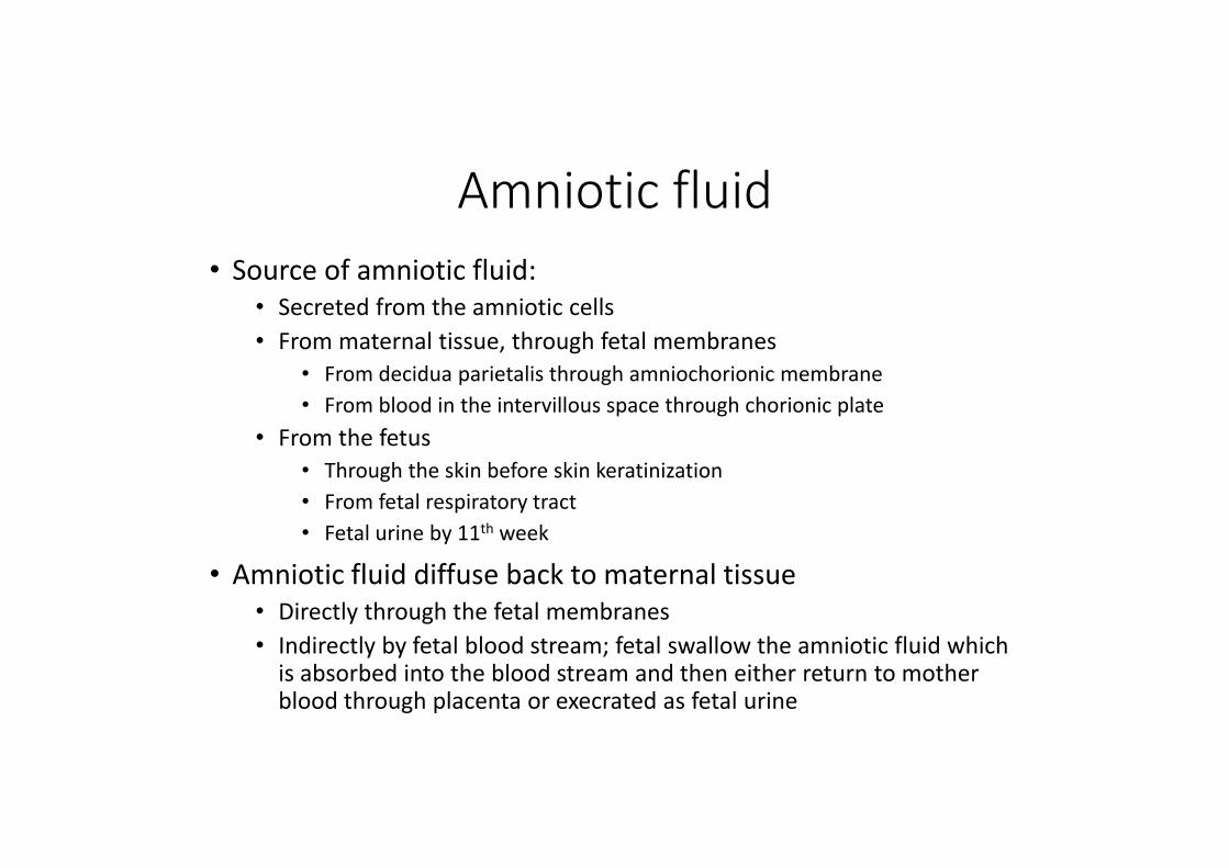
Amniotic fluid• Source of amniotic fluid:
• Secreted from the amniotic cells• From maternal tissue, through fetal membranes
• From decidua parietalis through amniochorionic membrane• From blood in the intervillous space through chorionic plate
• From the fetus• Through the skin before skin keratinization• From fetal respiratory tract• Fetal urine by 11th week
• Amniotic fluid diffuse back to maternal tissue• Directly through the fetal membranes• Indirectly by fetal blood stream; fetal swallow the amniotic fluid which is absorbed into the blood stream and then either return to mother blood through placenta or execrated as fetal urine
Amniotic fluid
• Amniotic fluid functions• Protection of the fetus• Helps control fetal temperature• Fetal fluid and electrolytes homeostasis• Aids in fetal development
• Symmetrical external growth • Muscular development through movement• Lung development
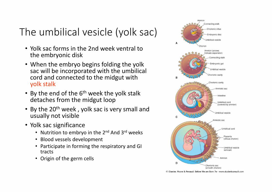
The umbilical vesicle (yolk sac)• Yolk sac forms in the 2nd week ventral to the embryonic disk
• When the embryo begins folding the yolk sac will be incorporated with the umbilical cord and connected to the midgut with yolk stalk
• By the end of the 6th week the yolk stalk detaches from the midgut loop
• By the 20th week , yolk sac is very small and usually not visible
• Yolk sac significance• Nutrition to embryo in the 2nd And 3rd weeks• Blood vessels development• Participate in forming the respiratory and GI tracts
• Origin of the germ cells
Related Documents