# 2007 The Authors Journal compilation # 2007 Blackwell Publishing Ltd doi: 10.1111/j.1600-0854.2006.00521.x Traffic 2007; 8: 259–269 Blackwell Munksgaard PKC Anchoring to GluR4 AMPA Receptor Subunit Modulates PKC-Driven Receptor Phosphorylation and Surface Expression Andre ´ R. Gomes 1,2,† , Susana S. Correia 1,3,4,† , Jose ´ A. Esteban 4 , Carlos B. Duarte 1,2 and Ana Luı´sa Carvalho 1,2, * 1 Center for Neuroscience and Cell Biology, University of Coimbra, Coimbra 3004-517, Portugal 2 Department of Zoology, University of Coimbra, 3004-517 Coimbra, Portugal 3 Department of Biochemistry, University of Coimbra, 3001-401 Coimbra, Portugal 4 Department of Pharmacology, University of Michigan, 48109 Ann Arbor, MI, USA *Corresponding author: Ana Luı ´sa Carvalho, [email protected] † These authors contributed equally to this work. Changes in the synaptic content of a-amino-3-hydroxy-5- methyl-4-isoxazolepropionate (AMPA)–type glutamate receptors lead to synaptic efficacy modifications, involved in synaptic plasticity mechanisms believed to underlie learning and memory formation. Early in devel- opment, GluR4 is highly expressed in the hippocampus, and GluR4-containing AMPA receptors are inserted into synapses. During synapse maturation, the number of AMPA receptors at the synapse is dynamically regulated, and both addition and removal of receptors from post- synaptic sites occur through regulated mechanisms. GluR4 delivery to synapses in rat hippocampal slices was shown to require protein kinase A (PKA)–mediated phosphorylation of GluR4 at serine 842 (Ser842). Protein kinase C (PKC) can also phosphorylate Ser842, and we have shown that PKCg can associate with GluR4. Here we show that activation of PKC in retina neurons, or in human embryonic kidney 293 cells cotransfected with GluR4 and PKCg, increases GluR4 surface expression and Ser842 phosphorylation. Moreover, mutation of amino acids R821A, K825A and R826A at the GluR4 C-terminal, within the interacting region of GluR4 with PKCg, abol- ishes the interaction between PKCg and GluR4 and prevents the stimulatory effect of PKCg on GluR4 Ser842 phosphorylation and surface expression. These data argue for a role of anchored PKCg in Ser842 phos- phorylation and targeting to the plasma membrane. The triple GluR4 mutant is, however, phosphorylated by PKA, and it is targeted to the synapse in CA1 hippocampal neurons in organotypic rat hippocampal slices. The pres- ent findings show that the interaction between PKCg and GluR4 is specifically required to assure PKC-driven phos- phorylation and surface membrane expression of GluR4. Key words: AMPA receptors, GluR4, phosphorylation, PKC, surface expression, synaptic delivery Received 11 December 2005, revised and accepted for publication 29 November 2006, published online 15 January 2007 a-Amino-3-hydroxy-5-methyl-4-isoxazolepropionate (AMPA)– type ionotropic glutamate receptors mediate the majority of fast excitatory synaptic transmission in the brain and are believed to be involved in learning and memory formation. It has been shown that these receptors can be added to and removed from the postsynaptic membrane, resulting in changes in synaptic efficacy (1). These changes in synaptic strength are involved in synaptic plasticity mech- anisms [long-term potentiation (LTP) and long-term depression (LTD)], which are believed to be the molecular basis of learning and memory (1,2). The AMPA receptors are heterooligomeric structures formed by four subunits [GluR1-4; (3)]. The combination of different receptor subunits results in distinct trafficking properties of the AMPA receptors (4,5). GluR4-containing AMPA receptors exhibit fast currents and are expressed in several regions of the central nervous system (CNS) (6–11). In the hippocampus, GluR4 is expressed mainly in early postnatal development, and GluR4-containing AMPA receptors are delivered to the synapse by spontaneous activity (11). In this brain region, synaptic delivery of GluR4- containing AMPA receptors is dependent on protein kinase A (PKA) phosphorylation of the serine 842 (Ser842) resi- due located at the GluR4 C-terminal domain. The GluR4 phosphorylation at Ser842 is believed to relieve a reten- tion interaction that blocks delivery of the receptor into synapses (12). The details of the mechanism that regulates GluR4-con- taining AMPA receptor targeting to the synapse early in development, during synaptogenesis, is unknown; how- ever, interactions with the C-termini of AMPA receptor subunits as well as protein phosphorylation are believed to play a role in receptor dynamics (13). GluR4 was described to interact with stargazin (14), 4.1N (15), protein kinase C (PKC) g (16) and recently with a-actinin-1 and IQGAP-1 (17). The membrane protein stargazin is an AMPA receptor auxiliary subunit (18) and is believed to mediate synaptic trafficking of AMPA receptors by recruiting receptors from submembranous sites to the plasma membrane, and by associating with PDZ proteins to bring AMPA receptors to the synapse (14). Recently, stargazin was found to also modulate AMPA receptor kinetics (19,20). Stargazin is phosphorylated by Ca 2þ /calmodulin-dependent kinase II (CaMKII) and by PKC, and stargazin phosphorylation pro- motes synaptic trafficking of AMPA receptors and is required for LTP at hippocampal synapses (21). 4.1N, a protein that associates with the actin cytoskeleton, was shown to bind GluR4, and the association was www.traffic.dk 259

Welcome message from author
This document is posted to help you gain knowledge. Please leave a comment to let me know what you think about it! Share it to your friends and learn new things together.
Transcript

# 2007 The Authors
Journal compilation# 2007 Blackwell Publishing Ltd
doi: 10.1111/j.1600-0854.2006.00521.xTraffic 2007; 8: 259–269Blackwell Munksgaard
PKC Anchoring to GluR4 AMPA Receptor SubunitModulates PKC-Driven Receptor Phosphorylation andSurface Expression
Andre R. Gomes1,2,†, Susana S. Correia1,3,4,†,
Jose A. Esteban4, Carlos B. Duarte1,2 and
Ana Luısa Carvalho1,2,*
1Center for Neuroscience and Cell Biology, University ofCoimbra, Coimbra 3004-517, Portugal2Department of Zoology, University of Coimbra,3004-517 Coimbra, Portugal3Department of Biochemistry, University of Coimbra,3001-401 Coimbra, Portugal4Department of Pharmacology, University of Michigan,48109 Ann Arbor, MI, USA*Corresponding author: Ana Luısa Carvalho, [email protected]†These authors contributed equally to this work.
Changes in the synaptic content of a-amino-3-hydroxy-5-
methyl-4-isoxazolepropionate (AMPA)–type glutamate
receptors lead to synaptic efficacy modifications,
involved in synaptic plasticity mechanisms believed to
underlie learning and memory formation. Early in devel-
opment, GluR4 is highly expressed in the hippocampus,
and GluR4-containing AMPA receptors are inserted into
synapses. During synapse maturation, the number of
AMPA receptors at the synapse is dynamically regulated,
and both addition and removal of receptors from post-
synaptic sites occur through regulated mechanisms.
GluR4 delivery to synapses in rat hippocampal slices
was shown to require protein kinase A (PKA)–mediated
phosphorylation of GluR4 at serine 842 (Ser842). Protein
kinase C (PKC) can also phosphorylate Ser842, and we
have shown that PKCg can associate with GluR4. Here we
show that activation of PKC in retina neurons, or in
human embryonic kidney 293 cells cotransfected with
GluR4 and PKCg, increases GluR4 surface expression and
Ser842 phosphorylation. Moreover, mutation of amino
acids R821A, K825A and R826A at the GluR4 C-terminal,
within the interacting region of GluR4 with PKCg, abol-
ishes the interaction between PKCg and GluR4 and
prevents the stimulatory effect of PKCg on GluR4
Ser842 phosphorylation and surface expression. These
data argue for a role of anchored PKCg in Ser842 phos-
phorylation and targeting to the plasma membrane. The
triple GluR4 mutant is, however, phosphorylated by PKA,
and it is targeted to the synapse in CA1 hippocampal
neurons in organotypic rat hippocampal slices. The pres-
ent findings show that the interaction between PKCg and
GluR4 is specifically required to assure PKC-driven phos-
phorylation and surface membrane expression of GluR4.
Key words: AMPA receptors, GluR4, phosphorylation,
PKC, surface expression, synaptic delivery
Received 11 December 2005, revised and accepted
for publication 29 November 2006, published online 15
January 2007
a-Amino-3-hydroxy-5-methyl-4-isoxazolepropionate (AMPA)–
type ionotropic glutamate receptors mediate the majority
of fast excitatory synaptic transmission in the brain and are
believed to be involved in learning and memory formation.
It has been shown that these receptors can be added to
and removed from the postsynaptic membrane, resulting
in changes in synaptic efficacy (1). These changes in
synaptic strength are involved in synaptic plasticity mech-
anisms [long-term potentiation (LTP) and long-term
depression (LTD)], which are believed to be the molecular
basis of learning and memory (1,2).
The AMPA receptors are heterooligomeric structures
formed by four subunits [GluR1-4; (3)]. The combination
of different receptor subunits results in distinct trafficking
properties of the AMPA receptors (4,5). GluR4-containing
AMPA receptors exhibit fast currents and are expressed
in several regions of the central nervous system (CNS)
(6–11). In the hippocampus, GluR4 is expressed mainly in
early postnatal development, and GluR4-containing AMPA
receptors are delivered to the synapse by spontaneous
activity (11). In this brain region, synaptic delivery of GluR4-
containing AMPA receptors is dependent on protein kinase
A (PKA) phosphorylation of the serine 842 (Ser842) resi-
due located at the GluR4 C-terminal domain. The GluR4
phosphorylation at Ser842 is believed to relieve a reten-
tion interaction that blocks delivery of the receptor into
synapses (12).
The details of the mechanism that regulates GluR4-con-
taining AMPA receptor targeting to the synapse early in
development, during synaptogenesis, is unknown; how-
ever, interactions with the C-termini of AMPA receptor
subunits as well as protein phosphorylation are believed to
play a role in receptor dynamics (13). GluR4 was described
to interact with stargazin (14), 4.1N (15), protein kinase C
(PKC) g (16) and recently with a-actinin-1 and IQGAP-1
(17). The membrane protein stargazin is an AMPA receptor
auxiliary subunit (18) and is believed to mediate synaptic
trafficking of AMPA receptors by recruiting receptors from
submembranous sites to the plasma membrane, and by
associating with PDZ proteins to bring AMPA receptors to
the synapse (14). Recently, stargazin was found to also
modulate AMPA receptor kinetics (19,20). Stargazin is
phosphorylated by Ca2þ/calmodulin-dependent kinase II
(CaMKII) and by PKC, and stargazin phosphorylation pro-
motes synaptic trafficking of AMPA receptors and is
required for LTP at hippocampal synapses (21). 4.1N,
a protein that associates with the actin cytoskeleton,
was shown to bind GluR4, and the association was
www.traffic.dk 259

suggested to play a role in the receptor expression at the
cell surface (15). a-Actinin-1 and IQGAP1 are also actin-
binding proteins, which were shown to bind to the
C-terminus of GluR4 at the region containing the Ser842
phosphorylation site (17). Phosphorylation of GluR4 dis-
rupts the interaction with a-actinin-1, whereas the interac-
tion with IQGAP-1 is preserved. The authors of the study
suggest that a-actinin-1 retains GluR4 in intracellular pools,
and that following synaptic activity and GluR4 phosphory-
lation, the interaction with a-actinin-1 is disrupted to
release GluR4 to the synapse (17).
PKCg is a CNS-specific PKC isoform shown to bind GluR4
AMPA receptor subunit at the C-terminal membrane-
proximal domain (16). PKCg was suggested to bind
directly to GluR4, facilitating receptor phosphorylation at
Ser842. Kinase targeting to specific subcellular micro-
domains is known to be necessary for signaling efficiency
and specificity, and is, in many cases, accomplished by
kinase interactions with protein partners. CaMKII interacts
with various subunits of N-methyl-D-aspartate receptors
(22); the interaction between CaMKII and NR2B can trap
an active Ca2þ-independent form of CaMKII (23) and was
recently shown to be required for different forms of
synaptic enhancement (24). Moreover, A-kinase anchoring
protein (AKAP)79/150-anchored PKA associates with
GluR1 through the adaptor proteins SAP97 or PSD-95
and promotes phosphorylation of GluR1 at Ser845 (25).
Disruption of the PKA–AKAP interaction is sufficient to
cause a long-lasting reduction in synaptic AMPA receptors
in cultured neurons and occludes synaptically induced
LTD in hippocampal slices (26). To further clarify the role
of PKCg–GluR4 interaction in the delivery of GluR4-
containing AMPA receptors to the plasma membrane,
we studied the cell surface targeting of a mutated form
of GluR4 protein, unable to bind PKCg. Our results
show that the PKCg targeting to GluR4 is essential for
PKCg phosphorylation of Ser842 and GluR4 surface
expression.
Results
Effect of PKC on phosphorylation and surface
expression of native GluR4-containing
AMPA receptors
In a previous study, we have shown that PKA activation in
primary cultured chick retinal neurons increases the
phosphorylation and surface expression of native GluR4-
containing AMPA receptors (27). Using the same prepa-
ration, which expresses high levels of GluR4 (28), we
tested whether PKC activation can trigger GluR4 phos-
phorylation and surface expression. After 5 days in
culture, cells were stimulated for 10 min with 200 nM
phorbol 12-myristate 13-acetate (PMA), and the phospho-
rylation of GluR4 was analysed using a phosphospecific
antibody against GluR4 phosphorylated at Ser842 (12).
We found that PMA stimulation of cultured chick retina
neurons significantly increased (25.7 � 2.3%) GluR4
Ser842 phosphorylation, when compared with the control
situation (Figure 1A). To test whether PKC activation can
lead to GluR4 surface expression, cells were stimulated
with 200 nM PMA, for 10 min, and were then incubated
with a biotinylation reagent. Biotinylated proteins were
purified with a streptavidin gel and analysed by immuno-
blotting in order to quantify plasma membrane GluR4.
Activation of PKC with PMA in chick retinal neurons
significantly increased (22.5 � 1.3%) membrane surface
GluR4, when compared with the control condition (Figure 1
B). Moreover, cell treatment with PMA increased the
phosphorylation of GluR4 at the plasma membrane
(41.9 � 10.8%, Figure 1C), whereas the phosphorylation
of intracellular GluR4 remained unchanged (Figure 1D).
These results suggest that PKC activation leads to phos-
phorylation of GluR4 Ser842 and targeting of GluR4-
containing AMPA receptors to the plasma membrane.
Accordingly, it was previously described that Ser842
phosphorylation is necessary and sufficient for GluR4
delivery to synapses early in development in the rat
hippocampus (12).
Mapping the interaction between GluR4 and PKCg
We have previously found that GluR4 interacts with PKCg
through the membrane-proximal C-terminal amino acid
segment E815–K828 in GluR4. This segment shows
sequence homology to the PKCg pseudosubstrate
domain, suggesting that the catalytic domain of PKCg
may bind to GluR4 through the amino acid segment 815–
828, thereby positioning the phosphorylation site Ser842
for preferential phosphorylation by PKCg (16). In order to
understand the role of this interaction in the regulation of
GluR4 phosphorylation by PKC, we started by identifying
the crucial amino acids for the interaction. Recombinant
glutathione S-transferase (GST)-GluR4 C-terminal fusion
protein with a triple point mutation at the amino acids
R821A, K825A and R826A (Figure 2B) within the GluR4-
PKCg binding domain (GluR4AAA C-terminus) was pro-
duced. The mutated residues were selected in order to
destroy the homology between this region of GluR4 and
the PKCg pseudosubstrate. The effect of these GluR4
point mutations on the interaction between GluR4 and
PKCg was tested by performing GST pull-down assays.
Rat brain extract (RBE) was incubated with GST fused to
GluR4WT C-terminus or GluR4AAA C-terminus, as indi-
cated. Glutathione-Sepharose beads were used to pull
down GST fusion proteins and their binding partners. PKCg
was copurified when the brain extract was incubated with
GST-GluR4 C-terminus but not when incubated with GST-
GluR4AAA C-terminus (Figure 2C). These data show that
R821, K825 and R826 in GluR4 are critical amino acids for
the interaction between GluR4 and PKCg to occur and
allow us to make constructs encoding a GluR4 form that
does not bind PKCg.
260 Traffic 2007; 8: 259–269
Gomes et al.

Effect of blocking the PKCg–GluR4 interaction on
PKC-driven phosphorylation and plasma membrane
expression of GluR4
Previous evidences suggest that PKCg bound to GluR4 can
phosphorylate Ser842 in GluR4, and cotransfection of
PKCg with GluR4, in human embryonic kidney (HEK) 293
cells, increased GluR4 subunit surface expression, follow-
ing PKC activation with PMA (16), Figure 3). To examine
the role of the association between PKCg and GluR4 in the
effect of PKC on GluR4 phosphorylation and AMPA
receptor trafficking to the plasma membrane, we trans-
fected HEK 293 cells with GluR4 or with the GluR4 triple
point mutant (GluR4AAA), which is unable to bind PKCg.
Cells were cotransfected with PKCg and stimulated with
200 nM PMA for 10 min (when indicated). Quantitative
immunoblotting was used to compare phosphorylated
versus total GluR4 (Figure 3A) and surface versus total
GluR4 (Figure 3B).
In cells expressing GluR4 or GluR4AAA, in the absence of
cotransfected PKCg, no significant differences between
the two proteins were observed in terms of either phos-
phorylation or plasma membrane expression. When
endogenous PKC is activated with PMA, there is increased
GluR4 phosphorylation at Ser842 (Figure 3A) and surface
expression (Figure 3B), which nevertheless fail to reach
statistical significance, suggesting that the amount of
endogenous PKC is limiting. In cells coexpressing GluR4
and PKCg and stimulated with PMA, dramatic increases in
both GluR4 phosphorylation (122.1 � 16.4%, above con-
trol) and GluR4 delivery to the plasma membrane (59.9 �15.8%, above control) were observed. These data are in
agreement with previous studies, showing that PKA
phosphorylation of GluR4 at this site is necessary and
sufficient for GluR4 surface and synaptic delivery (12,27).
The PKC-stimulated GluR4 surface expression was also
observed when the experiments were performed in cells
incubated with 0.35 M sucrose to block endocytosis,
suggesting that the effect of PKC is directly on the surface
insertion of the receptors (data not shown). However, no
significant changes in the phosphorylation or surface
expression of GluR4AAA were observed upon coexpres-
sion and activation (with PMA) of PKCg (Figure. 3A,B),
indicating that the interaction between PKC and GluR4 is
necessary for PKCg-mediated GluR4 phosphorylation and
receptor delivery to the cell surface.
Figure 1: Effect of PKC on GluR4 phosphory-
lation and surface expression in primary cul-
tures of retina amacrine-like neurons. A) Effect
of PKC activation on GluR4 phosphorylation at
Ser842. Cells were incubated with the PKC
activator, PMA (200 nM) for 10 min. Cell extracts
were prepared, subjected to SDS–PAGE and
immunoblotted against phosphorylated GluR4 at
Ser842 and total GluR4. The amount of phosphor-
ylated GluR4 was normalized to the total amount
of GluR4 in each lane. B) Effect of PKC activation
on GluR4 surface expression. Biotinylation was
performed as described in Materials and Meth-
ods. Streptavidin-retained protein complexes
were collected and run on SDS–PAGE. Immuno-
blot was performed using an antibody against
GluR4 C-terminal region. Surface GluR4 was
normalized to the total amount of GluR4 in each
condition. C, D) Effect of PKC activation on the
phosphorylation of surface (C) or intracellular (D)
GluR4. All data are expressed as percentage of
control and plotted as the mean � SEM for the
indicated number of experiments performed in
independent preparations (*p< 0.05, **p < 0.01,
Bonferroni’s test). Representative Western blots
are shown.
Traffic 2007; 8: 259–269 261
PKCg Targeting to GluR4
Effect of blocking the PKCg–GluR4 interaction on
PKA-driven phosphorylation and plasma membrane
expression of GluR4
To determine whether the triple point mutations at the
C-terminal membrane-proximal domain of GluR4 have an
effect on protein folding that could impair GluR4 phosphory-
lation and surface expression, we tested whether GluR4-
AAA can be phosphorylated by PKA and targeted to the
membrane upon PKA activation in HEK 293 cells. The HEK
293 cells were transfected with GluR4 or GluR4AAA and
stimulated with 10 mM forskolin (FSK) for 10 min (when
indicated) followed by incubation with the biotinylation
reagent. Cell extracts were collected as described in
Materials and Methods and quantitative immunoblotting
was used to compare phosphorylated versus total GluR4
(Figure 4A) and surface versus total GluR4 (Figure 4B).
Stimulation by FSK induced a robust increase in Ser842
phosphorylation both in HEK 293 cells expressing GluR4 or
GluR4AAA (159.7 � 40.1% and 175.9 � 45.0% above
control, respectively; Figure 4A). Plasma membrane
expression of GluR4 or GluR4AAA was also significantly
increased in FSK-stimulated cells [GluR4 (59.6 � 18.1%),
GluR4AAA (68.1 � 18.2%) above control; Figure 4B].
Synaptic targeting of GluR4 in rat hippocampal CA1
neurons occurs early in development and has been shown
to be driven by spontaneous synaptic activity and to
depend on PKA activity (11,12). Therefore, we tested
whether the GluR4 C-terminus triple mutant, which can be
phosphorylated by PKA in HEK 293 cells, is phosphory-
lated and targeted to the synapse in CA1 neurons in
hippocampal slice cultures. Green fluorescent protein
(GFP)-tagged GluR4 or GFP-tagged GluR4AAA was ex-
pressed in CA1 neurons of rat hippocampal organotypic
slices obtained from P5-6 animals, using the Sindbis virus
expression system. Slices were then maintained in culture
for approximately 36 h. The recombinant proteins showed
homogeneous dendritic expression (Figure 4D). Delivery
of GFP-GluR4 to synapses was monitored using the
inward rectification properties of the overexpressed re-
combinant receptors (electrophysiological tagging,
(4,11,12,29)). Recombinant GFP-GluR4 expressed in CA1
cells mainly forms homomeric receptors, which do not
conduct outward currents. Hence, their delivery into
synapses can be quantified as a decrease in the ratio of
the evoked postsynaptic current at þ40 mV relative to
the current at �60 mV [rectification index (RI) ¼ Iþ40/I�60].
The RI values obtained from GFP-GluR4AAA-infected cells
were similar to those obtained from GFP-GluR4-infected
cells, indicating that the PKA-driven synaptic incorporation
of GluR4AAA is not compromised by the triple mutation
(Figure 4C). Phosphorylation of GluR4 and GluR4AAA was
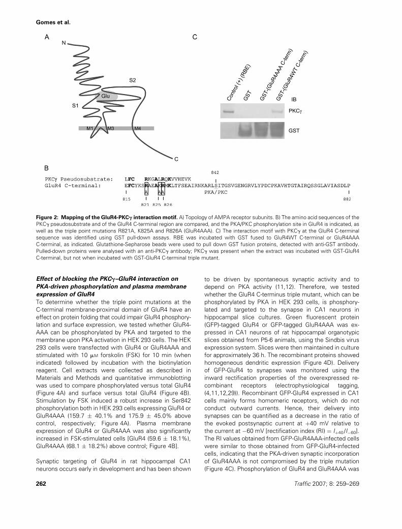
Figure 2: Mapping of the GluR4-PKCg interaction motif. A) Topology of AMPA receptor subunits. B) The amino acid sequences of the
PKCg pseudosubstrate and of the GluR4 C-terminal region are compared, and the PKA/PKC phosphorylation site in GluR4 is indicated, as
well as the triple point mutations R821A, K825A and R826A (GluR4AAA). C) The interaction motif with PKCg at the GluR4 C-terminal
sequence was identified using GST pull-down assays. RBE was incubated with GST fused to GluR4WT C-terminal or GluR4AAA
C-terminal, as indicated. Glutathione-Sepharose beads were used to pull down GST fusion proteins, detected with anti-GST antibody.
Pulled-down proteins were analysed with an anti-PKCg antibody; PKCg was present when the extract was incubated with GST-GluR4
C-terminal, but not when incubated with GST-GluR4 C-terminal triple mutant.
262 Traffic 2007; 8: 259–269
Gomes et al.
also compared in hippocampal slices. The GFP-tagged
GluR4 recombinant proteins were immunoprecipitated
with an anti-GFP antibody (Figure 4E, bottom panel). Both
GFP-GluR4 and GFP-GluR4AAA were phosphorylated on
Ser842 (Figure 4E, upper panel). No significant differences
in phosphorylation were detected between wild-type
GluR4 and the mutant in infected hippocampal slices.
These results indicate that the triple point mutation in
GluR4AAA does not affect PKA phosphorylation and
consequent plasma membrane or synaptic expression of
GluR4, or its synaptic function, and exclude the possibility
that the impairment in PKC-mediated phosphorylation and
surface expression of GluR4AAA is the result of altered
protein folding. Moreover, the results show that the
interaction between PKCg and GluR4 is specifically
required to assure PKC-driven phosphorylation of GluR4.
Experiments in hippocampal slice cultures were also
performed using a deletion mutant of GluR4, which lacks
a 14-amino-acid juxtamembrane region just past the
fourth transmembrane domain of the receptor subunit
(GluR4D815–828). The RI values obtained from neurons
infected with GFP-GluR4D815–828 were significantly high-
er than those obtained from GFP-GluR4-infected cells
(Figure 4C), indicating compromised synaptic incorpora-
tion of this mutant protein, although homogeneous den-
dritic expression was observed (Figure 4D). Moreover, the
deletion mutant showed impaired phosphorylation at
Ser842 (Figure 4E), but a serine to aspartate mutation at
residue 842 to mimic GluR4 phosphorylation could not re-
scue GluR4 synaptic delivery (GFP-GluR4D815–828S842D,Figure 4C), suggesting that this membrane-proximal region
of the receptor is necessary for synaptic delivery of GluR4.
Discussion
We have previously shown that PKCg binds directly to the
GluR4 membrane-proximal C-terminal domain and that
GluR4 is phosphorylated on Ser842 by bound kinases,
including PKCg, suggesting that the interaction maintains
the kinase in close proximity to GluR4, facilitating receptor
phosphorylation at Ser842 (16). The present data show that
PKC activation leads to GluR4 phosphorylation and surface
expression in cultured retina neurons, and that PKCg
coexpression in GluR4-transfected HEK 293 cells increases
GluR4 surface expression upon stimulation with PMA.
These evidences argue for a role of PKC in GluR4 receptor
subunit phosphorylation and targeting to the plasma
membrane. Moreover, the presented results show that
point mutations in GluR4 that disrupt the interaction site for
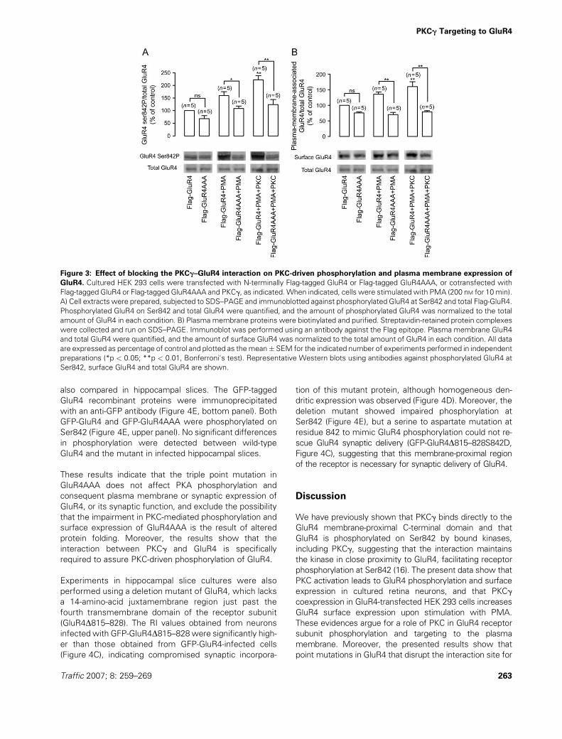
Figure 3: Effect of blocking the PKCg–GluR4 interaction on PKC-driven phosphorylation and plasma membrane expression of
GluR4. Cultured HEK 293 cells were transfected with N-terminally Flag-tagged GluR4 or Flag-tagged GluR4AAA, or cotransfected with
Flag-tagged GluR4 or Flag-tagged GluR4AAA and PKCg, as indicated.When indicated, cells were stimulated with PMA (200 nM for 10 min).
A) Cell extracts were prepared, subjected to SDS–PAGE and immunoblotted against phosphorylated GluR4 at Ser842 and total Flag-GluR4.
Phosphorylated GluR4 on Ser842 and total GluR4 were quantified, and the amount of phosphorylated GluR4 was normalized to the total
amount of GluR4 in each condition. B) Plasma membrane proteins were biotinylated and purified. Streptavidin-retained protein complexes
were collected and run on SDS–PAGE. Immunoblot was performed using an antibody against the Flag epitope. Plasma membrane GluR4
and total GluR4 were quantified, and the amount of surface GluR4 was normalized to the total amount of GluR4 in each condition. All data
are expressed as percentage of control and plotted as themean� SEM for the indicated number of experiments performed in independent
preparations (*p < 0.05; **p < 0.01, Bonferroni‘s test). Representative Western blots using antibodies against phosphorylated GluR4 at
Ser842, surface GluR4 and total GluR4 are shown.
Traffic 2007; 8: 259–269 263
PKCg Targeting to GluR4

PKCg in the GluR4 C-terminal membrane-proximal region
impair PKC-driven GluR4 phosphorylation and surface
expression in HEK 293 cells. These mutations do not alter
protein folding or compromise the Ser842 phosphorylation
site because the mutant protein is functional at synapses in
hippocampal CA1 neurons,where synaptic targeting of GluR4
has been shown to depend on GluR4 phosphorylation by
PKA and to be induced by spontaneous neuronal activity.
When we expressed a deletion mutant of GluR4
(GluR4D815–828) in organotypic rat hippocampal slices,
impaired synaptic delivery and no protein phosphorylation
at Ser842 were observed (Figure 4C,E). The C-terminal
membrane-proximal region of GluR4 was previously impli-
cated in the basal surface expression of GluR4 in HEK 293
cells and in dissociated hippocampal neurons in culture
(15). The authors showed that deletion of the C-terminal
domain up to the juxtamembrane region of GluR4 blocks
receptor surface expression, which could be partially
rescued if a C-terminal 14-amino-acid membrane-proximal
segment, the same domain that PKCg binds to in GluR4,
was present (15). The correspondent sequence in GluR1
was previously shown to bind the 4.1N protein, the
neuronal homologue of the erythrocyte membrane cyto-
skeletal protein 4.1 (30). Coleman et al. found that 4.1N
can also associate to GluR4 and that a deletion of the
C-terminal, membrane-proximal 14-amino-acid segment of
GluR4, or mutation of the R821, K825 and R826 residues at
Figure 4: Legend on next page.
264 Traffic 2007; 8: 259–269
Gomes et al.
this region, disrupts 4.1N binding and significantly reduces
basal GluR4 surface expression (15). Our results obtained
using infected hippocampal slices further show that synaptic
delivery of GluR4 is blocked in the absence of the GluR4
juxtamembrane C-terminal domain (Figure 4C). Moreover,
the deletion mutant is not phosphorylated in hippocampal
slices (Figure 4E), indicating that the deletion may com-
promise the efficiency of GluR4 phosphorylation by several
kinases, eventually by having an impact on the protein
folding at the C-terminus. Early in the development of rat
hippocampus, spontaneous activity drives GluR4-contain-
ing AMPA receptors to the synapse (11) by a mechanism
mediated by PKA phosphorylation of GluR4 Ser842 (12).
PKA phosphorylation of GluR4 Ser842 was suggested to
relieve a retention interaction, driving receptors to synap-
ses (12). Mutation of Ser842 to an aspartate was described
to drive GluR4-containing AMPA receptors to synapses,
bypassing the need for spontaneous activity (12). However,
replacement of Ser842 for an aspartate on GluR4D(815–828) recombinant protein was unable to revert the effect
of the GluR4 membrane-proximal segment deletion on
GluR4 delivery to synapses in rat hippocampal slices
(Figure 4C). Our data indicate that Ser842-phosphorylation-
mediated targeting of GluR4 to synapses is blocked by
deletion of the GluR4 membrane-proximal domain.
The GluR4 triple mutant (GluR4 R821A, K825A, R826A)
was targeted to the synapse in CA1 neurons as efficiently
as wild-type GluR4 (Figure 4C), and in HEK 293 cells, there
was no significant difference between the basal surface
expression of the wild-type and mutant forms of GluR4
(Figures 3B and 4B). Moreover, the increase in GluR4
surface expression in HEK 293 cells triggered by PKA
activation was the same for wild-type GluR4 and the triple
mutant (Figure 4B). Because the R821, K825 and R826
residues are critical for GluR4 binding to 4.1N (15), the
GluR4 triple mutant (GluR4 R821A, K825A, R826A) is
unable to bind 4.1N (data not shown); our results do not
support the idea that GluR4 binding to 4.1N is required for
GluR4 surface expression or synaptic targeting. However,
our data and those of others suggest that other determin-
ants exist at the membrane-proximal segment of the
C-terminus of the receptor, apart from the R821, K825
and R826 residues, with a role in the surface expression of
GluR4. A recent study identified C817 at the C-terminal
region of GluR4 (and equivalent Cys residues at the
C-terminus of the other AMPA receptor subunits) as a
palmitoylation site and found that depalmitoylated AMPA
receptors show a stronger association with 4.1N (31).
Moreover, palmitoylation of this Cys residue at the C-
terminus of AMPA receptors was shown to be necessary
for agonist-induced internalization of the receptors, but not
for their steady-state surface expression (31).
Our data point to a clear role for the basic R821, K825 and
R826 residues at the C-terminus of GluR4 in receptor
binding to PKCg, in GluR4 phosphorylation by PKC and in
PKC-driven surface expression of GluR4 (Figure 5). When
PKCg was coexpressed with GluR4, there was a striking
difference between surface expression of GluR4 and
GluR4AAA (Figure 3B). This suggests that the interaction
of GluR4 with PKCg through the C-terminal juxtamembrane
domain of GluR4 is necessary for surface delivery promoted
by PKC activation. Moreover, the triple mutation impairs
efficient GluR4 phosphorylation by PKC in HEK 293 cells
(Figure 3A), indicating that the kinase anchoring through the
815–828 GluR4 region is crucial for receptor phosphoryla-
tion by this kinase. Interestingly, the sequence in GluR1
(RSESKR) homologous to the PKC interaction site on GluR4
(RAEAKR) contains a PKC phosphorylation site, which con-
trols synaptic incorporation of GluR1 during LTP (32).
Taken together, these results point to a dual role for the
membrane-proximal region of the C-terminus of GluR4:
Figure 4: Effect of blocking the PKCg–GluR4 interaction on PKA-driven phosphorylation, plasma membrane expression and
synaptic delivery of GluR4. A) Cultured HEK 293 cells were transfected with N-terminally Flag-tagged GluR4 or Flag-tagged GluR4AAA.
When indicated, cells were stimulatedwith FSK (10mM for 10min). Cell extractswere prepared, subjected to SDS–PAGE and immunoblotted
against phosphorylated GluR4 at Ser842 and total Flag-GluR4. The amount of phosphorylated GluR4 was normalized to the total amount of
GluR4 in each condition. B) Plasma-membrane-associated GluR4 and total GluR4 were quantified in transfected HEK 293 cells, and the
amount of surface GluR4 was normalized to the total amount of GluR4 in each condition. All data are expressed as percentage of control and
plotted as the mean� SEM for the indicated number of experiments performed in independent preparations (**p< 0.01, Bonferroni’s test).
RepresentativeWestern blots using antibodies against phosphorylated GluR4 at Ser842, surface GluR4 and total GluR4 are shown. C) GluR4
expression and delivery to synapses in CA1 neurons of rat hippocampal slices. Cultured rat hippocampal slices expressing GFP-GluR4, GFP-
GluR4AAA, GFP-GluR4D(815–828) or GluR4D(815–828)S842D were used to collect electrophysiological data. GFP-GluR4 is delivered to
synapses when expressed in rat hippocampal slices [normalized average rectification value (Iþ40mV/I�60mV) was 46.4% � 7.1% for GluR4
infected]. GFP-GluR4AAA delivery to synapses is not significantly different when compared with GFP-GluR4 (average rectification value was
41.1% � 3.7% of control uninfected cells). GFP-GluR4D(815–828) or GFP-GluR4D(815–828)S842D delivery to synapses is impaired when
comparedwithGFP-GluR4.Normalized average rectification valueswere70.8%� 5.4%forGFP-GluR4D(815–828) and 87.1%� 6.3%forGFP-
GluR4D(815–828)S842D. Results are presented as mean � SEM, and statistical significance was determined by the t-test (assuming un-
equal variances). D) GFP fluorescence shows homogeneous dendritic expression of GFP-GluR4, GFP-GluR4AAA, GFP-GluR4(815–828) and
GFP-GluR4(815–828)S842D. Scale bar represents 10 mM. E) Phosphorylation of GFP-GluR4, GFP-GluR4AAA, GFP-GluR4D(815–828) or GFP-GluR4D(815–828)S842D in rat hippocampal slices. Cell extracts from cultured rat hippocampal slices expressing GFP-GluR4 or GFP-GluR4-
AAA, GFP-GluR4D(815–828) or GFP-GluR4D(815–828)S842D were prepared and used for immunoprecipitation with an anti-GFP antibody.
Immunoprecipitated proteins were analysed by Western blotting with antibodies against phosphorylated GluR4 at Ser842 and total GluR4.
Traffic 2007; 8: 259–269 265
PKCg Targeting to GluR4

(i) on the one hand, the R821, K825 and R826 residues
present in this region are necessary for the Ser842
residue in GluR4 to be phosphorylated by PKC and (ii)
on the other hand, other determinants at the membrane-
proximal segment of the C-terminus are required for
localization of Ser842-phosphorylated GluR4 at the syn-
apse or the plasma membrane. The interaction of GluR4
with PKCg plays a role in receptor phosphorylation,
whereas the interaction of this region of GluR4 with other
proteins could be important in stabilizing the receptor at
synapses and at the plasma membrane, after the phos-
phorylated receptor has been driven to the membrane,
eventually regulating receptor internalization. In fact,
members of the 4.1N protein family have been involved
in linking plasma-membrane-associated proteins to the
actin cytoskeleton (33), and polymerized actin was pre-
viously shown to be important for immobilization and
clustering of AMPA receptors (34,35). The phosphatidyli-
nositol 3-kinase (PI3K) was also reported to be clustered
with AMPA receptors at synapses and was shown to bind
directly to GluR1 and GluR2 (36). The 21 amino acids at the
GluR2 juxtamembrane C-terminal domain are responsible
for the interaction of PI3K (36), and the first 14 amino acids
of this segment in GluR2 are common to GluR4. It is
possible, therefore, that PI3K interacts with GluR4. Man et
al. (36) suggested that the accumulation of PI3K products,
like PtdIns(3,4,5)P3, near AMPA-receptor-containing vesi-
cles may facilitate the fusion of these vesicles with the
postsynaptic membrane.
We have previously shown that PKC up-regulates AMPA
receptor activity in chick embryo retinal cultures (37).
Activation of PKC with PMA in cultured chick embryo
retinal neurons significantly increased plasma membrane
GluR4 expression (Figure 1A) and GluR4 Ser842 phospho-
rylation (Figure 1B,C). Previous work showed that PKA
phosphorylation of Ser842 on GluR4 is necessary and
sufficient for GluR4 delivery to synapses (12); however,
PKC can also phosphorylate GluR4 Ser842 (38). Our results
show a role of PKC phosphorylation of GluR4 Ser842
in GluR4 targeting to the plasma membrane, in retinal
neurons, and suggest that phosphorylation-regulated deliv-
ery of GluR4 could be mediated by PKA or PKC in different
regions of the brain. The PMA-induced increase in the
surface expression of GluR4 in retina neurons indicates that
PKC regulates the targeting of GluR4-containing AMPA
receptors to the plasma membrane in neurons. Moreover,
several physiological stimuli could result in increased PKC
activity inamacrineneurons.Group Imetabotropicglutamate
receptors, as well as metabotropic acetylcholine receptors,
are expressed and functional in these cells and elicit inositol
phospholipids hydrolysis and [Ca2þ]i elevations (39–41).
In conclusion, our results support the involvement of GluR4-
anchored PKCg in Ser842 phosphorylation and in the PKC-
regulated delivery of GluR4 to the plasma membrane.
Materials and Methods
MaterialsTrypsin was purchased from Life Technologies Ltd (Paisley, UK), and fetal
calf serum was from Biochrom KG (Berlin, Germany) or from Biowhittaker
(Walkersville, MD, USA). The PMA was from Calbiochem (San Diego, CA,
USA). Forskolin was obtained from BIOMOL Research Laboratories, Inc
(Plymouth Meeting, PA, USA). Complete Mini protease inhibitor cocktail,
microporous polyvinylidene difluoride (PVDF) membranes and the mouse
anti-GFP antibody were obtained from Roche Diagnostics GmbH (Basel,
Switzerland). EZ-link Sulfo-NHS-SS-biotin, UltraLink Plus Immobilized
Streptavidin Gel and the bicinchoninic acid (BCA) protein assay reagent
kit were from Pierce (Rockford, IL, USA). pGEX4T-2 vector, glutathione-
Sepharose 4B, goat polyclonal anti-GST antibody, alkaline-phosphatase-
conjugated anti-rabbit and anti-mouse secondary antibodies, and the
Enhanced Chemifluorescence (ECF) immunodetection substrate were all
obtained from Amersham Biosciences (Uppsala, Sweden). Anti-goat
alkaline-phosphatase-conjugated antibody and mouse monoclonal anti-
transferrin receptor antibody were from Zymed Laboratories Inc
(San Francisco, CA, USA). The mouse monoclonal anti-PKCg antibody
was purchased from Transduction Laboratories (Lexington, KY, USA).
Isopropyl-1-thio-b-galactopyranoside was from Promega (Madison, WI,
USA). The rabbit polyclonal anti-GluR4 antibody was purchased from
Chemicon (Temecula, CA, USA), and the rabbit polyclonal anti-GluR4
phosphorylated at Ser842 antibody was a gift from Dr Richard L. Huganir.
This antibody was produced against the chemically phosphorylated
peptide (RNKARLSPITGSV) corresponding to rat GluR4 C-terminal 12 amino
acids (836–847) phosphorylated at Ser842 (12). The expression construct
Flag-tagged GluR4 was a kind gift from Dr Kari Keinanen (15), and the Quick
Change mutagenesis kit was purchased from Stratagene (Amsterdam, the
Netherlands). All the other reagents were obtained from Sigma (St Louis,
MO, USA) or from Merck (Darmstadt, Germany).
Embryonic chick retina cell cultureMonolayer cultures of chick retina amacrine-like cells were prepared as
previously described (39,42). Briefly, the retinas from 8-day-old chick
embryos (white leghorn) were dissected and digested with 0.1% trypsin
in Ca2þ- and Mg2þ-free Hank‘s balanced salt solution for 15 min at 378C.
Figure 5: Model for the mechanism of GluR4 AMPA receptor
trafficking and how it depends on the interaction between
GluR4 and PKC.
266 Traffic 2007; 8: 259–269
Gomes et al.
The cells were cultured on poly-D-lysine tissue culture dishes in basal
medium of Eagle, buffered with 25 mM HEPES and 10mM NaHCO3, pH 7.4,
and supplemented with 5% heat-inactivated fetal calf serum, penicillin
(100 U/mL) and streptomycin (100 mg/mL). The cells were maintained at
378C in a humidified incubator with 95% air and 5% CO2, and used after
5 days in culture.
Recombinant proteins expressed in Escherichia coliGlutathione S-transferase fused to the GluR4 C-terminal [GST-(GluR4 C-
term)] construct was cloned in pGEX4T-2 vector as described in Correia et
al. (16). The triple mutant GST-(GluR4AAA C-term), with the R821A, K825A
and R826A substitutions (Figure 2B), was prepared with the Quick Change
mutagenesis kit, using GST-(GluR4 C-term) construct as a template and
the primers 50-ttctgttacaagtccgctgcagaggcggcggcaatgaagctgact-30 and
50-agtcagcttcattgccgccgcctctgcagcggacttgtaacagaa-30.
Recombinant proteins were expressed in BL21 Escherichia coli trans-
formed with the constructs described above. Bacteria grown to A600 ¼ 0.8
were induced with isopropyl-1-thio-b-galactopyranoside (500 mM) for 30 min
at 308C and then lysed with PBS containing 1% Triton X-100 and supple-
mented with protease inhibitors. The cells were sonicated and shaken for
30min at 48C, and the insoluble fractionwas then removed by centrifugation
at 12 000 � g for 10 min at 48C. Glutathione S-transferase fusion proteins
were purified by glutathione-Sepharose affinity chromatography. Protein
concentration was measured using the BCA protein assay reagent kit.
Glutathione S-transferase binding assaysWhole RBE were prepared by homogenizing the tissue in lysis buffer
[20 mM Tris, 2 mM EGTA, 2 mM ethylenediaminetetraacetic acid (EDTA), 1%
Triton-X-100 and supplemented with protease inhibitors] followed by
centrifugation at 1000 � g for 10 min at 48C. The resulting supernatant
was recentrifuged at 1000 � g for 10 min at 48C, and the soluble brain
proteins were used for binding studies. Fifteen micrograms of fusion protein
was incubated with rat brain homogenates overnight at 48C. Themixture was
incubated with 50% glutathione-Sepharose beads for 30 min at 48C. Beadswere washed extensively with radioimmunoprecipitation assay (RIPA) buffer
(150 mM NaCl, 50 mM Tris–HCl, 5 mM EGTA, 1% Triton X-100, 0.5% sodium
deoxycholate, 0.1% SDS; pH 7.5) supplemented to 0.5 M NaCl, and binding
proteins were eluted with SDS–PAGE sample buffer [0.125 M Tris, 2% (w/v)
SDS, 5% (v/v) glycerol, 5% (v/v) b-mercaptoethanol] and boiled for 10 min.
Samples were analysed by Western blotting.
Recombinant proteins expressed in mammalian cellsThe construct encoding N-terminally Flag-tagged GluR4 was a kind gift from
Dr Kari Keinanen (15), and Flag-tagged GluR4AAA (construct coding for the
Flag-GluR4 sequence with C-terminal triple mutations R821A, K825A and
R826A, Figure 2B) was prepared with the Quick Change mutagenesis kit,
using the Flag-tagged GluR4 construct as a template and the primers
50-ttctgttacaagtccgctgcagaggcggcggcaatgaagctgact-30 and 50-agtcagcttcatt-
gccgccgcctctgcagcggacttgtaacagaa-30. The pBK-CMV-PKCg construct was
prepared as previously described (16). Cultured HEK 293 cells were
transfected with Flag-GluR4 or Flag-GluR4AAA, or cotransfected with Flag-
GluR4 or Flag-GluR4AAA and pBK-CMV-PKCg by the calcium phosphate
method (38).
To produce Sindbis virus containing GFP-GluR4D(815–828), GFP-
GluR4D(815–828)S842D, the C-terminal coding sequence of GluR4 with
a deletion of the 42 bp coding for the 14 juxtamembrane amino acids was
amplified using the primers 50-ggcatctctatcatgatc-30 and 50-cgacgggccct-
tatggtaggtccgatgcaatgacagc-30 (which include the restriction sites for
bclI and ApaI, respectively) and the pGW1-GluR4D(815–828) and pGW1-
GluR4D(815–828)S-842D constructs as a template. To obtain Sindbis virus
expressing the GFP-tagged GluR4 with the C-terminal triple mutations
R821A, K825A and R826A, the C-terminal sequence of Flag-tagged GluR4-
AAA was amplified using the same set of primers as above. The amplified
complementary DNAs were subcloned into the pSinRep5-GFP-GluR4
construct (11) previously digested with bclI and ApaI endonucleases to
remove the C-terminal segment of GluR4.
Organotypic cultures of hippocampal slices [made as described by Zhu et al.
(11)] were prepared from P5-6 rats and infected with Sindbis virus
expressing GFP-tagged GluR4 or its mutants within 12 h of their preparation
to mimic the expression profile of endogenous GluR4 and kept in culture
for 36 h.
Biotinylation of plasma-membrane-associated
proteinsTransfected HEK 293 cells and chick retina amacrine-like neurons in culture
were subjected to a membrane surface biotinylation assay, as previously
described (27). After stimulation, cells were washed twice with PBS with
calcium andmagnesium (PBS/Ca2þ/Mg2þ: 137 mM NaCl; 2.7 mM KCl; 1.8 mM
KH2PO4; 10 mM Na2HPO4; plus 0.5 mM MgCl2; 1 mM CaCl2; pH 7.4) and
incubated with 1 mg/mL NHS-SS-Biotin for 30 min at 48C under mild shaking.
Cells were then rinsed three times with PBS/Ca2þ/Mg2þ supplemented with
glycine (100 mM) and a fourth time with PBS supplemented with protease
inhibitors. Cells were then lysed with RIPA buffer supplemented with
protease and phosphatase inhibitors (10 mM Na4PO7; 50 mM NaF; 1 mM
Na3VO4), scraped off the plates and centrifuged at 14 000 � g for 10 min at
48C. The supernatants were transferred to clean tubes. A sample was
collected corresponding to the input, which was used as a control for
transfection efficiency and to determine the level of GluR4 Ser842 phosphor-
ylation in each experimental condition. UltraLink Streptavidin Plus was added
to equal amounts of supernatant and incubated for 2 h at 48C with mild
shaking (orbital shaker). Complexes were centrifuged (2500 � g, 3 min) and
washed further with RIPA buffer for four times. Proteins were eluted from
streptavidin beads by boiling for 10 min in SDS–PAGE sample buffer. The
efficiency of purification of biotinylated proteins was compared among
different samples by performingWestern blot against the transferrin receptor.
SDS–PAGE and immunoblottingThe extracts obtained were resolved by SDS–PAGE in 10% polyacrylamide
(43). This was followed by overnight electrotransfer to PVDF membranes at
40 V complemented by 30 min at 200 V. Membranes were then blocked for
1 h with 1% (w/v) BSA in Tris-buffered saline with 0.1% Tween-20 (TBS-T)
and probed for 1 h with the primary anti-GluR4 (1:400), anti-GluR4 phos-
phorylated at Ser842 (1:2,500), anti-Flag (1:1000), anti-transferrin (1:1000)
receptor, anti-PKCg (1:1000) or anti-GST (1:2500) antibodies. Following five
washes (5 min) in 1% BSA/TBS-T, the membranes were incubated for 1 h
with alkaline-phosphatase-conjugated secondary anti-rabbit, anti-mouse or
anti-goat (1:20 000) antibodies. The membranes were then washed again
five times (5 min), incubated with ECF for 5 min and scanned with the
Storm� 860 scanner (Amersham Biosciences). The scanned digital images
were quantified using the IMAGEQUANT 5 software (Amersham Biosciences).
ElectrophysiologyOrganotypic cultures of rat hippocampal slices infected with the Sindbis
virus expressing recombinant GluR4 or mutants tagged with GFP (as
indicated) were used to collect electrophysiological data. Voltage-clamp
whole-cell recordings were obtained from nearby infected and uninfected
CA1 pyramidal neurons, under visual guidance using fluorescence and
transmitted light illumination. External solution contained 119 mM NaCl,
2.5 mM KCl, 1 mM NaH2PO4, 11 mM glucose, 26 mM NaHCO3, 4 mM MgCl2,
4 mM CaCl2, 100 mM picrotoxin, 2 mM 2-chloroadenosine and 100 mM DL-2-
amino-5-phosphonopentanoic acid (APV), pH 7.4, and was gassed with
95% O2 and 5% CO2. Patch recording pipettes (4–7 MV) were filled with
internal solution containing 115 mM CsMeSO3, 20 mM CsCl, 10 mM HEPES,
2.5 mM MgCl2, 4 mM Na2-ATP, 0.4 mM Na-GTP, 10 mM sodium phospho-
creatine, 0.6 mM EGTA and 0.1 mM spermine, pH 7.25. Bipolar stimulating
electrodes were placed over Schaffer collateral fibres between 250 and 300
mM from the CA1 recorded cell, and synaptic responses were evoked with
single-voltage pulses (200 ms, up to 30 V). Whole-cell recordings were
made with a Multiclamp 700A amplifier (Axon Instruments, Union City, CA,
USA). Synaptic AMPA-receptor-mediated responses at�60mV andþ40mV
Traffic 2007; 8: 259–269 267
PKCg Targeting to GluR4

were averaged from 50 to 70 trials, and their ratio was used as an index of
rectification.
Hippocampal slices protein extract preparation and
immunoprecipitationOrganotypic cultures of hippocampal slices infected with Sindbis virus
expressing GFP-tagged GluR4 or its mutants were kept in culture for 36 h
and were then homogenized in 10 mMHEPES, pH 7.4, 500mM NaCl, 10 mM
NaF, 1 mM microcystin LR, 0.5 mM calyculin A, 10 mM EDTA, 0.1 mM
phenylmethylsulphonyl fluoride, 2 mg/mL CLAP (cocktail of chymostatin,
leupeptin, pepstatin A and antipain) and 1% Triton X-100. GFP immuno-
precipitates were obtained by incubation of 4 mg of anti-GFP monoclonal
antibody with 40 ml of protein G-sepharose beads (50%) and the rat
hippocampal homogenates for 4 h at 48C. These samples were then
washed, and immunoprecipitated proteins were eluted by boiling in 1�Laemmli sample buffer.
Immunohistochemistry for confocal microscopyCultured rat hippocampal slices infected with Sindbis virus expressing GluR4,
GluR4D(815–828) or GluR4D(815–828)S842D recombinant proteins tagged to
GFP were kept in culture for 36 h and were then fixed with 4% para-
formaldehyde and 4% sucrose for 2 h at 48C. Cells were washed in PBS after
fixation and were then incubated with 2% goat serum and 0.3% Triton X-100
in PBS (for blocking and permeabilization of neurons, respectively) for 1 h at
room temperature. Cells were incubated with a rabbit polyclonal anti-GluR4
antibody overnight at 48C. Neuronswere then incubatedwith biotinylated anti-
rabbit secondary antibody for 1 h at room temperature and then labelled with
avidin-Alexa594 for 1 h. Antibody excesswaswashedwith PBS. Imageswere
obtained with a Zeiss confocal microscope.
Statistical analysisResults are presented as means � SEM of the indicated number of
experiments carried out in different preparations. Statistical significance
was determined by one-way ANOVA followed by Bonferroni’s test, or in the
electrophysiology experiments by the t-test (assuming unequal variances).
Acknowledgments
We thank Richard L. Huganir for the phosphospecific anti-GluR4 antibody.
We acknowledge support from FCT, Portugal, to A. R. G., S. S. C., C. B. D.
and A. L. C. (POCI/SAU-NEU/58955/2004), and the NIH grant support
MH070417 to J .A. E.
References
1. Malinow R, Malenka RC. AMPA receptor trafficking and synaptic
plasticity. Annu Rev Neurosci 2002;25:103–126.
2. Malenka RC, Bear MF LTP and LTD: an embarrassment of riches.
Neuron 2004;44:5–21.
3. Rosenmund C, Stern-Bach Y, Stevens CF. The tetrameric structure of
a glutamate receptor channel. Science 1998;280:1596–1599.
4. Shi S, Hayashi Y, Esteban JA, Malinow R. Subunit-specific rules
governing AMPA receptor trafficking to synapses in hippocampal
pyramidal neurons. Cell 2001;105:331–343.
5. Passafaro M, Piech V, Sheng M. Subunit-specific temporal and spatial
patterns of AMPA receptor exocytosis in hippocampal neurons. Nat
Neurosci 2001;4:917–926.
6. Ghosh KK, Haverkamp S, Wassle H. Glutamate receptors in the
rod pathway of the mammalian retina. J Neurosci 2001;21:8636–8647.
7. Grunder T, Kohler K, Guenther E. Distribution and developmental
regulation of AMPA receptor subunit proteins in rat retina. Invest
Ophthalmol Vis Sci 2000;41:3600–3606.
8. Ravindranathan A, Donevan SD, Sugden SG, Greig A, Rao MS, Parks
TN. Contrasting molecular composition and channel properties of
AMPA receptors on chick auditory and brainstem motor neurons.
J Physiol 2000;523 (Pt 3):667–684.
9. Vandenberghe W, Bindokas VP, Miller RJ, Robberecht W, Brorson JR.
Subcellular localization of calcium-permeable AMPA receptors in spinal
motoneurons. Eur J Neurosci 2001;14:305–314.
10. Condorelli DF, Dell‘Albani P, Corsaro M, Barresi V, Giuffrida Stella AM.
AMPA-selective glutamate receptor subunits in astroglial cultures.
J Neurosci Res 1993;36:344–356.
11. Zhu JJ, Esteban JA, Hayashi Y, Malinow R. Postnatal synaptic
potentiation: delivery of GluR4-containing AMPA receptors by sponta-
neous activity. Nat Neurosci 2000;3:1098–1106.
12. Esteban JA, Shi SH, Wilson C, Nuriya M, Huganir RL, Malinow R. PKA
phosphorylation of AMPA receptor subunits controls synaptic traffick-
ing underlying plasticity. Nat Neurosci 2003;6:136–143.
13. Song I, Huganir RL. Regulation of AMPA receptors during synaptic
plasticity. Trends Neurosci 2002;25:578–588.
14. Chen L, Chetkovich DM, Petralia RS, Sweeney NT, Kawasaki Y,
Wenthold RJ, Bredt DS, Nicoll RA. Stargazin regulates synaptic target-
ing of AMPA receptors by two distinct mechanisms. Nature 2000;
408:936–943.
15. Coleman SK, Cai C, Mottershead DG, Haapalahti JP, Keinanen K.
Surface expression of GluR-D AMPA receptor is dependent on an
interaction between its C-terminal domain and a 4.1 protein. J Neurosci
2003;23:798–806.
16. Correia SS, Duarte CB, Faro CJ, Pires EV, Carvalho AL. Protein kinase C
gamma associates directly with the GluR4 alpha-amino-3-hydroxy-5-
methyl-4-isoxazole propionate receptor subunit. Effect on receptor
phosphorylation. J Biol Chem 2003;278:6307–6313.
17. Nuriya M, Oh S, Huganir RL. Phosphorylation-dependent interactions
of alpha-actinin-1/IQGAP1 with the AMPA receptor subunit GluR4.
J Neurochem 2005;95:544–552.
18. Vandenberghe W, Nicoll RA, Bredt DS. Stargazin is an AMPA receptor
auxiliary subunit. Proc Natl Acad Sci USA 2005;102:485–490.
19. Tomita S, Adesnik H, Sekiguchi M, Zhang W, Wada K, Howe JR,
Nicoll RA, Bredt DS. Stargazin modulates AMPA receptor gating and
trafficking by distinct domains. Nature 2005;435:1052–1058.
20. Turetsky D, Garringer E, Patneau DK. Stargazin modulates native
AMPA receptor functional properties by two distinct mechanisms.
J Neurosci 2005;25:7438–7448.
21. Tomita S, Stein V, Stocker TJ, Nicoll RA, Bredt DS. Bidirectional
synaptic plasticity regulated by phosphorylation of stargazin-like TARPs.
Neuron 2005;45:269–277.
22. Colbran RJ. Targeting of calcium/calmodulin-dependent protein kinase
II. Biochem J 2004;378(Pt 1):1–16.
23. Bayer KU, De Koninck P, Leonard AS, Hell JW, Schulman H. Interaction
with the NMDA receptor locks CaMKII in an active conformation.
Nature 2001;411:801–805.
24. Barria A, Malinow R. NMDA receptor subunit composition controls synap-
tic plasticity by regulating binding to CaMKII. Neuron 2005;48:289–301.
25. Colledge M, Dean RA, Scott GK, Langeberg LK, Huganir RL, Scott JD.
Targeting of PKA to glutamate receptors through a MAGUK-AKAP
complex. Neuron 2000;27:107–119.
26. Snyder EM, Colledge M, Crozier RA, Chen WS, Scott JD, Bear MF.
Role for A kinase-anchoring proteins (AKAPS) in glutamate receptor
trafficking and long term synaptic depression. J Biol Chem 2005;
280:16962–16968.
27. GomesAR, Cunha P, NuriyaM, Faro CJ, Huganir RL, Pires EV, CarvalhoAL,
Duarte CB. Metabotropic glutamate and dopamine receptors co-regu-
late AMPA receptor activity through PKA in cultured chick retinal
neurones: effect on GluR4 phosphorylation and surface expression.
J Neurochem 2004;90:673–682.
268 Traffic 2007; 8: 259–269
Gomes et al.

28. Carvalho AL, Correia S, Faro CJ, Duarte CB, Carvalho AP, Pires EM.
Phosphorylation of GluR4 AMPA-type glutamate receptor subunit by
protein kinase C in cultured retina amacrine neurons. Eur J Neurosci
2002;15:465–474.
29. Hayashi Y, Shi SH, Esteban JA, Piccini A, Poncer JC, Malinow R.
Driving AMPA receptors into synapses by LTP and CaMKII: require-
ment for GluR1 and PDZ domain interaction. Science 2000;287:
2262–2267.
30. Shen L, Liang F, Walensky LD, Huganir RL. Regulation of AMPA
receptor GluR1 subunit surface expression by a 4.1N-linked actin
cytoskeletal association. J Neurosci 2000;20:7932–7940.
31. Hayashi T, Rumbaugh G, Huganir RL. Differential regulation of AMPA
receptor subunit trafficking by palmitoylation of two distinct sites.
Neuron 2005;47:709–723.
32. Boehm J, Kang MG, Johnson RC, Esteban J, Huganir RL, Malinow R.
Synaptic incorporation of AMPA receptors during LTP is controlled by
a PKC phosphorylation site on GluR1. Neuron 2006; 51:213–225.
33. Hoover KB, Bryant PJ. The genetics of the protein 4.1 family:
organizers of the membrane and cytoskeleton. Curr Opin Cell Biol
2000;12:229–234.
34. Allison DW, Gelfand VI, Spector I, Craig AM. Role of actin in anchoring
postsynaptic receptors in cultured hippocampal neurons: differential
attachment of NMDA versus AMPA receptors. J Neurosci 1998;
18:2423–2436.
35. Kim CH, Lisman JE. A role of actin filament in synaptic transmission
and long-term potentiation. J Neurosci 1999;19:4314–4324.
36. Man HY, Wang Q, Lu WY, Ju W, Ahmadian G, Liu L, D’Souza S, Wong
TP, Taghibiglou C, Lu J, Becker LE, Pei L, Liu F, Wymann MP,
MacDonald JF, Wang YT. Activation of PI3-kinase is required for AMPA
receptor insertion during LTP of mEPSCs in cultured hippocampal
neurons. Neuron 2003;38:611–624.
37. Carvalho AL, Duarte CB, Faro CJ, Carvalho AP, Pires EV. Calcium influx
through AMPA receptors and through calcium channels is regulated by
protein kinase C in cultured retina amacrine-like cells. J Neurochem
1998;70:2112–2119.
38. Carvalho AL, Kameyama K, Huganir RL. Characterization of phosphor-
ylation sites on the glutamate receptor 4 subunit of the AMPA
receptors. J Neurosci 1999;19:4748–4754.
39. Duarte CB, Santos PF, Carvalho AP. [Ca2þ]i regulation by glutamate
receptor agonists in cultured chick retina cells. Vision Res 1996;36:
1091–1102.
40. Kreimborg KM, Lester ML, Medler KF, Gleason EL. Group I metabo-
tropic glutamate receptors are expressed in the chicken retina and by
cultured retinal amacrine cells. J Neurochem 2001;77:452–465.
41. Rego AC, Duarte EP, Oliveira CR. Oxidative stress in acidic conditions
increases the production of inositol phosphates in chick retinal cells in
culture. Free Radic Biol Med 1996;20:175–187.
42. Duarte CB, Ferreira IL, Santos PF, Oliveira CR, Carvalho AP. Ca(2þ)-
dependent release of [3H]GABA in cultured chick retina cells. Brain Res
1992;591:27–32.
43. Laemmli UK. Cleavage of structural proteins during the assembly of the
head of bacteriophage T4. Nature 1970;227:680–685.
Traffic 2007; 8: 259–269 269
PKCg Targeting to GluR4
Related Documents

![Reduced aggression in AMPA-type glutamate receptor GluR …0].pdf · Reduced aggression in AMPA-type glutamate receptor GluR-A subunit-deficient mice ... conditioning responses ...](https://static.cupdf.com/doc/110x72/5aa910cb7f8b9a72188c6780/reduced-aggression-in-ampa-type-glutamate-receptor-glur-0pdfreduced-aggression.jpg)


