8TH INTERNATIONAL SYMPOSIUM ON PARTICLE IMAGE VELOCIMETRY - PIV09 Melbourne, Victoria, Australia, August 25-28, 2009 In Vitro Flow Modelling for Mitral Valve Leakage Quantification Mathias Vermeulen 1,2 , Radoslav Kaminsky 1 , Benjamin Van Der Smissen 1,2 , Tom Claessens 1,2 , Patrick Segers 2 , Pascal Verdonck 2 and Peter Van Ransbeeck 1,2 1 Biomech, Department of Mechanics, University College Ghent, Ghent, BELGIUM 2 IBiTech bioMMeda, Faculty of Engineering, Ghent University, Ghent, BELGIUM ABSTRACT In this study particle image velocimetry (PIV) is used to measure and visualise the blood flow through a leaking mitral heart valve. The results are compared with the results from Doppler echocardiography and computational fluid dynamics (CFD). Using CAD, five-axis milling and Rapid Prototyping Machining (RPM) technology, a hydraulic in vitro flow model was developed and constructed which is compatible with flow investigation with 2D normal speed PIV and 2D Doppler echocardiography. The same CAD model was used to conduct the CFD analysis. PIV results compared successfully with Doppler echo and CFD results, both in the upstream converging region and downstream the turbulent regurgitated jet zone. These results are expected to improve the assessment of mitral valve regurgitation severity with Doppler echocardiography in clinical practice. Keywords- Particle Image Velocimetry, mitral regurgitation, heart valve, blood flow, five-axis milling, rapid prototyping, computational fluid dynamics, Doppler echocardiography. 1. INTRODUCTION Mitral regurgitation (MR) is a very common valve injury in modern clinical practice. It occurs when there is an abnormal backflow of blood through the mitral valve, i.e. from the left ventricle to the left atrium. In clinical practice patients with suspected or known MR are consistently evaluated using two-dimensional (Doppler) echocardiography. The assessment of MR severity via echocardiography, however, is complicated and all currently used methods have inherent weaknesses in one form ore another. Accordingly, it is difficult to obtain an accurate quantification of MR, which is of primary importance for guiding the patient’s subsequent management. Real-time three-dimensional echocardiography (RT3DE) has the potential to improve the quantification of MR because of its capability to facilitate visualisation of intracardiac flow events. The clinical use of RT3DE, however, still limited due to the relatively low temporal resolution and the interpretation of the images which is rather complicated. The aim of this study is to (i) design and construct a hydraulic model of the left atrium and ventricle using CAD, 5-axis CNC milling and rapid prototyping machining (RPM) technology to simulate the hemodynamic conditions encountered in typical MR, to (ii) design an identical in numero model and (iii) to investigate the complex three-dimensional flow phenomena with two reliable research techniques: Particle Image Velocimetry (PIV) and Computational Fluid Dynamics (CFD) and compare these with 2D Doppler echocardiography measurements. The knowledge gained from these experimental and numerical investigations should help to understand and interpret the phenomena observed on clinical echocardiography images. In addition it gives us the opportunity to refine the existing, or introduce new algorithms for examining the severity of MR by means of echocardiography. 2. MATERIALS AND METHODS 2.1 Design and construction of the hydraulic model To be able to compare the PIV results with echocardiography, a hydraulic in vitro model has been built which complies with the requirements for both PIV and echocardiography. The main requirements for PIV are the transparency of the model for optical access of lasersheet and camera. The PIV camera has to stand perpendicular to the plane in which velocities are measured. The PIV experimental assessment is performed naturally in the same plane which is measured with the 2D (Doppler) echocardiography. The main requirements for echocardiography are to obtain appropriate acoustic transparency of the used materials and the use of a test liquid containing acoustic scatterers. The ultrasound transducer needs to be oriented apically and along the bulk of the measured velocities. The developed CAD model (Solidworks 2008, Dassault Systèmes S.A., Vélizy-Villacoublay, France) consists of the left ventricle (LV, the rectangular cavity (figure 1A, (2)), the left atrium (LA, a circular cavity (3)) connected to the four pulmonary veins (PV). The mitral valve (MV) is represented as a small orifice of 4 mm (1). (A) 4 4

Welcome message from author
This document is posted to help you gain knowledge. Please leave a comment to let me know what you think about it! Share it to your friends and learn new things together.
Transcript
8TH INTERNATIONAL SYMPOSIUM ON PARTICLE IMAGE VELOCIMETRY - PIV09
Melbourne, Victoria, Australia, August 25-28, 2009
In Vitro Flow Modelling for Mitral Valve Leakage Quantification
Mathias Vermeulen1,2
, Radoslav Kaminsky1, Benjamin Van Der Smissen
1,2, Tom Claessens
1,2,
Patrick Segers2, Pascal Verdonck
2 and Peter Van Ransbeeck
1,2
1Biomech, Department of Mechanics, University College Ghent, Ghent, BELGIUM
2IBiTech bioMMeda, Faculty of Engineering, Ghent University, Ghent, BELGIUM
ABSTRACT
In this study particle image velocimetry (PIV) is used to
measure and visualise the blood flow through a leaking mitral
heart valve. The results are compared with the results from
Doppler echocardiography and computational fluid dynamics
(CFD). Using CAD, five-axis milling and Rapid Prototyping
Machining (RPM) technology, a hydraulic in vitro flow model
was developed and constructed which is compatible with flow
investigation with 2D normal speed PIV and 2D Doppler
echocardiography. The same CAD model was used to conduct
the CFD analysis. PIV results compared successfully with
Doppler echo and CFD results, both in the upstream
converging region and downstream the turbulent regurgitated
jet zone. These results are expected to improve the assessment
of mitral valve regurgitation severity with Doppler
echocardiography in clinical practice.
Keywords- Particle Image Velocimetry, mitral regurgitation,
heart valve, blood flow, five-axis milling, rapid prototyping,
computational fluid dynamics, Doppler echocardiography.
1. INTRODUCTION
Mitral regurgitation (MR) is a very common valve injury in
modern clinical practice. It occurs when there is an abnormal
backflow of blood through the mitral valve, i.e. from the left
ventricle to the left atrium.
In clinical practice patients with suspected or known MR are
consistently evaluated using two-dimensional (Doppler)
echocardiography. The assessment of MR severity via
echocardiography, however, is complicated and all currently
used methods have inherent weaknesses in one form ore
another. Accordingly, it is difficult to obtain an accurate
quantification of MR, which is of primary importance for
guiding the patient’s subsequent management. Real-time
three-dimensional echocardiography (RT3DE) has the
potential to improve the quantification of MR because of its
capability to facilitate visualisation of intracardiac flow
events. The clinical use of RT3DE, however, still limited due
to the relatively low temporal resolution and the interpretation
of the images which is rather complicated.
The aim of this study is to (i) design and construct a hydraulic
model of the left atrium and ventricle using CAD, 5-axis CNC
milling and rapid prototyping machining (RPM) technology to
simulate the hemodynamic conditions encountered in typical
MR, to (ii) design an identical in numero model and (iii) to
investigate the complex three-dimensional flow phenomena
with two reliable research techniques: Particle Image
Velocimetry (PIV) and Computational Fluid Dynamics (CFD)
and compare these with 2D Doppler echocardiography
measurements.
The knowledge gained from these experimental and numerical
investigations should help to understand and interpret the
phenomena observed on clinical echocardiography images. In
addition it gives us the opportunity to refine the existing, or
introduce new algorithms for examining the severity of MR by
means of echocardiography.
2. MATERIALS AND METHODS
2.1 Design and construction of the hydraulic model
To be able to compare the PIV results with echocardiography,
a hydraulic in vitro model has been built which complies with
the requirements for both PIV and echocardiography. The
main requirements for PIV are the transparency of the model
for optical access of lasersheet and camera. The PIV camera
has to stand perpendicular to the plane in which velocities are
measured. The PIV experimental assessment is performed
naturally in the same plane which is measured with the 2D
(Doppler) echocardiography. The main requirements for
echocardiography are to obtain appropriate acoustic
transparency of the used materials and the use of a test liquid
containing acoustic scatterers. The ultrasound transducer
needs to be oriented apically and along the bulk of the
measured velocities.
The developed CAD model (Solidworks 2008, Dassault
Systèmes S.A., Vélizy-Villacoublay, France) consists of the
left ventricle (LV, the rectangular cavity (figure 1A, (2)), the
left atrium (LA, a circular cavity (3)) connected to the four
pulmonary veins (PV). The mitral valve (MV) is represented
as a small orifice of 4 mm (1).
(A)
4
4
(B)
Figure 1 (A) Mitral regurgitation modelling.
Left: schematic drawing of MR, 1: Mitral valve plane; 2: left
ventricle; 3: left atrium; Right: sectional view of the
corresponding CAD model
(B) Front view of the hydraulic model assembly including the
positioned ultrasound transducer (in red).
Figure 1B shows the assembly of the hydraulic model
including the ultrasound transducer placed in the ‘apical’
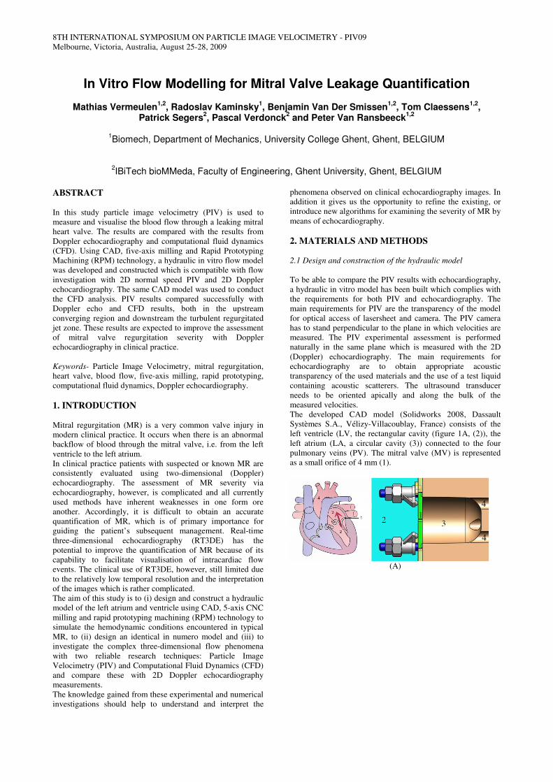
position. The mould for the atrium was made from necuron
651 (Necumer GmbH, Bohmte, Germany), constructed using
5-axis milling, and polished to obtain a smooth surface. After
the pre-processing with Magics software (Materialise NV,
Leuven, Belgium) Rapid Prototyping Machining technology
such as Selective Laser Sintering (SLS) and Stereolithography
(SLA) were used for manufacturing the components of the
hydraulic in vitro model. The casting of the silicone around
the left atrium was adapted from [1]. Vacuum casting was
used for casting the transparent silicone (Sylgard 184 silicone,
Dow Corning) around the necuron 651 mould. The rigid mitral
valve disk with an orifice of 4 mm was made from polymethyl
methacrylate (PMMA).
Figure 2 illustrates the vacuum casting of the left atrium.
Figure 2 casting of the left atrium; left: the silicon block cast
around the mould; right: the left atrium silicon block
2.2 Experimental PIV model
A 4 mm orifice was investigated with PIV in a closed-circuit
mock loop including reservoir, pump and the model. The PIV
measurements were performed at a constant inlet (LV)
pressure of 120mmHg and an outlet (PV) resistance pressure
of 60mmHg. According to our preliminary results [2] this
pressure drop results in a Reynolds number of:
based on the maximum orifice velocity. A water-glycerin
mixture was used as working fluid. The water-glycerin volume
ratio was experimentally determined to be 41% water, 59%
glycerine to match the refracting index of the used silicon
material (RI = 1.43). This mixture has a density of 1156 kg/m³
and a kinematic viscosity of 12.945 10-6 m²/s.
A standard 2D normal speed PIV system (ILA GmbH, Juelich,
Germany) was employed to perform the experiments. A
pulsed Nd:YAG laser reaching the maximum energy of 25mJ
was used as a light source. A CCD Sensicam QE camera with
a maximum resolution of 1376 x 1040 pixels was used to
record images which were stored in bitmap format afterwards.
The pulse distance varied between 200 µs and 1500 µs.
Polyamid microspheres particles with a density of 1016 kg/m³
and an average diameter of 57 µm were used as tracer
particles. Post processing of the images was done with
commercial VidPIV 4.6 software (ILA GmbH, Juelich,
Germany). In the region of interest, ‘in plane’ velocity vectors
and magnitudes were computed. The results are computed
from the statistical average of 1000 instantaneous flow field
measurements.
2.3 Two-dimensional Doppler echocardiography model
The 2D Doppler echocardiography measurements were
performed on the same hydraulic model that was used for the
PIV measurements. The echocardiography measurements
were done using a commercially available ultrasound system
(Vivid 7, GE Vingmed Ultrasound, Horten, Norway) equipped
with a cardiac M3 1.7/3.4-MHz matrix transducer. The
transducer was positioned in the apical position as illustrated
in figure 1B. The reflection of the ultrasound waves was
improved by adding polyamid microspheres particles to the
working fluid.
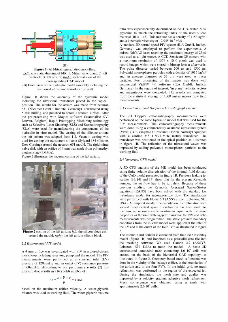
2.4 Numerical CFD model
A 3D CFD analysis of the MR model has been conducted
using finite volume discretisation of the internal fluid domain
of the CAD model presented in figure 1B. Previous leaking jet
studies [3], [4] and [5] show that for the present Reynolds
number, the jet flow has to be turbulent. Because of these
previous studies, the Reynolds Averaged Navier-Stokes
equations (RANS) have been solved with the standard k-ε
turbulence model for incompressible flow. The simulations
were performed with Fluent 6.3 (ANSYS, Inc., Lebanon, NH,
USA). An implicit steady time calculation in combination with
second order central space discretisation has been used. As
medium, an incompressible newtonian liquid with the same
properties as the used water-glycerin mixture for PIV and echo
measurements was programmed. The static pressure boundary
conditions from the in-vitro model were applied at the inlet of
the LV and at the outlet of the four PV’s as illustrated in figure
3.
The internal fluid domain is extracted from the CAD assembly
model (figure 1B) and imported as a parasolid data file into
the meshing software. We used Gambit 2.2 (ANSYS,
Lebanon, NH, USA) to mesh the model. A basic 3D
unstructured tetrahedral mesh containing 1.6 106 cells was
created on the basis of the hierarchal CAD topology, as
illustrated in figure 3. Geometry based mesh refinement was
done in the vicinity of the leakage orifice, at the boundaries of
the atrium and in the four PV’s. In the initial grid, no mesh
refinement was performed in the region of the expected jet.
During the simulation, the mesh size and quality was
improved by a velocity gradient adaptive mesh refinement.
Mesh convergence was obtained using a mesh with
approximately 2.6 106 cells.
Figure 3 Section view along the midplane of the initial
unstructured tetrahedral mesh of the numerical model.
3. RESULTS
Results of the PIV measurements and CFD simulations are
qualitatively compared to each other before they are
quantitatively compared with 2D Doppler echocardiography.
Figure 4 shows (A) the results of the PIV measurement, (B)
the results obtained by CFD (k-ε turbulence model) and (C)
the (Doppler) echo results.
Depending of the used camera settings, particles and other
measurement settings, the 2D PIV images were obtained with
a pulse distance of 500 µs. The 2D PIV image shows the 2D
velocity vectors in the midplane.
The 3D CFD results are obtained on the finest grid of 2.6 106
cells and show the projected 2D velocity vectors are shown in
figure 4 (panel B). The colour represents the velocity
magnitude of the 2D vectors ranging from blue to red
corresponding to low and high velocities.
(A) PIV
(B) CFD
(C) 2D doppler echocardiography
Figure 4 Results of the MR model obtained by: (A)
PIV, (B) CFD (in m/s) and (C) 2D Doppler
echocardiography (in m/s).
Both PIV and CFD results capture the following flow
phenomena:
• Turbulent leaking jet flow in the LA
• Flow convergence zone in front of the MV
• Stagnation point at the back of the LA
• A 3D recirculation zone or vortex in the LA
• Separation zones at the entrance of the PV’s.
The maximum velocity measured with PIV was 2.5 m/s and
was found at the beginning of the jet. Figure 5 shows the
velocity contour plot measured with PIV.
Figure 5 velocity contour plot measured with PIV
The maximum velocity obtained with CFD is 3.5 m/s. This
velocity is computed inside the orifice. Because optical access
to that location is not possible in the in vitro model, PIV
velocities remain unknown. However CFD results do confirm
PIV results in the region of the highest PIV velocities (2.6
m/s).
The maximum velocity of the 3D vortices at the end of the LA
was measured in this test case with PIV with a pulse distance
of 1ms. The maximum velocity measured with PIV was 0.3
m/s while the CFD value was considerably higher (0.6 m/s).
The colour flow map obtained with 2D Doppler
echocardiography is shown in figure 4C. The colour coded
map represents velocities along the ultrasound beam and
shows the jet and recirculation zones. The red colours
represent the flow towards the transducer while the blue
colours represent the velocity in the opposite direction. Due to
aliasing, the beginning of the jet is coloured red. This means
that the velocity at the beginning of the jet exceeded the
maximum velocity of the colour scale, resulting in inversion
of the colour scale in this region. However, pulsed wave
Doppler confirmed the maximum velocity to be 2.4 m/s at the
beginning of the jet. We can clearly distinguish three
phenomena; the leakage jet, the flow convergence zone in
front of the MV and the reversed flow in the 3D vortex.
DISCUSSION AND CONCLUSIONS
PIV leaking jet studies are often performed in a simple straight
tube. By considering a new hydraulic model of mitral
regurgitation which includes the left atrium and the four
pulmonary veins, a more realistic flow pattern can be
achieved. Using CAD, CNC and RPM, a new hydraulic in
vitro model has been designed and constructed for analysing
MR flow with PIV and 2D Doppler echocardiography. The
flow field caused by the leaking mitral valve was investigated
with PIV, 2D echo and CFD. As working fluid a water-
glycerin mixture was used which kinematic viscosity is three
times higher than the kinematic viscosity of blood. Following
table summarises the measured velocities
PIV echo CFD
Jet velocity (m/s) 2.5 2.4 2.6
Vortex velocity (m/s) 0.3 / 0.6
Table 1 mitral regurgitation velocity comparison
The results show a good quantitative agreement between PIV,
CFD and (pulsed Doppler) echo of the velocity of the leaking
jet. However the velocity of the 3D vortex at the back of the
atrium, showed a considerably higher velocity simulated with
CFD than obtained with PIV. This difference could be due to
the fact that the simulated jet is wider than the jet measured
with PIV. An explanation could be that the used turbulence
model and its parameters are suboptimal for this model.
ACKNOWLEDGMENTS The authors would like to thank Antoine De Henau and Frank
De Mets from the university college Ghent for their help in
constructing the mould of the atrium, Jurgen Deviche from
bioMMeda for the trust to let me use the vacuum casting
machine and the Rapid Prototyping Laboratory at the
University College Ghent for the construction of the in vitro
model parts.
REFERENCES
[1] Hopkins L. M. et al., 2000, “Particle Image Velocimetry
Measurements in Complex Geometries,” Exp. Fluids, 29,
pp. 91–95.
[2] Van Ransbeeck P. et al., 2008, “Experimental and
Numerical Flow Modeling towards Refinement of Three-
dimensional Echocardiography for Heart Valve Leakage
Quantification”, eMBEC, Antwerpen, Belgium, pp:2644-
2647. [3] Fallon A.M., 2006, “The Development of a Novel in vitro
Flow System to Evaluate Platelet Activation and
Procoagulant Potential Induced by Bileaflet Mechanical
Heart Valve Leakage Jets”, PhD dissertation, Georgia
Institute of Technology.
[4] Fallon A.M. et al., 2008, “Procoagulant Properties of
Flow Fields in Stenotic and Expansive Orifices, Annals
of Biomedical Engineering, 36(1), pp. 1–13.
[5] Amatya D.M., Longmire E.K., 2007, “Simultaneous
Measurements of Velocity and Deformation in Flows
Through Compliant Diaphragms Used as Heart Valve
Analogues”, Proceeding of the ASME 2007 Summer
Bioengineering Conference, Colorado, USA, 2007.
Related Documents